Embed Size (px)
Citation preview
NeuroToxicology 28 (2007) 886–891
Brief communication
Resveratrol attenuates oxidative-induced DNA damage
in C6 Glioma cells
Andre Quincozes-Santos, Ana Cristina Andreazza, Patricia Nardin,Claudia Funchal, Carlos-Alberto Goncalves, Carmem Gottfried *
Universidade Federal do Rio Grande do Sul, Instituto de Ciencias Basicas da Saude, Departamento de Bioquımica,
Rua Ramiro Barcelos 2600 Anexo, 90035-003 Porto Alegre, RS, Brazil
Received 12 December 2006; accepted 27 March 2007
Available online 31 March 2007
Abstract
The antioxidant compound, trans-resveratrol, is found in substantial amounts in several types of red wine and has been proposed to have
beneficial effects in brain pathologies that may involve oxidative stress. The objective of the present study was to investigate the genoprotective
effects of resveratrol under conditions of oxidative stress induced by hydrogen peroxide in C6 glioma cells. DNA damage was assessed by the
alkaline single-cell gel electrophoresis assay or comet assay. In order to investigate the genoprotective effects of resveratrol against oxidative stress
induced by hydrogen peroxide on DNA damage, two models of oxidative stress induction were utilized. (I) 1 mM hydrogen peroxide for 0.5 h (10–
250 mM of resveratrol) and (II) 0.1 or 0.5 mM hydrogen peroxide for 6 h (10–100 mM of resveratrol). Resveratrol was able to prevent oxidative
damage to cellular DNA, induced in model I, at all concentrations tested; however, at 6 h of incubation, resveratrol prevented DNA damage only
partially. After 6 h of incubation (up to 48 h) resveratrol per se induced a slight time and dose-dependent DNA damage. In conclusion, these results
provide evidence that resveratrol may act as a significantly bioactive compound, supporting the possibility that, due to its antioxidant properties, it
may be important in health and disease for protecting against DNA damage through oxidative stress.
# 2007 Elsevier Inc. All rights reserved.
Keywords: Resveratrol; Genotoxicity; Neuroprotection; Hydrogen peroxide; Comet assay; C6 glioma
1. Introduction
Resveratrol (3,5,40-trihydroxy-stilbene), a polyphenol pre-
sent in grapes and red wine, has antioxidant proprieties and a
wide range of other biological effects (Fremont, 2000; Baur and
Sinclair, 2006).
Many studies now attest the cardioprotective (Das et al.,
2006; Stef et al., 2006; Shen et al., 2006) and chemopreventive
(Pervaiz, 2004; Signorelli and Ghidoni, 2005; Delmas et al.,
2006) effects of this compound. In addition, epidemiological
evidence shows that moderate consumption of red wine is
inversely correlated with the incidence of dementia and
Alzheimer’s disease (Tredici et al., 1999; Marambaud et al.,
2005). Furthermore, direct neuroprotective effects of resver-
atrol against oxidative stress have been demonstrated in vitro
(Chanvitayapongs et al., 1997; Savaskan et al., 2003). The
* Corresponding author. Tel.: +55 51 3316 5565; fax: +55 51 3316 5535.
E-mail address: [email protected] (C. Gottfried).
0161-813X/$ – see front matter # 2007 Elsevier Inc. All rights reserved.
doi:10.1016/j.neuro.2007.03.008
underlying mechanisms of the neuroprotective action of
resveratrol, however, are not fully understood. We recently
demonstrated that resveratrol is able to modulate glial
parameters such as glutamate uptake, glutamine synthetase
and S100B secretion in C6 glioma cells (Dos Santos et al.,
2006) and in primary astrocyte cultures (L.M.V. Almeida,
unpublished results). Brain tissue is particularly vulnerable to
oxidative damage, possibly due to its high consumption of
oxygen and the consequent generation of high quantities of
reactive oxygen species (ROS) during oxidative phosphoryla-
tion (Castagne et al., 1999). Several regions of the brain are
particularly rich in iron, which promotes the production of
damaging oxygen free radical species such as �OH radicals
(Halliwell and Gutteridge, 1990). Furthermore, the brain is
relatively poorly endowed with protective antioxidant enzymes
or antioxidant compounds. ROS formation has been implicated
in damage to nervous tissue in several brain pathologies, such
as ischemia-reperfusion injury and neurodegenerative disorders
(Rego and Oliveira, 2003; Manton et al., 2004). Excessive ROS
(e.g. hydrogen peroxide, H2O2) can lead to lipid, protein and
A. Quincozes-Santos et al. / NeuroToxicology 28 (2007) 886–891 887
DNA oxidation, causing cell damage to all cellular constituents.
Irreparable DNA damage is involved in carcinogenesis, aging
and other degenerative diseases (Cozzi et al., 1997).
The C6 glioma cell line was originally derived from rat brain
tumors induced by N-nitrosomethylurea (Benda et al., 1968);
these cells have oligodendrocytic, astrocytic and neuronal
properties (Parker et al., 1980) and are widely used as an
astrocyte-like cell line (Mangoura et al., 1989; Feng and Zhang,
2004; Cechin et al., 2005; Funchal et al., 2005; Chen et al.,
2006a,b; Dos Santos et al., 2006). In this study, the effects of
resveratrol on H2O2-mediated DNA damage in C6 glioma cells
were determined using alkaline single-cell gel electrophoresis
(the comet assay).
2. Materials and methods
2.1. Materials
trans-Resveratrol, propidium iodide, ethidium bromide and
material for cell culture were purchased from Sigma (St. Louis,
MO, USA). Dulbecco’s modified Eagle’s medium (DMEM)
was purchased from Gibco BRL (Carlsbad, CA, USA) and fetal
bovine serum (FBS) was from Cultilab (Campinas, SP, Brazil).
All other chemicals were purchased from regular commercial
suppliers.
2.2. C6 glioma culture and resveratrol treatment
C6 glioma cells were performed essentially accordingly to
the procedure previously described (Dos Santos et al., 2006).
Late passage cells (i.e. after at least 100 passages) were seeded
in flasks and cultured in DMEM (pH 7.4) supplemented with
5% fetal bovine serum, 2.5 mg/mL Fungizone1 and 100 U/L
gentamicin. After cells reached confluence, the culture medium
was removed by suction and the cells were incubated for 1, 6,
12, 24 or 48 h at 37 8C in an atmosphere of 5% CO2/95% air in
DMEM (pH 7.4) without serum in the absence or presence of
resveratrol (10, 50, 100 or 250 mM), taking cells exposed to
vehicle (0.25% ethanol) as controls. The concentrations of
resveratrol used in these experiments were obtained from
previous determinations (Dos Santos et al., 2006). Importantly,
in all parameters analyzed, the results obtained with vehicle
were not different from those obtained in basal conditions
without ethanol.
2.3. Hydrogen peroxide treatment
In order to investigate the protective effects of resveratrol
against oxidative stress induced by H2O2, two models of
oxidative stress induction were used. Before H2O2 exposure,
cells were pre-incubated with different concentrations of
resveratrol (10, 50, 100 or 250 mM) for 1 h at 37 8C in an
atmosphere of 5% CO2/95% air in DMEM (pH 7.4) without
serum. After this, the medium was maintained and H2O2 was
added as follows: model I—1 mM H2O2 for 0.5 h (in the
presence or absence of 10, 50, 100 and 250 mM of resveratrol)
and model II—0.1 or 0.5 mM H2O2 for 6 h (in the presence or
absence of 10, 50 and 100 mM of resveratrol). During
incubations, cells were maintained at 37 8C in an atmosphere
of 5% CO2/95% air.
2.4. Comet assay
A standard protocol for comet assay preparation and
analysis was adapted (Singh et al., 1988; Tice et al., 2000).
After different treatments as described above, C6 glioma
cells were detached by incubating in the presence of trypsin/
EDTA 0.05%. During trypsinization, cells were carefully
manipulated to avoid mechanical stress. Slides were prepared
by mixing 50 mL of C6 glioma suspension with 70 mL of low
melting point agarose (0.75%). The mixture (cells/agarose)
was added to a fully frosted microscope slide coated with a
layer of 300 mL of normal melting agarose (1%). After
solidification, the cover slip was gently removed, and the
slides were placed in lysing solution (2.5 M NaCl, 100 mM
disodium EDTA and 10 mM Tris, pH 10.0, with freshly
added 1% Triton X-100 and 10% dimethyl sulfoxide) for up
to 24 h. Subsequently, the slides were incubated in freshly
prepared alkaline buffer (300 mM NaOH and 1 mM EDTA,
pH 12.6) for 10 min and electrophoresed. Following
electrophoresis, slides were immersed in neutralizing buffer
(0.4 M Tris–HCl, pH 7.5, 4 8C) for 5 min, before finally
applying 50 mL of 5 mg/mL ethidium bromide to them and
leaving in the dark for 20 min to stain the DNA. Negative and
positive controls were used for each electrophoresis assay in
order to ensure the reliability of the procedure. Images of 100
randomly selected nuclei (50 nuclei from two replicated
slides) were analyzed for each treatment. Nuclei were scored
visually for comet tail size based on an arbitrary scale of 0–4,
i.e. ranging from no damage to extensive damage of DNA.
Thus, a group damage index could range from 0 (all nuclei
without tail, 100 cells � 0) to 400 (all nuclei with maximally
long tails, 100 cells � 4) (Collins et al., 1996, 1997). During
electrophoresis any relaxed or broken DNA fragments
migrate further than supercoiled, undamaged DNA. Slides
were viewed on a Nikon inverted microscope using a TE-FM
Epi-Fluorescence accessory and images were transferred to a
computer with a digital camera (Sound Vision Inc., Wayland,
MA).
2.5. Morphological studies and propidium iodide uptake in
C6 glioma cells
During treatments, cells were analyzed and photographed
with Nikon inverted microscope phase-contrast optics. Cellular
damage was assessed by fluorescent image analysis using the
propidium iodide (PI) method (Pringle et al., 1996), which
measures the incorporation of PI by the cells. Cells were treated
with 7.5 mM PI concomitantly with H2O2 addition and
maintained until the end of incubation, after which cells were
analyzed and photographed with a Nikon inverted microscope
using a TE-FM Epi-Fluorescence accessory. Optical density
was determined with the Optiquant version 02.00 software
(Packard Instrument).
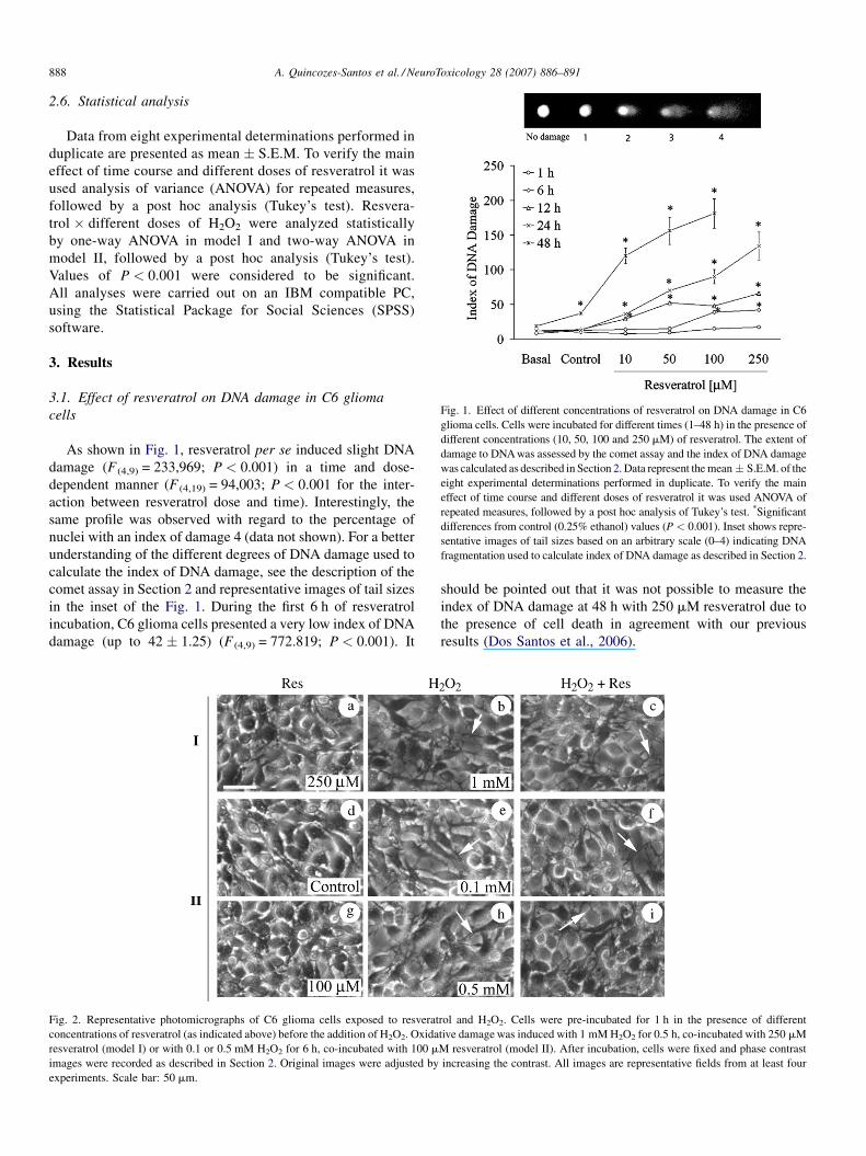
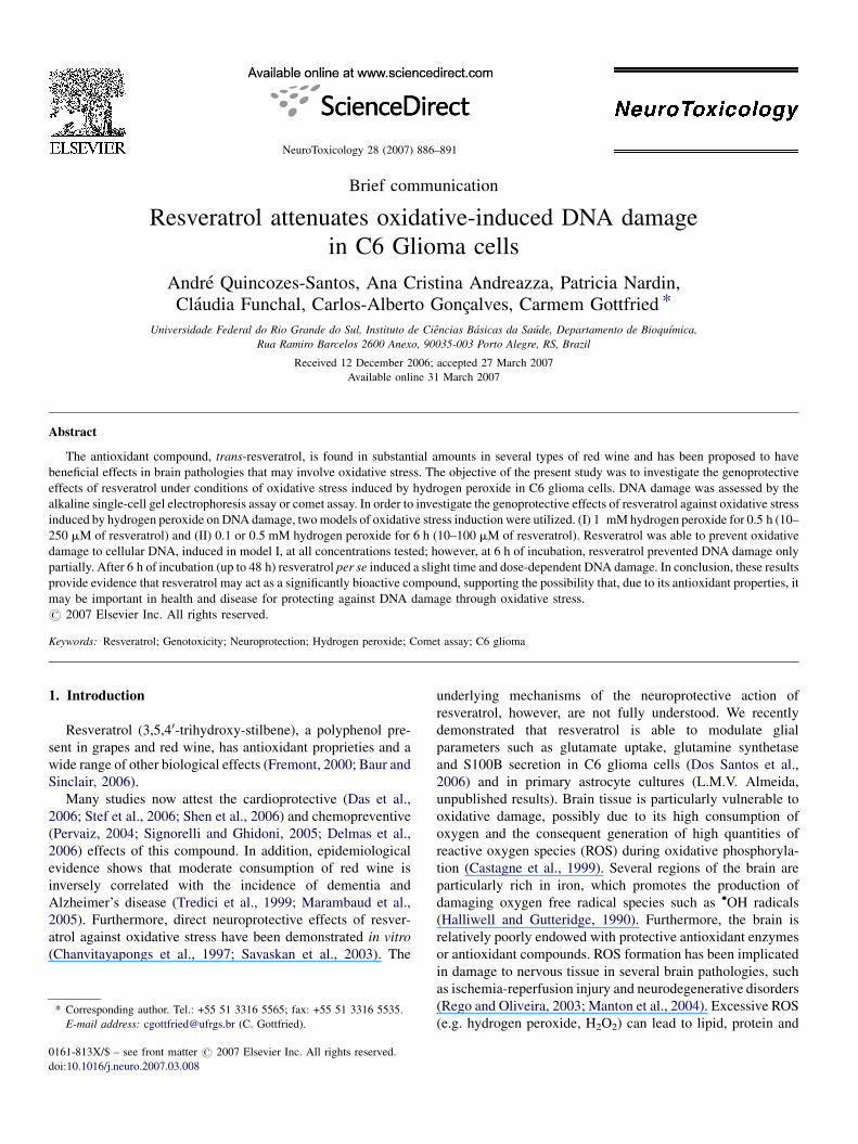
Fig. 1. Effect of different concentrations of resveratrol on DNA damage in C6
glioma cells. Cells were incubated for different times (1–48 h) in the presence of
different concentrations (10, 50, 100 and 250 mM) of resveratrol. The extent of
damage to DNA was assessed by the comet assay and the index of DNA damage
was calculated as described in Section 2. Data represent the mean� S.E.M. of the
eight experimental determinations performed in duplicate. To verify the main
effect of time course and different doses of resveratrol it was used ANOVA of
repeated measures, followed by a post hoc analysis of Tukey’s test. *Significant
differences from control (0.25% ethanol) values (P < 0.001). Inset shows repre-
sentative images of tail sizes based on an arbitrary scale (0–4) indicating DNA
fragmentation used to calculate index of DNA damage as described in Section 2.
A. Quincozes-Santos et al. / NeuroToxicology 28 (2007) 886–891888
2.6. Statistical analysis
Data from eight experimental determinations performed in
duplicate are presented as mean � S.E.M. To verify the main
effect of time course and different doses of resveratrol it was
used analysis of variance (ANOVA) for repeated measures,
followed by a post hoc analysis (Tukey’s test). Resvera-
trol � different doses of H2O2 were analyzed statistically
by one-way ANOVA in model I and two-way ANOVA in
model II, followed by a post hoc analysis (Tukey’s test).
Values of P < 0.001 were considered to be significant.
All analyses were carried out on an IBM compatible PC,
using the Statistical Package for Social Sciences (SPSS)
software.
3. Results
3.1. Effect of resveratrol on DNA damage in C6 glioma
cells
As shown in Fig. 1, resveratrol per se induced slight DNA
damage (F (4,9) = 233,969; P < 0.001) in a time and dose-
dependent manner (F (4,19) = 94,003; P < 0.001 for the inter-
action between resveratrol dose and time). Interestingly, the
same profile was observed with regard to the percentage of
nuclei with an index of damage 4 (data not shown). For a better
understanding of the different degrees of DNA damage used to
calculate the index of DNA damage, see the description of the
comet assay in Section 2 and representative images of tail sizes
in the inset of the Fig. 1. During the first 6 h of resveratrol
incubation, C6 glioma cells presented a very low index of DNA
damage (up to 42 � 1.25) (F (4,9) = 772.819; P < 0.001). It
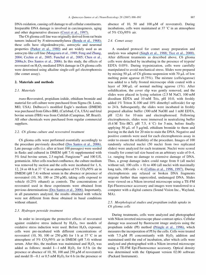
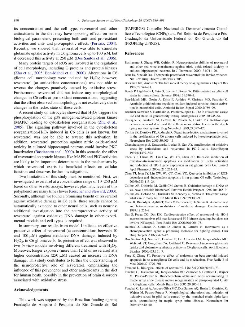
Fig. 2. Representative photomicrographs of C6 glioma cells exposed to resverat
concentrations of resveratrol (as indicated above) before the addition of H2O2. Oxida
resveratrol (model I) or with 0.1 or 0.5 mM H2O2 for 6 h, co-incubated with 100 m
images were recorded as described in Section 2. Original images were adjusted by
experiments. Scale bar: 50 mm.
should be pointed out that it was not possible to measure the
index of DNA damage at 48 h with 250 mM resveratrol due to
the presence of cell death in agreement with our previous
results (Dos Santos et al., 2006).
rol and H2O2. Cells were pre-incubated for 1 h in the presence of different
tive damage was induced with 1 mM H2O2 for 0.5 h, co-incubated with 250 mM
M resveratrol (model II). After incubation, cells were fixed and phase contrast
increasing the contrast. All images are representative fields from at least four
A. Quincozes-Santos et al. / NeuroToxicology 28 (2007) 886–891 889
3.2. Cell morphology and membrane integrity evaluation
under conditions of oxidative stress induced by H2O2
in C6 glioma cells
C6 cells were morphologically analyzed by phase contrast
microscopy after different exposure times of exposure to H2O2,
as indicated in Fig. 2. Under basal culture conditions, as
demonstrated previously (Funchal et al., 2006), C6 glioma cells
presented a rounded appearance, and this characteristic shape
was not altered even in control medium containing 0.25%
ethanol vehicle (panel d) or different times of exposure to
resveratrol (panels a and g). Importantly, 0.25% ethanol did not
alter basal morphology (data not shown) in model I of DNA
oxidative damage. Panels b, e, h show H2O2-induced
morphological alterations in C6 cells, consisting of process-
bearing cells, as indicated by the arrows, and resveratrol was
not able to prevent this effect (panels c, f, i). In order to confirm
the absence of cell death in all parameters investigated in both
models of H2O2-iduced DNA damage, cell integrity was
evaluated by PI uptake. There was no cell integrity alteration
(data not shown).
3.3. Genoprotective effects of resveratrol against
H2O2-iduced DNA damage in C6 glioma cells
The genoprotective effects of resveratrol against H2O2 are
shown in Fig. 3. In model I of treatment, the index of DNA
damage observed when C6 glioma cells were incubated in the
presence of H2O2 without resveratrol was 39 � 2.05. In this
Fig. 3. Inhibitory effect of resveratrol on H2O2-induced DNA damage in C6
glioma cells. Cells were pre-incubated for 1 h in the presence of different
concentrations of resveratrol (as indicated above) before the addition of H2O2.
Oxidative damage was induced with 1 mM H2O2 for 0.5 h, co-incubated with
10, 50, 100 or 250 mM resveratrol (model I) or with 0.1 or 0.5 mM H2O2 for 6 h,
co-incubated with 10, 50 or 100 mM resveratrol (model II). The extent of
damage to DNA was assessed by the comet assay and calculated as described in
Section 2. All data represent means � S.E.M. of the eight experimental
determinations performed in duplicate. Resveratrol � different doses of
H2O2 were analyzed statistically by one-way ANOVA, in model I and two-
way ANOVA in model II, followed by a post hoc analysis (Tukey’s test). (a)
Significant differences from control (0.25% ethanol) values; (b) significantly
different from 50 mM resveratrol in the presence of 0.5 mM of H2O2
(P < 0.001). Lines indicate index of DNA damage in the control samples.
model, resveratrol was able to almost totally prevent oxidative
damage to cellular DNA induced by H2O2 at all concentrations
tested (F (10,21) = 77.33; P < 0.001). For model II, H2O2 induced
increased DNA damage (F (2,24) = 135.75, P < 0.001), while
resveratrol on its own also had an effect (F (3,24) = 193.3,
P < 0.001). In general, resveratrol protects against H2O2
oxidative damage (as shown by interaction between resveratrol
and H2O2, F (6,24) = 132.172; P < 0.001). However, the effective
concentrations of resveratrol against oxidative 0.1 mM H2O2-
induced DNA damage (73� 2.56) ranged from 50 to 100 mM
(29 � 1.88 and 31 � 1.99, respectively) and in the presence of
0.5 mM H2O2, resveratrol presented a partial dose-dependent
prevention of H2O2-induced DNA damage (89 � 2.89) in C6
glioma cells. Interestingly, the percentage of nuclei containing
damage 4 showed the same profile as the DNA damage index
determined in Fig. 3 (data not shown).
4. Discussion
The present results constitute, to our knowledge, the first
evidence of a neuroprotective role of resveratrol against
oxidative DNA damage evaluated by the comet assay. H2O2 is
the major mediator of oxidative stress and a potent mutagen.
Large quantities of H2O2 can be generated by the respiratory
chain and several other metabolic pathways. Moreover,
activated inflammatory cells (e.g. microglia) are able to
produce this molecule via the oxidative burst mechanism.
Although this peroxide is a weak oxidant, it can be converted, in
the presence of reduced transition metals such as ferrous and
cuprous ions, to highly reactive hydroxyl radicals that are
believed to mediate the genotoxicity of this compound
(Halliwell and Gutteridge, 1990).
Model I of H2O2 exposure was used to induce an intense
(1 mM H2O2) and acute (30 min) damage. Cells pretreated with
resveratrol (for 1 h) were fully protected from H2O2 damage.
Model II of H2O2 exposure was used to induce a less intense
(0.1–0.5 mM H2O2), but lasting (6 h) damage. Pre-incubation
with resveratrol at 10 mM was not able to prevent DNA damage
induced by H2O2, but at 50–100 mM, resveratrol was
protective. However, caution is necessary to interpret results
of the comet assay. This assay is very sensitive for detecting low
levels of DNA damage, but this damage is usually not
necessarily correlated with cell viability (Brendler-Schwaab
et al., 2005). In fact, based on PI uptake, cell integrity was
maintained under our models (I and II) after oxidative insult
(data not shown).
Our results support the idea that resveratrol has a potential
role for neuroprotection in vitro and could be beneficial in brain
disorders involving oxidative stress. In neuropathologies, as
well as in the natural process of aging, the involvement of
oxygen free radical overproduction is strongly suspected as a
major factor related to the progressive derangement from
normal function (Beckman and Ames, 1998) and could involve
several mechanisms of cell signaling.
The effect of resveratrol per se on DNA damage was clear,
depending on its concentration and time of incubation.
Accordingly, many other data suggest that, depending upon
A. Quincozes-Santos et al. / NeuroToxicology 28 (2007) 886–891890
its concentration and the cell type, resveratrol and other
antioxidants in the diet may have opposing effects on some
biological parameters, presenting both anti- and pro-oxidant
activities and anti- and pro-apoptotic effects (Pervaiz, 2004).
Recently, we showed that resveratrol was able to stimulate
glutamate uptake activity in C6 glioma cells up to 100 mM, but
it decreased this activity at 250 mM (Dos Santos et al., 2006).
Many protein targets of ROS are involved in the regulation
of cell morphology, including G proteins and protein kinases
(Zhu et al., 2005; Ben-Mahdi et al., 2000). Alterations in C6
glioma cell morphology were induced by H2O2; however,
resveratrol (at antioxidant concentrations) was not able to
reverse the changes putatively caused by oxidative stress.
Furthermore, resveratrol did not induce any morphological
changes in C6 cells at pro-oxidant concentrations, suggesting
that the effect observed on morphology is not exclusively due to
changes in the redox state of these cells.
A recent study on astrocytes showed that H2O2 triggers the
phosphorylation of the p38 mitogen-activated protein kinase
(MAPK) leading to cytoskeleton reorganization (Zhu et al.,
2005). The signaling pathway involved in the cytoskeleton
reorganization-H2O2 induced in C6 cells is not known, but
resveratrol was not be able to reverse this activation. In
addition, resveratrol protection against nitric oxide-related
toxicity in cultured hippocampal neurons could involve PKC
inactivation (Bastianetto et al., 2000). In this scenario, the effect
of resveratrol on protein kinases like MAPK and PKC activities
are likely to be important determinants in the mechanisms by
which resveratrol exerts its beneficial effects on cellular
function and deserves further investigations.
Two limitations of this study must be mentioned. First, we
investigated resveratrol at a concentration range of 10–250 mM
based on other in vitro assays; however, plasmatic levels of this
polyphenol are many times lower (Gescher and Steward, 2003).
Secondly, although we found a promising benefit of resveratrol
against oxidative damage in C6 cells, these results cannot be
automatically extended to other neural cells, such as neurons;
additional investigation into the neuroprotective activity of
resveratrol against oxidative DNA damage in other experi-
mental models and cell types is required.
In summary, our results from model I indicate an effective
protective effect of resveratrol (at concentrations between 10
and 100 mM) against oxidative DNA damage, induced by
H2O2, in C6 glioma cells. Its protective effect was observed in
two in vitro models involving different treatment with H2O2.
Moreover, longer exposure (more than 12 h) of resveratrol at a
higher concentration (250 mM) caused an increase in DNA
damage. This study contributes to further the understanding of
the neuroprotective role of resveratrol, emphasizing the
influence of this polyphenol and other antioxidants in the diet
for human heath, possibly in the prevention of brain disorders
associated with oxidative stress.
Acknowledgements
This work was supported by the Brazilian funding agents;
Fundacao de Amparo a Pesquisa do Rio Grande do Sul
(FAPERGS) Conselho Nacional de Desenvolvimento Cientı-
fico e Tecnologico (CNPq) and Pro-Reitoria de Pesquisa e Pos-
Graduacao da Universidade Federal do Rio Grande do Sul
(PROPESq-UFRGS).
References
Bastianetto S, Zheng WH, Quirion R. Neuroprotective abilities of resveratrol
and other red wine constituents against nitric oxide-related toxicity in
cultured hippocampal neurons. Br J Pharmacol 2000;131:711–20.
Baur JA, Sinclair DA. Therapeutic potential of resveratrol: the in vivo evidence.
Nat Rev Drug Discov 2006;5:493–506.
Beckman KB, Ames BN. The free radical theory of aging matures. Physiol Rev
1998;78:547–81.
Benda P, Lightbody J, Sato G, Levine L, Sweet W. Differentiated rat glial cell
strain in tissue culture. Science 1968;161:370–1.
Ben-Mahdi MH, Gozin A, Driss F, Andrieu V, Christen MO, Pasquier C.
Anethole dithiolethione regulates oxidant-induced tyrosine kinase activa-
tion in endothelial cells. Antioxid Redox Signal 2000;2:789–99.
Brendler-Schwaab S, Hartmann A, Pfuhler S, Speit G. The in vivo comet assay:
use and status in genotoxicity testing. Mutagenesis 2005;20:245–54.
Castagne V, Gautschi M, Lefevre K, Posada A, Clarke PG. Relationships
between neuronal death and the cellular redox status. Focus on the devel-
oping nervous system. Prog Neurobiol 1999;59:397–423.
Cechin SR, Dunkley PR, Rodnight R. Signal transduction mechanisms involved
in the proliferation of C6 glioma cells induced by lysophosphatidic acid.
Neurochem Res 2005;30:603–11.
Chanvitayapongs S, Draczynska-Lusiak B, Sun AY. Amelioration of oxidative
stress by antioxidants and resveratrol in PC12 cells. NeuroReport
1997;8:1499–502.
Chen YC, Chow JM, Lin CW, Wu CY, Shen SC. Baicalein inhibition of
oxidative-stress-induced apoptosis via modulation of ERKs activation
and induction of HO-1 gene expression in rat glioma cells C6. Toxicol
Appl Pharmacol 2006a;216:263–73.
Chen TJ, Jeng JY, Lin CW, Wu CY, Chen YC. Quercetin inhibition of ROS-
dependent and -independent apoptosis in rat glioma C6 cells. Toxicology
2006b;223:113–26.
Collins AR, Dusinska M, Gedik CM, Stetina R. Oxidative damage to DNA: do
we have a reliable biomarker? Environ Health Perspect 1996;104:465–9.
Collins AR, Dobson VL, Dusinska M, Kennedy G, Stetina R. The comet assay:
what can it really tell us? Mutat Res 1997;29:183–93.
Cozzi R, Ricordy R, Aglitti T, Gatta V, Perticone P, De Salvia R. Ascorbic acid
and beta-carotene as modulators of oxidative damage. Carcinogenesis
1997;18:223–8.
Das S, Fraga CG, Das DK. Cardioprotective effect of resveratrol via HO-1
expression involves p38 map kinase and PI-3-kinase signaling, but does not
involve NFkappaB. Free Radic Res 2006;40:1066–75.
Delmas D, Lancon A, Colin D, Jannin B, Latruffe N. Resveratrol as a
chemopreventive agent: a promising molecule for fighting cancer. Curr
Drug Targets 2006;7:423–42.
Dos Santos AQ, Nardin P, Funchal C, De Almeida LM, Jacques-Silva MC,
Wofchuk ST, Goncalves CA, Gottfried C. Resveratrol increases glutamate
uptake and glutamine synthetase activity in C6 glioma cells. Arch Biochem
Biophys 2006;453:161–7.
Feng Z, Zhang JT. Protective effect of melatonin on beta-amyloid-induced
apoptosis in rat astroglioma C6 cells and its mechanism. Free Radic Biol
Med 2004;37:1790–801.
Fremont L. Biological effects of resveratrol. Life Sci 2000;66:663–73.
Funchal C, Dos Santos AQ, Jacques-Silva MC, Zamoner A, Gottfried C, Wajner
M, Pessoa-Pureur R. Branched-chain alpha-keto acids accumulating in
maple syrup urine disease induce reorganization of phosphorylated GFAP
in C6-glioma cells. Metab Brain Dis 2005;20:205–17.
Funchal C, Latini A, Jacques-Silva MC, Dos Santos AQ, Buzin L, Gottfried C,
Wajner M, Pessoa-Pureur R. Morphological alterations and induction of
oxidative stress in glial cells caused by the branched-chain alpha-keto
acids accumulating in maple syrup urine disease. Neurochem Int
2006;49:640–50.

A. Quincozes-Santos et al. / NeuroToxicology 28 (2007) 886–891 891
Gescher AJ, Steward WP. Relationship between mechanisms, bioavailibility,
and preclinical chemopreventive efficacy of resveratrol: a conundrum.
Cancer Epidemiol Biomarkers Prev 2003;12:953–7.
Halliwell B, Gutteridge JM. Role of free radicals and catalytic metal ions in
human disease: an overview. Methods Enzymol 1990;186:1–85.
Mangoura D, Sakellaridis N, Jones J, Vernadakis A. Early and late passage C-6
glial cell growth: similarities with primary glial cells in culture. Neurochem
Res 1989;14:941–7.
Manton KG, Volovik S, Kulminski A. ROS effects on neurodegeneration in
Alzheimer’s disease and related disorders: on environmental stresses of
ionizing radiation. Curr Alzheimer Res 2004;1:277–93.
Marambaud P, Zhao H, Davies P. Resveratrol promotes clearance of Alzhei-
mer’s disease amyloid-beta peptides. J Biol Chem 2005;280:37377–82.
Parker KK, Norenberg MD, Vernadakis A. ‘‘Transdifferentiation’’ of C6 glial
cells in culture. Science 1980;208:179–81.
Pervaiz S. Chemotherapeutic potential of the chemopreventive phytoalexin
resveratrol. Drug Resist Update 2004;7:333–44.
Pringle AK, Benham CD, Sim L, Kennedy J, Iannotti F, Sundstrom LE.
Selective N-type calcium channel antagonist omega conotoxin MVIIA is
neuroprotective against hypoxic neurodegeneration in organotypic hippo-
campal-slice cultures. Stroke 1996;27:2124–30.
Rego AC, Oliveira CR. Mitochondrial dysfunction and reactive oxygen species
in excitotoxicity and apoptosis: implications for the pathogenesis of neu-
rodegenerative diseases. Neurochem Res 2003;28:1563–74.
Savaskan E, Olivieri G, Meier F, Seifritz E, Wirz-Justice A, Muller-Spahn F.
Red wine ingredient resveratrol protects from beta-amyloid neurotoxicity.
Gerontology 2003;49:380–3.
Shen M, Jia GL, Wang YM, Ma H. Cardioprotective effect of resveratrol
pretreatment on myocardial ischemia-reperfusion induced injury in rats.
Vasc Pharmacol 2006;45:122–6.
Signorelli P, Ghidoni R. Resveratrol as an anticancer nutrient: molecular basis,
open questions and promises. J Nutr Biochem 2005;16:449–66.
Singh NP, McCoy MT, Tice RR, Schneider EL. A simple technique for
quantitation of low levels of DNA damage in individual cells. Exp Cell
Res 1988;175:184–91.
Stef G, Csiszar A, Lerea K, Ungvari Z, Veress G. Resveratrol inhibits aggrega-
tion of platelets from high-risk cardiac patients with aspirin resistance. J
Cardiovasc Pharmacol 2006;48:1–5.
Tice RR, Agurell E, Anderson D, Burlinson B, Hartmann A, Kobayashi H,
Miyamae Y, Rojas E, Ryu JC, Sasaki YF. Single cell gel/comet assay:
guidelines for in vitro and in vivo genetic toxicology testing. Environ Mol
Mutagen 2000;35:206–21.
Tredici G, Miloso M, Nicolini G, Galbiati S, Cavalletti G, Bertelli A. Resver-
atrol, map kinases and neuronal cells: might wine be a neuroprotectant?
Drugs Exp Clin Res 1999;25:99–103.
Zhu D, Tan KS, Zhang X, Sun AY, Sun GY, Lee JC. Hydrogen peroxide alters
membrane and cytoskeleton properties and increases intercellular connec-
tions in astrocytes. J Cell Sci 2005;118:3695–703.


