Embed Size (px)
Citation preview
Molecular Biology of the CellVol. 15, 1746–1759, April 2004
Bidirectional Transepithelial IgG Transport by a StronglyPolarized Basolateral Membrane Fc�-Receptor□D
Steven M. Claypool,*† Bonny L. Dickinson,‡ Jessica S. Wagner,‡Finn-Eirik Johansen,‡ Nanda Venu,‡ Jason A. Borawski,‡Wayne I. Lencer,‡¶# and Richard S. Blumberg†§¶#
Harvard Medical School, *Program in Immunology and Departments of §Medicine and†Gastroenterology Division, Brigham and Women’s Hospital, Boston, Massachusetts 02115; and‡Gastrointestinal Cell Biology and Department of Medicine, Children’s Hospital, BostonMassachusetts 02115; and ¶the Harvard Digestive Diseases Center, Boston, Massachusetts 02115
Submitted November 20, 2003; Revised January 5, 2004; Accepted January 18, 2004Monitoring Editor: Keith Mostov
The human MHC class I–related neonatal Fc receptor, hFcRn, mediates bidirectional transport of IgG across mucosalbarriers. Here, we find that at steady state hFcRn distributes predominantly to an apical intracellular compartment andalmost exclusively to the basolateral cell surface of polarized epithelial cells. It moves only transiently to the apicalmembrane. Ligand binding does not redistribute the steady state location of the receptor. Removal of the cytoplasmic tailthat contains di-leucine and tryptophan-based endocytosis motifs or incubation at low temperature (18°C) redistributesthe receptor apically. The rates of endocytosis of the full-length hFcRn from the apical or basolateral membrane domains,however, are equal. Thus, the strong cell surface polarity displayed by hFcRn results from dominant basolateral sortingby motifs in the cytoplasmic tail that nonetheless allows for a cycle of bidirectional transcytosis.
INTRODUCTION
The numerous studies on the cell biology of the polymericimmunoglobulin receptor (pIgR) and transferrin receptor(TfnR) as expressed in the Madin-Darby canine kidney(MDCK) cell line have provided a detailed characterizationof the itineraries of these proteins undergoing such pro-cesses as basolateral recycling, basolateral to apical transcy-tosis, and apical recycling in MDCK cells (Mostov andDeitcher, 1986; Leung et al., 1999; Brown et al., 2000; Wang etal., 2000). Comparatively little is known about trafficking inthe apical to basolateral direction because of the lack of amodel protein that physiologically harnesses this pathway,and whether the trafficking of pIgR and TfnR can be gener-alized to the trafficking of other proteins in polarized cellsremains to be tested.
The MHC class I–related neonatal Fc receptor, FcRn, isresponsible for the absorption of maternal IgG across therabbit and rodent yolk sac, the human placenta, and theproximal small intestine of the neonatal rodent (Rodewald,1970; Simister and Mostov, 1989; Roberts et al., 1990; Mede-san et al., 1996; Firan et al., 2001). Absorption of IgG dependson the ability of FcRn to bind IgG and traffic bidirectionallyacross the epithelial cells that line the lumen of these tissues(Jones and Waldmann, 1972; Abrahamson and Rodewald,1981; Dickinson et al., 1999; McCarthy et al., 2000; Kobayashiet al., 2002). Almost nothing is known about the cellularmechanisms that explain how any membrane receptor canmove bidirectionally across polarized epithelial cells.
Like most other MHC class I–related molecules, FcRn is anobligate heterodimer consisting of a glycosylated heavy (�)chain (40–44 kDa in humans, 48–50 kDa in rodents) thatassociates noncovalently with �2-microglobulin (�2m; Si-mister and Mostov, 1989). The association with �2m is spe-cies dependent (Claypool et al., 2002), and the functionalreceptor is likely a dimer of heterodimers that binds one IgGmolecule (Burmeister et al., 1994; Kim et al., 1994). The Fcfragment of IgG binds to FcRn at acidic pH (pH �6.5) andreleases from the receptor at neutral pH (Rodewald, 1976).In cells expressing FcRn, the bulk of FcRn is located intra-cellularly at steady state (Berryman and Rodewald, 1995;Dickinson et al., 1999; Ober et al., 2001). Ligand binding toFcRn can occur either at the cell surface or in the acidicendosome (Dickinson et al., 1999; Wu and Simister, 2001;Kobayashi et al., 2002). Trafficking of the rat FcRn in polar-ized cells depends on sorting motifs in the cytoplasmic tail(Stefaner et al., 1999; McCarthy et al., 2001; Wu and Simister,2001). Mutation of a di-leucine motif and a tryptophan res-idue in the cytoplasmic tail of rat FcRn impairs endocytosisof the receptor from both apical and basolateral membrane
Article published online ahead of print. Mol. Biol. Cell 10.1091/mbc.E03–11–0832. Article and publication date are available atwww.molbiolcell.org/cgi/doi/10.1091/mbc.E03–11–0832.
□D Online version of this article contains supplemental figures.Online version is available at www.molbiolcell.org.
# Corresponding author. Mailing address: Gastroenterology Divi-sion, Department of Medicine, Brigham and Women’s Hospital,Harvard Medical School, 75 Francis Street, Boston, MA 02115.E-mail address: [email protected] or Gastrointestinal Celland Developmental Biology, Gastroenterology and NutritionDivision, Children’s Hospital, 300 Longwood Ave, Boston, MA02115. E-mail address: [email protected] used: �2m, �2-microglobulin; h�2m, human �2m;hFcRn, human FcRn; hFcRntl�, tailless hFcRn; IMCD, innermedullary collecting duct; MDCK, Madin-Darby canine kidney;NIP, 5-iodo-4-hydroxy-3-nitro-phenylacetyl; NIP-hIgG1, hu-manized, NIP-specific human IgG1 monoclonal antibody; pIgR,polymeric immunoglobulin receptor; TfnR, transferrin receptor.
1746 © 2004 by The American Society for Cell Biology http://www.molbiolcell.org/content/suppl/2004/02/04/E03-11-0832.DC1.htmlSupplemental Material can be found at:

domains, with a more severe reduction in endocytosis ob-served from the apical surface. Removal of the entire cyto-plasmic tail strongly inhibits basolaterally directed IgGtransport but has no detectable effect on the apically directedtransport pathway (Wu and Simister, 2001). There are con-flicting results, however, on whether the FcRn cytoplasmictail affects sorting in the biosynthetic pathway (Stefaner etal., 1999; Wu and Simister, 2001).
In mammals that absorb IgG from breast milk, expressionof FcRn in the intestine is strongly downregulated afterweaning (Brambell, 1966; Simister and Mostov, 1989). Inhumans and nonhuman primates, the intestine continues toexpress FcRn into adult life (Dickinson et al., 1999; Zhu et al.,2001). FcRn is also expressed in the epithelium lining thelung in adult mice, humans, and nonhuman primates(Spiekermann et al., 2002). At these sites, FcRn mediates IgGtransport across the mucosal barrier and may function inimmune surveillance and host defense. In all species studiedso far, FcRn has also been shown to extend IgG half-life bybinding IgG in endosomes of endothelial cells (Borvak et al.,1998; Ward et al., 2003) and recycling the bound IgG back outof the cell, away from the late endosome and lysosomewhere degradation of unbound IgG occurs.
Nothing is known about how hFcRn operates in a polar-ized cell except that it is capable of mediating the bidirec-tional transport of IgG via a transcytotic mechanism thatrequires endosomal acidification (Dickinson et al., 1999;Claypool et al., 2002; Kobayashi et al., 2002). Previous studieson hFcRn in MDCK cells are not conclusive because thehuman heavy chain was expressed in cells lacking human�2m (Praetor et al., 1999). Chimeras of the human FcRnheavy chain and canine �2m do not traffic normally (Clay-pool et al., 2002).
Here, we study the trafficking of hFcRn in polarized epi-thelial cells by heterologous expression of the hFcRn heavychain in a h�2m-positive MDCK cell line. We find thattrafficking of human FcRn differs from that of the rat FcRn(Stefaner et al., 1999; McCarthy et al., 2000; McCarthy et al.,2001; Wu and Simister, 2001) and from previous claims onthe human receptor (Praetor et al., 1999). Like the rat recep-tor, human FcRn predominantly localizes to a supra-nuclearintracellular compartment and mediates bidirectional trans-cytosis of IgG, but unlike the rat receptor, the fraction ofhuman FcRn on the cell surface at steady state is almostexclusively basolateral and the efficiency of transport isstrongly apically directed. Although ligand binding does notaffect the distribution of hFcRn, either removal of the cyto-plasmic tail or incubation at low temperature (18°C) redis-tributes the receptor apically, and the fact that the rates ofendocytosis of the full-length receptor from the apical andbasolateral membranes are equal suggests that the steadystate distribution of hFcRn is maintained by a dominantbasolateral sorting signal(s) contained in the tail domain thatis functionally temperature sensitive. Unlike other rapidlyrecycling receptors that sort strongly to the basolateral mem-brane by motifs contained in the cytoplasmic tail, however,the bidirectional trafficking by FcRn through the transcytoticpathway is not stochastic and occurs at physiological rates.
MATERIALS AND METHODS
Plasmid Construction and Recombinant ProteinExpressionThe h�2m and hFcRn expression vectors have been described previously(Claypool et al., 2002). Generation of a tailless hFcRn containing an NH2-terminal hemaglutinin (HA) tag (5�-YPYDVPDYA-3�) was performed as de-scribed (Claypool et al., 2002) by introducing a stop translation site immedi-
ately after the first four predicted amino acids of the hFcRn cytoplasmic tail(RRMR; Story et al., 1994). All constructs described were verified by sequenc-ing.
To generate recombinant human IgG1 with specificity for the hapten5-iodo-4-hydroxy-3-nitro-phenylacetyl (NIP), pLNOH2-IgG1 (gift from T.E.Michalesen, Norwegian Institute of Public Health) containing an exon encod-ing a NIP-specific variable domain spliced to a downstream human IgG1heavy-chain constant region (Norderhaug et al., 1997) was transfected into themurine myeloma cell line J558L, and clonal lines selected. J558L cells expressendogenous � light chain that can combine with the chimeric heavy chain toform NIP-specific antibody with the human IgG1 Fc region (NIP-hIgG1).NIP-hIgG1 affinity purification was performed as described (Johansen et al.,1999).
AntibodiesThe mouse monoclonal antibodies (mAbs) 12CA5, reactive against the influ-enza HA epitope, and BBM1, specific for h�2m, and the rabbit anti-hFcRncytoplasmic tail domain polyclonal antiserum have been described (Claypoolet al., 2002). The guinea pig anti-hFcRn cytoplasmic tail domain antiserumwas a kind gift of Syntonix Pharmaceuticals (Waltham, MA). Pooled humanIgG was obtained from Lampire Biological Laboratories (Pipersville, PA) andChicken IgY from Gallus Immunotech Inc. (Wildwood, MO). Other anti-bodies used were: rabbit anti-h�2m and anti-HA polyclonal antisera, mouseanti–�-actin and anti–ZO-1 mAbs (Sigma Chemical Co., St. Louis, MO),chicken anti-human IgG Alexa 488 (Molecular Probes, Eugene, OR), mouseanti-transferrin receptor (Zymed, South San Francisco, CA), mouse anti–E-cadherin mAb (clone E4.6, kind gift of Drs. John Higgins and Michael Bren-ner; Cepek et al., 1994), mouse anti-GP135 mAb (Ojakian and Schwimmer,1988), and rabbit antiserum reactive to human CD55 (Santa Cruz Biotechnol-ogy, Santa Cruz, CA). Alexa-conjugated, horseradish peroxidase (HRP)-con-jugated, and alkaline phosphatase–conjugated IgG were from MolecularProbes, Pierce (Rockford, IL) and Sigma, respectively.
Cell CultureStable MDCK cell transfectants were prepared and cultured as previouslydescribed (Claypool et al., 2002). A stable h�2m-positive parental MDCK clonewas supertransfected with either full-length HA-tagged hFcRn or taillessHA-tagged hFcRn plasmids. MDCK II cells transfected with the parentalpcDNA3.1 and pEF6/V5-HisA vectors (Invitrogen, Carlsbad, CA) were gen-erated as negative controls.
T84 cells were passaged weekly and maintained in a 1:1 mixture of low-glucose DMEM and F-12 Nutrient Mixture containing 0.014 M NaHCO3, 0.015M HEPES, pH 7.3, and 6% heat-inactivated FBS at 37°C, 5% CO2. Caco-2 cellswere passaged weekly and maintained in high-glucose DMEM containing10% FBS and 15 mM HEPES, pH 7.4, at 37°C, 5% CO2. For the selective cellsurface biotinylation studies, T84 and Caco-2 cells were seeded at confluentdensity onto 24-mm, 3-�m pore size, semipermeable polycarbonate Transwellfilters (Corning Inc., Corning, NY) that were collagen coated (mouse collagenType IV; BD Biosciences, Bedford, MA) 24 h earlier. Transepithelial resis-tances were recorded daily for each filter, medium was replenished every 2–3days, and experiments were performed 11–15 days postplating at which pointthe resistances measured �1000 and 250–300 �cm2 for T84 and Caco-2monolayers, respectively.
MDCK clones were plated at confluent density on 24-mm diameter, 0.4-�mpore size, semipermeable polycarbonate Transwell filters or on 6.5-mm di-ameter, 0.4-�m pore size, optically clear semipermeable polyester Transwellfilters for biochemical studies and immunoflourescence experiments, respec-tively. Experiments were performed day 3 or 4 postplating. For serum star-vation, filters were incubated for 1 h in serum-free medium buffered to pH 7.3with 20 mM HEPES. To assess the effect of ligand with or without a pHgradient on the steady state distribution of hFcRn, human IgG or chicken IgY(100 �g/ml) was added to DMEM containing 2% FBS buffered to either pH 7.3with 20 mM HEPES or buffered to pH 6.0 with 20 mM MES. Ligand wasadded to the input chamber. DMEM containing 2% FBS, 20 mM HEPES, pH7.3, was added to the output chamber, and the filters incubated for 12 h at37°C, 5% CO2 before domain selective biotinylation. For colocalization, aftera 1-h serum starvation, DMEM containing 0.2% BSA buffered at pH 7.3 or 6.0and supplemented with 100 �g/ml human IgG1/� (Sigma) was applied asindicated. IgG uptake was visualized after incubation at 37°C, 5% CO2 for 20min. All 18°C incubations were performed for the indicated times utilizing acooled H2O bath.
IgG BindingCell lysates were prepared as described (Claypool et al., 2002). Definedquantities of lysate (Figure 1 caption) were incubated for 4 h with 200 �ghuman IgG, rotating gently at 4°C, and were captured on protein G-Sepharosebeads (Amersham Biosciences, Piscataway, NJ). After three successive washeswith 1 mg/ml CHAPS, pH 6.0 or 8.0, bound proteins were resolved on 12%SDS-PAGE gels under nonreducing conditions. All immunoblot analyses,including stripping and reprobing of nitrocellulose membranes (Schleicher &Schuell, Keene, NH) and densitometric analyses of films were performed aspreviously described (Claypool et al., 2002).
Basolateral Membrane Targeting of Human FcRn
Vol. 15, April 2004 1747

Transcytosis Assay and ELISATranscytosis assays were performed as described (Claypool et al., 2002) withthe following modifications. Monolayers were washed with Hanks’ balancedsalt solution at pH 7.4 with 10 mM HEPES (HBSS�, pH 7.4), followed by a20-min incubation at 37°C, 5% CO2 with either 1% BSA in HBSS�, pH 6.0(HBSS� buffered with 10 mM MES) or 1% BSA in HBSS�, pH 7.4, on theinput surface and HBSS�, pH 7.4, on the opposite, output surface. NIP-hIgG1was added to the input surface (60 nM final concentration), and the cells weremaintained for 90 min at 37°C, 5% CO2. For blocking experiments, rabbit IgGwas included in the buffer applied to the input surface 20 min before theaddition of NIP-hIgG1. After the 90-min incubation, the entire output solutionwas collected and transferred to a 96-well plate previously coated withBSA-NIP and blocked with BSA. Bound NIP-hIgG1 was detected by a humanIgG-specific ELISA, and data were analyzed using SigmaStat 1.0 Software(Jandel Corporation, San Rafael, CA).
Immunocytochemistry and Confocal MicroscopyImmunocytochemistry of MDCK cells grown on Transwell inserts was per-formed as described (Claypool et al., 2002). All images were collected using a60�/1.4 NA oil immersion objective and Nikon Axiophot (Garden City, NY)coupled to a MRC1024 laser scanning confocal system (Bio-Rad Laboratories,Hercules, CA). All images were gathered using the same laser power, gain,and pinhole size for the respective channels. For each insert analyzed, two ormore separate fields of cells were imaged (92 ı̀m2 each, later cropped to 34ı̀m2). Serial x-y scans were collected using ZO-1 as a reference point. Finalimage processing and labeling were performed using Adobe Photoshop(Mountain View, CA).
Cell Surface BiotinylationDomain selective biotinylation of filter-grown cells was performed as de-scribed (Casanova et al., 1991). Cell lysis and immunoprecipitation of eitherfull-length hFcRn or hFcRntl� was performed as described (Claypool et al.,2002) using a rabbit anti-hFcRn cytoplasmic tail antiserum and 12CA5, re-spectively. For T84 and Caco-2 cells, the immunoprecipitates were analyzeddirectly by SDS-PAGE, and surface hFcRn was detected by immunoblottingwith HRP-conjugated neutravidin (Pierce, 1:10,000). For the MDCK clones,immunoprecipitated proteins were eluted by boiling for 10 min in 24 �L 10%SDS. Ten percent of the eluate was set aside for direct analysis, and the
remaining 90% was diluted in 1 ml reprecipitation buffer (50 mM Tris, pH 7.4,150 mM NaCl, 1% NP-40, 0.5% sodium deoxycholate, 0.2% BSA), and surfaceproteins were captured with avidin-agarose (Pierce). In every experiment,total surface proteins were captured either from a separate portion of theoriginal biotinylated lysate or from the nonbinding flow through fraction afterFcRn immunoprecipitation, by the addition of avidin-agarose.
Antibody CaptureMonolayers were serum-starved and equilibrated in DMEM, 0.2% BSA, pH7.3, at the indicated temperatures for 20 min before the addition of 10 �l12CA5 ascites to either the apical or basolateral chambers. After 90-minincubations, unbound antibody was removed by four successive 10-minwashes with PBS, 0.1 mM CaCl2, 1 mM MgCl2, and 1% BSA at 4°C. Equalvolumes of cell lysates (described above) were added directly to proteinA-Sepharose and rotated at 4°C for 90 min. Twenty microliters of each samplewas used for a �-actin immunoblot analysis.
Endocytosis AssayEndocytosis of hFcRn was assayed as described (Graeve et al., 1989). Equalquantities of lysate were added to avidin-agarose to capture total biotinylatedsurface proteins. Two filters were maintained at 4°C throughout the entireexperiment, one serving as the biotinylation control (no reduction, time 0 min)and the other serving as a control for the reduction step. After extensivewashing, the samples were resolved under reducing conditions on 10%SDS-PAGE gels, and immunoblot analyses were performed as described(Claypool et al., 2002). Twenty microliters of each sample was collected for�-actin immunoblot analysis.
RESULTS
Functional Expression of hFcRn in MDCK CellsTo study trafficking of hFcRn in polarized epithelia, weprepared a stable MDCK cell line expressing h�2m and thensupertransfected this cell line with a plasmid encoding thefull-length hFcRn heavy chain containing an N-terminal HAtag. To demonstrate FcRn function, we first assessed the
Figure 1. Both endogenous and transfectedhFcRn bind human IgG with strict pH depen-dency. (A) Human IgG was preincubatedwith cell lysates prepared in CHAPS lysisbuffer, pH 6.0 or 8.0, and subsequently cap-tured by the addition of protein G-Sepharose.Bound proteins were analyzed by SDS-PAGEunder nonreducing conditions and immuno-blotted for hFcRn (top) and h�2m (bottom).The following cell lines were examined forIgG binding with the quantity of lysate indi-cated in parentheses: T84 cells (1 mg), Caco-2(1 mg), a control h�2m�/hFcRn� MDCKclone (1 mg), and two h�2m�/hFcRn�MDCK clones (0.2 mg). (B) Total hFcRn wasimmunoblotted in each of the following ly-sates: T84 (50 �g), Caco-2 (50 �g), a controlh�2m�/hFcRn� MDCK clone (50 �g), andtwo individual h�2m�/hFcRn� MDCKclones (10 �g). The bottom panel is the samemembrane reprobed for �-actin. The locationof bands consistent with the predicted sizes ofhFcRn (open arrow) and h�2m (gray arrow)are indicated on the left of each gel. Sizemarkers (kDa) are indicated on the left.
S.M. Claypool et al.
Molecular Biology of the Cell1748

pH-dependent binding of IgG to FcRn (Figure 1). The hu-man intestinal epithelial cell lines, T84 and Caco-2, whichexpress endogenous hFcRn, served as positive controls.MDCK cells transfected with empty vectors provided neg-ative controls. Equal quantities of cell lysates were bufferedat pH 6.0 or 8.0 and incubated with human IgG. IgG-bindingproteins were then captured by incubation with proteinG-Sepharose and analyzed for hFcRn and h�2m by SDS-PAGE and immunoblot. At acidic pH, IgG coprecipitatedwith both hFcRn (Figure 1A, top panel) and h�2m (Figure1A, bottom panel, lanes 2, 4, 8, and 10). There was no FcRnisolated by IgG added to lysates of MDCK cells transfectedwith vector alone (lanes 5 and 6) or from any cell lysatebuffered at pH 8.0 (lanes 1, 3, 7, and 9). FcRn was, however,present in the total cell lysates from all cells transfected withFcRn, whether buffered at pH 6.0 or 8.0 (Figure 1B, toppanel). The migration of the HA-tagged hFcRn heavy chainin MDCK cells was slightly delayed compared with endog-enous hFcRn in T84 and Caco-2 cells, consistent with theaddition of the HA-tag, and the protein bands appearedmore heterogeneous, consistent with the differential patternsof glycosylation observed in proteins heterologously ex-pressed in MDCK cells (Singer and Mostov, 1998). Immu-noblot for �-actin shows that each lane was loaded with anequal mass of total cell protein (Figure 1B, bottom panel).These data show that the hFcRn/h�2m heterodimer ex-pressed in canine MDCK cells can bind human IgG at acidicbut not basic pH, a hallmark of FcRn function.
To test for FcRn-mediated transcytosis, we used a recom-binantly expressed and purified chimeric mouse/humanIgG1 antibody reactive to the haptene 5-iodo-4-hydroxy-3-nitro-phenylacetyl (NIP-hIgG1, see MATERIALS ANDMETHODS). When tested in vitro, the recombinant NIP-hIgG1 antibody coprecipitated with hFcRn and h�2m indetergent lysates of MDCK cells at pH 6.0 but not at pH 8.0(our unpublished results). FcRn-mediated transcytosis ofNIP-hIgG1 was measured by ELISA for transport in both theapical to basolateral and basolateral to apical directions (Fig-ure 2, A and B, respectively). In some studies, the pH of theinput reservoir was buffered to 6.0 to increase IgG uptake byFcRn (McCarthy et al., 2001). The output reservoir was al-ways buffered at pH 7.4 to favor release of NIP-hIgG1 aftertranscytosis.
In the apical to basolateral direction, FcRn-mediatedtransport of NIP-hIgG1 was observed for FcRn-expressingcell lines when the input reservoir was buffered at pH 6.0.Small amounts of Nip-hIgG1 were transported when theinput reservoir was buffered to pH 7.4. Transport was spe-cific for FcRn because it was not observed in cell lines thatlacked hFcRn, and it was inhibited dose-dependently bycompetition with rabbit IgG that also binds hFcRn (Figure2A). In the basolateral to apical direction (Figure 2B), recep-tor-mediated transport of NIP-hIgG1 was observed whenthe input reservoir was buffered at pH 7.4, but transcytosisof NIP-hIgG1 was 10-fold greater when the input reservoirwas buffered to pH 6.0. Transport in this direction was alsospecific for hFcRn because no transport was observed in celllines lacking hFcRn, and it was inhibited dose-dependentlyby competition with rabbit IgG. Consistent with our initialstudies on FcRn in MDCK cells (Claypool et al., 2002), themass of NIP-hIgG1 transported was greater in the basolat-eral to apical direction. Thus, hFcRn expressed in the h�2m-containing MDCK cell line functions in bidirectional IgGtransport. These data are consistent with our previous find-ings that FcRn functions to transcytose IgG in both direc-tions across polarized T84 cells expressing FcRn endog-enously (Dickinson et al., 1999).
Human FcRn Localizes Predominantly to IntracellularCompartments and Colocalizes with Internalized IgGWe next analyzed the location of hFcRn in polarized MDCKcells expressing hFcRn by confocal microscopy. In the pres-ence of IgG, the bulk of hFcRn localized to the apical regionof the cell, beginning just below the level of the tight junctionand extending �4 �m down toward the basolateral plasmamembrane (Figures 3 and 4, red). The same distribution wasobserved in the absence of added IgG (our unpublishedresults). Thus, ligand binding to FcRn does not redistributethe steady state localization of hFcRn in MDCK cells. Thesubapical distribution of hFcRn in MDCK cells is also con-sistent with the predominantly apical intracellular distribu-tion of FcRn in human intestine and lung (Dickinson et al.,1999; Spiekermann et al., 2002) and rat intestine in vivo(Berryman and Rodewald, 1995).
IgG internalized for 20 min at pH 6.0 from the apicalsurface (Figure 3) or at pH 6.0 or 7.4 from the basolateral cell
Figure 2. Bidirectional transcytosis of IgG by hFcRn expressed inh�2m-positive MDCK cells. Regardless of the direction, transcytosisassays were performed with the pH of the input chamber bufferedat either pH 7.4 (� pH gradient) or pH 6.0 (� pH gradient), whereasthe output chamber was always buffered at pH 7.4. For blockingexperiments, the indicated concentration of rabbit IgG relative toNIP-hIgG1 was included in the input chamber that was buffered atpH 6.0. (A) Transcytosis of NIP-hIgG1 (60 nM final concentration) inthe apical to basolateral direction. (B) Transcytosis of NIP-hIgG1 inthe basolateral to apical direction. For ease of representation, datafrom two individual h�2m�/hFcRn� clones were combined.ANOVA on Ranks, p � 0.00001. The asterisk indicates statisticalsignificance relative to NIP-hIgG1 transport with a pH gradient butin the absence of competitive ligand at p � 0.05 by multiple com-parison procedures. The number sign indicates statistical signifi-cance relative to transport of NIP-hIgG1 in the absence of a pHgradient by the h�2m�/hFcRn� MDCK clone at p � 0.05 bymultiple comparison procedures (n � 3).
Basolateral Membrane Targeting of Human FcRn
Vol. 15, April 2004 1749

surface (Figure 4) colocalized with hFcRn. There was moreIgG internalized at pH 6.0 than at pH 7.4, consistent with ourfunctional studies (see Figure 2). Only very little IgG accu-mulated inside of MDCK cells lacking FcRn (Figure 3C and4C, green). As expected for a transporting receptor, IgGcolocalized with hFcRn at several planes of the cell, includ-ing the lateral aspects below the tight junction when loadedapically (Figure 3A, arrows in merged panels) and at thelevel of the tight junction when loaded basolaterally (Figure4, A and B, arrows in merged panels). Taken together, the
biochemical, morphologic, and functional studies describedabove show that MDCK cells coexpressing h�2m and hFcRnheavy chain accurately model the biology of FcRn in polar-ized human epithelial cells in vitro and in vivo.
The Fraction of hFcRn on the Cell Surface LocalizesAlmost Exclusively to the Basolateral Plasma MembraneWe next analyzed the cell surface distribution of hFcRn(Figure 5). MDCK cells expressing the hFcRn heterodimer(lanes 4–9), h�2m alone (lanes 10–12), or not expressing
Figure 3. IgG internalization is enhanced from the apical plasma membrane and colocalizes with hFcRn when buffered at pH 6.0 but notat pH 7.3. After serum starvation, 625 nM human IgG1/k buffered at pH 6.0 (A and C) or pH 7.3 (B) was internalized from the apical chambersof filter-grown h�2m�/hFcRn� (A and B) and control (C) MDCK cells for 20 min at 37°C. The cells were fixed, and hFcRn (red), ZO-1 (blue),and human IgG (green) were detected using Alexa-conjugated secondary antibodies. Serial red, green, and blue images were separatelycollected utilizing a confocal microscope and merged images generated using Adobe Photoshop. (A) For internalization of IgG at pH 6.0 byh�2m�/hFcRn� MDCK cells, the position of hFcRn (red) and IgG (green) with respect to ZO-1 (blue) are presented individually (left twocolumns) and merged (right column). (B and C) For simplicity, only merged images are presented. The position of each merged X-Y planerelative to the tight junction (ZO-1) is indicated at the right. Arrows highlight regions of intense hFcRn and IgG colocalization (n � 3). Bars,5 �m.
S.M. Claypool et al.
Molecular Biology of the Cell1750

either chain (lanes 1–3) were treated at 4°C with sulfo-NHS-biotin applied selectively to apical or basolateral cell sur-faces, or to both cell surfaces, to label apical or basolateralmembrane proteins or proteins on both membranes. Somemonolayers were serum starved for 1 h at pH 7.3 to disso-ciate bovine IgG from FcRn before domain-specific biotiny-lation (lanes 13–18). The monolayers were then lysed indetergent, and hFcRn was immunoprecipitated. Each immu-noprecipitate was eluted by boiling in SDS, and 10% was setaside to assess total FcRn content. The remaining 90% wasincubated with avidin-agarose beads to affinity-isolate bio-
tinylated cell surface-proteins. Both sets of samples wereanalyzed for HA-tagged FcRn by SDS-PAGE and immuno-blotting for the hFcRn heavy chain (using an anti-HA mAb).All MDCK cells expressing hFcRn contained hFcRn in theimmunoprecipitates (Figure 5A, bottom panel, lanes 4–9and 13–18). However, biotinylated hFcRn was only detectedon the basolateral plasma membrane, whether in the pres-ence or absence of bovine IgG (serum; Figure 5A, top panel,lanes 14–15 and 17–18, � serum, and lanes 5–6 and 8–9, �serum). The endogenous apical membrane protein GP135was detected in the same cell lysates only when biotin was
Figure 4. IgG is internalized from the basolateral plasma membrane and colocalizes with hFcRn when buffered at pH 6.0 and pH 7.3. Serumstarvation and IgG internalization were performed as described in Figure 3 except that the IgG buffered at pH 6.0 (A and C) or pH 7.3 (B)was added to the basolateral compartment of filter-grown h�2m�/hFcRn� (A and B) and control (C) MDCK cells. Detection of hFcRn (red),IgG (green), and ZO-1 (blue) and image collection and processing were performed as in Figure 3. (A) For internalization of IgG at pH 6.0 byh�2m�/hFcRn� MDCK cells, the position of hFcRn (red) and IgG (green) with respect to ZO-1 (blue) are presented individually (left twocolumns) and merged (right column). (B and C) For simplicity, only merged images are presented. The position of each merged X-Y planerelative to the tight junction (ZO-1) is indicated at the right. Arrows highlight regions of intense hFcRn and IgG colocalization. Bars, 5 �m.(n � 3)
Basolateral Membrane Targeting of Human FcRn
Vol. 15, April 2004 1751

applied to apical cell surfaces (Figure 5B), providing evi-dence for the specificity of cell surface biotin-labeling. Iden-tical results were obtained when monolayers were exposedto human IgG (supplemental Figure 1). Thus at steady state,the cell surface fraction of hFcRn in MDCK cells localizesalmost exclusively to the basolateral cell surface, and this isnot affected by IgG binding to the receptor. These resultsdiffer from those found for the rat receptor (Stefaner et al.,1999; McCarthy et al., 2000; McCarthy et al., 2001; Wu andSimister, 2001).
We next asked if endogenously expressed hFcRn displaysa similar polarized cell surface distribution in the T84 andCaco-2 human intestinal epithelial cell lines. Selective cellsurface biotinylation was performed with (Figure 6C) orwithout serum starvation (Figure 6A), and total FcRn wasimmunoprecipitated from cell lysates with a polyclonal rab-bit antiserum specific for the human FcRn cytoplasmic tail.The fraction of immunoprecipitated FcRn containing biotinwas analyzed by SDS-PAGE and ligand blot using HRP-labeled neutravidin (neutravidin-HRP). Biotinylated pro-teins, consistent in size with that predicted for hFcRn andh�2m were detected by neutravidin-HRP ligand blot in ba-solaterally biotinylated samples only (Figure 6C, lanes 3, 5,9, and 11). Exposure to bovine IgG had no effect on the cellsurface polarity of hFcRn (Figure 6A, lanes 4, 6, 10, and 12).The nitrocellulose membranes were then stripped and re-probed for hFcRn and h�2m by immunoblot to demonstratethe presence of hFcRn and h�2m in all cell lysates (Figure 6,B and D). Further evidence for the specificity of domain-specific biotin-labeling is provided by the same studies on aresident GPI-anchored apical membrane protein, CD55(Brown et al., 1989). CD55 was detected by immunoblot afteravidin-agarose precipitation from lysates of cells that wereapically but not basolaterally biotinylated (Figure 6F). Sim-
ilar results were obtained for the resident basolateral mem-brane protein E-cadherin, which was detected only afterbasolateral membrane biotinylation (Figure 6E). Thus, thecell surface expression of native hFcRn in the human intes-tinal cell lines T84 and Caco-2 displays strong basolateralpolarity, consistent with our results in MDCK cells.
Human FcRn Moves Transiently though the ApicalMembraneBecause hFcRn mediates bidirectional transport across epi-thelial monolayers, we tested if hFcRn may move transientlyto the apical membrane. A mouse mAb against the HA-tagon the N-terminal ectodomain of FcRn was applied to apicalor basolateral reservoirs of MDCK cells expressing hFcRnbuffered at pH 7.3. The Fc-fragment of mouse IgG2b, theisotype of the anti-HA mAb, does not bind human FcRnappreciably (Ober et al., 2001). After 90-min incubations atthe indicated temperatures, the cells were lysed and themouse anti-HA antibody-antigen complex was precipitatedwith protein A-Sepharose. At 4°C, hFcRn is immunoprecipi-tated by mouse anti-HA only when the anti-HA mAb isadded to basolateral reservoirs (Figure 7A, bottom panel,lanes 1–2), consistent with our results that at steady state,hFcRn is expressed in detectable quantities on the basolat-eral but not apical cell surface. When the anti-HA mAb wasincubated with MDCK cells at 37°C (bottom panel, lanes3–4), however, the anti-HA mAb immunoprecipitated hF-cRn from both cell surfaces. Given that very little human IgGis internalized from the apical membrane at neutral pH(Figure 3) and that the anti-HA mAb is a significantlyweaker ligand for hFcRn than human IgG (Ober et al., 2001),these results suggest that hFcRn moves transiently to theapical membrane where it can bind the anti-HA mAb in the
Figure 5. Serum starvation does not stimulate hFcRn redistribution to the apical plasma membrane of MDCK cells. The apical, basolateral,or both membrane domains of confluent MDCK clones were either directly biotinylated (no serum starvation, lanes 1–12), or, alternatively,the cells were first serum starved for 1 h before selective cell surface biotinylation (1 h serum starvation, lanes 13–18). (A) After FcRnimmunoprecipitation from 0.5 mg precleared lysate, 10% of the recovered protein was prepared for direct analysis (bottom panel), whereasthe remaining 90% was reprecipitated with avidin-agarose (top panel). The samples were analyzed by SDS-PAGE under reducing conditionsand FcRn detected by immunoblotting for the N-terminal HA tag. Lysates were derived from a control hFcRn�/h�2m� MDCK clone, onehFcRn�/h�2m� clone, and two hFcRn�/h�2m� clones. (B) Remaining surface proteins in the FcRn immunoprecipitation flow-throughwere precipitated with avidin-agarose. The samples were analyzed by SDS-PAGE under reducing conditions and immunoblotted using themouse anti-GP135 mAb (n � 3).
S.M. Claypool et al.
Molecular Biology of the Cell1752

apical reservoir. Similar results were obtained when MDCKcells were incubated at 18°C to slow membrane dynamics(bottom panel, lanes 5–6). Immunoblots for �-actin showthat equal amounts of total cell lysates were analyzed (Fig-ure 7B). To provide control for cell polarity, antibody cap-ture by the apical membrane protein GP135 was also ana-lyzed. In these experiments, protein A-Sepharose isolatedthe apically restricted GP135 antigen only after addition ofthe anti-GP135 mAb to apical reservoirs (Figure 7A, toppanel, lanes 3 and 4). These results, together with our ob-servations that low pH at the apical surface increases IgGuptake and transcytosis by hFcRn (Figures 2 and 3), suggestthat hFcRn moves transiently to the apical cell surface whereit can bind IgG.
Human FcRn Redistributes to the Apical Membrane afterIncubation at 18°C and upon Deletion of the CytoplasmicTailBecause incubation at 18°C has been demonstrated to blockcertain membrane trafficking events, including egress of nas-cent proteins from the trans-Golgi network (Matlin and Si-mons, 1983) and exit of circulating proteins from early andrecycling endosomes (Galloway et al., 1983; Dunn et al.,
1989), we tested whether low-temperature incubationsmight affect the strict basolateral cell surface distribution ofhFcRn (Figure 8). Filter-grown MDCK cells expressinghFcRn were incubated at 18°C for the indicated times beforeselective cell surface biotinylation. After 45 min at 18°C,hFcRn was detected on the apical plasma membrane (Figure8A, top panel, lane 4). The amount of hFcRn located on theapical membrane increased with longer incubation times at18°C (Figure 8A, top panel, lanes 6–11, and quantified in8B). The redistribution of hFcRn to apical membranes afterlow-temperature incubations was not dependent on proteinsynthesis as assessed using cycloheximide (Figure 8C, andquantified in 8E). Additionally, we examined the effect oflowered temperature on the cell surface distribution of thebasolateral membrane trafficking receptor for transferrin(TfnR). Like hFcRn, incubations at 18°C also redistributedTfnR to the apical membrane (Figure 8D, and quantified in8E). This observation is consistent with previously pub-lished results (Gibson et al., 1998) that detailed a tempera-ture-induced redistribution of TfnR in the endosomal sys-tem and shows that TfnR moves from the basolateralmembrane all the way to the apical membrane under suchconditions. In contrast to hFcRn and TfnR, however, the cell
Figure 6. Human FcRn endogenously expressed in the polarized intestinal epithelial cell lines, T84 and Caco-2, is only detected on thebasolateral plasma membrane. T84 and Caco-2 cells were grown on Transwell filters until confluent. (A and B) Selective cell surfacebiotinylation was performed as described in Figure 5 in the absence of prior serum starvation. (C and D) Selective cell surface biotinylationwas performed after a 1-h serum starvation. (A and C) FcRn was immunoprecipitated from 1 mg precleared lysates. The precleared material(�) and immunoprecipitates (�) were analyzed by SDS-PAGE under reducing conditions and immunoblotted with neutravidin-HRP. Thelocation of bands consistent with the predicted sizes of hFcRn (open arrow) and h�2m (gray arrow) are indicated on the left of each gel. (Band D) The membranes from A and C, respectively, were stripped and reprobed for hFcRn (top) and h�2m (bottom) as previously described.(E) Surface proteins from 0.1 mg of each lysate were captured by the addition of avidin-agarose. The precipitated surface proteins wereanalyzed by SDS-PAGE under reducing conditions and immunoblotted with anti–E-cadherin mAb. (F) Surface proteins from 1 mg of lysatesfollowing selective cell surface biotinylation were captured by avidin-agarose. The precipitated surface proteins were analyzed by SDS-PAGEunder reducing conditions and immunoblotted using a rabbit anti-human CD55 antiserum (n � 3).
Basolateral Membrane Targeting of Human FcRn
Vol. 15, April 2004 1753

surface distribution of the resident basolateral membraneprotein E-cadherin is not affected by incubation at 18°C(Figure 8F). Thus, the trafficking receptors hFcRn and TnfRredistribute to the apical membrane upon incubation at18°C. These data imply the presence of a temperature-sen-sitive step in the normal itinerary of hFcRn and TfnR traf-ficking that affects the strong basolateral polarity of theseproteins at steady state.
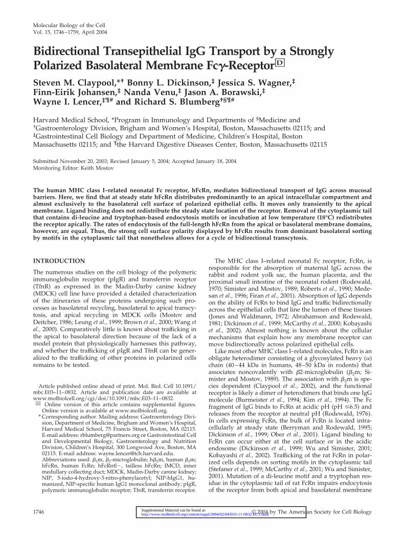
Because the cytoplasmic tail of many proteins, includingFcRn (Stefaner et al., 1999; McCarthy et al., 2001; Wu andSimister, 2001) and TfnR (Odorizzi et al., 1996; Odorizzi andTrowbridge, 1997), contains basolateral targeting motifs, wetested whether removal of the hFcRn cytoplasmic tail wouldalter its cell surface distribution. Two independently derivedstable MDCK cell clones expressing a tailless hFcRn(hFcRntl�) and h�2m were prepared and examined for cellsurface distribution as described above. In contrast to wild-type hFcRn, the hFcRntl� was detected on both the apicaland basolateral membrane domains in roughly equal quan-tities (Figure 9A). That the strict apical distribution of GP135was maintained in these clones indicated specificity for cellsurface labeling and that all of the MDCK clones analyzedmaintained both polarity and junctional integrity through-out the experimental protocol (Figure 9B). Thus, the cyto-plasmic tail of hFcRn contains one or more sorting motif(s)that when removed permits redistribution of a large amountof hFcRn (50% of the fraction on the cell surface) from analmost exclusively basolateral cell membrane localization tothe apical membrane.
Cell Surface Polarity of hFcRn Does Not Depend onDifferential Rates of Endocytosis from Apical orBasolateral MembranesThe strong basolateral cell surface polarity of hFcRn mayresult from different rates of endocytosis from apical and
basolateral membranes. To test this idea, we examined therates of hFcRn endocytosis from both cell surfaces. In thesestudies, we used disulfide-linked biotin (sulfo-NHS-SS-bi-otin) to label apical or basolateral cell surface proteins at 4°Cand then shifted the temperature to 18 or 37°C to allow forendocytosis. Using a membrane-impermeant reducingagent, the biotin tag is removed from only those proteins atthe cell surface, whereas biotinylated proteins inside the cellremain labeled with biotin. Thus in these studies, the hFcRn-SS-biotin that is detected by immunoblot after avidin-agar-ose precipitation represents the fraction of hFcRn that wasinternalized by endocytosis. As control, the rates of hFcRnendocytosis were compared with that of TfnR using thesame method.
The time course of hFcRn endocytosis from the basolateralmembrane (Figure 10, A–C) was rapid and similar to that ofTfnR at both 18 and 37°C (compare top and bottom panels).Endocytosis of both proteins was slower at 18°C. Immuno-blots for �-actin show that equal amounts of total cell lysateswere present in each sample during affinity precipitation(Figure 10B). These data are quantified in Figure 10C.
To measure endocytosis of hFcRn and TnfR from theapical membrane, MDCK cell monolayers expressing hFcRnwere first incubated overnight at 18°C to redistribute bothreceptors from the basolateral membrane to the apical mem-brane. The rates of apical hFcRn and TnfR endocytosis at 37and 18°C were measured as described above (Figure 10,D–F). As for basolateral hFcRn, the time course of apicalhFcRn endocytosis was rapid and similar to that of TfnR atboth temperatures (Figure 10D, middle and bottom panels).Endocytosis of the resident apical membrane protein GP135,which is anchored to the cytoskeleton, was not apparent ateither temperature (Figure 10D, top panel). Again, immuno-blots for �-actin show that equal amounts of total cell lysateswere used in each condition (Figure 10E). Figure 10F quan-tifies these data. These data show that endocytosis of hFcRnfrom the apical and basolateral membranes occurs at thesame rate and with similar efficiency to that of TfnR, consis-tent with endocytosis of FcRn by clathrin-mediated mecha-nisms (Rodewald and Kraehenbuhl, 1984). Thus, the redis-tribution of hFcRn to apical membranes seen duringincubations at 18°C or after removal of the hFcRn cytoplas-mic tail parallels that of TfnR and cannot be explained bydifferential rates of endocytosis. These results suggest thatthe cytoplasmic tail of hFcRn contains a dominant basolat-eral sorting motif(s) that distributes the receptor basolater-ally at steady state.
DISCUSSION
The results of these studies indicate that the fraction of humanFcRn on the cell surface of epithelial cells lining mucosal sur-faces is strictly polarized to the basolateral membrane. This isexplained by the presence of a dominant basolateral sortingmotif located in the cytoplasmic tail of the human receptor.Such a strict basolateral polarity of the human FcRn is observedfor hFcRn heterologously expressed in canine MDCK cells andendogenously expressed in human intestinal T84 and Caco-2cell lines. In contrast, the rat FcRn expressed in rat inner med-ullary collecting duct (IMCD) cells is only weakly polarized tothe basolateral plasma membrane (McCarthy et al., 2001; Wuand Simister, 2001), and the opposite polarity is observed forthe low-affinity Fc receptor Fc�RIIb fused to the rat FcRn cy-toplasmic tail in MDCK cells (Stefaner et al., 1999). We alsobelieve that the studies on FcRn in rat IMCD cells (McCarthy etal., 2000, 2001; McCarthy et al., 2001) are not conclusive as asignificant fraction of the rat FcRn found on the cell surface
Figure 7. Evidence for a dynamic pool of hFcRn on the apicalplasma membrane. Anti-HA mAb, with or without mouse anti-GP135 mAb, was added to the apical or basolateral chamber ofconfluent monolayers buffered at pH 7.3. After incubation for 90min at the indicated temperature and subsequent extensive wash-ing, antigen-antibody complexes were captured from equal vol-umes of lysate with protein A-Sepharose. (A) The recovered sam-ples were analyzed by anti-GP135 (top panel) and anti-HA (bottompanel) immunoblotting as described in Figure 5. (B) A 20-�L aliquotof each lysate, sampled just before washing the protein A-Sepharoseprecipitates, was analyzed for total protein content by anti–�-actinimmunoblotting as described in Figure 1. These data were con-firmed utilizing two different hFcRn�/h�2m� MDCK clones. Dataare presented from a single clone (n � 3).
S.M. Claypool et al.
Molecular Biology of the Cell1754

contains the ER form of N-linked oligosaccharides, suggestingthe presence of nonphysiologic trafficking in this model sys-tem.
The different cell surface polarities displayed by the full-length rat and human receptor correlates with different cel-lular physiology. Here, we find that the hFcRn transportsIgG more efficiently in the basolateral to apical direction.This is opposite to that found for the rat receptor (McCarthyet al., 2000), but consistent with our previous work (Claypool
et al., 2002) and with the function of hFcRn in placentalendothelial cells (Antohe et al., 2001). The reason for thesedifferences in trafficking of the human and rat receptor arenot known, though perhaps they are related to a strongerapical membrane-targeting motif located somewhere in thestructure of the rat receptor. A candidate apical-sorting mo-tif might be the four N-linked glycosylation sites present inthe ectodomain of the rat FcRn, three of which are lacking inthe human receptor.
Figure 8. Loss of strict hFcRn basolateral polarity upon prolonged incubation at 18°C. Confluent MDCK monolayers were incubated at 18°Cfor the indicated times before selective cell surface biotinylation. (A) FcRn immunoprecipitation, avidin-agarose reprecipitation, and anti-HAimmunoblotting were performed as described in Figure 5. (B) Data from three separate experiments with two individual hFcRn�/h�2m�MDCK clones were quantitated using Quantitation One software (Bio-Rad). After normalization to the amount of hFcRn present in eachimmunoprecipitate, the relative apical and basolateral distribution of hFcRn was calculated as percentages of the total surface hFcRn for eachtimepoint as follows: A/(A�B) � 100 and B/(A�B) � 100, where A is the normalized volume of hFcRn detected on the apical surface andB is the normalized volume of hFcRn on the basolateral membrane. (C and D) Where indicated, 10 �M cycloheximide was added to themedium during the overnight incubation at 18°C. Surface proteins were captured by the addition of avidin-agarose and FcRn (C) or TfnR(D) detected by immunoblot as described in Figure 5. (E) Data from three separate experiments and two different hFcRn�/h�2m� MDCKclones were quantitated as in B. After �-actin normalization, the relative apical and basolateral distribution of hFcRn and TfnR on the apicalor basolateral cell surface at 37, 18, and 18°C in the presence of cycloheximide was calculated as in B. (F) Avidin-agarose precipitation of theanti-FcRn flow-through from (A) and anti–E-cadherin immunoblotting were performed as previously described (n � 3).
Basolateral Membrane Targeting of Human FcRn
Vol. 15, April 2004 1755
Our studies show that human FcRn has a strong basolat-eral targeting motif located in the cytoplasmic tail, as re-moval of the cytoplasmic tail by mutagenesis redistributesthe receptor apically. This cannot be explained by differen-tial rates of endocytosis from apical and basolateral mem-brane domains. Thus, the strong cell surface polarity dis-played by FcRn results from dominant basolateral sorting.At least one step in the sorting of hFcRn to basolateralmembranes, however, must be differentially sensitive to lowtemperature because incubations at 18°C redistributes thewild-type receptor apically. Redistribution of hFcRn stilloccurs in the absence of protein synthesis, indicating thatdominant basolateral sorting for hFcRn occurs in the endo-some rather than in the secretory pathway.
Very similar results were observed with the membranetrafficking receptor for transferrin. The cytoplasmic tail ofTnfR also contains a strong basolateral targeting motif, andthe receptor is strictly localized to the basolateral membraneat steady state (Odorizzi et al., 1996; Odorizzi and Trow-bridge, 1997). Like hFcRn, a small fraction of the TfnR canmove bidirectionally across polarized MDCK cell monolay-ers by sorting through a common endosomal compartment(Odorizzi et al., 1996). Thus, the strong basolateral polarity ofa protein at steady state does not preclude the possibility ofbidirectional transcytosis. It is possible that the cytoplasmictails of TfnR and hFcRn share a functionally similar struc-ture. In contrast to TfnR, however, FcRn localizes predomi-nantly to an apical intracellular compartment, which mayrepresent the common endosome. Also trafficking by FcRnthrough the transcytotic pathway cannot be the same as forTfnR because it is more efficient, delivering physiologicallevels of IgG across the monolayer in both directions (Spiek-ermann et al., 2002 and our unpublished results).
Interestingly, TfnR, like hFcRn, was demonstrated to un-dergo a temperature-induced redistribution to the apical cellsurface. Another basolateral membrane protein, E-cadherin,maintained its strict basolateral polarity even after an over-night incubation at 18°C, demonstrating that the observed18°C effect is not common to all basolaterally restrictedreceptors. Thus, the cytoplasmic tails of TfnR and hFcRnshare a functionally similar motif that is active in the endo-somal system as a basolateral cell surface retrieval signal
and that is temperature sensitive. Mutagenesis studies hadpreviously determined that the cytoplasmic tail of TfnRcontained separate basolateral sorting motifs that were func-tional either in the biosynthetic or endocytic pathways(Odorizzi and Trowbridge, 1997). Moreover, residues 29–35of the cytoplasmic tail were identified as the motif operativein the biosynthetic pathway. In contrast, the molecular na-ture of the endocytic basolateral motif eluded identification.Similarly, although previous work on rat FcRn revealed thepresence of a basolateral sorting signal in the cytoplasmictail of rat FcRn (Stefaner et al., 1999; Wu and Simister, 2001),this motif has not been identified, although a di-leucinemotif in the cytoplasmic tail has been clearly demonstratedto not encode basolateral sorting information. Perhaps, theobserved redistribution of both TfnR and hFcRn at 18°C willprovide a useful assay that combined with site-directedmutagenesis will identify these elusive endocytically active,basolateral determinants.
Even with the strict basolateral membrane polarity ofFcRn at steady state, we find that hFcRn moves transiently tothe apical plasma membrane. This is evidenced most clearlyby the increase in internalization and transcytosis of IgGwhen apical reservoirs are clamped at pH 6.0, which ispermissive for FcRn binding, and by capture of an apicallyapplied antibody at 37 but not 4°C. The rapid and transientappearance of hFcRn at the apical cell surface may reflect anadaptation to protect hFcRn from digestive enzymes presentin luminal secretions. In support of this view, it was previ-ously demonstrated that pretreatment of neonatal rat prox-imal small intestinal loops with luminal trypsin drasticallyreduced the quantity of pH-dependent IgG binding to apicalmembranes (Borthistle et al., 1977).
The immediate luminal environment adjacent to the apicalmembrane of cells lining the intestine and probably all otherNa�-absorbing mucosal surfaces in the human and othermammals is acidic because of the activity of the sodium-hydrogen exchanger NHE3 (Tse et al., 1993; Noel et al., 1996).The NHE3 creates an inwardly directed proton gradientacross the brush border membrane of intestinal epithelialcells that is harnessed by proton-coupled solute-transportersto drive peptide and Fe2� absorption (Liang et al., 1995;Gunshin et al., 1997). Because FcRn releases IgG only very
Figure 9. Tailless hFcRn is detected on boththe apical and basolateral plasma membranedomains. Selective cell surface biotinylationof confluent MDCK monolayers was per-formed as described in Figure 5. (A) FcRnimmunoprecipitation, detection of mem-brane-associated hFcRn, and FcRn immuno-blotting were performed as described in Fig-ure 5. Lysates were derived from a controlhFcRn�/h�2m� MDCK clone and twoclones coexpressing hFcRntl� and h�2m(hFcRn�/h�2m�). Ten micrograms of wholecell lysate was analyzed directly to confirmthe presence and migration of tailless hFcRnafter immunoprecipitation (lane 10). (B) Avi-din-agarose precipitation of the anti-FcRnflow-through and anti-GP135 immunoblot-ting were performed as described in Figure 5(n � 3).
S.M. Claypool et al.
Molecular Biology of the Cell1756

slowly at acidic pH, we propose that in vivo the FcRn-IgGcomplex may remain intact after transport to the apicalmembrane. Here, the FcRn-IgG complex may bind cognateluminal antigens and then efficiently recycle back into andacross the epithelial barrier where the immune complex isreleased for processing by dendritic cells located in thesubepithelial space. In this way, FcRn and IgG may partic-ipate in immune surveillance at mucosal surfaces.
Finally, our results show that even though hFcRn inMDCK cells mediates the bidirectional transport of IgGacross the monolayer, hFcRn does not appear to redistributefrom its predominant intracellular or basolateral cell surfacedistribution after ligand binding. The lack of any markedredistribution of hFcRn at the cell surface or within vesiclesinduced by bovine IgG was perhaps not unexpected giventhe low affinity of hFcRn for bovine IgG (Ober et al., 2001)and the recent retraction of studies claiming otherwise (Re-traction, 2002; Ramalingam et al., 2002). Although the lack ofeffect on hFcRn distribution after receptor binding to humanIgG is somewhat counterintuitive, as the receptor presum-
ably moves with its ligand into and across the cell, it isconsistent with the fact that the Fc�RII/rat FcRn chimerawas demonstrated capable of bidirectional transport in theabsence of ligand (Stefaner et al., 1999). Thus, it may beargued that IgG binding is not a strict requirement for FcRntranscytosis. Further, it is possible that the bidirectionaltrafficking of hFcRn, and thus transport of IgG, is constitu-tive with no regulation by bound cargo. Directional trans-port might therefore be specified by the presence of gradi-ents external to FcRn, such as ligand and pH gradients. Thatthese types of gradients are capable of powerfully harness-ing this receptor system is perhaps best exemplified in theclassical setting for FcRn; the neonatal rat intestine.
ACKNOWLEDGMENTS
S.M.C. thanks Drs. Karl Matlin, Hidde Ploegh, and Cox Terhorst for criticallyimportant guidance and thoughtful discussions and Daniel T. Bailey and EwaMicewicz for technical assistance. This work was supported by NationalInstitutes of Health Grants DK53056 (to R.S.B. and W.I.L.), DK44319 and
Figure 10. Endocytosis of hFcRn from the apical and basolateral plasma membranes is delayed at low temperature. (A–C) Confluent MDCKmonolayers were biotinylated on the basolateral plasma membrane with the cleavable biotin analog, sulfo-NHS-SS-biotin. (D–F) Toaccumulate hFcRn on the apical surface, an overnight 18°C incubation was performed before biotinylation. (A and D) After quenching ofunreacted biotin, filters were incubated for the indicated times at 37 or 18°C and then rapidly cooled to 4°C. Remaining membrane-associatedbiotin was then removed from the apical and basolateral membranes with a glutathione-based reducing solution (lanes 3–12). Two filters perexperiment were maintained at 4°C throughout the experiment, serving as a biotinylation control (lane 1) and a control for the efficiency ofbiotin reduction (lane 2). Total biotinylated surface proteins were precipitated with avidin-agarose and analyzed by immunoblot. (B and E)�-actin immunoblot of each lysate (10 �g) was performed as previously described. (C and F) Data from two different hFcRn�/h�2m� MDCKclones were quantitated as in Figure 8. After normalization to �-actin, the relative amounts of hFcRn and TfnR endocytosed at 37 and 18°Cat each time point were expressed as a percentage of the total initial cell surface receptor (n � 2).
Basolateral Membrane Targeting of Human FcRn
Vol. 15, April 2004 1757
DK51362 (to R.S.B.), DK48106 and DK57827 (to W.I.L.) and DK34854 to theHarvard Digestive Diseases Center.
REFERENCES
Retraction. (2002). EMBO J. 21, 5953.
Abrahamson, D.R., and Rodewald, R. (1981). Evidence for the sorting ofendocytic vesicle contents during the receptor-mediated transport of IgGacross the newborn rat intestine. J. Cell Biol. 91, 270–280.
Antohe, F., Radulescu, L., Gafencu, A., Ghetie, V., and Simionescu, M. (2001).Expression of functionally active FcRn and the differentiated bidirectionaltransport of IgG in human placental endothelial cells. Hum. Immunol. 62,93–105.
Berryman, M., and Rodewald, R. (1995). Beta 2-microglobulin co-distributeswith the heavy chain of the intestinal IgG-Fc receptor throughout the trans-epithelial transport pathway of the neonatal rat. J. Cell Sci. 108, 2347–2360.
Borthistle, B.K., Kubo, R.T., Brown, W.R., and Grey, H.M. (1977). Studies onreceptors for IgG on epithelial cells of the rat intestine. J. Immunol. 119,471–476.
Borvak, J., Richardson, J., Medesan, C., Antohe, F., Radu, C., Simionescu, M.,Ghetie, V., and Ward, E.S. (1998). Functional expression of the MHC classI-related receptor, FcRn, in endothelial cells of mice. Int. Immunol. 10, 1289–1298.
Brambell, F.W. (1966). The transmission of immunity from mother to youngand the catabolism of immunoglobulins. Lancet 2, 1087–1093.
Brown, D.A., Crise, B., and Rose, J.K. (1989). Mechanism of membrane an-choring affects polarized expression of two proteins in MDCK cells. Science245, 1499–1501.
Brown, P.S., Wang, E., Aroeti, B., Chapin, S.J., Mostov, K.E., and Dunn, K.W.(2000). Definition of distinct compartments in polarized Madin-Darby caninekidney (MDCK) cells for membrane-volume sorting, polarized sorting andapical recycling. Traffic 1, 124–140.
Burmeister, W.P., Huber, A.H., and Bjorkman, P.J. (1994). Crystal structure ofthe complex of rat neonatal Fc receptor with Fc [see comments]. Nature 372,379–383.
Casanova, J.E., Apodaca, G., and Mostov, K.E. (1991). An autonomous signalfor basolateral sorting in the cytoplasmic domain of the polymeric immuno-globulin receptor. Cell 66, 65–75.
Cepek, K.L., Shaw, S.K., Parker, C.M., Russell, G.J., Morrow, J.S., Rimm, D.L.,and Brenner, M.B. (1994). Adhesion between epithelial cells and T lympho-cytes mediated by E-cadherin and the alpha E beta 7 integrin. Nature 372,190–193.
Claypool, S.M., Dickinson, B.L., Yoshida, M., Lencer, W.I., and Blumberg, R.S.(2002). Functional reconstitution of human FcRn in Madin-Darby caninekidney cells requires co-expressed human beta 2-microglobulin. J. Biol. Chem.277, 28038–28050.
Dickinson, B.L., Badizadegan, K., Wu, Z., Ahouse, J.C., Zhu, X., Simister, N.E.,Blumberg, R.S., and Lencer, W.I. (1999). Bidirectional FcRn-dependent IgGtransport in a polarized human intestinal epithelial cell line. J. Clin. Invest.104, 903–911.
Dunn, K., McGraw, T., and Maxfield, F. (1989). Iterative fractionation ofrecycling receptors from lysosomally destined ligands in an early sortingendosome. J. Cell Biol. 109, 3303–3314.
Firan, M., Bawdon, R., Radu, C., Ober, R.J., Eaken, D., Antohe, F., Ghetie, V.,and Ward, E.S. (2001). The MHC class I-related receptor, FcRn, plays anessential role in the maternofetal transfer of gamma-globulin in humans. Int.Immunol. 13, 993–1002.
Galloway, C.J., Dean, G.E., Marsh, M., Rudnick, G., and Mellman, I. (1983).Acidification of macrophage and fibroblast endocytic vesicles in vitro. Proc.Natl. Acad. Sci. USA 80, 3334–3338.
Gibson, A. et al. (1998). Sorting mechanisms regulating membrane proteintraffic in the apical transcytotic pathway of polarized MDCK cells. J. Cell Biol.143, 81–94.
Graeve, L., Drickamer, K., and Rodriguez-Boulan, E. (1989). Polarized endo-cytosis by Madin-Darby canine kidney cells transfected with functionalchicken liver glycoprotein receptor. J. Cell Biol. 109, 2809–2816.
Gunshin, H., Mackenzie, B., Berger, U.V., Gunshin, Y., Romero, M.F., Boron,W.F., Nussberger, S., Gollan, J.L., and Hediger, M.A. (1997). Cloning andcharacterization of a mammalian proton-coupled metal-ion transporter. Na-ture 388, 482–488.
Johansen, F.E., Natvig Norderhaug, I., Roe, M., Sandlie, I., and Brandtzaeg, P.(1999). Recombinant expression of polymeric IgA: incorporation of J chainand secretory component of human origin. Eur. J. Immunol. 29, 1701–1708.
Jones, E., and Waldmann, T. (1972). The mechanism of intestinal uptake andtranscellular transport of IgG in neonatal rat. J. Clin. Invest. 51, 2916–2927.
Kim, J.K., Tsen, M.F., Ghetie, V., and Ward, E.S. (1994). Localization of the siteof the murine IgG1 molecule that is involved in binding to the murineintestinal Fc receptor. Eur. J. Immunol. 24, 2429–2434.
Kobayashi, N., Suzuki, Y., Tsuge, T., Okumura, K., Ra, C., and Tomino, Y.(2002). FcRn-mediated transcytosis of immunoglobulin G in human renalproximal tubular epithelial cells. Am. J. Physiol. Renal Physiol. 282, F358–F365.
Leung, S.M., Rojas, R., Maples, C., Flynn, C., Ruiz, W.G., Jou, T.S., andApodaca, G. (1999). Modulation of endocytic traffic in polarized Madin-Darbycanine kidney cells by the small GTPase RhoA. Mol. Biol. Cell 10, 4369–4384.
Liang, R., Fei, Y.J., Prasad, P.D., Ramamoorthy, S., Han, H., Yang-Feng, T.L.,Hediger, M.A., Ganapathy, V., and Leibach, F.H. (1995). Human intestinalH�/peptide cotransporter. Cloning, functional expression, and chromosomallocalization. J. Biol. Chem. 270, 6456–6463.
Matlin, K.S., and Simons, K. (1983). Reduced temperature prevents transfer ofa membrane glycoprotein to the cell surface but does not prevent terminalglycosylation. Cell 34, 233–243.
McCarthy, K.M., Lam, M., Subramanian, L., Shakya, R., Wu, Z., Newton, E.E.,and Simister, N.E. (2001). Effects of mutations in potential phosphorylationsites on transcytosis of FcRn. J. Cell Sci. 114, 1591–1598.
McCarthy, K.M., Yoong, Y., and Simister, N.E. (2000). Bidirectional transcy-tosis of IgG by the rat neonatal Fc receptor expressed in a rat kidney cell line:a system to study protein transport across epithelia. J. Cell Sci. 113, 1277–1285.
Medesan, C., Radu, C., Kim, J.K., Ghetie, V., and Ward, E.S. (1996). Localiza-tion of the site of the IgG molecule that regulates maternofetal transmission inmice. Eur. J. Immunol. 26, 2533–2536.
Mostov, K.E., and Deitcher, D.L. (1986). Polymeric immunoglobulin receptorexpressed in MDCK cells transcytoses IgA. Cell 46, 613–621.
Noel, J., Roux, D., and Pouyssegur, J. (1996). Differential localization ofNa�/H� exchanger isoforms (NHE1 and NHE3) in polarized epithelial celllines. J. Cell Sci. 109 (Pt 5), 929–939.
Norderhaug, L., Olafsen, T., Michaelsen, T.E., and Sandlie, I. (1997). Versatilevectors for transient and stable expression of recombinant antibody moleculesin mammalian cells. J. Immunol. Methods 204, 77–87.
Ober, R.J., Radu, C.G., Ghetie, V., and Ward, E.S. (2001). Differences inpromiscuity for antibody-FcRn interactions across species: implications fortherapeutic antibodies. Int. Immunol. 13, 1551–1559.
Odorizzi, G., Pearse, A., Domingo, D., Trowbridge, I.S., and Hopkins, C.R.(1996). Apical and basolateral endosomes of MDCK cells are interconnectedand contain a polarized sorting mechanism. J. Cell Biol. 135, 139–152.
Odorizzi, G., and Trowbridge, I.S. (1997). Structural requirements for baso-lateral sorting of the human transferrin receptor in the biosynthetic andendocytic pathways of Madin-Darby canine kidney cells. J. Cell Biol. 137,1255–1264.
Ojakian, G.K., and Schwimmer, R. (1988). The polarized distribution of anapical cell surface glycoprotein is maintained by interactions with the cy-toskeleton of Madin-Darby canine kidney cells. J. Cell Biol. 107, 2377–2387.
Praetor, A., Ellinger, I., and Hunziker, W. (1999). Intracellular traffic of theMHC class I-like IgG Fc receptor, FcRn, expressed in epithelial MDCK cells.J. Cell Sci. 112, 2291–2299.
Ramalingam, T.S., Detmer, S.A., Martin, W.L., and Bjorkman, P.J. (2002). IgGtranscytosis and recycling by FcRn expressed in MDCK cells reveals ligand-induced redistribution. EMBO J. 21, 590–601.
Roberts, D.M., Guenthert, M., and Rodewald, R. (1990). Isolation and charac-terization of the Fc receptor from the fetal yolk sac of the rat. J. Cell Biol. 111,1867–1876.
Rodewald, R. (1970). Selective antibody transport in the proximal smallintestine of the neonatal rat. J. Cell Biol. 45, 635–640.
Rodewald, R. (1976). pH-dependent binding of immunoglobulins to intestinalcells of the neonatal rat. J. Cell Biol. 71, 666–669.
Rodewald, R., and Kraehenbuhl, J.P. (1984). Receptor-mediated transport ofIgG. J. Cell Biol. 99, 159s–164s.
Simister, N.E., and Mostov, K.E. (1989). An Fc receptor structurally related toMHC class I antigens. Nature 337, 184–187.
S.M. Claypool et al.
Molecular Biology of the Cell1758

Singer, K.L., and Mostov, K.E. (1998). Dimerization of the polymeric immu-noglobulin receptor controls its transcytotic trafficking. Mol. Biol. Cell 9,901–915.
Spiekermann, G.M., Finn, P.W., Ward, E.S., Dumont, J., Dickinson, B.L.,Blumberg, R.S., and Lencer, W.I. (2002). Receptor-mediated immunoglobulinG transport across mucosal barriers in adult life: functional expression ofFcRn in the mammalian lung. J. Exp. Med. 196, 303–310.
Stefaner, I., Praetor, A., and Hunziker, W. (1999). Nonvectorial surface trans-port, endocytosis via a Di-leucine-based motif, and bidirectional transcytosisof chimera encoding the cytosolic tail of rat FcRn expressed in Madin-Darbycanine kidney cells. J. Biol. Chem. 274, 8998–9005.
Story, C.M., Mikulska, J.E., and Simister, N.E. (1994). A major histocompati-bility complex class I-like Fc receptor cloned from human placenta: possiblerole in transfer of immunoglobulin G from mother to fetus. J. Exp. Med. 180,2377–2381.
Tse, C.M., Levine, S.A., Yun, C.H., Brant, S.R., Pouyssegur, J., Montrose, M.H.,and Donowitz, M. (1993). Functional characteristics of a cloned epithelialNa�/H� exchanger (NHE3): resistance to amiloride and inhibition by pro-tein kinase C. Proc. Natl. Acad. Sci. USA 90, 9110–9114.
Wang, E., Brown, P.S., Aroeti, B., Chapin, S.J., Mostov, K.E., and Dunn, K.W.(2000). Apical and basolateral endocytic pathways of MDCK cells meet inacidic common endosomes distinct from a nearly-neutral apical recyclingendosome. Traffic 1, 480–493.
Ward, E.S., Zhou, J., Ghetie, V., and Ober, R.J. (2003). Evidence to support thecellular mechanism involved in serum IgG homeostasis in humans. Int.Immunol. 15, 187–195.
Wu, Z., and Simister, N.E. (2001). Tryptophan- and dileucine-based endocy-tosis signals in the neonatal fc receptor. J. Biol. Chem. 276, 5240–5247.
Zhu, X. et al. (2001). MHC class I-related neonatal Fc receptor for IgG isfunctionally expressed in monocytes, intestinal macrophages, and dendriticcells. J. Immunol. 166, 3266–3276.
Basolateral Membrane Targeting of Human FcRn
Vol. 15, April 2004 1759