Embed Size (px)
Citation preview
Neuroscience 217 (2012) 154–171
SELECTIVITY AND PERSISTENT FIRING RESPONSES TO SOCIALVOCALIZATIONS IN THE BASOLATERAL AMYGDALA
D. C. PETERSON � AND J. J. WENSTRUP *
Department of Anatomy and Neurobiology, Northeast Ohio
Medical University, 4209 State Route 44, Rootstown, Ohio
44272-0095, United States
Abstract—This study examined responsiveness to acoustic
stimuli among neurons of the basolateral amygdala. While
recording from single neurons in awake mustached bats
(Pteronotus parnellii), we presented a wide range of acoustic
stimuli including tonal, noise, and vocal signals. While many
neurons displayed phasic or sustained responses locked to
effective auditory stimuli, the majority of neurons (n= 58)
displayed a persistent excitatory discharge that lasted well
beyond stimulus duration and filled the interval between
successive stimuli. Persistent firing usually began seconds
(median value, 5.4 s) after the initiation of a train of repeated
stimuli and lasted, in the majority of neurons, for at least
2 min after the end of the stimulus train. Auditory-responsive
amygdalar neurons were generally excited by one stimulus
or very few stimuli. Most neurons did not respond well to
synthetic stimuli including tones, noise bursts or fre-
quency-modulated sweeps, but instead responded only to
vocal stimuli (82 of 87 neurons). Furthermore, most neurons
were highly selective among vocal stimuli. On average, neu-
rons responded to 1.7 of 15 different syllables or syllable
sequences. The largest percentage of neurons responded
to a hiss-like rectangular broadband noise burst (rBNB) call
associated with aggressive interactions. Responsiveness
to effective vocal stimuli was reduced or eliminated when
the spectrotemporal features of the stimuli were altered in
a subset of neurons. Chemical activation of themedial genic-
ulate body (MG) increased both background and evoked fir-
ing. Among 39 histologically localized recording sites, we
saw no evidence of topographic organization in terms of
temporal response pattern, habituation, or the affect of calls
to which neurons responded. Overall, these studies
0306-4522/12 $36.00 � 2012 IBRO. Published by Elsevier Ltd. All rights reservehttp://dx.doi.org/10.1016/j.neuroscience.2012.04.069
*Corresponding author. Tel: +1-(330)-325-6630; fax: +1-(330)-325-5916.
E-mail addresses: [email protected] (D. C. Peterson), [email protected] (J. J. Wenstrup).
� Present address: Department of Biomedical Sciences, Iowa StateUniversity, 2086 Veterinary Medicine, Ames, IA 50011, United States.Abbreviations: ASP, aspartate; BBN, broadband noise; bDFM, bentdownward frequency modulated; BIC, bicuculline; bUFM, bent upwardfrequency modulated; cDFM, checked downward frequency modu-lated; FM, frequency modulated; fRFM, fixed rippled frequency mod-ulated; fSFM, fixed sinusoidal frequency modulated; GLU, glutamate;hRFM, humped rippled frequency modulated; MG, medial geniculatebody; NBN, narrowband noise; NNBs, short, narrowband noise burst;PSTH, peri-stimulus time histogram; QCFI, long, quasi constant freq-uency; QCFs, short quasi constant frequency; rBNB, rectangular bro-adband noise burst; sAFM, single arched frequency modulated; s.d.,standard deviations; sHFM, short humped frequency modulated;sRFM, stretched rippled frequency modulated; TCFs, short, true con-stant frequency.
154
demonstrate that amygdalar neurons in the mustached bat
show high selectivity to vocal stimuli, and suggest that per-
sistent firing may be an important feature of amygdalar
responses to social vocalizations. � 2012 IBRO. Published
by Elsevier Ltd. All rights reserved.
Key words: bat, Pteronotus parnellii, persistent firing, acous-
tic communication, basolateral amygdala.
INTRODUCTION
The amygdala, a collection of diverse and interconnected
nuclei of the medial temporal lobe, is involved in establish-
ing the biological relevance of sensory stimuli and in
mediating many elements of emotional responses to
those stimuli (Cardinal et al., 2002; Sah et al., 2003; Pare
et al., 2004; Phelps and LeDoux, 2005; Heimer and Van
Hoesen, 2006). Although one aspect of the amygdala’s
role is to generate stereotypic responses to learned aver-
sive stimuli, a broader perspective is that the amygdala
participates in evaluating the biological significance or sal-
ience of a broad range of sensory stimuli. This includes
stimuli with either positive or negative valence or affect
(Heimer and Van Hoesen, 2006; Costafreda et al.,
2008), particularly sensory stimuli associated with social
interactions (Sander and Scheich, 2005; Ball et al.,
2007; Gothard et al., 2007; Sergerie et al., 2008; Van
Bavel et al., 2008; Andics et al., 2010). This study exam-
ines responses of neurons in the basolateral amygdala to
social vocalizations and other acoustic signals.
Given the salience of acoustic communication in
humans and some other animals, it is not surprising that
the amygdala may play a significant role in the analysis
of and response to social vocalizations. In humans, the
amygdala is implicated in processing the prosodic fea-
tures of speech. For example, increased amygdalar acti-
vation is associated with angry vs. neutral prosody
(Sander et al., 2005; Wiethoff et al., 2009), speech con-
taining either positive or negative emotions (Fecteau
et al., 2007), the intensity of emotion in speech (Leitman
et al., 2010), and the identity of voices (Andics et al.,
2010). Further, the amygdala is involved in disorders that
include an altered emotional response to speech, such as
schizophrenia (Sanjuan et al., 2007; Escarti et al., 2010),
autism (Gabis et al., 2008; Kim et al., 2010), and some
forms of post-traumatic stress (Protopopescu et al.,
2005; Shin et al., 2006).
In other species, early work showed that amygdalar
neurons respond to social vocalizations (Sawa and
d.
D. C. Peterson, J. J. Wenstrup /Neuroscience 217 (2012) 154–171 155
Delgado, 1963; O’Keefe and Bouma, 1969; Jacobs and
McGinty, 1972), but there have been few systematic stud-
ies of responses to social vocalizations by amygdalar
neurons. In recent work on mustached bats (Naumann
and Kanwal, 2011), big brown bats (Gadziola et al.,
2012), and rats (Parsana et al., 2012), conspecific vocal
signals evoked a variety of temporal response patterns
among basolateral amygdalar neurons, but neurons gen-
erally showed stronger excitatory discharge in response
to vocal signals with negative affect. Further, amygdalar
neurons in each species showed some evidence of dis-
crimination or selectivity among vocal calls. In big brown
bats, high neuronal discriminability is based on the dura-
tion of response to different vocal stimuli. In most of these
neurons, responses to some vocalizations extended well
beyond the duration of the stimulus. This persistent firing
may be a key feature of amygdalar responses. The pres-
ent study examines responses to vocal stimuli in the
amygdala of mustached bats, and shows that many amy-
gdalar neurons express selective responses to vocal sig-
nals through persistent firing.
EXPERIMENTAL PROCEDURES
We describe auditory responses of amygdalar neurons obtained
in 20 awake mustached bats (Pteronotus parnellii), captured in
Trinidad and Tobago. All procedures were approved by the Insti-
tutional Animal Care and Use Committee of the Northeast Ohio
Medical University (formerly, Northeastern Ohio Universities
College of Medicine) and were performed in accordance with
the National Institutes of Health guidelines for the care and use
of laboratory animals.
Surgery
Each bat was sedated with butorphanol (5 mg/kg, Fort Dodge
Animal Health, Fort Dodge, IA, USA), anesthetized with isoflu-
rane (1.5–2.0%; Abbott Laboratories, North Chicago, IL, USA),
and placed in a custom-made stereotaxic holder that we have
used extensively in studies throughout the auditory system of this
species (Wenstrup and Grose, 1995; Wenstrup, 1999; Portfors
and Wenstrup, 2001; Marsh et al., 2006). Depilatory lotion was
used to remove hair over the skull, and the skin was disinfected
with betadine. A midline incision was made in the skin over the
dorsal surface of the skull, and the underlying muscles were
reflected laterally. A metal pin was then cemented onto the skull
to secure the head during physiological experiments, and a tung-
sten wire was cemented through a small opening in the skull to
serve as a ground for electrophysiological recordings. Using sur-
face and stereotaxic coordinates, a small hole (<0.5 mm in
diameter) was opened in the skull to expose cerebral cortex dor-
sal to the amygdala. For the basolateral amygdala, we used coor-
dinates 5–6 mm rostral to the internucal crest, 3.2–3.6 mm lateral
to the mid-sagittal crest, and 4.5–5.2 mm below the cortical
surface.
After surgery, a local anesthetic (4% Lidocaine, Ferndale
Laboratories, Inc., Ferndale, MI, USA) and an antibiotic (Neospo-
rin, Pfizer, Morris Plains, NJ, USA) were applied to the surgical
area and the bat was returned to the holding cage. Two or three
days after surgery, physiological experiments were initiated.
Physiological experiments
To minimize distress, bats were lightly sedated with butorphanol
(2.5 mg/kg, i.p.) before they were placed in a stereotaxic holder
for physiological experiments. The apparatus was located within
a heated, single-walled acoustic chamber. To allow time for the
animals to fully recover from the sedative, physiological recording
began at least one hour after the drug was injected. Recording
sessions did not exceed 6 h/day and occurred no more than
every other day.
Acoustic stimulation. Acoustic stimuli were computer synthe-
sized or previously recorded and digitized at 250 kHz. All sounds
were downloaded to a digital signal processor (Microstar
DAP5216a, Microstar Laboratories, Bellevue, WA, USA), con-
verted to analog signals (sampling rate 400 kHz for synthesized
stimuli; 250 kHz for vocalizations), filtered (model FT6–2; Tuck-
er-Davis Technologies, Alachua, FL, USA), attenuated (model
PA5; Tucker-Davis Technologies, Alachua, FL, USA), and ampli-
fied (Parasound model HCA-10000A). Sounds were transmitted
through an EMIT-B (Infinity, www.infinitysystems.com) tweeter
that was placed 10 cm from the ear and 25� into the sound field
contralateral to the recorded amygdala.
Speaker performance was tested with a calibrated micro-
phone (Bruel and Kjaer, model 4135) placed 10 cm from the
speaker and 0� azimuth. From 10 to 120 kHz the speaker perfor-
mance showed a smooth decrease (approximately 3 dB/10 kHz).
Distortion components were not detectable �55 dB below the
signal level. For sounds with multiple frequency components
(i.e., communication calls, broadband noise, narrowband noise,
and upward/downward sweeps), sound levels are shown in dB
relative to maximum speaker output. For tonal signals, maximum
output at 20 kHz and 60 kHz was 99 and 87 dB SPL,
respectively.
Physiological recording. Neural recordings were obtained
using glass micropipette electrodes filled with physiological saline
(resistances typically 5–15 MO). The electrodes were stereotaxi-
cally inserted into the amygdala from the dorsal surface of the
brain. Neuronal activity was amplified, bandpass filtered (600–
6000 Hz), and digitized at a sampling rate of 40 kHz (Microstar
DAP5216a). Custom-made software calculated the time of occur-
rence of spikes and displayed peri-stimulus time histograms
(PSTHs), raster plots, and basic statistics of the neural
responses in real time.
An extensive set of natural and artificial sounds was pre-
sented at each recording site. Stimuli included tones, combina-
tions of tones, broadband noise (BBN), narrowband noise
(NBN), frequency-modulated (FM) upward and downward
sweeps, synthesized echolocation pulses, echoes, pulse–echo
combinations, and social vocalizations. For each of these catego-
ries, initial tests were followed by more targeted, additional tests
if an auditory response was detected. Stimuli were presented in
test sequences consisting of repetitions of the same stimulus at
a rate of 2 Hz.
Pure tones were presented at 82 combinations of frequency
and sound level (i.e., 18–100 kHz in 1-kHz steps, 0.5-ms rise/fall)
at levels 20 and 40 dB below maximum speaker output (78–47
and 58–27 dB SPL). BBN bursts (1–120 kHz, 20-dB attenuation)
were presented at durations ranging from 11 to 61 ms in steps of
10 ms. NBN (bandwidths: 2 or 10 kHz) was presented at center
frequencies between 25 and 95 kHz in steps of 10 kHz at
20- and 40-dB attenuation. In addition to these stimuli, 15 differ-
ent communication calls [obtained from Drs. J. Kanwal (Kanwal
et al., 1994) and C. Portfors] were presented to the animals at
levels ranging from 0 to 80 dB below maximum speaker output
in steps of 5 or 10 dB. To examine the influence of upward and
downward FM sweeps, we presented sweeps (band-
width = 10 kHz) over a range of center frequencies (95–25 kHz
in steps of 10 kHz; 0.5-ms rise/fall). Additional tests of FM
sweeps altered sweep duration (11–61 ms in steps of 10 ms)
and/or sound level (in 10-dB steps). Pulse–echo sequences were
generated to mimic recordings of sonar calls emitted by mus-
tached bats. Each pulse and echo had four harmonic elements
with relative levels based on previous work (Kawasaki et al.,
156 D. C. Peterson, J. J. Wenstrup /Neuroscience 217 (2012) 154–171
1988; Vater et al., 2003). Simulated echoes were shifted up by
1–3 kHz and delayed after the pulse by 2–10 ms to mimic differ-
ent Doppler shifts and sonar target distances.
To be considered responsive to a particular stimulus, a neu-
ron was required to display, in at least two test sequences, a dis-
charge significantly above background over 20 or more stimulus
repetitions (see Data analysis). This conservative criterion was
adopted to avoid false-positive responses in neurons that could
display altered background discharge rates for reasons that
appeared to be unrelated to acoustic stimuli. As a result, we
did not consider further neurons that habituated rapidly to
repeated acoustic stimuli.
Additional tests were performed on those neurons demon-
strating responsiveness to sounds. For tones, BBN, and NBN,
the sound level was varied in 10-dB steps and the duration of
the stimulus was varied in 5-ms steps (11–101 ms). For neurons
that responded to one or more communication calls, additional
tests were performed if time permitted. For these tests, acoustic
stimuli included: (1) time-reversed calls, (2) frequency-shifted
calls, (3) calls with removal of specific temporal or spectral ele-
ments, and (4) combinations of communication calls. All manipu-
lations of calls were performed using custom software created by
D. Gans.
Drug application in medial geniculate body (MG). In five ani-
mals, one or more excitatory drugs (glutamate (GLU); aspartate
(ASP); bicuculline (BIC)) (GABAa receptor antagonist) were ion-
tophoretically injected into MG in order to increase auditory exci-
tation to the amygdala. In these animals, single neuron recordings
were obtained fromMGusing amicropipette electrode (physiolog-
ical saline) mounted on a five-barrel pipette (Havey and Caspary,
1980). The tip of the multibarrel pipette was broken to a diameter
of 30 lm, and the unbroken tip of the single electrode extended
10–20 lm beyond the multibarrel pipette. To balance all currents
used to apply or retain drugs, the center barrel of the multibarrel
pipette was filled with physiological saline and connected to a
sum channel (Dagan programmable current generator, model
6400). Other barrels were filled with a cocktail of L-glutamate
(S)-2-aminopentanedioic acid (GLU) (500 mM, pH 8; Sigma, St.
Louis, MO, USA) and aspartate (S)-(+)-aminosuccinic acid
(ASP) (500 mM, pH 8; Sigma, St. Louis, MO, USA), or GABAa
receptor antagonist BIC (10 mM, pH 3.0, Sigma, St. Louis, MO,
USA). BIC was retained with negative current (�15 nA) and
ejected using positive currents (+15 to +30 nA), while GLU
and ASP were retained with positive current (+15 nA) and
ejected using negative currents (�15 to �30 nA). Iontophoresis
currents for drug application and retention were based on previ-
ous studies by Nataraj and Wenstrup (2005, 2006) and Bauer
et al. (2000). Pilot tests established iontophoretic current levels
that altered spike discharge of neurons in the MG.
For placement of multi-barreled pipettes in the mustached
bat’s MG, we used stereotaxic coordinates developed in previous
studies (Wenstrup and Grose, 1995; Wenstrup, 1999) but modi-
fied for this subspecies: 3.0–3.2 mm lateral from the midline,
2.2–3.0 mm in depth. Caudo-rostral position was 3.0 mm rostral
to the center of the surface of the inferior colliculus. To confirm
location within MG, auditory-evoked activity was recorded.
Successful application of drugs was confirmed by alterations in
these evoked responses. Location within MG, confirmed by
observation of the multi-barreled pipette track in Nissl-stained
material, was in the medial MG.
Data analysis
Most data were obtained from well-isolated single neurons
defined by spikes of constant waveform and amplitude, with peak
voltage exceeding background noise by a factor of five. When
necessary, custom offline spike-sorting software (D. Gans) was
used to segregate spikes into single units on the basis of wave-
form morphology.
To test whether spike counts differed significantly from back-
ground discharge, we used t-tests (paired or unpaired, depending
on the type of trial). Background activity was measured in 20 or 32
samples of a 200-ms time window with no sound presentation.
Spike counts that were significantly different from (greater or less
than) the background discharge (p< 0.05) were defined as a
response. As described above, we used a conservative criterion
requiring that the response be observed in more than one test
sequence for a given stimulus. Because background discharge
was often low (<5 spikes/s), observation of suppressive effects
of acoustic stimuli was infrequent. While we note suppressive
effects when observed, we have refrained from population
analyses.
In test sequences, spike discharge was obtained over 20 or
more presentations of a stimulus in a repeated pattern, and ana-
lyzed in 2- or 3-ms bins. The first-spike latency, or beginning of a
suppressive response, was calculated as the first bin after sound
presentation in which the number of spikes per second was sta-
tistically different from baseline. After the response was initiated,
the cessation of the response was calculated as the time in which
the spike count was no longer statistically different from baseline.
The duration of the excitatory or suppressive response was mea-
sured as the difference between the first-spike latency and the
end of the response.
Some neurons displayed slowly acting response habituation
(over several seconds), in which the spike count was initially sig-
nificantly above background, but later changed to become no dif-
ferent than the background spike count. The ‘‘latency’’ of
habituation was calculated as the time from initial response to
the time at which a response ceased to meet statistical criterion
for responsiveness. If the response returned, the duration of habit-
uation was calculated from the beginning of the habituated
response to the time when the response returned. To ensure that
the loss of recording stability would not influence our observation
of habituation, we required that the response recovered within
20 min.
Histological reconstruction
In each animal, up to four auditory responsive recording sites
received iontophoretic deposits of neuronal tracers: biotinylated
dextran amine (MW 10,000, 10% in saline, Molecular Probes,
Eugene, OR, USA); FluoroGold (4% in saline; FluoroChrome,
Inc., Englewood, CO, USA); FluoroRuby (tetramethylrhodamine
dextran, MW 10,000, 10% in saline, Molecular Probes, Eugene,
OR, USA); or fluorescein dextran (MW 10,000, 10% in saline,
Molecular Probes, Eugene, OR, USA). Depending on the tracer,
we used positive or negative current (5–7 lA; variable on/off) for
5–10 min. Because these deposits were also used for axoplas-
mic transport studies, no more than four sites were marked in
each animal. To localize recording sites, we combined use of
the stereotaxic coordinates with at least one tracer deposit in
each recorded amygdala.
After tracer deposits, animals survived for 5–14 days, then
were euthanized with an overdose of Fatal Plus (100 mg/kg,
i.p., Vortech, Dearborn, MI, USA). Following loss of corneal
and withdrawal reflexes, an animal was perfused with 0.1 M
phosphate-buffered saline, followed by 4% paraformaldehyde in
0.1 M phosphate buffer, pH 7.4 (PB). The brain was removed
and stored overnight at 4 �C in 4% paraformaldehyde with 30%
sucrose. After that time, brains were frozen, sectioned at
50 lm in the transverse plane, and collected into three series.
One or two series (to be used for fluorescence) were mounted
on gelatin-coated slides and coverslipped with DPX. One series
was mounted on gelatin-coated slides and stained with cresyl vio-
let for cytoarchitecture. In cases with BDA deposits, one series
was treated with avidin–biotin–peroxidase and then stained with
diaminobenzidine enhanced with nickel ammonium sulfate
(Adams, 1981). The sections were then mounted on gelatin-
coated slides, air-dried, and coverslipped with DPX.
C 8
Minimum First Spike Latency (ms)
Num
ber o
f Neu
rons
0
2
4
6
17010 30 50 70 90 110 130 150
Freq
. (kH
z)
Tria
ls
71
50 100 150 200Time (ms)
0
BfSFM
0
50
100
Time (ms) 0 50 100 150 200
Tria
ls
68
A
0
50
100
Freq
. (kH
z) hRFM
32
0
32
0
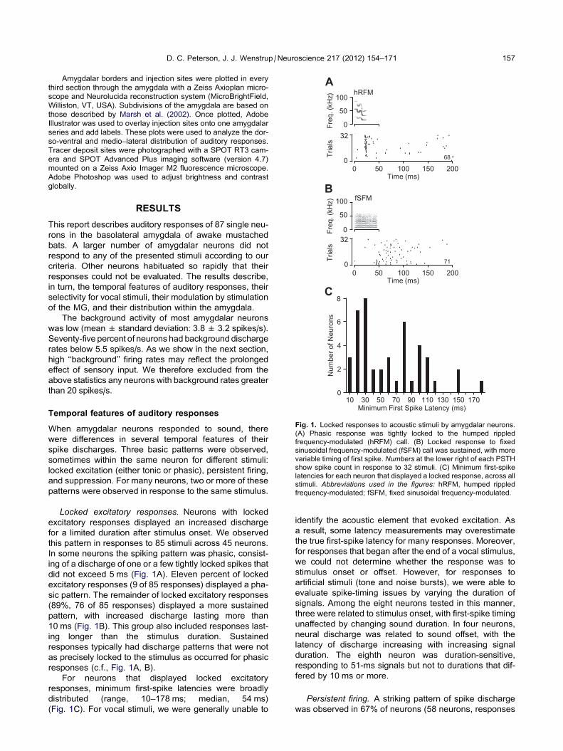
Fig. 1. Locked responses to acoustic stimuli by amygdalar neurons.
(A) Phasic response was tightly locked to the humped rippled
frequency-modulated (hRFM) call. (B) Locked response to fixed
sinusoidal frequency-modulated (fSFM) call was sustained, with more
variable timing of first spike. Numbers at the lower right of each PSTH
show spike count in response to 32 stimuli. (C) Minimum first-spike
latencies for each neuron that displayed a locked response, across all
stimuli. Abbreviations used in the figures: hRFM, humped rippled
frequency-modulated; fSFM, fixed sinusoidal frequency-modulated.
D. C. Peterson, J. J. Wenstrup /Neuroscience 217 (2012) 154–171 157
Amygdalar borders and injection sites were plotted in every
third section through the amygdala with a Zeiss Axioplan micro-
scope and Neurolucida reconstruction system (MicroBrightField,
Williston, VT, USA). Subdivisions of the amygdala are based on
those described by Marsh et al. (2002). Once plotted, Adobe
Illustrator was used to overlay injection sites onto one amygdalar
series and add labels. These plots were used to analyze the dor-
so-ventral and medio–lateral distribution of auditory responses.
Tracer deposit sites were photographed with a SPOT RT3 cam-
era and SPOT Advanced Plus imaging software (version 4.7)
mounted on a Zeiss Axio Imager M2 fluorescence microscope.
Adobe Photoshop was used to adjust brightness and contrast
globally.
RESULTS
This report describes auditory responses of 87 single neu-
rons in the basolateral amygdala of awake mustached
bats. A larger number of amygdalar neurons did not
respond to any of the presented stimuli according to our
criteria. Other neurons habituated so rapidly that their
responses could not be evaluated. The results describe,
in turn, the temporal features of auditory responses, their
selectivity for vocal stimuli, their modulation by stimulation
of the MG, and their distribution within the amygdala.
The background activity of most amygdalar neurons
was low (mean± standard deviation: 3.8 ± 3.2 spikes/s).
Seventy-five percent of neurons had background discharge
rates below 5.5 spikes/s. As we show in the next section,
high ‘‘background’’ firing rates may reflect the prolonged
effect of sensory input. We therefore excluded from the
above statistics any neurons with background rates greater
than 20 spikes/s.
Temporal features of auditory responses
When amygdalar neurons responded to sound, there
were differences in several temporal features of their
spike discharges. Three basic patterns were observed,
sometimes within the same neuron for different stimuli:
locked excitation (either tonic or phasic), persistent firing,
and suppression. For many neurons, two or more of these
patterns were observed in response to the same stimulus.
Locked excitatory responses. Neurons with locked
excitatory responses displayed an increased discharge
for a limited duration after stimulus onset. We observed
this pattern in responses to 85 stimuli across 45 neurons.
In some neurons the spiking pattern was phasic, consist-
ing of a discharge of one or a few tightly locked spikes that
did not exceed 5 ms (Fig. 1A). Eleven percent of locked
excitatory responses (9 of 85 responses) displayed a pha-
sic pattern. The remainder of locked excitatory responses
(89%, 76 of 85 responses) displayed a more sustained
pattern, with increased discharge lasting more than
10 ms (Fig. 1B). This group also included responses last-
ing longer than the stimulus duration. Sustained
responses typically had discharge patterns that were not
as precisely locked to the stimulus as occurred for phasic
responses (c.f., Fig. 1A, B).
For neurons that displayed locked excitatory
responses, minimum first-spike latencies were broadly
distributed (range, 10–178 ms; median, 54 ms)
(Fig. 1C). For vocal stimuli, we were generally unable to
identify the acoustic element that evoked excitation. As
a result, some latency measurements may overestimate
the true first-spike latency for many responses. Moreover,
for responses that began after the end of a vocal stimulus,
we could not determine whether the response was to
stimulus onset or offset. However, for responses to
artificial stimuli (tone and noise bursts), we were able to
evaluate spike-timing issues by varying the duration of
signals. Among the eight neurons tested in this manner,
three were related to stimulus onset, with first-spike timing
unaffected by changing sound duration. In four neurons,
neural discharge was related to sound offset, with the
latency of discharge increasing with increasing signal
duration. The eighth neuron was duration-sensitive,
responding to 51-ms signals but not to durations that dif-
fered by 10 ms or more.
Persistent firing. A striking pattern of spike discharge
was observed in 67% of neurons (58 neurons, responses
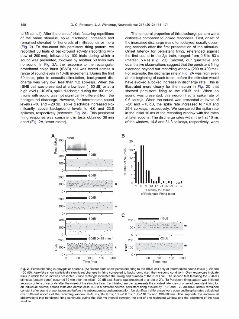
158 D. C. Peterson, J. J. Wenstrup /Neuroscience 217 (2012) 154–171
to 85 stimuli). After the onset of trials featuring repetitions
of the same stimulus, spike discharge increased and
remained elevated for hundreds of milliseconds or more
(Fig. 2). To document this persistent firing pattern, we
recorded 50 trials of background activity (recording win-
dow at 200 ms), followed by 100 trials during which a
sound was presented, followed by another 50 trials with
no sound. In Fig. 2A, the response to the rectangular
broadband noise burst (rBNB) call was tested across a
range of sound levels in 10-dB increments. During the first
50 trials, prior to acoustic stimulation, background dis-
charge was very low, less than 1.2 spikes/s. When the
rBNB call was presented at a low level (�50 dB) or at a
high level (�10 dB), spike discharge during the 100 repe-
titions with sound was not significantly different from the
background discharge. However, for intermediate sound
levels (�30 and �20 dB), spike discharge increased sig-
nificantly above background levels to 4.0 and 23.6
spikes/s, respectively (asterisks, Fig. 2A). This persistent
firing response was consistent in tests obtained 39 min
apart (Fig. 2A, lower raster).
200
100
0
200
100
0
200
100
0
200
100
0
-10dB
-30dB
-20dB
-50dB
A
*
*
Tria
ls
C
200
100
0 *
-20dB (+ 39 mins.)
0 50 100 150 200Time (ms)
B
0 Freq
. (kH
z)
100rBNB
Fig. 2. Persistent firing in amygdalar neurons. (A) Raster plots show persist
�30 dB). Asterisks show statistically significant changes in firing compared t
trials in which the sound was presented. Black rectangle indicates the timing
stimulus (bottom panel) occurred 39 min after the initial �20-dB test. Sound w
seconds or tens of seconds after the onset of the stimulus train. Each histogra
an individual neuron, across tests and across calls. (C) In a different neuron,
constant after sound presentation and before the subsequent sound presenta
over different epochs of the recording window: 0–10 ms, 0–50 ms, 100–20
observations that persistent firing continued during the 300-ms interval betw
window.
The temporal properties of this discharge pattern were
distinctive compared to locked responses. First, onset of
the increased discharge was often delayed, usually occur-
ring seconds after the first presentation of the stimulus.
Onset latency for persistent firing, referenced against
the first sound in the 2/s train, ranged from 0.5 to 63 s
(median 5.4 s) (Fig. 2B). Second, our qualitative and
quantitative observations suggest that the persistent firing
extended beyond our recording window (200 or 400 ms).
For example, the discharge rate in Fig. 2A was high even
at the beginning of each trace, before the stimulus would
have evoked a locked increase in discharge rate. This is
illustrated more clearly for the neuron in Fig. 2C that
showed persistent firing to the rBNB call. When no
sound was presented, this neuron had a spike rate of
0.6 spike/s. When the sound was presented at levels of
�20 and �10 dB, the spike rate increased to 14.0 and
29.6 spikes/s, respectively. We compared the spike rate
in the initial 10 ms of the recording window with the rates
at later epochs. The discharge rates within the first 10 ms
of the window, 14.8 and 31.3 spikes/s, respectively, were
200
100
0
-20dB
* Tria
ls
200
100
0
-10dB
*
Time (ms) 0 50 100 150 200
Latency to Onset of Prolonged Firing (sec)
Num
ber o
f Neu
rons
0
4
8
12
16
1 5 9 13 17 21 25 29 63
20
33
ent firing to the rBNB call only at intermediate sound levels (�20 and
o background (i.e., the no-sound condition). Gray rectangles indicate
and duration of the rBNB call. The second test featuring the �20-dBas presented at a rate of 2/s. (B) Persistent firing pattern was initiated
m bar represents the shortest latencies of onset of persistent firing for
persistent firing evoked by �10- and �20-dB rBNB stimuli remained
tion. No significant differences were observed in spike rates calculated
0 ms, 100–110 ms and 190–200 ms. This supports the audiovisual
een the end of one recording window and the beginning of the next
D. C. Peterson, J. J. Wenstrup /Neuroscience 217 (2012) 154–171 159
similar to those after the sound presentation. This result
suggests that the elevated discharge rate remained high
for the �400 ms between the offset of one stimulus and
the onset of the next. A third temporal feature of the per-
sistent firing pattern was related to firing after the stimulus
train ended. In 35 of the 85 responses featuring persistent
firing, the increased discharge lasted between 1 and
120 s. However, in the majority of cases (45/85
responses), persistent firing extended beyond 120 s and
lasted as long as 11 min beyond the last presented
stimulus. These results suggest that some amygdalar
neurons respond to optimal acoustic stimuli with an
increased discharge that lasts for hundreds of millisec-
onds, seconds, or even minutes.
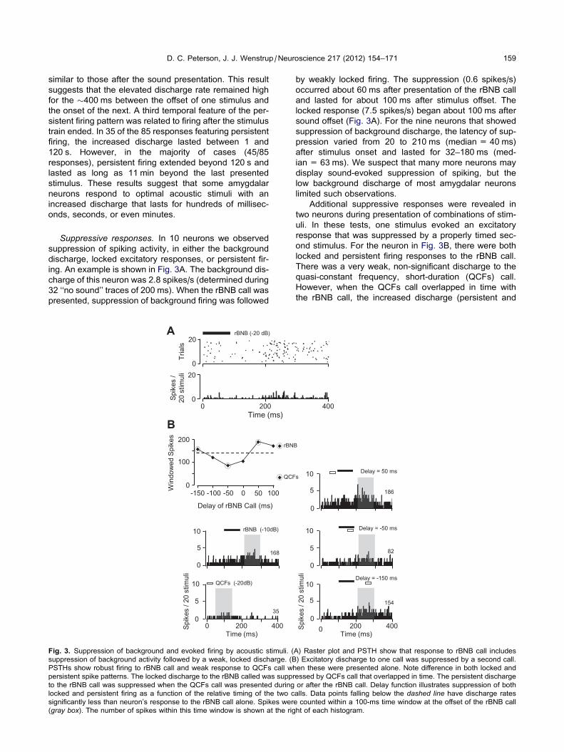
Suppressive responses. In 10 neurons we observed
suppression of spiking activity, in either the background
discharge, locked excitatory responses, or persistent fir-
ing. An example is shown in Fig. 3A. The background dis-
charge of this neuron was 2.8 spikes/s (determined during
32 ‘‘no sound’’ traces of 200 ms). When the rBNB call was
presented, suppression of background firing was followed
B
0
QCFs (-20dB)
0
5
10
rBNB (-10dB)
5
10
Spik
es /
20 s
timul
i
0
35
168
Time (ms) 200 400
Win
dow
ed S
pike
s
Delay of rBNB Call (ms)
-150 -100 -50 0 50 1000
100
200rBN
QCF
0
20
Spik
es /
20 s
timul
i
A
Time (ms) 0 200
Tria
ls 20
0
rBNB (-20 dB)
Fig. 3. Suppression of background and evoked firing by acoustic stimuli. (
suppression of background activity followed by a weak, locked discharge. (B
PSTHs show robust firing to rBNB call and weak response to QCFs call wh
persistent spike patterns. The locked discharge to the rBNB called was suppr
to the rBNB call was suppressed when the QCFs call was presented during
locked and persistent firing as a function of the relative timing of the two c
significantly less than neuron’s response to the rBNB call alone. Spikes wer
(gray box). The number of spikes within this time window is shown at the rig
by weakly locked firing. The suppression (0.6 spikes/s)
occurred about 60 ms after presentation of the rBNB call
and lasted for about 100 ms after stimulus offset. The
locked response (7.5 spikes/s) began about 100 ms after
sound offset (Fig. 3A). For the nine neurons that showed
suppression of background discharge, the latency of sup-
pression varied from 20 to 210 ms (median = 40 ms)
after stimulus onset and lasted for 32–180 ms (med-
ian = 63 ms). We suspect that many more neurons may
display sound-evoked suppression of spiking, but the
low background discharge of most amygdalar neurons
limited such observations.
Additional suppressive responses were revealed in
two neurons during presentation of combinations of stim-
uli. In these tests, one stimulus evoked an excitatory
response that was suppressed by a properly timed sec-
ond stimulus. For the neuron in Fig. 3B, there were both
locked and persistent firing responses to the rBNB call.
There was a very weak, non-significant discharge to the
quasi-constant frequency, short-duration (QCFs) call.
However, when the QCFs call overlapped in time with
the rBNB call, the increased discharge (persistent and
0
5
10
0
5
10
0
5
10
82
186
Delay = 50 ms
0 200 400
154
Time (ms)
B
s
Spik
es /
20 s
timul
i
Delay = -50 ms
Delay = -150 ms
400
A) Raster plot and PSTH show that response to rBNB call includes
) Excitatory discharge to one call was suppressed by a second call.
en these were presented alone. Note difference in both locked and
essed by QCFs call that overlapped in time. The persistent discharge
or after the rBNB call. Delay function illustrates suppression of both
alls. Data points falling below the dashed line have discharge rates
e counted within a 100-ms time window at the offset of the rBNB call
ht of each histogram.
9085
132
453
2629
403
150
Time (ms)
178
No sound (30 sweeps)
sweep 268-298
sweep 30-60
sweep 379-409
sweep 138-168
sweep 457-487
0 400200100 300
BBN (-20 dB)
0 400200100 300
0
0
A
C
B
D
E
F
G
H
0
0
15
0
15
0
15
0 Spik
es /
30 s
timul
i
60
Spik
es /
500
stim
uli
15
15
15
0
500
Swee
ps
100200300400
Spik
es /
30 s
timul
i
Fig. 4. Response habituation in an amygdalar neuron. (A) Raster
display of spike discharge during presentation of a broadband noise
burst over 500 consecutive trials. This neuron displayed both locked
and persistent response patterns. (B) PSTH of entire 500-trial
recording. (C) No-sound test immediately preceding the recordings
in (A) and (B). (D) PSTH shortly after sound presentation was
initiated. (E) Habituation preferentially affected persistent responses.
(F) Habituation affected both locked and persistent responses. (G)
Partial dishabituation, (H) Renewed habituation. Black bar indicatesthe timing and duration of the broadband noise stimulus. Number ofspikes in each histogram is shown at right.
160 D. C. Peterson, J. J. Wenstrup /Neuroscience 217 (2012) 154–171
locked) to the rBNB call was reduced or eliminated. No
suppression was observed when the two signals did not
overlap in time.
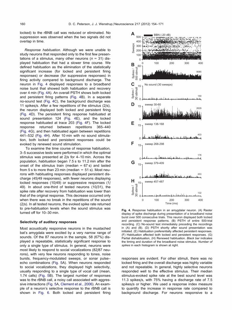
Response habituation. Although we were unable to
study neurons that responded only to the first few presen-
tations of a stimulus, many other neurons (n= 31) dis-
played habituation that had a slower time course. We
defined habituation as the elimination of the statistically
significant increase (for locked and persistent firing
responses) or decrease (for suppressive responses) in
firing activity compared to background discharge. The
neuron in Fig. 4 displayed responses to a broadband
noise burst that showed both habituation and recovery
over 4 min (Fig. 4A). An overall PSTH shows both locked
and persistent firing patterns (Fig. 4B). In a separate
no-sound test (Fig. 4C), the background discharge was
11 spikes/s. After a few repetitions of the stimulus (2/s),
the neuron displayed both locked and persistent firing
(Fig. 4D). The persistent firing response habituated at
sound presentation 124 (Fig. 4E), and the locked
response habituated at trace 203 (Fig. 4F). The locked
response returned between repetitions 365–440
(Fig. 4G), and then habituated again between repetitions
441–532 (Fig. 4H). After 10 min with no sound stimula-
tion, both locked and persistent responses could be
evoked by renewed sound stimulation.
To examine the time course of response habituation,
3–5 successive tests were performed in which the optimal
stimulus was presented at 2/s for 4–10 min. Across the
population, habituation began 7.5 s to 11.2 min after the
onset of the stimulus train (median = 67 s) and lasted
from 5 s to more than 23 min (median = 51 s). Most neu-
rons with habituating responses displayed persistent dis-
charge (45/49 responses), with fewer neurons displaying
locked responses (10/49) or suppressive responses (1/
49). In about one-third of tested neurons (10/31), the
spike rate after recovery from habituation was lower than
that of the original response. This decrease occurred only
when there was no break in the repetitions of the sound
(2/s). In all tested neurons, the evoked spike rate returned
to pre-habituation levels when the sound stimulus was
turned off for 10–30 min.
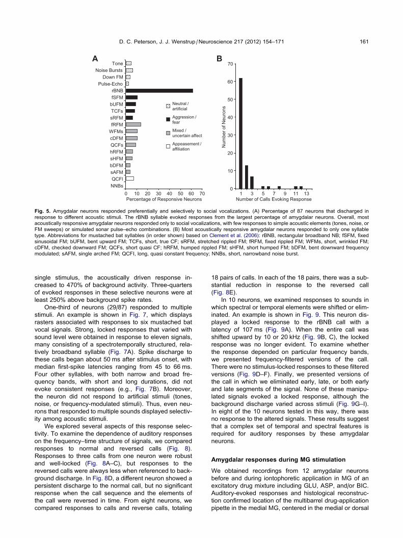
Selectivity of auditory responses
Most acoustically responsive neurons in the mustached
bat’s amygdala were excited by a very narrow range of
sounds. Of the 87 neurons in the sample, 58 (67%) dis-
played a repeatable, statistically significant response to
only a single type of stimulus. In general, neurons were
most likely to respond to social vocalizations (82/87 neu-
rons), with very few neurons responding to tones, noise
bursts, frequency-modulated sweeps, or sonar pulse–
echo combinations (Fig. 5A). When neurons responded
to social vocalizations, they displayed high selectivity,
usually responding to a single type of vocal call (mean,
1.74 calls) (Fig. 5B). The largest number of responses
was to the rBNB call, a noisy call associated with aggres-
sive interactions (Fig. 5A, Clement et al., 2006). An exam-
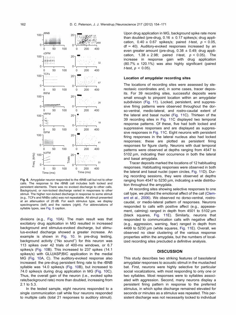
ple of a neuron’s selective response to the rBNB call is
shown in Fig. 6. Both locked and persistent firing
responses are evident. For other stimuli, there was no
locked firing and the overall discharge was highly variable
and not repeatable. In general, highly selective neurons
responded well to the effective stimulus. Their median
stimulus-evoked spike rate at the best sound level was
11.3 spikes/s, with 75% having a discharge rate of 7.5
spikes/s or higher. We used a response index measure
to quantify the increase in response rate compared to
background discharge. For neurons responsive to a
B70
0
10
20
30
40
50
60
1 3 5 9 11 13Number of Calls Evoking Response
7
Num
ber o
f Neu
rons
A
Percentage of Responsive Neurons 0 10 20 40 60 705030
fSFM
sRFM
bUFM TCFs
QCFl
Tone Noise Bursts
Down FM Pulse-Echo
rBNB
NNBs
WFMs
hRFM sHFM
sAFM
fRFM
cDFM
bDFM
QCFs
Neutral / artificial
Aggression / fear
Appeasement / affiliation
Mixed / uncertain affect
Fig. 5. Amygdalar neurons responded preferentially and selectively to social vocalizations. (A) Percentage of 87 neurons that discharged in
response to different acoustic stimuli. The rBNB syllable evoked responses from the largest percentage of amygdalar neurons. Overall, most
acoustically responsive amygdalar neurons responded only to social vocalizations, with few responses to simple acoustic elements (tones, noise, or
FM sweeps) or simulated sonar pulse–echo combinations. (B) Most acoustically responsive amygdalar neurons responded to only one syllable
type. Abbreviations for mustached bat syllables (in order shown) based on Clement et al. (2006): rBNB, rectangular broadband NB; fSFM, fixed
sinusoidal FM; bUFM, bent upward FM; TCFs, short, true CF; sRFM, stretched rippled FM; fRFM, fixed rippled FM; WFMs, short, wrinkled FM;
cDFM, checked downward FM; QCFs, short quasi CF; hRFM, humped rippled FM; sHFM, short humped FM; bDFM, bent downward frequency
modulated; sAFM, single arched FM; QCFI, long, quasi constant frequency; NNBs, short, narrowband noise burst.
D. C. Peterson, J. J. Wenstrup /Neuroscience 217 (2012) 154–171 161
single stimulus, the acoustically driven response in-
creased to 470% of background activity. Three-quarters
of evoked responses in these selective neurons were at
least 250% above background spike rates.
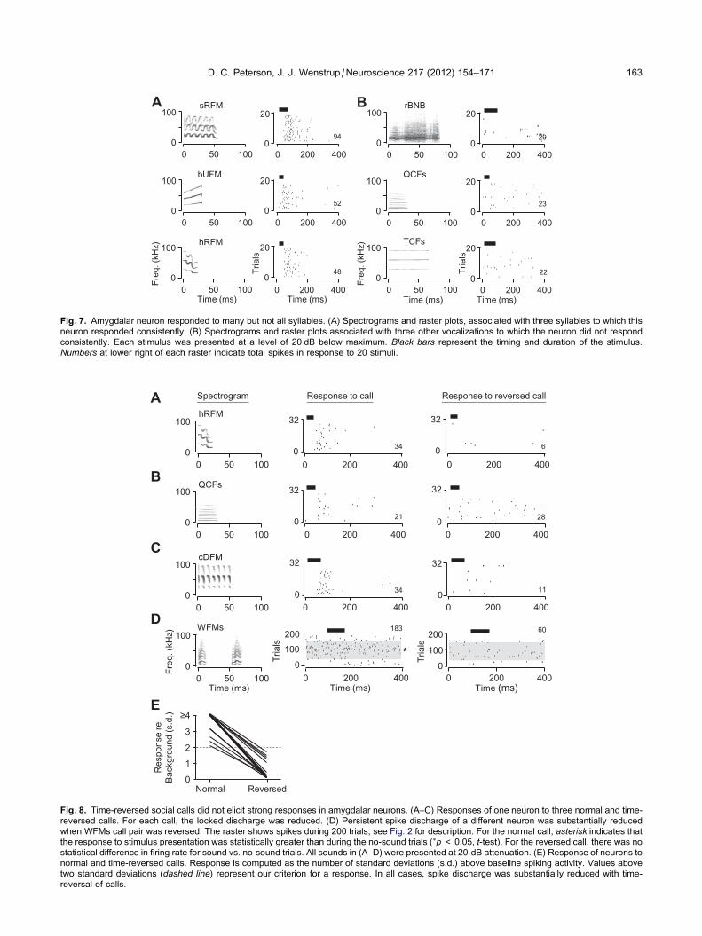
One-third of neurons (29/87) responded to multiple
stimuli. An example is shown in Fig. 7, which displays
rasters associated with responses to six mustached bat
vocal signals. Strong, locked responses that varied with
sound level were obtained in response to eleven signals,
many consisting of a spectrotemporally structured, rela-
tively broadband syllable (Fig. 7A). Spike discharge to
these calls began about 50 ms after stimulus onset, with
median first-spike latencies ranging from 45 to 66 ms.
Four other syllables, with both narrow and broad fre-
quency bands, with short and long durations, did not
evoke consistent responses (e.g., Fig. 7B). Moreover,
the neuron did not respond to artificial stimuli (tones,
noise, or frequency-modulated stimuli). Thus, even neu-
rons that responded to multiple sounds displayed selectiv-
ity among acoustic stimuli.
We explored several aspects of this response selec-
tivity. To examine the dependence of auditory responses
on the frequency–time structure of signals, we compared
responses to normal and reversed calls (Fig. 8).
Responses to three calls from one neuron were robust
and well-locked (Fig. 8A–C), but responses to the
reversed calls were always less when referenced to back-
ground discharge. In Fig. 8D, a different neuron showed a
persistent discharge to the normal call, but no significant
response when the call sequence and the elements of
the call were reversed in time. From eight neurons, we
compared responses to calls and reverse calls, totaling
18 pairs of calls. In each of the 18 pairs, there was a sub-
stantial reduction in response to the reversed call
(Fig. 8E).
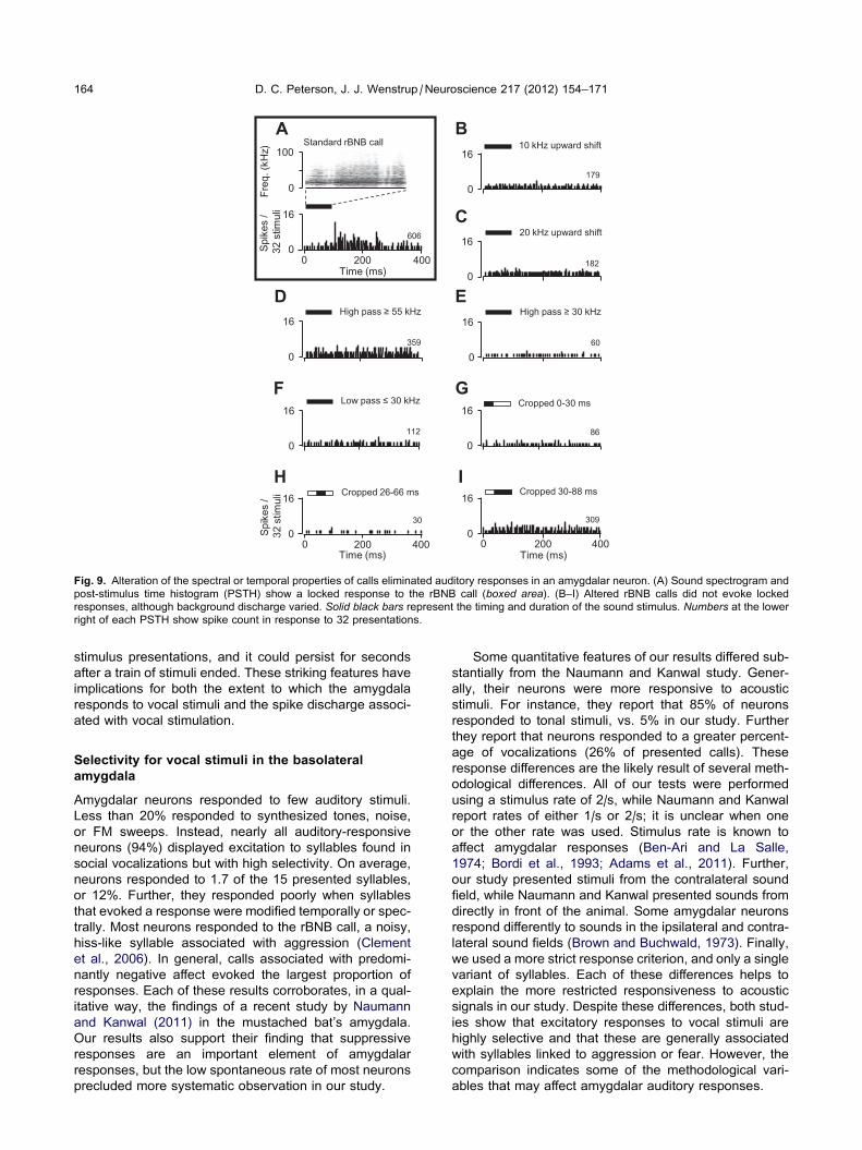
In 10 neurons, we examined responses to sounds in
which spectral or temporal elements were shifted or elim-
inated. An example is shown in Fig. 9. This neuron dis-
played a locked response to the rBNB call with a
latency of 107 ms (Fig. 9A). When the entire call was
shifted upward by 10 or 20 kHz (Fig. 9B, C), the locked
response was no longer evident. To examine whether
the response depended on particular frequency bands,
we presented frequency-filtered versions of the call.
There were no stimulus-locked responses to these filtered
versions (Fig. 9D–F). Finally, we presented versions of
the call in which we eliminated early, late, or both early
and late segments of the signal. None of these manipu-
lated signals evoked a locked response, although the
background discharge varied across stimuli (Fig. 9G–I).
In eight of the 10 neurons tested in this way, there was
no response to the altered signals. These results suggest
that a complex set of temporal and spectral features is
required for auditory responses by these amygdalar
neurons.
Amygdalar responses during MG stimulation
We obtained recordings from 12 amygdalar neurons
before and during iontophoretic application in MG of an
excitatory drug mixture including GLU, ASP, and/or BIC.
Auditory-evoked responses and histological reconstruc-
tion confirmed location of the multibarrel drug-application
pipette in the medial MG, centered in the medial or dorsal
Time (ms)
fSFM
20
0 4002000
20
0 4002000
20
0 4002000
20
0 4002000
20
0 4002000
20
0 4002000
20
0 4002000
rBNB
Time (ms)
fRFM
cDFM
Freq
. (kH
z)
hRFM
bUFM
TCFs
Tria
ls
0
100
10050 0
0
100
10050 0
0
100
10050 0
0
100
10050 0
0
100
10050 0
0
100
10050 0
0
100
10050 0
32
51
67
26
18
41
468
Fig. 6. Amygdalar neuron responded to the rBNB call but not to other
calls. The response to the rBNB call includes both locked and
persistent elements. There was no evoked discharge to other calls.
Background, or non-locked discharge varied in responses to other
stimuli. The higher non-locked discharge in response to some stimuli
(e.g., TCFs and NNBs calls) was not repeatable. All stimuli presented
at an attenuation of 20 dB. For each stimulus type, we display
spectrograms (left) and the rasters (right). For abbreviations of
syllable types, see Fig. 5 caption.
162 D. C. Peterson, J. J. Wenstrup /Neuroscience 217 (2012) 154–171
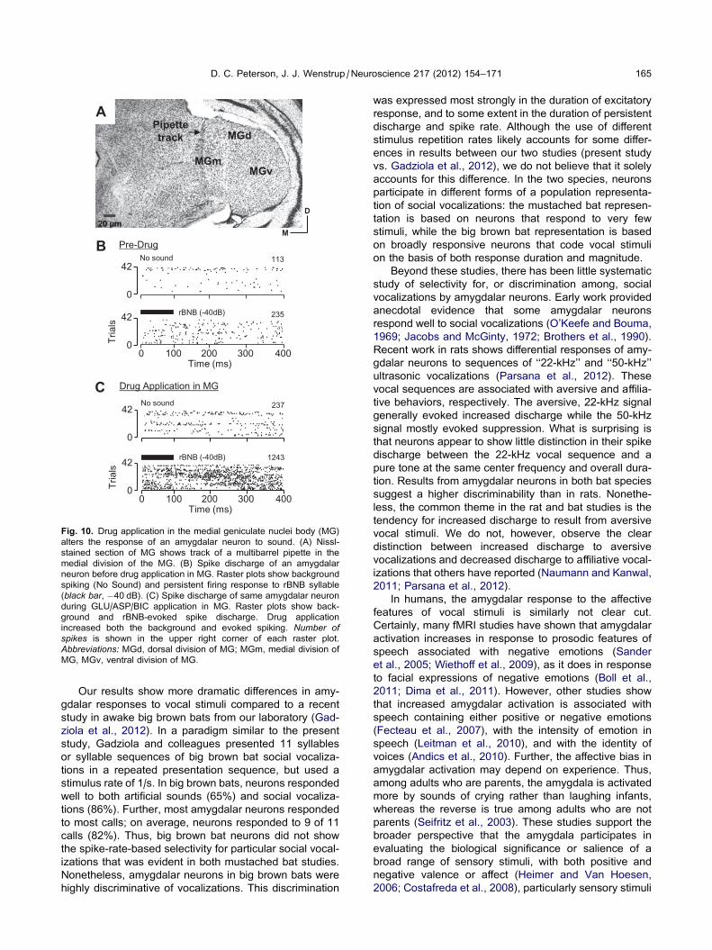
divisions (e.g., Fig. 10A). The main result was that
excitatory drug application in MG resulted in increased
background and stimulus-evoked discharge, but stimu-
lus-evoked discharge showed a greater increase. An
example is shown in Fig. 10. In pre-drug testing,
background activity (‘‘No sound’’) for this neuron was
113 spikes over 42 trials of 400-ms windows, or 6.7
spikes/s (Fig. 10B). This increased to 237 spikes (14.1
spikes/s) with GLU/ASP/BIC application in the medial
MG (Fig. 10A, C). The auditory-evoked response also
increased: the pre-drug persistent firing rate to the rBNB
syllable was 14.0 spikes/s (Fig. 10B), but increased to
74.0 spikes/s during drug application in MG (Fig. 10C).
Thus, the overall gain of the neuron (i.e., evoked spike
rate/background rate) more than doubled, increasing from
2.1 to 5.3.
In the tested sample, eight neurons responded to a
single communication call while four neurons responded
to multiple calls (total 21 responses to auditory stimuli).
Upon drug application in MG, background spike rate more
than doubled (pre-drug, 0.16 ± 0.17 spikes/s; drug appli-
cation, 0.40 ± 0.67 spikes/s; paired t-test, p< 0.05,
df = 40). Auditory-evoked responses increased by an
even greater amount (pre-drug, 0.38 ± 0.49; drug appli-
cation, 1.38 ± 2.98; paired t-test, p< 0.05). The
increase in response gain with drug application
(80.7%± 120.1%) was also highly significant (paired
t-test, p< 0.05).
Location of amygdalar recording sites
The locations of recording sites were assessed by ste-
reotaxic coordinates and, in some cases, tracer depos-
its. For 39 recording sites, successful deposits were
small enough to pinpoint location within an amygdalar
subdivision (Fig. 11). Locked, persistent, and suppres-
sive firing patterns were observed throughout the dor-
so-ventral, medio-lateral, and rostro-caudal extent of
the lateral and basal nuclei (Fig. 11C). Thirteen of the
39 recording sites in Fig. 11C displayed two temporal
response patterns. Of these, five had both locked and
suppressive responses and are displayed as suppres-
sive responses in Fig. 11C. Eight neurons with persistent
firing responses in the lateral nucleus also had locked
responses; these are plotted as persistent firing
responses for figure clarity. Neurons with dual temporal
patterns were observed at depths ranging from 4547 to
5102 lm, indicating their occurrence in both the lateral
and basal amygdala.
Tracer deposits marked the locations of 12 habituating
responses. Habituating responses were observed in both
the lateral and basal nuclei (open circles, Fig. 11D). Dur-
ing recording sessions, they were observed at depths
ranging from 4547 to 5230 lm, indicating a wide distribu-
tion throughout the amygdala.
At recording sites showing selective responses to one
call type, we plotted the emotional affect of the call (Clem-
ent et al., 2006). We observed no dorso-ventral, rostro-
caudal, or medio-lateral pattern of responses. Neurons
responded to calls with positive affect (e.g., appease-
ment, calming) ranged in depth from 4501 to 5102 lm(black squares, Fig. 11E). Similarly, neurons that
responded to communication calls with negative affect
(e.g., aggression, warning, fear) ranged in depth from
4499 to 5230 lm (white squares, Fig. 11E). Overall, we
observed no clear clustering of the various response
properties within the amygdala, but the numbers of local-
ized recording sites precluded a definitive analysis.
DISCUSSION
This study describes two striking features of basolateral
amygdalar responses to acoustic stimuli in the mustached
bat. First, neurons were highly selective for particular
social vocalizations, with most responding to only one or
two syllables. Most responses were to syllables associ-
ated with aggression. Second, many neurons display a
persistent firing pattern in response to the preferred
stimulus, in which spike discharge remained elevated for
seconds or minutes as a stimulus was repeated. This per-
sistent discharge was not necessarily locked to individual
sRFM A B
Time (ms) Time (ms)
29
20
0 4002000
Freq
. (kH
z)
22
400200
20
0 0
23
20
0 4002000
94
52
48
20
0 4002000
20
0 4002000
20
0 4002000
Time (ms)
Freq
. (kH
z)
Time (ms)
Tria
ls
Tria
ls
0
100
10050 0
0
100
10050 0
0
100
10050 0
0
100
10050 0
0
100
10050 0
0
100
10050 0
bUFM
hRFM
rBNB
QCFs
TCFs
Fig. 7. Amygdalar neuron responded to many but not all syllables. (A) Spectrograms and raster plots, associated with three syllables to which this
neuron responded consistently. (B) Spectrograms and raster plots associated with three other vocalizations to which the neuron did not respond
consistently. Each stimulus was presented at a level of 20 dB below maximum. Black bars represent the timing and duration of the stimulus.
Numbers at lower right of each raster indicate total spikes in response to 20 stimuli.
B
D
SpectrogramA
C
QCFs
Time (ms)
Freq
. (kH
z)
hRFM
cDFM
WFMs
E
Response to call
200
Tria
ls
*
Time (ms)
34
21
34
100
0
183
0 200 400
0 200 400
0 200 400
32
0
32
0
32
0
0 200 400
Res
pons
e re
Ba
ckgr
ound
(s.d
.)
Normal Reversed 0
1
2
3
≥4
Response to reversed call
400
6
28
11
200
100
0 0
0 200
0 200 400
0 200 400
32
0
32
0
32
0
400200Time (ms)
Tria
ls
60
0
100
10050 0
0
100
10050 0
0
100
10050 0
0
100
10050 0
Fig. 8. Time-reversed social calls did not elicit strong responses in amygdalar neurons. (A–C) Responses of one neuron to three normal and time-
reversed calls. For each call, the locked discharge was reduced. (D) Persistent spike discharge of a different neuron was substantially reduced
when WFMs call pair was reversed. The raster shows spikes during 200 trials; see Fig. 2 for description. For the normal call, asterisk indicates that
the response to stimulus presentation was statistically greater than during the no-sound trials (⁄p< 0.05, t-test). For the reversed call, there was no
statistical difference in firing rate for sound vs. no-sound trials. All sounds in (A–D) were presented at 20-dB attenuation. (E) Response of neurons to
normal and time-reversed calls. Response is computed as the number of standard deviations (s.d.) above baseline spiking activity. Values above
two standard deviations (dashed line) represent our criterion for a response. In all cases, spike discharge was substantially reduced with time-
reversal of calls.
D. C. Peterson, J. J. Wenstrup /Neuroscience 217 (2012) 154–171 163
High pass ≥ 55 kHz
359
0
16
D High pass ≥ 30 kHz
E
0 60
16
1820
16
C 20 kHz upward shift
179
0
16
B10 kHz upward shift
Low pass ≤ 30 kHzF
112
16
0 86
0
16
G Cropped 0-30 ms
0 400Time (ms)
309
0
16
200
I Cropped 30-88 ms
0 400Time (ms)
300
16
200
H Cropped 26-66 ms
Spik
es /
32
stim
uli
Spik
es /
32
stim
uli
0 400Time (ms)
0
16
200
606
AStandard rBNB call
Freq
. (kH
z)
0
100
Fig. 9. Alteration of the spectral or temporal properties of calls eliminated auditory responses in an amygdalar neuron. (A) Sound spectrogram and
post-stimulus time histogram (PSTH) show a locked response to the rBNB call (boxed area). (B–I) Altered rBNB calls did not evoke locked
responses, although background discharge varied. Solid black bars represent the timing and duration of the sound stimulus. Numbers at the lower
right of each PSTH show spike count in response to 32 presentations.
164 D. C. Peterson, J. J. Wenstrup /Neuroscience 217 (2012) 154–171
stimulus presentations, and it could persist for seconds
after a train of stimuli ended. These striking features have
implications for both the extent to which the amygdala
responds to vocal stimuli and the spike discharge associ-
ated with vocal stimulation.
Selectivity for vocal stimuli in the basolateralamygdala
Amygdalar neurons responded to few auditory stimuli.
Less than 20% responded to synthesized tones, noise,
or FM sweeps. Instead, nearly all auditory-responsive
neurons (94%) displayed excitation to syllables found in
social vocalizations but with high selectivity. On average,
neurons responded to 1.7 of the 15 presented syllables,
or 12%. Further, they responded poorly when syllables
that evoked a response were modified temporally or spec-
trally. Most neurons responded to the rBNB call, a noisy,
hiss-like syllable associated with aggression (Clement
et al., 2006). In general, calls associated with predomi-
nantly negative affect evoked the largest proportion of
responses. Each of these results corroborates, in a qual-
itative way, the findings of a recent study by Naumann
and Kanwal (2011) in the mustached bat’s amygdala.
Our results also support their finding that suppressive
responses are an important element of amygdalar
responses, but the low spontaneous rate of most neurons
precluded more systematic observation in our study.
Some quantitative features of our results differed sub-
stantially from the Naumann and Kanwal study. Gener-
ally, their neurons were more responsive to acoustic
stimuli. For instance, they report that 85% of neurons
responded to tonal stimuli, vs. 5% in our study. Further
they report that neurons responded to a greater percent-
age of vocalizations (26% of presented calls). These
response differences are the likely result of several meth-
odological differences. All of our tests were performed
using a stimulus rate of 2/s, while Naumann and Kanwal
report rates of either 1/s or 2/s; it is unclear when one
or the other rate was used. Stimulus rate is known to
affect amygdalar responses (Ben-Ari and La Salle,
1974; Bordi et al., 1993; Adams et al., 2011). Further,
our study presented stimuli from the contralateral sound
field, while Naumann and Kanwal presented sounds from
directly in front of the animal. Some amygdalar neurons
respond differently to sounds in the ipsilateral and contra-
lateral sound fields (Brown and Buchwald, 1973). Finally,
we used a more strict response criterion, and only a single
variant of syllables. Each of these differences helps to
explain the more restricted responsiveness to acoustic
signals in our study. Despite these differences, both stud-
ies show that excitatory responses to vocal stimuli are
highly selective and that these are generally associated
with syllables linked to aggression or fear. However, the
comparison indicates some of the methodological vari-
ables that may affect amygdalar auditory responses.
ATr
ials
No sound
rBNB (-40dB)
113
235
237
1243
rBNB (-40dB)
No sound
Drug Application in MG
0 100 200 300 400Time (ms)
42
0
42
0
42
0
42
0
B
Pipette track
MGv
MGd
MGm
C
Tria
ls
20 µm
Pre-Drug
0 100 200 300 400Time (ms)
D
M
Fig. 10. Drug application in the medial geniculate nuclei body (MG)
alters the response of an amygdalar neuron to sound. (A) Nissl-
stained section of MG shows track of a multibarrel pipette in the
medial division of the MG. (B) Spike discharge of an amygdalar
neuron before drug application in MG. Raster plots show background
spiking (No Sound) and persistent firing response to rBNB syllable
(black bar, �40 dB). (C) Spike discharge of same amygdalar neuron
during GLU/ASP/BIC application in MG. Raster plots show back-
ground and rBNB-evoked spike discharge. Drug application
increased both the background and evoked spiking. Number ofspikes is shown in the upper right corner of each raster plot.
Abbreviations: MGd, dorsal division of MG; MGm, medial division of
MG, MGv, ventral division of MG.
D. C. Peterson, J. J. Wenstrup /Neuroscience 217 (2012) 154–171 165
Our results show more dramatic differences in amy-
gdalar responses to vocal stimuli compared to a recent
study in awake big brown bats from our laboratory (Gad-
ziola et al., 2012). In a paradigm similar to the present
study, Gadziola and colleagues presented 11 syllables
or syllable sequences of big brown bat social vocaliza-
tions in a repeated presentation sequence, but used a
stimulus rate of 1/s. In big brown bats, neurons responded
well to both artificial sounds (65%) and social vocaliza-
tions (86%). Further, most amygdalar neurons responded
to most calls; on average, neurons responded to 9 of 11
calls (82%). Thus, big brown bat neurons did not show
the spike-rate-based selectivity for particular social vocal-
izations that was evident in both mustached bat studies.
Nonetheless, amygdalar neurons in big brown bats were
highly discriminative of vocalizations. This discrimination
was expressed most strongly in the duration of excitatory
response, and to some extent in the duration of persistent
discharge and spike rate. Although the use of different
stimulus repetition rates likely accounts for some differ-
ences in results between our two studies (present study
vs. Gadziola et al., 2012), we do not believe that it solely
accounts for this difference. In the two species, neurons
participate in different forms of a population representa-
tion of social vocalizations: the mustached bat represen-
tation is based on neurons that respond to very few
stimuli, while the big brown bat representation is based
on broadly responsive neurons that code vocal stimuli
on the basis of both response duration and magnitude.
Beyond these studies, there has been little systematic
study of selectivity for, or discrimination among, social
vocalizations by amygdalar neurons. Early work provided
anecdotal evidence that some amygdalar neurons
respond well to social vocalizations (O’Keefe and Bouma,
1969; Jacobs and McGinty, 1972; Brothers et al., 1990).
Recent work in rats shows differential responses of amy-
gdalar neurons to sequences of ‘‘22-kHz’’ and ‘‘50-kHz’’
ultrasonic vocalizations (Parsana et al., 2012). These
vocal sequences are associated with aversive and affilia-
tive behaviors, respectively. The aversive, 22-kHz signal
generally evoked increased discharge while the 50-kHz
signal mostly evoked suppression. What is surprising is
that neurons appear to show little distinction in their spike
discharge between the 22-kHz vocal sequence and a
pure tone at the same center frequency and overall dura-
tion. Results from amygdalar neurons in both bat species
suggest a higher discriminability than in rats. Nonethe-
less, the common theme in the rat and bat studies is the
tendency for increased discharge to result from aversive
vocal stimuli. We do not, however, observe the clear
distinction between increased discharge to aversive
vocalizations and decreased discharge to affiliative vocal-
izations that others have reported (Naumann and Kanwal,
2011; Parsana et al., 2012).
In humans, the amygdalar response to the affective
features of vocal stimuli is similarly not clear cut.
Certainly, many fMRI studies have shown that amygdalar
activation increases in response to prosodic features of
speech associated with negative emotions (Sander
et al., 2005; Wiethoff et al., 2009), as it does in response
to facial expressions of negative emotions (Boll et al.,
2011; Dima et al., 2011). However, other studies show
that increased amygdalar activation is associated with
speech containing either positive or negative emotions
(Fecteau et al., 2007), with the intensity of emotion in
speech (Leitman et al., 2010), and with the identity of
voices (Andics et al., 2010). Further, the affective bias in
amygdalar activation may depend on experience. Thus,
among adults who are parents, the amygdala is activated
more by sounds of crying rather than laughing infants,
whereas the reverse is true among adults who are not
parents (Seifritz et al., 2003). These studies support the
broader perspective that the amygdala participates in
evaluating the biological significance or salience of a
broad range of sensory stimuli, with both positive and
negative valence or affect (Heimer and Van Hoesen,
2006; Costafreda et al., 2008), particularly sensory stimuli
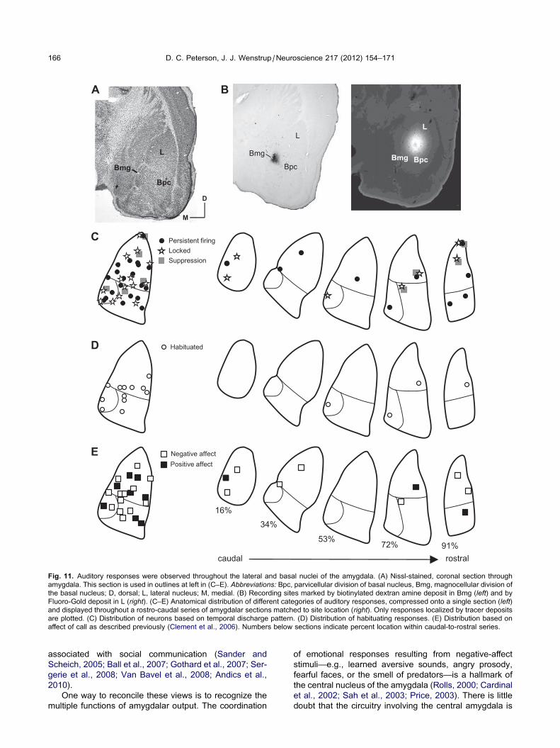
B
D
C
E
L
Bpc
L
Bmg Bpc
caudal rostral
16%34%
72% 91%53%
Persistent firing Locked Suppression
Habituated
Negative affect Positive affect
Bmg L
Bpc
Bmg
A
D
M
Fig. 11. Auditory responses were observed throughout the lateral and basal nuclei of the amygdala. (A) Nissl-stained, coronal section through
amygdala. This section is used in outlines at left in (C–E). Abbreviations: Bpc, parvicellular division of basal nucleus, Bmg, magnocellular division of
the basal nucleus; D, dorsal; L, lateral nucleus; M, medial. (B) Recording sites marked by biotinylated dextran amine deposit in Bmg (left) and by
Fluoro-Gold deposit in L (right). (C–E) Anatomical distribution of different categories of auditory responses, compressed onto a single section (left)and displayed throughout a rostro-caudal series of amygdalar sections matched to site location (right). Only responses localized by tracer deposits
are plotted. (C) Distribution of neurons based on temporal discharge pattern. (D) Distribution of habituating responses. (E) Distribution based on
affect of call as described previously (Clement et al., 2006). Numbers below sections indicate percent location within caudal-to-rostral series.
166 D. C. Peterson, J. J. Wenstrup /Neuroscience 217 (2012) 154–171
associated with social communication (Sander and
Scheich, 2005; Ball et al., 2007; Gothard et al., 2007; Ser-
gerie et al., 2008; Van Bavel et al., 2008; Andics et al.,
2010).
One way to reconcile these views is to recognize the
multiple functions of amygdalar output. The coordination
of emotional responses resulting from negative-affect
stimuli—e.g., learned aversive sounds, angry prosody,
fearful faces, or the smell of predators—is a hallmark of
the central nucleus of the amygdala (Rolls, 2000; Cardinal
et al., 2002; Sah et al., 2003; Price, 2003). There is little
doubt that the circuitry involving the central amygdala is
D. C. Peterson, J. J. Wenstrup /Neuroscience 217 (2012) 154–171 167
more closely tied to aversive emotional responses
(Hopkins and Holstege, 1978; Krettek and Price, 1978;
Pitkanen et al., 1997; Pare et al., 2004). On the other
hand, another major function of the amygdala seems to
be an analysis of stimulus salience, positive or negative,
that results in modulation of sensory processing (Bjordahl
et al., 1998; Kilgard and Merzenich, 1998; Ma and Suga,
2003; Chavez et al., 2009) through its direct projections
(Amaral and Price, 1984; McDonald and Jackson, 1987;
Kosmal et al., 1997; Marsh et al., 2002; Yukie, 2002)
and indirect projections through the basal cholinergic fore-
brain (Price et al., 1987; Pitkanen et al., 2000; Pare,
2003).
Persistent firing to vocal stimuli
A major finding of this study is that many amygdalar neu-
rons display their selective response to acoustic and vocal
signals through persistent discharge, i.e., the duration of
firing that extends beyond the duration of the stimulus.
This persistent firing usually occurred seconds or tens
of seconds after the beginning of a train of acoustic stimuli
and was not locked to individual stimulus presentations in
any obvious way. Rather, firing continued for at least the
interval between stimuli, thus lasting over 400 ms. Follow-
ing the termination of a stimulus train, neurons continued
to fire for periods of seconds to minutes. While these fea-
tures suggest an independence from the acoustic stimu-
lus, there was a clear dependence on both the stimulus
type and sound level. Our conclusion is that this firing is
the result of both the acoustic stimulus and some element
of the context surrounding the stimulus (e.g., the animal’s
emotional state) and represents a critical aspect of the
output of neurons in the basolateral amygdala.
Although Naumann and Kanwal (2011) report many
responses that feature some degree of persistent firing,
they do not describe the dramatic persistent firing behav-
ior that we report here. We propose that the difference
may relate to the sequencing of stimuli. We used a re-
peated stimulus paradigm, in which the same stimulus
was repeated for 20 or more repetitions. This preserves
at least some of the affective context of the syllables.
Naumann and Kanwal presented a sequence of syllables
based on syllable acoustics, and then repeated the
sequence. By presenting stimuli in such a sequence,
Naumann and Kanwal reduced the context associated
with particular vocal signals. If persistent firing depends
on both stimulus and context as we hypothesize, then it
would be less likely that amygdalar neurons display
persistent firing using their paradigm. We believe these
differing results show that syllable sequencing may be a
critical factor in the responses of amygdalar neurons to
vocal stimuli.
In the big brown bat, the duration of spike discharge
underlies the ability of lateral amygdalar neurons to dis-
criminate among vocal signals (Gadziola et al., 2012).
This is based in part on persistent firing, which could
extend for at least 250 ms beyond stimulus duration.
Furthermore, manipulations of signal features that
increased their salience, such as the modification of a
weakly aggressive call by addition of a tonal ending, could
substantially increase the duration of persistent firing.
Thus, both studies indicate that persistent firing is a major
feature of amygdala responses to social vocalizations.
They reinforce our view that stimulus sequencing may
have a major impact on amygdalar neuron discharge, par-
ticularly if the sequencing alters the salience of acoustic
signals. Ultimately, an understanding of the role of persis-
tent firing must be examined when syllables are
presented in the appropriate acoustic context, i.e., within
probabilistic sequences that can be associated with
particular behaviors or emotional states.
Persistent firing functions in several neural systems to
sustain representations of sensory stimuli for working
memory (Frank and Brown, 2003; Major and Tank,
2004). Such firing is present in auditory responses of corti-
cal (Pena et al., 1999; Romanski et al., 2005; Moshitch
et al., 2006; Bendor and Wang, 2008; Campbell et al.,
2010) and pontine (Miller and Covey, 2011) neurons.
Among basolateral amygdalar neurons, it has been
observed to auditory and other sensory stimuli (Bordi and
LeDoux, 1992; Bordi et al., 1993; Maeda et al., 1993;
Naumann and Kanwal, 2011), but it had not been related
until recently to discrimination of acoustic stimuli (Gadziola
et al., 2012) or to response selectivity (this study).
Persistent firing likely plays a crucial role in memory
operations associated with the amygdala (Pelletier et al.,
2005; Egorov et al., 2006), but may also serve a more gen-
eral function by transforming the time scale of acoustic
stimuli to time scales appropriate for control of emotional
expression and modulation of sensory processing.
In vitro studies of the amygdala, entorhinal cortex, and
endopiform nucleus have reported similar firing patterns,
termed graded persistent firing (Egorov et al., 2002,
2006; Frank and Brown, 2003). In these studies, depolar-
izing current in the presence of muscarinic agonists cre-
ates a persistent firing pattern that depends on the
strength and duration of the depolarizing current. The per-
sistent nature of the firing depends on a cholinergic input
to maintain the increased firing rate (Frank and Brown,
2003; Egorov et al., 2006). If the persistent firing response
that we observed has a similar mechanism, then it likely
depends on the combination of auditory input from MG
or auditory cortex (LeDoux et al., 1990; McDonald,
1998) and cholinergic inputs from the nucleus basalis
(Emson et al., 1979; Carlsen et al., 1985).
A further aspect of our results relevant to persistent fir-
ing is the finding that chemical activation of the medial
MG, a region that projects directly to the lateral amygdala,
raised firing rates of amygdala neurons. Particularly
intriguing is the observation that the sound-evoked, per-
sistent firing rates increased more than background firing,
and the increase in evoked firing was greater, in the pres-
ence of MG activation. This suggests that the auditory
environment may establish a level of amygdala input that
can enhance the gain of salient auditory stimuli. Because
the MG has projections to both auditory cortex and amyg-
dala, it is unclear whether direct MG projections, indirect
higher order projections, or a combination of multiple
amygdalar inputs are necessary to elicit this response.
There are several possible explanations of the fact
that chemical stimulation of the medial geniculate
168 D. C. Peterson, J. J. Wenstrup /Neuroscience 217 (2012) 154–171
influenced the background activity of non-auditory
responsive neurons. (1) It is possible that we did not
use appropriate auditory stimuli to elicit responses in
these neurons. (2) The excitation may have influenced
other multi-synaptic circuits that project to the amygdala
(e.g., nucleus basalis of Meynert, hippocampus, auditory
cortex, and/or association cortices). Combinations of
excitatory input from these projections may have influ-
enced the background activity. (3) The excitation from
MG activation may have had a direct excitatory effect on
local amygdalar circuits. (4) MG projections only activate
amygdalar neurons when large numbers of MG projection
neurons are activated simultaneously. The precise timing
of combinations of inputs (sensory, memory, association)
may then enable the amygdala to evaluate stimulus
significance.
Response habituation in the amygdala
Although our experimental design precluded study of
short-term adaptation effects acting on a time scale of a
few seconds or less, we did observe habituation with time
scales of seconds or minutes. In some neurons, the habit-
uation waxed and waned, while in others the habituation
continued until acoustic stimulation ended. Our interpreta-
tion of these effects is that the non-auditory elements of
input to the amygdala, including those that may activate
persistent firing, change over time.
Some amygdalar habituation is likely inherited from
the auditory inputs. Several studies in the past decade
have documented the presence of stimulus-specific adap-
tation in auditory responses. Repeated sounds evoke
diminished responses while those that are novel evoke
a stronger response (Ulanovsky et al., 2003; Perez-
Gonzalez et al., 2005). The major auditory inputs to the
amygdala, the medial and dorsal divisions of the MG
and the auditory cortex, each show stimulus-specific
adaptation (Ulanovsky et al., 2003; von der Behrens
et al., 2009; Anderson et al., 2009; Antunes et al.,
2010). This could well explain the poor response to artifi-
cial stimuli in our study. Moreover, the longer duration
forms of habituation in our study may correspond to the
longer time scales of adaptation observed in auditory
cortical neurons (Ulanovsky et al., 2004).
Nonetheless, the major time-varying response feature
that we observed among amygdalar neurons is very dif-
ferent from stimulus-specific adaption, in fact closer to
its opposite. In response to repeated presentations of
the same vocal stimulus, spike discharge increased and
became persistent. Further, we believe that this effect
only occurs when the stimulus is repeated, since stimulus
presentation with an interleaved stimulus paradigm
(Naumann and Kanwal, 2011) does not evoke the level
of persistent discharge that we observed. These results
are consistent, in our view, with the understanding that
the amygdala analyzes stimulus salience. Novel stimuli
(especially artificial stimuli) may be salient, but that sal-
ience is short-lived and is not likely to activate strongly
the emotional response circuits of the amygdala unless
the context surrounding the novel stimulus is appropriate.
In contrast, emotion-laden vocalizations may become
increasingly salient when they are repeated. Similarly,
repeated artificial stimuli that have become aversive
through conditioning evoke strong amygdalar responses
(Quirk et al., 1995; Amano et al., 2011). Thus, the overall
response is determined by context surrounding the imme-
diate stimulus, but novelty is only one of several contex-
tual factors. Others may include the temporal patterning
of vocalizations, other sensory input, and an animal’s
internal state. Habituation of seconds to minutes that we
observed is likely to depend on internal state factors
rather than novelty.
Overall functional view
The social acoustic environment of mustached bats
seems to require high selectivity to its social calls in brain
regions that establish the salience of acoustic stimuli. As
highly social mammals, mustached bats roost in large
groups (hundreds to thousands or more) within caves or
other structures (Bateman and Vaughan, 1974). Like
many neotropical species, these bats do not appear to
display daily torpor (Bonaccorso et al., 1992). Unlike
many other bats, these bats produce a nearly continuous
stream of echolocation signals (Kanwal et al., 1994). As a
result, these animals live in close quarters with conspecif-
ics, engage socially for 12 or more hours a day, and are
bombarded by high-amplitude echolocation signals of lim-
ited social salience. It is reasonable to expect that neu-
rons in the mustached bat amygdala, a brain region that
helps to establish the salience of acoustic stimuli, would
not respond to most echolocation signals. Instead, amy-
gdalar neurons appear to filter these signals out and
respond predominantly to signals that have social sal-
ience, i.e., the signals indicating aggressive interactions
with nearby bats as well as the signals indicating affiliative
or appeasing interactions. Appropriate behavioral
responses to these social signals are critical to an individ-
ual’s survival. The responses of basolateral amygdalar
neurons in these situations contribute to the amygdala’s
orchestration of emotional responses to these acousti-
cally based social interactions, as well as providing the
basis for altering auditory responsiveness to subsequent
signals. The persistent firing of mustached bat amygdalar
neurons to a particular salient call may serve to match the
time scale of the auditory response to desirable emotional
responses and modulation of subsequent auditory
responses.
Acknowledgements—These studies were supported by research
grants R01 DC00937 (J.J.W.) and National Research Service
Award F32 DC007786 (D.C.P.) from the National Institute on
Deafness and Other Communication Disorders of the U.S. Public
Health Service. Communication calls were provided through
research grant R01 DC04733 (C. Portfors, PI; J. Wenstrup and
J. Kanwal, co-investigators) from the National Institute on Deaf-
ness and Other Communication Disorders. The authors are pro-
foundly grateful to Don Gans (deceased) for his contributions to
this work. We thank Marie Gadziola and Jasmine Grimsley for
comments on the manuscript and sound analysis, Carol Grose
for assistance in data analysis and histological reconstruction,
the Wildlife Section of the Ministry of Agriculture, Land and
Marine Resources of Trinidad and Tobago for permission to
exports bats, and Brett Schofield for use of the Neurolucida
reconstruction system.
D. C. Peterson, J. J. Wenstrup /Neuroscience 217 (2012) 154–171 169
REFERENCES
Adams JC (1981) Heavy metal intensification of DAB-based HRP
reaction product. J Histochem Cytochem 29:775.
Adams RB, Jr., Franklin RG, Jr., Kveraga K, Ambady N, Kleck RE,
Whalen PJ, Hadjikhani N, Nelson AJ (2011) Amygdala responses
to averted vs. direct gaze fear vary as a function of presentation
speed. Soc Cogn Affect Neurosci. [Epub ahead of print].
Amano T, Duvarci S, Popa D, Pare D (2011) The fear circuit revisited:
contributions of the basal amygdala nuclei to conditioned fear. J
Neurosci 31:15481–15489.
Amaral DG, Price JL (1984) Amygdalo-cortical projections in the
monkey (Macaca fascicularis). J Comp Neurol 230:465–496.
Anderson LA, Christianson GB, Linden JF (2009) Stimulus-specific
adaptation occurs in the auditory thalamus. J Neurosci
29:7359–7363.
Andics A, McQueen JM, Petersson KM, Gal V, Rudas G,
Vidnyanszky Z (2010) Neural mechanisms for voice recognition.
Neuroimage 52:1528–1540.
Antunes FM, Nelken I, Covey E, Malmierca MS (2010) Stimulus-
specific adaptation in the auditory thalamus of the anesthetized
rat. PLoS One 5:e14071.
Ball T, Rahm B, Eickhoff SB, Schulze-Bonhage A, Speck O,
Mutschler I (2007) Response properties of human amygdala
subregions: evidence based on functional MRI combined with
probabilistic anatomical maps. PLoS One 2:e307.
Bateman GC, Vaughan TA (1974) Nightly activities of mormoopid
bats. J Mammal 55:45–65.
Bauer EE, Klug A, Pollak GD (2000) Features of contralaterally
evoked inhibition in the inferior colliculus. Hear Res 141:
80–96.
Ben-Ari Y, Le Gal la Salle G (1974) Lateral amygdala unit activity: II.
Habituating and non-habituating neurons. Electroencephalogr
Clin Neurophysiol 37:463–472.
Bendor D, Wang X (2008) Neural response properties of primary,
rostral, and rostrotemporal core fields in the auditory cortex of
marmoset monkeys. J Neurophysiol 100:888–906.
Bjordahl TS, Dimyan MA, Weinberger NM (1998) Induction of long-
term receptive field plasticity in the auditory cortex of the waking
guinea pig by stimulation of the nucleus basalis. Behav Neurosci
112:467–479.
Boll S, Gamer M, Kalisch R, Buchel C (2011) Processing of facial
expressions and their significance for the observer in subregions
of the human amygdala. Neuroimage 56:299–306.
Bonaccorso FJ, Arends A, Genoud M, Cantoni D, Morton T (1992)
Thermal ecology of moustached and ghost-faced bats,
(Mormoopidae) in Venezuela. J Mammal 73:365–378.
Bordi F, LeDoux J (1992) Sensory tuning beyond the sensory system:
an initial analysis of auditory response properties of neurons in the
lateral amygdaloid nucleus and overlying areas of the striatum. J
Neurosci 12:2493–2503.
Bordi F, LeDoux J, Clugnet MC, Pavlides C (1993) Single-unit activity
in the lateral nucleus of the amygdala and overlying areas of the
striatum in freely behaving rats: rates, discharge patterns, and
responses to acoustic stimuli. Behav Neurosci 107:757–769.
Brothers L, Ring B, Kling A (1990) Response of neurons in the
macaque amygdala to complex social stimuli. Behav Brain Res
41:199–213.
Brown KA, Buchwald JS (1973) Acoustic responses and plasticity of
limbic units in cats. Exp Neurol 40:608–631.
Campbell RA, Schulz AL, King AJ, Schnupp JW (2010) Brief sounds
evoke prolonged responses in anesthetized ferret auditory cortex.
J Neurophysiol 103:2783–2793.
Cardinal RN, Parkinson JA, Hall J, Everitt BJ (2002) Emotion and
motivation: the role of the amygdala, ventral striatum, and
prefrontal cortex. Neurosci Biobehav Rev 26:321–352.
Carlsen J, Zaborszky L, Heimer L (1985) Cholinergic projections from
the basal forebrain to the basolateral amygdaloid complex: a
combined retrograde fluorescent and immunohistochemical
study. J Comp Neurol 234:155–167.
Chavez CM, McGaugh JL, Weinberger NM (2009) The basolateral
amygdala modulates specific sensory memory representations in
the cerebral cortex. Neurobiol Learn Mem 91:382–392.
Clement MJ, Gupta P, Dietz N, Kanwal JS (2006) Audiovocal
communication and social behavior in mustached bats. In:
Kanwal JS, Ehret G, editors. Behavioral and neurodynamics for
auditory communication. Cambridge, NY: Cambridge University
press. p. 57–84.
Costafreda SG, Brammer MJ, David AS, Fu CH (2008) Predictors of
amygdala activation during the processing of emotional stimuli: a
meta-analysis of 385 PET and fMRI studies. Brain Res Rev
58:57–70.
Dima D, Stephan KE, Roiser JP, Friston KJ, Frangou S (2011)
Effective connectivity during processing of facial affect: evidence
for multiple parallel pathways. J Neurosci 31:14378–14385.
Egorov AV, Hamam BN, Fransen E, Hasselmo ME, Alonso AA (2002)
Graded persistent activity in entorhinal cortex neurons. Nature
420:173–178.
Egorov AV, Unsicker K, von Bohlen und Halbach O (2006) Muscarinic
control of graded persistent activity in lateral amygdala neurons.
Eur J Neurosci 24:3183–3194.
Emson PC, Paxinos G, Le Gal La Salle G, Ben-Ari Y, Silver A (1979)
Choline acetyltransferase and acetylcholinesterase containing
projections from the basal forebrain to the amygdaloid complex of
the rat. Brain Res 165:271–282.
Escarti MJ, de la Iglesia-Vaya M, Marti-Bonmati L, Robles M,
Carbonell J, Lull JJ, Garcia-Marti G, Manjon JV, Aguilar EJ,
Aleman A, Sanjuan J (2010) Increased amygdala and
parahippocampal gyrus activation in schizophrenic patients with
auditory hallucinations: an fMRI study using independent
component analysis. Schizophr Res 117:31–41.
Fecteau S, Belin P, Joanette Y, Armony JL (2007) Amygdala
responses to nonlinguistic emotional vocalizations. Neuroimage
36:480–487.
Frank LM, Brown EN (2003) Persistent activity and memory in the
entorhinal cortex. Trends Neurosci 26:400–401.
Gabis L, Wei H, Azizian A, DeVincent C, Tudorica A, Kesner-Baruch
Y, Roche P, Pomeroy J (2008) 1H-magnetic resonance
spectroscopy markers of cognitive and language ability in
clinical subtypes of autism spectrum disorders. J Child Neurol
23:766–774.
Gadziola MA, Grimsley JM, Shanbhag SJ, Wenstrup JJ (2012) A
novel coding mechanism for social vocalizations in the lateral
amygdala. J Neurophysiol 107:1047–1057.
Gothard KM, Battaglia FP, Erickson CA, Spitler KM, Amaral DG
(2007) Neural responses to facial expression and face identity in
the monkey amygdala. J Neurophysiol 97:1671–1683.
Havey DC, Caspary DM (1980) A simple technique for constructing
‘piggy-back’ multibarrel microelectrodes. Electroencephalogr Clin
Neurophysiol 48:249–251.
Heimer L, Van Hoesen GW (2006) The limbic lobe and its output
channels: implications for emotional functions and adaptive
behavior. Neurosci Biobehav Rev 30:126–147.
Hopkins DA, Holstege G (1978) Amygdaloid projections to the
mesencephalon, pons and medulla oblongata in the cat. Exp
Brain Res 32:529–547.
Jacobs BL, McGinty DJ (1972) Participation of the amygdala in
complex stimulus recognition and behavioral inhibition: evidence
from unit studies. Brain Res 36:431–436.
Kanwal JS, Matsumura S, Ohlemiller K, Suga N (1994) Analysis of
acoustic elements and syntax in communication sounds emitted
by mustached bats. J Acoust Soc Am 96:1229–1254.
Kawasaki M, Margoliash D, Suga N (1988) Delay-tuned combination-
sensitive neurons in the auditory cortex of the vocalizing
mustached bat. J Neurophysiol 59:623–635.
Kilgard MP, Merzenich MM (1998) Plasticity of temporal information
processing in the primary auditory cortex. Nat Neurosci
1:727–731.
Kim JE, Lyoo IK, Estes AM, Renshaw PF, Shaw DW, Friedman SD,
Kim DJ, Yoon SJ, Hwang J, Dager SR (2010) Laterobasal
170 D. C. Peterson, J. J. Wenstrup /Neuroscience 217 (2012) 154–171
amygdalar enlargement in 6- to 7-year-old children with autism
spectrum disorder. Arch Gen Psychiatry 67:1187–1197.
Kosmal A, Malinowska M,Woznicka A (1997) Diversity of connections
of the temporal neocortex with amygdaloid nuclei in the dog (Canis
familiaris). Acta Neurobiol Exp (Wars) 57:289–314.
Krettek JE, Price JL (1978) Amygdaloid projections to subcortical
structures within the basal forebrain and brainstem in the rat and
cat. J Comp Neurol 178:225–254.
LeDoux JE, Cicchetti P, Xagoraris A, Romanski LM (1990) The lateral
amygdaloid nucleus: sensory interface of the amygdala in fear
conditioning. J Neurosci 10:1062–1069.
Leitman DI, Wolf DH, Ragland JD, Laukka P, Loughead J, Valdez JN,
Javitt DC, Turetsky BI, Gur RC (2010) ‘‘It’s Not What You Say, But
How You Say it’’: a reciprocal temporo-frontal network for affective
prosody. Front Hum Neurosci 4:19.
Ma X, Suga N (2003) Augmentation of plasticity of the central
auditory system by the basal forebrain and/or somatosensory
cortex. J Neurophysiol 89:90–103.
Maeda H, Morimoto H, Yanagimoto K (1993) Response
characteristics of amygdaloid neurons provoked by emotionally
significant environmental stimuli in cats, with special reference to
response durations. Can J Physiol Pharmacol 71:374–378.
Major G, Tank D (2004) Persistent neural activity: prevalence and
mechanisms. Curr Opin Neurobiol 14:675–684.
Marsh RA, Fuzessery ZM, Grose CD, Wenstrup JJ (2002) Projection
to the inferior colliculus from the basal nucleus of the amygdala. J
Neurosci 22:10449–10460.
Marsh RA, Nataraj K, Gans D, Portfors CV, Wenstrup JJ (2006)
Auditory responses in the cochlear nucleus of awake mustached
bats: precursors to spectral integration in the auditory midbrain. J
Neurophysiol 95:88–105.
McDonald AJ (1998) Cortical pathways to the mammalian amygdala.
Prog Neurobiol 55:257–332.
McDonald AJ, Jackson TR (1987) Amygdaloid connections with
posterior insular and temporal cortical areas in the rat. J Comp
Neurol 262:59–77.
Miller K, Covey E (2011) Comparison of auditory responses in the
medial geniculate and pontine gray of the big brown bat,
Eptesicus fuscus. Hear Res 275:53–65.
Moshitch D, Las L, Ulanovsky N, Bar-Yosef O, Nelken I (2006)
Responses of neurons in primary auditory cortex (A1) to pure tones
in the halothane-anesthetized cat. J Neurophysiol 95:3756–3769.
Nataraj K, Wenstrup JJ (2005) Roles of inhibition in creating complex
auditory responses in the inferior colliculus: facilitated combination-
sensitive neurons. J Neurophysiol 93:3294–3312.
Nataraj K, Wenstrup JJ (2006) Roles of inhibition in complex auditory
responses in the inferior colliculus: inhibited combination-sensitive
neurons. J Neurophysiol 95:2179–2192.
Naumann RT, Kanwal JS (2011) Basolateral amygdala responds
robustly to social calls: spiking characteristics of single unit
activity. J Neurophysiol 105:2389–2404.
O’Keefe J, Bouma H (1969) Complex sensory properties of
certain amygdala units in the freely moving cat. Exp Neurol
23:384–398.
Pare D (2003) Role of the basolateral amygdala in memory
consolidation. Prog Neurobiol 70:409–420.
Pare D, Quirk GJ, Ledoux JE (2004) New vistas on amygdala
networks in conditioned fear. J Neurophysiol 92:1–9.
Parsana AJ, Li N, Brown TH (2012) Positive and negative ultrasonic
social signals elicit opposing firing patterns in rat amygdala.
Behav Brain Res 226:77–86.
Pelletier JG, Likhtik E, Filali M, Pare D (2005) Lasting increases in
basolateral amygdala activity after emotional arousal: implications
for facilitated consolidation of emotional memories. Learn Mem
12:96–102.
Pena JL, Perez-Perera L, Bouvier M, Velluti RA (1999) Sleep and
wakefulness modulation of the neuronal firing in the auditory
cortex of the guinea pig. Brain Res 816:463–470.
Perez-Gonzalez D, Malmierca MS, Covey E (2005) Novelty detector
neurons in the mammalian auditory midbrain. Eur J Neurosci
22:2879–2885.
Phelps EA, LeDoux JE (2005) Contributions of the amygdala to
emotion processing: from animal models to human behavior.
Neuron 48:175–187.
Pitkanen A, Savander V, LeDoux JE (1997) Organization of intra-
amygdaloid circuitries in the rat: an emerging framework for
understanding functions of the amygdala. Trends Neurosci
20:517–523.
Pitkanen A, Jolkkonen E, Kemppainen S (2000) Anatomic
heterogeneity of the rat amygdaloid complex. Folia Morphol
(Warsz) 59:1–23.
Portfors CV, Wenstrup JJ (2001) Responses to combinations of
tones in the nuclei of the lateral lemniscus. J Assoc Res
Otolaryngol 2:104–117.
Price JL (2003) Comparative aspects of amygdala connectivity. Ann
NY Acad Sci 985:50–58.
Price JL, Russchen FT, Amaral DG (1987) The limbic region. II. The
amygdaloid complex. In: Hofelt T, Swanson LW, Bjorkllund A,
editors. Handbook of chemical neuroanatomy. Amsterdam:
Elsevier. p. 279–388.
Protopopescu X, Pan H, Tuescher O, Cloitre M, Goldstein M,
Engelien W, Epstein J, Yang Y, Gorman J, LeDoux J,
Silbersweig D, Stern E (2005) Differential time courses and
specificity of amygdala activity in posttraumatic stress disorder
subjects and normal control subjects. Biol Psychiatry 57:464–473.
QuirkGJ,RepaC, LeDouxJE (1995) Fear conditioning enhances short-
latency auditory responses of lateral amygdala neurons: parallel
recordings in the freely behaving rat. Neuron 15: 1029–1039.
Rolls ET (2000) Precis of the brain and emotion. Behav Brain Sci
23:177–191 (discussion 192–233).
Romanski LM, Averbeck BB, Diltz M (2005) Neural representation of
vocalizations in the primate ventrolateral prefrontal cortex. J
Neurophysiol 93:734–747.
Sah P, Faber ES, Lopez De Armentia M, Power J (2003) The
amygdaloid complex: anatomy and physiology. Physiol Rev
83:803–834.
Sander K, Scheich H (2005) Left auditory cortex and amygdala, but
right insula dominance for human laughing and crying. J Cogn
Neurosci 17:1519–1531.
Sander D, Grandjean D, Pourtois G, Schwartz S, Seghier ML,
Scherer KR, Vuilleumier P (2005) Emotion and attention
interactions in social cognition: brain regions involved in
processing anger prosody. Neuroimage 28:848–858.
Sanjuan J, Lull JJ, Aguilar EJ, Marti-Bonmati L, Moratal D, Gonzalez
JC, Robles M, Keshavan MS (2007) Emotional words induce
enhanced brain activity in schizophrenic patients with auditory
hallucinations. Psychiatry Res 154:21–29.
Sawa M, Delgado JM (1963) Amygdala unitary activity in the
unrestrained cat. Electroencephalogr Clin Neurophysiol 15:
637–650.
Seifritz E, Esposito F, Neuhoff JG, Luthi A, Mustovic H, Dammann G,
von Bardeleben U, Radue EW, Cirillo S, Tedeschi G, Di Salle F
(2003) Differential sex-independent amygdala response to infant
crying and laughing in parents versus nonparents. Biol Psychiatry
54:1367–1375.
Sergerie K, Chochol C, Armony JL (2008) The role of the
amygdala in emotional processing: a quantitative meta-analysis
of functional neuroimaging studies. Neurosci Biobehav Rev 32:
811–830.
Shin RM, Tsvetkov E, Bolshakov VY (2006) Spatiotemporal
asymmetry of associative synaptic plasticity in fear conditioning
pathways. Neuron 52:883–896.
Ulanovsky N, Las L, Nelken (2003) Processing of low-probability
sounds by cortical neurons. Nat Neurosci 6:391–398.
Ulanovsky N, Las L, Farkas D, Nelken (2004) Multiple time scales of
adaptation in auditory cortex neurons. JNeurosci 24: 10440–10453.
Van Bavel JJ, Packer DJ, Cunningham WA (2008) The neural
substrates of in-group bias: a functional magnetic resonance
imaging investigation. Psychol Sci 19:1131–1139.
Vater M, Kossl M, Foeller E, Coro F, Mora E, Russell IJ (2003)
Development of echolocation calls in the mustached bat,
Pteronotus parnellii. J Neurophysiol 90:2274–2290.
D. C. Peterson, J. J. Wenstrup /Neuroscience 217 (2012) 154–171 171
von der BehrensW, Bauerle P, Kossl M, Gaese BH (2009) Correlating
stimulus-specific adaptation of cortical neurons and local field
potentials in the awake rat. J Neurosci 29: 13837–13849.
Wenstrup JJ (1999) Frequency organization and responses to
complex sounds in the medial geniculate body of the mustached
bat. J Neurophysiol 82:2528–2544.
Wenstrup JJ, Grose CD (1995) Inputs to combination-sensitive
neurons in the medial geniculate body of the mustached bat:
the missing fundamental. J Neurosci 15:4693–4711.
Wiethoff S, Wildgruber D, Grodd W, Ethofer T (2009) Response and
habituation of the amygdala during processing of emotional
prosody. Neuroreport 20:1356–1360.
Yukie M (2002) Connections between the amygdala and auditory
cortical areas in the macaque monkey. Neurosci Res 42:
219–229.
(Accepted 28 April 2012)(Available online 6 May 2012