Embed Size (px)
Citation preview
Talking in Fury: The Cortico-Subcortical Network Underlying Angry Vocalizations
Sascha Frühholz1,2, Hannah S. Klaas1, Sona Patel2 and Didier Grandjean1,2
1Neuroscience of Emotion and Affective Dynamics Laboratory (NEAD), Department of Psychology, University of Geneva, Geneva,Switzerland and 2Swiss Center for Affective Sciences, University of Geneva, Geneva, Switzerland
Sascha Frühholz and Hannah S. Klaas contributed equally to this study.
Address correspondence to Sascha Frühholz, University of Geneva, Swiss Center for Affective Sciences, 9 Chemin des Mines, CH-1202 Geneva,Switzerland. Email: [email protected]
Although the neural basis for the perception of vocal emotions hasbeen described extensively, the neural basis for the expression ofvocal emotions is almost unknown. Here, we asked participants bothto repeat and to express high-arousing angry vocalizations tocommand (i.e., evoked expressions). First, repeated expressions eli-cited activity in the left middle superior temporal gyrus (STG), point-ing to a short auditory memory trace for the repetition of vocalexpressions. Evoked expressions activated the left hippocampus,suggesting the retrieval of long-term stored scripts. Secondly, angrycompared with neutral expressions elicited activity in the inferiorfrontal cortex IFC and the dorsal basal ganglia (BG), specificallyduring evoked expressions. Angry expressions also activated theamygdala and anterior cingulate cortex (ACC), and the latter corre-lated with pupil size as an indicator of bodily arousal during emotion-al output behavior. Though uncorrelated, both ACC activity and pupildiameter were also increased during repetition trials indicating in-creased control demands during the more constraint production typeof precisely repeating prosodic intonations. Finally, different acousticmeasures of angry expressions were associated with activity in theleft STG, bilateral inferior frontal gyrus, and dorsal BG.
Keywords: ACC, amygdala, basal ganglia, fMRI, vocal emotions
Introduction
Emotional prosody is crucial for signaling affective statesduring social interactions. An impairment in the expression ofemotional prosody can have severe consequences in socialcontexts and for social and emotional development (Bell et al.1990). Early evidence for the neural basis of emotional vocalexpressions comes from patient studies. Brain lesions leadingto affective aprosodia point to a dominant role of the rightfrontal cortex in emotional prosody production (Borod et al.2002; Ross and Monnot 2008). These studies also point to aninvolvement of the basal ganglia (BG; Cohen et al. 1994).
The importance of the right frontal cortex and of the BG hasalso been supported by recent neuroimaging studies onemotional vocal productions. They reported activations in theinferior frontal gyrus (IFG; Aziz-Zadeh et al. 2010; Laukkaet al. 2011; Pichon and Kell 2013) and several subregions ofthe BG (Laukka et al. 2011; Pichon and Kell 2013). In addition,activations were reported in the STG (Dogil et al. 2002; Aziz-Zadeh et al. 2010; Laukka et al. 2011; Pichon and Kell 2013).These findings of STG involvement have been interpreted asarising from phonological feedback processing (Dogil et al.2002), as well as from the provision of articulatory maps (Aziz-Zadeh et al. 2010) in relation to acoustic features of emotionalvocal output, especially in the right STG (Pichon and Kell
2013). The IFG is supposed to have a role in articulatory moni-toring and modulation of vocal expressions (Dogil et al. 2002;Aziz-Zadeh et al. 2010; Laukka et al. 2011). The BG are assumedto be responsible for volitional control of vocally expressedaffect (Laukka et al. 2011), especially in their dorsal part, whilethe ventral part seems to add the emotional component duringthe preparation of vocal expressions (Pichon and Kell 2013).These results together provide evidence for an extendedcortico-subcortical network involved in the production ofemotional prosody that partly overlaps with a neural model ofmammalian vocalizations (Jurgens 2009; Hage 2010). Some in-consistencies remain in the previous findings, however, as wellas some open questions, especially concerning the BG, amyg-dala, anterior cingulate cortex (ACC), STG, and IFG, and alsoconcerning different types of vocal productions.
In this study, we addressed many of these open questionsconcerning these brain regions by investigating the neuralbasis of angry vocalizations. First, though the BG are an inte-gral part of the mammalian vocalizations network (Jurgens2009; Hage 2010), the importance of the BG for the expressionof emotional prosody in human vocalizations is still under dis-cussion (Ross and Monnot 2008). Laukka et al. (2011) claimedthat vocal affect is regulated by the BG by showing that increas-ing BG activation was inversely related to levels of nervousnessin the voice. Nervousness, however, is only one aspect of vocalaffect among several other important vocal acoustic features.Pichon and Kell (2013) found an involvement of the BG,especially of the ventral parts, during an emotional inductionphase prior to vocalizations, which showed a strong connec-tivity to the dorsal BG during the production phase of vocaliza-tions that followed. This is indicative of a functionalsegregation in the BG related to emotional and sensorimotorprocessing in the ventral and dorsal BG, respectively (Yelnik2008; Péron et al. 2013). However, Pichon and Kell (2013)were not able to properly validate the accuracy and validity ofvocal production during the experimental phase. Thus, theirresults only indirectly provide evidence for a role of differentBG subregions for the production of vocal emotions.
Hence, the results of previous studies provide only limitedevidence for direct involvement of the BG in producing vocalaffect. There are reasons, however, for a specific and importantrole of the BG during vocal output behavior and specificallyfor prosody production (Péron et al. 2013). It has been recentlysuggested that the BG have a specific role during propositionalspeech production, with particular involvement in the sequen-cing of speech units and their decoding (Riecker et al. 2002;Kotz and Schwartze 2010; Paulmann et al. 2011). This role hascurrently been proposed only for propositional, but not foremotional, speech. For emotional prosody and especially for
© The Author 2014. Published by Oxford University Press. All rights reserved.For Permissions, please e-mail: [email protected]
Cerebral Cortexdoi:10.1093/cercor/bhu074
Cerebral Cortex Advance Access published April 15, 2014 by guest on A
pril 16, 2014http://cercor.oxfordjournals.org/
Dow
nloaded from
anger prosody (Banse and Scherer 1996; Patel et al. 2011), thesequencing of speech units is especially relevant (Péron et al.2013). Emotional compared with neutral prosody can be de-scribed by a change in the timing of speech sequences indi-cated by the dynamics of acoustical features. Hence, weexpected to find especially activations in the dorsal BG in ourstudy according to the demands of dynamic speech patterning,because the dorsal BG seem to be strongly linked to the sen-sorimotor output components of vocal expressions (Pichonand Kell 2013).
Besides the BG, the amygdala is another important brainstructure that is strongly involved in the processing of emotion-al stimuli (LeDoux 2012). This structure is particularly involvedin the processing of high-arousal vocal emotions (Grandjeanet al. 2005; Sander et al. 2005; Wiethoff et al. 2009; Frühholzand Grandjean 2013a). In addition, it is also important foremotional output behavior by regulating the autonomousnervous system (Coccaro et al. 2011; LeDoux 2012). The amyg-dala also regulates autonomic reactions that support motorexecution, especially in emotional contexts (LeDoux 2000).Thus, it should also be involved in emotional output behavior,such as vocal emotions, but a strong link to the amygdala in arecent model of mammalian vocalizations is largely missing yet(Jurgens 2009; Hage 2010). One recent study in humans re-ported amygdala activity during emotional vocalizations, butonly during an emotion induction phase and not during the pro-duction of vocal expressions (Pichon and Kell 2013). The relativelack of support for amygdala activations during the productionof vocal expressions may have been due to the use of low-arousal emotions (such as neutral and sad) (Dogil et al. 2002;Aziz-Zadeh et al. 2010) or of less distinguished and vaguelydefined emotions (such as nervousness) (Laukka et al. 2011).
Here, we expected to find amygdala activation during wrath-ful vocal expressions of anger, a vocal expression of negativevalence and of high arousal. Being a phylogenetically oldemotion that is negative in valence and high in arousal andpower (Banse and Scherer 1996; Patel et al. 2011), vocal wrathor “hot anger” should be especially conducive to elicit acti-vations in subcortical structures during its expression. Inaddition, “hot” specifically compared with “cold” anger andgenerally compared with other emotional vocalizations can bereliably analyzed for acoustic and voice quality features (Patelet al. 2011). Vocal anger usually involves a strong activation inthe autonomous nervous system. A brain structure engaged inarousal and the generation of autonomous reactions duringemotional states is the ACC. Together with the amygdala, thisregion is implicated in a system of emotional control and affec-tive autonomic response generation (Critchley 2009). The ACCis also supposed to volitionally and motivationally control theinitialization of primate vocalizations in general (Jurgens 2009;Hage 2010). Thus, along with activation in the amygdaladuring the expression of high-arousal and negative vocalexpressions of anger, we expected activation in the ACC.
The final and crucial question we addressed here waswhether different types of emotional prosody production,specifically repetition (i.e., imitation) and evoked production(see Fig. 1), activate different neural regions. This has not beenstudied yet using functional neuroimaging, but patient studiesprovide some evidence for a neuronal dissociation of theseproduction types (Heilman et al. 2004; Ross and Monnot2008). All the reported patients had lesions in right frontalareas, with medial frontal lobe lesions leading to stronger
impairments in evoked expressions of prosody (Heilman et al.2004), while especially small focal lesions in the lateral frontaloperculum (fOP) can lead to stronger impairments in repeat-ing prosody compared with larger posterior fOP lesionsleading to evoked production deficits (Ross and Monnot 2008).However, lesion studies were not able to precisely locate the 2different production types, because there was much variationin the size and location of lesions in the right frontal areas(Ross and Monnot 2008) and in the brain regions additionallyinvolved, such as the ACC (Heilman et al. 2004), the insula, orthe BG (Ross and Monnot 2008). For the latter we especiallymight expect higher dorsal BG activity during the evoked pro-duction of prosody, since this mode more strongly requiresself-generation of prosodic sequences and sensorimotorcontrol (Pichon and Kell 2013).
Materials and Methods
ParticipantsFifteen healthy, native French-speaking and right-handed volunteers(8 females, mean age 23.67 years, SD 3.50 years) participated in theexperiment. All participants had normal or corrected-to-normal visionand normal hearing, and no history of psychiatric or neurologic inci-dents. Participants gave their informed and written consent for theirparticipation in the experiment. The study was approved by the localethics committee in accordance with ethical and data security guide-lines of the University of Geneva. After a postevaluation of the stimulusrecordings (see below), it was determined that <40% of the angry re-cordings of 2 participants were categorized as being angry and <40%of their neutral recordings were categorized as being neutral. There-fore, these 2 participants were excluded from analyses, resulting in 13participants in the final sample (7 females, mean age 23.85 years, SD3.69 years, age range 19–32 years).
Stimulus MaterialDuring the main experiment, participants had to express neutral andangry prosody using 2-syllable, 5-letter pseudowords consisting of aC–V–C–V–C combination (C = consonant and V = vowel) as stimulusmaterial. Pseudowords were chosen to avoid any semantic andemotional meaning that might influence the production of emotionalintonations. Four different pseudowords (“belam,” “lagod,” “minad,”and “namil”) were selected, which were similar to pseudowords thatare already incorporated in the Geneva Multimodal Emotion Portrayalcorpus (Bänziger and Scherer 2010). The 4 pseudowords were chosenfrom a sample of different pseudowords (2-syllable pseudowords,voiced sounds, no fricatives) spoken by 2 male and 2 female actors in aneutral and angry tone before the experiment. Thirty-two pseudo-words (2 male actors/2 female actors × 4 pseudowords × 2 emotions)were selected after a behavioral evaluation of the database by 12 par-ticipants (9 females, mean age 27.17 years, SD 4.39 years). All selectedwords spoken in an angry tone were significantly evaluated as beingangry (F4,44 = 65.099, P < 0.001). All words spoken in a neutral tonewere significantly rated as being neutral (F4,44 = 148.731, P < 0.001).Angry words were judged as higher in arousal than in neutral words(F4,44 = 159.415, P < 0.001). The selected stimuli were then normalizedfor the mean energy across all stimuli.
Task ProcedurePrior to the experiment, each participant was trained with a shortversion of the experiment using pseudowords that were not includedin the main experiment. Participants were especially trained not tomove their heads while speaking. To further reduce head movementsduring scanning, participants’ heads were tightly fixed in the func-tional magnetic resonance imaging (fMRI) scanner.
The main experiment consisted of 4 experimental blocks (2 rep-etition blocks and 2 evoked production blocks), each consisting of 38
2 Cortico-Subcortical Network Underlying Angry Vocalizations • Frühholz et al.
by guest on April 16, 2014
http://cercor.oxfordjournals.org/D
ownloaded from
trials. Repetition and evoked production blocks alternated across theexperiment and the block sequence was counterbalanced across par-ticipants. The 38 trials of each block consisted of 32 trials that includedthe production of prosody and 6 null events without auditory stimu-lation and vocal productions. During null event trials, no stimuluswould appear and participants were told to rest. The order of the trialswas randomized for each participant.
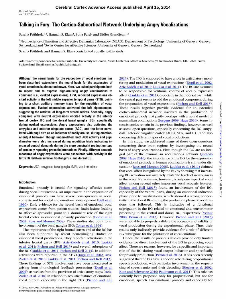
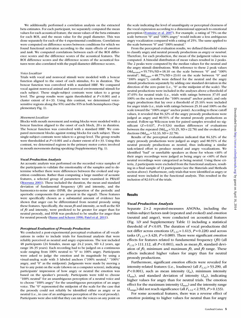
In repetition blocks (Fig. 1A), participants were asked to repeatthe prosodic intonations, which they had immediately heard spokenbeforehand by the actors. In evoked production blocks (Fig. 1B), par-ticipants had to produce the prosody freely. This evoked productiontask included a freely acted production of prosody with no constraintof imitating or repeating a certain prosodic style. We have to note,however, that the evoked task did not represent the production of vo-calizations resulting from really experiencing the underlying emotionor feeling of anger, but rather a relatively unconstraint production of
vocalizations on demand. In both the repetition and the evoked pro-duction blocks, the pseudoword was first presented on a gray screenfor 800 ms starting 250 ms after the last volume acquisition (Fig. 1C).It was presented either in uppercase letters (indicating angryprosody production), or in lowercase letters (indicating neutralprosody production). For the repetition trials, the word was pre-sented together with the voice of the actors. Afterwards, a black crossappeared on the screen during one volume acquisition (TA = 1580ms, see below). After the volume acquisition, the black cross turnedinto a white cross. The white cross indicated that participants shouldproduce the prosody asked for. The white cross stayed on the screenfor 1580 ms, after which the cross turned black again and the nextvolume was acquired. Participants’ prosody productions were re-corded in the silent gap during volume acquisitions using an fMRI-compatible Sennheiser optical microphone (16 bits and 44.1 kHz)and a digital voice recorder.
Figure 1. (A) During the repetition task, participants had to repeat and imitate the vocal production of neutral and angry prosody on 4 two-syllable pseudowords (“belam,” “lagod,”“minad,” and “namil”), which were presented visually on a screen and which they had heard immediately beforehand when listening to recordings of actors (“listening to actor”).The production of neutral prosody was indicated by words written in lowercase letters (e.g., “belam”) and the production of angry prosody was indicated by words written inuppercase letters (e.g., “BELAM”). The spectrograms show example recordings of the actors (left column in A and B) and of the participants (right column in A and B) for producingprosody on the pseudoword “belam.” (B) During the evoked task, participants were asked to freely produce prosodic intonations without hearing the recording of an actorbeforehand (“no actor”). This task included evoked production of prosody to the degree that participants could freely produce acted angry prosody without the constraint to imitate acertain prosodic style. (C) Shown are 2 example trials. The first trial depicts a repetition trial for the word “belam,” which had to be spoken in neutral tone (indicated by visuallypresented lower case letters) as previously heard from listening to an actor recording (auditory stimulation). The second trial depicts an evoked trial for the word “namil” spoken inan angry tone (indicated by visually presented upper case letters). Both auditory stimulation and vocal recording occurred during the silent gap between image acquisitions. (D)Results of the acoustical feature analysis of prosody productions during the repetition task (red) and the evoked task (green), separately for pitch-related acoustic features (leftpanel), for intensity-related features (middle panel), and for other voice quality features (right panel). The bars represent the ratio between feature scores for angry trials divided bythe same feature scores for neutral trials. The “e” indicates significant effects of the factor emotion (angry > neutral) for a 2 × 2 repeated-measures ANOVA, “-e” indicates thereverse effect (neutral > anger); “t” indicates a significant effect for the factor task, and “t*e” indicates a significant task * emotion interaction. Acoustical features: f0m, mean ofpitch; f0sd, standard deviation of pitch; f0min, minimum of pitch; f0max, maximum of pitch; f0range, range of pitch; Im, mean of intensity; Isd, standard deviation of intensity; Imin,minimum of normalized intensity; Imax, maximum of normalized intensity; Irange, range of intensity; ENdur, relative energy normalized to total duration; REN, relative energy in 0–500Hz band to 0–8 kHz band; HNRm, mean of the harmonic-to-noise ratio.
Cerebral Cortex 3
by guest on April 16, 2014
http://cercor.oxfordjournals.org/D
ownloaded from
Functional Voice Localizer ScanningWe used 8 s sound clips taken from an existing database (see http://vnl.psy.gla.ac.uk/) (Belin and Zatorre 2000) to determine human voice-sensitive regions in the bilateral superior temporal cortex. The soundclips consisted of 20 sequences of nonemotional human voices and 20sequences of animal or environmental sounds. Each sound clip was pre-sented once with a fixation cross on the screen and a 4-s gap betweeneach clip. The scanning sequence also contained twenty 8 s silentevents. Participants listened passively to the stimuli.
MouthMovement Localizer ScanningWe were interested only in the activations related primarily to the pro-duction of emotional prosody, not to the movement of the vocal appar-atus during speaking. Thus, to be able to exclude sensorimotor regionsshowing activations due to mouth movement only, we conducted amovement localizer scanning in the experiment. The movement locali-zer consisted of 8 movement blocks and 8 resting blocks. In eachblock, the same word appeared 10 times, alternating with a cross. Theword and the cross each appeared for 1 s on the screen. In movementblocks, the color of the words and crosses was green, and participantswere instructed to form the word with their lips as soon as it appearedon the screen. In resting blocks, words and crosses were red and par-ticipants were instructed not to move their lips and watch. Betweeneach block, there was a 5-s gap indicated by a blank screen. The 4stimulus words of the main experiment were used. Each word wasused in 2 movement blocks and in 2 resting blocks.
Image Acquisition and Image ProcessingAll functional imaging data were recorded on a 3-T Siemens TrioSystem (Siemens, Erlangen, Germany) using a T2*-weighted gradientecho-planar imaging sequence [time to repetition (TR) = 3290 ms, timeof acquisition TA = 1580 ms, time to echo (TE) = 30 ms, flip angle FA =90°, 28 slices, slice thickness 4 mm, distance factor = 20%, 64 matrix(3 × 3 mm)]. We used a sparse temporal acquisition protocol for themain experiment, which allowed presentation of auditory stimuli inthe silent gap between volume acquisitions. It also allowed us torecord the prosody productions of the participants (see below), whichare unaffected by the background scanner noise. A high-resolutionmagnetization-prepared rapid acquisition gradient echo, T1-weightedsequence [1 mm slices, TR = 1900 ms, TE = 2.27 ms, time to inversion(TI) = 900 ms, FoV 296 mm, in-plane 1 × 1 mm] was obtained in sagittalorientation to obtain structural brain images from each subject.
Images from the main experiment and from both localizer scanswere preprocessed and analyzed using the Statistical ParametricMapping software SPM8 (Welcome Department of Cognitive Neurol-ogy, London, UK). Functional images were realigned and coregisteredto the anatomical image. During realignment we ensured that headmotion in any spatial dimension of each participant was <1.5 mm,which is less than half of the voxel size used for image acquisition. Seg-mentation of the anatomical image revealed warping parameters thatwere used to normalize the functional images to the Montreal Neuro-logical Institute (MNI) stereotactic template brain. Functional imageswere resampled to a 2-mm3 voxel size and spatially smoothed using anisotropic Gaussian kernel of 8 mm3 full-width at half-maximum.
Pupil Diameter Measurement and AnalysisWe recorded the pupil diameter of each participant continuouslythroughout the main experiment by using an MRI-compatible long-range eye tracker system (EyeTrac 6, Applied Science Laboratories,USA) at a sampling rate of 60 Hz. Eye blinks in the pupil data wereinterpolated. The pupil diameter was supposed to be an indicator ofthe bodily arousal states (Partala and Surakka 2003) during theemotional vocalizations of the participants. For the cases in whichblinks affected >20% of a trial, the entire trial was excluded fromfurther statistical analyses. The average percentage of valid trials was86.95% (SD = 7.24). For valid trials, the time course of the pupil diam-eter was extracted for a window of −1000– to 3000 ms, time locked tothe appearance of the white fixation cross (the signal to the partici-pants to produce prosody). The time courses were baseline corrected
according to the mean signal in the baseline period −1000 to 0 ms. Themean pupil diameter was scored in the time period 0–1580 ms. Thiswas the silent gap interval during which participants were asked toproduce prosody. The mean pupil size was determined separately foreach experimental condition. Two participants had to be excludedfrom this analysis because of bad or missing pupil data due to acqui-sition problems during the experiment. The mean scores were sub-sequently subjected to a 2 × 2 repeated-measures analysis of variance(ANOVA) with the within-subject factors task (repeated and evoked)and emotion (neutral and anger). A statistical threshold of P < 0.05 wasused for this analysis.
Statistical Analyses of Functional Data
Main ExperimentEach trial was modeled with a boxcar function defined by the onset ofauditory stimulation and the onset of vocal productions, including theduration of each event. The boxcar function was convolved with a stan-dard hemodynamic response function (HRF) on a single-subject leveltaking into account the temporal sparse acquisition pattern (see Kumaret al. 2007; Frühholz et al. 2012). For the main experiment, separate re-gressors were created for each of the 4 experimental conditions (2tasks × 2 emotions) for correct trials as defined by the perceptual evalu-ation of the prosody productions (see below). Only trials were in-cluded in the first 4 regressors, which were reliably classified as neutralor angry prosody in the perceptual evaluation of the recorded prosodyproductions. A fifth regressor modeled all trials with unreliable classifi-cation. Six motion correction parameters were finally included as re-gressors of no interest to minimize false-positive activations that weredue to task-correlated motion. Simple contrasts for each experimentalcondition for each participant were taken to a second-level random-effects ANOVA group analysis.
To obtain the differences in blood oxygen level-dependent responses,the following contrasts were computed for the group analysis using asingle ANOVA (i.e., the flexible factorial design option in SPM) includingall experimental conditions. To reveal the main effect of task, we com-pared repetition trials with evoked trials and vice versa. To reveal themain effect of emotion, we compared angry production trials withneutral production trials. The effect of angry compared with neutral trialswas also computed separately for each the repetition and evoked task.Finally, we also computed interaction contrasts to find specific activationfor angry trials during the repetition and during the evoked task. All con-trasts were thresholded at P < 0.001 and a cluster extent of k = 33. Thiscombined voxel and cluster threshold corresponds to P < 0.05 correctedat the cluster level and was determined by the 3DClustSim algorithmimplemented in the AFNI software (http://afni.nimh.nih.gov/afni) usingthe estimated smoothness of the data across all contrasts computed.Across all contrasts, this procedure resulted in a maximum k = 33, andthis was set as cluster threshold for all contrasts.
We extracted beta estimates in several ROIs, including the bilateralIFG (left IFG, right IFG, and right IFGor), the bilateral STG [left mSTG,left posterior STG (pSTG), and right pSTG], the bilateral amygdala, lefthippocampus (HC), and bilateral BG (left putamen and right caudatenucleus), and the ACC. Peak activations for these ROIs were takenfrom the analysis of experimental main effects to directly investigateexperimental interaction effects in these ROIs additionally to the inter-action analyses described above. The ROIs were created in regionswhich were of primary interest in this study. Multiple ROIs werecreated in the IFG and the STG, because it was previously shown thatsubregions in these areas can have different functional roles during theprocessing and production of vocalizations (Frühholz and Grandjean2013b, c). For these ROI analyses, we scored mean beta estimates in 3mm radius spheres around peak activations. Since we did not findpeak activations for the amygdala in the main analysis, we extractedmean beta estimates in an anatomical amygdala mask as defined by theAAL brain atlas (Tzourio-Mazoyer et al. 2002), because the amygdalawas one of our main ROIs. Beta estimates were subjected to a 2 × 2repeated-measures ANOVA with the within-subject factors task (re-peated and evoked) and emotion (neutral and anger), especially to de-termine interaction effects in these ROIs between the experimentalfactors.
4 Cortico-Subcortical Network Underlying Angry Vocalizations • Frühholz et al.
by guest on April 16, 2014
http://cercor.oxfordjournals.org/D
ownloaded from
We additionally performed a correlation analysis on the extractedbeta estimates. For each participant, we separately computed the meanvalues for each acoustical feature, the mean values of the beta estimatesfor each ROI, and the mean value for the pupil diameter. This wasdone separately for each of the 4 experimental conditions. Correlationswere computed on difference scores between conditions for which wefound functional activation according to the main effects of emotionand task. We computed correlations between each of the ROI differ-ence scores and the difference scores of the acoustical features. TheROI difference scores and the difference scores of the acoustical fea-tures were also correlated with the pupil diameter difference scores.
Voice LocalizerTrials with vocal and nonvocal stimuli were modeled with a boxcarfunction aligned to the onset of each stimulus, 8 s in duration. Theboxcar function was convolved with a standard HRF. We comparedvocal against nonvocal animal and nonvocal environmental stimuli foreach subject. These single-subject contrasts were taken to a grouplevel. The group results were again thresholded at P < 0.001 and acluster extent of k = 33. Using this contrast, we determined voice-sensitive regions along the STG and the STS in both hemispheres (Sup-plementary Fig. 1).
Movement LocalizerBlocks with mouth movement and resting blocks were modeled with aboxcar function aligned to the onset of each block, 20 s in duration.The boxcar function was convolved with a standard HRF. We com-pared movement blocks against resting blocks for each subject. Thesesingle-subject contrasts were taken to a group level. The group resultswere thresholded at P < 0.001 and a cluster extent of k = 33. Using thiscontrast, we determined regions in the primary motor cortex involvedin mouth movements during speaking (Supplementary Fig. 1).
Vocal Production AnalysisAn acoustic analysis was performed on the recorded voice samples ofthe participants to validate the emotionality of the samples and to de-termine whether there were differences between the evoked and rep-etition conditions. Rather than computing a large number of acousticfeatures, a selected group of parameters were examined in PRAAT(Boersma 2001). These included the duration, the mean and standarddeviation of fundamental frequency (f0) and intensity, and theharmonics-to-noise ratio (HNR; the proportion of the periodic andaperiodic components that are present in the signal). These featureswere specifically chosen as a substantial amount of evidence hasshown that anger can be differentiated from neutral prosody usingthese features. Specifically, the mean f0 and intensity, as well as the SDof f0 and intensity, were predicted to be greater for anger than forneutral prosody, and HNR was predicted to be smaller for anger thanfor neutral prosody (Banse and Scherer 1996; Patel et al. 2011).
Perceptual Evaluation of Prosody ProductionWe conducted a post-experimental perceptual evaluation of all vocali-zations in order to include trials for functional analysis that werereliably perceived as neutral and angry expressions. The test included48 participants (24 females, mean age 24.2 years, SD 4.2 years, agerange 18–35 years). Each recording had to be judged on a continuousscale ranging from 100% neutral to “0” to 100% angry. Participantswere asked to judge the emotion and its magnitude by using avisual-analog scale with 3 labeled anchors (“100% neutral,” “100%”angry, and “0” as the midpoint). Judgments were made by moving abar to any point on the scale (shown on a computer screen), indicatingparticipants’ impression of how angry or neutral the emotion wasbased on the speaker’s prosody. Participants were told to choose“100% neutral” for an unambiguous perception of a neutral voice andto choose “100% angry” for the unambiguous perception of an angryvoice. The “0” represented the midpoint of the scale for the case thatthe prosody could not reliably be identified either as angry or asneutral (i.e., in case of an ambiguous perception of the vocal prosody).Participants were also told that they can rate the voices on any point on
the scale indicating the level of unambiguity or perceptual clearness ofthe vocal expression according to a dimensional approach to emotionalperception (Fontaine et al. 2007). For example, a rating of 75% on thescale between “0” and “100% angry” would indicate a less ambiguousangry vocalization compared with a rating of 25%. The same applied tothe scale between “0” and “100% neutral.”
From the perceptual evaluation results, we defined threshold valuesto classify angry and neutral prosody productions as angry or neutral.Therefore, for each production, the mean of the judgment values wascomputed. A bimodal distribution of mean values resulted in 2 peaks.The 2 peaks were computed by the median values for the neutral andthe angry stimuli distributions. With reference to these 2 peak values(Mdneutral = 55.75%/SD = 18.10 on the scale between “0” and “100%neutral”; Mdanger = 48.77%/SD = 23.61 on the scale between “0” and“100% angry”), cutoffs were defined for the neutral and the angrystimuli productions separately by adding one standard deviation in thedirection of the zero point (i.e., “0” as the midpoint of the scale). Theneutral productions were included in the analyses above a threshold of37.65% for neutral trials (i.e., trials with ratings between 37.65 and100% on the scale toward the “100% neutral” anchor point), and onlyangry productions that lay over a threshold of 25.16% were includedfor anger trials (i.e., trials with ratings between 25.16 and 100% on thescale toward the “100% anger” anchor point). According to these cutoffvalues, it revealed that 84.32% of the angry prosody productions werejudged as angry and 80.91% of the neutral prosody productions asneutral. Follow-up Wilcoxon tests for paired samples revealed no sig-nificant (Z = 0.637, P = 0.524) median differences of evaluationsbetween the repeated (Mdrep = 53.25, SD = 22.78) and the evoked pro-ductions (Mdspo = 52.33, SD = 22.78).
Results of the perceptual evaluation indicated that 84.32% of theangry prosody productions were judged as angry and 80.91% of theneutral prosody productions as neutral, thus indicating a similartask-related effort to produce neutral and angry vocalizations. Weidentified “bad” or unreliable speakers as those for whom <40% oftheir angry recordings were judged as being angry or <40% of theirneutral recordings were categorized as being neutral. Using these cri-teria, 2 participants were excluded from the functional analyses, result-ing in a final sample of 13 participants (see the Materials and Methodssection above). Furthermore, only trials that were identified as angry orneutral were included in the functional analysis. This resulted in theinclusion of 82.61% of the trials.
Results
Vocal Production AnalysisSeparate 2 × 2 repeated-measures ANOVAs, including thewithin-subject factors task (repeated and evoked) and emotion(neutral and anger), were conducted on acoustical features(Fig. 1D and Supplementary Table 1) including a statisticalthreshold of P < 0.05. The duration of vocal productions didnot differ across emotions (F1,12 = 1.613, P = 0.228) and acrosstasks (F1,12 = 3.420, P = 0.089). There were significant emotioneffects for features related to fundamental frequency (f0) (allF1,12 > 111.112, all P < 0.001), such as mean f0, standard devi-ation of f0, minimum and maximum f0, and f0 range. Theseeffects indicated higher values for angry than for neutralprosody productions.
Furthermore, significant emotion effects were revealed forintensity-related features (i.e., loudness) (all F1,12 > 53.290, allP < 0.001), such as mean intensity (Im), minimum intensity(Imin), and standard deviation of intensity (Isd), indicatinghigher values for angry than for neutral trials. The emotioneffect for the maximum intensity (Imax) and the intensity range(Irange) did not reach significance (all F1,12 < 2.593, P > 0.133).
For some acoustical features, there was a reverse effect ofemotion pointing to higher values for neutral than for angry
Cerebral Cortex 5
by guest on April 16, 2014
http://cercor.oxfordjournals.org/D
ownloaded from
trials. The acoustical measures of jitter (F1,12 = 15.236, P =0.002) and shimmer (F1,12 = 20.672, P = 0.001) revealed suchan effect. Furthermore, also the HNR revealed higher valuesduring neutral than during angry trials (F1,12 = 37.036, P < 0.001).For this feature, also the interaction between the factors emotionand task reached significance (F1,12 = 6.972, P = 0.022), indicat-ing a higher mean HNR during the repetition of neutralprosody compared with that of angry prosody (t12 = 5.159,P < 0.001). There were no other significant interactions (allF1,12 < 3.360, all P > 0.090).
There was one task effect for Isd indicating higher values forrepetition than for evoked trials (F1,12 = 4.893, P = 0.047). Allother task effects for the remaining acoustical features did notreach significance (all F1,12 < 4.386, all P > 0.058).
Functional Brain DataBecause we were only interested in functional activity whichwas specifically related to the production of angry prosody andnot to general activation due to mouth movements, we onlyreport functional activations here, which were located outsidethe primary sensorimotor regions as determined by the mouthmovement localizer scan (Supplementary Fig. 1).
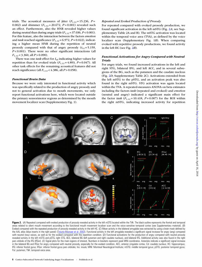
Repeated and Evoked Production of ProsodyFor repeated compared with evoked prosody production, wefound significant activation in the left mSTG (Fig. 2A; see Sup-plementary Table 2A and B). The mSTG activation was locatedwithin the temporal voice area (TVA), as defined by the voicelocalizer scan (Supplementary Fig. 1B). When comparingevoked with repetitive prosody productions, we found activityin the left HC (see Fig. 2B).
Functional Activations for Angry Compared with NeutralTrialsFor angry trials, we found increased activations in the left andright STG, bilateral IFG, and left ACC, and in several subre-gions of the BG, such as the putamen and the caudate nucleus(Fig. 2D; Supplementary Table 2C). Activations extended fromthe left mSTG to the pSTG, and an activation peak was alsofound in the right mSTG. STG activation was again locatedwithin the TVA. A repeated-measures ANOVA on beta estimatesincluding the factors task (repeated and evoked) and emotion(neutral and angry) indicated a significant main effect forthe factor task (F1,12 = 10.418, P = 0.007) for the ROI withinthe right mSTG, indicating increased activity for repetition
Figure 2. (A) Repeated compared with evoked production of prosody revealed activity in the left mSTG located within the TVA. The black outline represents the frontal and temporalareas related to silent mouth movement according to the functional mouth movement localizer scan and the voice-sensitive temporal cortex (see Supplementary material). (B)Evoked compared with the repeated production of prosody revealed activity in the left HC. (C) Mean activity in the bilateral amygdala was extracted by using a brain mask defined bythe AAL atlas (blue inserts in the right panel) (Tzourio-Mazoyer et al. 2002). Functional activity in the left amygdala revealed a significant signal increase for angry (ang) comparedwith neutral (neu) voices, as well as for the evoked compared with the repetition condition. (D) Functional activations for the production of angry compared with neutral prosodyrevealed activity in the left mSTG and pSTG, right STG, ACC, bilateral BG (left putamen and right caudate nucleus), and bilateral IFG. Additional activity was also found in the rightpars orbitals of the IFG (IFGor). (E) Signal plots for the main regions of interest. Numbers in brackets represent peak MNI coordinates. Asterisks indicate a significant signal increasein the bilateral BG and IFGor for angry compared with neutral prosody, especially for the evoked condition. ACC, anterior cingulate cortex; Cd: caudate nucleus; HC: hippocampus;IFG: inferior frontal gyrus; IFGor: inferior frontal gyrus pars orbitalis; Ins: insula; MNI: Montreal Neurological Institute; mSTG: middle temporal gyrus; pSTG: posterior temporal gyrus;Put: putamen; TVA: temporal voice area.
6 Cortico-Subcortical Network Underlying Angry Vocalizations • Frühholz et al.
by guest on April 16, 2014
http://cercor.oxfordjournals.org/D
ownloaded from
compared with evoked trials. There was no significant inter-action (F1,12 = 0.087, P = 0.774). The reverse contrasts ofneutral compared with angry trials did not reveal any signifi-cant functional activations.
Furthermore, activations were found in the bilateral IFG forangry productions (Fig. 2D). One peak was located in the leftIFG and 2 peaks were found in the right IFG. For the right IFG,one peak of activity was located in the pars orbitalis of theIFG (IFGor) and the other peak was located more posterior inthe pars opercularis of the IFG. Only in the IFGor peak did wefind a significant interaction between the factors emotion andtask (F1,12 = 12.437, P = 0.004) (Fig. 2E). Here, the activationdifference was especially pronounced during evoked angrycompared with evoked neutral prosody production (t12 =4.381, P < 0.001). No other post hoc tests revealed a significantdifference between experimental conditions (all t’s < 1.941, allP’s > 0.076).
Regarding the left ACC (Fig. 2D), there was a significantmain effect for emotion, indicating an increase of activity inthis region when prosody was produced angrily comparedwith neutral productions. An ANOVA on ROI beta estimates inthe ACC also revealed a significant main effect for task (F1,12 =9.938, P = 0.008), indicating greater involvement of the ACC inthe repetition compared with the evoked production. We alsofound activity in the right insula in angry compared withneutral production.
Furthermore, for angry productions, we found activity in thebilateral dorsal BG. There were activation peaks in the leftputamen and the right caudate nucleus (Fig. 3C). According tothe ROI analysis, this effect was especially pronounced forevoked angry prosody production in both the left putamen(F1,12 = 26.538, P < 0.001) and the right caudate nucleus (F1,12-= 10.902, P = 0.006), as indicated by significant interactioneffects. For the left putamen, paired post hoc t-tests revealed asignificant difference between angry and neutral productionsfor evoked prosody (t12 = 8.664, P < 0.001). For the rightcaudate nucleus, post hoc tests revealed a significant differencebetween angry and neutral productions for evoked prosody(t12 = 6.926, P < 0.001). No other post hoc test revealed a sig-nificant difference between experimental conditions (all t’s <1.465, all P’s > 0.169).
Functional Activations for Angry Compared with NeutralTrials Within Each TaskFor angry trials in the repetition task, we obtained activationsin the bilateral IFG, bilateral insula, and middle and left dorsalACC (Supplementary Table 3B). For angry trials in evokedtasks, there were activation peaks in the right IFG, the pars or-bitalis of the IFG (IFGor), the right insula, the left middle cin-gulate gyrus, the right STG, the bilateral putamen, and theright caudate nucleus (Supplementary Table 3A). We further-more computed interaction contrasts to find activity during theproduction of angry prosody that was specific to each task. Nosignificant activation was revealed in this analysis.
Functional Amygdala ActivityOur whole-brain analysis did not reveal significant activity inthe bilateral amygdala. However, since the amygdala was oneof our primary ROIs, we performed a ROI analysis on meanbeta estimates extracted from the bilateral amygdala masks(Fig. 2C). Repeated-measures ANOVAs on the beta estimatesrevealed a significant effect for the factor emotion for the leftamygdala (F1,12 = 4.899, P = 0.047), indicating higher activationin the amygdala during angry than during neutral prosody pro-duction. There was also a significant effect for the factor task(F1,12 = 2.957, P = 0.031), indicating a greater involvement ofthe amygdala during evoked prosody production than duringrepetition of prosody. An interaction did not reach significance(F1,12 = 2.290, P = 0.278). No significant main effects or inter-actions were found in the right amygdala (all F1,12 < 2.947, allP > 0.112).
Correlations of Functional Activations with Features of VocalProductionsTo find out which functional activations in our primary ROIsare related to specific acoustic features of vocal productions,we computed correlations between functional activations inour ROIs and vocal features of the emotional prosody pro-ductions. Significant correlations are reported in Table 1, allother correlations were not significant (all r’s < |0.533|, allP’s > 0.061). Correlations were computed on difference scoresthat resulted from comparing the different experimental con-ditions. During the repetition compared with the evoked
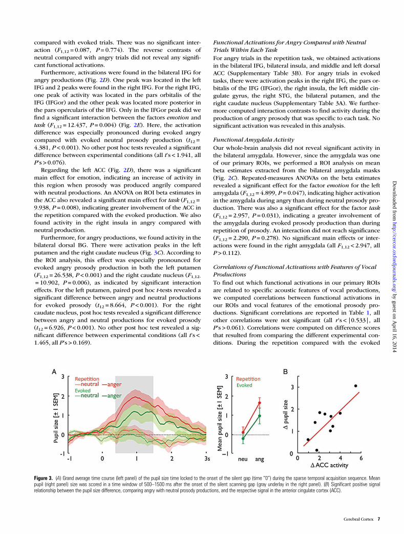
Figure 3. (A) Grand average time course (left panel) of the pupil size time locked to the onset of the silent gap (time “0”) during the sparse temporal acquisition sequence. Meanpupil (right panel) size was scored in a time window of 500–1500 ms after the onset of the silent scanning gap (gray underlay in the right panel). (B) Significant positive signalrelationship between the pupil size difference, comparing angry with neutral prosody productions, and the respective signal in the anterior cingulate cortex (ACC).
Cerebral Cortex 7
by guest on April 16, 2014
http://cercor.oxfordjournals.org/D
ownloaded from
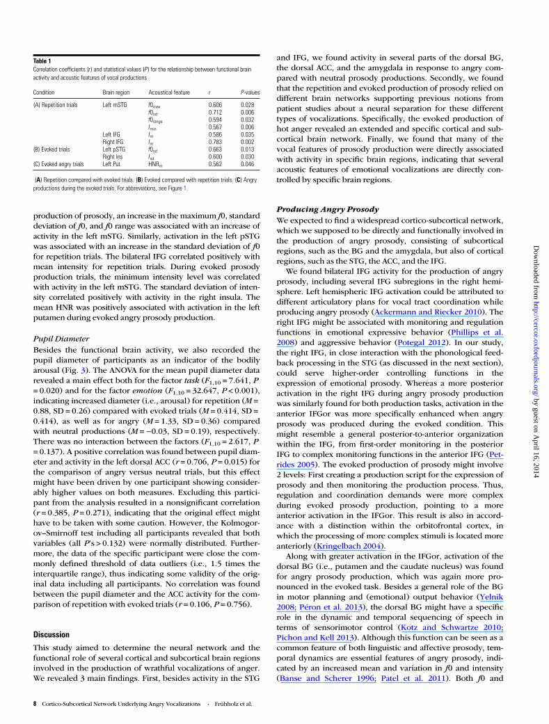
production of prosody, an increase in the maximum f0, standarddeviation of f0, and f0 range was associated with an increase ofactivity in the left mSTG. Similarly, activation in the left pSTGwas associated with an increase in the standard deviation of f0for repetition trials. The bilateral IFG correlated positively withmean intensity for repetition trials. During evoked prosodyproduction trials, the minimum intensity level was correlatedwith activity in the left mSTG. The standard deviation of inten-sity correlated positively with activity in the right insula. Themean HNR was positively associated with activation in the leftputamen during evoked angry prosody production.
Pupil DiameterBesides the functional brain activity, we also recorded thepupil diameter of participants as an indicator of the bodilyarousal (Fig. 3). The ANOVA for the mean pupil diameter datarevealed a main effect both for the factor task (F1,10 = 7.641, P= 0.020) and for the factor emotion (F1,10 = 32.647, P < 0.001),indicating increased diameter (i.e., arousal) for repetition (M =0.88, SD = 0.26) compared with evoked trials (M = 0.414, SD =0.414), as well as for angry (M = 1.33, SD = 0.36) comparedwith neutral productions (M =−0.03, SD = 0.19), respectively.There was no interaction between the factors (F1,10 = 2.617, P= 0.137). A positive correlation was found between pupil diam-eter and activity in the left dorsal ACC (r = 0.706, P = 0.015) forthe comparison of angry versus neutral trials, but this effectmight have been driven by one participant showing consider-ably higher values on both measures. Excluding this partici-pant from the analysis resulted in a nonsignificant correlation(r = 0.385, P = 0.271), indicating that the original effect mighthave to be taken with some caution. However, the Kolmogor-ov–Smirnoff test including all participants revealed that bothvariables (all P’s > 0.132) were normally distributed. Further-more, the data of the specific participant were close the com-monly defined threshold of data outliers (i.e., 1.5 times theinterquartile range), thus indicating some validity of the orig-inal data including all participants. No correlation was foundbetween the pupil diameter and the ACC activity for the com-parison of repetition with evoked trials (r = 0.106, P = 0.756).
Discussion
This study aimed to determine the neural network and thefunctional role of several cortical and subcortical brain regionsinvolved in the production of wrathful vocalizations of anger.We revealed 3 main findings. First, besides activity in the STG
and IFG, we found activity in several parts of the dorsal BG,the dorsal ACC, and the amygdala in response to angry com-pared with neutral prosody productions. Secondly, we foundthat the repetition and evoked production of prosody relied ondifferent brain networks supporting previous notions frompatient studies about a neural separation for these differenttypes of vocalizations. Specifically, the evoked production ofhot anger revealed an extended and specific cortical and sub-cortical brain network. Finally, we found that many of thevocal features of prosody production were directly associatedwith activity in specific brain regions, indicating that severalacoustic features of emotional vocalizations are directly con-trolled by specific brain regions.
Producing Angry ProsodyWe expected to find a widespread cortico-subcortical network,which we supposed to be directly and functionally involved inthe production of angry prosody, consisting of subcorticalregions, such as the BG and the amygdala, but also of corticalregions, such as the STG, the ACC, and the IFG.
We found bilateral IFG activity for the production of angryprosody, including several IFG subregions in the right hemi-sphere. Left hemispheric IFG activation could be attributed todifferent articulatory plans for vocal tract coordination whileproducing angry prosody (Ackermann and Riecker 2010). Theright IFG might be associated with monitoring and regulationfunctions in emotional expressive behavior (Phillips et al.2008) and aggressive behavior (Potegal 2012). In our study,the right IFG, in close interaction with the phonological feed-back processing in the STG (as discussed in the next section),could serve higher-order controlling functions in theexpression of emotional prosody. Whereas a more posterioractivation in the right IFG during angry prosody productionwas similarly found for both production tasks, activation in theanterior IFGor was more specifically enhanced when angryprosody was produced during the evoked condition. Thismight resemble a general posterior-to-anterior organizationwithin the IFG, from first-order monitoring in the posteriorIFG to complex monitoring functions in the anterior IFG (Pet-rides 2005). The evoked production of prosody might involve2 levels: First creating a production script for the expression ofprosody and then monitoring the production process. Thus,regulation and coordination demands were more complexduring evoked prosody production, pointing to a moreanterior activation in the IFGor. This result is also in accord-ance with a distinction within the orbitofrontal cortex, inwhich the processing of more complex stimuli is located moreanteriorly (Kringelbach 2004).
Along with greater activation in the IFGor, activation of thedorsal BG (i.e., putamen and the caudate nucleus) was foundfor angry prosody production, which was again more pro-nounced in the evoked task. Besides a general role of the BGin motor planning and (emotional) output behavior (Yelnik2008; Péron et al. 2013), the dorsal BG might have a specificrole in the dynamic and temporal sequencing of speech interms of sensorimotor control (Kotz and Schwartze 2010;Pichon and Kell 2013). Although this function can be seen as acommon feature of both linguistic and affective prosody, tem-poral dynamics are essential features of angry prosody, indi-cated by an increased mean and variation in f0 and intensity(Banse and Scherer 1996; Patel et al. 2011). Both f0 and
Table 1Correlation coefficients (r) and statistical values (P) for the relationship between functional brainactivity and acoustic features of vocal productions
Condition Brain region Acoustical feature r P-values
(A) Repetition trials Left mSTG f0max 0.606 0.028f0sd 0.712 0.006f0range 0.594 0.032Imin 0.567 0.006
Left IFG Im 0.586 0.035Right IFG Im 0.783 0.002
(B) Evoked trials Left pSTG f0sd 0.663 0.013Right Ins Isd 0.600 0.030
(C) Evoked angry trials Left Put HNRm 0.562 0.046
(A) Repetition compared with evoked trials. (B) Evoked compared with repetition trials. (C) Angryproductions during the evoked trials. For abbreviations, see Figure 1.
8 Cortico-Subcortical Network Underlying Angry Vocalizations • Frühholz et al.
by guest on April 16, 2014
http://cercor.oxfordjournals.org/D
ownloaded from
intensity dynamics determine the temporal and rhythmic un-folding of emotional prosody, and this rhythmic aspect duringvocal productions can be impaired in individuals with BGlesions (Péron et al. 2010). In our study, the repetition task in-volved actor recordings as an external cue, thereby minimizingthe need to self-generate the temporal dynamics for prosodyproduction. In contrast, the dorsal BG showed stronger activityin the evoked task for producing angry prosody, which mayreflect stronger self-generated prosody dynamics.
While the BG might be associated with the temporal anddynamic sequencing of emotional speech output, the ACCmight have 2 different functions during emotional vocaliza-tions. First, the emotion effect that we observed in the ACCmight point to its function of regulating the arousal level andthus in the control of the autonomous nervous system foremotional output behavior (Critchley et al. 2003). Activity inthe ACC was positively correlated with the individual’s pupildiameter as a physiological measure of bodily arousal (Partalaand Surakka 2003) during the production of angry comparedwith neutral prosody. This result is further supported by ourfinding of activation in the right insula together with the ACCduring angry prosody production, since the insula is similarlyassociated with the generation of autonomic responses (Ull-sperger et al. 2010). Thus, both the ACC and the insula seem toregulate autonomic arousal when people speak in an aggres-sive angry tone.
Though uncorrelated, the increased activity of the ACC andthe increased pupil size for repetition compared with evokedtrials together might point to the second function of ACCserving increased performance monitoring during the moredemanding repetition/imitation of emotional vocalizations.The pupil size has been shown to be an indicator of cognitiveload especially during language-related tasks (Hyona et al.1995), and the ACC seems a central brain area serving perform-ance and error monitoring during increased demands of cogni-tive control (Kerns et al. 2004). The more constraintproduction type of exactly repeating prosodic intonationshould have involved increased cognitive load and perform-ance and error monitoring demands. Thus, beyond the func-tions of the ACC for volitional initiation of vocalizations(Jurgens 2009; Hage 2010), our data seem to suggest that theACC is also involved regulating the arousal level as well asmonitoring the vocal performance depending on the pro-duction type.
The present study also found activation in the amygdaladuring the production of angry prosody and provides strongevidence to extend the general vocalization network (Jurgens2009; Hage 2010) by the limbic brain system, which plays animportant role during emotional vocalizations. Our results em-phasize the importance of the amygdala underlying emotionaloutput behavior, but contradict the prevailing view of theamygdala as a structure mainly involved in the detection ofstimuli and conditioning (Cardinal et al. 2002). One importantrole of the amygdala is its involvement in the expression ofemotionally relevant behavior. The amygdala is known to beinvolved in the expression and regulation of angry behavior,probably in the experience of an aggressive impulse (Coccaroet al. 2011). The amygdala was also more activated in ourstudy during evoked productions, indicating that evokedprosody might be associated with stronger emotional regulat-ory effects by the amygdala. This interpretation is supportedby recent findings of amygdala involvement during emotional
prosody preparation (Pichon and Kell 2013). One alternativeinterpretation might be that, instead of regulating emotionaloutput behavior, amygdala activity might also originate fromauditory feedback processing of own emotional vocalizations.While this might partly explain higher amygdala activityduring angry compared with neutral vocalizations, evokedcompared with repetition trials also revealed stronger amygda-la activity, and both are balanced in terms of expressingneutral and angry vocalizations. A considerable proportion ofthe amygdala activity thus might be modulated by the vocalproduction type. In the present study, evoked trials were lessconstraint than repetition trials in terms of how to vocalize.This provides some evidence for its differential regulatory roleunderlying emotional and especially angry vocalizations(Coccaro et al. 2011) under conditions of less constraint pro-duction modes, which might also have been a more stressfuland thus more “emotional” production mode. This is also sup-ported by the important regulatory role of the amygdala inpathological expressions of vocal emotions (Lauterbach et al.2013).
The final brain structure found for producing angry prosodywas the STG. During the production of prosody, specific pro-sodic speech elements have to be derived and translated intoautonomous motor output. Evidence suggests that the STGsupports sensory-motor integration (Hickok 2009; Peschkeet al. 2009), the gating of amygdala connections (Pehrs et al.2013), and phonological feedback processing (Zheng et al.2010), which are necessary for accurate production of angryprosody. This is supported by our finding that left STG activitycorrelated with pitch features of vocal productions, supportingthe online adjustments of vocal output behavior by auditoryfeedback processes (Aziz-Zadeh et al. 2010). The STG mightalso serve as a phonological short-term store facilitatingprosody production, especially in cases when there is a delaybetween the perception and production of prosody, such asduring the repetition task.
Repeated and Evoked Vocal ExpressionsBesides our first experimental questions about the brainnetwork underlying angry vocalizations, the second questionsconcerned the differential modulation of brain activity depend-ing on the type of vocal productions. We thus compared brainactivity during prosody production for the repetition and theevoked production of prosody. The repetition task elicitedactivity in the left mSTG and pSTG. The STG seems to functionas a phonological store during the repetition of prosody,which is corroborated by recent findings for a short-termstorage system in the STG (Ravizza et al. 2010; Acheson et al.2011) and the closely located planum temporale (Hickok et al.2009). The planum temporale was found in a recent studyduring the nonaffective repetition of pseudowords (McGetti-gan et al. 2011), and the same region has been proposed forsensory-motor integration that maps perceived speech directlyto motor speech output. During the repetition task in thepresent study, the actors’ intonations had to be perceived andstored for subsequent repetition. Hence, it is likely that leftSTG activation is due to phonological short-term storage of thepseudoword before its production.
The evoked task elicited activity in the left HC. The HC isgenerally involved in long-term memory functions, as well asemotional regulation functions (Fanselow and Dong 2010).
Cerebral Cortex 9
by guest on April 16, 2014
http://cercor.oxfordjournals.org/D
ownloaded from
Our finding of HC activity might indicate the retrieval of long-term stored production rules during evoked prosody pro-duction for speech. During the evoked task, participants hadto produce prosody naturally without relying on a prosodytemplate that was immediately heard beforehand. For thisevoked production, the participants had to retrieve a prototypescript from long-term memory following the production rulesfor prosody intonations. This might be also explained by thefact that the evoked task did not involve vocal productions trig-gered by underlying emotional states, but rather required rela-tively unconstraint vocal productions on demand, whichmakes the use of prototype scripts more likely. Another expla-nation for the HC activation might be that the pseudowordswere learned by being processed and constantly repeated(Paulesu et al. 2009). Yet, as the same words were used in boththe repetition and evoked production conditions, learningshould have occurred in both conditions and not only in theevoked production of prosody. This, however, is not the casesince there was no HC activation during the repeated pro-duction of prosody.
Linking Brain Activity in the STG, BG, and IFG withSpecific Voice FeaturesOur final experimental question concerned the involvement ofspecific brain areas, which underlie the production of specificacoustic features during angry prosody. We accordingly ob-served that some of the neural activations mentioned earlierwere associated with specific acoustical features of the vocalexpressions. Activation within the STG was positively associ-ated with pitch-related features, such as the maximum, vari-ation, and range of the f0. As the STG is responsible forsensory-motor integration (Hickok 2009; Peschke et al. 2009)and phonological feedback processing (Zheng et al. 2010),this association might indicate regulatory effort and online ad-justments for accurate f0 production in angry prosody. Thus,emotional vocalizations strongly depend on auditory feedbackand perceptual processing of own-vocalizations in the auditorycortex. Our results strongly suggest to extend recent models ofmammalian vocalizations (Jurgens 2009; Hage 2010) byadditionally including auditory-motor loops as an integral partof vocal expressions. Activation in the IFG was positivelyassociated with the intensity of vocal productions, potentiallydue to the monitoring and regulation functions of the IFG. In-tensity was higher in angry than in neutral trials. The pro-duction of angry prosody probably required to a greater extentthe regulation and monitoring of intensity, resulting in anassociation of intensity and IFG activation. Finally, dorsal BGactivations were positively correlated with the HNR of the pro-duced speech, confirming results from BG lesion studies (VanLancker Sidtis et al. 2010).
ConclusionsThe production of high-arousing emotional prosody comprisesa cortical–subcortical network encompassing not only thebilateral IFG and STG, but also the bilateral dorsal BG, the leftamygdala, and the ACC. Several of these structures directlyregulate the bodily arousal or activation level as well as theacoustic properties underlying emotional vocalization. Theresults implicate both left and right hemispheric structures inthe production of emotional prosody, which contrasts with aprominent model stating that affective prosody is processed
dominantly by the right hemisphere and that its organization isanalogous to propositional prosody in the left hemisphere(Ross and Monnot 2008). The results about the central role ofthe BG might inspire research on the neural basis of impair-ments in the production of emotional prosody such as theyoccur in individuals with Parkinson disease (Péron et al. 2010,2013). The results might also inspire future research on theneural basis of angry vocalizations that are based on realexperiences of emotions or feelings instead of vocalizationsproduced on command.
Supplementary MaterialSupplementary material can be found at: http://www.cercor.oxford-journals.org/.
Funding
This study was supported by the Swiss National Science Foun-dation (SNSF, 105314_124572/1—D.G.) and by the NCCR inAffective Sciences at the University of Geneva (51NF40-104897—D.G.).
ReferencesAcheson DJ, Hamidi M, Binder JR, Postle BR. 2011. A common neural
substrate for language production and verbal working memory. JCogn Neurosci. 23:1358–1367.
Ackermann H, Riecker A. 2010. The contribution(s) of the insula tospeech production: a review of the clinical and functional imagingliterature. Brain Struct Funct. 214:419–433.
Aziz-Zadeh L, Sheng T, Gheytanchi A. 2010. Common premotorregions for the perception and production of prosody and corre-lations with empathy and prosodic ability. PLoS ONE. 5. e8759.
Banse R, Scherer KR. 1996. Acoustic profiles in vocal emotionexpression. J Pers Soc Psychol. 70:614–636.
Bänziger T, Scherer KR. 2010. Introducing the Geneva MultimodalEmotion Portrayal (GEMEP) Corpus. In: Bänziger T, Scherer KR,Roesch EB, editors. Blueprint for affective computing: a source-book Oxford. UK: Oxford University Press. p. 271–294.
Belin P, Zatorre RJ. 2000. Voice-selective areas in human auditorycortex. Nature. 403:309.
Bell WL, Davis DL, Morgan-Fisher A, Ross ED. 1990. Acquired aproso-dia in children. J Child Neurol. 5:19.
Boersma P. 2001. Praat, a system for doing phonetics by computer.Glot Int. 5:341–345.
Borod JC, Bloom RL, Brickman AM, Nakhutina L, Curko EA. 2002.Emotional processing deficits in individuals with unilateral braindamage. Appl Neuropsychol. 9:23–36.
Cardinal RN, Parkinson JA, Hall J, Everitt BJ. 2002. Emotion and motiv-ation: the role of the amygdala, ventral striatum, and prefrontalcortex. Neurosci Biobehav Rev. 26:321–352.
Coccaro EF, Sripada CS, Yanowitch RN, Phan KL. 2011. Corticolimbicfunction in impulsive aggressive behavior. Biol Psychiatry.69:1153–1159.
Cohen MJ, Riccio CA, Flannery AM. 1994. Expressive aprosodia follow-ing stroke to the right basal ganglia: a case report. Neuropsychol-ogy. 8:242–245.
Critchley HD. 2009. Psychophysiology of neural, cognitive and affec-tive integration: fMRI and autonomic indicants. Int J Psychophysiol.73:88–94.
Critchley HD, Mathias CJ, Josephs O, O’Doherty J, Zanini S, Dewar BK,Cipolotti L, Shallice T, Dolan RJ. 2003. Human cingulate cortex andautonomic control: converging neuroimaging and clinical evi-dence. Brain. 126:2139–2152.
Dogil G, Ackermann H, Grodd W, Haider H, Kamp H, Mayer J, RieckerA, Wildgruber D. 2002. The speaking brain: a tutorial introductionto fMRI experiments in the production of speech, prosody andsyntax. J Neurolinguistics. 15:59–90.
10 Cortico-Subcortical Network Underlying Angry Vocalizations • Frühholz et al.
by guest on April 16, 2014
http://cercor.oxfordjournals.org/D
ownloaded from
Fanselow MS, Dong H-W. 2010. Are the dorsal and ventral hippo-campus functionally distinct structures? Neuron. 65:7–19.
Fontaine JR, Scherer KR, Roesch EB, Ellsworth PC. 2007. The world ofemotions is not two-dimensional. Psychol Sci. 18:1050–1057.
Frühholz S, Ceravolo L, Grandjean D. 2012. Specific brain networksduring explicit and implicit decoding of emotional prosody. CerebCortex. 22:1107–1117.
Frühholz S, Grandjean D. 2013a. Amygdala subregions differentiallyrespond and rapidly adapt to threatening voices. Cortex.49:1394–1403.
Frühholz S, Grandjean D. 2013b. Multiple subregions in superior tem-poral cortex are differentially sensitive to vocal expressions: a quan-titative meta-analysis. Neurosci Biobehav Rev. 37:24–35.
Frühholz S, Grandjean D. 2013c. Processing of emotional vocalizationsin bilateral inferior frontal cortex. Neurosci Biobehav Rev.37:2847–2855.
Grandjean D, Sander D, Pourtois G, Schwartz S, Seghier ML, SchererKR, Vuilleumier P. 2005. The voices of wrath: brain responses toangry prosody in meaningless speech. Nat Neurosci. 8:145–146.
Hage SR. 2010. Neuronal networks involved in the generation of voca-lization. In: Brudzynski SM, editor. Handbook of mammalian voca-lization—an integrative neuroscience approach. Oxford: AcademicPress. p. 339–349.
Heilman KM, Leon SA, Rosenbek JC. 2004. Affective aprosodia from amedial frontal stroke. Brain Lang. 89:411–416.
Hickok G. 2009. The functional neuroanatomy of language. Phys LifeRev. 6:121–143.
Hickok G, Okada K, Serences JT. 2009. Area Spt in the human planumtemporale supports sensory-motor integration for speech proces-sing. J Neurophysiol. 101:2725–2732.
Hyona J, Tommola J, Alaja AM. 1995. Pupil dilation as a measure ofprocessing load in simultaneous interpretation and other languagetasks. Q J Exp Psychol A. 48:598–612.
Jurgens U. 2009. The neural control of vocalization in mammals: areview. J Voice. 23:1–10.
Kerns JG, Cohen JD, MacDonald AW 3rd, Cho RY, Stenger VA, CarterCS. 2004. Anterior cingulate conflict monitoring and adjustments incontrol. Science. 303:1023–1026.
Kotz SA, Schwartze M. 2010. Cortical speech processing unplugged: atimely subcortico-cortical framework. Trends Cogn Sci.14:392–399.
Kringelbach M. 2004. The functional neuroanatomy of the human orbi-tofrontal cortex: evidence from neuroimaging and neuropsychol-ogy. Prog Neurobiol. 72:341–372.
Kumar S, Stephan KE, Warren JD, Friston KJ, Griffiths TD. 2007. Hier-archical processing of auditory objects in humans. PLoS ComputBiol. 3:e100.
Laukka P, Åhs F, Furmark T, Fredrikson M. 2011. Neurofunctional cor-relates of expressed vocal affect in social phobia. Cogn AffectBehav Neurosci. 11:413–425.
Lauterbach EC, Cummings JL, Kuppuswamy PS. 2013. Toward a moreprecise, clinically—informed pathophysiology of pathologicallaughing and crying. Neurosci Biobehav Rev. 37:1893–1916.
LeDoux J. 2012. Rethinking the emotional brain. Neuron. 73:653–676.LeDoux JE. 2000. Emotion circuits in the brain. Annu Rev Neurosci.
23:155–184.McGettigan C, Warren JE, Eisner F, Marshall CR, Shanmugalingam P,
Scott SK. 2011. Neural correlates of sublexical processing in phono-logical working memory. J Cogn Neurosci. 23:961–977.
Partala T, Surakka V. 2003. Pupil size variation as an indication of affec-tive processing. Int J Hum Comput Studies. 59:185–198.
Patel S, Scherer KR, Bjorkner E, Sundberg J. 2011. Mapping emotionsinto acoustic space: the role of voice production. Biol Psychol.87:93–98.
Paulesu E, Vallar G, Berlingeri M, Signorini M, Vitali P, Burani C, PeraniD, Fazio F. 2009. Supercalifragilisticexpialidocious: how the brainlearns words never heard before. Neuroimage. 45:1368–1377.
Paulmann S, Ott DVM, Kotz SA. 2011. Emotional speech perceptionunfolding in time: the role of the basal ganglia. PLoS ONE. 6:e17694.
Pehrs C, Deserno L, Bakels JH, Schlochtermeier LH, Kappelhoff H,Jacobs AM, Fritz TH, Koelsch S, Kuchinke L. 2013. How musicalters a kiss: superior temporal gyrus controls fusiform-amygdalareffective connectivity. Soc Cogn Affect Neurosci. doi: 10.1093/scan/nst169.
Péron J, Frühholz S, Verin M, Grandjean D. 2013. Subthalamic nucleus:a key structure for emotional component synchronization inhumans. Neurosci Biobehav Rev. 37:358–373.
Péron J, Grandjean D, Le Jeune F, Sauleau P, Haegelen C, Drapier D,Rouaud T, Drapier S, Verin M. 2010. Recognition of emotionalprosody is altered after subthalamic nucleus deep brain stimulationin Parkinson’s disease. Neuropsychologia. 48:1053–1062.
Peschke C, Ziegler W, Kappes J, Baumgaertner A. 2009. Auditory–motor integration during fast repetition: the neuronal correlates ofshadowing. Neuroimage. 47:392–402.
Petrides M. 2005. Lateral prefrontal cortex: architectonic and functionalorganization. Philos Trans R Soc Lond B Biol Sci. 360:781–795.
Phillips ML, Ladouceur CD, Drevets WC. 2008. A neural model of vo-luntary and automatic emotion regulation: implications for under-standing the pathophysiology and neurodevelopment of bipolardisorder. Mol Psychiatry. 13:833–857.
Pichon S, Kell CA. 2013. Affective and sensorimotor components ofemotional prosody generation. J Neurosci. 33:1640–1650.
Potegal M. 2012. Temporal and frontal lobe initiation and regulation ofthe top-down escalation of anger and aggression. Behav Brain Res.231:386–395.
Ravizza SM, Hazeltine E, Ruiz S, Zhu DC. 2010. Left TPJ activity inverbal working memory: implications for storage-and sensory-specific models of short term memory. Neuroimage. 55:1836–1846.
Riecker A, Wildgruber D, Dogil G, Grodd W, Ackermann H. 2002. Hemi-spheric lateralization effects of rhythm implementation during sylla-ble repetitions: an fMRI study. NeuroImage. 16:169–176.
Ross ED, Monnot M. 2008. Neurology of affective prosody and itsfunctional-anatomic organization in right hemisphere. Brain Lang.104:51–74.
Sander D, Grandjean D, Pourtois G, Schwartz S, Seghier ML, SchererKR, Vuilleumier P. 2005. Emotion and attention interactions insocial cognition: brain regions involved in processing angerprosody. Neuroimage. 28:848–858.
Tzourio-Mazoyer N, Landeau B, Papathanassiou D, Crivello F, Etard O,Delcroix N, Mazoyer B, Joliot M. 2002. Automated anatomical label-ing of activations in SPM using a macroscopic anatomical parcella-tion of the MNI MRI single-subject brain. Neuroimage. 15:273–289.
Ullsperger M, Harsay HA, Wessel JR, Ridderinkhof KR. 2010. Con-scious perception of errors and its relation to the anterior insula.Brain Struct Funct. 214:629–643.
Van Lancker Sidtis D, Rogers T, Godier V, Tagliati M, Sidtis JJ. 2010.Voice and fluency changes as a function of speech task and deepbrain stimulation. J Speech Lang Hear Res. 53:1167.
Wiethoff S, Wildgruber D, Grodd W, Ethofer T. 2009. Response andhabituation of the amygdala during processing of emotionalprosody. Neuroreport. 20:1356–1360.
Yelnik J. 2008. Modeling the organization of the basal ganglia. RevNeurol (Paris). 164:969–976.
Zheng ZZ, Munhall KG, Johnsrude IS. 2010. Functional overlapbetween regions involved in speech perception and in monitoringone’s own voice during speech production. J Cogn Neurosci.22:1770–1781.
Cerebral Cortex 11
by guest on April 16, 2014
http://cercor.oxfordjournals.org/D
ownloaded from


