Embed Size (px)
Citation preview
Modulation of Basolateral Amygdala Neuronal Firing and AfferentDrive by Dopamine Receptor Activation In Vivo
J. Amiel Rosenkranz and Anthony A. Grace
Departments of Neuroscience and Psychiatry, University of Pittsburgh, Pittsburgh, Pennsylvania 15260
The basolateral amygdala (BLA) is implicated in responding toaffective stimuli. Dopamine (DA) is released in the BLA duringnumerous conditions; however, the neurophysiological effectsof DA in the BLA have not been examined in depth. In thisstudy, the effects of DA receptor manipulation on spontaneousand afferent-driven neuronal firing were examined using in vivoextracellular single-unit recordings in parallel with systemic andiontophoretic drug application, and stimulation of the substan-tia nigra/ventral tegmental area in the rat. The effects of DAreceptor activation in the BLA were found to depend on thecharacteristics of the BLA neuron examined, causing an in-crease in the firing rate of putative interneurons and a decreasein the firing of identified projection neurons. Additionally, DAreceptor activation attenuated short-latency spikes evoked byelectrical stimulation of prefrontal cortical and mediodorsal tha-
lamic inputs to the BLA while potentiating the responsesevoked by electrical stimulation of sensory association cortex.
DA receptor activation can thus attenuate BLA projectionneuron firing via two mechanisms: (1) by a direct inhibition, and(2) by indirect actions mediated via activation of BLA interneu-rons. This is hypothesized to lead to a global filtration of weakerinputs. Moreover, DA potentiates sensory inputs and attenuatesmedial prefrontal cortex inputs to the BLA. Conditions in whichDA is released in the BLA, such as during the presentation of anaffective stimulus, will lead to a potentiation of the strongestsensory input and a dampening of cortical inhibition over theBLA, thus augmenting the response to affective sensory stimuli.
Key words: dopamine; basolateral amygdala; projection neu-ron; interneuron; afferent; electrophysiology; iontophoresis;substantia nigra; ventral tegmental area; modulation
The importance of the amygdala in autonomic, endocrine, andmotor responses to affective stimuli and affective conditioning haslong been recognized (for review, see Aggleton, 1992; Rolls,1992; Davis et al., 1994). The amygdala may be divided intoseveral nuclei based on cytoarchitectural, histochemical, connec-tional, and functional criteria (for review, see Price et al., 1987;Swanson and Petrovich, 1998). Amygdalar areas that are key toaffective conditioning and responding include the basolateralamygdala (BLA) nuclei (lateral, basolateral, and basomedial nu-cleus) and the central nuclei (Roozendaal et al., 1991; Falls andDavis, 1995; Maren et al., 1996; Killcross et al., 1997; Sajdyk andShekhar, 1997; Soltis et al., 1997). The BLA receives projectionsfrom areas including the medial prefrontal cortex (mPFC; Otter-son, 1989; McDonald et al., 1996), sensory association cortex(McDonald and Mascagni, 1996; Shi and Cassell, 1997), andthalamus (Van Vulpen and Verwer, 1989), whose integrity arenecessary to permit affective conditioning (Gaffan et al., 1988,1993; Gaffan and Murray, 1990; Campeau and Davis, 1995;Poremba and Gabriel, 1997). It is suggested that the primary flowof information through the amygdala during affective respondinginvolves sensory input to the BLA, which projects to the centralnucleus, and onto autonomic and neuroendocrine centers (Pit-kanen et al., 1997) to produce autonomic and neuroendocrineresponses (LeDoux et al., 1988; Hitchcock and Davis, 1991;
Yeomans and Pollard, 1993). Moreover, the BLA projections tothe nucleus accumbens (NAc; McDonald, 1991), a limbic regioninvolved in the production of affective motor behavior (Mogen-son et al., 1980; Le Moal and Simon, 1991), may be involved in themotor response to affective stimuli (Cador et al., 1989).
Systemic and direct alterations of dopamine (DA) transmissionwithin the BLA are known to produce significant effects onaffective conditioning and responding (Weldon et al., 1991;Hitchcott et al., 1997; Munro and Kokkinidis, 1997; Lamont andKokkinidis, 1998; Nader and LeDoux, 1999). Furthermore, DAhas been implicated in either the etiology or treatment of certainsymptoms of schizophrenia, depression, and anxiety (Reynolds,1983; Seeman, 1990; Grace, 1991, 1992; Deutch, 1992; Pitchot etal., 1992; Brown and Gershon, 1993; Wingerson et al., 1996). Eachof these disorders is associated with symptoms that are charac-teristic of amygdala dysfunction, and furthermore, these patientsare reported to exhibit anatomical abnormalities in the amygdalaand in areas synaptically connected with the BLA (Berman et al.,1986; Arnold et al., 1991; Drevets et al., 1992, 1997; Pakkenberg,1992; Raine et al., 1992; Bogerts et al., 1993).
There is converging evidence that DA plays a significant role inamygdala function. Thus, the elements necessary for functionalDA transmission have been shown to be present in the BLA(Swanson, 1982; Loughlin and Fallon, 1983; Scibilia et al., 1992;Revay et al., 1996; Asan, 1997). Furthermore, studies have shownthat DA levels are increased in the BLA during learning (Hori etal., 1993) and in response to stressful or predictive stimuli (Her-man et al., 1982; Coco et al., 1992; Harmer and Phillips, 1999;Inglis and Moghaddam, 1999). Nonetheless, the neurophysiologyof DA in the BLA has not been examined in detail. Previousstudies examining this aspect of DA action (Wepsic and Austin,1971; Ben-Ari and Kelly, 1976; Bashore et al., 1978; Spehlmann
Received July 26, 1999; revised Sept. 10, 1999; accepted Sept. 30, 1999.This work was supported by National Institutes of Health Grants MH57440,
MH45156 (A.A.G.), and NS 07433 (J.A.R.). We acknowledge the excellent techni-cal assistance of Nicole MacMurdo and Brian Lowry.
Correspondence should be addressed to J. Amiel Rosenkranz, Department ofNeuroscience, 446 Crawford Hall, University of Pittsburgh, Pittsburgh, PA 15260.E-mail: [email protected] © 1999 Society for Neuroscience 0270-6474/99/1911027-13$05.00/0
The Journal of Neuroscience, December 15, 1999, 19(24):11027–11039
and Norcross, 1984; Wang and Rebec, 1996) have reported het-erogenous results. Moreover, even though the BLA consists oftwo basic neuronal subtypes [i.e., the pyramidal-like projectionneuron and the inhibitory interneuron (McDonald, 1985, 1992)],previous studies have not segregated the effects of DA based onthe neuron subtype examined. Additionally, there has been vir-tually no examination of the effects of DA on the neurophysiologyof afferent inputs to this region.
In this study, we examine the electrophysiological effects of DAreceptor activation on afferents to the BLA, such as mediodorsalthalamus (MD), mPFC, and sensory association cortex (Te3).Additionally, the effects of DA receptor activation on the twobasic neuronal subtypes of the BLA are examined.
Parts of this paper have been presented in abstract form(Rosenkranz and Grace, 1998).
MATERIALS AND METHODSMaterialsDA, the nonselective DA agonist apomorphine, the DA D1 agonistSKF-38393, the D1 antagonist SCH-23390, the D2 agonist quinpirole,and the D2 antagonist raclopride, were purchased from Research Bio-chemicals (Natick, MA). The nonselective DA antagonist haloperidolwas a generous gift from McNeil Laboratories. L-glutamic acid (gluta-mate) was purchased from Sigma (St. Louis, MO).
Electrophysiological recordingsIn vivo extracellular single-unit or population field potential electrophys-iological recordings were performed in anesthetized male Sprague Daw-ley rats (250–400 gm). All procedures followed the National Institutes ofHealth Guide for the Care and Use of Laboratory Animals, and wereapproved by the Institutional Animal Care and Use Committee at theUniversity of Pittsburgh. Animals were housed in pairs, supplied withfood and water ad libitum, and maintained on a 12 hr light /dark cycle.Rats were anesthetized with 8% chloral hydrate (400 mg/kg, i.p). Addi-tional supplemental doses of chloral hydrate were administered intra-peritoneally when necessary. Temperature was monitored with a rectaltemperature probe (model 4600; Precision Thermometer, YellowSprings, OH), and maintained at 37–38°C using a heat control unit andheating pad (Fintronics, Orange, CT). The rat was mounted in a stereo-taxic device (Narishige, Tokyo, Japan), incisions were made in the scalpto expose the skull, burr holes were drilled into the skull, and the durawas removed in an area overlying the BLA [25.3 lateral (L), 23.3 caudal(C) from bregma]. Depending on the experiment, an additional hole forthe stimulating electrode was drilled in the skull overlying one of thefollowing: the mPFC [infralimbic /prelimbic cortex, 12.7 rostral (R),20.7 L, 24.3 ventral (V)], mediodorsal thalamus [MD; 22.1 C, 20.5 L,25.3 V), secondary sensory cortex (Te3; 26.5 L, 25.0 C, 25.2 V),substantia nigra/ventral tegmental area (SN/VTA; 25.6 C, 20.8 L, 28.0V) or NAc (12.2 R, 21.4 L, 27.2 V). Structures were localized using astereotaxic atlas (Paxinos and Watson, 1997). Single-barrel electrodeswere constructed using a vertical microelectrode puller (PE-2; Narish-ige), and the recording barrel filled with 2% Pontamine sky blue in 2 MNaCl (impedance measured in vivo ranged between 10 and 20 MV forsingle-unit recording and 4–8 MV for population field potential record-ings, both measured at 1 kHz). Recording electrodes were slowly loweredinto the amygdala via a micromanipulator (MO-8; Narishige). Bipolarconcentric stimulating electrodes were lowered into one of the otherremaining structures, and stimulation was delivered using a Grass S88stimulator (Quincy, MA), with the intensity ranging between 75 and 900mA with a duration of 0.2–0.3 msec. Stimulation pulses were photoelec-trically isolated (PSIU6G; Grass). At the completion of each experiment,recording sites were marked by ejection of Pontamine sky blue to markthe recording site.
Data collectionSignals from the recording electrode were amplified by a headstageconnected to the preamplifier before being fed into a window discrimi-nator (Fintronics discriminator/amplifier) and displayed on an oscillo-scope (V-134; Hitachi, Tokyo, Japan) and an audio monitor (AM5;Grass). The data were also stored on video tapes after being digitized(DR-390, Neurocorder; NeuroData, New York, NY). Data were simul-
taneously collected using a Microstar board for data acquisition andonline data monitoring using software developed in this laboratory(Neuroscope), and stored on a personal computer (Gateway 2000, modelP5–100XL) for subsequent off-line analysis.
Iontophoretic application of drugMultibarrel microelectrodes (five barrels; Activational Systems, Warren,MI) were constructed using a vertical microelectrode puller (PE-2;Narishige), and the tip was broken back under microscopic control. Thecentral barrel of the microelectrode was filled with 2% Pontamine skyblue in 2 M NaCl for electrophysiological recordings. One of the outerbarrels was filled with 4 M NaCl for automatic current balancing, andvarious drug solutions were used in the remaining barrels. Drug barrelsof the multibarrel pipette were filled with 50 or 100 mM DA, pH 4.5,20–100 mM glutamate, pH 8.0, 20–40 mM NMDA, pH 8.0, 10–30 mMSKF 38393, pH 4.5, 10 mM quinpirole, pH 4.5, or 100–200 mM GABA,pH 4.0. All drugs were dissolved in 10 mM NaCl. Drugs dissolved insolutions of acidic pH were ejected with (1) iontophoretic current, anddrugs dissolved in solutions of basic pH were ejected with (2) ionto-phoretic current (E104B; Fintronics). Retaining currents of the oppositepolarity ranged between 8 and 10 nA. Iontophoretic drug currents rangedbetween 2 and 32 nA, although currents of up to 110 nA were tested ina few cases to ensure consistency of effects at high ejection currents. Theejection of glutamate or NMDA was often done using timed, repetitivepulses with a duration of 15 or 30 sec, with a 30 or 45 sec delay betweenpulses.
Systemic drug administrationDrugs were dissolved in distilled water, or in the case of haloperidol,dilute lactic acid, to a final concentration of 0.5 mg/ml. Drugs wereadministered via a lateral tail vein (or in a few cases intraperitoneally) involumes of 0.05–0.4 ml in ascending doses in a dose–response fashion.Saline was administered as a control.
Data analysisThe particulars of the data analysis depended on the type of neuronalactivity monitored:Spontaneous spike discharge. Single units were isolated (with a signal-to-noise ratio of $3:1, and a minimal duration of 1.0 msec was set to excludespikes that were not of somatodendritic origin; Humphrey, 1979), andstable baseline firing rates were obtained for a minimum of 1 min(typically 5 min) before drug administration. After stable baseline datawere collected, systemic or iontophoretic drug administration was per-formed. After each dose of systemically administered drug, neuronalactivity was recorded for a minimum of 4 min before a subsequentadministration occurred. These predrug and postdrug administrationepochs were compared using paired t tests (a 5 0.05 for significance). Ifa DA agonist was administered first, antagonists were administered10–30 min after the final agonist administration, while continuouslyrecording neuronal activity.
Additionally, the duration of the action potentials recorded from BLAunits was quantified as the time from the initial change from baseline tothe return to baseline. The distribution of firing rates was also examinedand fit to population curves (Jandel Table Curve). Furthermore, thedistribution of firing rates was examined as a function of action potentialduration. Using firing rate population distributions and firing rate dis-tributions as a function of action potential duration, a cutoff of 0.5 Hzwas used to segregate fast- and slow-firing neurons.
Electrically evoked responses. Electrical stimulus pulses were oftendelivered during electrode penetration to search for units that exhibitedevoked responses (0.6 Hz, 0.2–0.7 mA, 0.2 msec duration). Evokedresponses consisted of single-unit responses or evoked field potentials.Single units were operationally defined as monosynaptic if their latencywas ,25 msec; they showed very little shift in latency when increasing thestimulus intensity, yet they showed some range (1–2 msec) in latencydistribution (“jitter”), ruling out antidromic activation. Stimulus intensi-ties were varied to determine an evoked spike response probability of;50% in the case of single units, or half-maximal amplitude in the caseof evoked field potentials. The magnitude of the evoked field potentialwas quantified as the absolute voltage change from baseline to the peakof the positive deflections, or the trough of the negative deflections. Afterstable baselines were recorded, drugs were administered systemically asabove, and drug-induced changes in the evoked spike probability andfield potential amplitude were measured. A minimum of 150 sweeps wasobtained before and after drug administration at various time points. If
11028 J. Neurosci., December 15, 1999, 19(24):11027–11039 Rosenkranz and Grace • Dopaminergic Modulation of Basolateral Amygdala Neuronal Activity
the neuron was spontaneously spiking, a minimum of 1–2 min was givenafter electrical stimulation before basal firing rate was recorded.
Substantia nigra/VTA stimulation. After stable baselines were obtainedfrom spontaneously spiking neurons or neurons activated by ionto-phoretic application of glutamate, the substantia nigra/ventral tegmentalarea was electrically stimulated to induce dopamine release in the amyg-dala. The stimulus parameters used consisted of pulse trains of 10–20 Hz,0.2 msec duration, 0.5–0.6 mA, for a period of 1–2 sec. Prestimulus firingrate values were compared to firing rate values of a 15 sec epoch thatimmediately followed the SN/VTA stimulation. In many cases, thisprocess was repeated for the same neuron after several minutes hadelapsed, and firing had returned to baseline levels. In some of theserepeated stimulation trials, haloperidol was administered systemically inan attempt to block the effects of later SN/VTA stimulation trains as away of confirming dopaminergic mediation of the response.
Antidromically evoked activity. Electrical stimulations were performedas above. An antidromic response was defined as the ability of evokedspikes to follow stimulation frequencies of . 250 Hz, displayed constantresponse latency, display collision with spontaneously occurring spikeswhen possible, and be evoked from an area receiving BLA projections. Inthe majority of cases, a test for collision of evoked and spontaneousspikes was not feasible because of the lack of spontaneous spiking inmany antidromically activated units.
Glutamate-evoked activity. Responses to glutamate were tested in oneof two ways: (1) constant glutamate ejection (110 to 140 nA) was usedto maintain a stable baseline of neuronal activity, or (2) glutamate wasejected in pulses, as described above. After obtaining a stable firing rate,or a stable response to pulsed glutamate, DA or DA agonists wereco-iontophoresed (as described above), and changes in firing rate thatwere induced by the iontophoretic application of DA or DA agonists wasexamined. During constant glutamate iontophoresis, the glutamate-induced spike discharge (minimum 60 sec) was compared to the firingrate during DA co-iontophoresis. Alternatively, when glutamate wasapplied using 15 or 30 sec duration pulses, several consecutive pulseswere averaged, and the responses compared to the effects obtained topulsed glutamate recorded during DA co-iontophoresis.
To examine the statistical significance of the drug effects, Student’s ttests were used. A group t test was applied, and if the results were notstatistically significant, individual t tests were used on the results fromsingle neurons to determine how many neurons of the particular treat-ment group displayed a significant change concomitant with the treat-ment. Post hoc layered Bonferroni was used to maintain multiple com-parisons at a 5 0.05. Where violations of assumptions of normality andhomogeneity of variance were present, appropriate nonparametric testswere used.
HistologyVerification of recording and stimulating electrode sites was obtainedhistologically. Rats were killed by overdose with anesthetic, decapitated,and the brains were removed and fixed in 10% formalin for a minimumof 24 hr. Brains were cryoprotected with 30% sucrose in 0.1 M phosphatebuffer and were then frozen and sliced with a cryostat or with a slidingmicrotome into 40 mm coronal sections. Mounted sections were thenstained with cresyl violet. Recording sites were identified by the blue spotcaused by ejection of Pontamine sky blue (see Electrophysiological re-cordings). Stimulating sites were identified as the end of the stimulatingelectrode track.
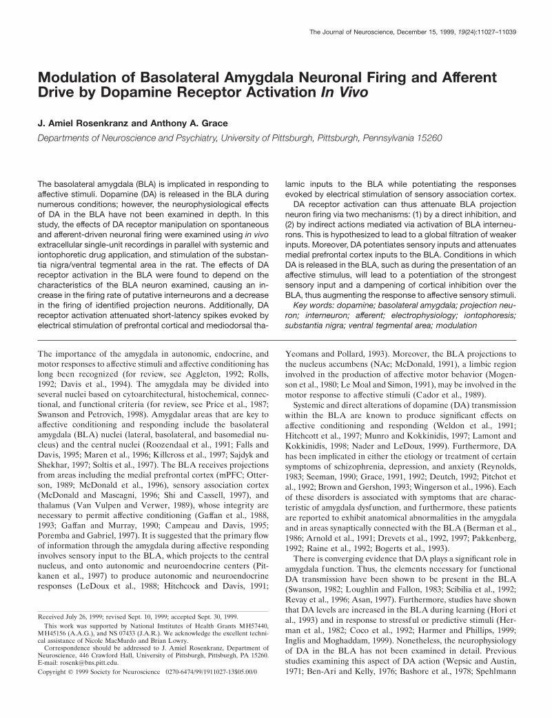
RESULTSEvidence for two subtypes of BLA neuronsAs seen in previous reports, the basal firing rate of most sponta-neously spiking neurons in the amygdala was very low (mean 6SEM, 0.91 6 2.47 Hz; n 5 179; range, 0–22.3 Hz), with evidenceof a large population of neurons that did not fire spontaneouslyduring the recording period. After closer examination, neuronscould be placed into one of two normal distributions (r 2 . 0.98)with significantly different mean firing rates ( p , 0.05; group ttest; Fig. 1): fast-firing (mean, 2.81 6 0.52 Hz; n 5 55; range,0.51–22.3 Hz) and slow-firing (mean, 0.071 6 0.013 Hz; n 5 124;range, 0–0.50 Hz) neurons. Based on the distribution of neuronalfiring rates, a cutoff of 0.5 Hz was used to segregate the twopopulations. Hence “slow-firing” will be used to indicate neurons
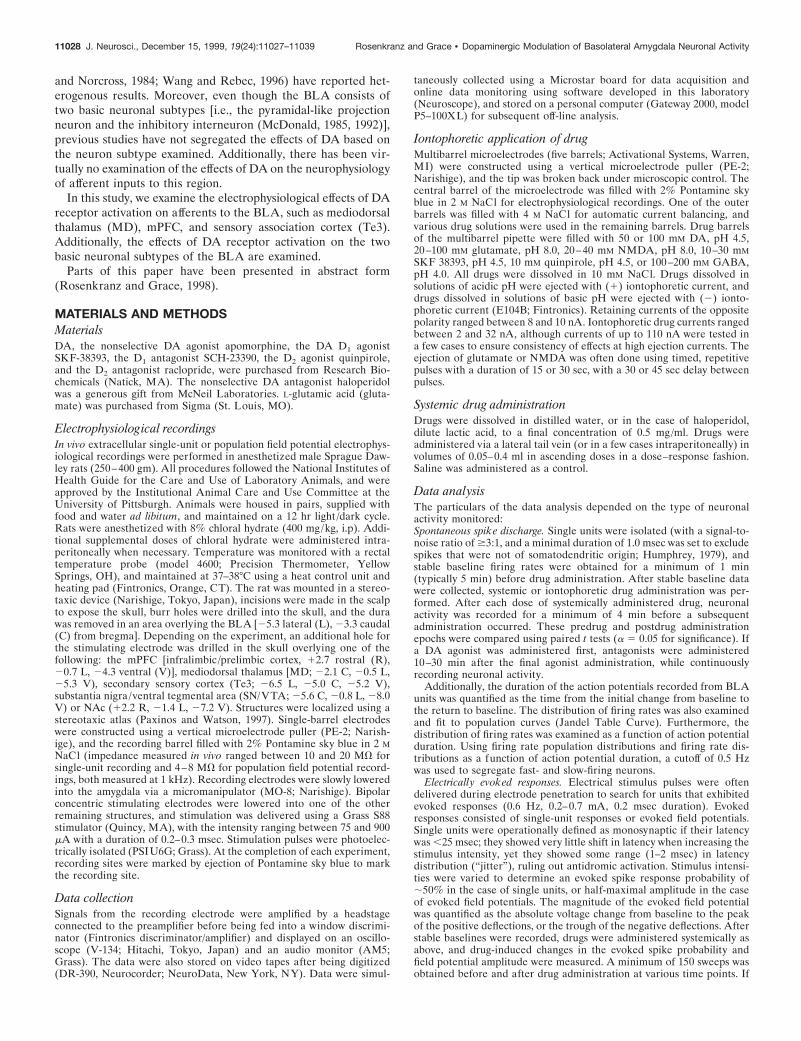
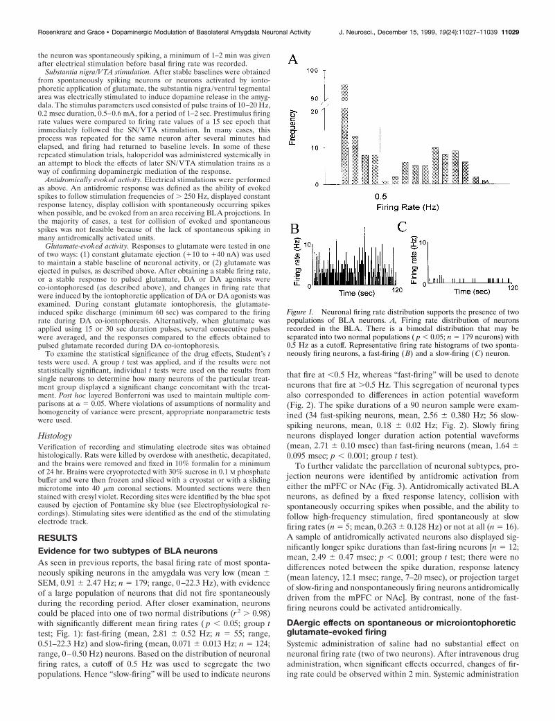
that fire at ,0.5 Hz, whereas “fast-firing” will be used to denoteneurons that fire at .0.5 Hz. This segregation of neuronal typesalso corresponded to differences in action potential waveform(Fig. 2). The spike durations of a 90 neuron sample were exam-ined (34 fast-spiking neurons, mean, 2.56 6 0.380 Hz; 56 slow-spiking neurons, mean, 0.18 6 0.02 Hz; Fig. 2). Slowly firingneurons displayed longer duration action potential waveforms(mean, 2.71 6 0.10 msec) than fast-firing neurons (mean, 1.64 60.095 msec; p , 0.001; group t test).
To further validate the parcellation of neuronal subtypes, pro-jection neurons were identified by antidromic activation fromeither the mPFC or NAc (Fig. 3). Antidromically activated BLAneurons, as defined by a fixed response latency, collision withspontaneously occurring spikes when possible, and the ability tofollow high-frequency stimulation, fired spontaneously at slowfiring rates (n 5 5; mean, 0.263 6 0.128 Hz) or not at all (n 5 16).A sample of antidromically activated neurons also displayed sig-nificantly longer spike durations than fast-firing neurons [n 5 12;mean, 2.49 6 0.47 msec; p , 0.001; group t test; there were nodifferences noted between the spike duration, response latency(mean latency, 12.1 msec; range, 7–20 msec), or projection targetof slow-firing and nonspontaneously firing neurons antidromicallydriven from the mPFC or NAc]. By contrast, none of the fast-firing neurons could be activated antidromically.
DAergic effects on spontaneous or microiontophoreticglutamate-evoked firingSystemic administration of saline had no substantial effect onneuronal firing rate (two of two neurons). After intravenous drugadministration, when significant effects occurred, changes of fir-ing rate could be observed within 2 min. Systemic administration
Figure 1. Neuronal firing rate distribution supports the presence of twopopulations of BLA neurons. A, Firing rate distribution of neuronsrecorded in the BLA. There is a bimodal distribution that may beseparated into two normal populations ( p , 0.05; n 5 179 neurons) with0.5 Hz as a cutoff. Representative firing rate histograms of two sponta-neously firing neurons, a fast-firing (B) and a slow-firing (C) neuron.
Rosenkranz and Grace • Dopaminergic Modulation of Basolateral Amygdala Neuronal Activity J. Neurosci., December 15, 1999, 19(24):11027–11039 11029
of the dopaminergic agonists apomorphine (n 5 10), SKF-38393(n 5 6), or quinpirole (n 5 4) had variable effects on the firingrate of spontaneously spiking neurons (10 of 20 neurons increasedfiring rate, 7 of 20 neurons decreased firing rate). However, theresponse observed could be differentiated based on the neuronalfiring rate. Because of the similarity in responses, all DA agonistswere grouped together. Fast-firing (.0.5 Hz) neurons displayedan increase in firing rate after systemic DA agonist administration(Fig. 4; 10 of 11 neurons; 10 rats; p 5 0.016; Wilcoxon; mean firingrate 6 SEM; pre-DA agonist, 2.57 6 0.749 Hz; post-DA agonist,5.44 6 1.898 Hz; maximum change, .3000% of baseline; meanchange, 256% of baseline), whereas the firing rate of slowly firingneurons was attenuated (Fig. 4; seven of nine neurons; eight rats;p 5 0.0156; Wilcoxon; pre-DA agonist, 0.371 6 0.199 Hz;post-DA agonist, 0.191 6 0.092 Hz; maximum change, 0% ofbaseline; mean change, 33% of baseline) by similar doses of thesame drugs [doses as follows: (1) DA agonists (initial doses of0.05–0.15 mg): apomorphine, mean initial dose, 0.14 6 0.016mg/kg, ranging from 0.09 to 0.31 mg/kg, and a final dose as highas 0.61 mg/kg; SKF-38393, initial dose, 0.05–0.15 mg, corre-sponding to a mean initial dose 0.12 6 0.002 mg/kg, ranging from0.11 to 0.13 mg/kg and a final dose as high as 0.50 mg/kg;quinpirole, initial dose, 0.16 6 0.03 mg/kg, ranging from 0.13 to0.290 mg/kg, and a final dose as high as 0.58 mg/kg; (2) DA
antagonists (initial doses of 0.1–0.25 mg): haloperidol, initialdose, 0.30 6 0.050 mg/kg, ranging from 0.13 to 0.65 mg/kg, anda final dose as high as 0.65 mg/kg; raclopride, initial dose, 0.26 60.086 mg/kg, ranging from 0.13 to 0.49 mg/kg, with a final dose as
Figure 2. Parcellation of fast- and slow-firing neurons of the BLA byspike duration. A, Example trace of a fast-spiking neuron that displays ashort spike duration (1.2 msec). B, Example trace of a slow-firing neuronthat displays a long spike duration (4.0 msec). C, A plot of spike duration byfiring rate for a sample of 90 BLA neurons demonstrates that fast-firingneurons (.0.5 Hz) tend to display short spike durations, whereas slow-firing neurons (,0.5 Hz) tend to display longer duration spikes ( p , 0.01).
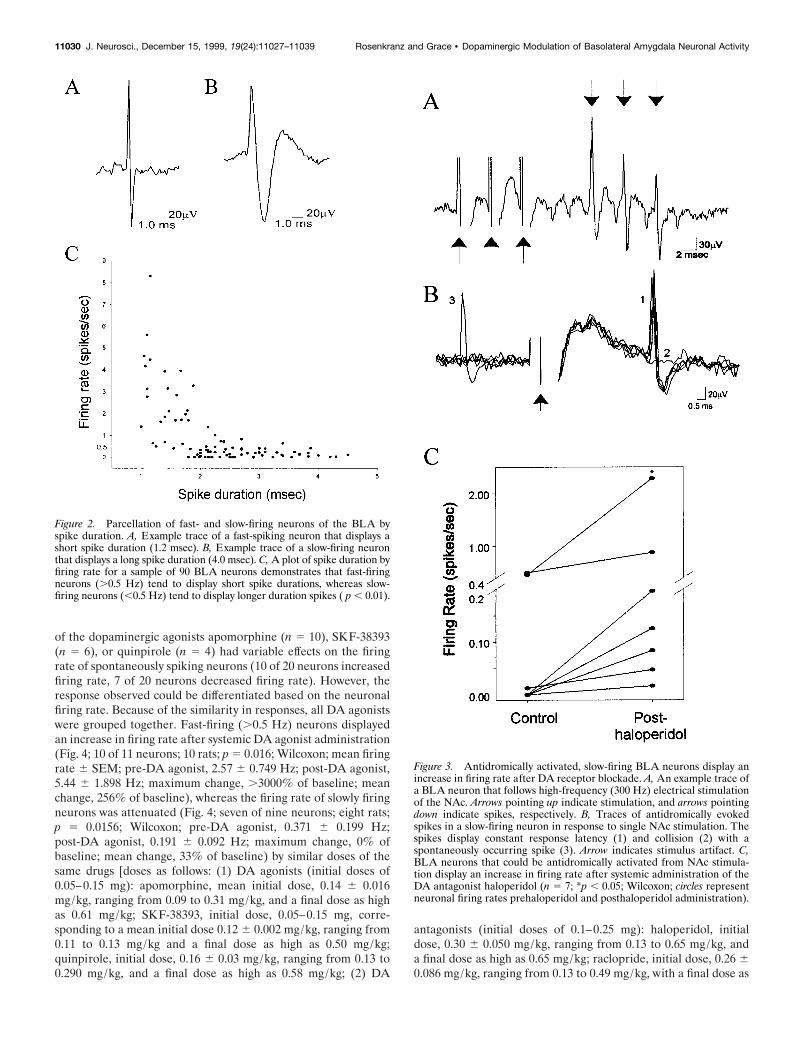
Figure 3. Antidromically activated, slow-firing BLA neurons display anincrease in firing rate after DA receptor blockade. A, An example trace ofa BLA neuron that follows high-frequency (300 Hz) electrical stimulationof the NAc. Arrows pointing up indicate stimulation, and arrows pointingdown indicate spikes, respectively. B, Traces of antidromically evokedspikes in a slow-firing neuron in response to single NAc stimulation. Thespikes display constant response latency (1) and collision (2) with aspontaneously occurring spike (3). Arrow indicates stimulus artifact. C,BLA neurons that could be antidromically activated from NAc stimula-tion display an increase in firing rate after systemic administration of theDA antagonist haloperidol (n 5 7; *p , 0.05; Wilcoxon; circles representneuronal firing rates prehaloperidol and posthaloperidol administration).
11030 J. Neurosci., December 15, 1999, 19(24):11027–11039 Rosenkranz and Grace • Dopaminergic Modulation of Basolateral Amygdala Neuronal Activity
high as 0.58 mg/kg; SCH 23390, initial dose, 0.21 6 0.017 mg/kg,ranging from 0.13–0.23 mg/kg, with a final dose as high as 0.75mg/kg]. In several cases, the initial dose was repeated with sub-sequent injections given at double the previous dose, in a dose–response manner. In the instances in which more than one dose ofDA agonist was administered, supplementary doses potentiated
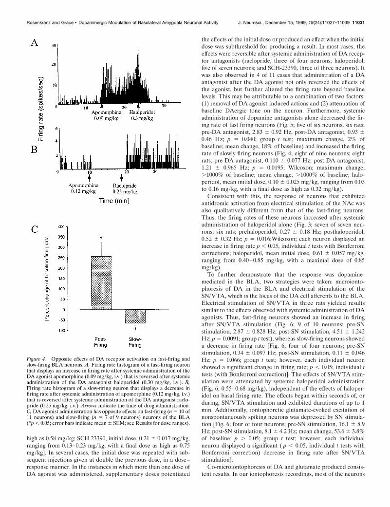
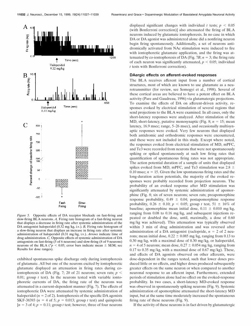
the effects of the initial dose or produced an effect when the initialdose was subthreshold for producing a result. In most cases, theeffects were reversible after systemic administration of DA recep-tor antagonists (raclopride, three of four neurons; haloperidol,five of seven neurons; and SCH-23390, three of three neurons). Itwas also observed in 4 of 11 cases that administration of a DAantagonist after the DA agonist not only reversed the effects ofthe agonist, but further altered the firing rate beyond baselinelevels. This may be attributable to a combination of two factors:(1) removal of DA agonist-induced actions and (2) attenuation ofbaseline DAergic tone on the neuron. Furthermore, systemicadministration of dopamine antagonists alone decreased the fir-ing rate of fast firing neurons (Fig. 5; five of six neurons; six rats;pre-DA antagonist, 2.83 6 0.92 Hz, post-DA antagonist, 0.93 60.46 Hz; p 5 0.040; group t test; maximum change, 2% ofbaseline; mean change, 18% of baseline) and increased the firingrate of slowly firing neurons (Fig. 4; eight of nine neurons; eightrats; pre-DA antagonist, 0.110 6 0.077 Hz; post-DA antagonist,1.21 6 0.965 Hz; p 5 0.0195; Wilcoxon; maximum change,.1000% of baseline; mean change, .1000% of baseline; halo-peridol, mean initial dose, 0.10 6 0.025 mg/kg, ranging from 0.03to 0.16 mg/kg, with a final dose as high as 0.32 mg/kg).
Consistent with this, the response of neurons that exhibitedantidromic activation from electrical stimulation of the NAc wasalso qualitatively different from that of the fast-firing neurons.Thus, the firing rates of these neurons increased after systemicadministration of haloperidol alone (Fig. 3; seven of seven neu-rons; six rats; prehaloperidol, 0.27 6 0.18 Hz; posthaloperidol,0.52 6 0.32 Hz; p 5 0.016;Wilcoxon; each neuron displayed anincrease in firing rate p , 0.05, individual t tests with Bonferronicorrections; haloperidol, mean initial dose, 0.61 6 0.057 mg/kg,ranging from 0.40–0.85 mg/kg, with a maximal dose of 0.85mg/kg).
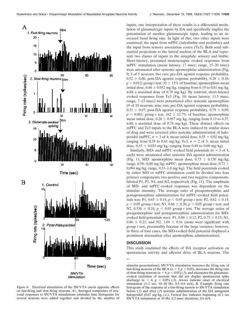
To further demonstrate that the response was dopamine-mediated in the BLA, two strategies were taken: microionto-phoresis of DA in the BLA and electrical stimulation of theSN/VTA, which is the locus of the DA cell afferents to the BLA.Electrical stimulation of SN/VTA in three rats yielded resultssimilar to the effects observed with systemic administration of DAagonists. Thus, fast-firing neurons showed an increase in firingafter SN/VTA stimulation (Fig. 6; 9 of 10 neurons; pre-SNstimulation, 2.87 6 0.828 Hz; post-SN stimulation, 4.51 6 1.242Hz; p 5 0.0091; group t test), whereas slow-firing neurons showeda decrease in firing rate [Fig. 6; four of four neurons; pre-SNstimulation, 0.34 6 0.097 Hz; post-SN stimulation, 0.11 6 0.046Hz; p 5 0.066; group t test; however, each individual neuronshowed a significant change in firing rate; p , 0.05; individual ttests (with Bonferroni correction)]. The effects of SN/VTA stim-ulation were attenuated by systemic haloperidol administration(Fig. 6; 0.55–0.68 mg/kg), independent of the effects of haloper-idol on basal firing rate. The effects began within seconds of, orduring, SN/VTA stimulation and exhibited durations of up to 1min. Additionally, iontophoretic glutamate-evoked excitation ofnonspontaneously spiking neurons was depressed by SN stimula-tion [Fig. 6; four of four neurons; pre-SN stimulation, 16.1 6 8.9Hz; post-SN stimulation, 8.1 6 4.2 Hz; mean change, 53.6 6 3.8%of baseline; p . 0.05; group t test; however, each individualneuron displayed a significant ( p , 0.05, individual t tests withBonferroni correction) decrease in firing rate after SN/VTAstimulation].
Co-microiontophoresis of DA and glutamate produced consis-tent results. In our iontophoresis recordings, most of the neurons
Figure 4. Opposite effects of DA receptor activation on fast-firing andslow-firing BLA neurons. A, Firing rate histogram of a fast-firing neuronthat displays an increase in firing rate after systemic administration of theDA agonist apomorphine (0.09 mg/kg, i.v.) that is reversed after systemicadministration of the DA antagonist haloperidol (0.30 mg/kg, i.v.). B,Firing rate histogram of a slow-firing neuron that displays a decrease infiring rate after systemic administration of apomorphine (0.12 mg/kg, i.v.)that is reversed after systemic administration of the DA antagonist raclo-pride (0.25 mg/kg, i.v.). Arrows indicate the time of drug administration.C, DA agonist administration has opposite effects on fast-firing (n 5 10 of11 neurons) and slow-firing (n 5 7 of 9 neurons) neurons of the BLA(*p , 0.05; error bars indicate mean 6 SEM; see Results for dose ranges).
Rosenkranz and Grace • Dopaminergic Modulation of Basolateral Amygdala Neuronal Activity J. Neurosci., December 15, 1999, 19(24):11027–11039 11031
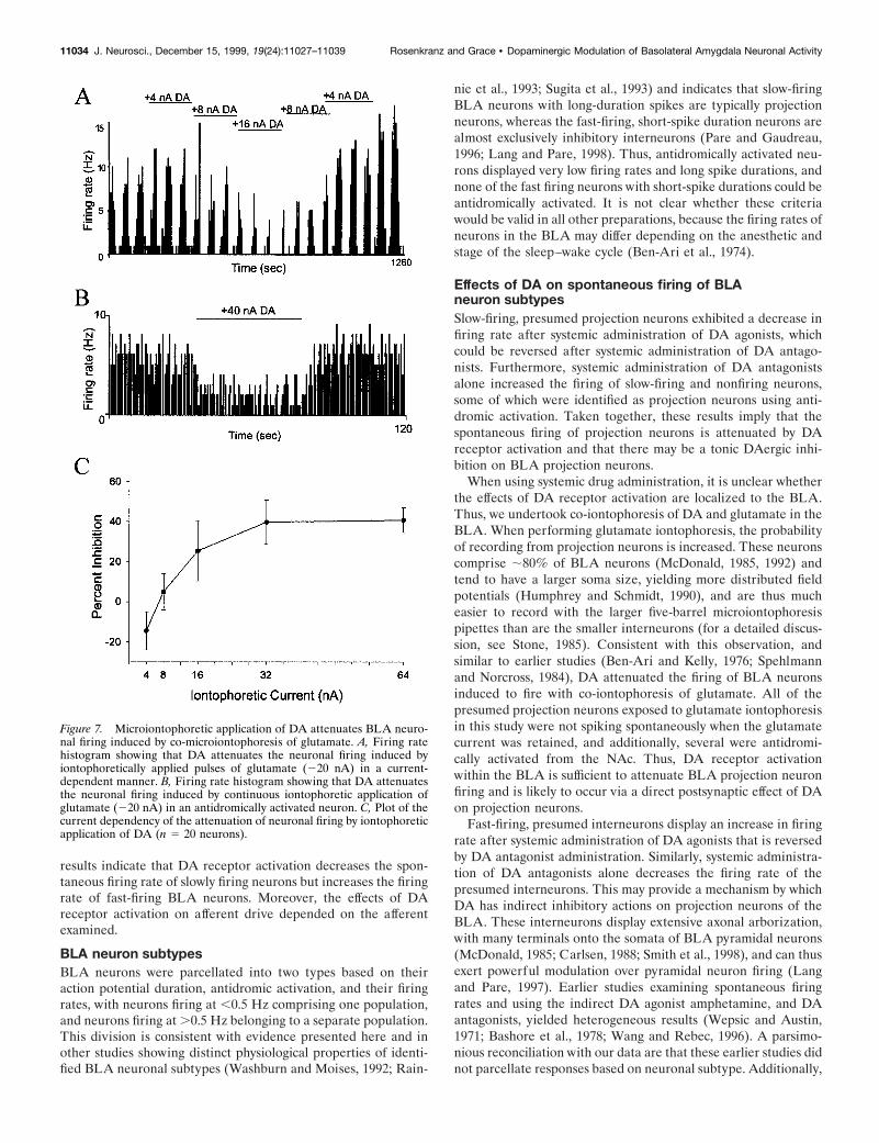
exhibited spontaneous spike discharge only during iontophoresisof glutamate. All but one of the neurons excited by iontophoreticglutamate displayed an attenuation in firing rates during co-iontophoresis of DA (Fig. 7; 20 of 21 neurons; seven rats; p ,0.01; group t test). In the neurons tested with variable ionto-phoretic currents of DA, the firing rate of the neurons wasattenuated in a current-dependent manner (Fig. 7). The effects ofiontophoretic DA were attenuated by systemic administration ofhaloperidol (n 5 2 of 2). Iontophoresis of the specific DA agonistsSKF-38393 (n 5 4 of 5; p 5 0.013; group t test) and quinpirole[n 5 3 of 4; p 5 0.11; group t test; however, three of four neurons
displayed significant changes with individual t tests; p , 0.05(with Bonferroni correction)] also attenuated the firing of BLAneurons induced by glutamate iontophoresis. In no case in whichDA or DA agonist was administered alone did a nonfiring neuronbegin firing spontaneously. Additionally, a set of neurons anti-dromically activated from NAc stimulation were induced to firewith iontophoretic glutamate application, and the firing was at-tenuated by co-iontophoresis of DA (Fig. 7B; n 5 3; the firing rateof each neuron was significantly attenuated, p , 0.05; individualt tests with Bonferroni correction).
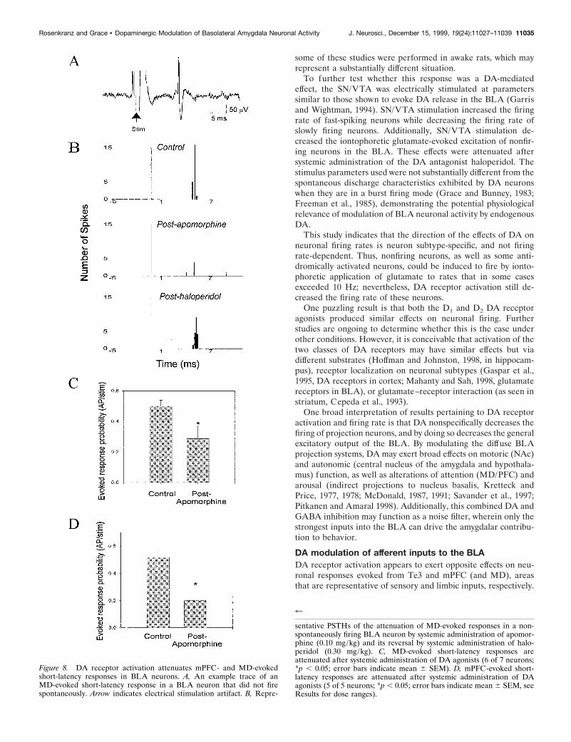
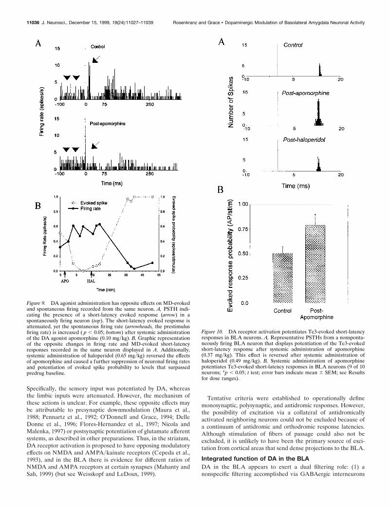
DAergic effects on afferent-evoked responsesThe BLA receives afferent input from a number of corticalstructures, most of which are known to use glutamate as a neu-rotransmitter (for review, see Somogyi et al., 1998). Several ofthese cortical areas are believed to have a potent effect on BLAactivity (Pare and Gaudreau, 1996) via glutamatergic projections.To examine the effects of DA on afferent-driven activity, re-sponses evoked by electrical stimulation of several regions thatsend projections to the BLA were examined. In all cases, only theshort-latency responses were analyzed. After stimulation of theMD, short-latency, putative monosynaptic (Fig. 8; n 5 15; meanlatency, 16.9 msec; range, 5–26 msec), and occasionally multisyn-aptic responses were evoked. Very few neurons that displayedboth antidromic and orthodromic responses were encountered,and these were not included in this study. Except where noted,the responses evoked from electrical stimulation of MD, mPFC,and Te3 were recorded from neurons that were not spontaneouslyspiking or spiked spontaneously at such low firing rates thatquantification of spontaneous firing rates was not appropriate.The action potential duration of a sample of units that displayedspikes evoked from MD, mPFC, and Te3 stimulation was 2.8 60.10 msec; n 5 15. Given the low spontaneous firing rates and thelong-duration action potentials, the majority of the evoked re-sponses were probably recorded from projection neurons. Theprobability of an evoked response after MD stimulation wassignificantly attenuated by systemic administration of apomor-phine (Fig. 8; six of seven neurons; seven rats; preapomorphineresponse probability, 0.49 6 0.04; postapomorphine responseprobability, 0.26 6 0.10; p , 0.05; group t test, 51 6 16% ofbaseline; apomorphine mean initial dose, 0.11 6 0.010 mg/kg,ranging from 0.08 to 0.16 mg/kg, and subsequent injections re-peated or doubled the dose, until, maximally, a dose of 0.60mg/kg was achieved). This attenuation was typically observedwithin 3 min of drug administration and was reversed afteradministration of a DA antagonist (raclopride, n 5 2 of 2 neu-rons; mean initial dose, 0.22 6 0.085 mg/kg, ranging from 0.13 to0.30 mg/kg, with a maximal dose of 0.30 mg/kg, or haloperidol,n 5 4 of 5 neurons; mean dose, 0.27 6 0.054 mg/kg, ranging from0.17 to 0.35 mg/kg, with a maximal dose of 0.67 mg/kg). These,and effects of DA agonists observed on other afferents, weredose-dependent in the ranges tested, such that lower doses pro-duced little or no effects, and higher doses produced subsequentlygreater effects on the same neuron or when compared to anotherneuronal response to an afferent input. Furthermore, extendedperiods of stimulation alone had no effect on the evoked-responseprobability. In two cases, a short-latency MD-evoked responsewas observed in spontaneously spiking neurons (Fig. 9). Systemicadministration of apomorphine attenuated the short-latency MDinput, but at the same time moderately increased the spontaneousfiring rate of these neurons (Fig. 9).
If the activity of these neurons is in fact driven by glutamatergic
Figure 5. Opposite effects of DA receptor blockade on fast-firing andslow-firing BLA neurons. A, Firing rate histogram of a fast-firing neuronthat displays a decrease in firing rate after systemic administration of theDA antagonist haloperidol (0.32 mg/kg, i.v.). B, Firing rate histogram ofa slow-firing neuron that displays an increase in firing rate after systemicadministration of haloperidol (0.31 mg/kg, i.v.). Arrows indicate time ofdrug administration. C, Opposite effects of systemic administration of DAantagonists on fast-firing (5 of 6 neurons) and slow-firing (8 of 9 neurons)neurons of the BLA (*p , 0.05; error bars indicate mean 6 SEM; seeResults for dose ranges).
11032 J. Neurosci., December 15, 1999, 19(24):11027–11039 Rosenkranz and Grace • Dopaminergic Modulation of Basolateral Amygdala Neuronal Activity
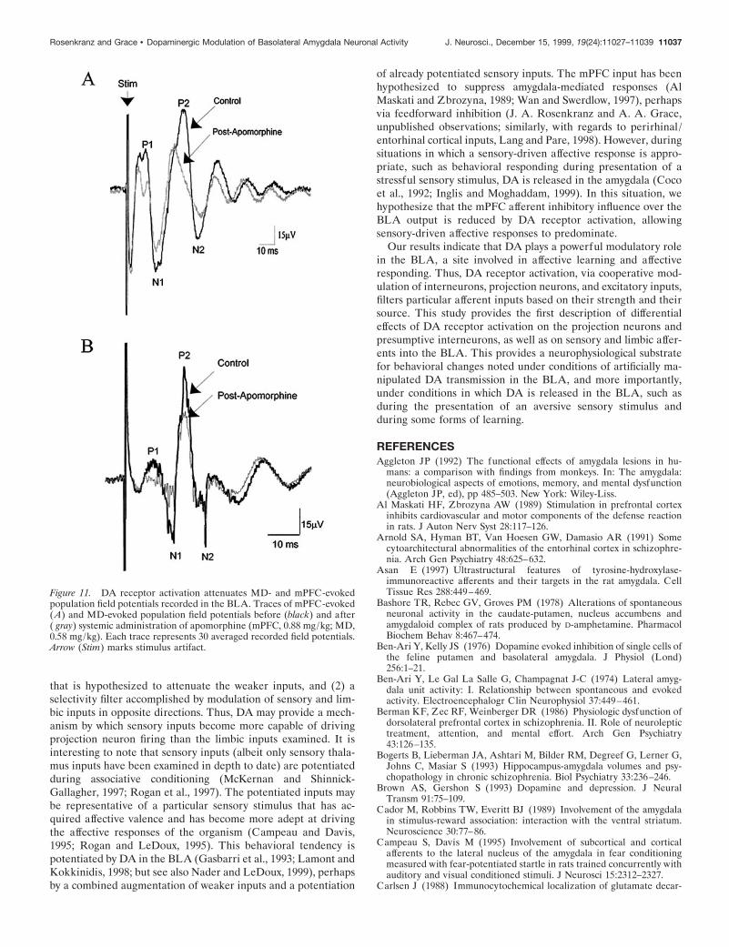
inputs, one interpretation of these results is a differential modu-lation of glutamatergic inputs by DA and specifically implies thepotentiation of another glutamatergic input, leading to an in-creased basal firing rate. In light of this, two other inputs wereexamined; the input from mPFC (infralimbic and prelimbic) andthe input from sensory association cortex (Te3). Both send sub-stantial projections to the lateral nucleus of the BLA and repre-sent two classes of inputs to the amygdala: sensory and limbic.Short-latency, presumed monosynaptic evoked responses frommPFC stimulation (mean latency, 17 msec; range, 15–20 msec)were attenuated after systemic apomorphine administration (Fig.9; 5 of 5 neurons; five rats; pre-DA agonist response probability,0.52 6 0.06; post-DA agonist response probability, 0.20 6 0.10;p 5 0.012; group t test; 35 6 13% of baseline; apomorphine meaninitial dose, 0.46 6 0.052 mg/kg, ranging from 0.19 to 0.61 mg/kg,with a maximal dose of 0.78 mg/kg). By contrast, short-latencyevoked responses from Te3 (Fig. 10; mean latency, 11.5 msec;range, 7–13 msec) were potentiated after systemic apomorphine(9 of 10 neurons; nine rats; pre-DA agonist response probability,0.51 6 0.07; post-DA agonist response probability, 0.79 6 0.09;p 5 0.001; group t test, 162 6 12.7% of baseline; apomorphinemean initial dose, 0.28 6 0.057 mg/kg, ranging from 0.13 to 0.37,with a maximal dose of 0.74 mg/kg). These distinct effects onmPFC and Te3 inputs to the BLA were induced by similar dosesof drug and were reversed after systemic administration of halo-peridol (mPFC, n 5 3 of 4; mean initial dose, 0.55 6 0.02 mg/kg,ranging from 0.39 to 0.61 mg/kg; Te3, n 5 2 of 3; mean initialdose, 0.53 6 0.035 mg/kg, ranging from 0.49 to 0.60 mg/kg).
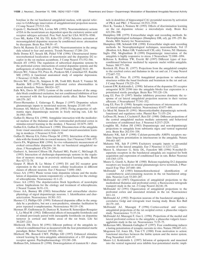
Similarly, MD- and mPFC-evoked field potentials (n 5 3 of 4,each) were attenuated after systemic DA agonist administration(Fig. 11; MD: apomorphine mean dose, 0.73 6 0.150 mg/kg;range, 0.58–0.88 mg/kg; mPFC: apomorphine mean dose, 0.72 60.094 mg/kg; range, 0.51–1.0 mg/kg). The field potentials evokedby either MD or mPFC stimulation could be divided into fourprimary components; two positive and two negative components,labeled P1, P2, N1, and N2, respectively (Fig. 11). The amplitudeof MD- and mPFC-evoked responses was dependent on thestimulus intensity. The average ratio of preapomorphine andpostapomorphine administration for mPFC-evoked field poten-tials was: P1, 0.65 6 0.14, p , 0.05 group t test; P2, 0.62 6 0.14,p , 0.05 group t test; N1, 0.66 6 0.20, p . 0.05 group t test; andN2, 0.536 6 0.18, p , 0.05 group t test. The average ratios ofpreapomorphine and postapomorphine administration for MD-evoked field potentials were: P1, 0.88 6 0.12; P2, 0.73 6 0.15; N1,0.86 6 0.21; and N2, 1.04 6 0.41 (none were significant withgroup t test, presumably because of the large variance; however,in three of four cases, the MD-evoked field potential displayed aprominent attenuation after apomorphine administration).
DISCUSSIONThis study examined the effects of DA receptor activation onspontaneous activity and afferent drive of BLA neurons. The
Figure 6. Electrical stimulation of the SN/VTA exerts opposite effectson fast-firing and slow-firing neurons. A1, Averaged composites of neu-ronal responses to SN/VTA stimulations (stimulus time histograms forseveral neurons were added together and divided by the number of
4
stimulus presentations). SN/VTA stimulation increases the firing rate offast-firing neurons of the BLA (n 5 5; p , 0.05), decreases the firing rateof slow-firing neurons (n 5 4; p , 0.05) ( 2), and attenuates the glutamate-evoked excitation of neurons that did not display spontaneous spikedischarge (n 5 4; p , 0.05) (3). Arrows indicate onset of electricalstimulation (1–2 sec; 10–20 Hz; 0.5–0.6 mA). B, Example firing ratehistogram of the response of a fast-firing neuron to SN/VTA stimulationbefore (1) and after (2) systemic administration of the DA antagonisthaloperidol (0.67 mg/kg, i.v.). Vertical line indicates beginning of 1 secSN/VTA stimulation at 10 Hz, 0.2 msec durations, 0.6 mA.
Rosenkranz and Grace • Dopaminergic Modulation of Basolateral Amygdala Neuronal Activity J. Neurosci., December 15, 1999, 19(24):11027–11039 11033
results indicate that DA receptor activation decreases the spon-taneous firing rate of slowly firing neurons but increases the firingrate of fast-firing BLA neurons. Moreover, the effects of DAreceptor activation on afferent drive depended on the afferentexamined.
BLA neuron subtypesBLA neurons were parcellated into two types based on theiraction potential duration, antidromic activation, and their firingrates, with neurons firing at ,0.5 Hz comprising one population,and neurons firing at .0.5 Hz belonging to a separate population.This division is consistent with evidence presented here and inother studies showing distinct physiological properties of identi-fied BLA neuronal subtypes (Washburn and Moises, 1992; Rain-
nie et al., 1993; Sugita et al., 1993) and indicates that slow-firingBLA neurons with long-duration spikes are typically projectionneurons, whereas the fast-firing, short-spike duration neurons arealmost exclusively inhibitory interneurons (Pare and Gaudreau,1996; Lang and Pare, 1998). Thus, antidromically activated neu-rons displayed very low firing rates and long spike durations, andnone of the fast firing neurons with short-spike durations could beantidromically activated. It is not clear whether these criteriawould be valid in all other preparations, because the firing rates ofneurons in the BLA may differ depending on the anesthetic andstage of the sleep–wake cycle (Ben-Ari et al., 1974).
Effects of DA on spontaneous firing of BLAneuron subtypesSlow-firing, presumed projection neurons exhibited a decrease infiring rate after systemic administration of DA agonists, whichcould be reversed after systemic administration of DA antago-nists. Furthermore, systemic administration of DA antagonistsalone increased the firing of slow-firing and nonfiring neurons,some of which were identified as projection neurons using anti-dromic activation. Taken together, these results imply that thespontaneous firing of projection neurons is attenuated by DAreceptor activation and that there may be a tonic DAergic inhi-bition on BLA projection neurons.
When using systemic drug administration, it is unclear whetherthe effects of DA receptor activation are localized to the BLA.Thus, we undertook co-iontophoresis of DA and glutamate in theBLA. When performing glutamate iontophoresis, the probabilityof recording from projection neurons is increased. These neuronscomprise ;80% of BLA neurons (McDonald, 1985, 1992) andtend to have a larger soma size, yielding more distributed fieldpotentials (Humphrey and Schmidt, 1990), and are thus mucheasier to record with the larger five-barrel microiontophoresispipettes than are the smaller interneurons (for a detailed discus-sion, see Stone, 1985). Consistent with this observation, andsimilar to earlier studies (Ben-Ari and Kelly, 1976; Spehlmannand Norcross, 1984), DA attenuated the firing of BLA neuronsinduced to fire with co-iontophoresis of glutamate. All of thepresumed projection neurons exposed to glutamate iontophoresisin this study were not spiking spontaneously when the glutamatecurrent was retained, and additionally, several were antidromi-cally activated from the NAc. Thus, DA receptor activationwithin the BLA is sufficient to attenuate BLA projection neuronfiring and is likely to occur via a direct postsynaptic effect of DAon projection neurons.
Fast-firing, presumed interneurons display an increase in firingrate after systemic administration of DA agonists that is reversedby DA antagonist administration. Similarly, systemic administra-tion of DA antagonists alone decreases the firing rate of thepresumed interneurons. This may provide a mechanism by whichDA has indirect inhibitory actions on projection neurons of theBLA. These interneurons display extensive axonal arborization,with many terminals onto the somata of BLA pyramidal neurons(McDonald, 1985; Carlsen, 1988; Smith et al., 1998), and can thusexert powerful modulation over pyramidal neuron firing (Langand Pare, 1997). Earlier studies examining spontaneous firingrates and using the indirect DA agonist amphetamine, and DAantagonists, yielded heterogeneous results (Wepsic and Austin,1971; Bashore et al., 1978; Wang and Rebec, 1996). A parsimo-nious reconciliation with our data are that these earlier studies didnot parcellate responses based on neuronal subtype. Additionally,
Figure 7. Microiontophoretic application of DA attenuates BLA neuro-nal firing induced by co-microiontophoresis of glutamate. A, Firing ratehistogram showing that DA attenuates the neuronal firing induced byiontophoretically applied pulses of glutamate (220 nA) in a current-dependent manner. B, Firing rate histogram showing that DA attenuatesthe neuronal firing induced by continuous iontophoretic application ofglutamate (220 nA) in an antidromically activated neuron. C, Plot of thecurrent dependency of the attenuation of neuronal firing by iontophoreticapplication of DA (n 5 20 neurons).
11034 J. Neurosci., December 15, 1999, 19(24):11027–11039 Rosenkranz and Grace • Dopaminergic Modulation of Basolateral Amygdala Neuronal Activity
some of these studies were performed in awake rats, which mayrepresent a substantially different situation.
To further test whether this response was a DA-mediatedeffect, the SN/VTA was electrically stimulated at parameterssimilar to those shown to evoke DA release in the BLA (Garrisand Wightman, 1994). SN/VTA stimulation increased the firingrate of fast-spiking neurons while decreasing the firing rate ofslowly firing neurons. Additionally, SN/VTA stimulation de-creased the iontophoretic glutamate-evoked excitation of nonfir-ing neurons in the BLA. These effects were attenuated aftersystemic administration of the DA antagonist haloperidol. Thestimulus parameters used were not substantially different from thespontaneous discharge characteristics exhibited by DA neuronswhen they are in a burst firing mode (Grace and Bunney, 1983;Freeman et al., 1985), demonstrating the potential physiologicalrelevance of modulation of BLA neuronal activity by endogenousDA.
This study indicates that the direction of the effects of DA onneuronal firing rates is neuron subtype-specific, and not firingrate-dependent. Thus, nonfiring neurons, as well as some anti-dromically activated neurons, could be induced to fire by ionto-phoretic application of glutamate to rates that in some casesexceeded 10 Hz; nevertheless, DA receptor activation still de-creased the firing rate of these neurons.
One puzzling result is that both the D1 and D2 DA receptoragonists produced similar effects on neuronal firing. Furtherstudies are ongoing to determine whether this is the case underother conditions. However, it is conceivable that activation of thetwo classes of DA receptors may have similar effects but viadifferent substrates (Hoffman and Johnston, 1998, in hippocam-pus), receptor localization on neuronal subtypes (Gaspar et al.,1995, DA receptors in cortex; Mahanty and Sah, 1998, glutamatereceptors in BLA), or glutamate–receptor interaction (as seen instriatum, Cepeda et al., 1993).
One broad interpretation of results pertaining to DA receptoractivation and firing rate is that DA nonspecifically decreases thefiring of projection neurons, and by doing so decreases the generalexcitatory output of the BLA. By modulating the diffuse BLAprojection systems, DA may exert broad effects on motoric (NAc)and autonomic (central nucleus of the amygdala and hypothala-mus) function, as well as alterations of attention (MD/PFC) andarousal (indirect projections to nucleus basalis, Kretteck andPrice, 1977, 1978; McDonald, 1987, 1991; Savander et al., 1997;Pitkanen and Amaral 1998). Additionally, this combined DA andGABA inhibition may function as a noise filter, wherein only thestrongest inputs into the BLA can drive the amygdalar contribu-tion to behavior.
DA modulation of afferent inputs to the BLADA receptor activation appears to exert opposite effects on neu-ronal responses evoked from Te3 and mPFC (and MD), areasthat are representative of sensory and limbic inputs, respectively.
Figure 8. DA receptor activation attenuates mPFC- and MD-evokedshort-latency responses in BLA neurons. A, An example trace of anMD-evoked short-latency response in a BLA neuron that did not firespontaneously. Arrow indicates electrical stimulation artifact. B, Repre-
4
sentative PSTHs of the attenuation of MD-evoked responses in a non-spontaneously firing BLA neuron by systemic administration of apomor-phine (0.10 mg/kg) and its reversal by systemic administration of halo-peridol (0.30 mg/kg). C, MD-evoked short-latency responses areattenuated after systemic administration of DA agonists (6 of 7 neurons;*p , 0.05; error bars indicate mean 6 SEM). D, mPFC-evoked short-latency responses are attenuated after systemic administration of DAagonists (5 of 5 neurons; *p , 0.05; error bars indicate mean 6 SEM, seeResults for dose ranges).
Rosenkranz and Grace • Dopaminergic Modulation of Basolateral Amygdala Neuronal Activity J. Neurosci., December 15, 1999, 19(24):11027–11039 11035
Specifically, the sensory input was potentiated by DA, whereasthe limbic inputs were attenuated. However, the mechanism ofthese actions is unclear. For example, these opposite effects maybe attributable to presynaptic downmodulation (Maura et al.,1988; Pennartz et al., 1992; O’Donnell and Grace, 1994; DelleDonne et al., 1996; Flores-Hernandez et al., 1997; Nicola andMalenka, 1997) or postsynaptic potentiation of glutamate afferentsystems, as described in other preparations. Thus, in the striatum,DA receptor activation is proposed to have opposing modulatoryeffects on NMDA and AMPA/kainate receptors (Cepeda et al.,1993), and in the BLA there is evidence for different ratios ofNMDA and AMPA receptors at certain synapses (Mahanty andSah, 1999) (but see Weisskopf and LeDoux, 1999).
Tentative criteria were established to operationally definemonosynaptic, polysynaptic, and antidromic responses. However,the possibility of excitation via a collateral of antidromicallyactivated neighboring neurons could not be excluded because ofa continuum of antidromic and orthodromic response latencies.Although stimulation of fibers of passage could also not beexcluded, it is unlikely to have been the primary source of exci-tation from cortical areas that send dense projections to the BLA.
Integrated function of DA in the BLADA in the BLA appears to exert a dual filtering role: (1) anonspecific filtering accomplished via GABAergic interneurons
Figure 9. DA agonist administration has opposite effects on MD-evokedand spontaneous firing recorded from the same neuron. A, PSTH indi-cating the presence of a short-latency evoked response (arrow) in aspontaneously firing neuron (top). The short-latency evoked response isattenuated, yet the spontaneous firing rate (arrowheads, the prestimulusfiring rate) is increased ( p , 0.05; bottom) after systemic administrationof the DA agonist apomorphine (0.10 mg/kg). B, Graphic representationof the opposite changes in firing rate and MD-evoked short-latencyresponses recorded in the same neuron displayed in A. Additionally,systemic administration of haloperidol (0.65 mg/kg) reversed the effectsof apomorphine and caused a further suppression of neuronal firing ratesand potentiation of evoked spike probability to levels that surpassedpredrug baseline.
Figure 10. DA receptor activation potentiates Te3-evoked short-latencyresponses in BLA neurons. A, Representative PSTHs from a nonsponta-neously firing BLA neuron that displays potentiation of the Te3-evokedshort-latency response after systemic administration of apomorphine(0.37 mg/kg). This effect is reversed after systemic administration ofhaloperidol (0.49 mg/kg). B, Systemic administration of apomorphinepotentiates Te3-evoked short-latency responses in BLA neurons (9 of 10neurons; *p , 0.05; t test; error bars indicate mean 6 SEM; see Resultsfor dose ranges).
11036 J. Neurosci., December 15, 1999, 19(24):11027–11039 Rosenkranz and Grace • Dopaminergic Modulation of Basolateral Amygdala Neuronal Activity
that is hypothesized to attenuate the weaker inputs, and (2) aselectivity filter accomplished by modulation of sensory and lim-bic inputs in opposite directions. Thus, DA may provide a mech-anism by which sensory inputs become more capable of drivingprojection neuron firing than the limbic inputs examined. It isinteresting to note that sensory inputs (albeit only sensory thala-mus inputs have been examined in depth to date) are potentiatedduring associative conditioning (McKernan and Shinnick-Gallagher, 1997; Rogan et al., 1997). The potentiated inputs maybe representative of a particular sensory stimulus that has ac-quired affective valence and has become more adept at drivingthe affective responses of the organism (Campeau and Davis,1995; Rogan and LeDoux, 1995). This behavioral tendency ispotentiated by DA in the BLA (Gasbarri et al., 1993; Lamont andKokkinidis, 1998; but see also Nader and LeDoux, 1999), perhapsby a combined augmentation of weaker inputs and a potentiation
of already potentiated sensory inputs. The mPFC input has beenhypothesized to suppress amygdala-mediated responses (AlMaskati and Zbrozyna, 1989; Wan and Swerdlow, 1997), perhapsvia feedforward inhibition (J. A. Rosenkranz and A. A. Grace,unpublished observations; similarly, with regards to perirhinal /entorhinal cortical inputs, Lang and Pare, 1998). However, duringsituations in which a sensory-driven affective response is appro-priate, such as behavioral responding during presentation of astressful sensory stimulus, DA is released in the amygdala (Cocoet al., 1992; Inglis and Moghaddam, 1999). In this situation, wehypothesize that the mPFC afferent inhibitory influence over theBLA output is reduced by DA receptor activation, allowingsensory-driven affective responses to predominate.
Our results indicate that DA plays a powerful modulatory rolein the BLA, a site involved in affective learning and affectiveresponding. Thus, DA receptor activation, via cooperative mod-ulation of interneurons, projection neurons, and excitatory inputs,filters particular afferent inputs based on their strength and theirsource. This study provides the first description of differentialeffects of DA receptor activation on the projection neurons andpresumptive interneurons, as well as on sensory and limbic affer-ents into the BLA. This provides a neurophysiological substratefor behavioral changes noted under conditions of artificially ma-nipulated DA transmission in the BLA, and more importantly,under conditions in which DA is released in the BLA, such asduring the presentation of an aversive sensory stimulus andduring some forms of learning.
REFERENCESAggleton JP (1992) The functional effects of amygdala lesions in hu-
mans: a comparison with findings from monkeys. In: The amygdala:neurobiological aspects of emotions, memory, and mental dysfunction(Aggleton JP, ed), pp 485–503. New York: Wiley-Liss.
Al Maskati HF, Zbrozyna AW (1989) Stimulation in prefrontal cortexinhibits cardiovascular and motor components of the defense reactionin rats. J Auton Nerv Syst 28:117–126.
Arnold SA, Hyman BT, Van Hoesen GW, Damasio AR (1991) Somecytoarchitectural abnormalities of the entorhinal cortex in schizophre-nia. Arch Gen Psychiatry 48:625–632.
Asan E (1997) Ultrastructural features of tyrosine-hydroxylase-immunoreactive afferents and their targets in the rat amygdala. CellTissue Res 288:449–469.
Bashore TR, Rebec GV, Groves PM (1978) Alterations of spontaneousneuronal activity in the caudate-putamen, nucleus accumbens andamygdaloid complex of rats produced by D-amphetamine. PharmacolBiochem Behav 8:467–474.
Ben-Ari Y, Kelly JS (1976) Dopamine evoked inhibition of single cells ofthe feline putamen and basolateral amygdala. J Physiol (Lond)256:1–21.
Ben-Ari Y, Le Gal La Salle G, Champagnat J-C (1974) Lateral amyg-dala unit activity: I. Relationship between spontaneous and evokedactivity. Electroencephalogr Clin Neurophysiol 37:449–461.
Berman KF, Zec RF, Weinberger DR (1986) Physiologic dysfunction ofdorsolateral prefrontal cortex in schizophrenia. II. Role of neuroleptictreatment, attention, and mental effort. Arch Gen Psychiatry43:126–135.
Bogerts B, Lieberman JA, Ashtari M, Bilder RM, Degreef G, Lerner G,Johns C, Masiar S (1993) Hippocampus-amygdala volumes and psy-chopathology in chronic schizophrenia. Biol Psychiatry 33:236–246.
Brown AS, Gershon S (1993) Dopamine and depression. J NeuralTransm 91:75–109.
Cador M, Robbins TW, Everitt BJ (1989) Involvement of the amygdalain stimulus-reward association: interaction with the ventral striatum.Neuroscience 30:77–86.
Campeau S, Davis M (1995) Involvement of subcortical and corticalafferents to the lateral nucleus of the amygdala in fear conditioningmeasured with fear-potentiated startle in rats trained concurrently withauditory and visual conditioned stimuli. J Neurosci 15:2312–2327.
Carlsen J (1988) Immunocytochemical localization of glutamate decar-
Figure 11. DA receptor activation attenuates MD- and mPFC-evokedpopulation field potentials recorded in the BLA. Traces of mPFC-evoked(A) and MD-evoked population field potentials before (black) and after( gray) systemic administration of apomorphine (mPFC, 0.88 mg/kg; MD,0.58 mg/kg). Each trace represents 30 averaged recorded field potentials.Arrow (Stim) marks stimulus artifact.
Rosenkranz and Grace • Dopaminergic Modulation of Basolateral Amygdala Neuronal Activity J. Neurosci., December 15, 1999, 19(24):11027–11039 11037
boxylase in the rat basolateral amygdaloid nucleus, with special refer-ence to GABAergic innervation of amygdalostriatal projection neuron.J Comp Neurol 273:513–526.
Cepeda C, Buchwald NA, Levine MS (1993) Neuromodulatory actionsof DA in the neostriatum are dependent upon the excitatory amino acidreceptor subtypes activated. Proc Natl Acad Sci USA 90:9576–9580.
Coco ML, Kuhn CM, Ely TD, Kilts CD (1992) Selective activation ofmesoamygdaloid dopamine neurons by conditioned stress: attenuationby diazepam. Brain Res 590:39–47.
Davis M, Rainnie D, Cassell M (1994) Neurotransmission in the amyg-dala related to fear and anxiety. Trends Neurosci 17:208–214.
Delle Donne KT, Sesack SR, Pickel VM (1996) Ultrastructural immu-nocytochemical localization of neurotensin and the dopamine D2 re-ceptor in the rat nucleus accumbens. J Comp Neurol 371:552–566.
Deutch AY (1992) The regulation of subcortical dopamine systems bythe prefrontal cortex: interactions of central dopamine systems and thepathogenesis of schizophrenia. J Neural Transm [Suppl] 36:61–89.
Drevets WC, Videen TO, Price JL, Preskorn SH, Carmichael ST, RaichleME (1992) A functional anatomical study of unipolar depression.J Neurosci 12:3628–3641.
Drevets WC, Price JL, Simpson Jr JR, Todd RD, Reich T, Vannier M,Raichle ME (1997) Subgenual prefrontal cortex abnormalities inmood disorders. Nature 386:824–827.
Falls WA, Davis M (1995) Lesions of the central nucleus of the amyg-dala block conditioned excitation, but not conditioned inhibition of fearas measured with the fear-potentiated startle effect. Behav Neurosci109:379–387.
Flores-Hernandez J, Galarraga E, Bargas J (1997) Dopamine selectsglutamatergic inputs to neostriatal neurons. Synapse 25:185–195.
Freeman AS, Meltzer LT, Bunney BS (1985) Firing properties of sub-stantia nigra dopaminergic neurons in freely moving rats. Life Sci36:1983–1994.
Gaffan D, Murray EA (1990) Amygdalar interaction with the mediodor-sal nucleus of the thalamus and the ventromedial prefrontal cortex instimulus-reward learning in the monkey. J Neurosci 10:3479–3493.
Gaffan EA, Gaffan D, Harrison S (1988) Disconnection of the amygdalafrom visual association cortex impairs visual reward-association learn-ing in monkeys. J Neurosci 8:3144–3150.
Gaffan D, Murray EA, Fabre-Thorpe M (1993) Interaction of the amyg-dala with the frontal lobe in reward memory. Eur J Neurosci 5:968–975.
Garris PA, Wightman RM (1994) In vivo voltammetric measurement ofevoked extracellular dopamine in the rat basolateral amygdaloid nu-cleus. J Neurophysiol 478:239–249.
Gasbarri A, Introini-Cillison IB, Packard MG, Pacitti C, McGaugh JL(1993) Interaction of cholinergic-dopaminergic systems in the regula-tion of memory storage in aversively motivated learning tasks. BrainRes 627:72–78.
Gaspar P, Bloch B, Le Moine C (1995) D1 and D2 receptor geneexpression in the rat frontal cortex: cellular localization in differentclasses of efferent neurons. Eur J Neurosci 7:1050–1063.
Grace AA (1991) Phasic versus tonic dopamine release and the modu-lation of dopamine system responsitivity: a hypothesis for the etiologyof schizophrenia. Neuroscience 41:1–23.
Grace AA (1992) The depolarization block hypothesis of neurolepticaction: Implications for the etiology and treatment of schizophrenia.J Neural Transm 36:91–131.
Grace AA, Bunney BS (1983) Intracellular and extracellular electro-physiology of nigral dopaminergic neurons. 1. Identification and char-acterization. Neuroscience 10:301–315.
Harmer CJ, Phillips GD (1999) Enhanced dopamine efflux in the amyg-dala by a predictive, but not a non-predictive, stimulus: facilitation byprior repeated D-amphetamine. Neuroscience 90:119–130.
Herman JP, Guillonneau D, Dantzer R, Scatton B, Semerdjian-RouquierL, Le Moal M (1982) Differential effects of inescapable footshocks andof stimuli previously paired with inescapable footshocks on dopamineturnover in cortical and limbic areas of the rat brain. Life Sci30:2207–2214.
Hitchcock JM, Davis M (1991) Efferent pathway of the amygdala in-volved in conditioned fear as measured with the fear-potentiated startleparadigm. Behav Neurosci 105:826–842.
Hitchcott PK, Bonardi CMT, Phillips GD (1997) Enhanced stimulus-reward learning by intra-amygdala administration of a D3 dopaminereceptor agonist. Psychopharmacology 133:240–248.
Hoffman DA, Johnston D (1998) Downregulation of transient K1 chan-
nels in dendrites of hippocampal CA1 pyramidal neurons by activationof PKA and PKC. J Neurosci 18:3521–3528.
Hori K, Tanaka J, Nomura M (1993) Effects of discrimination learningon the rat dopamine release: a microdialysis study. Brain Res621:296–300.
Humphrey DR (1979) Extracellular single unit recording methods. In:Electrophysiological techniques (Humphrey DR, ed), pp 199–259. Be-thesda, MD: Society for Neuroscience.
Humphrey DR, Schmidt EM (1990) Extracellular single-unit recordingmethods. In: Neurophysiological techniques, neuromethods, Vol 15(Boulton AA, Baker GB, Vanderwolf CH, eds). Totowa, NJ: Humana.
Inglis FM, Moghaddam B (1999) Dopaminergic innervation of theamygdala is highly responsive to stress. J Neurochem 72:1088–1094.
Killcross S, Robbins TW, Everitt BJ (1997) Different types of fear-conditioned behaviour mediated by separate nuclei within amygdala.Nature 388:377–380.
Kretteck JE, Price JL (1977) Projections from the amygdaloid complexto the cerebral cortex and thalamus in the rat and cat. J Comp Neurol172:687–722.
Kretteck JE, Price JL (1978) Amygdaloid projections to subcorticalstructures within the basal forebrain and brainstem in the rat and cat.J Comp Neurol 178:225–254.
Lamont EW, Kokkinidis L (1998) Infusion of the dopamine D1 receptorantagonist SCH 23390 into the amygdala blocks fear expression in apotentiated startle paradigm. Brain Res 795:128–136.
Lang EJ, Pare D (1997) Similar inhibitory processes dominate the re-sponse of cat lateral amygdaloid projection neurons to their variousafferents. J Neurophysiol 77:341–352.
Lang EJ, Pare D (1998) Synaptic responsiveness of interneurons of thecat lateral amygdaloid nucleus. Neuroscience 83:877–889.
Le Moal M, Simon H (1991) Mesocorticolimbic dopaminergic network:functional and regulatory roles. Physiol Rev 71:155–234.
LeDoux JE, Iwata J, Cicchetti P, Reis DJ (1988) Different projections ofthe central amygdaloid nucleus mediate autonomic and behavioralcorrelates of conditioned fear. J Neurosci 8:2517–2529.
Loughlin SE, Fallon JH (1983) Dopaminergic and non-dopaminergicprojections to amygdala from substantia nigra and ventral tegmentalarea. Brain Res 262:334–338.
Mahanty NK, Sah P (1998) Calcium-permeable AMPA receptors me-diate long-term potentiation in interneurons in the amygdala. Nature394:683–687.
Mahanty NK, Sah P (1999) Excitatory synaptic inputs to pyramidalneurons of the lateral amygdala. Eur J Neurosci 11:1217–1222.
Maren S, Aharonov G, Stote DL, Faneslow MS (1996) N-methyl-D-aspartate receptors in the basolateral amygdala are required for bothacquisition and expression of conditioned fear in rats. Behav Neurosci110:1365–1374.
Maura G, Giardi A, Raiter M (1988) Release-regulating D-2 dopaminereceptors are located on striatal glutamatergic nerve terminals. J Phar-macol Exp Ther 247:680–684.
McDonald AJ (1985) Immunohistochemical identification ofg-aminobutyric acid-containing neurons in the rat basolateral amyg-dala. Neurosci Lett 53:203–207.
McDonald AJ (1987) Organization of amygdaloid projections to themediodorsal thalamus and prefrontal cortex: a fluorescence retrogradetransport study in the rat. J Comp Neurol 262:46–58.
McDonald AJ (1991) Organization of amygdaloid projections to theprefrontal cortex and associated striatum in the rat. Neuroscience44:1–14.
McDonald AJ (1992) Projection neurons of the basolateral amygdala: acorrelative Golgi and retrograde tract tracing study. Brain Res Bull28:179–185.
McDonald AJ, Mascagni F (1996) Cortico-cortical and cortico-amygdaloid projections of the rat occipital cortex: a phaseolus vulgarisstudy. Neuroscience 71:37–54.
McDonald AJ, Mascagni F, Guo L (1996) Projections of the medial andlateral prefrontal cortices to the amygdala: a phaseolus vulgaris leuco-agglutinin study in the rat. Neuroscience 71:55–75.
McKernan MG, Shinnick-Gallagher P (1997) Fear conditioning inducesa lasting potentiation of synaptic currents in vitro. Nature 390:607–611.
Mogenson GJ, Jones DL, Yim CY (1980) From motivation to action:functional interface between the limbic system and the motor system.Prog Neurobiol 14:69–97.
Munro LJ, Kokkinidis L (1997) Infusion of quinporole and muscimolinto the ventral tegmental area inhibits fear-potentiated startle: impli-
11038 J. Neurosci., December 15, 1999, 19(24):11027–11039 Rosenkranz and Grace • Dopaminergic Modulation of Basolateral Amygdala Neuronal Activity
cations for the role of dopamine in fear expression. Brain Res746:231–238.
Nader K, LeDoux J (1999) The dopaminergic modulation of fear: quin-porole impairs the recall of emotional memories in rats. Behav Neuro-sci 113:152–165.
Nicola SM, Malenka RC (1997) Dopamine depresses excitatory and in-hibitory synaptic transmission by distinct mechanisms in the nucleusaccumbens. J Neurosci 17:5697–5710.
O’Donnell P, Grace AA (1994) Tonic D2-mediated attenuation of cor-tical excitation in nucleus accumbens neurons recorded in vitro. BrainRes 634:105–112.
Otterson OP (1989) Connections of the amygdala of the rat. IV: Corti-coamygdaloid and intraamygdaloid connections as studies with axonaltransport of horseradish peroxidase. J Comp Neurol 205:30–48.
Pakkenberg B (1992) The volume of the mediodorsal thalamic nucleus intreated and untreated schizophrenics. Schizophrenia Res 7:95–100.
Pare D, Gaudreau H (1996) Projection neurons and interneurons of thelateral and basolateral amygdala: distinct firing patterns and differentialrelation to theta and delta rhythms in conscious cats. J Neurosci16:3334–3350.
Paxinos G, Watson C (1997) The rat brain in stereotaxic coordinates, Ed3. San Diego: Academic.
Pennartz CMA, Dolleman-Van der Weel MJ, Kitai ST, Lopes da SilvaFH (1992) Presynaptic dopamine D1 receptors attenuate excitatoryand inhibitory limbic inputs to the shell region of the rat nucleusaccumbens studies in vitro. J Neurophysiol 67:1325–1334.
Pitchot W, Ansseau M, Gonzalez Moreno A, Hansenne M, von FrenckellR (1992) Dopaminergic function in panic disorder: comparison withmajor and minor depression. Biol Psychiatry 32:1004–1011.
Pitkanen A, Amaral DG (1998) Organization of the intrinsic connec-tions of the monkey amygdaloid complex: projections originating in thelateral nucleus. J Comp Neurol 398:431–458.
Pitkanen A, Savander V, LeDoux JE (1997) Organization of intra-amygdaloid circuitries in the rat: an emerging framework for under-standing functions of the amygdala. Trends Neurosci 20:517–523.
Poremba A, Gabriel M (1997) Amygdalar lesions block discriminativeavoidance learning and cingulothalamic training-induced neuronalplasticity in rabbits. J Neurosci 17:5237–5244.
Price JL, Russchen FT, Amaral DG (1987) The limbic region. II: Theamygdaloid complex. In: Handbook of chemical neuroanatomy, Vol 5,Integrated systems of the CNS, Part I (Bjorklund A, Swanson LW, eds).Amsterdam: Elsevier Science.
Raine A, Lencz T, Reynolds GP, Harrison G, Sheard C, Medley I,Reynolds LM, Cooper JE (1992) An evaluation of structural andfunctional prefrontal deficits in schizophrenia: MRI and neuropsycho-logical measures. Psychiatry Res 45:123–137.
Rainnie DG, Asprodini EK, Shinnick-Gallagher P (1993) Intracellularrecordings from morphologically identified neurons of the basolateralamygdala. J Neurophysiol 69:1350–1362.
Revay R, Vaughan R, Grant S, Kuhar MJ (1996) Dopamine transporterimmunohistochemistry in median eminence, amygdala, and other areasof the rat brain. Synapse 22:93–99.
Reynolds GP (1983) Increased concentrations and lateral asymmetry ofamygdala dopamine in schizophrenia. Nature 305:527–529.
Rogan MT, LeDoux JE (1995) LTP is accompanied by commensurateenhancement of auditory-evoked responses in a fear conditioning cir-cuit. Neuron 15:127–136.
Rogan MT, Staubli UV, LeDoux JE (1997) Fear conditioning inducesassociative long-term potentiation in the amygdala. Nature390:604–607.
Rolls ET (1992) Neurophysiology and functions of the primate amyg-dala. In: The amygdala: neurobiological aspects of emotions, memory,and mental dysfunction, pp 143–165. New York: Wiley-Liss.
Roozendaal B, Koolhaus JM, Bohus B (1991) Attenuated cardiovascu-lar, neuroendocrine, and behavioral responses after a single footshockin central amygdala lesioned male rats. Physiol Behav 50:771–775.
Rosenkranz JA, Grace AA (1998) Afferent inputs into the basolateralamygdala from the mediodorsal thalamus are modulated by dopamine.Soc Neurosci Abstr 24:690.
Sajdyk TJ, Shekhar A (1997) Excitatory amino acid receptor antagonistsblock the cardiovascular and anxiety responses elicited by gamma-aminobutyric acid A receptor blockade in the basolateral amygdala ofrats. J Pharmacol Exp Ther 283:969–977.
Savander V, LeDoux JE, Pitkanen A (1997) Interamygdaloid projectionsof the basal and accessory basal nuclei of the rat amygdaloid complex.Neuroscience 76:725–735.
Scibilia RJ, Lachowicz JE, Kilts CD (1992) Topographic nonoverlappingdistribution of D1 and D2 dopamine receptors in the amygdaloidnuclear complex of the rat brain. Synapse 11:146–154.
Seeman P (1990) Atypical neuroleptics: role of multiple receptors, en-dogenous dopamine, and receptor linkage. Acta Psychiatry Scand 82:14.
Shi C-J, Cassell MD (1997) Cortical, thalamic, and amygdaloid projec-tions of the rat temporal cortex. J Comp Neurol 382:153–175.
Smith Y, Pare JF, Pare D (1998) Cat intraamygdaloid inhibitory net-work: ultrastructural organization of parvalbumin-immunoreactive el-ements. J Comp Neurol 391:164–179.
Soltis RP, Cook JC, Gregg AE, Sanders BJ (1997) Interaction of GABAand excitatory amino acids in the basolateral amygdala: role in cardio-vascular regulation. J Neurosci 17:9367–9374.
Somogyi P, Tamas G, Lujan R, Buhl EH (1998) Salient features ofsynaptic organisation in the cerebral cortex. Brain Res Rev 26:113–135.
Spehlmann R, Norcross K (1984) Decreased sensitivity of neurons in thebasolateral amygdala to dopamine and noradrenaline iontophoresisafter a kindling stimulus. Exp Neurol 83:204–210.
Stone TW (1985) Microiontophoresis and pressure injection. IBROhandbook series: methods in neuroscience. New York:Wiley-Interscience.
Sugita S, Tanaka E, North RA (1993) Membrane properties and synap-tic potentials of three types of neurone in rat lateral amygdala. J Physiol(Lond) 460:705–718.
Swanson LW (1982) The projections of the ventral tegmental area andadjacent regions: a combined fluorescent retrograde tracer and immu-nofluorescence study in the rat. Brain Res Bull 9:321–353.
Swanson LW, Petrovich GD (1998) What is the amygdala? Trends Neu-rosci 21:323–331.
Van Vulpen EHS, Verwer RWH (1989) Organization of projectionsfrom the mediodorsal nucleus of the thalamus to the basolateral com-plex of the amygdala in the rat. Brain Res 500:389–394.
Wan F-J, Swerdlow NR (1997) The basolateral amygdala regulates sen-sorimotor gating of acoustic startle in the rat. Neuroscience 76:715–724.
Wang Z, Rebec GV (1996) Amygdaloid neurons respond to clozapinerather than haloperidol in behaving rats pretreated with intra-amygdaloid amphetamine. Brain Res 711:64–72.
Washburn MS, Moises HC (1992) Electrophysiological and morpholog-ical properties of rat basolateral amygdaloid neurons in vitro. J Neuro-sci 12:4066–4079.
Weisskopf MG, LeDoux JE (1999) Distinct populations of NMDA re-ceptors at subcortical and cortical inputs to principal cells of the lateralamygdala. J Neurophysiol 81:930–934.
Weldon DA, Travis ML, Kennedy DA (1991) Posttraining D1 receptorblockade impairs odor conditioning in neonatal rats. Behav Neurosci105:450–458.
Wepsic JG, Austin GM (1971) The neurophysiological effects of amphet-amine upon the cat amygdala. In: Neurobiology of the amygdala(Eleftheriou BE, ed), pp 623–640. New York: Plenum.
Wingerson DK, Cowley DS, Kramer GL, Petty F, Roy-Byrne PP (1996)Effect of benzodiazepines on plasma levels of homovanillic acid inanxious patients and control subjects. Psychiatry Res 65:53–59.
Yeomans JS, Pollard BA (1993) Amygdala efferents mediating electri-cally evoked startle-like responses and fear potentiation of acousticstartle. Behav Neurosci 107:596–610.
Rosenkranz and Grace • Dopaminergic Modulation of Basolateral Amygdala Neuronal Activity J. Neurosci., December 15, 1999, 19(24):11027–11039 11039


