Embed Size (px)
Citation preview

Genito-Urinary Tumors
Episode 5
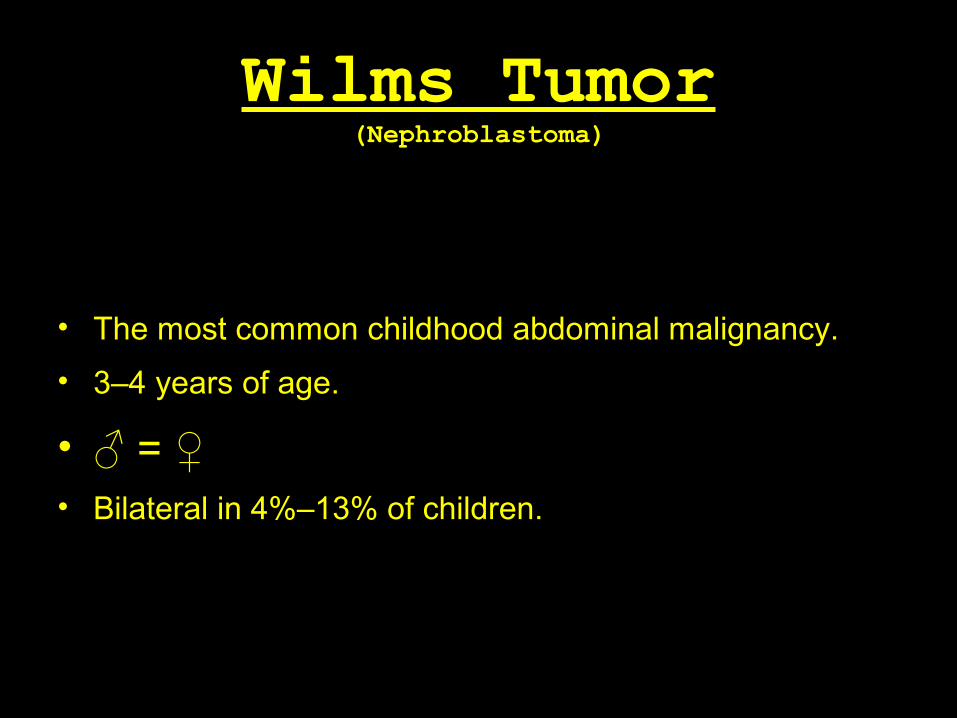
Wilms Tumor(Nephroblastoma)
• The most common childhood abdominal malignancy.
• 3–4 years of age.
• ♂ = ♀• Bilateral in 4%–13% of children.
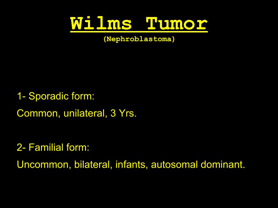
Wilms Tumor(Nephroblastoma)
1- Sporadic form:
Common, unilateral, 3 Yrs.
2- Familial form:
Uncommon, bilateral, infants, autosomal dominant.

Wilms Tumor(Nephroblastoma)
• May be associated with congenital anomalies
such as: - Cryptorchidism (2.8% of cases). - Hemihypertrophy (2.5%). - Hypospadias (1.8%). - Sporadic aniridia.

Wilms Tumor(Nephroblastoma)
• WAGR syndrome (Wilms tumor, aniridia,
genitourinary abnormalities, mental retardation).
• Drash syndrome (male pseudohermaphroditism,
progressive glomerulonephritis)
• Beckwith-Wiedemann syndrome
(hemihypertrophy).

Wilms Tumor(Nephroblastoma)
• Arises from mesodermal precursors of the
renal parenchyma (metanephric Blastema).
Wilms Tumor(Nephroblastoma)
Gross Pathology:
• Large solid spherical mass with areas of
hemorrhage and necrosis surrounded with
pseudocapsule.

Wilms Tumor(Nephroblastoma)
Gross Pathology:Spread:
1. Extend into the renal vein and, subsequently, into the
inferior vena cava.
2. Direct local invasion of adjacent structures.
3. Local regional lymph node metastases.
4. Hematogenous metastases.
Wilms Tumor(Nephroblastoma)
• Palpable mass.
• Hypertension may be present in up to 25% of
cases due to renin production by the tumor
• Hematuria and pain are infrequent clinical
findings.
Wilms Tumor(Nephroblastoma)
Radiological Picture

Wilms Tumor(Nephroblastoma)
Plain X-Ray:
Large soft tissue mass
displacing bowel gas

Wilms Tumor(Nephroblastoma)
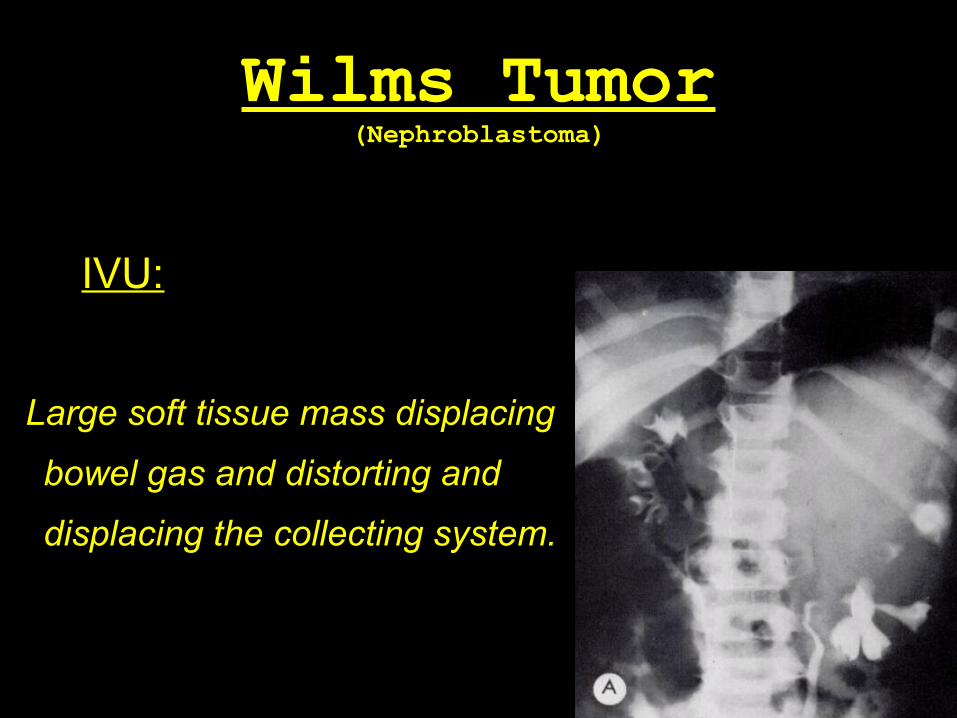
IVU:
Large soft tissue mass displacing
bowel gas and distorting and
displacing the collecting system.
Wilms Tumor(Nephroblastoma)
IVU:
Large soft tissue mass displacing
bowel gas and distorting and
displacing the collecting system.

Wilms Tumor(Nephroblastoma)
US:
• Large soft tissue mass
heterogeneous echogenicity, which
represents hemorrhage, necrosis,
or calcification.
• Vascular Invasion.

Wilms Tumor(Nephroblastoma)
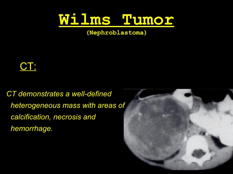
CT:
CT demonstrates a well-defined
heterogeneous mass with areas of
calcification, necrosis and
hemorrhage.
Wilms Tumor(Nephroblastoma)
CT:
CT demonstrates a well-defined
heterogeneous mass with areas of
calcification, necrosis and
hemorrhage.

Wilms Tumor(Nephroblastoma)
CT:
CT demonstrates a well-defined
heterogeneous mass with areas of
calcification, necrosis and
hemorrhage.

Wilms Tumor(Nephroblastoma)
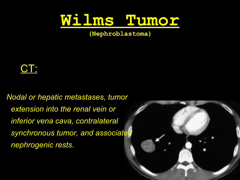
CT:
Nodal or hepatic metastases, tumor
extension into the renal vein or
inferior vena cava, contralateral
synchronous tumor, and associated
nephrogenic rests.

Wilms Tumor(Nephroblastoma)
CT:
Nodal or hepatic metastases, tumor
extension into the renal vein or
inferior vena cava, contralateral
synchronous tumor, and associated
nephrogenic rests.
Wilms Tumor(Nephroblastoma)
CT:
Nodal or hepatic metastases, tumor
extension into the renal vein or
inferior vena cava, contralateral
synchronous tumor, and associated
nephrogenic rests.


Wilms Tumor(Nephroblastoma)
MRI:
Well-defined heterogeneous mass with low signal intensity
on T1-weighted images and high signal intensity on T2-
weighted images.

Wilms Tumor(Nephroblastoma)
Angiography: