Embed Size (px)
Citation preview

Stomatološki vjesnikStomatološki vjesnikStomatological reviewStomatological review
Stomatološki vjesnik 2016; 5 (1-2)

Stomatološki vjesnik
Stomatološki vjesnik 2016; 5 (1-2)
CONTENTS / SADRŽAJ
ORIGINAL SCIENTIFIC ARTICLES / ORIGINALNI NAUČNI RADOVI
OUTCOME OF IMPLANT SUPPORTED SINGLE-TOOTH CROWNS IN ANTERIOR MAXILLA
USING OBJECTIVE INDICES AND PATIENTS' PERCEPTIONS: A 1-YEAR PROSPECTIVE STUDY
Tosum S., Ajanović M., Kamber-Ćesir A., Dervišević A., Kazazić L.
PERIODONTAL STATUS AND CANDIDA CARRIAGE IN TYPE 1 DIABETES MELLITUS
Huseinbegović A., Fazlić Imamović R., Bektaš S., Bajrić E., Dedić A., Šečić S.
CORRELATION BETWEEN PSYHOACTIVE SUBSTANCE ABUSE AND PERIODONTAL ALTERATION
Hadžić S., Gojkov-Vukelić M., Pašić E., Šečić S.
THE RANGE OF MAXIMUM MOUTH OPENING IN PARTIALLY EDENTULOUS PATIENTS
WITH SYMTPOMS OF TEMPOROMANDIBULAR JOINT DYSFUNCTION
Strujić-Porović S., Ajanović M., Kazazić L., Berhamović L., Đonlagić A., Kamber-Ćesir A.
ANALYSIS OF CONCORDANCE BETWEEN CLINICAL AND RADIOGRAPHICAL DEPTH
OF OCCLUSAL CARIES LESIONS – A PILOT STUDY
Bajsman A., Haračić A., Vuković A., Zukić S.
REVIEW ARTICLE / PREGLEDNI ČLANAK
DISCOLORATION OF DECIDUOUS AND PERMANENT TEETH:
CLASSIFICATION, ETIOLOGY AND TREATMENT OPTIONS
Selimović-Dragaš M., Kovačević N.
CASE REPORT / PRIKAZ SLUČAJA
CLINICAL EVALUATION OF THE EFFECTS FROM SURGICAL REGENERATIVE PERIODONTAL THERAPY
WITH THE USE OF BIOMATERIALS
Todoroska S., Atanasovska-Stojanovska A., Popovska M., Ivanovski K.
BOOK REVIEW / PRIKAZ KNJIGE
BASICS OF GNATHOLOGY
Ajanović M., Strujić-Porović S., Šuljak-Lončarević A., Hadžović Džuvo A.,
Kazazić L., Tosum S., Berhamović L., Hamzić A., Đonlagić A., Kapur E.
3
11
19
27
33
41
53
63
Izdavač / Publisher:
Za izdavača / For publisher: Muhamed Ajanović
ČLANOVI UREĐIVAČKOG ODBORA / EDITORIAL BOARD :
Glavni urednik / Editor in chief: Sadeta Šečić
Sekretar uređivačkog odbora / Secretary of editorial board: Selma Zukić
Članovi /Members: Sead Redžepagić, Muhamed Ajanović, Sedin Kobašlija, Amra Vuković, Enita Nakaš,
MEĐUNARODNI UREĐIVAČKI ODBOR / INTERNATIONAL EDITORIAL BOARD:Anwar Barakat Bataineh (Irbid, Jordan), Jasenka Živko-Babić (Zagreb, Hrvatska), Andrija Petar Bošnjak (Rijeka, Hrvatska), Hrvoje Brkić (Zagreb , Hrvatska), Dolores Biočina Lukenda (Split, Hrvatska), Davor Katanec (Zagreb, Hrvatska), Šahza Hatibović Koffman (London Ontario Kanada), Mladen Kuftinec (USA), Darko Macan (Zagreb, Hrvatska), Berislav Perić (Zagreb, Hrvatska), Tore Solheim (Oslo, Norveška), Dragoslav Stamenković (Beograd, Srbija), Marin Vodanović (Zagreb, Hrvatska)
Lektor za engleski jezik / English language editor: Nermana Bičakčić
Tehničko uređenje / Technical editor: Branislav Trogrančić
Štampa / Printed by: Štamparija Fojnica
Dizajn naslovnice / Cover page design: Lana Malić
Tiraž/ Number of copies:
KONTAKT / CONTACT:Stomatološki vjesnikStomatološki fakultet sa klinikamaBolnička 4a, 71000 SarajevoBosna i HercegovinaTelefon: + 387(33)214 294e-mail: [email protected] Web: www.stomatoloskivjesnik.ba
TRANSAKCIJSKI RACUN / TRANSFER ACCOUNT:33386902296551066UniCredit Bank dd
Stomatološki fakultet Univerziteta u Sarajevu / Faculty of Dentistry, University of Sarajevo
200
Sanja Hadžić, Alma Konjhodžić Prcić, Lejla Kazazić
ISSN 0350-5499 UDK 616.31
Svrha i i cilj :Stomatološki vjesnik je neprofitni naučno stručni časopis koji publicira originalne naučne radove, prikaze slučajeva, pisma uredniku, savremene perspektive, editorijale, preliminarne komunikacije u oblasti stomatologije i drugih biomedicinskih nauka. Radovi su na Bosanskom/Hrvatskom/Srpskom jeziku sa naslovom, sažetkom i ključnim riječima bilingvalnim B/H/S i engleskom jeziku. Radovi se mogu koristiti u edukacijske svrhe bez predhodnog odobrenja, a uz obavezno navođenje izvora. Korištenje cijelih ili dijelova članaka u komercijalne svrhe nije dozvoljeno bez predhodnog pismenog odobrenja izdavača Autorska prava posjeduje izdavač: Stomatološki fakultet sa klinikama Univerziteta u Sarajevu.
Aim and Scope:Stomatološki vijesnik / Stomatological review is a non-profit scientific journal that publishes original articles, case reports, letters to the editors, current perspectives, editorials, fast-track articles in a field of dentistry and other bio-medical sciences. Papers are in Bosnian/ Croatian/Serbian language with at least title, abstract and key words bilingual in B/C/S and English language. All manuscripts undergo the peer review process before can be accepted for publishing in Stomatološki vjesnik/ Stomatolgical review. Papers can be used for educational purposes without prior consent only with adequate citation of the sources. Using whole or parts of articles for commercial purposes is not permitted without prior written permission of the publisher. Copyright owns the publisher: Faculty of Dentistry with Clinics, University of Sarajevo.
Časopis Stomatološki vjesnik je oslobođen poreza na promet prema Mišljenju Federalnog ministarstva obrazovanja, nauke, kulture i sporta br: 04-15-661/2002.
Journal Stomatological review is tax exempt according to the opinion of the Federal Ministry of Education Science Culture and Sports no: 04-15-661/2002.
Printed on acid free paper
Indexed in: (Index Copernicus International), (Directory of Open Access Journal), SJIF (Scientific Journal Impact Factor Value 2.502), EBSCO, HINARI, INFOBASE, AE GLOBAL INDEX, DRJI ORG, SJIF, ONE SEARCH, PUBGET, RESEARCH BIBLE, SIS
IIC DOAJGOOGLE SCHOLAR, EZB (Electronishe Zeitschriftenbibliothek),

Stomatološki vjesnik 2016; 5 (1-2) 3
ORIGINAL SCIENTIFIC ARTICLE / ORIGINALNI NAUČNI RAD
OUTCOME OF IMPLANT SUPPORTED SINGLE-TOOTH CROWNS IN ANTERIOR MAXILLA USING OBJECTIVE INDICES AND PATIENTS' PERCEPTIONS: A YEAR PROSPECTIVE STUDY
1 1Tosum S , Ajanović M , Kamber-Ćesir A ,2 1Dervišević A. , Kazazić L.
1.* . .
1 Department of Prosthodontics, Faculty of Dentistry, University of Sarajevo, Sarajevo, Bosnia and Herzegovina2 Department of Maxillofacial Surgery, Faculty of Dentistry, University of Sarajevo, Sarajevo, Bosnia and Herzegovina
ABSTRACT
Aim: The main purpose of this study was to determine the degree of success of oral rehabilitation in patients with implant therapy using implants bredent blueSKY (bredent medical GmbH & Co.KG Senden, Germany).
Materials and methods: The study evaluated 32 implants a year after the implant therapy. The observed patients were of both sexes, aging from twenty to seventy years and randomly sampled. All patients underwent the OHIP-CRO49 index in order to determine the level of their satisfaction with implant therapy, implant type bredent blueSKY. The ICAI index was assessed aiming to determine the degree of success in aesthetic and functional rehabilitation.
Results: In 23 individuals of both sexes with an average age of 60.13 ± 13.1 (21-73) who were included in the study, a total of 32 implants retaining a single-crown tooth restoration were inserted. In our research we used a conventional (two-phase) restoration of implants, without immediate loading. The crown was installed 90 days after the placement of the implants. With ICAI (Implant Crown Aesthetic Index), in the results of our study with the largest number of the implant-supported single-tooth restoration, labial gingival margin did not differ from the labial edge of the control natural tooth. In significantly smaller number of compensation (3.13%), the deviation was greater than 1.5 mm compared to the control natural tooth (chi-square test, p = 0.001). No deviation was found in 56.25% cases and in 40.63% there was a deviation less than 1.5 mm. Chi square test showed a statistically significant difference. The highest total OHIP score was 142 and was recorded in one patient. Most of the patients had a total OHIP score 0 meaning a favorable quality of life.
Conclusions: A statistically significant difference was found in a distribution of the implant-supported single-tooth restorations cemented on implants type bredent blueSKY, with varying degrees of aesthetic success / failure. The largest number of single tooth crowns achieved satisfactory aesthetic outcome one year after being in function.
Key words: dental implant, oral rehabilitation, single tooth crown, gingiva.
*Corresponding author
Tosum Selma, PhD
Department of Prosthodontics
Faculty of Dentistry
University of Sarajevo
Bolnička 4a
71000 Sarajevo
Bosnia and Herzegovina
Phone: +387 (33) 214 249
e-mail: [email protected]

Excellent (Score=0)
Satisfactory (Score =1 or 2)
Moderate (Score =3 or 4)
Poor aesthetics (Score =5 or more)
Total
6 (18,8%)
22 (68,8%)
2 (6,2%)
2 (6,2%)
32 (100%)
Aesthetic success (total ICAI score) N (%)
4
OUTCOME OF IMPLANT SUPPORTED SINGLE-TOOTH CROWNS IN ANTERIOR MAXILLA USING OBJECTIVE INDICES AND PATIENTS' PERCEPTIONS: A YEAR PROSPECTIVE STUDY
5
Tosum S., Ajanović M., Kamber-Ćesir A., Dervišević A., Kazazić L.
Introduction
The goal of dentistry is to restore lost functions of
stomatognathic system such as: chewing, speech,
esthetic, comfort. Regardless of the bone atrophy,
disease, or injury of the stomatognathic system, im-
plant dentistry could achieve an ideal goal [1].
The use of implants prosthetically restores func-
tion and aesthetics. The loss of a single tooth has be-
come a common treatment alternative to conventio-
nal tooth supported reconstruction, mainly due to
the benefit of avoiding sacrificing the intact tooth
substance of adjacent teeth.
The use of implants prosthetically restores func-
tion and aesthetics. The loss of a single tooth has
become a treatment option instead of conventional
fixed prosthesis, mainly due to the benefit of avoiding
the preparation of adjacent teeth [2,3]. As the pre-
dictability of dental implants has been proved be-
yond doubt, achieving a good success rate in terms of
stability is no longer a big concern among dentist, but
aesthetic success of therapy is of mayor concern now
[4].
Restoration of the missing tooth should not be
designed only to withstand the forces of occlusion
and mastication, but also to create an acceptable
aesthetic result [5].
For many patients, an aesthetic result is the main
motivating factor when deciding in favor of dental
implantation [6-8]. In visible anterior region priority
is given to aesthetics while in the posterior region the
function can be emphasized as the most important
factor. But requirements for aesthetics and function
must be balanced with regard to predictable longe-
vity, minimal biological effects and cost aspects [9].
Achieving an aesthetically optimal result is deter-
mined by a number of factors: tooth related factors
such as tooth dimensions, form and color contribute
to aesthetics as well as soft tissue-related factors in-
cluding inter-dental and mid-facial soft tissue dimen-
sions, texture and color [10]. When estimating
aesthetic of the crown restoration, it should be consi-
dered that the crown restoration has to be in har-
mony and symmetry with the crown form adjacent
natural teeth as well as with the one of the contra-
lateral tooth [11]. Soft tissue management is one of
the final esthetic result, with the need to harmonize
color, form, and contour with that of the adjacent
tissues [12].
The concern from an aesthetic point of view is also
the topography of the surrounding soft tissues i.e.,
the position of the soft tissue margin at the facial
aspect of the crown and the degree of inter-dental
papillae fill in the embrasure spaces lateral to the
implant-supported crown [11,13].
Meijer et al. introduced a new index for rating
aesthetics of implant-supported single crowns and
adjacent soft tissue – the Implant Crown Aesthetic
Index (ICAI). This index is important for considering
parameters for the evaluation of the implant crown
and peri-implant mucosa [14].
The recent concepts of implant dentistry is not
restricted to the basic needs, but has evolved to
cosmetic or aesthetic correction to uplift the self-
esteem and confidence of people [4].
To evaluate patients' view regarding the change in
their quality of life after receiving dental implant
treatment, the Oral Health Impact Profile-49 (OHIP-
49) was developed. The OHIP-49 is a reliable and va-
lid instrument for detailed measurement of the social
impact of oral disorders and has potential benefits
for clinical decision making and research [15,16].
The main purpose of this study was to determine
the degree of success of oral rehabilitation in patients
with implant therapy using implants bredent blue-
SKY (bredent medical GmbH & Co.KG Senden, Ger-
many).
Materials and methods
In this prospective study, patients with tooth loss
in the aesthetic region in maxilla that were referred
to the Faculty of Dentistry in Sarajevo for prosthetic
rehabilitation were included. This study was appro-
ved by the Ethics Committee of the Faculty of Dentis-
try, and it was conducted in accordance with the
ethical principles of the Helsinki. Patients were infor-
med of the options for tooth replacement including
the risks and benefits of dental implants. All included
patients required replacements with single-tooth
restorations. The inclusion criteria were: age >18,
single tooth loss in the maxillary front, presence of
contra-lateral natural tooth, good oral hygiene. The
exclusion criteria were: history of radiotherapy in
head and neck, current chemotherapy, metabolic bo-
ne disorders, absence of any serious systemic disease
which would jeopardize bone healing, glucocorti-
coids and bisphosphonates medication. The study
evaluated 32 implants a year after the implant
therapy. Observed patients were of both sexes, aging
from twenty to seventy years (mean age: 60.13±13.1)
and randomly sampled. Surgical treatment was
carried out under local anesthesia by specialist in
prosthodontics and oral implantology, following
manufactures instructions. Metal-ceramic crowns
and metal alloy abutments were used for the coronal
part in all constructions.
All patients underwent the Croatian version of the
Oral Health Impact Profile questionnaire (OHIP-
CRO49 index) to determine the level of patient satis-
faction with implant therapy, implant type bredent
blueSKY® (bredent medical GmbH & Co. KG Senden,
Germany). The OHIP-CRO49 consisted of 49 ques-
tions. Each answer was scored with a Likert response
scale from 0 (never experienced problem) to 4 (pro-
blem experienced very often). Higher scores indica-
ted more impaired oral health. The questionnaire
was filled in by hand at the Clinic without assistance
[16].
Patients were clinically examined one year after
completion of implant-supported restorations. The
contra-lateral/natural tooth was used as a reference
when a single implant rehabilitation was assessed
[7]. The ICAI index was assessed, with a goal to deter-
mine the degree of success in aesthetic and functional
rehabilitation. The ICAI evaluates five variables for
the crown restoration ( hard tissue ) of the implants
and four variables for the mucosa ( soft tissue )
surrounding the implant ( color, anatomic contour
and surface texture ). These nine variables were: 1.
Mesiodistal dimension of the crown, 2. Position of the
incise edge of the crown, 3. Labial convexity of the
crown, 4. Color and translucency of the crown, 5.
Surface of the crown, 6. Position of the labial margin
of the peri-implant mucosa, 7. Position of mucosa in
the approximate embrasures, 8. Contour of the labial
surfaces of the mucosa, 9. Color and surface of the
labial mucosa. Each item is given score I, while major
/ gross deviation takes 5 penalty points. The total
score leads to judgment regarding aesthetics: 0
penalty points = excellent; I or 2 points = satisfactory;
3 or 4 points = moderate; 5 or more points = poor
aesthetics.
The score ranging from 0 to 45, where 0 repre-
sents the most positive score. Statistical analysis was
performed using software package SPSS for Win-
dows version 18.0. The descriptive analysis of data
was presented as frequency and mean ± standard
deviation, minimal and maximal value. Che-square
test was performed to determine differences for
variables: mesio-distal dimension of the crown, labial
convexity of the crown, the color and translucency of
the implant-supported crown, the position of the
labial margin of the peri-implant mucosa, the
position of mucosa in approximate embrasures, the
color and surface of the labial mucosa of the implant-
supported crown. Spearman rank correlation was
used to determine correlation between the quality of
life and three variables: age, gender and smoking
habits. The level of statistical significance was set at
5%.
The study included 23 patients (age: 21-73 years)
treated for tooth loss with 32 dental implants. All the
patients had received an implant-supported crown
following tooth loss due to TDI (traumatic dental in-
jury). 53.1% were female and 46.9% male. According
to smoking habits, 78.1% were non-smokers and
21.9% smokers.
Excellent aesthetic (score O) was carried out by
18,8% evaluated metal-ceramic crowns on implant
supported single tooth restorations; 68,8% showed
satisfactory aesthetic success (score 1 or 2); 6.1%
moderate aesthetic success (score 3 or 4) and only
two patients (6.1%) had poor aesthetics success
(score 5 and 10 )(Table 1.).
Results
Stomatološki vjesnik 2016; 5 (1-2) Stomatološki vjesnik 2016; 5 (1-2)
Table 1.The frequency of total ICAI scores as indicators of aesthetic success of prosthetic restorations

6
According to mesio-distal dimension of the crown,
there were 75.0 % without deviation, 18.8% were
slightly under-contoured compared to contra-lateral
tooth, 6.3 % were slightly over-contoured compared
to contra-lateral tooth. Chi square test revealed sta-
tistical significance (p<0.001).
According to position of incisal edge of the im-
plant-supported crown, there were no deviation.
Labial convexity of the crown was in the most ca-
ses ( 93.8% ) in harmony with the adjacent tooth and
in 6.2 % was big mismatch. Chi square test revealed
statistical significance (p<0.001).
The color and translucency of the implant-sup-
ported crown were in harmony with contra-lateral
tooth in the most cases ( 81.3% ) and in 18.8% was a
slight mismatch. Chi square test revealed statistical
significance (p<0.001).
Analysis of the position of the labial margin of the
peri-implant mucosa showed no deviation in
56.25%, 40.63% deviation was less than 1.5 mm and
3.13% deviation was more than 1.5 mm (Chi square
test, p=0.001).
Analysis of the position of mucosa in the approxi-
mate embrasures showed no deviation in 87.5% and
12.5% deviation less than 1.5 mm (Chi square test,
p<0.001).
Analysis of the contour of the labial surface of the
mucosa showed no deviation in 87.5%, 9.4% slightly
under-contoured and 3.1% rather over-contoured.
The color and surface of the labial mucosa of the
implant-supported crown were statistically different
compared to contra-lateral natural tooth; 78.1% had
no mismatch, 21.9% had slight mismatch (Chi square
test, p= 0.001) (Table 2.).
Spearman rank correlation revealed no statisti-
cally significant correlation between aesthetic
success prosthetic restoration and: age (rho=-0.283,
p=0.117), gender (rho=-0.166, p=0.365) and smo-
king habits (rho=-0.200, p=0.272).
Analysis of seven domains of OHIP-49 showed no
statistical significance in two domains: functional
limitation and physical discomfort (Table 3.).
The highest total OHIP score was 142 (one pati-
ent). 12 patients had total OHIP score 0 (Table 4.).
The mean value and standard deviation of total
OHIP score were presented in Table 5. and Table 6.
Spearman rank correlation revealed no statistical
significant correlation between the quality of life of
implant supported single-tooth restoration in
7
aesthetic zone and: age (rho=-0.302, p=0.093),
gender (rho=-0.081, p=0.660) and smoking habits
(rho=-0.059, p=0.750).
Logistic regression showed no statistically signi-
ficant influence of gender, age, smoking habits as a
predictor of quality of life with implant supported
single tooth restorations (Table 7.).
Discussion
In our study, the mesio-distal width of the most
implant-supported crown was different compared to
mesio-distal width of clinical crown of contra-lateral
tooth and the difference was statistically significant.
Chang et al. found no statistically significant diffe-
rences in mesio-distal width of the implant-suppor-
ted crown and the clinical crown of contra-lateral tooth [7]. In study from 2009., after guided bone
regeneration and early implant placement (Nobel-
replace tapered Ti Unite® Nobel Biocare, Göteborg,
Sweden), Cosyn at al. found the mean disparity width
of 0.2 mm between the implant crown and teeth [10].
Vilhjálmur et al. evaluated aesthetic outcome of
implant-supported crowns in the anterior maxilla
and periimplant soft tissue using three different in-
dexes: ICAI, modified version of the ICAI (mod-ICAI),
Pink Aesthetic Score (PES), and the index of Califor-
nian Dental Association (CDA), one year after the
crown placement. Implants used in this study were
Brånemark ( Nobel Biocare, Göteborg, Sweden ) or
Astra ( Astra Tech AB, Mödalen, Sweden). They found
correlation between color of the new crown and the
sum of hard tissue related items of the ICAi and the
mod-ICAI was significant [17].
In our study, the position of incision edge of the
implant-supported crown was in harmony with con-
tra-lateral tooth. From available literature we didn't
find relevant results for the position of incision edge
but Chang et al. measured the distance between the
soft tissue margin and the incision edge and found
statistically significance difference in clinical crown
length in aesthetic maxillary region. The crowns
supported by implants (Brånemark, Nobel Biocare,
Göteborg, Sweden ) were on the average 1.0 mm
longer than the clinical crown of contra-lateral teeth
[7]. To the contrary, Cosyn et al. found no statistically
significant difference in clinical crown length [10].
OUTCOME OF IMPLANT SUPPORTED SINGLE-TOOTH CROWNS IN ANTERIOR MAXILLA USING OBJECTIVE INDICES AND PATIENTS' PERCEPTIONS: A YEAR PROSPECTIVE STUDY
Smokers
Women
Non-smokers
Men
Total
Total
32 (100%)
32 (100%)
0 (0,0%)
4 (12,5%)
3 (9,4%)
1 (3,1%)
7 (21,9%)
21 (65,6%)
14 (43,8%)
14 (43,8%)
Aesthetic success*
Aesthetic failure**
Smokers
Women
Non-smokers
Men
Total
Total
32 (100%)
32 (100%)
3 (9,36%) 4 (12,50 %)
9 (28,13%) 16 (50,0 %)
7 (21,88%) 10 (31,25 %)
5 (15,63%) 10 (31,25 %)
Favorable* Unavorable**
Quality of life
* Aesthetic success: total ICAI=0-2; ** Aesthetic failure: total ICAI ≥ 3
Table 2. Aesthetic success of prosthetic restoration according gender and smoking habits
N= number of implant-supported single-tooth restorationsa Chi square test
Table 3. Evaluation of quality of life according OHIP domains
Functional limitation
Physical discomfort
Psychological discomfort
Physical disability
Psychological disability
Social disability
Handicap
20
20
27
28
29
30
29
62.5
62.5
84.4
87.5
90.6
93.8
90.6
12
12
5
4
3
2
3
37.5
37.5
15.6
12.5
9.4
6.2
9.4
Domain
Quality of life
Favorable
N % N %
Unfavorable
0
1
2
6
8
36
49
142
12
10
4
2
1
1
1
1
Total OHIP score
Total 32
N
Mean Value
95% confidence interval
Median
Standard deviation
Minimum
Maximum
8,28
from -1,29 to 17,85
1,00
26,536
0
142
Total OHIP score
Table 4. Frequency of total OHIP score
Table 5. Descriptive statistics of total OHIP score
*Total OHIP score=0; ** Total OHIP score ≥1
Table 6. The quality of life of patients with implant supported single-tooth restoration according gender and smoking habits
Table 7. Logistic regression: Influence of gender, age and smoking habits on the quality of life
-,469
-,094
-,404
Gender
Age
Smoking habits
,794
,060
,928
,554
,115
,663
,626
,910
,667
Exp(B)Sig.S.E.BPredictor
Tosum S., Ajanović M., Kamber-Ćesir A., Dervišević A., Kazazić L.
Stomatološki vjesnik 2016; 5 (1-2) Stomatološki vjesnik 2016; 5 (1-2)

6
According to mesio-distal dimension of the crown,
there were 75.0 % without deviation, 18.8% were
slightly under-contoured compared to contra-lateral
tooth, 6.3 % were slightly over-contoured compared
to contra-lateral tooth. Chi square test revealed sta-
tistical significance (p<0.001).
According to position of incisal edge of the im-
plant-supported crown, there were no deviation.
Labial convexity of the crown was in the most ca-
ses ( 93.8% ) in harmony with the adjacent tooth and
in 6.2 % was big mismatch. Chi square test revealed
statistical significance (p<0.001).
The color and translucency of the implant-sup-
ported crown were in harmony with contra-lateral
tooth in the most cases ( 81.3% ) and in 18.8% was a
slight mismatch. Chi square test revealed statistical
significance (p<0.001).
Analysis of the position of the labial margin of the
peri-implant mucosa showed no deviation in
56.25%, 40.63% deviation was less than 1.5 mm and
3.13% deviation was more than 1.5 mm (Chi square
test, p=0.001).
Analysis of the position of mucosa in the approxi-
mate embrasures showed no deviation in 87.5% and
12.5% deviation less than 1.5 mm (Chi square test,
p<0.001).
Analysis of the contour of the labial surface of the
mucosa showed no deviation in 87.5%, 9.4% slightly
under-contoured and 3.1% rather over-contoured.
The color and surface of the labial mucosa of the
implant-supported crown were statistically different
compared to contra-lateral natural tooth; 78.1% had
no mismatch, 21.9% had slight mismatch (Chi square
test, p= 0.001) (Table 2.).
Spearman rank correlation revealed no statisti-
cally significant correlation between aesthetic
success prosthetic restoration and: age (rho=-0.283,
p=0.117), gender (rho=-0.166, p=0.365) and smo-
king habits (rho=-0.200, p=0.272).
Analysis of seven domains of OHIP-49 showed no
statistical significance in two domains: functional
limitation and physical discomfort (Table 3.).
The highest total OHIP score was 142 (one pati-
ent). 12 patients had total OHIP score 0 (Table 4.).
The mean value and standard deviation of total
OHIP score were presented in Table 5. and Table 6.
Spearman rank correlation revealed no statistical
significant correlation between the quality of life of
implant supported single-tooth restoration in
7
aesthetic zone and: age (rho=-0.302, p=0.093),
gender (rho=-0.081, p=0.660) and smoking habits
(rho=-0.059, p=0.750).
Logistic regression showed no statistically signi-
ficant influence of gender, age, smoking habits as a
predictor of quality of life with implant supported
single tooth restorations (Table 7.).
Discussion
In our study, the mesio-distal width of the most
implant-supported crown was different compared to
mesio-distal width of clinical crown of contra-lateral
tooth and the difference was statistically significant.
Chang et al. found no statistically significant diffe-
rences in mesio-distal width of the implant-suppor-
ted crown and the clinical crown of contra-lateral tooth [7]. In study from 2009., after guided bone
regeneration and early implant placement (Nobel-
replace tapered Ti Unite® Nobel Biocare, Göteborg,
Sweden), Cosyn at al. found the mean disparity width
of 0.2 mm between the implant crown and teeth [10].
Vilhjálmur et al. evaluated aesthetic outcome of
implant-supported crowns in the anterior maxilla
and periimplant soft tissue using three different in-
dexes: ICAI, modified version of the ICAI (mod-ICAI),
Pink Aesthetic Score (PES), and the index of Califor-
nian Dental Association (CDA), one year after the
crown placement. Implants used in this study were
Brånemark ( Nobel Biocare, Göteborg, Sweden ) or
Astra ( Astra Tech AB, Mödalen, Sweden). They found
correlation between color of the new crown and the
sum of hard tissue related items of the ICAi and the
mod-ICAI was significant [17].
In our study, the position of incision edge of the
implant-supported crown was in harmony with con-
tra-lateral tooth. From available literature we didn't
find relevant results for the position of incision edge
but Chang et al. measured the distance between the
soft tissue margin and the incision edge and found
statistically significance difference in clinical crown
length in aesthetic maxillary region. The crowns
supported by implants (Brånemark, Nobel Biocare,
Göteborg, Sweden ) were on the average 1.0 mm
longer than the clinical crown of contra-lateral teeth
[7]. To the contrary, Cosyn et al. found no statistically
significant difference in clinical crown length [10].
OUTCOME OF IMPLANT SUPPORTED SINGLE-TOOTH CROWNS IN ANTERIOR MAXILLA USING OBJECTIVE INDICES AND PATIENTS' PERCEPTIONS: A YEAR PROSPECTIVE STUDY
Smokers
Women
Non-smokers
Men
Total
Total
32 (100%)
32 (100%)
0 (0,0%)
4 (12,5%)
3 (9,4%)
1 (3,1%)
7 (21,9%)
21 (65,6%)
14 (43,8%)
14 (43,8%)
Aesthetic success*
Aesthetic failure**
Smokers
Women
Non-smokers
Men
Total
Total
32 (100%)
32 (100%)
3 (9,36%) 4 (12,50 %)
9 (28,13%) 16 (50,0 %)
7 (21,88%) 10 (31,25 %)
5 (15,63%) 10 (31,25 %)
Favorable* Unavorable**
Quality of life
* Aesthetic success: total ICAI=0-2; ** Aesthetic failure: total ICAI ≥ 3
Table 2. Aesthetic success of prosthetic restoration according gender and smoking habits
N= number of implant-supported single-tooth restorationsa Chi square test
Table 3. Evaluation of quality of life according OHIP domains
Functional limitation
Physical discomfort
Psychological discomfort
Physical disability
Psychological disability
Social disability
Handicap
20
20
27
28
29
30
29
62.5
62.5
84.4
87.5
90.6
93.8
90.6
12
12
5
4
3
2
3
37.5
37.5
15.6
12.5
9.4
6.2
9.4
Domain
Quality of life
Favorable
N % N %
Unfavorable
0
1
2
6
8
36
49
142
12
10
4
2
1
1
1
1
Total OHIP score
Total 32
N
Mean Value
95% confidence interval
Median
Standard deviation
Minimum
Maximum
8,28
from -1,29 to 17,85
1,00
26,536
0
142
Total OHIP score
Table 4. Frequency of total OHIP score
Table 5. Descriptive statistics of total OHIP score
*Total OHIP score=0; ** Total OHIP score ≥1
Table 6. The quality of life of patients with implant supported single-tooth restoration according gender and smoking habits
Table 7. Logistic regression: Influence of gender, age and smoking habits on the quality of life
-,469
-,094
-,404
Gender
Age
Smoking habits
,794
,060
,928
,554
,115
,663
,626
,910
,667
Exp(B)Sig.S.E.BPredictor
Tosum S., Ajanović M., Kamber-Ćesir A., Dervišević A., Kazazić L.
Stomatološki vjesnik 2016; 5 (1-2) Stomatološki vjesnik 2016; 5 (1-2)

98
Choquet et al. concluded that the shape of the
crown may be more difficult to fit with the neigh-
boring teeth and the contra-lateral tooth especially
when there is lack of alveolar bone and soft tissue in
the implant region [18].
In the study of 53 implant supported single-tooth
replacements Scheller et al. reported that a majority
(75%) of the implant-supported crowns the soft tis-
sue margin remained stable during a 5-year follow-
up period and that recession of the soft tissue margin
occurred at only 10% of the single implant resto-
rations [19]. These results are in accordance with our
results from 1-year follow-up period.
In the highest percentage in our study inter-dental
papillae of implant-supported single crowns were in
harmony with contra-lateral tooth (87.5%), which is
consistent with the results reported by Cosyn et al.
They found a borderline significant difference ( p=
0.053) in the mesial papilla height between implant
crowns and contra-lateral teeth. The mesial papilla
was on average 0.4 mm and the distal papilla 1mm
shorter at implant restoration in comparison with
the corresponding site at contra-lateral teeth [10].
Three years after treatment, 59 patients with 98
implant supported single-tooth , Hosseini et al. eva-
luated aesthetic outcome of peri-implant mucosa
using Copenhagen Index Score. They used three abut-
ment materials (zirconia, titanium and gold alloy)
and the results showed that the color of the marginal
mucosa was comparable at zirconia and metal abut-
ments. When titanium abutments were used, in 52 %
cases there was no marginal discoloration. In our
study, in 87.5% cases, the color and surface of the
labial mucosa of implant-supported single crowns
were in harmony with contra-lateral tooth [20].
In this study, a possible impact on oral health-
related quality of life after implant treatment was
evaluated using OHIP-CRO49 index. Oral Health
Impact Profile is explicitly aimed to collect a variety
of impacts ranging from oral functional effects to
pain and personal affects as well as social interaction.
The theoretical model and empirical methods are
used to constitute the aspects of the OHIP which are
distinctive from other indices such as those develo-
ped by Cushing et al., Atchinson and Dolan and
Strauss and Hunt [16,21,22,23]. The OHIP-49 is
suitable for use as an outcome measure in a clinical
trial in which different treatment protocols are com-
pared, and it is regarded as the best descriptor of the consequences of oral disorders [16]. Treatment with
dental implants results in a major change of the oral
status for patients, but implants will never precisely
replicate the feeling of natural teeth. On the other
hand, implant therapy will most certainly influence
appearance, speech, chewing function, temper, and
self-esteem thus affecting a patient's quality of life [24,25]. Erkapers et al. reported that the patients
treated with immediately loaded implants in the
edentulous maxilla generally reported statistically
significant increased satisfaction. All seven domains
showed a statistically significant improvement in the
post-treatment period[26]. The quality of life was
estimated as better in five domains (psychological
discomfort, physical disability, psychological disabi-
lity, social disability, handicap) than functional limi-
tation and physical discomfort. However, responses
for these two domains were more favorable than
unfavorable.
The equal number of male and female patients
estimated their quality of life as unfavorable (Table
6). Factors such as age, gender, education, self-
esteem, functionality and past dental history may
also contribute to a patient's opinion regarding the
aesthetics of the single crown of implants and its
adjacent mucosa.
Conclusion
A statistically significant difference was found in
the distribution of the implant-supported single-
tooth restorations cemented on implants type bre-
dent blueSKY, with varying degrees of aesthetic
success / failure. The largest number of single tooth
crowns achieved satisfactory aesthetic outcome one
year after being in function.
Declaration of interest
No conflict of interest as declared by authors.
References
1. Tatum OH: The Omni implant system. Procee-
dings of the Alabama Implant Congress, Bir-
mingham, Ala, May 1988.
2. Andreasson, B. Implants for single-tooth replace-
ment. A clinical and experimental study on the
Brånemark CeraOne system. Swedish Dental
Journal, supplement 1995;108; 7-41.
3. Misch CE. Dental Implant Prosthetics. 1st ed. St
Louis, Missouri. Mosby publisher. 2005. Pg 35.
4. Elias AC, Sheiham A. The relationship between
satisfaction with mouth and number and position
of teeth. J Oral Rehab 1998; (25):649–61.
5. Qualtrough AJ, Burke FJ.A look at dental esthetics.
Quinn. Int. 1994: (25):7-14.
6. Chang M, Odman PA, Wennstrom JL, Andersson B.
Esthetic outcome of implant-supported single-
tooth replacements assessed by the patient and
by prosthodontics. Int J Prosthodont 1999; 12 :
335-341.
7. Chang M, Wennstrom JL, Odman PA, Andersson B.
Implant supported single-tooth replacements
compared to contralateral natural teeth. Crown
and soft tissue dimensions. Clin Oral Implants Res
1999; 10: 185-194.
8. Sammartino G, Marenzi G, di Lauro AE, Paoloan-
toni G. Aesthetics in oral implantology: biological,
clinical, surgical and prosthetic aspects. Implant
Dent. 2007; 16:54-65.
9. Priest GF. Esthetis comparison of alternatives for
replacement of a single missing tooth. J of Esth
Dent 1996; (8):58-65.
10. Jan Cosyn, Tim De Rouck. Aesthetic outcome of
single-tooth implant restorations following early
implant placement and guided bone regenera-
tion: crown and soft tissue dimensions compared
with contralateral teeth. Clin Oral Impl Res 2009;
(20): 1063–1069.
11. Phillips, K. & Kois, J.C. Aesthetic peri-implant site
development. The restorative connection. Dental
Clinics of North America 1998; (42); 57-70.
12. Croll BM. Emergence profiles in natural tooth
contour. Part I: Photographic observations. J
Prosthet Dent 1989; (62):4–10.
13. Takei H, Yamada H, Han T. Maxillary anterior
estethics. Preservation of the interdental papilla.
Dental Clinics of North America 1989; (33); 263-
273.
14. Meijer HJA, Stellingsma K, Meijndert L, Ragho-
ebar GM. A new index for rating aesthetics of
implant-supported single crowns and adjacent
soft tissue- The Implant Crown Aesthetic Index. A
pilot study on validation of a new index. Clin. Oral
Impl. Res 2005; (16 ): 645-649
17. Vilhjálmur VH, Kristin SK, Kjell S,Asgeir B. Aes-
thetics of implant-supported single anterior
maxillary crowns evaluated by objective indices
and participants' perceptions. Clin. Oral Impl.
Res. 2011; (22): 1399-1403.
18. Choquet V, Hermans M, Adriaenssens P, Daele-
mans P, Tarnow DP, Malevez C. Clinical and radio-
graphic evaluation of the papilla level adjacent to
single-tooth dental implants. A retrospective stu-
dy in the maxillary anterior region. J Periodontol.
2001; 72:1364-71.
19. Scheller H, Urgell JP, Kultje C, Klineberg I, Gold-
berg PV, Stevenson Moore P, Alonso JMN, Schaller
M, Corria RM, Engquist B, Toreskog S, Kasten-
baum F & Smith CR. A 5-year multicenter study on
implant-supported single crown restorations.
Intern Journal of Oral and Maxilofacial Implants
1998; (13): 212-218.
20. Hosseini M,Worsaae N, Schiødt M, Gotfredsen K. A
3-year prospective study of implant –supported,
single-tooth restorations of all-ceramic and
metal-ceramic materials in patients with tooth
agenesis. 2012: 1-10.
15. Allen PF, McMillan AS, Walshaw D, Locker D. A
comparison of the validity of generic- and di-
sease-specific measures in the assessment of oral
health–related quality of life. Community Dent
Oral Epidemiol 1999; (27):344–352.
16. Slade GD, Spencer AJ. Development and evalua-
tion of the Oral Health Impact Profile. Community
Dent Health 1994; (11):3–11.
OUTCOME OF IMPLANT SUPPORTED SINGLE-TOOTH CROWNS IN ANTERIOR MAXILLA USING OBJECTIVE INDICES AND PATIENTS' PERCEPTIONS: A YEAR PROSPECTIVE STUDY Tosum S., Ajanović M., Kamber-Ćesir A., Dervišević A., Kazazić L.
Stomatološki vjesnik 2016; 5 (1-2) Stomatološki vjesnik 2016; 5 (1-2)
10 11
ORIGINAL SCIENTIFIC ARTICLE / ORIGINALNI NAUČNI RAD
PERIODONTAL STATUS AND CANDIDA CARRIAGE IN TYPE 1 DIABETES MELLITUS
1 2 3Huseinbegović A.* , Fazlić Imamović R. , Bektaš S. , 1 4 5Bajrić E. , Dedić A. , Šečić S.
1 Department of Preventive Dentistry and Pedodontics , Faculty of Dentistry, University of Sarajevo, Sarajevo, Bosnia and Herzegovina 2 Dental department of Health centre of the Sarajevo Canton, Sarajevo, Bosnia and Herzegovina3 Department of Microbiology, Institute for Public Health of the Sarajevo Canton, Sarajevo, Bosnia and Herzegovina4 Department of Oral Medicine and Periodontology, Faculty of Dentistry, University of Sarajevo, Sarajevo, Bosnia and Herzegovina5 Department of Oral Surgery, Faculty of Dentistry, University of Sarajevo, Sarajevo, Bosnia and Herzegovina
ABSTRACT
Objective: The aim of our study was to evaluate the association of
type 1 diabetes mellitus as well as of metabolic control of the
disease, with the periodontal health and Candida carriage in
diabetic adolescents.Methods: The study sample consisted of 90 school adolescents
aged 12-18 years, divided into two groups. Diabetic group consisted
of 60 patients with type 1 diabetes mellitus divided into two sub-
groups: D1 with individuals having well controlled glycemia and D2
having poorer metabolic control, each consisting of 30 patients.
Control group consisted of 30 healthy individuals. Plaque index and
gingival index as described by Sillnes and Löe, as well as Community
Periodontal Index, were used to assess the level of oral hygiene and
periodontal health. Cotton swab samples from oral mucosa were
taken and cultured in order to determine Candida carriage.Results showed that the average plaque and gingival index
values for the diabetic patients were significantly higher (p<0.01)
than one of the control group. Poorly controlled diabetics also had
significantly more plaque accumulations and gingival inflammation
when compared with well-controlled diabetics group. CPI
categories had different distributions among groups, with
CPI0=16,67%, CPI1=40,0% , CPI2=36,7% , CPI3=6,7% for the
controls and CPI0=8,3%, CPI1=23,3% , CPI2=31,7% , CPI3= 36,7%
for diabetics. Candida was more prevalent in Diabetic group.Conclusion: According to the results obtained, type 1 diabetes
mellitus considerably affects periodontal health in adolescents.
Poorer metabolic control may be a precipitating factor for increased
oral colonization of Candida.
Key words: Type 1 diabetes mellitus, periodontal health,
Candida
*Corresponding author
Amina Huseinbegović, Ph.D
Faculty of Dentistry
Bolnička 4a
71000 Sarajevo
Bosnia &Herzegovina
Phone: +387 (33) 216-219
e-mail:
21. Cushing AM, Sheiham A, Maizels J. Developing
sociodental indicators- the socialimpact of dental
disease. Com Dent Health 1986; (3):3-17.
22. Atchinson KA, Dolan TA. Development of the
Geriatric Oral Health Assesment Index. J of Dent
Edu 1009; (54):680-687.
23. Strauss RP, Hunt RJ. Understanding the value of
teeth to older adults: influencs on the quality of
life. J of the American Dent Ass 1993; (124):105-
110.
24. Reisine ST, Fertig J, Weber J,Leder S. Impact of
dental conditions on patient's quality of life.
Community Dent Oral Epidemiol 1989;17:7-10.
25. Awad MA, Locker D, Korner-Bitensky N, Feine JS.
Measuring the effect of intra-oral implant reha-
bilitation on health-related quality of life in a
randomized controlled clinical trial. J Dent Res
2000; 79:1659-1663.
26. Erkapers M, Ekstrand K, Baer RA, Toljanic JA,
Thor A. Patient satisfaction following Dental
Implant Treatment with Immediate Loading in
the Edentulous Atrophic Maxilla. Int J Oral
Maxillofac Implants 2011;(26):356–364.
OUTCOME OF IMPLANT SUPPORTED SINGLE-TOOTH CROWNS IN ANTERIOR MAXILLA USING OBJECTIVE INDICES AND PATIENTS' PERCEPTIONS: A YEAR PROSPECTIVE STUDY
Stomatološki vjesnik 2016; 5 (1-2) Stomatološki vjesnik 2016; 5 (1-2)
12 13
PERIODONTAL STATUS AND CANDIDA CARRIAGE IN TYPE 1 DIABETES MELLITUS Huseinbegović A., Fazlić Imamović R., Bektaš S., Bajrić E., Dedić A., Šečić S.
Available literature evidence on the oral health
status of children and adolescents with type 1 dia-
betes suggest that they are at increased risk of
developing periodontal disease at an early age. The
only study that assesses correlation between glyce-
mic control and periodontal health in B&H popula-
tion is the study of Dedić [12] which was performed
among adult patients in 1994. Our study was conduc-
ted to provide information on possible influence of
T1DM on periodontal health in adolescents in the
Sarajevo Canton area.
Metabolic control of the disease in the study was
assessed by the mean values of glycated haemoglobin
A1c (HbA1c), which is widely used marker of chronic
glycaemia, reflecting average blood glucose levels
over a 2- to 3-month period of time. The test plays a
critical role in the management of the patient suffe-
ring from diabetes, since it correlates well with both
microvascular and, to a lesser extent, macrovascular
complications and is used as the standard bio-
marker for the adequacy of glycaemic management
[13,14].
The aim of our study was to evaluate the associa-
tion of T1DM, as well as of metabolic control of the
disease, with the periodontal health and Candida
carriage in diabetic adolescents.
Material and Methods
This prospective study was conducted in the
Public Institution for Health Care of the Sarajevo
Canton from January 2014 to February 2015.
Ethical considerations
The study design was approved by Ethical
Committee of Faculty of Dentistry, University of
Sarajevo. The nature and purpose of the research was
presented to the parents/guardians of all individuals
participated in the study, who then signed a consent
form. At the end of the research, all patients received
dental treatment.
Individuals
The study sample consisted of 90 school
adolescents aged 12-18 years, who reside in the area
of the Sarajevo Canton, divided into two groups:
diabetic group (D) and a control group (C).
Group D consisted of 60 patients who had been
diagnosed with T1DM according to the diagnostic
protocol of Endocrinology Department of Pediatric
Clinic in Sarajevo, at least two years before.
This group of individuals was further divided into
two subgroups each comprising of 30 persons,
according to their glycemic control: subgroup D1
consisted of patients with well controlled glycaemia
(HbA1c≤ 7%); whereas subgroup D2 consisted of
patients with poorer metabolic control, with
HbA1c>7% [13,14]. These data were obtained from
patient medical records.
The recruitment of the individuals was done at the
Endocrinology Department in the period from Janua-
ry 2014 to February 2015. All the consecutive pati-
ents of the corresponding age were handed written
information about the study, and the invitation for
dental examination. We were able to recruit a total of
30 patients with poorer metabolic control (subgroup
D2) who matched inclusion criteria and agreed to
participate in the study. They were scheduled for the
dental examination at the Dental Department of the
Public Institution for Health Care of the Sarajevo Can-
ton. As well, 30 consecutively attending individuals
with well controlled glycaemia (subgroup D1), who
accepted to participate in the study and matched the
criteria, were scheduled for the examination. The
inclusion criteria were: age from 12-18 years, written
parental consent and those being diagnosed with
T1DM at least 2 years earlier. Individuals were exclu-
ded for the following criteria: undergoing active or-
thodontic therapy, having professionally applied to-
pical fluorides in the previous 6 months, using antibi-
otics and/or oral antiseptics in the previous 2 weeks.
The Control group (C), comprised of 30 age-
matched healthy subjects not affected with any sys-
temic diseases, as determined through medical histo-
ry. This group consisted of the adolescents who were
regular patients of Dental Department of the Public
Institution for Health Care of the Sarajevo Canton.
The inclusion criteria for controls were: age, parental
consent, and no history of systemic illness, whereas
the exclusion criteria were the same as those for the
Diabetic group.
Dental examinations
Clinical examinations were performed at the
Dental Department of Health Care of the Sarajevo
Canton, by a single dentist, using straight dental
mirror and World Health Organization (WHO)
periodontal probe.
The level of oral hygiene was assessed by means of
a plaque index (PI) and gingival index (GI), using
scores 0-3, as described by Silness and Löe [15, 16].
The evaluation was performed on 6 index teeth: 16,
11, 26, 36, 31 and 46. Scores were recorded for 4 sites
per tooth: mesial, distal, buccal, and lingual surface.
Plaque quantity was assessed at the cervical tooth
area.
The Community Periodontal Index (CPI) was used
for periodontal evaluation. The same index teeth
were used as for PI and GI. Three indicators of
periodontal status were used for this assessment:
bleeding gums, periodontal calculus and periodontal
pockets. According to WHO recommendations, when
examining adolescents under the age of 15, only
bleeding gums and calculus were recorded to avoid
falsely scoring deepened sulci due to eruption as
periodontal pockets [17].
Candida sample collection and cultivation
After the dental examination, the samples were
collected from the oral mucosa using sterile cotton
swab and transported immediately to laboratory for
microbiology analysis. Swab cultures were inocula-
ted on Sabouraud dextrose agar (Difco Becton
Dickinson, Franklin Lakes, NJ, USA) (SDA). Inoculated
plates were incubated for 48 hours at 37°C. After that,
the presence of Candida was confirmed by colony
characteristics on SDA.
Statistical analysis
Data were analyzed using Microsoft Office XP
Excel. Descriptive data were presented as Mean ± SD
(standard deviation) or the percentages. The diffe-
rences between groups were assessed by using the
Student t-test. Differences were considered to be sta-
tistically significant at p <0.05.
Results
The individuals enrolled in the study aged from 12
to 18 years. The mean age in Diabetic group was
15.8±1.79, while of those in Control group it was
15.2±1.99 respectively. Mean ages from subgroups
D1 and D2 were 15.97±1.59 and15.63±1.99
respectively.
Introduction
Diabetes mellitus represents a group of metabolic
diseases characterized by hyperglycemia due to a
relative or absolute insufficiency of insulin secretion
and/or concomitant resistance to the metabolic ac-
tion of insulin on target tissues [1,2].
Type 1 diabetes (T1DM) is the result of an absolu-
te insulin deficiency. This type of diabetes is usually
diagnosed during childhood or early adolescence [3].
The classic micro- and macro-vascular complica-
tions associated with diabetes include retinopathy,
neuropathy, nephropathy, cardiovascular complica-
tions and delayed wound healing [4]
The oral complications of T1DM include xerosto-
mia, periodontal disease (gingivitis and periodon-
titis), dental abscesses, tooth loss, soft tissue lesions
and burning mouth syndrome. [5]. Periodontal
diseases were considered as the sixth most prevalent
complication of diabetes mellitus following other
diabetic complications [6].
Several mechanisms have been proposed to ex-
plain the increased susceptibility of diabetic patients
to periodontal disease, including alterations in host
response, subgingival microflora, collagen metabo-
lism, vascular and gingival crevicular fluid, as well as
compromised neutrophil function, decreased phago-
cytosis and leukotaxis [7].
There is emerging evidence to support the exi-
stence of a two-way relationship between diabetes
and periodontitis, with diabetes increasing the risk
for periodontitis, and periodontal inflammation
negatively affecting glycaemic control [8]. That is,
particularly if glycaemic control is poor, diabetes is
associated with an increased prevalence and severity
of periodontitis, and, severe periodontitis is asso-
ciated with compromised glycaemic control [9].
It is also frequently claimed that uncontrolled gly-
cemia predisposes to a variety of local and systemic
infections. Oral candidiasis is known to be more pre-
valent among diabetic patients, with the course of
infection being more complicated than in healthy
individuals [10]. Candida is a normal commensal in
the oral cavity, but during hyperglycemic episodes,
the environmental alteration in the oral cavity may
occur, such as: immune dysfunction, increased sali-
vary glucose and acid production favour, the transi-
tion of the harmless commensal to a pathogen [11].
Stomatološki vjesnik 2016; 5 (1-2) Stomatološki vjesnik 2016; 5 (1-2)

14 15
Average duration of illness in subgroup D1 was
8.87±3.13 year, while in subgroup D2 it was
8.37±3.12. This is not regarded as statistically
significant difference.
Plaque index
The mean PI score was higher in the Diabetic
group than in the Control group (diabetics subjects
1.60 ± 0.74; healthy subjects 0.87 ± 0.73), which was a
significant difference (t=4.39, p<0.01). Also,
significant difference was disclosed in mean PI score
between well and poorly controlled diabetics
(t=2.92, p<0.01) – namely; subgroup D2 subjects had
a higher PI than those from subgroup D1(1.87± 0.73
versus 1.33±0.66, respectively).
Gingival index
Adolescents with diabetes had significantly more
gingival inflammation than controls. Mean GI for the
Diabetic group was 1.62±0.74, whereas for the
Control group it was 0.87±0.78 (t=4.33, p<0.01).
Distribution of of sites with gingival inflammation
among diabetic and healthy subjects is shown in
Figures 1. and 2.
The differences in mean GI remained significant
between subgroups D2 and D1 as well (1.87±0.73
and 1.37±0.67 respectively; t=2.72, p<0.01).
CPI score
The proportion of subjects with a healthy
periodontium in Control group was 16.67% versus
8.3% in Diabetic group. The most frequent CPI score
in healthy adolescents was score 1 (bleeding on
probing) and among diabetics score was 3
(periodontal pocket 4 - 5 mm). In subgroup D1, the
most frequent finding was bleeding, as well as in
healthy individuals, while 4-5mm periodontal
pockets were mostly found in subgroup D2. Table1.
shows distribution of CPI categories among groups in
detail.
Candida carriage
Candida was isolated in 60.0% diabetic and in
43.3% healthy adolescents. Regarding metabolic
control, this difference was considerable, with 73.3%
positive yeast growth in poorly controlled patients
and 46.7% in well controlled patients.
Discussion
Increased susceptibility to periodontal disease in
diabetes patients has been subject of big interest for
long time. Epidemiological studies have clearly
identified that diabetes is a major risk factor for
periodontitis, increasing the risk approximately
three times compared to non-diabetic individuals,
particularly if glycaemic control is poor [9]. The
meta-analysis of Khader et al. [18] based on 23
comparative studies, demonstrated that diabetics
had poor oral hygiene as measured by the average of
PI, higher severity of the gingival disease as
measured by the average of GI, and higher severity of
periodontal disease. The most prevalent periodontal
disease among children is gingivitis, and it usually
becomes more severe in adolescence [19].
In our study Plaque index and Gingival index as
described by Sillnes and Löe [15,16], and Community
Periodontal Index [17] were used to assess the level
of oral hygiene and periodontal health of diabetic and
healthy adolescents.
According to results obtained, oral hygiene of
controls can be categorized as good, while that of
T1DM patients as fair [20]. Diabetics had
significantly higher mean PI values than healthy
adolescents regardless of metabolic control. The
evidence that our study presents coincides with that
of Aren et al. [21], which showed that both newly
diagnosed diabetic patients and those with longer
duration of the disease had significantly more plaque
accumulations than healthy subjects. The study of
Lalla et al. [22] also demonstrated that children with
diabetes had significantly more dental plaque than
non-diabetic control children, and when the
percentage of sites that had visible plaque was
calculated (plaque index >2) a significant difference
was found again. Other studies [23,24,25] also
revealed higher PI scores in patients with diabetes
mellitus.
To the contrary, study of Tagelsir [26] disclosed no
statistically significant difference in PI between
diabetics and non-diabetics. Furthermore, in the
study of Siudikiene [27], diabetics even had less
plaque on their teeth, but significantly more calculus
than controls.
Increased plaque accumulation could be
explained by increased levels of glucose in the saliva
and the gingival cervical fluid of diabetic patients,
which enhances biofilm growth; as well as by the
impaired salivary secretion, which prolongs oral
clearance [28].
On the other hand, non-significant results might
be attributed to regular follow up and the well-
controlled metabolic level of the diabetic children
[29], but also to the fact that the oral hygiene practice
of diabetic patients may be similar to that of non-
diabetic controls.
Multiple studies showed that children with
diabetes had s ignificantly more gingival
inflammation than control children measured by GI
[21,22,24,25,30], which corresponds with our
results. This difference in our study remained
significant between subgroups D2 and D1, mean GI
values being higher in adolescents with poor
metabolic control. In Lithuanian study [27], higher
values of GI in poorly controlled subjects were also
determined, although the differences were not
statistically significant. Some studies showed no
significant difference in GI between the T1DM
patients and controls, as those of Luczaj [31] and
Firatli [32].
In our study, 64% of diabetic subjects had mean GI
scores 2 or 3, that denotes a bleeding site. Although
this finding is an indicator of inflammation, it is
possible that vascular changes in diabetes mellitus
add to increased gingival bleeding [24]. Also, it has
been shown that patients with T1DM mount an
exaggerated gingival inflammatory response to a
bacterial challenge compared to that found in non-
diabetics [30], which might be one of the
explanations for our results.
Considering CPI score results in our study,
percentage of subjects with healthy periodontium
was two times higher in Control than in Diabetic
group, and furthermore, while periodontal pockets
were registered in only about 7% healthy
adolescents, the same score was found in almost 37%
diabetic patients.
Earlier epidemiological study conducted on
national and regional levels in Bosnia and
Herzegovina among 15-year-old from Sarajevo [33]
reported somewhat less favorable distributions of
CPI categories (CPI0=18%, CPI1=38%, CPI2=28%,
CPI3=14%, CPI4=1%) with larger proportion of
individuals with registered periodontal pockets than
in healthy ones in our study, but even so, our findings
still present an indication of greater severity of
periodontal disease in diabetic patients.
Figure 1. Distribution of sites with gingival inflamationin diabetic subjects
Moderateinflammation
57%
Severeinflammation
7%
Absence ofinflammation
8%
Mildinflammation
28%
Moderateinflammation
23%
Absence ofinflammation
37%
Mildinflammation
40%
Figure 2. Distribution of of sites with gingival inflamation
in healthy individuals
Table1. Distribution of CPI categories among groups
0
1
2
3
16,67
40,00
36,67
6,67
8,33
23,33
31,67
36,67
10,00
33,33
36,67
30,00
6,67
13,33
36,67
43,33
CPIscore
Controlgroup
(%)
Diabeticgroup
(%)
SubgroupD1(%)
SubgroupI
(%)
PERIODONTAL STATUS AND CANDIDA CARRIAGE IN TYPE 1 DIABETES MELLITUS Huseinbegović A., Fazlić Imamović R., Bektaš S., Bajrić E., Dedić A., Šečić S.
Stomatološki vjesnik 2016; 5 (1-2) Stomatološki vjesnik 2016; 5 (1-2)

17
Study of Lalla [22] found that attachment loss, the
hallmark of periodontitis, calculated as either a
subject based mean, or as the number of affected
teeth, was also significantly higher in diabetic
children compared to non-diabetic control
individuals. Similar results were presented by Firatli
et al. [32].
Besides posing a risk to periodontal disease,
diabetes is often considered a precipitating factor for
increased oral colonization of Candida. This yeast is a
normal commensal of the oral cavity but increased
salivary glucose and acid production favor its
transition to a pathogen [11]. In our study Candida
was isolated in 43.3% of healthy adolescents, in
46.7% well controlled diabetics and in even 73.3%
poorly controlled T1DM patients. A study comparing
Candida carriage in adult diabetic patients [34] found
positive growth of Candida colonies in 30% T1DM
patients as opposed to only 7% in healthy
individuals. The CFU/ml values in diabetics
compared to controls were also significantly higher.
In another study, 67.8% of diabetic patients and
50.4% of healthy individuals were Candida positive,
and the difference was statistically significant [35].
Regarding metabolic control, Siudikiene et al.
showed an inverse correlation of salivary microbial
counts of Candida and the level of metabolic control
of T1DM. Poorly controlled diabetics had
significantly higher counts of yeasts, when compared
to the group of well-to-moderately controlled
diabetics [36].
Conclusion
The results presented in this study demonstrate
that young patients with type 1 diabetes mellitus are
more prone to periodontal disease, with higher
severity exhibited in patients with poorer metabolic
control. Furthermore, higher rate of isolation of
Candida in diabetics implies potential for increased
susceptibility to oral mucosal candidiasis.
In light of this findings it is clear that early oral
screening of paediatric T1DM patients is necessary in
order to timely prevent or stop further progression of
periodontal disease and oral infection.
Conflict of interest
The authors declare no conflict of interest.
References
1. Al-Maskari AY, Al-Maskari MY, Al-Sudairy S. Oral
manifestations and complications of diabetes
mellitus. A review. Sultan Qaboos Univ Med J.
2011;11(2):179-86.
2. Garber A. Diabetes mellitus. In: Stein JH, editor.
Internal Medicine. 5th Ed. Vol. 1. St Louis: Mosby;
1998. p. 1850–4.
3. Loghmani E. Diabetes mellitus: type 1 and type 2.
In: Stang J, Story M, editors. Guidelines for adoles-
cent nutrition services. Minneapolis, MN: Center
for Leadership, Education and Training in Mater-
nal and Child Nutrition, Division of Epidemiology
and Community Health, School of Public Health,
University of Minnesota; 2005. p. 167-82.
4. Ismail AF, McGrath CP, Yiu CK. Oral health of
children with type 1 diabetes mellitus: a syste-
matic review. Diabetes Res Clin Pract. 2015;
108(3):369-81.
5. Carneiro VL, Fraiz FC, Ferreira Fde M, Pintarelli
TP, Oliveira AC, Boguszewski MC. The influence of
glycemic control on the oral health of children
and adolescents with diabetes mellitus type 1.
Arch Endocrinol Metab. 2015;59(6):535-40.
6. Löe H. Periodontal disease: The sixth complica-
tion of diabetes mellitus. Diabetes Care 1993;
16:329–34.
7. Ship JA. Diabetes and oral health: An overview. J
Am Dent Assoc 2003;134:4S-10S.
8. Preshaw PM, Alba AL, Herrera D, Jepsen S, Kon-
stantinidis A, Makrilakis K, Taylor R. Periodontitis
and diabetes: a two-way relationship. Diabeto-
logia. 2012 Jan;55(1):21-31.
9. Casanova L, Hughes FJ, Preshaw PM. Diabetes and
periodontal disease: a two-way relationship. Br
Dent J. 2014 Oct;217(8):433-7. doi: 10.1038/
sj.bdj.2014.907.
16
10. Tekeli A, Dolapci I, Emral R, Cesur S. Candida
carriage and Candida dubliniensis in oropharyn-
geal samples of type-1 diabetes-mellitus patients.
Mycoses 2004; 47: 315–318.
11. Balan P, B Gogineni S, Kumari N etal.Candida
Carriage Rate and Growth Characteristics of Sali-
va in Diabetes Mellitus Patients: A Case‒Control
Study. J Dent Res Dent Clin Dent Prospects. 2015;
9(4):274-9. doi: 10.15171/joddd.2015.048.
12. Dedić A. Korelacija između visine glikoziliranog
hemoglobina HbA1c i promjena na parodontu i
drugim organima kod pacijenata sa diabetes
mellitusom-tip1. Doktorska disertacija, Univer-
zitet u Sarajevu; 1994.
13. International Expert Committee. International
Expert Committee report on the role of the A1C
assay in the diagnosis of diabetes. Diabetes Care.
2009;32:1327-34.
14. American Diabetes Association. Diagnosis and
classification of diabetes mellitus. Diabetes Care.
2012;35(Suppl 1):S64-71.
15. Silness J, Löe H. Periodontal disease in pregnancy
II. Correlation between oral hygiene and perio-
dontal condition. Acta Odontol Scand 1964;
22:121-35.
16. Löe H, Silness J. Periodontal disease in pregnancy
I. Prevalence and severity. Acta Odontol Scand
1963;21:533-51.
17. World Health Organization. Oral Health Surveys:
Basic Methods,4th ed. Geneva: World Health
Organization;1997.
18. Khader YS, Dauod AS, El-Qaderi SS, Alkafajei A,
Batayha WQ. Periodontal status of diabetics
compared with nondiabetics: A meta-analysis. J
Diabetes Complications 2006;20:59-68.
19. Nakagawa S, Fujii H, Machida Y, Okuda K: A longi-
tudinal study from prepuberty to puberty of gin-
givitis. Correlation between the occurrence of
Prevotella intermedia and sex hormones. J Clin
Periodontol 1994;21:658– 665.
20. Marya CM. Dental indices. In: Marya CM, editor. A
textbook of public health dentistry. New Delhi :
Jaypee Brothers Medical Publisher; 2011. p.185-
211.
21. Aren G, Sepet E, Ozdemir D, Dinccag N, Guvener B,
Firatli E. Periodontal health, salivary status, and
metabolic control in children with type 1 diabetes
mellitus. J Periodontol 2003;74(12):1789–95.
22. Lalla E, Cheng B, Lal S, Tucker S, et al. Periodontal
changes in children and adolescents with diabe-
tes: a case-control study. Diabetes Care. 2006;
29(2):259-99.
23. Lopez del Valle LM, Ocasio-Lopez C. Comparing
the oral health status of diabetic and non-diabetic
children from Puerto Rico: a case–control pilot
study. P R Health Sci J. 2011;30:123-27.
24. Orbak R, Simsek S, Orbak Z, Kavrut F, Colak M. The
influence of type-1 diabetes mellitus on dentition
and oral health in children and adolescents.
Yonsei Med J. 2008;49(3):357-65.
25. Dakovic D, Pavlovic MD. Periodontal disease in
children and adolescents with type 1 diabetes in
Serbia. J Periodontol 2008;79:987–92. doi:
10.1902/jop.2008.070549
26. Tagelsir A, Cauwels R, van Aken S, Vanobbergen J,
Martens LC. Dental caries and dental care level
(restorative index) in children with diabetes me-
llitus type 1. Int J Pedriatic Dent. 2011;21(1):13-
22. doi: 10.1111/j.1365-263X.2010.01094.x.
27. Siudikiene J, Maciulskiene V, Dobrovolskiene R,
Nedzelskiene I. Oral hygiene in children with type
I diabetes mellitus. Stomatologija. 2005;7(1):24-
7.
28. Siudikiene J, Machiulskiene V, Nyvad B, Tenovuo J,
Nedzelskiene I. Dental caries increments and re-
lated factors in children with type 1 diabetes
mellitus. Caries Res. 2008 ;42(5):354-62. doi:
10.1159/000151582.
29. Sbordone L, Ramaglia L, Barone A, Ciaglia RN,
Iacono VJ. Periodontal status and subgingival mi-
crobiota of insulin-dependent juvenile diabetics:
a 3-year longitudinal study. J Periodontol. 1998;
69(2):120–8.
30. Al-Khabbaz AK, Al-Shammari KF, Hasan A, Abdul
Rasoul M. Periodontal health of children with
type 1 diabetes mellitus in Kuwait: a case–control
study. Med Princ Pract. 2013;22(2):144-9. doi:
10.1159/000342624.
PERIODONTAL STATUS AND CANDIDA CARRIAGE IN TYPE 1 DIABETES MELLITUS Huseinbegović A., Fazlić Imamović R., Bektaš S., Bajrić E., Dedić A., Šečić S.
Stomatološki vjesnik 2016; 5 (1-2) Stomatološki vjesnik 2016; 5 (1-2)

18
31. Luczaj-Cepowicz E, Marczuk-Kolada G, Waszkiel
D. Evaluation of periodontal status in young
patients with insulin-dependent diabetes melli-
tus (type 1). Adv Med Sci. 2006;51 (Suppl 1):134-
7.
32. Firatli E. The relationship between clinical perio-
dontal status and insulin-dependent diabetes
mellitus. Results after 5 years. J Periodontol.
1997;68(2):136–40.
33. Marković N, Arslanagić-Muratbegović A, Kobaš-
lija S, Bajrić E, Selimović-Dragaš M, Huseinbe-
gović A. Caries prevalence of children and ado-
lescents in Bosnia and Herzegovina. Acta Med
Acad. 2013;42(2):108-16.
34. Shenoy MP, Puranik RS, Vanaki SS, Puranik SR,
Shetty P, Shenoy R. A comparative study of oral
candidal species carriage in patients with type1
and type2 diabetes mellitus. J Oral Maxillofac
Pathol. 2014;18:S60-5. doi: 10.4103/0973-
029X.141361.
35. Zomorodian K, Kavoosi F, Pishdad GR, Mehriar P,
Ebrahimi H, Bandegani A, Pakshir K. Prevalence
of oral Candida colonization in patients with
diabetes mellitus. Journal De Mycologie Médicale.
2016; http://dx.doi.org/10.1016/j.mycmed.
2015.12.008
36. Siudikiene J, Machiulskiene V, Nyvad B, Tenovuo J,
Nedzeiskiene I. Dental caries and salivary status
in children with type 1 diabetes mellitus related
to the metabolic control of the disease. Eur J Oral
Sci. 2006; 114: 8–14.
PERIODONTAL STATUS AND CANDIDA CARRIAGE IN TYPE 1 DIABETES MELLITUS
19
ORIGINAL SCIENTIFIC ARTICLE / ORIGINALNI NAUČNI RAD
CORRELATION BETWEEN PSYHOACTIVE SUBSTANCE ABUSE AND PERIODONTAL ALTERATION
1Hadžić S.* , Gojkov-Vukelić M.¹, Pašić E.¹, Šečić S.²
1 Department of Oral Medicine and Periodontology, Faculty of Dentistry, University of Sarajevo, Sarajevo, Bosnia and Herzegovina² Department of Oral Surgery and Dental Implantology, Faculty of Dentistry, University of Sarajevo, Sarajevo, Bosnia and Herzegovina
ABSTRACT
Introduction: The World Health Organization has defined drugs or psychoactive substances as all substances of natural or synthetic origin which, when introduced into the organism, may alter one or many various bodily functions, and which, after repeated use, may lead to development of a mental or physical addiction. In oral cavities of drug addicts, drug addiction may induce direct or indirectly caused alterations. Incidence of periodontal diseases is higher among drug addicts than among those who do not consume psychoactive substances.
Objective: Evaluate the effect of psychoactive substances among persons with alcohol and opiate addiction on the level of oral hygiene and periodontal alteration.
Materials and Methods: The sample consisted of 60 subjects of both sexes, from 18 to 60 years of age, divided into 2 groups: Group A: subjects addicted to psychoactive substance - alcohol, and Group B: subjects addicted to psychoactive substance - opiates. Following an extensive anamnesis and clinical examination of the mucosa, periodontal examination was performed. Plaque index and gingival bleeding index were used for quantification of periodontium.
Results and Conclusions: Data obtained by clinical examination was statistically processed: oral hygiene, plaque index, gingival index, and presence of gingivitis and periodontitis in psychoactive substance abusers. Statistical analysis confirmed (p = 0.00397) a higher incidence of periodontal disease in alcohol-dependent patients (60%), compared to opiate addicts (23%). Regardless of age, type of psychoactive substance, or length of substance abuse, there is a correlation between the abuse of psychoactive substances (drugs) and periodontal alterations.
Key words: psychoactive substances (opiates, alcohol), periodontal diseases, oral hygiene.
*Corresponding author
Sanja Hadžić, PhD
Faculty of Dentistry,
University of Sarajevo
Bolnička 4a, 71000 Sarajevo
Bosnia and Herzegovina
Phone: +387 33 210 683
e-mail : [email protected]
Stomatološki vjesnik 2016; 5 (1-2) Stomatološki vjesnik 2016; 5 (1-2)

CORRELATION BETWEEN PSYHOACTIVE SUBSTANCE ABUSE AND PERIODONTAL ALTERATION Hadžić S., Gojkov-Vukelić M., Pašić E., Šečić S.
Introduction
Dependence on psychoactive substances is
currently a leading social and health problem world-
wide. The World Health Organization has defined
drugs or psychoactive substances as all substances of
natural or synthetic origin which, when introduced
into the organism, may alter one or many various
bodily functions 1, 2.
Drugs are natural or synthetic substances that
may cause alterations or disorders of one or more
somatic or mental functions, and are used in medical
or non-medical purposes. Drugs are divided into
hard and soft drugs. Hard drugs cause severe mental
and physical dependence (heroin, cocaine). Soft
drugs have a moderate potential for psychological
dependence, and a small risk of physical dependence.
These include alcohol, amphetamines, nicotine,
hallucinogens, caffeine, marijuana (hashish), organic
solvents, and others 3, 4.
Psychoactive substance can enter a body in seve-
ral ways: orally (alcohol, tablets), by smoking, inha-
lation (marijuana, cocaine, opium), sniffing (cocaine,
heroin), subcutaneously, and intravenously (heroin,
cocaine). Psychoactive substances (drugs) have a
major impact on oral health. Oral alterations depend
on the type of drug. Incidence of caries, periodontal
diseases, and fungal infections is higher among drug
addicts than among those who do not consume
psychoactive substances.
Alterations in the oral mucosa are not pathogno-
monic for those addicted to psychoactive substances,
but the following may be observed: Cheilitis angu-
laris, Candidiasa mucosae oris, Glossitis rhombica
mediana, Gingivostomatitis ulceromembranacea,
Gingivitis catharalis, Linear gingival erythema, Paro-
dontitis agresiva, Lingua nigra villosa, Leukoplakia,
Hyperceratisis mucousae oris, Stomatitis aphthosis
recurrens (SAR), Erosiae mucosae oris, Herpes
simplex, Morsicatio mucosae oris, caries, bruxism,
and other 5, 6. In patients addicted to psychoactive
substances (alcohol, opiates), immunosuppression
of the immune system is observed, giving rise to an
increased incidence of opportunistic infections. Pati-
ents who use methamphetamine display the so-
called "Meth mouth", characterized by: poor oral
hygiene, xerostomia, caries, gingivitis, and aggressive
periodontitis (AGP) 7.
Research done by many authors points to the
interdependence between psychoactive substance
(drugs) intake, length of addiction, and types of
drugs, and poor motivation for oral hygiene and
periodontal disease. Mc Grath, et al. (2005) examined
the influence of amphetamines on oral health, and
the most common diseases they recorded were: rapid
caries, reminiscent of nursing caries, decaying tooth
surface, aggressive periodontitis, and oral candidia-
sis, probably caused by xerostomia, toxic effects of
drugs, and poor oral hygiene.
Angular cheilitis, oral candidiasis, and gingival
lacerations are often observed in cocaine addicts
(Brunnswich 2005), while patients addicted to alco-
hol more frequently display severe periodontitis 8, 9.
Numerous oral, periodontal, and dental diseases
occur with an increased incidence in the group of psy-
choactive substance abusers. Periodontal diseases
can be an initial or secondary symptom of AIDS; hen-
ce, the importance of symptomatology is preventive
and therapeutic 10.
Aim of this research is to evaluate the effect of
psychoactive substances among persons with
alcohol and opiate addiction on the level of oral
hygiene and periodontal alteration.
Materials and methods
The study included 60 patients who were hospi-
talized at the Institute for Alcoholism and Substance
Abuse of Canton Sarajevo, who have provided their
consent to participate in this research. They are
divided into two groups: Group A – alcohol addicts,
and Group B - opiate addicts; both sexes were inclu-
ded and the participants were 18 to 60 years of age
(Figure 1,2).
Extensive anamnesis was carried out prior to
clinical examination. All anamnestic data and obser-
ved pathological alterations were noted in a working
chart specifically designed for the purpose of this stu-
dy. The chart records data on the types of psycho-
active substance used by the patient, commencement
of substance intake, and duration of addiction. Cli-
nical examination included an extraoral (assessment
of exterior appearance, constitution, examination of
the head and neck), and intraoral examination
(examination of the labial mucosa, cheeks, palate,
tongue, floor of the mouth, periodontium, and teeth)
(Figures 3, 4, 5, 6, 7, 8).
20 21
Figure 1. Alcohol addict with carious teeth, plaque, and periodontitis
Figure 2. Opiate addict with decayed crowns, carious teeth, and periodontitis
Figure 4. Examination of the tongue
Figure 3. Examination of the lips
Figure 5. Examination of the buccal mucosa
Figure 6. Caries profunda, gingivitis catarrhalis
Stomatološki vjesnik 2016; 5 (1-2) Stomatološki vjesnik 2016; 5 (1-2)

CORRELATION BETWEEN PSYHOACTIVE SUBSTANCE ABUSE AND PERIODONTAL ALTERATION Hadžić S., Gojkov-Vukelić M., Pašić E., Šečić S.
Periodontium is a functional organ composed of
gingiva, periodontal ligament, alveolar bone, and ce-
mentum. Gingiva is the only visible part of periodon-
tium. During the clinical examination of the gingiva,
we observed its size, shape, colour, and consistency.
We used the Pitting-test (verification of oedema or
gingival hyperplasia) to estimate the size of the
gingiva.
The following indices were used for quantification
of periodontium:
Plaque index (Silness and Löe)
0: No plaque on tooth, scraping the neck of the
tooth produces no plaque on the end of the
probe;
1: No plaque on tooth, but scraping the neck of
the tooth produces plaque;
2: Plaque is visible on tooth;
3: Tooth contains a lot of plaque;
Plaque index was used to quantify the presence of
dental biofilm (plaque) on teeth.
Gingival bleeding index (Löe and Silness)
0: Change in colour, shape, and size of gingiva
not observed, no inflammation;
1: Colour of gingiva changed, blunt pressure
does not cause bleeding, mild inflamma-
tion;
2: Blunt pressure causes gingival bleeding,
moderate inflammation;
3: Gingiva bleeding spontaneously, significant
erythema, hypertrophy, ulcerations, severe
inflammation.
Normal and pathological conditions of the gingiva
are quantitatively expressed using gingival index.
Results
The study included 60 patients of both sexes, aged
18-60 years, addicted to alcohol (Group A) and opia-
tes (Group B), with various degrees of periodontal
alteration.
State of oral hygiene is a reflection of the general
habits of psychoactive substance abusers to tooth
brushing.
Table 1 shows the frequency (percentage) of
tooth brushing in examined groups, and in total.
Chi-square test (p = 0.0021) has confirmed that
there are significant differences in the frequency of
tooth brushing in groups. In Group A, 12 patients
(40.00%) do not brush their teeth, while of 24
patients (80.00%) in Group B brush their teeth once
or twice a day.
Table 2 shows the frequency (percentage) of
plaque index in examined groups, and in total.
In Group A, 20 patients had plaque index 2, 8
patients had plaque index 3, and only 1 patient had
plaque index 1. In Group B, 18 patients had plaque
index 2, 6 patients had plaque index 3, and 6 patients
had plaque Index 1. Chi-square test (p = 0.30256)
confirmed that there are no significant differences in
frequencies of plaque index in groups. 38 patients
(63.33%) had plaque index of 2. In Group A, 20
patients (66.67%) had index 2, while there were 18
such patients (60.00%) in Group B.
Table 3 shows the frequency (percentage) of
gingival index in examined groups, and in total.
22 23
Figure 7.Linear gingival erythema
Figure 8.Parodontitis agressiva
Table 1.Frequency of tooth brushing
Table 2.Plaque index frequencies
40,00
6,67
23,33
12
2
14
7
4
11
6
19
25
5
5
10
n
1 timeper day
Group A
Group A
Group A
Group B
Group B
Group B
Total
Total
Total
2 andmore times
per dayOccasionally
Doesnot brush
Plaqueindex
n n n% % % %
23,33
13,33
18,33
20,00
63,33
41,67
16,67
16,67
16,67
26,67
20,00
23,33
8
6
14
20
18
38
2
6
8
Index 1 Index 2 Index 3
n n n% % %
66,67
60,00
63,33
6,67
20,00
13,33
13,33
6,67
10,00
4
2
6
11
12
23
15
16
31
Index 1 Index 2 Index 3Plaqueindex
n n n% % %
36,67
40,00
38,33
50,00
53,33
51,67
Chi-square test (p = 0.68990) confirmed that there
are no significant differences in frequencies of gingi-
val index in groups. 31 patients (51.67%) had gingi-
val index 1. In Group A, 15 patients (50.00%) had
index 1, and there were 16 such patients (53.33%) in
Group B. Index 2 was determined in 23 patients
(38.33%), of which 11 patients (36.67%) in Group A,
and 12 patients (40.00%) in Group B.
Table 3. Gingival index frequency
Stomatološki vjesnik 2016; 5 (1-2) Stomatološki vjesnik 2016; 5 (1-2)

CORRELATION BETWEEN PSYHOACTIVE SUBSTANCE ABUSE AND PERIODONTAL ALTERATION Hadžić S., Gojkov-Vukelić M., Pašić E., Šečić S.
Table 4 shows the frequency (percentage) of
occurrence by type of gingivitis, in groups.
Gingivitis catarrhalis was present in 23.3% of pa-
tients in Group A, and 13.3% patients in Group B. Gin-
givitis plaquosa was recorded in 16.67% of patients
in both Group A and B. Chi-square test (p = 0.78639)
confirmed that there are no significant differences in
frequency according to the type of gingivitis in
groups. Most common in both groups were gingivitis
plaquosa with 10, and catarrhalis with 11 patients.
Linear gingival erythema was recorded only in 4
patients (13.33%) from Group B.
Table 5 shows the frequency (percentage) of
occurrence of periodontitis in examined groups, and
in total.
Chi-square test (p = 0.00397) confirmed that there
are significant differences in the frequencies of
occurrence of periodontitis in groups. In Group A, 18
patients (60.00%) had periodontitis, while there
were 7 such patients (23.33%) in Group B. It is
evident that the group of alcohol addicts has clinically
validated periodontitis at the rate of 60% .
Discussion
Alcohol and opiate abuse is harmful to general and
oral health. In recent years, pathological aspect of
substance abuse on dental and oral tissues has been
the subject of research by many authors because of
the increased number of young addicts.
One of the major causes of periodontal disease is
low motivation of addicts to maintain good oral
hygiene. Patients in our sample, regardless of their
age were, not motivated for maintenance of good oral
hygiene, particularly the participants in Group A
(alcohol addicts). In Group A (alcohol addicts), 12
patients (40%) do not brush their teeth at all, while
24 patients (80%) in Group B (drug addicts) brush
their teeth once or twice a day. Supplemental pro-
ducts for oral hygiene are not used by 85% of
patients.
Due to poor oral hygiene, patients had a high fre-
quency of plaque and cavities, and had periodontal
diseases (gingivitis and periodontitis) due to the
24 25
absence of immunological components of salivary
and gingival fluid 11.
Psychoactive substances, or drugs, destroy the
internal structure of the teeth, and alcohol helps the
release of fluoride from certain nutrients thus
affecting the lower incidence of caries. In his study,
Harris (2010) confirmed that a group of drug addicts
had a higher incidence of caries (p <0.5) 1.3 (25) than
in the group of alcohol addicts 0.95 (1.7), and that the
understanding of the true effect of psychoactive
substances on dental health is the subject of further
research. In 2007, it was estimated that about 22.3
million Americans aged 12 years were classified as
addicted to some type of psychoactive substance 12.
Khockh (2009) examined the periodontium of 102
patients (addicts), mean age 41 years, using plaque
index and gingival index. The results showed that
there is a correlation between the frequency of tooth
brushing and plaque levels. Post-hoc analysis
showed that in the group of non-alcoholics, those
who brushed their teeth once or twice a day had less
plaque and fewer cavities 13.
Alcohol addicts have poorer oral hygiene, high
plaque index, and prevalence of periodontal diseases,
which was confirmed by Araujo (2004), who
recorded 47.7% of alcohol addicts and 61.8% of
smokers in his sample of 34 patients. Prevalence of
dental disease was 82%, and 70% of patients had
plaque index 3 14, 15.
Our results are consistent with the results of the
aforementioned authors, confirmed by 2 patients
(6.7%) in Group A who had plaque index 1, 20
patients (67%) in the same group had plaque index 2,
and 8 patients (27%) had plaque index 3. In Group B,
6 patients (20%) had plaque index 1, plaque index 2
was found in 18 patients (60%), and plaque index 3
was observed in 6 patients (20%).
Chi-square test confirmed that there is a
statistically significant difference in the occurrence
of periodontitis between the groups. In Group A, 18
patients (60%) had clinically recorded periodontitis,
compared to Group B, where periodontitis was
identified in 7 patients (23%).
Psychoactive substances - in our sample, alcohol
and opiates - have had a great influence on the inci-
dence of periodontal disease. By their chemical com-
position, psychoactive substances have directly influ-
enced the emergence of diseases. Low level of oral
hygiene, poor motivation, and lack of and education
influenced the increased adherence of plaque, and
were correlated with the occurrence of dental
diseases 16.
Conclusions
Patients addicted to psychoactive substances are
not motivated to maintain oral hygiene. Alcohol
addicts do not maintain proper oral hygiene in 40%
of cases.
Correlation between substance abuse (alcohol,
opiates) and alteration of periodontium (gingivitis
and periodontitis) has been confirmed.
A significantly higher incidence of periodontal
disease in patients who are addicted to alcohol
(60%), compared to patients addicted to opioids has
been demonstrated.
References
1. Cerić I, Mehić Basara N i sar. Zloupotreba psiho-
aktivnih supstanci i lijekova. Medicinski fakultet
Univerziteta u Sarajevu 2007.
2. The World Health Report. Media Information.
WHO Geneva 2001.
3. Health in the European Union. Health and Consu-
mer Protection. Report on the State of Young
Peoples. European Comission 2000.
4. Studija ispitivanja znanja i stavova vezanih za zlo-
upotrebu droga kod djece i omladine u Kantonu
Sarajevo. Zavod za javno zdravstvo Kantona
Sarajevo: Sarajevo 2001.
5. Sinanović O. Posljedice zloupotrebe psihoaktiv-
nih supstanci po tjelesno zdravlje korisnika u : Si-
nanović O. (urednik) Ovisnost o drogama/uzroci i
posljedice prevencija i liječnje. Behrambegova
medresa i Medicinski fakultet:Tuzla 2002;45-70.
6. Mehić Basara N, Cerić I, Hasečić H. Plan and Pro-
gram of fighting drug abuse in Federation of
Bosnia and Herzegovina;13 th International
Conference of Reduction of Drug Related Harm.
Ljubljana 3-7 March, 2002; Ljubljana Slovenia.
7. Hamamoto DT, Rhodus NL. Methamphetamine
abuse and dentistry. Journal Of Oral Disease.
Shool of Dentistry University of Minnesota USA
2010;15:27 37.
3,33
3,33
3,33
1
1
2
30
30
60
7
4
11
5
5
10
17
20
37
n
None
Group A
Group A
Chelitis Total
Group B
Group B
Total
Total
Plaquosa Catarrhalis Hyperplasia
n n n% % % %
23,33
13,33
18,33
16,67
16,67
16,67
56,67
66,67
61,67Table 4.Frequency of gingivitis by groups
30
30
60
12
23
35
18
7
25
Hasperiodontitis
Does not haveperiodontitis Total
n n% %
40,00
76,67
58,33
60,00
23,33
41,67Table 5. Frequency of periodontitisin groups
Stomatološki vjesnik 2016; 5 (1-2) Stomatološki vjesnik 2016; 5 (1-2)

27
ORIGINAL SCIENTIFIC ARTICLE / ORIGINALNI NAUČNI RAD
THE RANGE OF MAXIMUM MOUTH OPENING IN PARTIALLY EDENTULOUS PATIENTS WITH SYMTPOMS OF TEMPOROMANDIBULAR JOINT DYSFUNCTION
*1 1 1Strujić-Porović S. , Ajanović M. , Kazazić L. , 1 1 1Berhamović L. , Đonlagić A. , Kamber-Ćesir A.
1 Department of Prosthodontics, Faculty of Dentistry, University of Sarajevo, Sarajevo, Bosnia and Herzegovina
ABSTRACT
Objective: The objective of this research was to determine the range of active maximum mouth opening and the condylar posi-tion in partially edentulous patients with and without symptoms of temporomandibular joint dysfunction.
Subjects and Methods: The sample consists of partially edentulous patients with (30) and partially edentulous patients without symptoms of temporomandibular joint dysfunction (30). In all patients involved in the research a case history was taken accompanied by a detailed clinical intro-oral and extra-oral exa-mination. The range of active maximum mouth opening was mea-sured as a maximum distance between the incisal edges of the upper and lower central incisors at mid-line. Radio-graphs of the right and left temporomandibular joint in the position of the ma-ximally opened mouth were taken using the Ortopantomograph OP 100, program 6. On the obtained radio-graphs the analysis of condylar position in relation to the top of articular eminence and the linear measurements were performed.
Results: Partially edentulous patients with symptoms of tem-poromandibular joint dysfunction had a higher range of maxi-mum mouth opening (p<0.008) and the condyles significantly more in front of the top of articular eminence in the sagittal plane (p<0.001) compared to the partially edentulous patients without symptoms of temporomandibular joint dysfunction.
Conclusion: Partially edentulous patients with symptoms of temporomandibular joint dysfunction had a higher range of maxi-mum mouth opening and temporomandibular joint hypermobi-lity in comparison to partially edentulous patients without symptoms of temporomandibular joint dysfunction.
Key words : range of maximum mouth opening, temporoman-dibular joint hypermobility, condyle position.
*Corresponding author
Sanela Strujić - Porović, Ph.D
Department of Prosthodontics
Faculty of Dentistry
University of Sarajevo
Bolnička 4a
71 000 Sarajevo
Bosnia and Herzegovina
Phone: + 387 (61) 144 350
e-mail: [email protected]
CORRELATION BETWEEN PSYHOACTIVE SUBSTANCE ABUSE AND PERIODONTAL ALTERATION
26
8. Mc Grath C, Chan B. Oral health sensations asso-
ciated with illicit drug abuse. Aust Dent J.
2005;198(3):159-62.
9. Brunswick M. Meth users can look forward to
gumming their food. Chicago Sun Tim, January
10, 2005.
10. Jiantao Wang, Jian Lv, Wanchun Wang, Xiubo
Jiang, Alcohol consumption and risk of periodon-
titis: a meta-analysis, Journal of Clinical Perio-
dontology, 2016, 43, 7, 572
11. Amanda P, Warmakulasuriya S. Tooth decay in
alcohol abusers compared to alcohol and drug
abusers. Journal of dentistry 2010.
12. Harris CK, Warnakulasurya KA et al. Prevalence of
oral mucosal lesions in alcohol misusers in south
London. J Oral Pathl Med 2004;33(59):253-9.
13. Khocht A et al. Dental care and oral disease in
alcohol dependent person. Journal subs. Abuse
Treat 2009;37(2):214-218.
14. Araujo MW, Dermen K, Connors G et al. Oral and
health among in patients in treatment for alcohol
use disordes: pilot study. J Int. Acad Periodontol
2004;6(4):125-30.
15. Forner L, Larsen T, Kilian M, Holmestrip P.
Incidenca of bacteremia after chewing, tooth
brushing and scaling in individuals with
periodontalis inflammation. Journal of clinical
Periodontology 2006;138:227-230.
16. Susin C, Wagner MC, Haas AN, Oppermann RV,
Albandar JM. The association between alcohol
consumption and periodontitis in southern Bra-
zilian adults. J Periodontal Res 2015 Oct; 50(5):
622-8.
Stomatološki vjesnik 2016; 5 (1-2) Stomatološki vjesnik 2016; 5 (1-2)

28 29
THE RANGE OF MAXIMUM MOUTH OPENING IN PARTIALLY EDENTULOUS PATIENTS WITH SYMTPOMS OF TEMPOROMANDIBULAR JOINT DYSFUNCTION Strujić-Porović S., Ajanović M., Kazazić L., Berhamović L., Đonlagić A., Kamber-Ćesir A.
Introduction
The range of maximum mouth opening is consi-
dered as one of the parameters to evaluate the func-
tion of temporomandibular joint (TMJ) and the status
of masticatory musculature [1]. The maximum mo-
uth opening (MMO) shows a great variability because
it is related to age, gender, antropometric characte-
ristics, height, weight, race, size of mandible, but also
the size of the cranial base [2,3,4]. Both excessive and
reduced mandibular movement can show the signs
and symptoms of the muscular and TMJ dysfunction.
Maximum mouth opening in patients with myoge-
nous problems is reduced but in patients with ante-
rior disc discplacement with reduction is similar to
those without symptoms and signs of temporoman-
dibular disorders (TMD) [1,5].
Patients with the condylar hypermobility showed
wide range of mouth opening with an greater inter-
incisal distance [5]. Previous studies have shown that
a higher condylar translation may be related to the
disc displacement with reduction and symptoms of
TMJ dysfunction [6,7]. The findings of magnetic reso-
nance have shown that in patients with the symp-
toms of clicking and pain in the area of TMJ, and also
with the pain at palpitation of masticatory muscles
and the condyle position considerably in front of the
articular eminence, there exist structural and patho-
logical changes in the lateral pterygoid muscle. These
changes play an important role in the occurrence of
clinical symptoms [8]. Because of great variability in
the range of active maximum mouth opening (MMO)
in asymptomatic patients, the diagnostic value of this
parameter in patients with the symptoms of TMJ
dysfunction is questionable [9]. Therefore, the objec-
tive of this research was to determine the range of
active maximum mouth opening and the condyle po-
sition in partially edentulous patients with and
without symptoms of TMJ dysfunction.
Subjects and methods
The research included the patients who came to
the Department of Prosthodontics at the Faculty of
Dentistry University of Sarajevo. The research was
approved by the Ethical Committee of the Faculty of
Dentistry. Partially edentulous patients (60) of either
gender, aging from 40 to 60, with the eugnathic jaw
relationship were included in the study. Patients
without upper and lower anterior teeth, prosthetic
rehabilitated patients with crowns on the anterior
teeth, patients with abrasion of teeth, tumors in the
facial area have been excluded from the research. A
medical history of each patient was taken, clinical
examination was carried out as well as TMJ radio-
graphs at the maximum mouth opening.
The patients in this study were divided according
to the presence of clinical symptoms and signs of TMJ
dysfunction: pain in the TMJ area, painful reciprocal
clicking, painful terminal clicking, deviation and
deflection of the mandible, headache, ringing and
tinnitus into the experimental group – partially eden-
tulous patients with the symptoms of TMJ dysfunc-
tion (30) and the control group – partially edentulous
patients without symptoms of TMJ dysfunction (30).
The measurements were taken with patients in the
upright position who were given instructions to
maximally open their mouth three times as much as
possible.
The biggest interincisal distance was recorded as
a referential value. The range of active maximum
mouth opening was measured as a maximum distan-
ce between the incisal edges of the upper and lower
central incisors at mid-line without the overbite by
using a millimeter ruler. The TMJ radio-graphs in the
position of maximum mouth opening were taken by
using Ortopantomograph® OP100, Instrumenta-
rium Imaging (Finland), program 6. The lead apron,
covering the back, shoulders and the upper thorax,
were used as a protection of patients against
irradiation.
The patient's head was positioned by light lines so
that his mediosaggital plane was directed to the floor
while its Frankfurt plane was parallel to the floor. In
the medial line an appropriate nasal support was
placed. A temporomandibular pointer placed in line
with the external auditory canal was used to adjust
the direction of focal beam to TMJ. At this the
patient's head was fixed in cephalostat by a semi ring
holder in the area of the forehead. On the obtained
radio-graphs of both TMJs at the maximum mouth
opening linear measurements of the range of
articular surfaces in the sagittal and vertical planes
were carried out.
The range was measured between the two marked
points, ie. point A on the top of the articular eminence
as an anterior border of the upper articular surface of
mandibular fossa and point C on the top of the lower
articular surface (Figure1.). The results of all measu-
rements were statistically analyzed by using the stan-
dard methods of descriptive statistics. Statistically
significant differences between the two groups were
evaluated by using Mann-Whitney Rank Sum test, t-
test and Chi-square test. The significance level was p <
0.05.
Results
Out of the total of 60 partially edentulous patients
19 (32%) were males, while 41 (68%) were females.
In the group of partially edentulous patients with the
symptoms of TMJ dysfunction females were
prevalent (83%) compared to males (17%). In the
control group there were 53% of female subjects and
47% of male subjects (Figure 2.).
The difference with regards to gender between
the examined groups was statistically significant
(p<0.05). In partially edentulous patients with the
symptoms of TMJ dysfunction the mean of maximum
mouth opening was 48.700 mm (± 5.730) with
minimum and maximum mouth opening 40 and 60
mm, while in partially edentulous patients without
symptoms of TMJ dysfunction it amounted to 45.667
mm (± 2.758).
By statistical analysis it was established that there
is a statistically significant difference between the
examined groups at the level of probability p = 0.008
(Table 1.).
At the maximum mouth opening the difference in
the condyle position between the right and left TMJ in
the sagittal and vertical planes is neither statistically
significant in the experimental group (p=0.252 and
p=0.308) nor in the control group (p=0.535 and
p=0.250). At the maximum mouth opening in
partially edentulous patients with the symptoms of
TMJ dysfunction, the lower articular surface was on
average 6.400 mm (± 4.029) in front of the top of
articular eminence in the sagittal plane, while in
partially edentulous patients without symptoms of
TMJ dysfunction it amounted to 3.933 mm (± 2.883).
By analysis, it was established that there is a highly
significant difference in values (p<0.001) (Table 2.).
The difference in the position of the lower articular
surface superior to the top of articular eminence in
the vertical plane between the experimental group
(3.107 mm ± 1.720) and the control group (2.511 mm
±1.237) was not statistically significant (p=0.127)
(Table 3.) (Figure 3.).
N – number ; SD- standard deviation; SE- standard error
Table 1. Range of maximum mouth opening in partially edentulous patients with and without symptoms of TMJ dysfunction.
Figure 1. X –ray of TMJ at maximum mouth opening.
A – Top of the articular eminence C – Top of the lower articular surface.
Figure 2. Percentage of partially edentulous patients by gender.
Range of maximum mouth opening (mm)
40-60
30
48.700
5.730
1.046
50
40-50
30
45.667
2.758
0.504
45
Experimental group Control group
Interval
N
Mean
SD
SE
Median
Mann-Whitney
Rank Sum Testp = 0.008
90%
80%
70%
60%
50%
40%
30%
20%
10%
0%Partialy
edentulouspatient withsymptoms
of TMJdysfunction
Partialyedentulous
patient withoutsymptoms
of TMJdysfunction
Total
Male
Female
Stomatološki vjesnik 2016; 5 (1-2) Stomatološki vjesnik 2016; 5 (1-2)

30 31
Discussion
Maximum mouth opening is an important diagnostic parameter in the evaluation of patients with temporomandibular disorders (TMD). Limited mouth opening can be a result of extracapsular or intracapsular disorders [1,10] while increased maximum mouth opening is commonly an argument in diagnosing TMJ hypermobility [11,12], namely, subluxation. The previous research has shown a wide range of MMO values in asymptomatic subjects, from 40-60 mm, and depending on many factors, age, gender, race, ethnic origin, differences in body size, facial morphology, size of mandible, position of head
[2,13-15]. The average of MMO value in Americans
amounts to 48.8 mm [13], in Chinese 49.10 mm [14], in Croats 49.89 mm [16], in Irish 42.2 mm [3], in French 50.77 mm [17]. In this study, patients without symptoms of TMJ dysfunction had MMO value ranging from 40-50 mm, the average being 45.66 mm.
Patients with the symptoms and signs of TMJ dys-function and condylar hypermobility showed wide range of mouth opening with an interincisal distance greater than 4 cm [5]. The findings of this research have shown that partially edentulous patients with the symptoms of TMJ dysfunction and condylar hy-permobility had a significant higher range of maxi-mum mouth opening in relation to partially edentu-lous patients without symptoms (p=0.008) (Table 1. and 2.). In comparison to our findings Kitsoulis et al. (2011) found a statistically significant difference
(p<0.05) in the average value of MMO between those with symptoms (44.45 mm, SD 6.11) and asymptomatic ones (46.45 mm, SD 7.16) [18].
Also, Čelić et al. found a higher value of MMO in asymptomatic persons (50.8 mm) in relation to those with TMD (48.4 mm), but the difference was statisti-cally significant only between asymptomatic persons and the persons with symptoms of muscular disor-ders and disc displacement with reduction. The au-thors concluded that this data did not have a clinical significance because they were in the range of "nor-mal" MMO values [19]. In our research the average MMO values in both groups of individuals are also in the range of ''normal'' values. These differences in MMO values in asymptomatic persons and those with symptoms in our research and the previous ones co-uld be accounted for by the age difference of persons, gender influence, condylar hypermobility, great in-ter-individual variation, antropometric characteris-tics, overbite of incisors included in the MMO values along with a different methodology of measurement.
Contrary to our findings, Casanova et al. (2012) did not find a statistically significant difference in the average value of MMO between asymptomatic per-sons and those with symptoms such as miofacial pain and dislocation of the articular disc [2]. Gallagher et al. found that there were no apparent differences in mean maximum opening between the group with symptoms (TMJ "abnormal") and group without symptoms of TMJ dysfunction (TMJ "nor-mal") [3].
Sawair et al. (2010) found that there was a weak but significant negative correlation between the active maximum mouth opening and the number of
lost teeth [20]. Sarita et al. state that a statistically significant correlation between shortened dental arch and symptoms and signs of TMD does not exist. Only the complete absence of posterior occlusal support increases a risk of TMD development [21].
The range of maximum mouth opening is used as an indirect assessment of TMJ mobility while a more direct measurement is determined by the evaluation of condylar translation and the condyle position in relation to the top of articular eminence both clini-cally and radiographically [22]. The results of this research have shown that in partially edentulous patients with symptoms of TMJ dysfunction at the maximum mouth opening, in the sagittal plane, the condyles are found considerably higher in front of the articular eminence in relation to asymptomatic patients (p<0.001) (Table 2.). In the vertical plane, the difference in the condyle position between the examined groups was not statistically significant (Table 3.). Patients with symptoms of TMJ dysfunc-tion who had condyles considerably higher in front of the top of articular eminence had a statistically significant higher values of the range of the MMO in relation to patients without symptoms, which is in accordance with the findings of Juce et al. and Ste-genge et al. [12,22].
Gallagher et al. did not find a correlation between the reduced mouth opening and TMJ dysfunction
which corresponds to our findings [3]. Muto et al. have found a significant correlation between the MMO range and the condyle position in front of the top of articular eminence in the sagittal plane, while the correlation was not significant in the vertical
plane [23]. Obwegeser et al. showed that this
correlation did not exist [24]. Kalaykovom et al. indicate that the condylar position itself is not an indicator of symptomatic hypermobility of condyle [25]. In the absence of symptoms subluxation of TMJ is to be considered as variation of the normal [26] as it is a case with the individuals in the control group.
Table 2. The position of the lower articular surface in front of the top of articular eminence in partially edentulous
patients with and without symptoms of TMJ dysfunction.
Figure 3. X- Ray of patient with symptoms of TMJ dysfunction.
Table 3. The position of the lower articular surface superior from the top of articular eminence in partially edentulous patients with and without symptoms of TMJ dysfunction.
Maximum open mouth Maximum open mouth
1-18
60
6.400
4.029
0.520
6
0.5-3
42
3.107
1.720
0.265
3
0.5-12
60
3.933
2.883
0.372
3
1-4
44
2.511
1.237
0.186
2.5
Right and left TMJSagital plane
Experimental group
Right and left TMJVertical plane
Experimental group
Right and left TMJSagital plane
Control group
Right and left TMJVertical planeControl group
Interval
N
Mean
SD
SE
Median
Interval
N
Mean
SD
SE
Median
Mann-Whitney
Rank Sum Test
Mann-Whitney
Rank Sum Testp < 0.001 p = 0.127
N – number ; SD- standard deviation; SE- standard error N – number ; SD- standard deviation; SE- standard error
Nevertheless, condyle hypermobility causes acute and chronic trauma of the joint structure and asso-ciated muscles and ligaments, and it is one of the etiological factors of TMJ disorder and the occurren-ce of clinical symptoms [27].
Females show a higher degree of TMJ hypermo-bility and TMD symptoms in relation to males [28], which can be explained by a higher degree of TMJ hypermobility in the experimental group with the prevalent presence of females compared to the con-trol group. The relationship between the symptoms of TMJ dysfunction and condylar hypermobility was also confirmed by Şener et al. [29], Katzberg et al. [6],
Kavucun et al. [30].The loss of teeth, accompanied by chronic subluxation can later develop into a re-current TMJ dislocation [31].
Conclusion
Partially edentulous patients with symptoms of
temporomandibular joint dysfunction had a higher
range of maximum mouth opening and temporo-
mandibular joint hypermobility in comparison to
partially edentulous patients without symptoms of
temporomandibular joint dysfunction.
References
1. Miller VJ, Bo-
okhan V, Brummer D, Singh JC. A mouth opening index for patients with temporomandibular disorders. J Oral Rehabil. 1999;26:534-538.
2. Casanova –Rosado JF, Medina Solis CE, Casanova –Rosado AJ, Vallejos- Sánchez AA, Patiño- Marín, Maupome G, Gomez-Gomez V. Clinical characteri-zation of mouth opening among Mexican adoles-cents and yang adults. J Dent Sci. 2012;7:81-4
3. Gallagher C, Gallagher V, Whelton H, Cronin M. The normal range of mouth opening in an Irish population. J Oral Rehabil. 2004; 31(2) : 110-116.
4. Dijekstra PU, de Bond LG, Stegenga B, Boering G. Angle of mouth opening measurement: reliability of a technique for temporomandibular joint mo-bility assessment. J Oral Rehabil. 1995; 22:268-8.
5. Eberhard D,Bantleon HP, Steger W. Functional magnetic resonance imaging of temporomandi-bular joint disorders. European Journal of Orthodontics. 2000;22(5):489-97.
6. Katzberg RW, Keith DA, Guralnick WC, Ten Eric WR. Correlation of condilar mobility and arthro-tomography in patients with internal derange-
THE RANGE OF MAXIMUM MOUTH OPENING IN PARTIALLY EDENTULOUS PATIENTS WITH SYMTPOMS OF TEMPOROMANDIBULAR JOINT DYSFUNCTION Strujić-Porović S., Ajanović M., Kazazić L., Berhamović L., Đonlagić A., Kamber-Ćesir A.
Stomatološki vjesnik 2016; 5 (1-2) Stomatološki vjesnik 2016; 5 (1-2)

32
ments of the temporomandibular joint. Oral Surg Oral Med Oral Pathol Oral Radiol Endod. 1982; 54(6):622-7.
7. Holmlund AB, Gynther GW, Kardel R, Axelsson SE. Surgical treatment of temporomandibular joint luxation. Swed Dent J.1999;23(4):127-32.
8. Yang X, Pernu H, Phytien J. et al. MRI Findings Concerning the Lateral Pterygoid Muscle in Pa-tients with Symptomatic TMJ Hypermobility. J Craniomand Pract. 2001;19(4):261-8.
9. Hochstedler JL, Allen JD, Follmar MA. Temporo-mandibular joint range of motion: a ratio of inter-incisal opening to excursive movement in a heal-thy population. Cranio. 1996;14(4): 296 -300.
10. Dworkin SF, LeResche L, DeRouen T, Von Korff M. Assessing clinical signs of temporomandibular disorders: reliability of clinical examiners. J Prosthet Dent. 1990; 63(5):574-9.
11. Maruyama T, Kotani M, Ozaki H, Sumida N, Aka-nishi M, Kuroda T. A study on the condylar posi-tion of temporomandibular joint in the dysfunc-tion of the stomatognathic system using tomogra-phy. J Osaka Univ Dent Sch. 1984; 24:89-96.
12. Juca FK, Suazo Galdames IC, Guimarāes AS. Mandybular condyle position in maximum mouth opening. A magnetic resonance imaging evalua-tion. Int J Morphol. 2009;27(3):867-71.
13. Zawawi KH, Al-Badawi EA, LoboSL et al. A index for measurement of normal maximum mouth opening. J Can Dent Assoc. 2003;69(11):737-41.
14. Yao KT, Lin CC, Hung CH. Maximum mouth opening of ethnic Chinese in Taiwan. J Dent Sci. 2009; 4(1):40-44.
15. Visscher CM, Huddleston Slater JJ, Lobbezoo F, Naeije M. Kinematics of the human mandible for different head postures. J Oral Rehabil. 2000;27: 299-305.
16. Celic R, Jerolimov V, Knezovic D: Relationship of slightly limited mandibular movements to tem-poromandibular disorders. Braz Dent J. 2004; 15(2):151-154.
17. Placko G, Bellot-Samson V, Brunet S, Guyot L, Richard O, Cheynet F, Chossegros C, Ouaknine M: Normal mouth opening in the adult French polulation. Rev Stomatol Chir Maxillofac. 2005; 106:267-71.
18. Kitsoulis P, Marini A, Iiiou K, Galani V, Zimps A, Kanavaros P, Paraskevas G. Signs and symptoms of temporomandibular joint disorders related to the degree of mouth opening and hearing loss. Ear, Nose nd Throat Disorders. 2011; 11:5.
19. Čelić R, Jerolomov V, Knezović Zlatarić D, Klaić B. Measurements of mandibular movements in pa-tients with temporomandibular disorders and
asymptomatic subjects. Coll.Antropol. 2003; 27 Suppl 2:43-49.
20. Sawair FA, Hassoneh YM, Al-Zawawi BM, Baqain ZH. Maximum mouth opening. Associated factors and dental significance. Saudi Med J. 2010; 31(4): 369-73.
21. Sarita PTN, Kreulen CM,Witter DJ, Creugers NHJ. Signs and Symptoms Associated with TMD in Adults with Shortened Dental Arches. Int J Prosthodont. 2003;16(3): 265.
22. Stegenga B, de Bont L.G.M, de Leeuw R, Boering G. Assesment of mandibular function impairmant associated with temporomandibular joint osteo-arthrosis and internal derangement. J Orofac Pain. 1993; 7:183.
23. Muto T, Kohara M, Kanazawa M, Kawakami J.The position of the mandibular condyle at maximal mouth opening in normal subjects. J Oral Maxillofac Surg. 1994; 52(12):1269-72.
24. Obwegeser HL, Farmand M, Al-Majali F, Engelke W. Findings of mandibular movement and posi-tion of the mandibular condyles during maximal mouth opening.Oral Surg OralMed Oral Pathol 1987; 63 (5):517-25.
25. Kalaykova S, Naeije M, Huddleston Slater JJ, Lobbezoo F. Is condylar position a predictor for functional signs of TMJ hypermobility? J Oral Rehabil. 2006;33(5): 349-55.
26. Caminiti MF, Weinberg S. Chronic Mandibular Dislocation: The role of Non-Surgical and Surgical Treatment. J Canad Dent Assoc.1998; 64(7): 483-91.
27. Hong L, Zhou J, Hong M. The use of panoramic radiograph to observe abnormal movement of condyle. Zhonghua Kou Qiang Yi Xue Za Zhi. 1997;32(2):92-4.
28. Barrera-Mora JM, Espinar Escalona E, Abalos Labruzzi C, Llamas Carrera JM, Ballesteros EJ, Solano Reina E, Rocabado M.The relationship between malocclusion, benign joint hypermo-bility syndrome, condylar position and TMD symptoms. Cranio. 2012 Apr;30(2):121-30.
29. Sener S, Akgüniü F. Correlation of Diffeent MRI Characteristics of Anterior Disc Dispacement With Reduction and Without Reduction. J Contem Dent Pract. 2005;6(1):26- 36.
30. Kavucun V, Sahin S, Kamanli A,Karan A, Aksov C. The role of systemic hypermobility and condylar hypermobility in the temporomandibular joint dysfunction syndrome. Reumato Int. 2006; 26(3): 257-60.
31. Shorey CW, Campbell JH. Dislocation of the temporomandibular joint. Oral Surg Oral Med Oral Pathol Oral Radiol Endod. 2000; 89:662-8.
THE RANGE OF MAXIMUM MOUTH OPENING IN PARTIALLY EDENTULOUS PATIENTS WITH SYMTPOMS OF TEMPOROMANDIBULAR JOINT DYSFUNCTION
33
ORIGINAL SCIENTIFIC ARTICLE / ORIGINALNI NAUČNI RAD
ANALYSIS OF CONCORDANCE BETWEEN CLINICAL AND RADIOGRAPHICAL DEPTH OF OCCLUSAL CARIES LESIONS – A PILOT STUDY
1 2 1 1Bajsman A.* , Haračić A. , Vuković A. , Zukić S.
1 Department for Dental Morphology, Dental Anthropology and Forensic Dentistry, Faculty of Dentistry, University of Sarajevo, Sarajevo, Bosnia and Hercegovina2 Public Health Care Center, Kakanj, Bosnia and Herzegovina
ABSTRACT
Aim: The aim of the study was to investigate the influence of
complexity of occlusive morphology on radiographic detection of
occlusive caries lesions, and to analyze the reliability of radiography
in dentistry.
Material and methods: A sample of this study consisted of 40
teeth (20 molars and premolars 20), shown in 33 digital retroco-
ronal radio-graphs. Carious lesion depth was known for each tooth,
measured clinically, using graduated probe, and after cavity opening
and removal of carious masses. Lesion depth measurements on digi-
tal radio-graphs were performed using ImageJ software. Obtained
values (clinical and software) were correlated and analyzed by
Microsoft Office Excell 2007 and IBM SPSS Statistics 20.0.0.
Results: Analysis of obtained results shows that clinical depth
value is higher compared to measurements of radiolucent zone on
digital radiographs, in most cases. There are very few matched re-
sults of clinical and software measurements. Measurements on mo-
lars of total sample show concordance in 15% of cases. Measure-
ments on premolars of total sample show concordance in 25% of
cases. Discrepancies of measurements results are not large, but are
present in 80% of the sample.
Conclusion: The complexity of the occlusive morphology is one
of the main reasons for difficult radiological diagnosis of occlusive
caries lesions. Different occlusive relief, rich and various fissure
systems, the presence of filling materials, the superposition of lip
and lingual enamel represent reasons why radiography is not able to
clearly demonstrate the existence and exact boundaries of
propagation of carious lesions.
Key words: occlusal morphology, caries diagnosis, digital
radiography.
*Corresponding author
Anita Bajsman, Ph.D
Faculty of Dentistry
Bolnička 4a
71000 Sarajevo
Bosnia and Herzegovina
Tel: +387 33 214 249 (239)
e-mail: [email protected]
Stomatološki vjesnik 2016; 5 (1-2) Stomatološki vjesnik 2016; 5 (1-2)

34 35
ANALYSIS OF CONCORDANCE BETWEEN CLINICAL AND RADIOGRAPHICAL DEPTH OF OCCLUSAL CARIES LESIONS – A PILOT STUDY Bajsman A., Haračić A., Vuković A., Zukić S.
Introduction
Epidemiological studies recorded a decline preva-
lence of smooth surfaces dental caries since the early
1970's. The main contribution to the decrease
smooth surface caries has been given by the increa-
sed use of fluoride in the last 40 years. Occlusive
cavitations currently create the majority of new
lesions in dental younger "post - fluoride" generation.
Today, clinicians believe that the use of fluoride
slowed up the progress of occlusive carious lesions,
and at the same time "strengthened" the enamel so
that the sound of probing of the occlusive surface may
obscure relatively large carious lesions in dentin.
There are many terms used in literature to describe
this phenomenon, such as "occult", "hidden", "secret"
caries or even "fluoride syndrome" [1].
With the reduction of smooth surface dental ca-
ries there has been increase in the number of primary
carious lesions in pits and fissures. The prevalence of
caries in premolars and molars depend on the occlu-
sal morphology. Due to the existence of prominent
and depressed parts forming such a complex occlu-
sive relief, there are numerous micro-spaces that
represent an ideal "hidden" habitat for pathogens
where they are protected from mechanical removal
and possibility of being washed off by saliva [2, 3].
The diagnosis of early caries lesion is of great im-
portance because it enables timely action towards
stopping the progression of existing lesions and en-
courages the process of remineralization. Today, in
daily practice, caries diagnosis is most commonly
performed using visual - tactile method and radio-
graphy. This is why it is important to evaluate the
reliability of this diagnosis method.
There are wide areas of indication for dental x-ray,
for purpose of getting new diagnostic or therapeutic
information. Despite the existence of many benefits
of radiological methods in dentistry, there are certain
disadvantages. Radiography is unable to conclusively
distinguish occlusive lesions with and without cavi-
tations. In practice, only one-third of the initial occlu-
sive caries lesions are correctly diagnosed radiologi-
caly, until the lesions that progressed in dentin mini-
mally are properly recognized in two thirds of cases.
The reasons for difficult radiographic diagnosis of
occlusive caries lesions could be found in the complex
occlusive morphology and frequent presence of
filling materials. Also, due to the superposition of the
lips and lingual enamel, caries lesions of occlusive
enamel are not clearly visible and the beginning of
dentinal decay is difficult to determine [4, 5, 6, 7].
Radiography underestimates lesion depth. In vi-
tro experiments have shown that when the occlusive
lesion is clearly visible on the radiograph, deminera-
lization reached up to or beyond the middle third of
dentin. The effectiveness of radiological diagnosis of
caries lesions depends on good quality of image.
Radio-graphs are not always able to show caries in its
earliest stages, and besides that they have additional
restrictions, with the risk of false positive and false
negative diagnosis. Variations among observers in
the interpretation of images are also a limitation [6].
In recent times, due to widespread use of digital
radiography, every effort is made in order to improve
this diagnostic method. There are numerous advan-
tages of digital systems over conventional radiogra-
phy, and among the most important are: reduced
dose of ionizing radiation; the shorter time between
exposure and display of the image on a PC monitor;
presumed smaller number of repeated recordings
and mistakes (errors on conventional radiographs
are considered to be due to wet film processing);
wider dynamic range; facilitating the provision of
information to the patient, because the patient is able
to see the image on the monitor - that helps the un-
derstanding of the treatment plan; and save images in
computer files which saves space and allows you to
transfer recordings electronically. Also, the possibi-
lity of image post-processing, offered by digital sys-
tems, is of great importance. Pixel value can be
changed by applying mathematical operations thus
changing certain characteristics of the image with
possibility to displaying it clearer and allowing the
practitioner to easier identification of details. Soft-
ware is available to enable the practitioner such
processing [8].
Evidence suggests, although this is far from ma-
king a final conclusion, that some digital radiographic
methods allow better sensitivity, compared to con-
ventional radiography, both on proximal and occlu-
sive surfaces [9].
Pereira et al [10] consider that there is no diffe-
rence in the accuracy of post processed digital radio-
graphic images with manipulated contrast and
brightness, and those on which post-processing was
not done. In addition, dentists are able to use post-
processing of digital images very differently. If post-
processing is not used properly, it can reduce
diagnostic accuracy, and additional research is
needed in this regard. Results of Pereira et al indicate
that digital radiographic techniques are accurate in
detecting occlusive caries lesions as much as conven-
tional techniques.
Rapid technological progress is in favor of deve-
loping and improving diagnostic systems in denti-
stry, with the ultimate goal of achieving possibilities
of computer diagnosis to be correct and accurate.
Materials and methods
This study was approved by the Ethical Commit-
tee of Faculty of Dentistry, University of Sarajevo,
decision number 09-545-4/11.
The study involved patients who applied to the
Department of Dental Pathology and Endodontics,
Faculty of Dentistry, University of Sarajevo. The
inclusive criteria were the presence of at least one
fully erupted molar and / or premolar in the dental
arch, with occlusive surfaces with different degrees
of primary caries lesion, without the presence of
restorations and materials for fissure sealing. Teeth
with lesions in the oral, vestibular and proximal
surfaces were excluded, as well as teeth with set
orthodontic rings, developmental anomalies, teeth
with pathological abrasion and other structural de-
fects, teeth with any intrinsic or extrinsic discolora-
tion. Research procedure is described in detail to
every examine, and each respondent gave written
informed consent.
For all teeth that fulfilled the inclusion criteria,
clinical examination has been performed, as well as
laser fluorescence examination, and digital retro-
coronal radiographs were made. If two of the three
diagnostic methods confirmed the presence of caries
lesion, the cavity was opened and the carious masses
were removed. After that, clinical depth of caries le-
sion was measured by graduated probe, and obtained
data were recorded in research chart.
The study sample consisted of 40 teeth, 20 molars
and premolars 20, shown at 33 digital radio-graphs.
Clinical depth of caries propagation was known for
each tooth, expressed in millimeters.
Digital radiography
For exposure, analysis and processing of images X-
ray machine De Götzen xgenus® digital (De Götzen S.
r. l. Via Roma, 45 – 21057 Olgate Olona (VA) – Italy,
software version 1.30.113) was used. Xgenus®
digital system uses CCD sensor. For radiovisiographic
recording in this study sensor size 1 was used, and
the recording was performed in low resolution (LR)
mode.
All digital radio-graphs were analyzed as native
and post-processed. Different software tools were
used optionally, and most frequently the ones enable
the improvement of the contrast and brightness of
the image. Post-processing procedure facilitates the
identification and measurement of small depth
carious lesions to a large extent.
ImageJ software
Measurements of the depth of caries lesions on
the occlusive surfaces of the teeth, or measurement
radiolucent zone in the radio-graphs were performed
using ImageJ software (NIH, Maryland, USA), after
calibration, and expressed in millimeters (Picture
1.)
Results
Considering the entire sample of 40 teeth, con-
cordance in results in both variables existed in 20%of
cases, whilst discrepancies existed in 80% of cases.
Two cases of 0 value in radiographic measurement
were included in discrepancies – lesions were not
noticable on radiographs. The existence of these
Picture 1. Measurement in ImageJ software
Stomatološki vjesnik 2016; 5 (1-2) Stomatološki vjesnik 2016; 5 (1-2)

37
lesions was clinically observed, and their depth, in
both cases, was 1 mm. Possible reasons for the inabi-
lity to observe these lesions could be the existence of
errors in the exposition, which resulted in imprecise
recording, small depth of lesions, which are still in the
enamel, or subjectivity of interpreter of radiograph.
Figure 1. clearly illustrates noticeably higher va-
lues of clinical measurements compared to radio-
graphic measurement in molars of total sample.
Concordance of results is present in only 15% of ca-
ses. Also, higher valuse of radiographic measure-
ments, compared to clinical measurements were
recorded in 15% of cases. The remaining 70% of ca-
ses recorded higher values of clinical measurements.
Figure 2. presents evidently higer values of
clinical measurements, compared to radiographic
measurements in premolars of total sample. How-
ever, the difference between variables in premolars is
lower than in molars. In 25% of cases there is a
concordance of results of clinical and radiographic
measurements, i.e. 10% higher than in molars.
Higher values of clinical measurements were ob-
served in 55% of cases. Higher values of radiographic
measurements were recorded in 10% of cases, while
in 10% of cases false negative result occurred, i.e.
lesions were not seen on radiographs and measured
result was 0, but the existence of lesions was proven
clinically, and measured depths are equal (1mm).
Figure 3. clearly shows that the lesion depth is
higher in molars compared to premolars. Deeper
carious lesions facilitated the assessment of the same
on the radio-graphs. When evaluating the results, it
was noted that in all radio-graphs, molar lesions were
visible and it was possible to perform measurement,
contrary to specific cases of lesions in premolars that
are clinically recorded (at the level of the enamel) and
that could not be seen on the radio-graphs even after
post-processing. The cases of non recording enamel
lesions are mostly influenced by the complexity of
occlusal morphology, and due to the superposition of
enamel, smaller depth lesions failed to be seen clearly
in radio-graphs.
Table 1. shows the Spearman correlation coef-
ficient at the confidence level of p <0.01 for the tested
variables. This value shows a weak correlation bet-
ween clinical and radiographic value of caries lesion
depth in tested sample.
T - Test (Table 2.) showed that there is no sta-
tistically significant correlation between clinical and
radiographic value of caries lesion depth in tested
sample, at the level of significance p≤0.05
36
Figure 2. The values of clinical depth of caries lesion measured by graduated probe, and the values obtained by measuring the area of radiolucent zone corresponding to the carious lesion on radiographs using ImageJ, in premolars of total sample
Figure 3. The depth of carious lesions in premolars and in molars of total sample
1
1
1,5
1
2,3
2,5
2
1,7
2
2
2,1
3
3
1,1
1,5
3
2,7
3
4
1,5
1,5
4
1,6
2
5
1
1
5
2
2
6
1,5
1,5
6
3
3
7
1,2
1
7
1,3
2
8
1,2
1,5
8
2,1
1,5
9
2,1
1
9
1,7
2
10
1,3
1,5
10
1,8
2
11
1,5
1,5
11
2,7
2
12
1,3
1,5
12
2,1
2
13
0
1
13
1,7
2
14
1,6
2
14
2,2
3
15
1,2
1,5
15
1,8
2
16
1,5
1
16
1,8
2,5
17
1,2
1,5
17
2,1
3,5
18
1,4
1,5
18
2
2,5
19
0
1
19
2
2
20
1,5
1,5
20
2,1
2,5
2,5
2,0
1,5
1,0
0,5
0,0
4
3
2
1
0
Radiographic measurement
Clinical measurement
Radiographic measurement
Clinical measurement
4
2
0Caries lesion depht
in premolarsCaries lesion depht
in molars
Table 1. Spearman's correlation coefficient
Table 2. T - test of total sample
ANALYSIS OF CONCORDANCE BETWEEN CLINICAL AND RADIOGRAPHICAL DEPTH OF OCCLUSAL CARIES LESIONS – A PILOT STUDY
Correlationcoefficient
** Correlation is significant at the 0.01 level (2-tailed).
Spearman'srho
Clinicalmeasurement
Clinicalmeasurement
Radiographicmeasurement
Sig. (2-tailed)
N
Sig. (2-tailed)
N
Correlationcoefficient
1.000
.
40
.492**
.001
40
Figure 1 The values of clinical depth of caries lesion measured by graduated probe, and the values obtained by measuring the area of radiolucent zone corresponding to the carious lesion on radiographs using ImageJ, in molars of total sample
IndependentSamples Test
Levene's Testfor Equality of
Variances
T-test for Equality of Means
F Sig. t df
Sig.(2-
tailed)
MeanDifference
Std.Error
Difference
95% ConfidenceInterval of the
Difference
Lower
RVGValues
Equalvariancesassumed
6.356 .023 -3.158 16 .006 -.8667 .2744 -1.4484
-2.437 5.880 .052 -.8667 .3557 -1.7412Equalvariancesnot assumed
Bajsman A., Haračić A., Vuković A., Zukić S.
Stomatološki vjesnik 2016; 5 (1-2) Stomatološki vjesnik 2016; 5 (1-2)
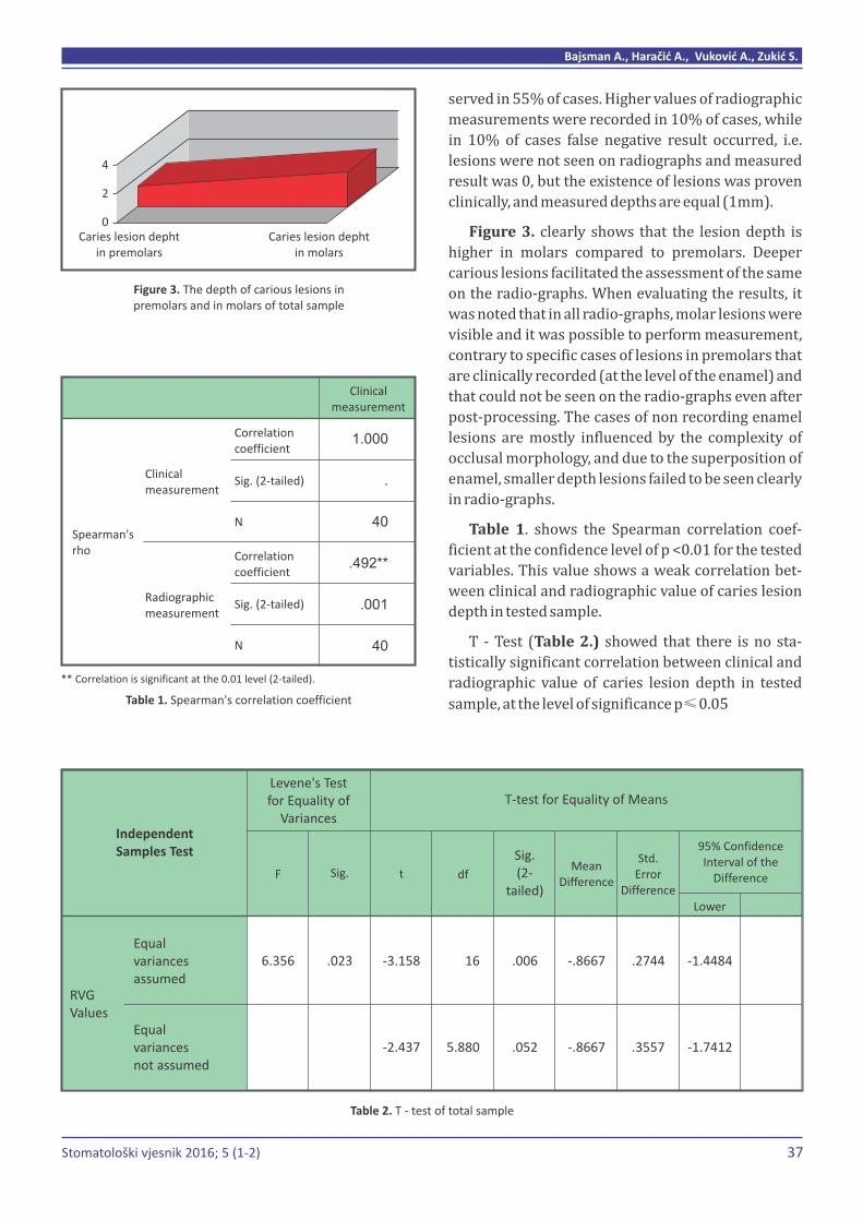
37
lesions was clinically observed, and their depth, in
both cases, was 1 mm. Possible reasons for the inabi-
lity to observe these lesions could be the existence of
errors in the exposition, which resulted in imprecise
recording, small depth of lesions, which are still in the
enamel, or subjectivity of interpreter of radiograph.
Figure 1. clearly illustrates noticeably higher va-
lues of clinical measurements compared to radio-
graphic measurement in molars of total sample.
Concordance of results is present in only 15% of ca-
ses. Also, higher valuse of radiographic measure-
ments, compared to clinical measurements were
recorded in 15% of cases. The remaining 70% of ca-
ses recorded higher values of clinical measurements.
Figure 2. presents evidently higer values of
clinical measurements, compared to radiographic
measurements in premolars of total sample. How-
ever, the difference between variables in premolars is
lower than in molars. In 25% of cases there is a
concordance of results of clinical and radiographic
measurements, i.e. 10% higher than in molars.
Higher values of clinical measurements were ob-
served in 55% of cases. Higher values of radiographic
measurements were recorded in 10% of cases, while
in 10% of cases false negative result occurred, i.e.
lesions were not seen on radiographs and measured
result was 0, but the existence of lesions was proven
clinically, and measured depths are equal (1mm).
Figure 3. clearly shows that the lesion depth is
higher in molars compared to premolars. Deeper
carious lesions facilitated the assessment of the same
on the radio-graphs. When evaluating the results, it
was noted that in all radio-graphs, molar lesions were
visible and it was possible to perform measurement,
contrary to specific cases of lesions in premolars that
are clinically recorded (at the level of the enamel) and
that could not be seen on the radio-graphs even after
post-processing. The cases of non recording enamel
lesions are mostly influenced by the complexity of
occlusal morphology, and due to the superposition of
enamel, smaller depth lesions failed to be seen clearly
in radio-graphs.
Table 1. shows the Spearman correlation coef-
ficient at the confidence level of p <0.01 for the tested
variables. This value shows a weak correlation bet-
ween clinical and radiographic value of caries lesion
depth in tested sample.
T - Test (Table 2.) showed that there is no sta-
tistically significant correlation between clinical and
radiographic value of caries lesion depth in tested
sample, at the level of significance p≤0.05
36
Figure 2. The values of clinical depth of caries lesion measured by graduated probe, and the values obtained by measuring the area of radiolucent zone corresponding to the carious lesion on radiographs using ImageJ, in premolars of total sample
Figure 3. The depth of carious lesions in premolars and in molars of total sample
1
1
1,5
1
2,3
2,5
2
1,7
2
2
2,1
3
3
1,1
1,5
3
2,7
3
4
1,5
1,5
4
1,6
2
5
1
1
5
2
2
6
1,5
1,5
6
3
3
7
1,2
1
7
1,3
2
8
1,2
1,5
8
2,1
1,5
9
2,1
1
9
1,7
2
10
1,3
1,5
10
1,8
2
11
1,5
1,5
11
2,7
2
12
1,3
1,5
12
2,1
2
13
0
1
13
1,7
2
14
1,6
2
14
2,2
3
15
1,2
1,5
15
1,8
2
16
1,5
1
16
1,8
2,5
17
1,2
1,5
17
2,1
3,5
18
1,4
1,5
18
2
2,5
19
0
1
19
2
2
20
1,5
1,5
20
2,1
2,5
2,5
2,0
1,5
1,0
0,5
0,0
4
3
2
1
0
Radiographic measurement
Clinical measurement
Radiographic measurement
Clinical measurement
4
2
0Caries lesion depht
in premolarsCaries lesion depht
in molars
Table 1. Spearman's correlation coefficient
Table 2. T - test of total sample
ANALYSIS OF CONCORDANCE BETWEEN CLINICAL AND RADIOGRAPHICAL DEPTH OF OCCLUSAL CARIES LESIONS – A PILOT STUDY
Correlationcoefficient
** Correlation is significant at the 0.01 level (2-tailed).
Spearman'srho
Clinicalmeasurement
Clinicalmeasurement
Radiographicmeasurement
Sig. (2-tailed)
N
Sig. (2-tailed)
N
Correlationcoefficient
1.000
.
40
.492**
.001
40
Figure 1 The values of clinical depth of caries lesion measured by graduated probe, and the values obtained by measuring the area of radiolucent zone corresponding to the carious lesion on radiographs using ImageJ, in molars of total sample
IndependentSamples Test
Levene's Testfor Equality of
Variances
T-test for Equality of Means
F Sig. t df
Sig.(2-
tailed)
MeanDifference
Std.Error
Difference
95% ConfidenceInterval of the
Difference
Lower
RVGValues
Equalvariancesassumed
6.356 .023 -3.158 16 .006 -.8667 .2744 -1.4484
-2.437 5.880 .052 -.8667 .3557 -1.7412Equalvariancesnot assumed
Bajsman A., Haračić A., Vuković A., Zukić S.
Stomatološki vjesnik 2016; 5 (1-2) Stomatološki vjesnik 2016; 5 (1-2)

39
Discussion
The morphology of the occlusive surfaces or
three-dimensional appearance of the fissure system
affects the formation of occlusive caries. Salman [11]
in his research of the impact of occlusive morphology
on the severity of occlusive caries points out that
there is a statistically significant correlation at the
level p≤0.001.
Valera et al [2] state that there is no statistically
significant difference between the premolars and
molars related to the presence or absence of pits and
fissures. Both in premolars and molars caries pre-
valence depends on the occlusive morphometry, with
a higher prevalence when the pits and fissures are
present. Analyzing the results of the study we can
conclude that the occlusive surfaces of molars are
largely destroyed with caries lesions than it is the
case with premolars. A complex occlusive surface of
molars enables retaining debris. Due to the presence
cusps and fissures, food remains are harder removal
mechanically, which favors the metabolic processes
in the biofilm. However, premolars are in a slightly
better position in the dental arch in relation to the
molars. Their position allows easier implementation
of oral hygiene, preventive measures and self-clea-
ning tooth surfaces compared to molars, which pre-
vents longer retention of biofilm and development of
caries lesion.
According to several studies (Carvalho et al 1989,
Ekstrand et al 1995) caries lesion usually begins in
the central fissure. On the other hand, following a
series of studies, carious lesions were mainly
detected in the pits and fissures (Gustafson, Nagano i
König) [2].
However, both groups of researchers agree that
the beginning and progression of carious lesion is in
connection with macro morphology of occlusive
surface.
There is no doubt that the complexity of the occlu-
sive surface favors the development of carious le-
sions and also constitutes an aggravating circum-
stance for adequate and timely diagnosis. Reviewing
the literature and analyzing the results of various
studies [12], we found statements that are proven in
this study. It is concluded that the efficacy of clinical
method is similar to radiographic methods, with
clinical methods having a lower number of false - ne-
gative results compared to the radiographic method.
Da Silva et al [13] compared the accuracy of direct
digital radiography with conventional radiography in
the analysis of occlusive caries in primary molars.
Taking into account the sensitivity and specificity, the
study showed that direct digital radiography has a
similar level of accuracy as conventional radiography.
Visual inspection showed greater accuracy of both
radiographic methods when it comes to occlusive ca-
ries lesions in the enamel, and digital radiography
had a similar effect as conventional radiography and
visual inspection for dentinal lesions. These results
correlate with the results of this study. We had several
clinically diagnosed and measured lesions (enamel
lesions) which were not visible, or were hardly visi-
ble on radio-graphs, after post-processing. When it
comes to lesions in dentin, they are clearly observed
on radio-graphs, and there is less discrepancy in va-
lues of clinical and radiographic depth.
The results of this study show that radiovisio-
graphs underestimate the depth of carious lesions. It
is evident that in most cases the values of clinical
measurements of carious lesions are higher compa-
red to the measurements of radiolucent zone in ra-
diographs. There are very few matching values obtai-
ned by clinical measurement and measurement on
digital radiographs. Discrepancies in measurements
could be explained by subjective observation during
measurement. There is also the possibility of error
due to post-processing of digital radiographs. Discre-
pancies of the measurement results are not large, but
they are present in 80% of the sample.
There are cases in this study where it was not
possible to observe lesions on digital radiographs.
The existence of these lesions was clinically proven
and they were measured using graduated probe, and
recorded depth in both cases was 1 mm. The reason
for the inability to observe these lesions may be the
possible existence of errors in the exposition, which
resulted in imprecise recording, small depth of
lesions, which are still in the enamel, or subjectivity
in interpreting digital radiographs.
In a study that compares two digital radiography
systems and conventional radiography in the diag-
nosis of occlusive and proximal caries, Da Rocha et al
[14] point out that there is no significant difference
between the digital systems (Digora® i DenOptix®)
and conventional films in diagnosis of occlusive
caries. It is also concluded that there are significant
variations between examiners. In this study, a group
of examiners was made up of students, and slight
variations have been reported within this group, with
levels of 0.43 to 0.55. However, comparing the values
obtained by the students with another group of exa-
miners made up of radiologists, significant variations
were observed, with levels of 0.28 to 0.54. This out-
come supports the statement that, in the interpreta-
tion of images, significant factor is the subjectivity of
each examiner.
Conclusions
The depth of occlusive caries lesion is noticeably
higher in molars comparing to premolars. Larger
depth of caries lesion facilitates its coping and
assessment in digital radio-graphs. Digital radio-
graphs underestimate the depth of carious lesions.
There are very few matches of clinical measurements
of caries lesion depth using graduated probe, and
measurements on digital radio-graphs, using ImageJ
software. Discrepancies of results are not large, but
are present in 80% of cases. Difficult radiological
diagnosis of occlusive caries lesions is justified by
rich relief of occlusive surface, with various elements
superimposed on radio-graphs, as well as possible
presence of filling materials. Radiography is a
valuable diagnostic tool. Although there is still no
perfect way to diagnose using radiography, it is
believed that, due to the rapid and unstoppable
progress of technology in medicine, we are on the
right track towards finding the ideal radiographic
diagnostic system.
Declaration of interest
Authors declare no conflict of interest.
References
1. Burrow MF, Burrow JF, Makinson OF. Pits and
fissures: etch resistance in prismless enamel
walls. Australian Dental Journal. 2001;46(4):258
– 262.
2. Valera F et al. Morphometric analysis of the
occlusal surface: the influence on the prevalence
of carious lesions. Salusvita 2005;24:301-308.
3. McComb D, Tam LE. Diagnosis of Occlusal Caries:
Part I. Conventional Methods. Journal of Canadian
Dental Association 2001;67(8):454 – 457.
4. Bloemendal E, de Vet HCW, Bouter LM. The value
of bitewing radiographs in epidemiological caries
research: a systematic review of the literature.
Journal of Dentistry.2004;32:255 – 264.
5. Wenzel A. Bitewing and digital bitewing radio-
graphy for detection caries lesions. Journal of
Dental Research 2004;83(Spec Iss):C72 – 75
6. Forner Navarro L, Llena Puy MC, Garcia Godoy F.
Diagnostic performance of radiovisiography in
combination with a diagnosis assisting program
versus conventional radiography and radiovisio-
graphy in basic mode and with magnification.
Med Oral Pathol Oral Cir Bucal 2008;13(4):261 –
265.
7. American Dental Association Council on Scien-
tific Affairs. The Use of Dental radiographs; Up-
date and recommendations. Journal of American
Dental Association 2006;137:1304 – 1312.
8. Gormez O, Yilmaz HH. Image Post – Processing in
Dental Practice. European Journal of Dentistry
2009;3:343 – 347.
9. Bader JD, Shugars DA, Bonito AJ. Systematic
reviews of selected dental caries diagnostic and
management methods. Journal of Dental Educa-
tion 2001;10:960-968.
10. Pereira AC, Eggertsson F, Moustafa A, Zero DT,
Eckert GJ, Mialhe FJ. Evaluation of three radio-
graphic methosd for detecting occlusal caries
lesions. Brazilian Journal of Oral Sciences
2009;2:67-70.
11. Salman FD. The effect of fissure morphology on
caries severity of adults in north Iraq. Al – Taqani
2011;24:29-37.
ANALYSIS OF CONCORDANCE BETWEEN CLINICAL AND RADIOGRAPHICAL DEPTH OF OCCLUSAL CARIES LESIONS – A PILOT STUDY
38
Bajsman A., Haračić A., Vuković A., Zukić S.
Stomatološki vjesnik 2016; 5 (1-2) Stomatološki vjesnik 2016; 5 (1-2)

ANALYSIS OF CONCORDANCE BETWEEN CLINICAL AND RADIOGRAPHICAL DEPTH OF OCCLUSAL CARIES LESIONS – A PILOT STUDY
40
12. Mestriner SF, Vinha D, Mestriner WJ. Comparison
of different methods for the occlusal dentine
caries diagnosis. Journal of Applied Oral Science
2005;13:28-34.
13. Dias da Silva PR, Martins Marques M, Steagall W,
Medeiros Mendes F, Lascala CA. Accuracy of
direct digital radiography for detecting occlusal
caries in primary teeth compared with conven-
tional radiography and visual inspection: an in
vitro study. Dentomaxillofacial Radiology
2010;39:362–367.
14. Da Rocha ASP, de Almeida SM, Boscolo FN, Haiter
Neto F. Interexaminer agreement in caries radio-
graphic diagnosis by conventional and digital ra-
diographs. Journal of Applied Oral Science
2005;13:329 – 333.
41
REVIEW ARTICLE / PREGLEDNI ČLANAK
DISCOLORATION OF DECIDUOUS AND
PERMANENT TEETH: CLASSIFICATION,
ETIOLOGY AND TREATMENT OPTIONS
1 2Selimović-Dragaš M.* , Kovačević N.
1 Department of Preventive and Pediatric Dentistry, Faculty of Dentistry, University of Sarajevo, Sarajevo, Bosnia and Herzegovina2 Public Health Center Lukavac, Department for Dental Health Care, Kakanj, Bosnia and Herzegovina
ABSTRACT
The aim of this review is to present various causes of deciduous
and permanent teeth discoloration, their classification and
treatment options. Having in mind the differences but also the
similarities in clinical features of different types of tooth staining, it
is very important to know and understand the mechanisms behind
tooth discolorations. That's why this review offers a new
classification based on etiological background and a clinical
appearance of tooth discoloration, considering it more suitable for
the proper selection of a treatment. Color and the shape of the teeth
are integrated part of facial aesthetics which plays a very important
role in the complex social, cultural and psychological interaction of
every person. Therefore, it is important that doctors of dental
medicine understand the background of different tooth
discolorations so, in cases with aesthetic problems, they are able to
offer proper information for the patients with the aim to include
them in the treatment planning.
Key words: tooth discoloration, extrinsic discoloration, intrinsic
discoloration, internalized discoloration.
*Corresponding author
Mediha Selimović-Dragaš, PhD
Bolnička 4a
71000 Sarajevo
Bosnia and Herzegovina
Phone: +387 33 214-249 (ext.338)
e-mail address:
Faculty of Dentistry
Stomatološki vjesnik 2016; 5 (1-2) Stomatološki vjesnik 2016; 5 (1-2)

42
DISCOLORATION OF DECIDUOUS AND PERMANENT TEETH: CLASSIFICATION, ETIOLOGY AND TREATMENT OPTIONS
Introduction
The tooth can be described as an anatomic and
functional complex of different tissues: enamel, den-
tin, pulp and cementum. Any changes of these struc-
tures will change its light transmitting and reflecting
properties which will result as an alteration in the
outward appearance of the tooth [1]. Tooth color re-
presents a range of colors with the visible gradation,
from the gingival margin to the incisal edge. Due to
the proximity of dentin, the tooth is darker in the
gingival area, canines are darker than the central and
lateral incisors, and younger people, especially in the
primary dentition, characteristically have lighter
teeth [1]. The color of the teeth is determined by the
yellowish color of dentin, modified by the thickness
and degree of mineralization of semitransparent ena-
mel [2].
Less mineralized and thus less transparent ena-
mel make the color of deciduous teeth bluish–white
comparing to the yellowish-white color of perma-
nent teeth caused by the better mineralized and bet-
ter light-permeable enamel of permanent teeth [2].
Any change in tooth color, different from the usual,
can be marked as a discoloration of teeth. Having in
mind the large number of variations in discoloration
of teeth, correct diagnosis of the cause of discolora-
tion is very important for the plan and the result of
therapy.
The aim of this review is to present a various cau-
ses of deciduous discoloration,
Materials and methods
An extensive search of PubMed data base for clini-
cal and in vitro studies in English, published between
1969. and 2015. was performed, using different com-
bination of key words: tooth discoloration, extrinsic
discoloration, intrinsic discoloration and internali-
zed discoloration. Aside from the time frame and
English dental literature, inclusive criteria for papers
were: online full-text available, detailed information
on study methodology and relevance for dental clini-
cal practice. Included studies were: clinical trials, cri-
tical reviews, longitudinal studies and case reports.
and permanent teeth
based on contemporary achievements in the process
of diagnosis of their etiology, their classification and
treatment options.
Clinical trials and recent reviews were considered
with highest quality value and in the cases where was
limited literature to find on a topic, case reports were
used. Independent screening of 63 unique titles
identified 40 studies that met the eligibility criteria.
Review of current literature and discussion
Tooth discoloration may be divided into three
main types, intrinsic, extrinsic and internalized.
The discolorations on the tooth surface or in the
acquired pellicle, caused by exogenous pigments we-
re defined as an extrinsic discoloration [1, 3]. Based
on cause, extrinsic discoloration can be divided into
direct and indirect tooth discoloration [3] (Table 1.).
The stain, in direct staining, is a result of the basic
color of the chromogen caused by compounds incor-
porated into the pellicle layer. Direct staining has
multi-factorial etiology, mainly caused by the chro-
mogens derived either from the diet or substances
habitually placed in the mouth. Accumulation of bac-
terial plaque is also classified as this type of dental
staining.
Indirect staining is caused by a chemical interac-
tion on the tooth surface usually associated with local
or systemic usage of different medicaments [3]. Indi-
rect staining can be a result of occupational metal
intoxication or environmental metal intoxication.
Gorlin and Goldman (1971.) classified extrinsic
tooth discoloration according to its origin, as metallic
or non-metallic [1]. In1997. Nathoo proposed classi-
fication of extrinsic tooth discoloration as a three ca-
tegories: Nathoo tip I, Nathoo tip II, i Nathoo tip III [4].
Intrinsic tooth discoloration was caused by chro-
mogens which have endogenous or exogenous origin
and it can be defined as a staining incorporated in
tooth structures during odontogenesis (pre-erup-
tive) or after odontogenesis (post-eruptive) [5].
(Table 2.)
Internalized discolorations are tooth discolora-
tion formed during the tooth development, when ex-
trinsic stain is incorporated within the tooth sub-
stance [1,3].
In literature, internalized discoloration was de-
scribed as a developmental internalized discoloration
when extrinsic stain was incorporate within the
tooth substance following dental development or
acquired internalized discoloration where changes
on teeth or surrounding tissues, during the life, di-
rectly or indirectly affect the change of tooth color
[1].(Table 3.)
For a dental practitioner knowing and understan-
ding the etiology of tooth discoloration is of great
importance because the correct diagnosis for the
cause of discoloration has a profound effect on
treatment outcomes. From the other side, it is impor-
tant to explain to the parents the origin of dental
discoloration in order to enable the appropriate
treatment to be carried out.
Intrinsic, extrinsic and internalized discoloration
clinically can be presented in eleven colors: white,
black, blue, brown, green, grey, yellow, orange, pink,
red and purple.
White tooth staining
Materia alba is a soft, mushy accumulation of bac-
teria, desquamated epithelial cells and salivary pro-
teins on the outer surface of the tooth. White in color,
it can be removed with a powerful jet of water [6].
Active early caries lesion is chalky-white with
matte surface. Two or three weeks old incipient le-
sions can be visible only after the enamel is dry, while
older lesions are seen on wet enamel. In this stage of
early caries lesion discrete porosity on enamel sur-
face also can be seen [7].
Selimović-Dragaš M,. Kovačević N.
43
Table1. Extrinsic discoloration, causes and types of stains
Color
White
Yellow
White, yellow, brown
Green
Brown, black, orange, blue, green
Brown, black
Greenish
Dark brown, black
Red-brown stain
Yellowish-brown
Dark brown stains
Yellowish-brown
Yellow
Green
Violet to black
Grey, black
Yellow-brown
Black
Yellow-brown
Yellow-brown
Brown
Black stain
Blue-green
Green stain
Deep orange stain
CausesClasification
Materia alba
Dental plaque/biofilm
Dental calculus
Chromogenic fungi
Chromogenic bacteria
Chromogens, acidic food. Intake of coffee,
red vine and tea The color is due to tannin.
Chlorophyll-Rich Diet
Tobacco smoking/chewing tobacco
Chewing of pan ( a combination of betel nut
of the areca palm, betel leaf, and lime)
Chewing of Khat leaves (Catha edulis)
Chewing of nuts of Cola plant (Cola nitida)
Cationic antiseptics (chlorhexidine)
Essential oils / phenol solutions
Mouth rinses containing Copper salts
Potassium permanganate
Silver nitrate
Stannous fluoride
Iron containing oral solutions
Co-amoxiclav
Doxycycline
Linezolid
Iron , Silver and manganese dust
Mercury and lead dust
Copper and Nickel
Chromic acid,fumes
Etiological factors
DENTAL DEPOSITS
CHROMOGENIC MICROORGANISMS
DIETARY HABITS
SUBSTANCES HABITUALLYPLACED IN THE MOUTH
LOCAL USAGE OF MEDICAMENTS
ORAL ADMINISTRATION OF DRUGS
OCCUPATION AND ENVIRONMENTAL FACTORS
IN
DI
RE
CT
DI
RE
CT
Stomatološki vjesnik 2016; 5 (1-2) Stomatološki vjesnik 2016; 5 (1-2)

45
ment, leading to enamel with lower mineral content
and increased porosity. The clinical appearance may
vary based on the severity from areas of white, opa-
que dots or stripes that occur on homologous teeth to
diffuse opaque mottling superimposed on chalky
white or dark brown/black areas. Opaque areas of
enamel are porous (hypomineralized), and the
degree of porosity depends on the concentration of
fluoride in the body fluids during tooth development
[9].
Amelogenesis imperfecta (AI) is a group of here-
ditary disorders, clinically and genetically hetero-
geneous, characterized by abnormal amelogenesis,
affecting both primary and permanent dentitions
[10]. Witkop in1988 presented the most widely used
Enamel defects are white colored but with the ad-
herence of pigments they become yellow or yellow-
brown and they always show a sharp demarcation
between sound and affected enamel. Enamel defects
on a single tooth or only a few teeth suggest a local
etiological factor. A systemic factor (both short and
longer term) may affect all the teeth that are deve-
loping during the time of the insult and lead to what is
described as a chronological defect [1]. The preva-
lence of developmental defects of enamel (DDE) at 12
years of age in Bosnia and Herzegovina was 32, 80%,
with significantly more DDE in Banja Luka [8].
Dental fluorosis is a structural disturbance of den-
tal enamel, caused by successive exposures to high
concentrations of fluoride during tooth develop-
classification system. Based on phenotypes and the
inheritance patterns AI is divided into four main
types and then into 15 subtypes that was clinically
described by Ng and Messer 2009. [11]. Ameloge-
nesis imperfecta type II and type IV, clinically can be
manifested as white colored enamel [12].
Black tooth staining
Theilade at al.(1973.) put forward the view that
external discoloration depend on oral hygiene, accor-
ding to which black coloration was associated with
children with good oral hygiene and low caries risk
[13].
Bandon at al. claimed that actinomyces, porphyro-
monas gingivalis and prevotella melaninogenicus
presents the microorganisms which are predomi-
nant in black tooth discolorations [14]., while Saba at
al. found that actinomyces species were dominant
comparing with other microorganisms [15]. Staining
was result of presence of ferric sulphate made by
reaction between hydrogen sulphide, which is a bac-
terial product and iron present in saliva and gingival
fluid [16].
There is a positive correlation between the
external discolorations and presence of trace
elements, particularly iron in water [3]. Exposure to
iron, manganese and silver can cause black external
discoloration of the teeth [3]. .
Hard inactive caries lesion and dental fluorosis
can be black due to adherence of the pigment from
the environment [3].
Epoxy resin-based sealers such as the AH26 can
cause black discoloration of the teeth, after a few
years of placement [17].
Medications, such as minocycline, aside from the
characteristic blue-gray color, can stain root of the
tooth, during its formation, in black color [18].
Blue, blue-green, blue-brown and
blue-grey tooth staining
Blue tooth staining can be found in children with Syndroma Wast [19]. According to one case report,
this staining was extrinsic caused by chromogenic
bacteria pseudomonas aeruginosa [19]. Rarely, blue
tooth staining can be found at patients with Parkin-
son's disease and cyanotic congenital heart diseases
[18].
Mercury intoxication and intoxication with lead
dust was a main cause of blue-green extrinsic disco-
loration of teeth, mainly as a result of bad oral hygie-
44
DISCOLORATION OF DECIDUOUS AND PERMANENT TEETH: CLASSIFICATION, ETIOLOGY AND TREATMENT OPTIONS
Table 2.Classification of intrinsic tooth discoloration, causes and types of stains
Table 3. Classification of internalized discoloration according to origin of the stains
Color
Color
White, yellow, brown, yellowish-brown
White, yellow, brown, yellowish-brown
Yellowish-brown, blue-grey
Yellow, brown, grey
Yellowish-brown, blue-grey
Yellow
Yellow-grey, dark brown
Red, reddish-brown
Brown
Yellow, green, yellow-green, brown
Yellow, grey-brownYellow
Blue-grey
Green Chalky-white, yellow-brown, dark brown, black
Chalky-white, brown, black Yellow
Yellow Yellow
Pink Blue-gray
Pink Yelow-brown
Grey, brown Yellow, grey, brown
Yellow, yellowish-brown Grey, brown, black, pink
Pink
Blue-greyGrey-brown
Causes
Causes
Clasifi-cation
Clasifi-cation
Hypomineralization of enamel (demarcated and diffuse opacities)
Hypoplasia of enamel (generalized and localized)
Molar Incisor Hypomineralization (MIH)
Amelogenesis imperfecta (AI)
Dentinogenesis imperfecta (DI)
Dysplasia dentinum (DD)
Osteogenesis imperfecta (OI)
Epidermolysis bullosa
Cystic fibrosis (CF)
Pre-eruptive incorporation of extrinsic stain within the tooth substance following dental development.It occurs in enamel defects like hypomineralization and hypoplasia and in the porous surface of exposed dentine. The enamel defects can vary from white to yellow to brownish areas and they always show a sharp demarcation between sound and affected enamel.
Congenital erythropoietin porphyria (CEP)
Alkaptonuria (AKU)
The breakdown products of haemolysis will lead to deposition of bile pigments in the calcifying dental hard tissues, particularly at the neonatal line.
Tetracycline Progressive loss of enamel and dentin due to attrition, abrasion and erosionMinocycline
Ciprofloxacin Dental caries
Dental fluorosis Physiological involution of tooth
Physiological involution of tooth Gingival recession
Cervical root resorption Amalgam
Pulpal hemorrhage Composite/GIC
Necrosis of dental pulp Intracanal medicaments
Obliteration of dental pulp Root canal sealers and obturating material
Internal root resorption
Minocycline
Incomplete removal of pulpal reminiscent during root canal treatment
Etiological factors
Etiological factors
NON-GENETIC DISTURBANCE OF TOOTH GERM
GENETIC DISORDER
METABOLIC DISORDERS
PRE-ERUPTIVE CAUSES OF INTERNALIZED TOOTH DISCOLORATION
CONGENITAL HYPERBILIRUB INAEMIA
CHANGES OF THE DENTAL HARD TISSUES
MEDICAMENTS
PERIODONTAL CHANGES
CHRONIC FLUORIDE INTOXICATION
CHANGES IN THE DENTAL HARD TISSUES
PULPAL CAUSES
DENTAL MATERIALS
MEDICAMENTSIATROGENIC FACTORS
PO
ST
ER
UP
TI
VE
AC
QU
IR
ED
PR
EE
RU
PT
IV
E
DE
VE
LOP
ME
NTA
L
Selimović-Dragaš M,. Kovačević N.
Stomatološki vjesnik 2016; 5 (1-2) Stomatološki vjesnik 2016; 5 (1-2)

46
Incomplete metabolism of tyrosine and pheny-
lalanine at the patients with alkaptonuria, results
with the excessive amount of homogentisic acid that
are deposited in the body and the teeth. This affects
the permanent dentition by causing a brown disco-
loration [1].
Dark brown tooth discoloration observed in
patients with cystic fibrosis (CF), autosomal reces-
sive disorder, can be related to ingestion of tetra-
cycline during tooth formation or to the metabolic
effects of the disease itself [23].
Pigmentation of the primary dentition may result
from hemolytic anemia of the fetus or newborn. He-
mosiderin, a degradation product of hemoglobin, is
deposited in forming dentin and enamel, causing
brown color of the teeth [2].
Arrested carious lesion is manifested as destruc-
tion of both enamel and dentin with the color that
ranges from light brown, to dark brown or almost
black and the nature of this discoloration is not
known yet. It is suggested that this color is exogenous
in origin deriving from chromogenic bacteria and
dietary habits. [3].
The brown/black discoloration of enamel, which
can be seen at dental fluorosis, enamel hypoplasia
and molar-incisor hypomineralization (MIH) is post-
eruptive and probably caused by the internalization
of extrinsic stain into the porous enamel [3].
Yellow and yellow-brown tooth discoloration
The most common yellowish tooth discoloration
is a result of precipitation of dietary chromogens
onto tooth surface deposits such as acquired pellicle
or the plaque. Dental plaque formed on tooth surfa-
ces is a complex ecosystem composed of diverse oral
bacteria and salivary components. If it is in the form
of acquired pellicle it can be seen after application of
various kind of disclosure agents or by the probe.
Excessive amount of dental plaque can be seen by
naked eye as a yellowish stain on tooth surface [6].
Dental calculus is calcified dental plaque, compo-
sed primarily of calcium phosphate mineral salts
deposited between and within remnants of formerly
viable microorganisms. Supragingival dental calcu-
lus formation is placed next to ginigival margin most-
ly at tooth surfaces adjacent to the salivary ducts,
colored yellow or yellow-brown [6].
Bad habits like smoking and chewing tobacco or
chewing of Khat leaves, associated with bad oral
ne [3], while blue-green intrinsic discoloration was
result of hemolytic disease of the newborn [20].
Minocycline is a semi-synthetic derivative of
tetracycline. Its prolonged ingestion can lead to
green-gray or blue-gray intrinsic staining of the teeth
during and after the complete formation and erup-
tion of teeth [3].
Crowns of teeth with dentinogenesis imperfecta
usually have a normal size. In the cervical portion of
the tooth there is a constriction that gives the crown a
characteristic bulbous appearance. Teeth with denti-
nogenesis imperfecta show a variety of colors, most
often a blue-gray or bluish brown, which demon-
strate opalescence on transillumination [3, 18].
Longstanding amalgam restorations can generate
stannous corrosion products, which migrates into
the dentinal tubules leaving a blue-gray, dark grey to
black color of dentine, especially in large cavity
preparations with undermined enamel [3, 21].
Intracanal medicaments, as triple antibiotic paste
(TAP), which contains ciprofloxacin, metronidazole
and minocycline according to case report can, cause a
blue-gray tooth discoloration [22].
Brown tooth staining
Chromogenic bacteria considered being a cause of
brown extrinsic stains, placed typically at the gingival
margin of the tooth, in children with good oral hygi-
ene and low caries experience [13].
Intake of coffee and tea can cause brown extrinsic
tooth discoloration due to tannin. The color seen on
the tooth is thought to be derived from polyphenolic
compounds that provide the color in food [3].
Some systemic medications are also shown to
cause brown extrinsic staining. One case report show
that linezolid 600 mg. of dose, which was ordinate
two times a day during four weeks, caused brown
extrinsic staining in a 11 year old girl [3].
Dark brown extrinsic stains, located on cervical
third or even one half of tooth surface can be the
result of tobacco from cigarettes, cigars, pipes, and
chewing tobacco. This pigmentation is the result of
the tar taken up by the pellicle, whose amount
depends not on the amount of smoking but on the
amount of present plaque, and during the time it
penetrates into the enamel [16].
Brown stains on the surface of the teeth also could
be due to the deposition of caffeine, tannins or
theobromine that is result of daily chewing of nuts of
Cola plant [16].
hygiene can cause yellowish-brown extrinsic tooth
discoloration [16]
Some genetic defects in enamel or dentin forma-
tion including amelogenesis imperfecta, dentinoge-
nesis imperfecta, dentinal dysplasia , molar incisor
hypomineralization ( MIH), osteogenesis imperfecta
(OI) and hypoplasia of enamel can be associated with
yellow to yellow-brown tooth discoloration.
Amelogenesis imperfecta, hereditary condition
with thin, hard enamel with more severe hereditary
hypoplasia has a yellow to yellow-brown appearance
With prevalence of 1:700 children in Europe to
1:14000 children in America, it can be seen alone or
as a symptom in some sindroms like Syndroma
Tricho-dento-osseus, Syndroma Kohlshütter-Tönz,
Syndroma McGibbon and Syndroma Jalili [24, 5, 2].
Dentinogenesis imperfecta (DI), autosomal domi-
nant genetic conditions is characterised by abnormal
dentine structure affecting either the primary or both
the primary and secondary dentitions. According to
the most current classification adopted by the
Mendelian Inheritance in Man (MIM) dentinogenesis
imperfecta type I or dentinogenesis imperfecta type
II (classification system formulated by Shields) re-
ported to have an incidence of 1 in 6,000 to 1 in 8,000
newborn children and can be connected with a
yellow-brown tooth discoloration [25,26].
According to current MIM classification DI-I can
be considered as a variable feature of Osteogenesis
imperfecta (OI), Syndroma Ehles-Danlos, Syndroma
Goldbatt, Syndroma Brachio-Skeletal-Genitalium
and Schimke immuno-osseous dysplasia [26].
Dentine dysplasia type II (Shields classification),
is an autosomal dominant genetic conditions which
has different clinical features depending on dentition
[3]. While permenent dentition can have a brown dis-
coloration of the teeth, primary dentition is usually
yellow to yellow-brown or has amber translucency
like the one at dentinogenesis imperfecta [2].
Molar-incisor hypomineralization (MIH] is an
idiopathic condition characterized by severe hypo-
mineralized enamel affecting incisors and perma-
nent first molars. Clinical features of enamel defects
can vary from white to yellow to brownish areas and
they always show a sharp demarcation between
sound and affected enamel [3].
The prevalence of MIH in 12-year-olds in Bosnia
and Herzegovina was 12, 3%, and study from 2009
showed that the prevalence of MIH in children from
Sarajevo, aged 12 was 11,5% [8, 27].
Enamel hypoplasia (EH) is defined as a deficiency
of enamel formation. A variety of environmental and
genetic factors have been shown to contribute to the
formation of these defects, including malnourish-
ment, mechanical trauma or inflammation. Enamel
hypoplasia can be localized or generalized and its
colour varies from white, yellow to brownish [8].
Epidermolysis bullosa (EB) is a rare group of inhe-
rited disorders that manifests as blistering or erosion
of the skin and, in some cases, the epithelial lining of
other organs, in response to little or no apparent
trauma. Patients with epidermolysis bullosa can have
a pitting of the enamel possibly caused by vesicu-
lation of the ameloblast layer which demonstrates a
yellow tooth discoloration [3].
Mouth washes such as chlorhexidine can cause a
yellow–brown extrinsic tooth discoloration. Having
in mind that those mouth washes are cationic anti-
septics most evidence indicates that the cause of stai-
ning is the precipitation of anionic dietary chromo-
gens onto the adsorbed cations [16]. Mouthwash that
contains stannous fluoride will often stain teeth in
gold-yellow color [3].
Essential oils or phenol solutions, if used, also can
be considered as a cause of yellow extrinsic tooth
discoloration [3].
Tetracycline, oxytetracycline and demethylchlor-
tetracycline are known to cause intrinsic discolo-
ration when prescribed during tooth development.
Teeth affected by tetracycline may have a yellowish
appearance that fluoresces under ultraviolet light,
giving off a bright yellow color [23].
Some cases presented in literature confirmed that
yellow-brown extrinsic tooth discoloration can be
found as a complication after a long-term usage of
doxycycline 200mg/ day. Poor oral hygiene and in-
tense sunlight exposure may be exacerbating factors
in prominent staining of permanent dentition caused
by doxycycline [28].
Several intracanal medicaments based on phenols
or iodoform, sealed in the root canal are in direct
contact with dentin. Penetration and oxidation of the
materials in dentinal tubules will result with yellow
or yellow –brown tooth discoloration [22].
A study by Mjor and Toffenetti reported that
yellow-brown margin discoloration suggests inade-
quate acid-etching of the enamel prior to placing the
bond agent, inadequate handling of the material
(placing, concentration, adaptation) and problems
associated with polymerization shrinkage [29].
47
DISCOLORATION OF DECIDUOUS AND PERMANENT TEETH: CLASSIFICATION, ETIOLOGY AND TREATMENT OPTIONS Selimović-Dragaš M,. Kovačević N.
Stomatološki vjesnik 2016; 5 (1-2) Stomatološki vjesnik 2016; 5 (1-2)

Pulp canal obliteration occurs commonly follo-
wing traumatic injuries to teeth. Radiographically
pulpal obliteration is characterized by the apparent
loss of the pulp space and yellow to yellow-brown
crown discolouration is a common clinical finding in
teeth with pulp canal obliteration [12].
As a result of progressive loss of enamel and den-
tin due to attrition, abrasion and erosion, enamel
thins and the tooth becomes darker as the color of the
dentin becomes more apparent. At that stage tooth
has yellow to dark yellow color. Once the dentin is
exposed, the potential for chromogens to enter the
body of the tooth increases and tooth color becomes
darker [3].
During the process of ageing, the enamel under-
goes both thinning and textural change with the
change in its light transmission properties. The natu-
ral darkening and the yellowing of the teeth, which
occur with age, can be due to the combination of the
factors involving both enamel and dentin. Deposi-
tions of secondary and tertiary dentin and pulp sto-
nes are the factors that contribute to the darkening of
the teeth as a result of aging [3].
Yellow-green tooth discoloration
A yellow-green tooth discoloration can be seen at
the cases of Congenital hyperbilirubinaemia when
the level of bilirubin in blood exceeds 30mg/100ml.
This will lead to deposition of bile pigments in the
calcifying dental hard tissues, particularly at the
neonatal line [30].
Green tooth discoloration
According to Theilade and associates [13] green
extrinsic discoloration in children are usually asso-
ciated with poor oral hygiene. Chromogenic bacteria
have also been suggested as an etiological factor in
the production of this kind of stains that are more
common in boys and have a tendency to reappear
after complete removal [16].
Chromogenic bacteria as penicillium and chromo-
genic fungi as aspergillus species that grow up only
on light are considered as a cause of green tooth
discoloration on vestibular surfaces of maxillary
teeth [16].
Green enamel discoloration can bee found after
the consumption of food rich in chlorophyll [18].
Intoxication by copper and nickel, as well as mouth-
wash copper salts may cause a greenish extrinsic
discoloration [3]. Some medications are also known
to cause intrinsic discoloration when prescribed
during tooth development.
As an example can be cited one case report from
1991. in which ciprofloxacin, second-generation of
fluoroquinolone synthetic antibiotic, has been asso-
ciated with greenish intrinsic discoloration of the
teeth [3].
Orange and orange-red tooth discoloration
Orange extrinsic discolorations as well as green
extrinsic discoloration in children are usually asso-
ciated with poor oral hygiene. Comparing with green
tooth discoloration, orange extrinsic discolorations
are not so common, and they are easier to remove
with tooth brush. Chromogenic bacteria, namely
serratia marcescens and flavobacterium lutescens
have been cited as possible etiological agents of those
discolorations typically at the gingival margin of
labial surfaces of mandibular and maxillary anterior
teeth [16].
Occupational intoxication with chromic acid
fumes may cause deep orange stain [3].
Also, red-orange tooth discoloration can be found
after the root canal therapy as an effect of eugenol
and phenolic compounds [31].
Pink tooth discoloration
It has been shown that the pinkish hue seen ini-
tially after trauma may disappear in 2 to 3 months if
the tooth becomes revascularized [12].
Cervical root resorption is a specific kind of
external resorption often presents a pink spot lesion
at the cemento-enamel junction in an otherwise
symptomless tooth. Internal root resorption always
begins from the pulpal side of the root surface, where
hyperplastic, vascular pulp tissue fills in the resorbed
areas. The first evidence of the lesion may be the
appearance of a pink-hued area on the crown of the
tooth. This condition is referred to as the "pink tooth
of Mummery" [32, 33]. Also, pink discoloration can
bee seen after the usage of some sealers like Roth
Elite Grade 801 that contain ingredients that will dis-
color over time even without tubule penetration [34].
Purple-pink post-mortem tooth discolorations
have been well documented in the literature. Some
authors have suggested that causes of death such as
carbon monoxide poisoning and drowning have an
influence on this kind of discoloration [35].
Red, Reddish brown, red-purple tooth
discoloration
In a single case report in the literature Rendall and
McDougall, (1976) described the phenomenon of red
discoloration of the upper central incisor teeth asso-
ciated with periapical granuloma in Lepromatous
Leprosy. The authors suggested that the upper
incisor area is relatively cool, which can be a factor
which may be of critical importance for the lodge-
ment and multiplication of this bacillus, as it is in
other body sites in Lepromatous Leprosy [36].
Congenital erythropoietin porphyria (Günther's
disease) is a rare, autosomal recessive disorder of
porphyrin metabolism, resulting in an increase in the
formation and excretion of porphyrins. The porphy-
rin pigments are incorporated into teeth during den-
tal formation resulting with a characteristic reddish-
brown discoloration of teeth, called erythrodontia.
The affected tooth shows a red fluorescence under
ultra-violet light [23].
Tay et al. (2006] reported red-purple staining of
light-exposed, root-treated dentin when root canals
were rinsed with 1.3% NaOCl as initial rinse followed
by the use of BioPure MTAD as final rinse. This intrin-
sic dentin staining occurred irrespective of whether
the root canals were filled, and could be observed
even in crown dentin when the latter was sequen-
tially immersed in NaOCl and MTAD [37].
Grey, brown-gray and yellow-grey tooth
discoloration
Systemic administration of the tetracycline, du-
ring pregnancy and during the mineralization of the
crowns of the teeth, results with a tetracycline
staining. Teeth affected by Chlortetracycline have a
brown-gray appearance that is worse on eruption
and diminishes with time. The affected teeth also
fluoresce under ultraviolet light, giving off a bright
yellow color [23].
Also, tetracycline intracanal medicament as Le-
dermix paste (triamcinolone acetonide and deme-
thylchlortetracycline), used for endodontic therapy
may cause dark gray-brown discoloration [3].
Some root canal sealers like Sealapex, can be the
cause of grey tooth discoloration [34].
Incomplete removal of obturating material and
sealer remnants in the pulp chamber, mainly those
containing metallic components, often result in grey
and grey-brown discoloration [3].
Necrosis of dental pulp, which occurs several
weeks or months after acute trauma, can change a
tooth color to grey-brown. This discoloration is a
result of hemolysis of the red blood cells that release
the hema group. Later, hema group combines with
the putrefying pulp tissue and form black iron sulfide
that changes a tooth color to grey-brown. Periapical
changes, as an additional sign of necrosis, can be seen
at the earliest three weeks after the trauma, and often
need to take up to a few months [12].
Also, the gray-yellow tooth discoloration can be
seen in cystic fibrosis [23].
Treatment planning and recommendations
for the therapy
For the planning of proper treatment of tooth
discoloration in children is important to know and
understand the etiology, appearance, composition,
location, severity and degree of adherence of dental
stains. In order to make a correct diagnosis and
enable the appropriate treatment to be carried out it
is necessary to provide a detailed patient's history of
tooth discoloration and clinical examination. The
scratch test is usually used to distinguish between
extrinsic and intrinsic discoloration [3]. Light scra-
tching with a dental instrument is used to assess sur-
face texture. Intrinsic discoloration cannot be remo-
ved by using the scratch test and distribution is either
generalized to all teeth or localized to certain teeth or
tooth surfaces when a single tooth is discolored.
In many cases of tooth discoloration in order to
determine the definitive diagnosis it is necessary to
perform additional tests, and some cases also require
a multidisciplinary approach.
After precise definition of the cause of tooth disco-
loration it is necessary to select a proper treatment,
which is different for external, internal and inter-
nalized discoloration.
According to the American Academy of Pediatric
Dentistry (AAPD), "toothbrush prophylaxis" is
effective treatment for the removing of dental plaque
but in a case of dental calculus and extrinsic discolo-
ration tooth brushing is not enough [38]. "Rubber cup
prophylaxis" is more effective in removal of bacterial
biofilm and extrinsic discoloration on tooth surfaces,
48 49
DISCOLORATION OF DECIDUOUS AND PERMANENT TEETH: CLASSIFICATION, ETIOLOGY AND TREATMENT OPTIONS Selimović-Dragaš M,. Kovačević N.
Stomatološki vjesnik 2016; 5 (1-2) Stomatološki vjesnik 2016; 5 (1-2)

but it cannot remove dental calculus [38]. Therefore
ultrasonic and sonic scaling is the recommended
treatment for removal of dental calculus [38]. The
small, quick vibrations in combination with a water
flow are proved to be very effective in removal of
deposits on the tooth surface. The benefits of ultra-
sonic scaling include increased efficiency of calculus
removal and less need for hand scaling. Treatment of
discolored dental fissures include air-jet polishing
with an abrasive powder but it has to be kept in mind
that this method can lead to enamel removal, so its
frequent use is not advised [7]. Also, gaseous ozone
can be used for elimination of the microorganisms
from unreachable places, what will lead to change the
conditions necessary for the growth and develop-
ment of microorganisms responsible for the tooth
discoloration and demineralization [7].
Treatment of internal tooth discoloration de-
pends primarily on the age of the patient and their
dentition and should be planned from simpler to
more complex procedures. Treatment of discolora-
tion of deciduous teeth caused by blood pigments,
porphyrins or tetracycline
[23].
According to most of the authors treatment of
tooth discoloration should be started with tooth whi-
tening [23, 39]. Depending of the vitality of the tooth,
recommended treatment is bleaching with carba-
mide peroxide for vital teeth and bleaching with
sodium perborate (tetrahydrate) with 3% hydrogen
peroxide or water for non-vital teeth, considering
that AAPD do not recommend bleaching of vital teeth
in mixed dentition stage [39]. In cases where the re-
sults of tooth whitening are not satisfied, next re-
commended treatment will be veneers, composite
one for school children, and porcelain veneers for
high school children and grown ups. Recommended
treatment for tooth discoloration of deciduous and
permanent teeth, which were result of hypoplasia, is
tooth restoration [40] preparing by GIC or
composites [12] or prosthetic one [40] using
stainless steel crowns [12]. Depending of the severity
of the problem, treatment of tooth discoloration of
frontal permanent teeth like enamel opacities, MIH,
mild and moderate dental fluorosis, according to
Dean index, includes microabrasion techniques
alone or in combination with vital bleaching
techniques [12].
is not recommended,
which means that most treatment refers to the
treatment of permanent dentition
Conclusion
This review summarizes the literature findings of
different etiological factors of tooth discoloration.
Having in mind that tooth staining influences the
treatment options and may have an effect on the
outcome of treatment, new comprehensive
classification of tooth discoloration based on
etiological background and consequently staining
are considered to be more understandable for dental
practitioners. An understanding of the mechanism
behind the discoloration is of high relevance to the
dental practitioner as it can be valuable in the
decision-making process when considering how to
treat different kinds of tooth discoloration.
Declaration of interest
The authors have no conflict of interest to declare.
References
1. Watts A, Addy M. Tooth discolouration and
staining: a review of the literature. Br Dent J 2001;
190(6): 309-316.
2. Beloica D, Vulović M, Gajić M, Stevanović R,
Carević M, Ivanović M, i sar. Dečja stomatologija.
Beograd: Elit Medica; 2000.
3. Manuel ST, Abhishek P, Kundabala M. Etiology of
tooth discoloration: a review. Nig Dent J. 2010;
18(2): 56-63.
4. Nathoo SA. The chemistry and mechanisms of
extrinsic and intrinsic discoloration. J Am Dent
Assoc. 1997; 128(Suppl): 6S-10S.
5. Plotino G, Buono L, Grande NM, Pameijer CH,
Somma F. Nonvital tooth bleaching: a review of
the literature and clinical procedures. J Endod.
2008; 34(4): 394-407.
6. Topić B. Parodontologija: Biologija – Imunopato-
geneza - Praksa. Sarajevo – Zagreb, Stomatološki
fakultet - Medicinska naklada; 2005.
7. Kobašlija S, Jurić H, Vulićević ZR, Anić I, Čuković-
Bagić I, Pavelić B, i sar. Minimalno invazivna
terapija. Sarajevo: Dobra knjiga; 2012.
50 51
DISCOLORATION OF DECIDUOUS AND PERMANENT TEETH: CLASSIFICATION, ETIOLOGY AND TREATMENT OPTIONS
8. Muratbegović A. Istraživanje razvojnih defekata
cakleni i molarno-incizivnih hipomineralizacija
kod dvanaestogodišnjaka [Magistarski rad].
Sarajevo: Univerzitet u Sarajevu, Stomatološki
fakultet; 2005.
9. Kobašlija S, Huseinbegović A, Selimović-Dragaš
M, Berhamović E. Karijes zuba - primarna preven-
cija i kontrola. Sarajevo: Stomatološki fakultet
Univerziteta u Sarajevu; 2010.
10. Cherkaoui Jaouad I, El Alloussi M, Chafai El Alaoui
S, Laarabi FZ, Lyahyai J, Sefiani A. Further eviden-
ce for causal FAM20A mutations and first case of
amelogensis imperfecta and gingival hyperpla-
sia syndrome in Morocco: a case report. BMC Oral
Healt. 2015;15:14. doi:10.1186/1472-6831-15-
14.
11. Ng FK, Messer LB. Dental management of amelo-
genesis imperfecta patients: a primer on geno-
type-phenotype correlations. Pediatr Dent. 2009;
31(1): 20-30.
12. Koch G, Poulsen S. Pediatric dentistry: a clinical
approach. 2 nd ed. Oxford: Blackwell Publishing
Ltd; 2009.
13. Theilade J, Slots J, Fejerskov O. The ultrasound of
black stain on human primary teeth. Scand J Dent
Res. 1973; 81(7): 528–32.
14. Bandon D, Chabane-Lembobub A, Le Gall M.
Exogenous tooth discoloration in childeren: black
stain. Arch Pediatr. 2011; 18(12): 1348-52. doi:
10.1016/j.arcped.2011.07.014. Epub 2011 Sep 6.
15. Saba C, Solidani M, Berlutti F, Vestri A, Ottolenghi
L, Polimeni A. Black stains in the mixed dentition:
a PCR microbiological study of the etiopathogenic
bacteria. J Clin Pediatr Dent. 2006; 30(3):219–24.
16. Hattab FN, Qudeimat MA, Al-Rimawi HS. Dental
discoloration: an overview. J Esthet Dent. 1999;
11(6): 291-310.
17. Thomson AD, Athanassiadis B, Kahler B, Walsh L.
Tooth discoloration: staining effects of various
sealers and medicaments. Aust Endod J. 2012; 38
(1):2-9. doi: 10.1111/j.1747-4477.2011.
00339.x.
18. Neville BW, Damm DD, Allen CM, Bouquot JE. Oral
and Maxillofacial Pathology. 3rd ed. St. Louis, Mo:
Saunders Elsevier; 2009.
19. Bussell RM, Deery C. Case report: Blue chromo-
genic dental staing in child with West syndrome.
Eur Arch Paediatr Dent. 2010; 11(6): 298-300.
20. Barta JE, King DL, Jorgensen RL. ABO blood group
incompatibility and primary tooth discoloration.
Pediatr Dent. 1989; 11(4): 316-318.
21. Wei SHY, Ingram MJ. Analyses of the Amalgam-
Tooth Interface Using the Electron Microprobe. J
Dent Res. 1969; 48(2): 317- 320.
22. Kim JH, Kim Y, Shin SJ, Park JW, Jung IY. Tooth
discoloration of immature permanent incisor
associated with triple antibiotic therapy: a case
report. J Endod. 2010; 36(6): 1086-91. doi:
10.1016/j.joen.2010.03.031.
23. Dean JA, Avery DR, McDonald RE. McDonald and
Avery's Dentistry for the Child and Adolescent.
9th ed. Maryland Heights, Mo: Mosby Elsevier;
2011.
24. Neto NL, Paschoal MA, Kobayashi TY, Rios D, Silva
SM. Early oral rehabilitation of a child with
amelogenesis imperfecta. J Health Sci Inst. 2010;
28:246–8.
25. Rajendran R. Developmental disturbances of oral
and paraoral structures. In: Sivapathasundharam
B, Rajendran R, editors. Shafer'S Textbook of Oral
Pathology. 6th ed. Noida: Elsevier (A Division of
Reed Elsevier India Pvt. Ltd.); 2009.
26. Barron MJ, McDonnell ST, MacKie I, Dixon MJ.
Hereditary dentine disorders: dentinogenesis
imperfecta and dentine dysplasia. Orphanet J
Rare Dis. 2008; 3:31. doi:10.1186/1750-1172-3-
31.
27. Arslanagić-Muratbegović A. Evaluacija kliničkih
parametara molarno incizivnih hipominerali-
zacija u predikciji posteruptivnog loma cakline
[Doktorska disertacija]. Sarajevo: Univerzitet u
Sarajevu, Stomatološki fakultet; 2011.
28. Ayaslioglu E, Erlek E, Oba AA, Cebecioĝlu E.
Doxycycline induced staing of permanent adult
dentition. Aust Dent J. 2005; 50(4): 273-275.
29. Mjör IA, Toffenetti F. Placement and replacement
of resinbased composite restorations in Italy.
Oper Dent. 1992; 17(3): 82-5.
30. Amaral TH, Guerra Cde S, Bombonato-Prado KF,
Garcia de Paula E Silva FW, de Queiroz AM. Tooth
pigmentation caused by bilirubin: a case report
and histological evaluation. Spec Care Dentist.
2008; 28(6): 254-257. doi: 10.1111/j.1754-
4505.2008.00048.x.
Selimović-Dragaš M,. Kovačević N.
Stomatološki vjesnik 2016; 5 (1-2) Stomatološki vjesnik 2016; 5 (1-2)

53
CLINICAL EVALUATION OF THE EFFECTS FROM SURGICAL REGENERATIVE PERIODONTAL THERAPY WITH THE USE OF BIO-MATERIALS
1 1Todoroska S.* , Atanasovska-Stojanovska A. , 1 1Popovska M. , Ivanovski K.
1 Department of Periodontology and Oral Pathology University Dental Clinical Center "St.Pantelejmon" Faculty of Dentistry, University "Ss. Cyril and Methodius" Skopje, Republic of Macedonia
ABSTRACT
Background: Conventional treatment of destructive periodontal
disease can arrest the disease, but does not lead to restoration of
connective tissues and bone support. Regenerative periodontal therapy
should result in formation of a new cement, connective tissue
attachment and alveolar bone.
Objective:The purpose of this study was to compare clinical and
radiological outcomes of regenerative surgical treatment using either
enamel matrix proteins (EMP) combined with bovineporous bone
mineral (BPBM) or with BPBM alone in the treatment of periodontal
intrabony defects in human patients.
Methods: Six paired intrabony defects were surgically treated using
a split-mouth design. Defects were treated with EMP(Emdogain®)
+BPBM (Bio-Oss®) in one group, and with BPBM in the second group.
Clinical and radiological evaluation was performed at 6 months.
Results: Preoperative probing depths, attachment levels were
similar for both groups. Post-surgical measurements taken at 6months
revealed significantly greater reduction in recession level in the first
group (EMP +BPBM). Improvement in all clinical parameters for both
methods were registered. But there was no significant difference
between the two methods for resolution of teeth mobility.
Conclusion: The results of this study showed that surgical
treatment with the use of bio-materials in combination or alone
ensures better bone filling and that clinical attachment gains when
compared to preoperative measurements. Significant reduction in the
degree of dental luxation was present in both groups. However, there
were significant lower values for soft tissue recession in the group with
EMP+BPBM.
Key words: regeneration, periodontal surgery, bio-materials, Bio-
Oss®, Emdogain®
*Corresponding author
Sashka Todoroska
Department of Periodontology
and Oral Pathology
University Dental Clinical Center
"St.Pantelejmon" Faculty of Dentistry
University "Ss. Cyril and
Methodius" Skopje
Mother Tereza 17, 1000 Skopje
Republic of Macedonia
Phone: +389(02)3299041
E mail: [email protected]
CASE REPORT / PRIKAZ SLUČAJA
52
DISCOLORATION OF DECIDUOUS AND PERMANENT TEETH: CLASSIFICATION, ETIOLOGY AND TREATMENT OPTIONS
31. Van der Burgt TP, Eronat C, Plasschaert AJM.
Staining patterns in teeth discolored by endo-
dontic sealers. J Endod. 1986; 12(5): 187-91.
32. Thomas P, Krishna Pillai R, Pushparajan Rama-
krishnan B, Palani J. An Insight into Internal
Resorption. ISRN Dentistry. 2014; 2014:759326.
doi: 10. 1155 /2014 /759326.
33. Fuss Z, Tsesis I, Lin S. Root resorption-diagnosis,
classification and treatment choices based on
stimulation factors. Dent Traumatol. 2003; 19(4):
175-82.
34. Davis MC, Walton RE, Rivera EM.Sealer distri-
bution in coronal dentin. J Endod. 2002;28:464-6
35. Gowda BC, Sivapathasundharam B, Chatterji A,
Chatterji BL. Histological appearance of postmor-
tem pink teeth: Report of two cases. J Forensic
Dent Sci. 2015; 7(2): 168-70. doi: 10.4103/0975-
1475.156200.
36. Rendall JR, McDougall AC. Reddening of the upper
central inciscors associated with periapical
granuloma in lepromatous leprosy. Br J Oral Surg.
1976; 13(3): 271-7.
37. Tay FR, Mazzoni A, Pashley DH, Day TE, Ngoh EC,
Breschi L. Potential iatrogenic tetracycline stai-
ning of endodontically treated teeth via NaOCl/
MTAD irrigation: a preliminary report. J Endod.
2006; 32(4):354-8.
38. American Academy of Pediatric Dentistry. Policy
on the role of dental prophylaxis in pediatric den-
tistry; 2012. Available at: http: //www.aapd.org/
media /Policies _ Guidelines /P_ Prophylaxis.pdf.
Accessed April 2, 2015.
39. American Academy of Pediatric Dentistry. Policy
on the Use of Dental Bleaching for Child and
Adolescent Patients; 2014. Available at: http://
www.aapd.org/media/ policies_guidelines /
p_bleaching.pdf. Accessed November 12, 2015.
40. Welbury RR, Duggal MS, Hosey MT. Paediatric
dentristry. 3 rd ed. New York: Oxford University
Press; 2005.
Stomatološki vjesnik 2016; 5 (1-2) Stomatološki vjesnik 2016; 5 (1-2)

54 55
CLINICAL EVALUATION OF THE EFFECTS FROM SURGICAL REGENERATIVE PERIODONTAL THERAPY WITH THE USE OF BIO-MATERIALS Todoroska S., Atanasovska-Stojanovska A., Popovska M., Ivanovski K.
Bone substitute BPBM(Bio-Oss®), or porous
bone mineral of beef origin is used in periodontology
and in oral and maxillofacial surgery. Experiments
showed that it is slowly absorbed and integrated into
the natural bone. Crystal structure of the mineral is
quite similar to human bone and easily fits into the
natural processes of bone modeling and remodeling.
Slow absorption of the particles enables long-term
preservation of volume, the body accepts these
particles and they fully integrate into living tissue
[9,10].
During embryonic development, amelogenins -
enamel matrix protein derivatives EMD, regulate
initiation, propagation, setting and final maturation
of hydroxyl-apatite crystallites of the enamel. Enamel
matrix proteins are temporarily deposited on the
surface of the dental root and provide initial and
essential step in the formation of cellular cement
[11]. When used in periodontal lesions EMD mimic
the development of the supporting apparatus of teeth
during their formation [12 - 14]. The origin of EMD
(Emdogain® -Straumann Basel, Switzerland) is from
dental rudiments from developing piglets six months
old. Since EMD has pork origin, it has the potential to
stimulate the defense reactions in humans. However
derivatives of enamel matrix are very similar in all
mammals and rarely act antigenic applied to the root
surface during the surgical procedure. Emdogain®
allows the layer of insoluble matrix to attract mesen-
chymal cells able to add new matrix components and
growth factors participating in obtaining dental attachment [13]. Results from previous clinical trials
show that Emdogain® upgrades clinical attachment
and bone in 93% of cases. Studies also indicate that
patients with deep periodontal pockets (over 6mm)
with a 1-and 2-wall infra-bone defects, can expect a
significant progress for alveolar bone and bone filling
up to 60-70%[15].
Case description
A 41 year old female patient reported to Clinic for
Oral Pathology and Periodontology complaining to
bleeding when brushing teeth, increased teeth
mobility and discomfort from the teeth in general.
Patient's dental history gave information about
previous non-regular periodontal treatments for the
past 10 to 11 years. Surgical or regenerative treat-
Introduction
Periodontal disease is an infectious disease
caused by bacteria of the dental plaque. It is charac-
terized by destruction of the periodontal tissues. In
order to help patients affected by periodontal disease
to acquire and maintain good oral health, function
and aesthetics, various therapeutic approaches have
been developed, depending on the degree and seve-
rity of the disease [1-3]. Durability of the tooth de-
pends on the pulp and periodontal health status,
periapical region and presence to the extent of re-
constructions. Choosing between periodontal rege-
neration to allow the preservation of the tooth, and
tooth extraction, is the most questionable decision in
dental clinical practice. It affects treatment plan and
refers to the future life style of the patient [4]. Prior to
regenerative therapy, treatment begins with initial,
hygienic phase which includes: patient education,
plaque control, scaling and root planning, antibacte-
rial therapy and elimination of plaque retentive fac-
tors. When these methods fail to prevent bone loss,
further steps are necessary leading to surgical and
regenerative therapy [5].
While repair is healing of a wound with tissue that
doesn't restore architecture and function of the lost
parts, regeneration is a reconstitution of injured or
lost parts. Periodontal regeneration refers to regene-
ration of the tooth supporting tissues: cementum,
periodontal ligament and alveolar bone on previous-
ly diseased root surface. New attachment comprises
union of connective tissue with root surface and it
may be epithelial adhesion or connective tissue adap-
tation and it may include new cementum. New
attachment differs from reattachment which compri-
ses of the reunion of epithelial and connective tissue
with a root surface. Bone fill comprises of clinical re-
storation of bone tissue in a treated periodontal de-
fect [6,7].
Objectives of the regenerative periodontal thera-
py are: Restoration of the complete apparatus atta-
ched to the tooth with bone, cement and periodontal
ligament; Prevention of long attached epithelium mi-
gration as a risk factor for recurrence of periodon-
titis; Long time retention of the teeth and acceptable
esthetics. Regenerative therapy has an advantage
over the extraction of the tooth. It was proven that
periodontal is compromised but treated teeth and
have survival rates equivalent to survival rates of
implants in well-maintained patients [8].
ment for the periodontal disease was never done
before. Medical history was not corrspondent to the
periodontal status. Patient smokes 2-3 cigarettes per
day not taking any other medications [4, 5]. Clinical
periodontal examination showed generalized gin-
gival inflammation according Bleeding Papilla Index
(BPI Saxer i Mȕhlemann, 1975) - grade 3. Unsatis-
fying oral hygiene according to Approximate Plaque
Index (API; Lange, 1986) of - 40% plaque was pre-
sent. In the upper jaw, recorded teeth mobility was at
mean 2.46, gingival recession at 4,85 mm, probing
depth at 5.8 mm, loss of clinical attachment level at
10,85 mm respectively and furcation involvement of I
grade according to the Glickman´s classification.
Panoramic radio-graph image (Figure 1.) showed a
great, general, horizontal loss of dental alveolar bone
with vertical bone destruction around teeth: 17, 14,
26, 25 and 47.
On the basis of clinical and para clinical findings
diagnosis of a chronic generalized,advanced perio-
dontitis was determined [2, 3].
Treatment procedure
Whole mouth treatment with detail scaling and
root planning was done at the first visit [5]. Expla-
nation and directions for meticulous oral hygiene
practice were presented as well, and recommenda-
tion for complete smoking cessation. One week later,
API was <20% and operative treatment was
scheduled 3 days later. (Figure 2.)
Figure1.
Panoramic radio-graph image before treatment
showing generalized horizontal bone destruction
and vertical bone loss around teeth:
17, 14, 26, 25 and 47.
Figure 2.
Clinical appearance of the upper right side
before the operative treatment.
Figure 3.
After reflection of the flap, detail scaling and root planning.
Stomatološki vjesnik 2016; 5 (1-2) Stomatološki vjesnik 2016; 5 (1-2)
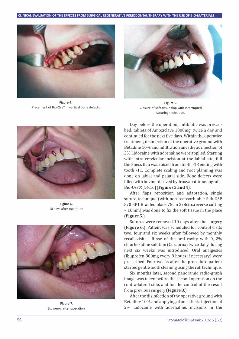
56 57
Day before the operation, antibiotic was prescri-
bed: tablets of Amoxiclave 1000mg, twice a day and
continued for the next five days. Within the operative
treatment, disinfection of the operative ground with
Betadine 10% and infiltration anesthetic injection of
2% Lidocaine with adrenaline were applied. Starting
with intra-crevicular incision at the labial site, full
thickness flap was raised from tooth -18 ending with
tooth -11. Complete scaling and root planning was
done on labial and palatal side. Bone defects were
filled with bovine-derived hydroxyapatite xenograft -
Bio-Oss®[14,16] (Figures 3 and 4).
After flaps reposition and adaptation, single
suture technique (with non-reabsorb able Silk USP
5/0 EP1 Braided black 75cm 3/8circ.reverse cutting
– 16mm) was done to fix the soft tissue in the place
(Figure 5.).
Sutures were removed 10 days after the surgery
(Figure 6.). Patient was scheduled for control visits
two, four and six weeks after followed by monthly
recall visits. Rinse of the oral cavity with 0, 2%
chlorhexidine solution (Curaprox) twice daily during
next six weeks was introduced. Oral analgesics
(ibuprofen 800mg every 8 hours if necessary) were
prescribed. Four weeks after the procedure patient
started gentle tooth cleaning using the roll technique.
Six months later, second panoramic radio-graph
image was taken before the second operation on the
contra-lateral side, and for the control of the result
from previous surgery (Figure 8.).
After the disinfection of the operative ground with
Betadine 10% and applying of anesthetic injection of
2% Lidocaine with adrenaline, incisions in the
gingival pockets were performed and mucoperio-
steal flaps were raised on vestibular and palatal side
(Figures 9. and 10.).
All granulation tissue was removed from the bone
defects and root surface was washed with physio-
logical saline (Figure11.).
Treatment of the defects with EMP was performed
following manufacturer´s instructions. Root surfaces
adjacent to the defects were treated with Pref gel®
(24%EDTA) for 2minutes. The area was then rinsed
with sterile saline solution. Emdogain® was then
mixed with Bio-Oss® and applied into the defects
[17, 18]. The graft material was packed in the defects
Figure 4.
Placement of Bio-Oss® in vertical bone defects.Figure 5.
Closure of soft tissue flap with interrupted
suturing technique.
Figure 6.
10 days after operation
Figure 7.
Six weeks after operation
Figure 8.
Control panoramic radiography
after six months
Figure 9.
Six months later, second operation
Figure10.
Raised full thickness flap
Figure11.
After debridement of the granulation tissue
Todoroska S., Atanasovska-Stojanovska A., Popovska M., Ivanovski K.
Stomatološki vjesnik 2016; 5 (1-2) Stomatološki vjesnik 2016; 5 (1-2)
CLINICAL EVALUATION OF THE EFFECTS FROM SURGICAL REGENERATIVE PERIODONTAL THERAPY WITH THE USE OF BIO-MATERIALS

58
using amalgam condensers to the level of the bony
walls (Figures 12. and 13.).
Flaps were repositioned and sutured using inter-
rupted suturing technique with non-reabsorb able
Silk USP 5/0 EP1 Braided black 75cm 3/8circ.reverse
cutting – 16mm.
Patient was instructed to rinsing the oral cavity
with 0, 2 % chlorhexidine solution (Curaprox) twice
daily. Oral analgesics (ibuprofen 800mg every 8 ho-
urs if necessary) were prescribed. Ten days postope-
rative, sutures were removed. Surgical wounds were
cleaned with iodine solution [20,21]. Two weeks af-
ter the procedure patient started gentle tooth clea-
ning using the roll technique (Figures14. and 15.).
Patient was examined weekly up to 1 month after
the operation and then at 3 and 6 months. Postope-
rative care included reinforcement of oral hygiene
and mechanical plaque removal.
Discussion
The results from our study confirm previous
findings for the improvement of clinical parameters
achieved with EMP and EMP combined with BPBM
[17,19,22]. At the baseline clinical periodontal exa-
mination showed generalized periodontal inflamma-
tion. After the improvement of the values for the
plaque index and PBI before the surgical treatment,
results have sustained during the next six months.
Results for the upper right side – control group,
showed significant improvement for the teeth
mobility from mean 2,37 to 1; GR mean from 4,25 mm
59
to 3,87 mm; PD mean from 5,87 mm to 5,25 mm; CAL
mean from 10,12 mm to 9,12. (Figure16.)
1 = Teeth mobility
2 = Gingival recession(mm)
3 = Probing depth(mm)
4 = Clinical attachment level(mm)
In the upper left side – test group, results showed
significant improvement for the clinical parameters.
TM mean from 2,14 to 0,28; GR mean from 4,14 mm
to3 mm; PD mean from 5,71 mm to 2,42 mm; CAL
mean from 9,85 mm to 5,42 mm (Figure18.).
In human, studies assessing histological quality of
the regenerated tissues and type of attachment are
limited because the block sections are necessary to
be examined. Even with the limitation of this study,
results indicate that we have gained better success
with the second treatment (EMP+BPBM) in stabili-
zing teeth mobility, improving gingival recession,
Figure12.
Pref gel® conditioning
Figure13.
Emdogain® +Bio-Oss®application
Figures14. and 15.
Ten days after operation
Figure16.
Radiography 12 months after the first and 6 months after the second treatment
Figure17.
Clinical findings before the regenerative treatment
(baseline) and six months after the regenerative
periodontal therapy with the use of Bio-Oss®.
Figure18.
Results presented at baseline and six months after regenerative
periodontal therapy with the use of Bio-Oss®and Emdogain®
Figure 19.
Difference between the results from
two treatment modalities.
Todoroska S., Atanasovska-Stojanovska A., Popovska M., Ivanovski K.
Stomatološki vjesnik 2016; 5 (1-2) Stomatološki vjesnik 2016; 5 (1-2)
CLINICAL EVALUATION OF THE EFFECTS FROM SURGICAL REGENERATIVE PERIODONTAL THERAPY WITH THE USE OF BIO-MATERIALS

60 61
improving probing depth values and significant
lower clinical attachment level loss than with the first
treatment (BPBM) in comparison to the values before
the surgical treatment (baseline). In the test group
we found 16% improvements for the GR, 40%
improvements for the PD and 30% improvements for
CAL. In the control group results show 4% impro-
vements for GR, 6% improvements for PD and 6%
improvements for CAL. It takes longer period of
result tracking to make the final assessment and to
bring conclusion over the treatment efficacy [23].
Conclusion
Minimally invasive surgical technique ensures
minimal intraoperative and postoperative pain and
morbidity. Additionally, use of bio-materials:
(in our case Bio-Oss®) and
Emdogain®) in treating bone defects, ensures better
bone filling, greater clinical attachment levels and
lower values for soft tissue recession. It brings a
significant reduction in the degree of dental luxation.
We found even better results when combining the
two materials but we will continue to follow these
case for the future period in order to record long-
term outcome.
BPBM
enamel matrix proteins (
Declaration of interest
The authors declare no conflict of interest. This
study was not sponsored by any external organi-
zation.
References
1. Lindhe J, et al,; Clinical Periodontology and
Implant Dentistry. Blackwell Munksgaard. Fourth
Edition 2003.
2. Newman MG, et al, Carranza's Clinical Periodon-
tology. Elsevier.
3. Rateitschak EM, Wolf HF: Farbatlanten der
Zahnmedizin.Band1: Parodontologie. Thieme
4. Andrew Dentino, Seokwoo Lee, Jason Mailhot and
Arthur F. Hefti. (2013) Principles of periodon-
tology. Periodontology 2000 61, 16-53.
5. (2011) Comprehensive Periodontal Therapy: A
Statement by the American Academy of Perio-
dontology. Journal of Periodontology 82:7, 943-
949.
6. Caton JG, Greenstein G. Factors related to perio-
dontal regeneration.Periodontol 2000 1993:1:9-
15.
7. Dieter D. Bosshardt and Anton Sculean. Does
periodontal tissue regeneration really work? Pe-
riodontology 2000 51:10.1111/prd.2009.51.
issue-1, 208-219.
8. Cortellini P, Tonetti MS. Clinical performance of a
regenerative strategy for intrabony defects:
scientific evidence and clinical experience. J
Periodontol 2005; 76:341-350
9. Camelo M, Nevins ML, Lynch SE, Schenk RK,
Simion M, Nevins M. Periodontal regeneration
with an autogenous bone- Bio-Oss composite
graft and a Bio-Gide membrane. Int J Periodontics
Restorative Dent 2001:21:109-119.
10. Camelo M, Nevins ML, Schenk RK, Simion M,
Rasperini G, Lynch SE, Nevins M. Clinical,
radiographic, and histologic evaluation of human
periodontal defects treated with Bio-Oss and Bio-
Gide. Int J Periodontics Restorative Dent
1998:18:321-331
11. Bosshardt DD. Biological mediators and perio-
dontal regeneration: a review of enamel matrix
proteins at the cellular and molecular levels.
Journal of Clinical Periodontology.2008; 35
(suplement8):87-105. [PubMed]
12. Esposito M, Grusovin MG, Coulthard P, Wor-
thington HV(2008)Enamel matrix derivative
(Emdogain®) for periodontal tissue regene-
ration in intrabony defects. The Cochrane Library
[online serial].Issue1:1-34(Accessed March3,
2008)
13. Hammarström L (1997) Enamel matrix, cemen-
tum development and regeneration. J Clin Perio-
dontol24:658-668
14. Bosshardt DD, Sculean A, Windisch P, Pjeturrson
BE, Lang NP. Effеcts of enamel matrix proteins on
tissue formation along the roots of human teeth. J
Periodontal Res 2005:40:158-167
15. Rathe, F; Junker, R; Chesnutt, B.M.; Jansen J.
A2009 The effect of enamel matrix derivative
(Emdogain) on bone formation
16. Kasaj A, Willershausen B, Reichert C, Röhrig B,
Smeets R, Schmidt M. Ability of nanocrystalline
hydroxyapatite paste to promote human perio-
dontal ligament cell proliferation. Journal of Oral
Science. 2008; 50(3):279-285. [PubMed]
17. Ivanovic A, Nikou G, Miron RJ, Nikolidakis D,
Sculean A. Which biomaterials may promote
periodontal regeneration in intrabony periodon-
tal defects? A systematic review of pre-clinical
studies. Quintessence Int 2014:45:385-395.
18. Trombelli L, Heitz-Mayfield LJA, Needleman I,
Moles D, Scabbia A. A systematic review of graft
materials and biological agents for periodontal
intraosseous defects. Journal of Clinical Perio-
dontology.2002; 29(supplement3):117-
135.[PubMed]
19. Silvestri M, Rasperini G, Milani S. 120 infrabony
defects treated with regenerative therapy: long-
term results. J Periodontol 2011:82:668-675.
20. Cortellini P, Tonetti MS. A minimally invasive
surgical technique with an enamel matrix deri-
vative in the regenerative treatment of intra-bony
defects: a novel approach to limit morbidity. J
ClinPeriodontol 2007; 34:87-93.
21. Cortellini P, Nieri M, Prato GP, Tonetti MS. Single
minimally invasive surgical technique with an
enamel matrix derivative to treat multiple
adjacent inttra-bony defects: clinical outcomes
and patient morbidity. J ClinPeriodontol 2008;
35:605-613.
22. Simone Domenico Aspriello, Luigi Ferrante,
Corrado Rubini and Matteo Piemontese. (2011)
Comparative study of DFDBA in combination
with enamel matrix derivative versus DFDBA
alone for treatment of periodontal intrabony
defects at 12 months post-surgery. Clinical Oral
Investigations 15, 225-232.
23. Tonetti MS, Fourmousis I, Suvan J, Cortellini P,
Bragger U, Lang NP.Healing, postoperative
morbidity and patient perception of outcomes
following regenerative therapy of deep intrabony
defects. J ClinPeriodontol 2004; 31:1092-1098.
Todoroska S., Atanasovska-Stojanovska A., Popovska M., Ivanovski K.
Stomatološki vjesnik 2016; 5 (1-2) Stomatološki vjesnik 2016; 5 (1-2)
CLINICAL EVALUATION OF THE EFFECTS FROM SURGICAL REGENERATIVE PERIODONTAL THERAPY WITH THE USE OF BIO-MATERIALS

63
The book Basics of Gnathology deals with issues of
occlusion and planned reconstruction of the same.
Taking into consideration its topic and as well as high
demands connected to function and restoration of
occlusion expected from therapist, this book
represents an inevitable reference for every doctor of
dental medicine.
The book is written in very legible and simple
manner, methodologically adjusted to the field it deals
with. It is systematically divided into 14 chapters,
subsequently elaborated according to unique pattern,
providing visibility and better perception for
memorizing.
The chapter titled "Stomatognathic System" clear-
ly describes anatomic and morphologic details not
only for bones but also of muscles of the system
including very clear picture of temporomandibular
joint. The following chapters are dealing with move-
ments in temporomandibular joint and central regu-
lation and control of the same.
BOOK REVIEW / PRIKAZ KNJIGE
Further on, one can find chapters about other
determinants of the jaw movements, anthropomor-
phic model and position of head in space. Conse-
quently, the authors direct our attention to the
characteristics of optimal occlusion and importance
of articulators and facebow as inevitable diagnostic
parts of rehabilitation of occlusion.
Such analytic approach to book writing speaks in
favor of high appreciation of the knowledge of authors
regarding the theme and also of long lasting clinical
and lecturing experience. That, in great extent,
contributes to overall originality of this text.
List of literature indicates that the book was
written according to the newest knowledge in this
field. Such systematical book, with chapters men-
tioned above, will be of a great use for students,
specialists and doctors of dental medicine in Bosnia
and Herzegovina as well as in the region, taking into
consideration insufficiency of domestic literature
dealing with the subject.
TITLE:
AUTHORS:
PUBLISHER:
LANGUAGE:
PAGES:
DATE OF ISSUE:
ISBN:
Basics of Gnathology
Muhamed Ajanović, Sanela Strujić-Porović, Azijada Šuljak-Lončarević, Almira Hadžović Džuvo, Lejla Kazazić, Selma Tosum, Lejla Berhamović, Adis Hamzić, Amela Đonlagić, Eldan Kapur
Faculty of Dentistry with Clinics, University of Sarajevo, Sarajevo, Bosnia and Herzegovina
Bosnian
314
2015
978-9958-9051-6-2
Stomatološki vjesnik 2016; 5 (1-2)

65
INSTRUCTIONS FOR THE AUTHORS
Submissions of manuscripts are made through
the submission form available at web page of
the Journal (www.stomatoloskivjesnik.ba) or
by sending the email to Editorial office at
radovi@stomatoloski vjesnik.ba
E-mail must be composed of:
A) Covering letter, in which authors explain the
importance of their study (Explanation why we
should publish your manuscript ie. what is new
and what is important about your manuscript,
etc).
B) Title of the manuscript
C) Authors' names and email addresses (mark
corresponding author with *)
D) Abstract
E) Attached file of the Copyright assignment form
and
F) Manuscript.
Authors should NOT in addition post a hard copy
of the manuscript and submission letter, unless they
are supplying artwork, letters or files that cannot be
submitted electronically, or have been instructed to
do so by the editorial office.
Please read Instructions carefully to improve
yours paper's chances for acceptance for publi-
shing.
Thank you for your interest in submitting an
article to Stomatološki vjesnik.
Type of papers suitable for publishing in Sto-
matološki vijesnik (Journal in following text):
Original Articles, Case Reports, Letters to the Edi-
tors, Current Perspectives, Editorials, and Fast-Track
Articles are suitable for publishing in Stomatološki
vjesnik. Papers must be fully written in English with
at least title, abstract and key words bilingual in Bos-
nian/Croatian/Serbian language (B/C/S) and Eng-
lish language.
Editorial process:
All submitted manuscripts are initially evaluated
by at least two scientific and academic members of
editorial board. An initial decision is usually reached
within 3–7 days.
Submitted manuscripts may be rejected without
detailed comments after initial review by editorial
board if the manuscripts are considered inappro-
priate or of insufficient scientific priority for publi-
cation in Stomatološki vjesnik.
If sent for review, each manuscript is reviewed by
scientists in the relevant field. Decisions on reviewed
manuscripts are usually reached within one month.
When submission of a revised manuscript is invited
following review, the revision must be received in
short time of the decision date.
Criteria for acceptance:
Submitted manuscripts may be rejected without
detailed comments after initial review by editorial
board if the manuscripts are considered inappropria-
te or of insufficient scientific priority for publication
in the Journal. All other manuscripts undergo a com-
plete review by reviewers or other selected experts.
Criteria for acceptance include originality, validity of
data, clarity of writing, strength of the conclusions,
and potential importance of the work to the field of
dentistry and similar bio-medical sciences. Submit-
ted manuscripts will not be reviewed if they do not
meet the Instructions for authors, which are based on
"Uniform Requirements for Manuscripts Submitted
to Biomedical Journals" (http://www.icmje.org/).
INSTRUCTIONS FOR THE AUTHORSmade in accordance with the recommendations of the International Committee of Medical Journal based on "Uniform Requirements for Manuscripts Submitted to Biomedical Journals" (http://www.icmje.org/).
Stomatološki vjesnik 2016; 5 (1-2)

6766
INSTRUCTIONS FOR THE AUTHORS
national bodies should be avoided. Use only standard
abbreviations. Avoid abbreviations in the title and
abstract. The full term for which an abbreviation
stands should precede its first use in the text unless it
is a standard unit of measure-ment.
The Title Page should carry the full title of the pa-
per and a short title to be used as a 'running head'
(and which should be so identified). The first name,
middle initial and last name of each author should
appear marked with superscript numbers or/and
symbols corresponding to their affiliation or/and
note. Affiliations of the authors should be written be-
low authors name list. Full name, address, phone and
fax number and e-mail of the author responsible for
correspondence should appear on the Title Page.
Please include the word count of the abstract and
word count of text on the title page.
The second page should carry a structured
abstract of no more than 250 words. The abstract
should state the Objective(s) of the study or investi-
gation, basic Methods (selection of study subjects or
laboratory animals; obser-vational and analytical
methods), main Results (giving specific data and
their statistical significance, if possible), and the
principal Conclusions. It should emphasize new and
important aspects of the study or observations.
The abstract should be followed by a list of 3–10
keywords or short phrases which will assist the
cross-indexing of the article and which may be publi-
shed. When possible, the terms used should be from
the Medical Subject Headings list of the National
Library of Medicine (http://www.nlm.nih.gov/
mesh/meshhome.html).
Body of the manuscript text of an experimental
or observational nature may be divided into sections
headed Introduction, Materials and Methods (inclu-
ding ethical and statistical information), Results,
Discussion and Conclusion, although reviews may
require a different format.
Original Articles: original experimental and clini-
cal studies should not exceed 4400 words (up to 15
pages) including tables and references.
With Case Reports: presentation of a clinical case
which may suggest novel working hypotheses, with a
short discussion on the pertinent literature. The text
should not exceed 2400 words (up to 8 pages).
Letters to the Editors should not exceed 500
words, should not be signed by more than three
authors and should not have more than 5 references.
Preferably, letters should be in reference to a Journal
article published within the last 3 months or to novel
hypotheses so as to stimulate comments on issues of
common interest. Authors of the letters accepted for
publication will receive the galley proofs. The Editors
will generally solicit replies. The Editors reserve the
right to modify the text.
Current Perspectives: invited articles by recogni-
zed authorities, to include position papers, reviews,
and special topics of general interest. Independent
submission will also be considered.
Editorials: invited articles or brief editorial com-
ments that represent opinions of recognized leaders
in biomedical research.
Fast-Track Articles: short articles on laboratory or
clinical findings, representing important new in-
sights or major advances, produced with established
methods or new applications of an established or
new method. The text should not exceed 2400 words.
Acknowledgements: One or more statements
should specify (a) contributions that need acknow-
ledging but do not justify authorship, such as general
support by a department chairman; (b) acknow-
ledgements of technical help; (c) acknowledgements
of financial and material support, specifying the
nature of the support. Persons who have contributed
intellectually to the paper but whose contributions
do not justify authorship may be named and their
function or contribution described – for example,
'scientific adviser', 'critical review of study proposal',
'data collection', 'participation in clinical trial'. Such
persons must have given their permission to be na-
med. Authors are responsible for obtaining written
permission from persons acknowledged by name
because readers may infer their endorsement of the
data and conclusions.
Declaration of Interest
A statement must be provided listing all financial
support received for the work and, for all authors, any
INSTRUCTIONS FOR THE AUTHORS
Authorship:
All authors have to sign the copyright assignment
form. We ask all authors to confirm that: they have
met the criteria for authorship as established by the
International Committee of Medical Journal Editors;
they believe that the paper represents honest work,
and are able to verify the validity of the results re-
ported.
Redundant or duplicate publication:
We ask the authors to confirm in the copyright
assignment form that the paper has not been publi-
shed in its current form or a substantially similar
form (in print or electronically, including on a web
site), that it has not been accepted for publication
elsewhere, and that it is not under consideration by
another publication. The International Committee of
Medical Journal Editors has provided details of what
is and what is not duplicate or redundant publi-
cation (http://www.icmje.org). In the submission
letter to the editors, authors are asked to draw
attention to any published work that concerns the
same patients or subjects as the present paper.
Author Contributions:
Authors are required to include a statement
to verify the contributions of each co-author in the
copyright assignment form (avai lable at
www.stomatoloskivjes nik.ba ).
Patient consent forms:
The protection of a patient's right to privacy is
essential. The authors may be asked by the editorial
board to send copy of patient consent forms on which
patients or other subjects of the experiments clearly
grant permission for the publication of photographs
or other material that might identify them. If the
consent form for the research did not specifically
include this, authors should obtain it or remove the
identifying material.
Ethics committee approval:
The authors must state clearly in the submission
letter and in the Methods section that the conducted
studies on human participants are with the approval
of an appro-priate named ethics committee. Please
also look at the latest version of the Declaration of
Helsinki (http:// www.wma.net/e/policy/b3.htm).
Similarly, the authors must confirm that experiments
involving animals adhered to ethical standards and
must state the care of animal and licensing guidelines
under which the study was perfor-med. The editorial
board may ask author(s) for copy of ethical
committee approval.
Declaration of Interest:
We ask the authors to state all potential financial
support received for the work. This applies to all
papers including editorials and letters to the editor
(see below). If you are sure that there is no conflict of
interest, please state that.
Copyright:
Copyright assignment form contains authors' sta-
tement that all the copyrights are transferred to the
publisher if and when the manuscript is accepted for
publishing.
Subscribers may reproduce tables of contents or
prepare lists of articles including abstracts for inter-
nal circulation within their institutions. Permission
of the Publisher is required for resale or distribution
outside the institution and for all other derivative
works, including compilations and translations. If
excerpts from other copyrighted works are included,
the author(s) must obtain written permission from
the publisher and credit the source(s) in the article.
Formatting requirements:
Manuscript should be written in Times New Ro-
man 12, Normal, double spacing.
Include the following sections, each starting on a
separate page: Title Page, Abstract and Keywords,
Text, Acknowledgements, Declaration of interest, Re-
ferences, Individual tables and figures with captions.
Margins should be not less than 3 cm. Pages
should be numbered consecutively, beginning with
the Title Page, and the page number should be placed
in the bottom right hand corner of each page.
Abbreviations should be defined on their first
appearance in the text; those not accepted by inter-
Stomatološki vjesnik 2016; 5 (1-2) Stomatološki vjesnik 2016; 5 (1-2)

6968
INSTRUCTIONS FOR THE AUTHORS
financial involvement (including employment, fees,
share ownership) or affiliation with any organization
whose financial interests may be affected by material
in the manuscript, or which might potentially bias it.
This applies to all papers including editorials and
letters to the editor. If you are sure that there is no
conflict of interest, please state this.
References
References should be numbered consecutively in
the order in which they first appear in the text. They
should be assigned Arabic numerals, which should be
given in brackets, e.g. [17]. References should include
the names of all authors when six or fewer; when
seven or more, list only the first six names and add et
al. References should also include full title and source
information (Vancouver style).
Journal names should be abbreviated as in
MEDLINE (http://www.medscape.com/Home/
Search/ IndexMedicus/IndexMedicus.html).
Examples of citation:
Standard journal article:
Tashiro H, Shimokawa H, Sadamatu K, Yamamoto
K. Prognostic significance of plasma concentra-
tions of transforming growth factor-ß. Coron
Artery Dis 2002; 13(3):139-143.
More than six authors:
Yetkin E, Senen K, Ileri M, Atak R, Tandogan I,
Yetkin Ö, et al. Comparison of low-dose dobuta-
mine stress echocardiography and echocardio-
graphy during glucose-insulin-potassium infu-
sion for detection of myocardial viability after
anterior myocardial infarction. Coron Artery Dis
2002; 13(3):145-149.
Books:
Heger JW, Niemann JT, Criley JM. Cardiology, 5th
ed. Philadelphia: Lippincott, Williams & Wilkins;
2003.
Chapter in a book:
Braunwald E, Perloff JK. Physical examination of
the heart and circulation. In; Braunwald E, Zipes
DP, Libby P (eds). Heart disease; a textbook of
cardiovascular medicine, 6th edn. Philadelphia:
WB Saunders; 2001, pp. 45-81.
Personal communications and unpublished work
should not feature in the reference list but should
appear in parentheses in the text. Unpublished work
accepted for publication but not yet released should
be included in the reference list with the words 'in
press' in parentheses beside the name of the journal
concerned. References must be verified by the
author(s) against the original documents.
Tables
• Provide each table on a separate page of the
manuscript after the references.
• Each table should be typed on a separate sheet in
double spacing.
• Number the table according to their sequence in
the text. The text should include references to all
tables. Each table should be assigned an Arabic
numeral, e.g. (Table 3).
• Include a brief and self-explanatory title with
explanations essential to the understanding of the
table at the bottom of the table.
• Identify statistical measures of variations, such as
standard deviation and standard error of the
mean or other where appropriate.
Figures
• Provide each figure on a separate page of the
manuscript after the references. Number the
figures according to their sequence in the text. The
text should include references to all figures.
• Graphs and figures should be in black/white or
greyscale format. Colour illustrations are accep-
table but not guaranteed. Minimal quality 300dpi,
figures should be filed in suitable format (*.JPG,
*.PNG, .*TIFF)
• If figures are not original provide source and
permition.
• All figures should be 100% of a suitable final size
and have the printing resolution of 300dpi and be
cropped to include the figure only (no blank
space).
INSTRUCTIONS FOR THE AUTHORS
Units of measurement
Measurements of length, height, weight, and volu-
me should be reported in metric units (meter, kilo-
gram, or liter) or their decimal multiples. All
hematologic and clinical chemistry measurements
should be reported in the metric system in terms of
the International System of Units (SI).
Post acceptance
All correspondence concerning the copy, editing
and production of accepted manuscripts should be
addressed to Stomatološki vjesnik.
Charges
Authors will not be charged fees in a first year of
publishing Stomatološki vjesnik. Optional charges
for colour reproduction of figures may apply. Authors
will be informed about this when the poof is supplied
to them. The charges must be completed before the
article is released.
Stomatološki vjesnik 2016; 5 (1-2) Stomatološki vjesnik 2016; 5 (1-2)