Embed Size (px)
Citation preview

Stomatološki vjesnikStomatološki vjesnikStomatological reviewStomatological review
Stomatološki vjesnik 2014; 3(1)

Stomatološki vjesnik
Stomatološki vjesnik 2014; 3 (1)
CONTENTS / SADRŽAJ
ORIGINAL SCIENTIFIC ARTICLES / ORIGINALNI NAUČNI RADOVI
REVIEW ARTICLE / PREGLEDNI ČLANAK
CASE REPORT / PRIKAZ SLUČAJA
BOOK REVIEW / PRIKAZ KNJIGE
MAXILLARY CRESTAL BONE LOSS AROUND BREDENT BLUESKY® IMPLANTS: ONE YEAR STUDY Ajanović M, Hamzić A, Redžepagić S,Kamber-Ćesir A, Kazazić L, Tosum S
FLUORIDE RELEASE AND CYTOTOXICITY OF RESIN-MODIFIED GLASS-IONOMER CEMENTSSelimović-Dragaš M, Hasić-Branković L, Huseinbegović A, Kobašlija S, Hatibović-Kofman Š
ANALYSIS OF METALS RELEASED FROM DENTAL AMALGAM ALLOY USING INDICTIVELY COUPLED PLASMA-MASS SPECTROMETRYBajsman A, Turkušić E, Vuković A, Zukić S, Zukanović A, Kahrović E
DISTRIBUTION OF WHITE SPOTS AFTER DE-BONDING IN ORTHODONTIC PATIENTSZabokova-Bilbilova E, Muratovska I, Stojanovska V, Stefanovska E, Dimova C
INTERPROXIMAL PLAQUE CONTROL IN PATIENTS WITH FIXED ORTHODONTIC APPLIANCESStefanovska E, Ivanovski K, Atanasovska-Stojanovska A, Zabokova-Bilbilova E, Zendeli-Bedzeti L
MICROBIOLOGICAL AND EXFOLIATIVE CYTOLOGICAL FINDING OF THE ORAL CANDIDA DUBLINIENSIS IN PATIENTS WHO ARE PSHYOACTIVE SUBSTANCES ADDICTSHadžić S, Dedić A, Gojkov-Vukelić M, Pašić E, Hodžić M, Topić B
PERIODONTAL STATUS IN THE PERMANENT DENTITION OF CHILDREN WITH CEREBRAL PALSYIhtijarević-Trtak S, Kobašlija S, Hadžagić-Ćatibušić F, Huseinbegović A
THE CLINICAL APPLICATION OF PLATELET-RICH FIBRIN (PRF) IN ORAL SURGERY: REVIEWŠečić S, Prohić S
DELAYED TOOTH REPLANTATION AFTER TRAUMATIC AVULSIONJovanovska M
FUNDAMENTALS OF TOOH MORPHOLOGY AND DENTAL ANTHROPOLOGYVuković A, Zukić S, Bajsman A, Selmanagić A
3
9
17
27
35
43
51
59
67
73
Izdavač / Publisher:
Za izdavača / For publisher: Sead Redžepagić
ČLANOVI UREĐIVAČKOG ODBORA / EDITORIAL BOARD :
Glavni urednik / Editor in chief: Sadeta Šečić
Sekretar uređivačkog odbora / Secretary of editorial board: Selma Zukić
Članovi /Members: Sead Redžepagić, Samir Prohić, Muhamed Ajanović, Amira Dedić, Sedin Kobašlija, Tarik Mašić, Amra Vuković, Enita Nakaš
MEĐUNARODNI UREĐIVAČKI ODBOR / INTERNATIONAL EDITORIAL BOARD:Anwar Barakat Bataineh (Irbid, Jordan), Jasenka Živko-Babić (Zagreb, Hrvatska), Andrija Petar Bošnjak (Rijeka, Hrvatska), Hrvoje Brkić (Zagreb , Hrvatska), Dolores Biočina Lukenda (Split, Hrvatska), Davor Katanec (Zagreb, Hrvatska), Šahza Hatibović Koffman (London Ontario Kanada), Mladen Kuftinec (USA), Darko Macan (Zagreb, Hrvatska), Berislav Perić (Zagreb, Hrvatska), Tore Solheim (Oslo, Norveška), Dragoslav Stamenković (Beograd, Srbija), Marin Vodanović (Zagreb, Hrvatska)
Lektor za engleski jezik / English language editor: Nermana Bičakčić
Tehničko uređenje / Technical editor: Branislav Trogrančić
Štampa / Printed by: Štamparija Fojnica
Dizajn naslovnice / Cover page design: Lana Malić
Tiraž/ Number of copies:
KONTAKT / CONTACT:Stomatološki vjesnikStomatološki fakultet sa klinikamaBolnička 4a, 71000 SarajevoBosna i HercegovinaTelefon: + 387(33)214 294e-mail: [email protected] Web: www.stomatoloskivjesnik.ba
TRANSAKCIJSKI RACUN / TRANSFER ACCOUNT:33386902296551066UniCredit Bank dd
Stomatološki fakultet Univerziteta u Sarajevu / Faculty of Dentistry, University of Sarajevo
200
ISSN 0350-5499 UDK 616.31
Svrha i i cilj :
Stomatološki vjesnik je neprofitni naučno stručni časopis koji publicira originalne naučne radove, prikaze slučajeva, pisma uredniku, savremene perspektive, editorijale, preliminarne komunikacije u oblasti stomatologije i drugih biomedicinskih nauka. Radovi su na Bosanskom/Hrvatskom/Srpskom jeziku sa naslovom, sažetkom i ključnim riječima bilingvalnim B/H/S i engleskom jeziku. Radovi se mogu koristiti u edukacijske svrhe bez predhodnog odobrenja, a uz obavezno navođenje izvora. Korištenje cijelih ili dijelova članaka u komercijalne svrhe nije dozvoljeno bez predhodnog pismenog odobrenja izdavača Autorska prava posjeduje izdavač: Stomatološki fakultet sa klinikama Univerziteta u Sarajevu.
Aim and Scope:
Stomatološki vijesnik / Stomatological review is a non-profit scientific journal that publishes original articles, case reports, letters to the editors, current perspectives, editorials, fast-track articles in a field of dentistry and other bio-medical sciences. Papers are in Bosnian/ Croatian/Serbian language with at least title, abstract and key words bilingual in B/C/S and English language. All manuscripts undergo the peer review process before can be accepted for publishing in Stomatološki vjesnik/ Stomatolgical review. Papers can be used for educational purposes without prior consent only with adequate citation of the sources. Using whole or parts of articles for commercial purposes is not permitted without prior written permission of the publisher. Copyright owns the publisher: Faculty of Dentistry with Clinics, University of Sarajevo.
Časopis Stomatološki vjesnik je oslobođen poreza na promet prema Mišljenju Federalnog ministarstva obrazovanja, nauke, kulture i sporta br: 04-15-661/2002.
Journal Stomatological review is tax exempt according to the opinion of the Federal Ministry of Education Science Culture and Sports no: 04-15-661/2002.
Printed on acid free paper
Indexed in: (Index Copernicus International), (Directory of Open Access Journal), EZB (Electronishe Zeitschriftenbibliothek), SJIF (Scientific Journal Impact Factor Value 2.502)IIC DOAJ

Stomatološki vjesnik 2014; 3 (1) 3
ORIGINAL SCIENTIFIC ARTICLE / ORIGINALNI NAUČNI RAD
MAXILLARY CRESTAL BONE LOSS AROUND BREDENT BLUESKY® IMPLANTS: ONE YEAR STUDY
1 1 1Ajanović M* , Hamzić A , Redžepagić S ,1 1 1Kamber-Ćesir A , Kazazić L , Tosum S
1 Department of Prosthodontics, Faculty of Dentistry, University of Sarajevo, Sarajevo, Bosnia and Herzegovina
ABSTRACT
The resorption of the alveolar crest is a parameter that is fre-
quently used in the investigation of endoosseous implants. The
aim of the study was to analyze the amount of crestal bone loss of
the upper jaw around Bredent BLUESKY® implants of different
dimension a year after functional loading.
This study analyzed total number of 88 implants type Bredent
BLUESKY®. The measurements were performed using Kodak
dental software 6.11.7.0 after implantation and a year after its
functional loading. The average value of the distal bone resorp-
tion around implant dimension 3.5 x 10 mm in front maxilla was
0.67 mm (± 0.098 mm), while the average mesial resorption was
0.57 mm (± 0.118 mm). The average value of the distal bone
resorption around implant size 4.0 x 8 mm in maxilla lateral was
0.52 mm (± 0.176 mm), while the average mesial resorption was
0.53 mm (± 0.176 mm). Bone resorption was greater at the distal
portion of the crest than mesial, although the differences were not
statistically significant.
Key words: maxilla, crestal bone loss, Bredent BLUESKY®
Implants.
*Corresponding author
Ajanović Muhamed, Professor, PhD
Department of Prosthodontics
Faculty of Dentistry
University of Sarajevo,
Bolnička 4a
71000 Sarajevo
Bosnia and Herzegovina
Phone: +387 61 134 522
e-mail: [email protected]

4
MAXILLARY CRESTAL BONE LOSS AROUND BREDENT BLUESKY® IMPLANTS: ONE YEAR STUDY
5
Ajanović M, Hamzić A, Redžepagić S, Kamber-Ćesir A, Kazazić L, Tosum S
Introduction
The ideal goal of modern dentistry is to restore the
patient to normal contour, function, comfort, esthe-
tics, speech, and health. What makes dental implant
dentistry unique is the ability to achieve this ideal
goal regardless of the atrophy, disease or injury of the
stomatognathic system [1].
The number of dental implants used in the United
States increased more than ten times from 1983 to
2002. More than 700.000 dental implants are inser-
ted each year. The number of implants continues to
increase steadily, with more than $ 150 million of
implant products sold to North American dentists in
2002 compared with $ 10 million in 1983, with an
expected growth sustained at 9.4% for the next seve-
ral years [2, 3]. More than 90% of interfacing surgical
specialty dentists provided implant treatment
routinely in their practices, 90% of prosthodontics
restored implants routinely, and more than 78% of
general dentists used implants to support fixed and
removable prosthesis compared to 65% 15 years ago
[4, 5, 6].
As the implant performs its function for five years
period, it may be considered as successful regarding
the fact that after its removal there is no significant
bone defect. Today, the criteria are stricter and re-
quire implants' survival for at least ten years. The
success of implantation depends also on the quality
of tissue types, materials, design and microstructure
implants, wound type, degree of stress, possible cor-
rosion, and number of other factors. It is understood
that many of the implantology problems have not
been definitively and satisfactorily resolved. There
are many studies on the relation the body – implant:
physical - chemical, experimental and clinical re-
searches. Nowadays, there are systems made of dif-
ferent materials, different shapes, different micro
and macrostructure, so the results ranging from
extraordinary success to the complete disappoint-
ment [7].
Endoosseous implantation particularly demands
finer bone structure, its structure and density. Dense
bone structure, more calcified and mineralized bone,
accepts better the implant than loose, unmineralized
bone with plenty haemotopoietic and fat tissue [8].
The resorption of the alveolar crest is a parameter
that is frequently used in the investigation of endoos-
seous implants. For such purposes, it is necessary to
establish precisely defined reference level on the
implant and to set the level of peri-implant alveolar
bone in relation to that reference plan [9].
The clinical success of implants in the upper jaw is
much smaller compared to implants in intercanine
part called [8]. At the maxilla, the front part of
alveolar crest to the second premolar is conditionally
favorable region for implantation. Unfavorable
region is the lateral part of the alveolar crest,
including the tuber maxillae [10].
The aim of this study was to evaluate crestal bone
resorption around dental implants in different
regions of maxilla one year after its functional
loading.
Patients and Methods
The Ethics Committee of the Faculty of Dentistry
(University of Sarajevo) approved this study. All the
examinees gave their informal consent.
This study analyzed total number of 88 implants
type Bredent BLUESKY®. 18 implants dimension 3.5
x 10 mm were inserted in the maxilla on the right
side, and 18 in the maxilla on the left side. 22 implants
dimension 4.0 x 8 mm were inserted in the maxilla on
the right side, and 30 in the maxilla on the left side.
The implants were placed into the maxilla according
to a strict surgical protocol following the manufactu-
rer's instructions. After healing phase of three
months without functional loading, gingiva former
was inserted. After 14 days, gingiva former was re-
moved and impressions were taken. The time place-
ment of prosthetic restorations on the implants was
four months after surgery. All the implants were used
as an abutment of individual crowns and bridges.
Dental panoramic radiographs were made before
surgery, immediately after surgery and a year after its
functional loading, using
Kodak 8000 c, XJAM530. Panoramic images were
calibrated using CliniView (version 5.2 Instrumenta-
rium Imaging). The measurements were performed
by comparing images using software Kodak dental
software 6.11.7.0. The mesial and distal to the im-
plant immediately after implant placement determi-
nes the highest level of bone resorption in the alveo-
lar part, which is denoted as point A. After a year, we
repeated OPG and determined the mesial and distal
bone loss, which is denoted as point B. The difference
Ortopantomograph type
between points A and B is expressed in millimeters
(mm) and indicates the level of bone resorption.
Statistical analysis
The data were analyzed using the IBM SPSS v.17
software package (descriptive statistics, paired
samples t-test).
Results
The study included total number of 42 male and
female patients. Among male patients, 43.5% were
smokers, while 56.5% were non-smokers. Among
females, 42.1% were smokers and 57.9% nonsmo-
kers.
Among male patients, 78.3% were partially den-
tate, while 21.7% were totally edentulous. 94.7% fe-
males were partially dentate, only 5.3% were totally
edentulous.
Table 1 shows the frequency of inserted implants
in the lateral and front region of maxilla on the left
and right side. It should be noted that there is no
implant dimension 4.0 x 8 mm in front region of
maxilla.
The mean of the distal bone resorption around
implant dimension 3.5 x 10 mm in maxilla on the
right side was 0.60 mm (± 0.137 mm) with standard
deviation of 0.32 mm, while the mean of mesial bone
resorption was 0.50 mm (± 0.176 mm) with standard
deviation of 0.36 mm. The differences between the
mesial and distal resorption is not statistically
significant (p = 0.254). The mean of the distal bone
resorption around implant dimension 3.5 x10 mm in
maxilla on the left side was 0.62 mm (± 0.118 mm)
with standard deviation of 0.28 mm, while the mean
of mesial bone resorption was 0.59 mm (± 0.137 mm)
with standard deviation of 0.29 mm.
Testing the difference between the values bet-
ween distal and mesial resorption using t-test we
found no statistically significant difference (p =
0.491). The mean of the distal bone resorption
around implant size 4.0 x 8 mm in maxilla on the right
side was 0.52 mm (± 0.176 mm) with standard
deviation of 0.41 mm, while the mean mesial resorp-
tion was 0.53 mm (± 0.176 mm) with a standard
deviation of 0.40 mm. The differences between me-
sial and distal resorption are not statistically
significant (p = 0.900). The mean of the distal bone
resorption around implant dimension 4.0 x 8 mm in
maxilla on the left side was 0.60 mm (± 0.157 mm)
with an standard deviation of 0.44 mm, while the
mean of the mesial resorption was 0.54 mm (± 0.137
mm) with a standard deviation of 0.39 mm. Testing
the difference between the mean distal and mesial
resorption using t-test, we found no statistically sig-
nificant difference (p = 0.366).
The average value of the distal bone resorption
around implant dimension 3.5 x 10 mm in front
maxilla was 0.67 mm (± 0.098 mm), while the average
mesial resorption was 0.57 mm (± 0.118 mm). The
average value of the distal bone resorption around
implant size 4.0 x 8 mm in maxilla lateral was 0.52
mm (± 0.176 mm), while the average mesial resorp-
tion was 0.53 mm (± 0.176 mm).
Tables 2, 3, 4 and 5 show the values for the level of
bone resorption mesially and distally around the
implant dimension 3.5 x 10 mm in different regions of
the maxilla. The differences between the mean
resorption on the mesially and distally sides are not
statistically significant.
Stomatološki vjesnik 2014; 3 (1) Stomatološki vjesnik 2014; 3 (1)
%nnn %%
13.6
14.8
31.8
39.8
100.0
0.0
0.0
42.3
57.7
100.0
33.3
36.1
16.7
13.9
100.0
12
13
28
35
88
0
0
22
30
52
12
13
6
5
36
Maxilla right front
Maxilla left front
Maxilla right lateral
Maxilla left lateral
Region
Dimension of implant
3.5 x 10 mm 4.0 x 8 mm Total
Total
Table 1Frequency of inserted implant by region
Table 2The level of bone resorption mesially and distally to
the implant dimension 3.5 x 10 mm - maxilla right front
Implant 3.5 x 10 mm (maxilla right front)
95% CI of MeanStandardDeviation
Distal resorption(n=12)
Mesial resorption(n=12)
0.69±0.157 mm
0.52±0.196 mm
0.26
0.36
Paired samples t-test (t= 1.428, df= 11, p= 0.181)
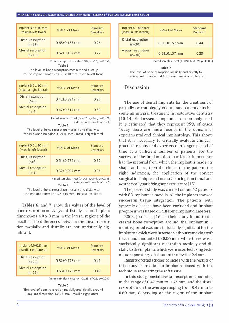
Table 3The level of bone resorption mesially and distally
to the implant dimension 3.5 x 10 mm - maxilla left front
Implant 3.5 x 10 mm (maxilla left front)
95% CI of MeanStandardDeviation
Distal resorption(n=13)
Mesial resorption(n=13)
0.65±0.137 mm
0.62±0.157 mm
0.26
0.27
Paired samples t-test (t= 0.602, df=12, p= 0.558)
Table 4The level of bone resorption mesially and distally to
the implant dimension 3.5 x 10 mm - maxilla right lateral
Implant 3.5 x 10 mm (maxilla right lateral)
95% CI of MeanStandardDeviation
Distal resorption(n=6)
Mesial resorption(n=6)
0.42±0.294 mm
0.47±0.314 mm
0.37
0.39
Paired samples t-test (t= -2.236, df=5, p= 0.076)(Note, a small sample of n = 6)
Table 5The level of bone resorption mesially and distally to
the implant dimension 3.5 x 10 mm - maxilla left lateral
Implant 3.5 x 10 mm (maxilla left lateral)
95% CI of MeanStandardDeviation
Distal resorption(n=5)
Mesial resorption(n=5)
0.54±0.274 mm
0.52±0.294 mm
0.32
0.34
Paired samples t-test (t= 0.343, df=4, p= 0.749) (Note, a small sample of n = 5)
Table 6The level of bone resorption mesially and distally around
implant dimension 4.0 x 8 mm - maxilla right lateral
Implant 4.0x0.8 mm(maxilla right lateral)
95% CI of MeanStandardDeviation
Distal resorption(n=22)
Mesial resorption(n=22)
0.52±0.176 mm
0.53±0.176 mm
0.41
0.40
Paired samples t-test (t= - 0.128, df=21, p= 0.900)
Table 7The level of bone resorption mesially and distally to
the implant dimension 4.0 x 8 mm – maxilla left lateral
Implant 4.0x0.8 mm(maxilla left lateral)
95% CI of MeanStandardDeviation
Distal resorption(n=30)
Mesial resorption(n=30)
0.60±0.157 mm
0.54±0.137 mm
0.44
0.39
Paired samples t-test (t= 0.918, df=29, p= 0.366)
6
MAXILLARY CRESTAL BONE LOSS AROUND BREDENT BLUESKY® IMPLANTS: ONE YEAR STUDY
Tables 6. and 7. show the values of the level of
bone resorption mesially and distally around implant
dimensions 4.0 x 8 mm in the lateral regions of the
maxilla. The differences between the mean resorp-
tion mesially and distally are not statistically sig-
nificant.
Discussion
The use of dental implants for the treatment of
partially or completely edentulous patients has be-
come an integral treatment in restorative dentistry
[10-14]. Endoosseous implants are commonly used.
It is estimated that they represent 95% of cases.
Today there are more results in the domain of
experimental and clinical implantology. This shows
that it is necessary to critically evaluate clinical -
practical results and experience in longer period of
time at a sufficient number of patients. For the
success of the implantation, particular importance
has the material from which the implant is made, its
shape and size, then the choice of the patient, the
right indication, the application of the correct
surgical technique and manufacturing functional and
aesthetically satisfying superstructure [15].
The present study was carried out on 42 patients
with 88 implants in maxilla. All the implants showed
successful tissue integration. The patients with
systemic diseases have been excluded and implant
prognosis was based on different implant diameters.
2008. Job et al. [16] in their study found that a
crestal bone resorption around the implant in 3
months period was not statistically significant for the
implants, which were inserted without removing soft
tissue and amounted to 0.06 mm, while there was a
statistically significant resorption mesially and di-
stally to the implants which were inserted using tech-
nique separating soft tissue at the level of 0.4 mm.
Results of cited studies coincide with the results of
this study in relation to implants placed with the
technique separating the soft tissue.
In this study, mesial crestal resorption amounted
in the range of 0.47 mm to 0.62 mm, and the distal
resorption on the average ranging from 0.42 mm to
0.69 mm, depending on the region of the implant
7
dimensions 3.5 x 10 mm. Around implant dimensions
4.0 x 8 mm mesial crestal resorption was 0.47 mm
and 0.62 mm, and the distal resorption was 0.42mm
and 0.69 mm.
2008. Jang et al. in his research found bone
resorption of 0.7 mm in the same period of evaluation
like this study. Mesial crestal resorption ranged from
0.4 mm to 1.2 mm with the mean 0.76 mm when mo-
dified platform while crestal resorption of 2.1 mm to
3.1 mm on average 2.53 mm when unmodified plat-
forms. Distal crestal resorption in modified platform
ranged from 0.3 mm to 1.3 mm with the mean of 0.77
mm while in the unmodified platforms distal crestal
resorption ranged from 2.2 mm to 2.9 mm, the mean
2.56 mm [17].
2010. Heinemann et al. in their longitudinal study
on a sample of 147 implants size 3.7 and 4.2 mm
inserted correctly in the maxilla found low crestal
resorption. For implant size 3.7 mm, crestal resorp-
tion was 0.16 mm per year, while for implant size 4.3
mm, crestal resorption was 0.09 mm per year. Results
of this study are different from the results of our
study mostly because of different implant systems
used and different implant dimensions leading to-
wards the conclusion that cervical implant topogra-
phy can significantly affect the crestal bone resorp-
tion [18].
2011. Hürzeler et al. found that the mean bone
resorption within one year was 0.40 mm (± 0.12 mm)
for the experimental group and 0.34 mm (± 0.29) for
the group that was studied retrospectively at the
control. It is important to note that the study was
conducted on 5.0 mm implants and the results partly
coincide with the results of our study [19].
Conclusion
Obtained results produced interesting findings
showing that the resorption was mainly greater at
the distal portion of the crest than mesial, although
the differences were not statistically significant.
Declaration of Interest
There is no conflict of interest.
References
1. Misch CE. Dental implant prosthetich; Library of Congres Cataloging in-Publicatioin Data. St Louis: Mosby, 2005.
2. Misch CE. Implant quality scale: a clinical asses-sment of the health - Disease Continuum. Oral Health 1998; 88:15-25.
3. Misch CE. Implant success or failure: clinical assessment in implant dentistry. In: Misch CE, ed. Contemporary implant dentistry. St Louis, Mo: Mosby Year Book, 1999.
4. Ten Bruggenkate C, Van der Kwast WAM, Ooster-beek HS. Success criteria ih oral implantology: a review of the literature. Int J Oral Implantol 1990; 7:45-53.
5. Watson MT. Implant dentistry: a 10-year retro-spective report. Dental Products Report 1997; 31:14-18.
6. Goff S. Trends in dentistry. Dental Products Re-port. 2002 ;36(1):16-24.
7. AAID Nomenclature Committee. Glossary of implant terms. J Oral Implantol 1990; 16: 57-63.
8. Bruggenkate F, Sutter JIA, Van den Berg, HS. Oosterbe- clz: Explantation Procedure with Spe-cial Emphasis on the IT1 I- mplant System, Int J Oral Maxillofac Implants 1994; 223-229.
9. Heinemann F, Bourauel C, Hasan I, Gedrange T. Influence of the implant cervical topography on the crestal bone resorption and immediate im-plant survival. J Physiol Pharmacol 2009; 60(8): 99-105.
10. Sulejmanagić H, Redžepagić S. Osnovi dentalne implantologije. Univerzitet u Sarajevu, Stomato-loški fakultet, 2002. p. 9-80.
11. Van Steeberghe D, Lekholm U, Bolender C, et al. The applicability of osseointegrated oral im-plants in the rehabilitation of partial edentulism: a prospeetive multi-center study on 558 fixtures. Int J Oral Maxillofac Implants 1990; 272-281.
12. Kirsch A, Ackerman KL. The IMZ osteointegrated implant system. Dent Clin North Am 1989; 33(4): 733-791.
13. Misch CE. The Core-Vent implant system. In Endosteal dental: Implants, St Louis, 1991. p. 64.
14. Schnitman PA. Education in implant dentistry. J Am Dent Assoc 1990; 330-332.
Stomatološki vjesnik 2014; 3 (1) Stomatološki vjesnik 2014; 3 (1)
Ajanović M, Hamzić A, Redžepagić S, Kamber-Ćesir A, Kazazić L, Tosum S
9Stomatološki vjesnik 2014; 3 (1) Stomatološki vjesnik 2014; 3 (1)
MAXILLARY CRESTAL BONE LOSS AROUND BREDENT BLUESKY® IMPLANTS: ONE YEAR STUDY
8
ORIGINAL SCIENTIFIC ARTICLE / ORIGINALNI NAUČNI RAD
FLUORIDE RELEASE AND CYTOTOXICITY OF RESIN-MODIFIED GLASS-IONOMERCEMENTS
1 2Selimović-Dragaš M* , Hasić-Branković L , 1 1 3Huseinbegović A , Kobašlija S , Hatibović-Kofman Š
1 Department of Preventive and Pediatric Dentistry, Faculty of Dentistry, University of Sarajevo, Bosnia and Herzegovina2 Department of Restorative Dentistry, Faculty of Dentistry, University of Sarajevo, Bosnia and Herzegovina3 Divisions of Orthodontics & Pediatrics Dentistry, Schulich School of Medicine & Dentistry, University of Western Ontario, London, Canada
ABSTRACT
Fluoride release from glass-ionomer cements presents an im-
portant advantage in the process of prevention of secondary caries
at surrounding surfaces. Biological activity of GICs can be partially
determined by the quantity of released fluoride ions. The objecti-
ves of this study were: to define the quantity of fluoride ions relea-
sed from the experimental resin modified glass-ionomer cements
and to define the effect of fluoride ions released from the experi-
mental RMGICs on their cyto-toxicity. Concentrations of the fluo-
ride ions were measured indirectly, by the fluoride-selective WTW,
F500 electrode potential. Statistical analyses of F-ion concentra-
tions released by experimental RMGICs was evaluated at two time
points, after 8 and 24 hours, showing statistically higher fluoride
releases from RMGIC Vitrebond. To evaluate cyto-toxicity of resin
modified glass-ionomer cements on NIH3T3 mouse fibroblasts,
specimens were divided into groups: RMGIC GC Fuji II LC, GC Fuji
Plus and Vitrebond; group 4. positive control was presented by
specimens of composite Vit-l-ecence® and negative control-group
5. was presented by DMEM. Cell cultures were exposed to 10% of
eluate for each single specimen and each experimental material.
After the incubation period, cell metabolism was evaluated using
methyltetrazolium assay. Kruskal-Wallis test and Tukey-Kramer
post hoc test showed significantly more cytotoxicity of Vitrebond
comparing to all other experimental materials including composite
Vit-l-ecence® as a positive control. Correlation between concen-
trations of fluoride ion released and cytotoxic response of NIH3T3
mouse fibroblast cell line after 8 and 24 hours is high, is positive
and statistically significant for Fuji II LC only.
Key words: fluoride release, resin modified glass-ionomers,
cytotoxicity.
*Corresponding author
Mediha Selimović-Dragaš
Bolnička 4ª
71 000 Sarajevo
Phone: +387 33 214-249 (138)
e-mail address:
University of Sarajevo,
Faculty of Dentistry
15. Schnitman PA, Shulman LB: Recommendations of the con-sensus development conference on den-tal implants, J Am Dent Assoc 1979; 98:373-377.
16. Job S, Bhat V, Naidu EM. In vivo evaluation of crestal bone heights following implant placement with 'flapless' and 'with-flap' techniques in sites of immediately loaded implants. Ind J dent Research. 2008; 19(4):320-325.
17. Jang BJ, Pena ML, Eskow R, Elian N; Cho SC, Tar-now D. The Effect of Implant Desing on Crestal Bone Levels. 23rd Annual Meeting of Academy of Osseointegration in Boston. 2008; 5:111-112.
18. Heinemann F, Hasansam, Schwahn C, Biffar R, T. Mundt. Crestal bone resorption around platform-switched dental implants with fine threaded neck after immediate and delayed loading. Biomed Tech. 2010; 55(6):317-21.
19. Hürzeler M, Fickl S, Zuhr O, Hannes C, Wachtel H. Peri-Implant Bone Level Around Implants With Platform-Switched Abutments: Preliminary Data From a Prospective Study. J Oral Maxillofac Surg. 2007; 65:33-39.

10 11
FLUORIDE RELEASE AND CYTOTOXICITY OF RESIN-MODIFIED GLASS-IONOMER CEMENTS Selimović-Dragaš M, Hasić-Branković L, Huseinbegović A, Kobašlija S, Hatibović-Kofman Š
Introduction
Capability of glass-ionomer cements to act as a
fluoride ion reservoir has been known for long time
[1]. This characteristic of glass-ionomer cements
presents an important advantage in the process of
prevention of secondary caries around restorative
margins as well as for surrounding surfaces [2]. Flu-
oride ions released by glass-ionomer cements helped
in reduction of demineralization of adjacent enamel,
enhancement of its re-mineralization and prevention
of secondary caries by inhibition of microbial growth
and metabolism [3, 4]. Fluorides represent the basic
component of glass powder and if it is to be efficiently
extracted by the polyacid it has to be in crystalline
form as fluorite [5].
Two mechanisms have been proposed by which
fluoride may be released from glass-ionomer cem-
ents. One mechanism is short term reaction presen-
ted by rapid dissolution from outer surface into solu-
tion. Second mechanism, presented with the sustai-
ned diffusion of ions trough the bulk cement, is more
gradual [4]. Quantity of fluoride ions released from
the glass-ionomer cements has major importance in
definition of their biological activity. Resin modified
glass-ionomer cements (RMGICs), hybrid version of
conventional glass-ionomers, combine the main ad-
vantages of glass-ionomer cements such as adhesion
to tooth structure, fluoride release and biocompatibi-
lity, with easy handling of light polymerized composi-
tes [6]. They also show some adverse properties
when used as restorative materials, and the level of
biocompatibility is not always satisfactory [7]. In a
view of the complex chemistry and physicochemistry
of RMGICs differences in the processes responsible
for the fluoride release can be expected [8].
The objectives of this in vitro study were: To define
the quantity of fluoride ions released from experi-
mental resin modified glass-ionomer cements and to
define the effect of fluoride ions released from the
experimental glass-ionomer cements on their cyto-
toxicity on cell cultures of NIH 3T3 mouse fibroblasts.
Material and Methods
Materials and manufacture of specimens
Three resin modified glass-ionomer cements: GC
Fuji II LC, GC Fuji plus (GC Corporation) and Vitre-
bond (3M/ESPE) were used as an experimental
materials in this study. Materials were prepared at
room temperature according to manufacturer's
instructions, packed into open silicon rings (internal
diameter 4mm and height 2 mm) between two cellu-
loid sheets. Resin modified glass-ionomer cements:
GC Fuji II LC, GC Fuji Plus (GC Corporation) and
Vitrebond (3MESPE) were polymerized for 40 sec. on TM
each surface with light activation lamp Elipar
FreeLight L (3MESPE) [9]. The whole sample consis-
ted of 108 discs of RMGICs and 36 discs of composite
Vit-l-ecence® (Ultradent Products, Inc. USA),
Elution samples
After sterilization, RMGICs samples were placed
in 96 well tissue culture plates (Falcon MICROTEST™
96 Tissue Culture Plate Becton Dickinson Labware).
Each chamber was filled with 100µl of Dulbecco's
Modified Eagle's Medium(DMEM Sigma Chemical Co.
St. Louis, MO) The medium, with the immersed speci-
mens, were maintained for 72 hours in humidified
incubator at 37 °C with 95% air and 5% CO The me-2.
dium was retained for toxicity testing.
Cell Culture
NIH3T3 mouse fibroblast cells (ATCC CCL 163,
clone A31; American Type culture collection, Rock-
ville, MD) used in this in vitro study were cultivated in
experimental culture flask T-25 on DMEM (Sigma
Chemical Co. St. Louis, MO), supplemented with 10%
(v/v) foetal calf serum (FCS, Collaborative research,
Bedford , MA) and 1% AA-liquid, (GIBCO® CO.USA)
containing 10,000 units/ml penicillin in G-sodium,
10,000 µg/ml streptomycin sulphate, 25µg/ml
amphotericin B as antimycotic diluted in 0,85%
saline.
Cultures were incubated at 37 °C in humidified
atmosphere with 95% air and 5% CO until confluent. 2
Cellular growth and medium pH were monitored
daily using phase contrast microscopy and pH meter. 4 2
Cells were grown to density 1x 10 cells / cm .
24 hours before experiment, NIH3T3 cell cultures 4 2 were plated at 3x 10 cells/ cm in 96-well tissue
culture plates (Falcon MICROTEST™ 96 Tissue Cultu-
re Plate Becton Dickinson Labware), containing 100
µl DMEM.
Test material and controls
After 24 hours of incubation, complete culture
medium in 96 well tissue culture plates with NIH3T3
mouse fibroblast cells were replaced with 90 µl of
fresh DMEM and 10 µl of extract DMEM representing
10% eluate of specimens of resin modified glass-
ionomer cements previously incubated 72 hours in
DMEM.
This way, all cell cultures were exposed to 10% of
eluate of each single specimen and of each experi-
mental material. Each experiment was performed
using 12 representative areas, for each material as
well as for positive and negative control group.
Experimental dishes were incubated for 24 hours at
37 °C with 5% CO and 95% air. In order to ensure 2
reproducibility, the experiment was conducted in
triplicate.
MTT [3-(4,5-dimethylthiazol-2-yl)-2,5 diphenyl
tetrazolium bromide] cytotoxicity assay
Cytotoxicity of resin modified glass-ionomer
cements, presented by GC Fuji II LC, GC Fuji Plus (GC
Corporation) and Vitrebond (3MESPE), was evalua-
ted by cell metabolic activity measured by succinic
dehydrogenase (SDH) activity, which is a measure of
the mitochondrial respiration of the cells [10]. Fol-
lowing the procedure, previously described in detail
by Mossman [10], citotoxicity of three experimental
RMGIC was evaluated by methyltetrazolium (MTT)
assay. 24 hours after the incubation of cells with the
RMGICs eluates, the basal MTT scores were obtained 2by spectrophotometer (Safire Tecan Group Ltd.)
using a test wavelength of 570 nm.
Measurement of concentration of fluoride ions
The concentration of fluoride ions in eluates of
each experimental RMGICs was assayed by means of
an electrode potential of ion specific electrode (WTW,
F 500) in combination with referent electrode R503 /
D, for all ion selective electrodes series 500. The
electrode was calibrated with three standard solu-
tions of 0, 00001 g/L; 0, 001 g/L and 0, 1 g/L of fluo-
ride. The whole sample for the measurement of fluo-
ride ions concentration consisted of 54 discs, 18 discs
for each experimental material. Discs were divided
into three groups out of which each one was divided
in two subgroups consisting from three specimens of
each experimental material.
After 24hrs, the test specimens were immersed in
a polypropylene container PP, 25x90mm/30ml
(Semikem: Cat. No.15.0597) completely covered
with 5 ml distilled, de-ionized water. The containers
were hermetically closed.
Stomatološki vjesnik 2014; 3 (1) Stomatološki vjesnik 2014; 3 (1)
First measurement of fluoride concentration in
eluates of three samples of each tested material was
conducted after 8 hours. The other three specimens
of each experimental RMGIC were eluted for 24 hours
at room temperature until the moment of second
measurement of fluoride concentration [11].
After 8 hours, for the first measurements, and
after 24 hours for the second measurements, 5ml. of
TISAB solution (total ionic strength adjustment
buffer solution- Modell: TISAB; Best.Nr.:140 100;
WTW D-82362 Weilheim) was added to each con-
tainer. The dishes were hermetically closed and agi-
tated at the speed of 60 Hz. for two minutes. Elutes,
prepared this way were set for 30min. in order to
achieve stabile solution before measurements [12].
A fluoride ion selective electrode WTW, F500,
combined with reference R503/D electrode was used
to quantify the amount of fluoride ion released from
each specimen into the buffer solution. The fluoride
ion concentrations of eluates of each experimental
RMGICs were measured in triplicate.
A total amount of fluoride released (expressed in
micrograms of fluoride released per gram of solu-
tion) into the buffer solution, after 8 and 24 hours
was calculated from the calibration curve [11]. Each
data point was the average of three samples. -Concentration of free F ions was determined by
potentiometer methods based on mathematic for-
mula:
Based on this formula and data obtained during
the experiment, calibration curve for fluoride selecti-
ve electrode was constructed. For quantitative deter-
mination of F- ions in the eluates (µg/g) standard ca-
libration curve was obtained by plotting the peak
heights of known concentration of the fluor solutions
and by calibration curve for fluoride selective electro-
de constructed previously. Calibration diagram con-
structed that way gave a mathematic formula for
calculation of concentration of fluoride ions expres-
sed in µg/g which is:
Statistical evaluation of cytotoxicity of RMGICs:
GC Fuji II LC, GC Fuji Plus (GC Corporation) and
Vitrebond (3MESPE) were performed by statistical
software SPSS for Windows 15.0 (SPSS Inc. USA).
Cytotoxic effect of RMGIC on NIH3T3 mouse fibro
blasts were evaluated by Kruskal-Wallis test and Tu
-
-

12 13Stomatološki vjesnik 2014; 3 (1) Stomatološki vjesnik 2014; 3 (1)
FLUORIDE RELEASE AND CYTOTOXICITY OF RESIN-MODIFIED GLASS-IONOMER CEMENTS Selimović-Dragaš M, Hasić-Branković L, Huseinbegović A, Kobašlija S, Hatibović-Kofman Š
key-Kramer post hoc test.
Spearman's rank correlation
coefficient.
Cytotoxicity:
Kruskal-Wallis test (Table 1) and Tukey-Kramer
post hoc test (Table 2) for the materials evaluated on
NIH3T3 mouse fibroblast cells, shows significantly
more cytotoxicity of Vitrebond (Z = -5.5835, p <
0,0001). The Vitrebond (3MESPE) showed signifi-
cantly higher cytotoxicity than all other materials as
well as positive and negative control.
Fluoride release:
Table 3 shows descriptive statistical values of
released fluoride ions from the respective RMGICs .
Distributions of results do not deviate significantly
from symmetry. To assess the equality of in
three groups of experimental materials Levene's test
was used and statistical difference between the
variances in the experimental materials was found
(F (5, 48) =20, 86, p=0, 0001; F (5, 48) =13, 39, 1 2
p=0, 0001).
Wilcoxon test was used to
determine significant differences between the con-
centrations of fluoride ions of each experimental
material released after 8 and 24 hours of elution time. -Cytotoxicity and F release were evaluated for signi-
ficant differences by
Results
variances
Considering a statistical difference between the
homogeneity of variances, for the evaluation of -statistical difference in the amount of released F ions
between two time points ( 8 and 24 hours) and for the
three different RMGICs non-parametric statistical
tests were used.
The Wilcoxon signed-rank test was used to test -statistical difference in the amount of released F ions
between two time points (8 and 24 hours) and sta-
tistical difference is significant Z= -2,897, p=0,004 so -
it can be concluded that the amount of released F
ions for all experimental RMGICs was greater after 24
hours (Figure 1). RMGIC Vitrebond released signifi--
cantly more F ions at each time interval than all other
experimental RMGICs.
Correlation between cytotoxicity and fluoride
release:
- Spearman's rank coefficient between F released
after 8 hours and cytotoxic reaction of NIH3T3 mouse
fibroblast cells indicated a low, negative and statisti-
cally insignificant correlation (ρ = -0,127) while cor--
relation between F released after 24 hours and cyto-
toxic reaction of NIH3T3 mouse fibroblast cells was
negative and significant (ρ = -0,342).
Although some values of correlation coefficient
are relatively high, no one reach the level of statistical
significance of 0, 05 probably because of insufficient
numbers of specimens in the sample (n=9).
Spearman's rank correlation coefficient between - F released by experimental materials and cytotoxic
reaction of NIH3T3 mouse fibroblast at both time
points ( 8 and 24 hours) was statistically significant
for RMGIC Fuji II LC only (8 hrs. ρ = 0,817; 24 hrs. ρ =
0,750) (Table 4).
Discussion
Fluoride release by RMGICs plays a major role in
its selection for specific clinical application [5]. The
pattern of released fluoride reflects in the greatest
amount of fluoride released during the first days and
then decreasing to nearly constant level [1]. Kan K.C. -
et al. [9] concluded that the greatest amount of F
release occurs in the first 24 hours, being confirmed
by present study were the difference in fluoride rele-
ase for all experimental materials was significantly
greater after 24 hours. (p= 0,004).
The present in vitro study evaluated the quantity
of fluoride ions released from the three resin modi-
fied glass-ionomer cements: GC Fuji II LC, GC Fuji plus
(GC Corporation) and Vitrebond (3M/ESPE) and
defined the effect of fluoride ions released from the
experimental resin modified glass-ionomer cements
on their cytotoxicity.
Table 1Results of Kruskal-Wallis test of cytotoxicity of experimental
glass-ionomer cements evaluated on NIH3T3 mouse fibroblast cells
Table 2Tukey-Kramer test of multiple comparisons for the experimental
glass ionomer cements evaluated on NIH3T3 mouse fibroblast cells
Table 4Matrice of correlation of F- release from experimental GICs
and cytotoxic response of NIH3T3 mouse fibroblast cells
Material
Material
Median
Differencesbetween
the groups
Z-value
Code
Meanrank
Meanvalue
Sum ofranks
No. ofmeasure-
ments
No. ofmeasure-
ments
Negative Control
Negative Control
Vitrebond
Vitrebond
Fuji plus
Fuji plus
Fuji II LC
Fuji II LC
Positive controlVit-l-escence®
Positive controlVit-l-escence®
12
12
12
12
12
12
12
12
12
12
B
C
E
D
A
1045.00
78.00
503.50
418.50
495.00
87.08
6.50
41.96
34.88
41.25
0.1032417
0.61465
0.6246417
0.635025
0.796475
5.1293
-5.5835
-0.8696
-1.8113
-0.9638
0.78365
0.10145
0.62655
0.6262
0.6336
A,C,D,E
B,A
B,A
B,A
B,C,D,E
Vitrebond was the most cytotoxic experimental material(Z= -5.5835 ; p< 0, 0001)
Table 3Descriptive statistic values of concentration of F- ions (μg/g) in the 8 and 24 hours eluates
Material Min Max Mean SDStatistic Std. error
Skewness
Statistic Std. error
Kurtosis
Fuji LCII_8h
Fuji LCII_24h
Fuji Plus_8h
Fuji Plus_24h
Vitrebond_8h
Vitrebond_24h
9
9
9
9
9
9
0,133
0,312
0,243
0,920
0,625
1,897
1,261
2,092
1,318
3,112
23,808
33,359
0,624
1,265
0,855
1,731
9,962
12,114
0,332
0,568
0,385
0,738
9,146
10,590
0,582
-0,484
-0,321
0,706
0,514
1,131
0,717
0,717
0,717
0,717
0,717
0,717
0,647
-0,627
-1,528
-0,254
-1,423
0,684
1,400
1,400
1,400
1,400
1,400
1,400
Values of concentration of F- ions (μg/g) released from experimental resin modified glass-ionomer cements were much higher in the 24 hours eluates for all experimental materials
Correlation coefficient demonstrated a high, positive and significant correlation between F- released after 8 hours and cytotoxic reaction
of NIH3T3 mouse fibroblast cells for RMGIC Fuji II LC (ρ = 0,817). Spearman's rank correlation coefficient between F- released
after 24 hours and cytotoxic reaction of NIH3T3 mouse fibroblast cells was statistically significant for RMGIC Fuji II LC only (ρ = 0,750)
The RMGIC Vitrebond showed significantly higher cytotoxicity than all other materials as well as positive and negative control
MaterialF- release
24hrsCytotoxicityNIH3T3 8hrs
CytotoxicityNIH3T3 24hrs
Fuji II LC F-8h
Fuji II LC F-24h
Fuji Plus F-8h
Fuji Plus F-24h
Vitrebond F-8h
Vitrebond F-24h
0,800(**)
-0,385
0,533
0,817(**)
-0,217
-0,233
0,750(*)
-0,561
0,250
0
5
10
15
20
25
30
35
40
8h 24h
**** *
* *
*
*
***
*
*
Figure 1 Box-plot distribution of the amount of released F- ions between
two time points (8 and 24 hours). Median of the amount of released F- ions after 8 hours is C=0,568, which is lower comparing with the median of the amount of released F- ions after 24 hours(C= 1,0485)
** ρ statistically significant at 0.01 * ρ statistically significant at 0.05
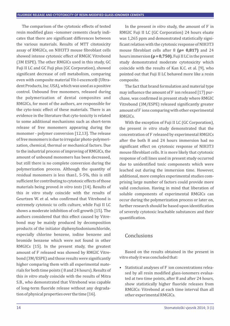
14 15Stomatološki vjesnik 2014; 3 (1) Stomatološki vjesnik 2014; 3 (1)
FLUORIDE RELEASE AND CYTOTOXICITY OF RESIN-MODIFIED GLASS-IONOMER CEMENTS Selimović-Dragaš M, Hasić-Branković L, Huseinbegović A, Kobašlija S, Hatibović-Kofman Š
The comparison of the cytotoxic effects of tested
resin modified glass –ionomer cements clearly indi-
cates that there are significant differences between
the various materials. Results of MTT citotoxicity
assay of RMGICs, on NIH3T3 mouse fibroblast cells
showed intense cytotoxic effect of RMGIC Vitrebond
(3M ESPE). The other RMGICs used in this study, GC
Fuji II LC and GC Fuji plus (GC Corporation), showed
significant decrease of cell metabolism, comparing
even with composite material Vit-l-escence® (Ultra-
dent Products, Inc. USA), which was used as a positive
control. Unbound free monomers, released during
the polymerization of dental composites and
RMGICs, for most of the authors, are responsible for
the cyto-toxic effect of these materials. There is an
evidence in the literature that cyto-toxicity is related
to some additional mechanisms such as short-term
release of free monomers appearing during the
monomer - polymer conversion [12,13]. The release
of free monomers is due to irregular photo-polymeri-
zation, chemical, thermal or mechanical factors. Due
to the industrial process of improving of RMGICs, the
amount of unbound monomers has been decreased,
but still there is no complete conversion during the
polymerization process. Although the quantity of
residual monomers is less than1, 5-5%, this is still
sufficient for contributing to cytotoxic effects of those
materials being proved in vitro tests [14]. Results of
this in vitro study coincide with the results of
Geurtzen W. et al. who confirmed that Vitrebond is
extremely cytotoxic to cells culture, while Fuji II LC
shows a moderate inhibition of cell growth [15]. The
authors considered that this effect caused by Vitre-
bond may be mainly produced by decomposition
products of the initiator diphenyliodoniumchloride,
especially chlorine benzene, iodine benzene and
bromide benzene which were not found in other
RMGICs [15]. In the present study, the greatest - amount of F released was showed by RMGIC Vitre-
bond (3M/ESPE) and those results were significantly
higher comparing them with all experimental mate-
rials for both time points ( 8 and 24 hours). Results of
this in vitro study coincide with the results of Mitra
S.B., who demonstrated that Vitrebond was capable
of long-term fluoride release without any degrada-
tion of physical properties over the time [16].
- In the present in vitro study, the amount of F in
RMGIC Fuji II LC (GC Corporation) 24 hours eluate
was 1,265 ppm and demonstrated statistically signi-
ficant relation with the cytotoxic response of NIH3T3
mouse fibroblast cells after 8 (ρ= 0,817) and 24
hours immersion (ρ = 0,750). Fuji II LC in the present
study demonstrated moderate cytotoxicity which
coincide with the results of Kan K.C. et al. [9], who
pointed out that Fuji II LC behaved more like a resin
composite.
The fact that brand formulation and material type -may influence the amount of F ion released [17] -
, was confirmed in present study where RMGIC
Vitrebond (3M/ESPE) released significantly greater -
amount of F ions comparing with other experimental
RMGICs.
With the exception of Fuji II LC (GC Corporation),
the present in vitro study demonstrated that the - concentration of F released by experimental RMGICs
after the both 8 and 24 hours immersion had no
significant effect on cytotoxic response of NIH3T3
mouse fibroblast cells. It is more likely that cytotoxic
response of cell lines used in present study occurred
due to unidentified toxic components which were
leached out during the immersion time. However,
additional, more complex experimental studies com-
prising large number of factors could provide more
valid conclusion. Having in mind that liberation of
soluble components of experimental RMGICs can
occur during the polymerization process or later on,
further research should be based upon identification
of severely cytotoxic leachable substances and their
quantification.
Conclusions
Based on the results obtained in the present in
vitro study it was concluded that:
-• Statistical analyses of F ion concentrations relea-
sed by all resin modified glass-ionomers evalua-
ted at two time points, after 8 and after 24 hours,
show statistically higher fluoride releases from
RMGICs: Vitrebond at each time interval than all
other experimental RMGICs.
pur
chase
• The evaluation of citotoxicity of resin modified
glass-ionomer cements on NIH3T3 mouse fibro-
blast cell lines showed that cytotoxic effect of
Vitrebond (3MESPE) was significantly higher
than all other materials as well as positive and
negative control.
• Correlation Coefficient between concentrations of
fluoride ion released by evaluated glass-ionomer
cements and cytotoxic response of NIH3T3 mouse
fibroblast cell line are relatively high, but do not
reach levels of biological significance.
According to the methodology employed in the
present study it can be concluded that experimental -
RMGICs liberated F as well as other soluble compo-
nents which diffused into the culture medium. These
components cannot be dismissed as possible
cytotoxic factors which contribute to the cytotoxicity
observed in this study.
Acknowledgements
This study was supported by Western University,
Canada and The Dental Faculty, University of Sara-
jevo on the basis of an international agreement bet-
ween both Universities. The authors gratefully
acknowledge colleagues at The Division of Oral
Biology at Schulich School of Medicine & Dentistry,
Western University, Canada and colleagues at the De-
partment of Medical Chemistry at the Medical Faculty
University of Sarajevo for their skilful technical
assistance.
Declaration of interest
There is not any conflict of interest for this mate-
rial in the manuscript for all authors, between the
authors, or for any organization.
References
7. Costa CAS, Hebling J, Garcia-Godoy F,Thomas Hanks C. In vitro cytotoxicity of five glass-ionomer cements. Biomaterials 2003; 24: 3853-58
10. Mosmann T. Rapid Colorimetric Assay for Cellular Growth and Survival: Application to Proliferation and cytotoxicity Assays. J Immunol Method 1983; 65: 55-63
1. Hatibović-Kofman S, Koch G, Ekstrand J. Glass ionomer materials as a rechargeable fluoride- release system. Int J Paediatr Dent. 1997; 7(2): 65-73
2. Hatibović-Kofman S, Koch G. Fluoride release from glass ionomer cement in vivo and in vitro. Swed Dent J 1991;15: 253-8
3. Forsten L. Fluoride release and uptake by glass ionomers and related materials and its clinical effect. Biomaterials 1998; Mar 19(6): 503-8
4. Selimović-Dragaš M. Jurić H. Glass-ionomer cements In: Z.R.Vuličević(ed) Materials for clinical application in children's dentistry Belgrade: Beobook ;2010.
5. Wiegard A, Buchalla W, Attin T. Review on fluoride releasing restorative materials – Fluoride release and uptake characteristics, antibacterial activity and influence on caries formation. Dental materials 2007; 23: 343-62
6. Stanislawski L, Daniau X, Lautiè A, Goldberg M. Factors Responsible for Pulp Cell Cytotoxicity Induced by Resin-Modified glass Ionomer Cements. J Biomed Mater Res ( Appl Biomater) 1999; 48: 277-88
8. Crisp S, Wilson A.D. Reactions in Glas Ionomer Cements: I decomposition of the powder. J Dent Res. 1974; 53(6): 1408- 13
9. Kan K.C, Messer L.B, Messer H.H. Variability in Cytotoxicity and Fluoride Release of Resin –modified Glass-ionomer Cements. J Dent Res 1997; 76(8):1502-07
11. Selimović-Dragaš M, Hasić-Branković L, Fehim
Korać F, Đapo N, Huseinbegović A, Kobašlija S,
Lekić M, Hatibović-Kofman Š. In vitro fluoride release from a different kind of conventional and resin modified glass-ionomer cements Bosn J Basic Med Sci 2013; 13 (3): 197-202

17Stomatološki vjesnik 2014; 3 (1) Stomatološki vjesnik 2014; 3 (1)
ORIGINAL SCIENTIFIC ARTICLE / ORIGINALNI NAUČNI RAD
ANALYSIS OF METALS RELEASED FROM DENTAL AMALGAM ALLOY USING INDICTIVELY COUPLED PLASMA-MASS SPECTROMETRY
1 2 1 1Bajsman A* , Turkušić E , Vuković A , Zukić S , 3 2Zukanović A , Kahrović E
1 Department for Dental Morphology, Dental Anthropology and Forensics, Faculty of Dentistry, University of Sarajevo, Sarajevo, Bosnia and Herzegovina2 Department for Chemistry, Faculty of Science, University of Sarajevo, Sarajevo, Bosnia and Herzegovina3 Department for Pediatric and Preventive Dentistry, Faculty of Dentistry, University of Sarajevo, Sarajevo, Bosnia and Herzegovina
ABSTRACT
Dental amalgam is a restorative material used in dentistry
from the very beginning. The basic chemical compounds of dental
amalgam alloy are silver, tin and mercury. In spite of the fact that
the interest or the esthetic restorative materials grow all over the
world, amalgam is still the most used material. The fact that one of
the main components of dental amalgam is elemental mercury
causes concern of patients as well as some governmental agencies
in a number of the world countries.
Aim of this paper is to analyze quantity of metals released from
four dental amalgams in artificial saliva solution in vitro, and to
estimate possible effects of those metals on health of patients
with dental amalgam fillings.
Material and method: Experimental material is presented by
four dental amalgams that differ in percentage of metals in
preamalgamated alloy. Electrodes were made of those amalgams
and immersed in artificial saliva solution (Duffo, Quezzada
Castillo), and elute was analyzed using inductively coupled
plasma - mass spectrometry (ICP-MS), for determination of
quantity of mercury, silver, tin, copper and zinc isotopes.
Conclusion: Quantities of mercury isotopes and isotopes of
other metals from dental amalgams in artificial saliva solution in
vitro could not be considered potentially harmful for patients
with dental amalgam fillings, regarding official statement of the
World Health Organization and other governmental agencies in
many countries around the world.
Key words: dental amalgam, ICP-MS, corrosion.
*Corresponding author
Anita Bajsman, PhD
Faculty of Dentistry,
University of Sarajevo
Bolnička 4a
71000 Sarajevo
Bosnia and Herzegovina
Phone: +387 33 214 249 (239)
e-mail: [email protected]
FLUORIDE RELEASE AND CYTOTOXICITY OF RESIN-MODIFIED GLASS-IONOMER CEMENTS
12. Beriat NC, Nalbant D.Water Absorption and HEMA Release of Resin-Modified Glass-Ionomers. Eur J Dent. 2009 Oct;3(4):267-72
13. Kranjevac T, Milovanović M, Volarević V, Lukić M.L, Arsenijević N, Marković D, et al. Cytotoxic effects of glass ionomer cements on human dental pulp stem cells correlate with fluoride release. Med Chem. 2012; 8(1): 40-45
14. Goldberg M. In vitro and in vivo studies on the toxicity of dental resin components: a review. Clin Oral Invest.2008; 12(1):1-8
15. Geurtzen W, Spahl W, Leyhausen G. Residual Monomer / Aditive Release and Variability in Cytotoxicity of Light –curing Glass-ionomer Cements and Compomers. J Dent Res 1998; 77(12): 2012-19
16. Mitra S.B. In vitro fluoride release from a light-cured glas-ionomer liner/base. J Dent Res 1991; 70(1):75-8
17. Marković D. Lj. Petrović B.B. Perić T.O. Fluoride content and recharge ability of five glass-ionomer dental materials. BMC Oral Health 2008; 8:21
16
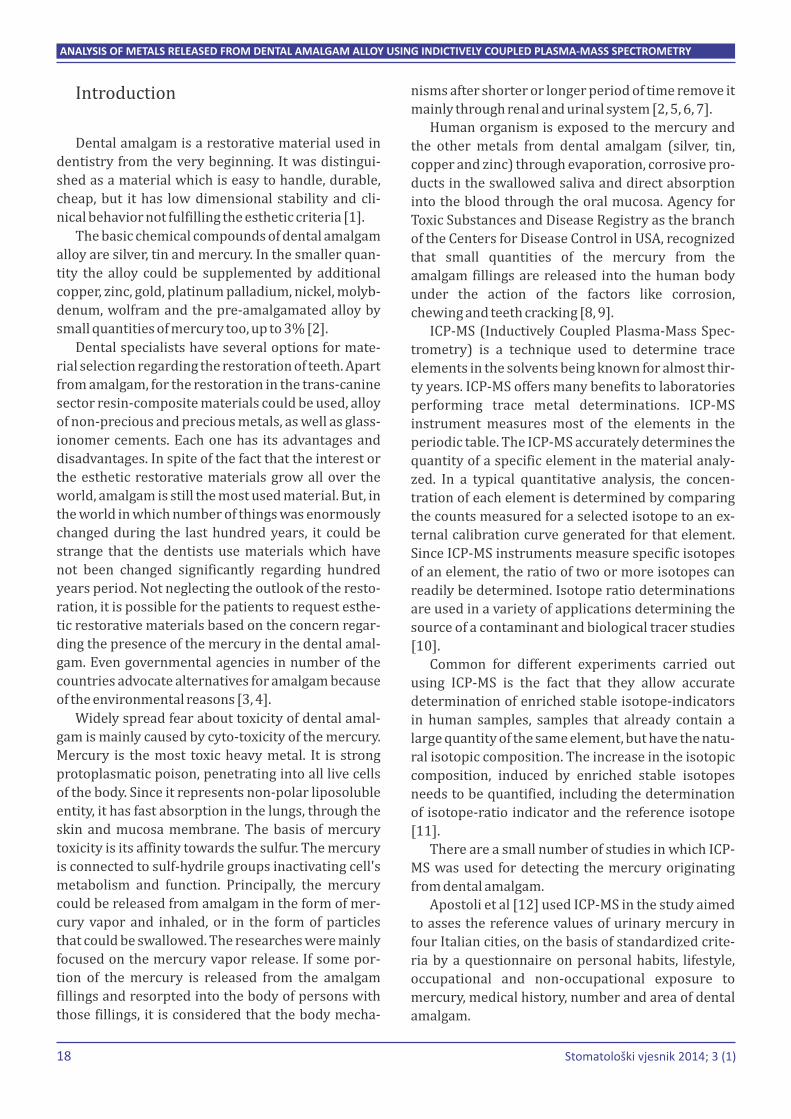
18 19
ANALYSIS OF METALS RELEASED FROM DENTAL AMALGAM ALLOY USING INDICTIVELY COUPLED PLASMA-MASS SPECTROMETRY Bajsman A, Turkušić E, Vuković A, Zukić S, Zukanović A, Kahrović E
Introduction
Dental amalgam is a restorative material used in
dentistry from the very beginning. It was distingui-
shed as a material which is easy to handle, durable,
cheap, but it has low dimensional stability and cli-
nical behavior not fulfilling the esthetic criteria [1].
The basic chemical compounds of dental amalgam
alloy are silver, tin and mercury. In the smaller quan-
tity the alloy could be supplemented by additional
copper, zinc, gold, platinum palladium, nickel, molyb-
denum, wolfram and the pre-amalgamated alloy by
small quantities of mercury too, up to 3% [2].
Dental specialists have several options for mate-
rial selection regarding the restoration of teeth. Apart
from amalgam, for the restoration in the trans-canine
sector resin-composite materials could be used, alloy
of non-precious and precious metals, as well as glass-
ionomer cements. Each one has its advantages and
disadvantages. In spite of the fact that the interest or
the esthetic restorative materials grow all over the
world, amalgam is still the most used material. But, in
the world in which number of things was enormously
changed during the last hundred years, it could be
strange that the dentists use materials which have
not been changed significantly regarding hundred
years period. Not neglecting the outlook of the resto-
ration, it is possible for the patients to request esthe-
tic restorative materials based on the concern regar-
ding the presence of the mercury in the dental amal-
gam. Even governmental agencies in number of the
countries advocate alternatives for amalgam because
of the environmental reasons [3, 4].
Widely spread fear about toxicity of dental amal-
gam is mainly caused by cyto-toxicity of the mercury.
Mercury is the most toxic heavy metal. It is strong
protoplasmatic poison, penetrating into all live cells
of the body. Since it represents non-polar liposoluble
entity, it has fast absorption in the lungs, through the
skin and mucosa membrane. The basis of mercury
toxicity is its affinity towards the sulfur. The mercury
is connected to sulf-hydrile groups inactivating cell's
metabolism and function. Principally, the mercury
could be released from amalgam in the form of mer-
cury vapor and inhaled, or in the form of particles
that could be swallowed. The researches were mainly
focused on the mercury vapor release. If some por-
tion of the mercury is released from the amalgam
fillings and resorpted into the body of persons with
those fillings, it is considered that the body mecha-
nisms after shorter or longer period of time remove it
mainly through renal and urinal system [2, 5, 6, 7].
Human organism is exposed to the mercury and
the other metals from dental amalgam (silver, tin,
copper and zinc) through evaporation, corrosive pro-
ducts in the swallowed saliva and direct absorption
into the blood through the oral mucosa. Agency for
Toxic Substances and Disease Registry as the branch
of the Centers for Disease Control in USA, recognized
that small quantities of the mercury from the
amalgam fillings are released into the human body
under the action of the factors like corrosion,
chewing and teeth cracking [8, 9].
ICP-MS (Inductively Coupled Plasma-Mass Spec-
trometry) is a technique used to determine trace
elements in the solvents being known for almost thir-
ty years. ICP-MS offers many benefits to laboratories
performing trace metal determinations. ICP-MS
instrument measures most of the elements in the
periodic table. The ICP-MS accurately determines the
quantity of a specific element in the material analy-
zed. In a typical quantitative analysis, the concen-
tration of each element is determined by comparing
the counts measured for a selected isotope to an ex-
ternal calibration curve generated for that element.
Since ICP-MS instruments measure specific isotopes
of an element, the ratio of two or more isotopes can
readily be determined. Isotope ratio determinations
are used in a variety of applications determining the
source of a contaminant and biological tracer studies
[10].
Common for different experiments carried out
using ICP-MS is the fact that they allow accurate
determination of enriched stable isotope-indicators
in human samples, samples that already contain a
large quantity of the same element, but have the natu-
ral isotopic composition. The increase in the isotopic
composition, induced by enriched stable isotopes
needs to be quantified, including the determination
of isotope-ratio indicator and the reference isotope
[11].
There are a small number of studies in which ICP-
MS was used for detecting the mercury originating
from dental amalgam.
Apostoli et al [12] used ICP-MS in the study aimed
to asses the reference values of urinary mercury in
four Italian cities, on the basis of standardized crite-
ria by a questionnaire on personal habits, lifestyle,
occupational and non-occupational exposure to
mercury, medical history, number and area of dental
amalgam.
Shraim et al [13] evaluated concentrations of
mercury and other metals in the wastewater of three
dental clinics and the influent of a wastewater
treatment plant in Al-Madinah Al-Munawarah using
ICP-MS.
The aim of this study was to analyze the quantity
of metals released from four dental amalgams in
artificial saliva solution, and on the basis of these
results to assess the potential impact of these metals
to the health of patients having the amalgam fillings.
Material and Methods
The experimental material in this study consisted
of four commercial dental amalgams, which differ in
the percentage share of individual metals in pre-alloy
(Table 1). Since those are encapsulated amalgams,
mercury and pre-alloy were mixed mechanically in
amalgamator, in a ratio 1:1, according to the manufac-
turer's instructions. Amalgam is manually condensed
into molds made of polyethylene as an inert material.
Moulds are cylindrical in shape, with the length of
140 mm and a diameter of 12 mm. Amalgam was
condensed into a prepared dock at one end of the
polyethylene mold, which is also cylindrical in shape
with a diameter of 8 mm, a depth of 2 mm. Those
electrodes, in fact, represented the artificial tooth.
Electrodes are numbered 1-4 (Figure 1).
Each electrode is inserted into a freshly prepared
solution of artificial saliva (Duffo, Quezada Castillo
[14], Table 2), at room temperature for 5 days. Every
24 hours, the sample of artificial saliva in which each
electrode was separately inserted was taken, and the
ICP-MS analysis was performed (Agilent ICPMS 7500
ce; Agilent, Waldbronn, Germany; Software: HP
ChemStation) (Figures 2 and 3).
Each sample of 1g elute was taken for analysis and
treated with 2 ml of nitric acid (for the determination
of silver). For each analysis three measurements we-
re performed and mean value for each element calcu-
lated, being shown as the final value. Thus, we get in-
formation regarding the presence and changes of
amounts of trace elements in artificial saliva solution,
which are released from any of the tested dental
amalgams. We determined the amount of corrosive
products released in the form of ions from each amal-
gam alloy, expressed in g/l - parts per billion (ppb).
Stomatološki vjesnik 2014; 3 (1) Stomatološki vjesnik 2014; 3 (1)
Table 1Experimental material
Table 2Composition of artificial saliva (Duffo, Quezada Castillo) (14)
Amalgam
Component
% Ag % Sn % Cu % Zn
Concentration (g/l)
Sample 1
Sample 2
Sample 3
Sample 4
NaCl
KCl
CaCl x 2H O2 2
KH PO2 4
Na HPO x 12H O2 4 2
KSCN
KHCO3
Citric acid
70
70
41
70
25.7
18
31
18.5
3.3
12
28
11
No data available
No data available
No data available
0.5
0,600
0,720
0,220
0,680
0,856
0,060
1,500
0,030
Figure 1 Amalgam electrodes
Figure 2 Agilent ICPMS 7500 ce
20 21Stomatološki vjesnik 2014; 3 (1) Stomatološki vjesnik 2014; 3 (1)
ANALYSIS OF METALS RELEASED FROM DENTAL AMALGAM ALLOY USING INDICTIVELY COUPLED PLASMA-MASS SPECTROMETRY Bajsman A, Turkušić E, Vuković A, Zukić S, Zukanović A, Kahrović E
Results
Tables 3, 4, 5 and 6 show the amounts of released
copper, zinc, tin and silver isotopes from four
experimental samples of dental amalgams, detected
by ICP-MS.
Although artificial saliva samples in which the
amalgams were immersed were taken each 24 hours
over five days, the analysis and interpretation of
results we observe showed only the results of isotope
concentration after the first 24 hours, considering
the human organism. Specifically, human saliva is a
very dynamic solution that is secreted during the day
and after some time becomes ingested in oral cavity.
None of the amalgam in the oral cavity can be con-
tinuously in contact with the same saliva during five
days, but data for the first 24 hours can give us insight
of the approximate quantities of certain elements
that can be released from dental amalgam during the
day. It is important to emphasize that in the interpre-
tation of results we observed total amount of dischar-
ged isotopes for each individual metals - copper, zinc,
silver, tin, mercury (Table 7). Considering data taken
from Al-Salehi et al [15], that average surface of amal-2
gam filling is 25 mm , and that surface of our amal-2
gam samples is 50,24 mm , that daily saliva produc-
tion is 770 ml, or 8 ml each 15 minutes [16], the
following quantities of discharged metals from amal-
gam fillings during the first 24 hours were obtained
(Table 7).
Discussion
While interpreting the results it was presumed
that, on the basis of the amount of detected isotopes
released into artificial saliva solution from dental
amalgam alloy, maximum allowable daily concentra-
tions of these elements and they impact to the human
organism can be predicted. But, we have to bear in
mind all the advantages and disadvantages of in vitro
studies.
From the standpoint of electrochemical corrosion,
it is important to emphasize that dental amalgams, in
the metallurgical point of view, are the most complex
biomaterials. Dental amalgam is a dynamic material
in which the reactions take place in solid state over a
long time period. Since these reactions occur in a
complex and variable oral environment, it is obvious
that in vitro studies can only reflect the actual in vivo
conditions to certain extent [17].
Determination of mercury released and adsorbed
from dental amalgam in vivo is difficult and complex.
Dodes [18] refers to studies of Olsson and Bergman
citing the following factors as variables that affect the
amount of mercury released from amalgam restora-
tions: the number of teeth, the number of restored
surfaces, basal release of mercury, the factors that
increase the release, as chewing and brushing teeth,
alimentary and hygiene habits, mouth breathing, the
ratio of oral and nasal respiration, ingestion, absorp-
tion, inhalation, ingestion absorption, and body
weight.
Most measurements of release of mercury and
mercury vapor from dental amalgams in the current
research were performed on dry amalgam surfaces,
and they may not be relevant to the restorations of
the oral environment. Amalgam restorations in the
oral cavity are normally covered with a film of saliva.
It was determined that the release of mercury vapor
was drastically inhibited by the presence of saliva
film on the surface of amalgam. The size of this
inhibition casts a shadow of doubt on the application
of in vitro measurements of mercury vapor from the
abraded amalgam surfaces, in terms of valuation
acceptable levels of mercury vapors in the respira-
tory environment defined by the World Health
Organization (WHO) [19].
Mercury is the most noble of the main elements in
dental amalgam. Under conditions dominating in the
Figure 3 Agilent ICPMS 7500 ce
Table 3Concentrations of released isotopes of copper, zinc, tin,
silver and mercury from amalgam sample 1.
Table 4Concentrations of released isotopes of copper, zinc, tin,
silver and mercury from amalgam sample 2.
Conc.(ppb)/day
Conc.(ppb)/day
Day 5
Day 5
Day 4
Day 4
Day 3
Day 3
Day 2
Day 2
Day 1
Day 1
63 Cu/ppb
65 Cu/ppb
66 Zn/ppb
68 Zn/ppb
107 Ag/ppb
109 Ag/ppb
118 Sn/ppb
120 Sn/ppb
200 Hg/ppb
202 Hg/ppb
63 Cu/ppb
65 Cu/ppb
66 Zn/ppb
68 Zn/ppb
107 Ag/ppb
109 Ag/ppb
118 Sn/ppb
120 Sn/ppb
200 Hg/ppb
202 Hg/ppb
24,5
24,4
341,7
346,7
6,6
6,7
50,6
50,2
7,7
7,9
38,5
38,0
696,3
705,7
3,3
3,3
275,5
271,4
9,1
8,4
112,7
112,0
92,7
94,9
10,5
10,6
93,6
93,3
6,6
6,6
44,4
44,2
1140,0
1156,0
4,8
4,8
37,9
37,3
14,2
14,9
29,9
28,5
50,0
50,0
5,9
5,9
166,0
166,2
5,5
5,8
63,9
63,1
617,2
627,6
8,1
8,1
63,5
63,2
15,1
15,1
37,4
36,4
50,0
50,0
9,2
9,4
290,3
287,1
4,8
5,5
86,4
85,6
637,0
648,7
8,5
8,4
76,5
76,8
13,5
13,7
48,6
47,8
144,5
148,4
10,8
10,9
404,5
404,5
8,5
8,6
98,1
97,5
718,5
728,1
16,5
16,5
104,7
103,7
29,2
29,8
Table 5Concentrations of released isotopes of copper, zinc, tin,
silver and mercury from amalgam sample 3.
Table 6Concentrations of released isotopes of copper, zinc, tin,
silver and mercury from amalgam sample 4.
Conc.(ppb)/day
Conc.(ppb)/day
Day 5
Day 5
Day 4
Day 4
Day 3
Day 3
Day 2
Day 2
Day 1
Day 1
63 Cu/ppb
65 Cu/ppb
66 Zn/ppb
68 Zn/ppb
107 Ag/ppb
109 Ag/ppb
118 Sn/ppb
120 Sn/ppb
200 Hg/ppb
202 Hg/ppb
63 Cu/ppb
65 Cu/ppb
66 Zn/ppb
68 Zn/ppb
107 Ag/ppb
109 Ag/ppb
118 Sn/ppb
120 Sn/ppb
200 Hg/ppb
202 Hg/ppb
34,4
34,1
221,2
226,4
3,9
3,9
46,6
46,6
0,8
1,1
96,8
97,1
372,3
381,4
0,7
0,7
5,0
4,9
5,0
5,0
45,0
44,3
261,5
269,9
12,0
12,1
146,9
149,8
5,0
5,0
29,9
29,7
63,1
65,1
1,7
1,7
315,4
312,1
5,0
5,0
49,9
49,1
50,0
50,0
3,3
3,2
579,7
736,2
5,0
5,0
40,0
39,0
50,0
50,0
14,9
14,9
495,3
470,0
17,3
18,1
61,4
60,1
17,0
21,5
6,5
6,5
108,2
108,5
1,1
1,5
96,2
95,0
3,6
4,1
12,8
12,6
18,6
17,7
11,7
12,3
75,6
75,0
50,0
50,0
8,0
8,0
145,4
145,7
5,4
5,2
61,9
61,2
802,5
816,2
1282,0
1388,0
46,1
46,0
27,1
27,9
Table 7Amounts of metals released from average sized amalgam
filling through 24 hours
Sample / Quantity of released metal (µg)
Sample 1 Sample 2 Sample 3 Sample 4
Cu
Zn
Ag
Sn
Hg
18,73
263,76
5,07
38,62
5,97
29,30
537,19
2,52
209,55
6,71
26,25
171,50
2,99
35,71
0,73
74,30
288,79
0,54
3,80
3,83

22 23
PRESENCE OF CANDIDA SPECIES IN SUPRA-GINGIVAL DENTAL PLAQUE AND CARIOUS DENTINE AT CHILDREN WITH EARLY CHILDHOOD CARIES
Stomatološki vjesnik 2014; 3 (1) Stomatološki vjesnik 2014; 3 (1)
ANALYSIS OF METALS RELEASED FROM DENTAL AMALGAM ALLOY USING INDICTIVELY COUPLED PLASMA-MASS SPECTROMETRY Bajsman A, Turkušić E, Vuković A, Zukić S, Zukanović A, Kahrović E
oral cavity, silver and mercury have the lowest elec-
trochemical activity. However, despite the limited
tendency to dissolve electrochemically, these ele-
ments can form almost insoluble sulfides [17]. Okabe
et al [20] refer to comprehensive review of the lite-
rature on in vivo and in vitro corrosion of dental
amalgam, which states that it is not even one product
of corrosion of dental amalgam in which the main
constituent is mercury. Therefore, states that, regard-
less to the composition of amalgam, mercury relea-
sed into an aqueous medium is usually kept in so-
lution, or is re-amalgamated with residual alloy.
According to American Dental Association (ADA),
mercury amalgam releases only 1-3 µg of mercury
per day, comparing to 5 – 6 µg that most humans
ingest daily, by food, water or air. Therefore, ADA
states that 500 fillings would be necessary in order to
achieve adverse effects on human organism. To the
contrary, according to WHO investigations, only one
filling can release between 3 and 17 µg of mercury
per day [8], which is consistent to the results of this
study.
Al-Salehi et al [15] state that Macker and Berglund
reported that non-stimulated amount of mercury
release from dental amalgam is on average 0, 4 μg per
surface, daily, and that it was calculated in six diffe-
rent in vivo studies. Assuming that amalgam surface
in vivo is 5mm x 5mm, they calculated that the
amount of mercury release from one amalgam is 1, 6 2μg/cm per day. Assuming that amalgam surface of 1
2cm equals four amalgam surfaces in vivo, value of the
release of mercury in our study is: Sample 1 - 23,7 2 2μg/cm , Sample 2 – 26,84 μg/cm , Sample 3 – 2,92 2 2
μg/cm , and Sample 4 – 15,32 μg/cm . The results are
within the maximum acceptable daily intake of 45 μg,
prescribed by the WHO, and also within the amount
of 300-500 mg per day, seven days a week, for which
the WHO considers not causing any effect. The
amount of 300-500 μg of mercury per day is 10 times
greater than the amount ingested by patient with 12
average-sized amalgam fillings, where calculated
level of fecal excretion is 60 μg [5, 21].
Particular attention should be paid to Sample 3,
which released the least amount of mercury in this
study. It is important to emphasize that it is amalgam
with very large proportion of copper (28%), leading
to the conclusion that in this way the elimination of γ2
phase, as the locus minoris resistentiae from the
aspect of corrosion process was achieved, and higher
proportion of η phase in the process of amalgamation
was achieved, thus improving corrosion resistance.
According to Al-Salehi et al, the patient should
have about 160 amalgam restorations so that mer-
cury release could reach the maximum acceptable
daily intake [15, 22].
Al-Salehi et al tested influences of carbamide-
peroxide [15] and hydrogen-peroxide [22], as tooth
bleaching agents, on mercury release from dental
amalgam, using ICP-MS. Concentrations of tested
agents were 1%, 3%, 10% i 30%. Concentrations of
released mercury after 24 hours on 37°C were bet-
ween 360 µg/l and 1428 µg/l. Those amounts are
much higher than the amounts in our investigation,
probably according to different test medium and
different temperature.
ADA in its report in 1998 related to the Halbach
research, who examined the release of mercury from
amalgam fillings in vivo in a group of 20 patients with
one to forty-six amalgam surfaces in the oral cavity
[23]. Dose of released mercury in that investigation
was between 0, 3-13, 9 μg per day, 4, 5 μg per day on
average. The same researcher came to essentially
same results with another group of subjects, where
average daily dose of mercury derived from amalgam
was 4, 8 μg. Halbach showed that the release of
mercury is linearly associated with time and the total
area of restorations. The Halbach results largely cor-
respond to the results of this study being very im-
portant, considering the fact that our study is in vitro.
Determination of quantity of mercury released
from dental amalgam in various scientific studies
varies significantly. It is obviously difficult to make
direct comparisons of different data, due to the lack
of standardization of experimental details and form
of data presentation [22]. The abovementioned
applies to the results of this study.
For the amount of mercury released from dental
amalgam in vivo, many factors, including number and
age of the restorations, amalgam type, total area and
the quality of restoration, mercury measurement
methods, individual sensitivity of subjects and access
to data analysis, may be responsible for different
results of the assessment exposure to mercury from
dental amalgam. The effect of meals consumed is also
important for the release of mercury, which is not
consistent. It is shown for some food to reduce intra-
oral mercury vapor [23].
Although the research focus regarding the poten-
tial toxicity of amalgam is oriented towards mercury,
amalgam contains other substances that can be
neuro-toxic in high doses. Amalgam is an alloy of
mer-cury, zinc, tin, copper and silver, and each of
these elements can be neuro-toxic in high doses.
Analysis of data collected by research of Lobner and
Asrari indi-cates that the toxicity of amalgam is not
conditioned by release of mercury. Each of amalgam
components (Zn, Sn, Cu, Ag) is toxic, although even
high concen-trations of tin (200 μM) cause only about
15% of cell death [24].
The idea that the release of zinc from dental
amalgam is toxic is not new. It has been shown that
the amalgams which contain zinc are more toxic than
those without zinc. Taking into account the percen-
tage of zinc in dental amalgam, it surprises that zinc is
the first toxic substance to be released. It has been
shown that zinc is basic corrosion product released
from amalgam. Amalgam contains enough zinc that
could be responsible for toxic influence on nerve cells
in culture [24].
Zinc is essential element. Daily intake of zinc is 12-
15 mg. It is estimated that human body contains total
amount of 2, 5 g of zinc. Poisoning due to excessive
zinc ingestion is rare. Evidence of hematological, he-
patic or renal toxicity was not observed in persons
who ingested 12 g of elemental zinc in 2 [6].
Average sized amalgam fillings made of amalgams
tested in this research release 171, 5 μg (Sample 3) to
573, 19 μg (Sample 2) of zinc per day. These amounts
are far below the maximum amount allowed.
Copper is essential trace element that catalyses
process of hem-synthesis and absorption of iron. It is
third of the most common trace elements in human
organism, after iron and zinc. Corrosion products of
copper from high-copper dental amalgam alloys after
exposure to saliva include oxides, chloride oxides and
hydroxides, chlorides and sulphides of copper. Ave-
rage daily intake of copper is between 1-3 mg, and 2-5
mg, and primary source is food. Emetic dose of
copper sulfate for adults is 0, 25 to 0, 5 g, and this cop-
per sulfate can be described as copper. Toxic con-
centrations of copper can be achieved after ingestion
of 1g of copper. Ingestion of fluids or solid food that
contains minimally 25 mg/l of copper is connected
with symptoms of acute gastroenteritis. Established
lethal dose of copper in untreated adults is about 10-
20 g Cu [6, 25, and 26].
Average sized amalgam fillings made of amalgams
tested in this research release 18, 73 μg (Sample 1), to
74, 3 μg (Sample 4) of copper per day. These amounts
are far below the amount that could cause adverse
effects on human organism.
Data of tin toxicity are very limited. Tin can be
detected in all human tissues and accumulated
amount increases with age. The largest part of the tin
in the tissues is a consequence of ingestion of metals.
It is estimated that daily intake is 1-38 mg, and the
intake is higher with greater consumption of canned
food, because the inside of the cans is coated with tin.
Except for rare reports of gastrointestinal disorders,
there is very little evidence of significant toxicity of
tin for humans [26].
Quantities of tin released from average sized
amalgam fillings made of amalgams tested in this
research are between 3, 8 µg (Sample 4) to 209, 55 µg
(Sample 2). There is a small possibility that this
quantity of ingested tin in vivo could lead to unin-
tended consequences.
One of the possible ways of entering the silver in
the human organism is its release from dental amal-
gam. Soluble silver compounds are more easily ab-
sorbed than metallic or insoluble silver, and may have
the potential to cause adverse effects on human
organism (i.e. argyria or argyrosis). However, it is im-
portant to emphasize that the silver in any form is not
considered toxic to the immune, cardiovascular, ner-
vous, or reproductive system, and it is not considered
carcinogenic [27].
Quantities of sliver released from average sized
amalgam fillings made of amalgams tested in this
research are between 0, 54 µg (Sample 4) to 2, 99 µg
(Sample 3). Those quantities participate in very small
degree in daily intake of silver in human body.
Conclusions
The quantity of release of certain metals, as iso-
topes, from dental amalgams was determined in this
research using ICP-MS.
1. The release of all four main metals (zinc,
copper, tin, silver and mercury) from tested dental
amalgam alloys was proved.
2. According to our in vitro results, quantities of
mercury released from each amalgam tested could
not be considered as hazard to patient's health. It has

24 25Stomatološki vjesnik 2014; 3 (1) Stomatološki vjesnik 2014; 3 (1)
ANALYSIS OF METALS RELEASED FROM DENTAL AMALGAM ALLOY USING INDICTIVELY COUPLED PLASMA-MASS SPECTROMETRY Bajsman A, Turkušić E, Vuković A, Zukić S, Zukanović A, Kahrović E
been shown that for each tested amalgam, the pre-
sence of large number of average sized amalgam
fillings in oral cavity would be necessary to achieve
the maximum allowed daily intake identified as such
by the WHO, which is 45 µg. Since the official position
of WHO is that the amount of 300-500 µg of mercury
per day, seven days a week, do not lead to any adverse
effects, it can be concluded that the patient could
have significantly higher number of amalgam fillings
in the oral cavity, made of amalgams tested, without
any health effects.
3. Release of the isotopes of mercury and other
metals from dental amalgam composition, in this in
vitro study, does not reach the amount that could be
considered potentially hazardous to the health of the
patient, considering the official position of WHO and
governmental agencies in many countries around the
world.
Acknowledgement
This research is part of scientific project No. 11-
14-21625.1/07, supported by Ministry of Education
and Science of the Sarajevo Canton. The authors
would like to express their gratitude to Ao. Univ. Prof.
Kurt Kalcher and Institute for Analytical Chemistry,
Karl-Franzens University, Graz, Austria, for extraor-
dinary help during the experimental procedure.
References:
1. Soler JI, Elacuria J, Triana R, Guinea E, Osborne JW. A History of Dental Amalgam. Journal of History of Dentistry, 2002;50:109-16.
2. Prpić-Mehičić G, Galić N. Dental amalgam. In: Jerolimov V, Editor: Basis of dentistry materials monograph on the internet. Zagreb: University in Zagrebu, Faculty of Dentistry; 2005 cited 2011 Nov 8. Available from: .
3. Edlich RFC, Cross CL, Dahlstrom JJ, Long WB, Newkirk AT. Implementation of revolutionary legislation for informed consent for dental patients receiving amalgam restorations. Journal of Environmental pathology, Toxicology and Oncology, 2008;27:1-3.
4. Burke FJT. Amalgam To Tooth Colored Materials – Implications for Clinical Practice and Dental Edu-cation: Governmental Restrictions and Amalgam Usage Survey Results. Journal of Dentistry, 2004; 32:343-350.
5. Mackay R. Is dental amalgam safe? Journal of Aus-tralian College of Nutrition and Environmental Medicine, 1993;12:5-12.
6. Hl PJ. Trace elements in persons with dental amalgam – the role of chelating agent PhD Thesis. Bergen: University of Bergen, 2003.
7. Clarkson TW, Mahos L. The toxicology of mercury and its chemical compounds. Critical Reviews in Toxicology, 2006;36:609-62.
8. Chirba-Martin MA, Welshhans CM, An uncertain risk and an uncertain future: assessing the legal implications of mercury amalgam fillings. Health Matrix, 2004:14;293-324.
9. FathiM, Mortazavi V. A review on dental amalgam corrosion and its consequences. Journal of Re-search in Medical Sciences, 2004:1;42-51.
10. PerkinElmer Internet. Waltham. The 30-Minute Guide to ICP-MS cited 2011 Nov 7 ; about 8 p.. Available from: http://www.icp.hacettepe.edu.tr/ A30MinutesGuideToICPMS.pdf.
11. Stürup S. The use of ICP-MS for stable isotope tracer studies in humans: a review. Analytical and Bioanalytical Chemistry, 2004:378;273-282.
12. Apostoli P, Cortesi I, Mangili A, Elia G, Drago I, Gagliardi T et al. Assessment of reference values for mercury in urine: the results of an Italian poly-centric study. Science of the Total Environment, 2002;289:13-24.
13. Shraim A, Alsuhaimi A, Al-Thakafy JT. Dental clinics: a point pollution source, not only of mercury but also of other amalgam constituents. Chemosphere, 2011;84:1133-1139.
14. Duffo GS, Quezada Castillo E. Development of an artificial saliva solution for studying the corro-sion behavior of dental alloys. Corrosion 2004;60:594-602.
15. Al-Salehi SK, Hatton PV, Miller CA, McLeod C, Joiner A. The effect of carbamide peroxide treat-ment on metal ion release from dental amalgam. Dental Materials 2006;22:948-953.
16. Malić M. Salivary glands diseases. In: Topić B, stEditor. Oral medicine 1 ed. Sarajevo: Faculty of
Dentistry University in Sarajevo; 2001.p.217-233.
17. Olson S, Berglund A, Bergman M. Release of elements due to electrochemical corrosion of dental amalgam. Journal of Dental research 1994;73:33-43.
18. Dodes JE. The amalgam controversy – an evi-dence-based analysis. JADA 2001;132:248-256.
19. Mahler DB, Adey JD, Simms LE, Marek M. Influ-ence of liquid films on mercury vapor loss from dental amalgam. Dental Materials 2002;18:407-412.
20. Okabe T, Elvebak B, Carrasco L, Ferracane JL, Keanini RG, Nakajima H. Mercury release from dental amalgams into continuously replenished liquids. Dental Materials 2003;19:38-45.
21. Osborne JW. Dental amalgam and mercury vapor release. Advances in Dental Research 1992; 6:135-138.
22. Al-Salehi SK, Hatton PV, McLeod CW, Cox AG. The effect of hydrogen peroxide concentration on
metal ion release from dental amalgam. Journal of Dentistry 2007;35:172-176.
23. ADA Council on Scientific Affairs. Dental amal-gam: Update on safety concerns. JADA 1998; 129:494-503.
24. Lobner D, Asrari M. Neurotoxicity of dental amalgam is mediated by zinc. Journal of Dental Research 2003;82:243-246.
25. Barceloux DG. Copper. Clinical Toxicology 1999; 37:217-230.
26. Louria DB, Joselow MM, Browder AA. The human toxicity of certain trace elements. Annals of Internal Medicine 1972;76:307-319.
27. Drake PL, Hazelwood KJ. Exposure-related health effects of silver and silver compounds: A review. Annals of Occupational Hygiene 2005;49:575-585.

27Stomatološki vjesnik 2014; 3 (1)
ORIGINAL SCIENTIFIC ARTICLE / ORIGINALNI NAUČNI RAD
DISTRIBUTION OF WHITE SPOTSAFTER DE-BONDING IN ORTHODONTIC PATIENTS
1 2Zabokova-Bilbilova E* , Muratovska I , 2 3 4Stojanovska V , Stefanovska E , Dimova C
1 Department of Pediatric and Preventive Dentistry, University Dental Clinical Centre "St. Pantelejmon", Faculty of Dentistry, Skopje, R. Macedonia2 Department of Cariology and Endodontology, University Dental Clinical Centre "St. Pantelejmon", Faculty of Dentistry, Skopje, R. Macedonia3 Department of Periodontology and Oral Pathology, University Dental Clinical Centre "St. Pantelejmon", Faculty of Dentistry, Skopje, R. Macedonia4 Faculty of Medical Sciences, General Stomatology, "Goce Delcev" University-Stip, R. Macedonia
ABSTRACT
Fixed orthodontic appliances can interfere with removing bacterial plaques from dental surfaces which can ultimately lead to white spot formation. The aim of this study was to evaluate the quantity of white spots lesions in orthodontic patients treated with fixed orthodontic appliances during different treatment periods and to reveal the teeth most commonly affected by white spot lesions.
Materials and Methods: A total of 30 patients undergoing or scheduled for fixed orthodontic treatment were divided into two groups. The patient's sample was divided according to the duration of fixed orthodontic appliance treatment. In the first group treatment time ranged from 1 to 1.5 year, and in the second group from 1.5 to 2 years respectively. The patients were selected randomly during regular recall visits to the Orthodontic Clinic at the University Dental Clinic Center in Skopje. The patients were examined for the presence of white spot before insertion of the appliance and after its removal using visual examination.
Results: This study showed that there was a significant increa-se in the formation of white spot lesions along with the increase in treatment duration. The first molars were the most affected teeth by white spots followed by canines and second premolars.
Conclusion: The results of the study showed an increase of white spots formation with the increase of orthodontic treatment duration. The orthodontists should educate their patients to per-form and maintain good oral hygiene by proper brushing techni-ques including all teeth not emphasizing anterior teeth only.
Key words: white spots lesions, fixed orthodontic appliance.
*Corresponding author
Dr Efka Zabokova-Bilbilova DDS, PhD
Department of Pediatric
and Preventive Dentistry,
University Dental Clinical
Centre "St. Pantelejmon",
Faculty of Dentistry Skopje
Vodnjanska 17, 1000 Skopje
Macedonia
e-mail: [email protected]

28 29Stomatološki vjesnik 2014; 3 (1) Stomatološki vjesnik 2014; 3 (1)
DISTRIBUTION OF WHITE SPOTS AFTER DE-BONDING IN ORTHODONTIC PATIENTS Zabokova-Bilbilova E, Muratovska I, Stojanovska V, Stefanovska E, Dimova C
Introduction
Dental caries is a common complication of ortho-
dontic treatment with fixed orthodontic appliances.
The clinical manifestation of early enamel caries is
presented white spot lesions (WSLs). WSLs are the
opaque white appearance of enamel lesion resulting
from subsurface demineralization which creates po-
res between enamel rods and results in changes of
the enamel optical properties [1, 2, 3]. Prevention of
demineralization during orthodontic treatment has
been a challenging issue orthodontists faced with
although caries prevention measures have improved
over the last years. Due to the presence of fixed ortho-
dontic appliances, it is difficult for patients to main-
tain adequate oral hygiene, as orthodontic appliances
increase the number of plaque retention areas on
teeth surfaces that are normally less susceptible to
caries development [4, 5, 6].
Fixed orthodontic appliances create stagnation
areas for plaque and make tooth cleaning difficult.
Brackets, arch wires, ligatures, and other orthodontic
appliances complicate the use of conventional oral-
hygiene measures. This often results in significant
plaque accumulation around the bracket bases.
Demineralization of enamel around brackets can be
an extremely rapid process and appears most fre-
quently on the cervical and middle thirds of the
buccal surfaces of the maxillary lateral incisors, the
mandibular canines, and the first premolars [7, 8].
De-mineralized enamel can re-mineralize after de-
bonding under favorable conditions [9, 10].
An insight into available literature on the preva-
lence of WSLs revealed that most relevant studies re-
ported the presence of these lesions at the comple-
tion of orthodontic treatment [11, 12]. Depending on
the examination technique used, the prevalence of
WSLs varies. Boersma et al. [13] using quantitative
light fluoroscopy investigated the prevalence of
WSLs at the end of orthodontic treatment and repor-
ted that 97% of patients had one or more lesions. In
light of these studies, one may conclude that demi-
neralization is a significant clinical problem resulting
in an unacceptable esthetic presentation that, in so-
me severe cases, may require restorative treatment.
Epidemiological studies found that the most
commonly affected teeth are molars, maxillary lateral
incisors, mandibular canines and premolars. A study
by Øgaard et al. found that the level of visible plaque
around the appliance, shortly after bonding, was the
best predictor for white spot lesion at de-bonding
[14].
Prevalence of WSLs in the literature ranges widely
from (2-97%). Sagarika et al. found a prevalence of
75.6% among orthodontic patients compared to
15.6% among control group [15]. Much of the varia-
tion in detecting WSLs was due to the method of de-
tection with the highest prevalence of demineraliza-
tion found when light induced fluorescent method
had been used. Similar devices mainly based on
optical phenomena, like digital imaging fiber-optic
trans-illumination (DIFOTI) and red laser-light indu-
ced fluorescence, are also considered as more accu-
rate methods in relation to direct visual examination.
Material and Methods
A total of 30 patients (14 female and 16 male)
undergoing or scheduled for fixed orthodontic treat-
ment were divided into two groups. The patient's
sample was divided according to the duration of fixed
orthodontic appliance treatment. In the first group,
treatment time ranged from 1 to 1.5 year, and in the
second group from 1.5 to 2 years respectively. The
patients were selected randomly during regular
recall visits to the Orthodontic Clinic at the University
Dental Clinic Center in Skopje. Selection criteria
were: aged of 16-18 years, healthy individuals and a
treatment period with fixed appliance. Only tooth
surfaces gingival to the arch wire were examined for
the presence of WSLs, as this is the area most prone to
enamel demineralization during orthodontic treat-
ment. The patients were seen regularly every 3-4
weeks for the orthodontic follow-up visits. During
each visit, the patients were motivated to perform
proper tooth brushing techniques and maintain good
oral hygiene. When orthodontic treatment was
finished, the bands and brackets were removed, the
remaining cement and composite was removed using
sharp hand instruments and all teeth were polished.
The facial surfaces of the teeth were examined in
order to detect white spot formation using a mirror
and an explorer. Even in non extraction cases the first
premolars were excluded from the study. The follo-
wing modified WSLs scoring system introduced by
Gorelick et al. [16] was used for the visual exami-
nation:
Score 0 = No visible white spots or surface dis-
ruption (no demineralization),
Score 1 = Visible white spot without surface dis-
ruption (mild demineralization),
Score 2 = Visible white spot lesion having a
rough-ened surface but not requiring a
restoration (moderate demineraliza-
tion),
Score 3 = Visible white spot lesion requiring
restoration (severe demineralization).
Visual examination was performed by the same
dentist for all patients. Each tooth was air-dried for 5
seconds and the buccal surface was examined
visually for enamel decalcification in the portion of
the crown gingival to the bracket using the following
scale.
Statistical Analysis
The increasing WSL index before and after a
treatment was determined. Statistical analyses were
performed using SPSS version 6. Since the scoring
system was used for determination, nonparametric
tests were used for statistical analysis. Descriptive
statistics were obtained for all groups. Kruskal-
Wallis and Mann-Whitney U tests were used to
compare the groups. A p-value, 0.05 was considered
to be statisti-cally significant.
Results
All the results are shown in tables 1, 2, 3, 4, 5 and 6.
Tables 1 and 2 showed the descriptive statistics of
white spots in upper and lower jaws of male subjects.
There was an increase in the scores of white spots
with the increase of treatment duration time. The
first molars were affected by white spots formation
more often than the other teeth followed by the
second premolars and canines. Tables 3 and 4
showed the descriptive statistics of white spots in
upper and lower jaws of female subjects.
Comparison of white spots among teeth at each
duration of time, right dental side versus left, upper
teeth versus lower and male versus female in each
treatment duration time is presented in Table 5.
There were significant differences in the frequency of
UpperRight
LowerRight
UpperLeft
LowerLeft
Totalupper
Totallower
teeth
1
2
3
5
6
1
2
3
5
6
8
8
8
8
8
8
8
8
8
8
80 92 800,11 0,340,25 0,67,5 12,5- -- -80
0
0
0
0
0
0
0
0
0
0
0 0
0
0
0
0
0
0
0
0
0
0
0
0
1
0
1
0
0
1
0
1
1 1
0
0
0,14
0
0,2
0
0
0,15
0
0,17
0
0
0,35
0
0,7
0
0
0,4
0
0,3
100
100
82,5
100
70
100
100
75,5
0
80,5
100
100
83
83
32
100
100
65
83
52
-
-
16
-
31
-
-
15
-
14
-
-
-
15,6
30
-
-
32
15,6
30
-
-
-
-
-
-
-
-
-
-
-
-
-
-
-
-
-
-
-
-
-
-
-
-
-
-
-
-
-
-
-
-
-
-
-
-
-
-
-
-
0
0
1,1
0,4
0,8
0
0
0,9
0,5
0,9
0
0
0,05
0,7
1
0
0
0,34
0,6
0,5
0
0
1
0
1
0
0
1
1
1
8
8
8
8
8
8
8
8
8
8
1
2
3
5
6
1
2
3
5
6
N min max mean SD0 01 12 23 3
SDmeanmaxminNteeth
Table 1Descriptive Analysis of White Spots in Male Group (Frequency of Score %)
Treatment time ranging from 1 to 1.5 year

31Stomatološki vjesnik 2014; 3 (1)
DISTRIBUTION OF WHITE SPOTS AFTER DE-BONDING IN ORTHODONTIC PATIENTS Zabokova-Bilbilova E, Muratovska I, Stojanovska V, Stefanovska E, Dimova C
white spots between teeth in upper and lower right
quadrant at duration [1] in female group, the first
molar shows the highest frequency of white spots
followed by the canine in the upper jaw. In the lower
right quadrant the first molars were most commonly
affected followed by canines and second premolars.
In duration [2] in male group the upper arch shows
significantly higher frequency of white spots than the
lower. In females the lower left quadrant displayed
significant difference in the distribution of white
spots among teeth and again the first molars were
affected by white spots more often than the other
teeth followed by the canines (Table 6).
Discussion
The first clinical evidence of the demineralization
is the white spot lesion (WSLs), which potentially can
become a cavitated carious lesion extending even
into the dentin. White spot lesions are no fluoridated
opacities having a more defined shape and are well
differentiated from surrounding enamel which is
often located in the middle of the tooth. The WSLs has
been defined as "subsurface enamel porosity from
carious demineralization" presenting itself as a "mil-
ky white opacity" when located on smooth surfaces.
In addition to the fact that WSLs is the first step to de-
UpperRight
LowerRight
UpperLeft
LowerLeft
Totalupper
Totallower
teeth
1
2
3
5
6
1
2
3
5
6
8
8
8
8
8
8
8
8
8
8
80 75 800,25 0,340,35 0,628 6,1- -- -80
0
0
0
0
0
0
0
0
0
0
0 0
0
0
0
0
0
0
0
0
0
0
0
1
1
0
1
0
1
1
1
1
1 1
0
0,3
0,3
0
0,3
0
0,3
0,2
0,2
0,2
0
0,4
0,4
0
0,4
0
0,4
0,4
0,4
0,4
100
65
65,5
100
65,5
100
65,5
65
0
65,5
100
100
100
100
65,5
100
100
65,5
100
100
-
32,3
32.3
-
32,3
-
32,3
32,2
32,3
32,3
-
-
-
-
32
-
-
32
-
-
-
-
-
-
-
-
-
-
-
-
-
-
-
-
-
-
-
-
-
-
-
-
-
-
-
-
-
-
-
-
-
-
-
-
-
-
-
-
-
-
0
0
1
0
0,8
0
0
0,9
0
0
0
0
0
0
1
0
0
0,34
0
0
0
0
0
0
1
0
0
1
0
0
8
8
8
8
8
8
8
8
8
8
1
2
3
5
6
1
2
3
5
6
N min max mean SD0 01 12 23 3
SDmeanmaxminNteeth
Table 2Descriptive Analysis of White Spots in Male Group (Frequency of Score %)
Treatment time ranging from1.5 to 2 years
UpperRight
LowerRight
UpperLeft
LowerLeft
Totalupper
Totallower
teeth
1
2
3
5
6
1
2
3
5
6
8
8
8
8
8
8
8
8
8
8
80 90 830,11 0,340,35 0,615,5 12,10,7 1,3- 1,880
0
0
0
0
0
0
0
0
0
0
0 0
0
0
0
0
0
0
0
0
0
0
0,2
0,2
0,1
0,1
0,3
0,1
0,2
0,2
0
0,1
0,12 3
0
0
0,14
0
0,7
0
0
0,15
0
0,67
0,4
0,4
0,35
0,3
0,45
0,3
0,4
0,3
0
0,4
100
100
82,5
100
60
100
100
82,5
0
82,5
100
100
83
83
32
100
100
65
51
52
11,5
12,5
11,5
11
12,5
12,5
11,5
16
-
11,2
-
-
12,5
15,6
32
-
-
32
-
32
-
-
-
-
-
-
-
-
-
5,9
-
-
-
-
32
-
-
-
-
-
-
-
-
-
-
-
-
-
-
-
-
-
-
-
-
-
-
-
-
11,2
0
0
1,3
0,4
0,8
0
0
0,9
0,8
0,9
0
0
0,5
0,7
1
0
0
0,25
0,2
0,5
0
0
1
1
2
0
0
1
1
3
8
8
8
8
8
8
8
8
8
8
1
2
3
5
6
1
2
3
5
6
N min max mean SD0 01 12 23 3
SDmeanmaxminNteeth
Table 4Descriptive Analysis of White Spots in Female Group (Frequency of Score %)
Treatment time ranging from1.5 to 2 years
UpperRight
LowerRight
UpperLeft
LowerLeft
Totalupper
Totallower
teeth
1
2
3
5
6
1
2
3
5
6
6
6
6
6
6
6
6
6
6
6
60 90 800,11 0,340,35 0,68,5 13,11,5 6- 1,860
0
0
0
0
0
0
0
0
0
0
0 0
0
0
0
0
0
0
0
0
0
0
0
0
2
0
2
0
0
1
0
2
2 3
0
0
0,14
0
0,7
0
0
0,15
0
0,67
0
0
0,35
0
0,9
0
0
0,4
0
0,4
100
100
82,5
100
60
100
100
82,5
0
82,5
100
100
83
83
32
100
100
65
83
52
-
-
16
-
32
-
-
16
-
16
-
-
-
15,6
32
-
-
32
15,6
32
-
-
-
-
16,1
-
-
-
-
-
-
-
-
-
32
-
-
-
-
15
-
-
-
-
-
-
-
-
-
-
-
-
15,6
-
-
-
-
-
-
-
0
0
1,1
0,4
0,8
0
0
0,9
0,5
0,9
0
0
0,5
0,7
1
0
0
0,34
0,6
0,5
0
0
3
2
1
0
0
1
1
2
6
6
6
6
6
6
6
6
6
6
1
2
3
5
6
1
2
3
5
6
N min max mean SD0 01 12 23 3
SDmeanmaxminNteeth
Table 3Descriptive Analysis of White Spots in Female Group (Frequency of Score %)
Treatment time ranging from 1 to 1.5 year
Duration Z-valueSidesSexChi-
SquareDentalarch
N
Table 5Comparison of White Spot among teeth at Each Duration Time
Treatment time ranging from 1 to 1.5 year
-
-
-0,48
-
-
-1,28
-
-
-
-
-
-0,15
-2,10*
Right
Left
Right vs. Left
Right
Left
Right vs. Left
Right
Left
Right vs. Left
Right
Left
Right vs. Left
5,65
8,34
-
8,6
3,5
-
9,4*
3,2
-
9,7*
7,5
-
-
8
8
16
8
8
16
6
8
14
6
8
14
14
UpperMale
Female
Female
Male
Male vs. Female
Lower
Lower
Upper
Duration Z-valueSidesSexChi-
SquareDentalarch
N
Table 6Comparison of White Spot among teeth at Each Duration Time
Treatment time ranging from 1.5 to 2 years
-
-
-0,06
-
-
-1,23*
-
-
-
-
-
-0,15
-0,25
Right
Left
Right vs. Left
Right
Left
Right vs. Left
Right
Left
Right vs. Left
Right
Left
Right vs. Left
1,37
1,37
-
3
3
-
2,55
2,44
-
7,78
11,4*
-
-
8
8
16
8
8
16
6
8
14
6
8
14
14
UpperMale
Female
Female
Male
Male vs. Female
Lower
Lower
Upper
30 Stomatološki vjesnik 2014; 3 (1)
32 33Stomatološki vjesnik 2014; 3 (1) Stomatološki vjesnik 2014; 3 (1)
DISTRIBUTION OF WHITE SPOTS AFTER DE-BONDING IN ORTHODONTIC PATIENTS Zabokova-Bilbilova E, Muratovska I, Stojanovska V, Stefanovska E, Dimova C
struction of the teeth, this enamel demineralization
associated with fixed orthodontic appliances pre-
sents other significant clinical problem for the
orthodontists.
Early detection of white spot lesions during ortho-
dontic treatment is also very important, as it would
allow clinicians to implement preventive measures to
control the demineralization process before progres-
sion of the lesions. The Gorelick index to measure
WSLs is so far the most used method to registration
WSL in orthodontic patients. This index is well suited
to registration of the specific area of WSL but may
have limitations in measuring the extent of WSLs.
In the literature, there are conflicting reports de-
scribing the distribution of WSLs; Gorelick et al.
reported that the most commonly affected tooth was
the maxillary lateral incisor. On the other hand, Miz-
rahi [17] concluded that the maxillary and mandibu-
lar first molars were the most commonly affected
teeth. In a later study, Øgaard [18] agreed with Miz-
rahi's conclusions. To the contrary, Geiger et al. re-
ported that lesions occurred most frequently on
maxillary lateral incisors and canines.
Detecting WSLs during active treatment can be
challenging for the clinician. The clinical crown must
be free from plaque and debris, and the presence of
excess gingival tissue can make visualization of WSLs
difficult. Furthermore, to detect incipient WSLs, the
tooth must be air-dried. If these steps are not follo-
wed, a WSL could easily be overlooked. Therefore, a
thorough examination of each patient should be done
at each appointment, and each patient should receive
a customized oral hygiene treatment regime to halt
the progression of any demineralization.
The result of this study showed that there was an
increase in white spot formation around orthodontic
brackets when the duration time of the treatment
increased. This result is in agreement with that of
Zavareh et al. (19) who found that duration of treat-
ment had a significant effect on the occurrence of
white spots. Tufekci et al. [20] reported a sharp in-
crease in the number of white spot lesions during the
first 6 months of treatment that continued to rise at a
slower rate to 12 months. The comparison of white
spot among teeth in each quadrant showed that the
first molars were most affected by white spot lesion
followed by the canines and second premolars. The
high incidence of white spots on first molars could be
attributed to the banding of these teeth and their role
in food accumulation and difficulties in maintaining
good oral hygiene on banded molars. The canines and
second premolars came in the second place regar-
ding white spots formation. This may be due to the
presence of hook at canine and premolar brackets
and the use of force accessories such as elastic chains
and coil springs aiding in plaque retention which
leads to more white spot formation. The fact is that
the patients usually concentrate their brushing more
on anterior teeth than on posterior being in accor-
dance to Øgaard who found that the most commonly
affected teeth are the molars, maxillary lateral inci-
sors, mandibular canines and premolars.
Most studies have also found either the maxillary
laterals or maxillary canines to be the most common-
ly affected teeth. Although there was no difference on
the right side, the maxillary left lateral incisors
developed more WSL than the canines on the left
side. In the mandibular arch, the canines have rou-
tinely been found as the most susceptible
[21,22,23].
Conclusion
The results of this study showed an increase of
white spots formation with the increase of ortho-
dontic treatment duration.
The buccal segments were affected more than the
anterior segment. The orthodontists should educate
their patients to perform and maintain good oral
hygiene by proper brushing techniques including all
teeth and not emphasizing anterior teeth only.
References
1. Readers' forum. White spot lesions: Prevention and treatment. Am J Orthod and Orthop 2010; 138:6.
2. Joshua A, Chapman W, Eugene R, Eckert GJ, Kula KS, Gonzalez-Cabezas C. Risk factors for incidence and severity of white spot lesions during treatment with fixed orthodontic appliances. Am J Orthod Dentofacial Orthop 2010; 138:188-194.
3. Chang HS, Walsh LJ, Freer TJ. Enamel deminera-lization during orthodontic treatment. Aetiology and prevention. Aust Dent J 1997; 42:5.
4. Akin M, Basciftci FA. Can white spot lesion be treated effectively. Angle Orthod 2012; 82:770-775.
5. Julien KC, Buschang PH, Campell PM. Prevalence of white spot lesion formation during orthodon-tic treatment. Angle Orthod 2013;83(4):641-7.
6. Richter AE, Arruda AO, Peters MC, Sohn W. Inci-dence of caries lesions among patients treated with comprehensive orthodontics. Am J Orthod Dentofacial Orthop 2011; 139:657-664.
7. Chapman Ј, Roberts Е, Eckertн G, Kula K, Gonza-lez-Cabezas C. Risk factors for incidence and severity of white spot lesions during treatment with fixed orthodontic appliances. Am J Orthod and Orthop 2010; 138:188-94.
8. O'Reilly MM, Featherstone JDB. Demineralization and remineralization around orthodontic appli-ances: an in vivo study. Am J Orthod and Orthop 1987; 92:33-40.
9. Mitchell L. An investigation into the effect of a fluoride releasing adhesive on the prevalence of enamel surface changes associated with directly bonded orthodontic attachments. Br J Orthod 1992; 19:207-14.
10. Lucchese A, Gherlone E. Prevalence of white-spot lesions before and during orthodontic treatment with fixed appliances. Eur J Orthod 2013; 35(5):664-8.
11. Robertson M, How Kau C, Jeryl D, Robert P, Powers J, and Jennifer T. MI Paste Plus to prevent demineralization in orthodontic patients: A pro-spective randomized controlled trial. Am J Orthod Dento-facial Orthop 2011; 140(5):660-68.
12. Hsieh TJ, Pinskaya Y, Roberts WE. Assessment of orthodontic treatment outcomes: early treat-ment versus late treatment. Angle Orthod 2005; 75:162-70.
13. Boersma JG, van der Veen MH, Lagerweij MD, Bokhout B, Parahl-Andersen B. Caries Prevalence Measured with QLF after Treatment with Fixed Orthodontic Appliances: Influencing Factors. Caries Res 2005; 39:41-47.
14. Øgaard B, Larsson E, Henriksson T, Birkhed D, Bishara SE. Effects of combined application of antimicrobial and fluoride varnishes in ortho-dontic patients. Am J Orthod Dentofacial Orthop 2001; 120:28-35.
15. Sagarika N, Suchindran S, Loganathan SC, Gopikrishna V. Prevalence of white spot lesion in a section of Indian population undergoing fixed orthodontic treatment: An in vivo assessment using the visual international caries detection and assessment system 2 criteria. J Cons Dent 2012; 15(2):104-8.
16. Goerlick L, Geiger AM, Gwinnett AJ. Incidence of white spot formation after bonding and banding. Am J Orthod 1982; 81:93-98.
17. Mizrahi E. Surface distribution of enamel opaci-ties following orthodontic treatment. Am J Orthod 1983; 84:323-331.
18. Øgaard B, Rølla G, Arends J, ten Cate JM. Ortho-dontic appliances and enamel demineralization. Part 2. Prevention and treatment of lesions. Am J Orthod Dentofacial Orthop 1988; 94:123-8.
19. Zavareh FA, Bouzari M, Nasr H, Kha-Razifard MJ. Distribution of White Spots after Debanding in Orthodontic Patients. J Dent 2006; 3(4):173-177.
20. Tufekci E, Dixon JS, Gunsolley JC, Lindauer SJ. Prevalence of white spot lesions during ortho-dontic treatment with fixed appliances. Angle Orthod 2011; 81:206–210.
21. Benson PE. Evaluation of white spot lesions on teeth with orthodontic brackets. Semin Orthod 2008; 14:200-208.
22. Enia M, Bock N, Ruf S. White-spot lesions during multibfacket appliances treatment: a challenge for clinical excellence. Am J Ortod Dentofacial Orthop 2011; 140:17-24.
23. Tarawneh A, Hyasat A. White Spot Formation un-der Banded Molars after Orthodontic Treatment and Suggested Preventive Measures. J R Med Ser 2010; 17(3):45-50.

35Stomatološki vjesnik 2014; 3 (1)
ORIGINAL SCIENTIFIC ARTICLE / ORIGINALNI NAUČNI RAD
INTERPROXIMAL PLAQUE CONTROL IN PATIENTS WITH FIXED ORTHODONTIC APPLIANCES
1 1 1Stefanovska E* , Ivanovski K , Atanasovska-Stojanovska A ,2 1Zabokova-Bilbilova E , Zendeli-Bedzeti L
1 Department of Periodontology and Oral Pathology, Faculty of Dentistry, Ss Cyril and Methodius” University, Skopje, Macedonia2 Department of Pediatric and Preventive Dentistry, Faculty of Dentistry, Ss Cyril and Methodius” University, Skopje, Macedonia
ABSTRACT
The aim of the study was to determine the influence of an inter-
proximal cleaning on the oral hygiene status and to compare the
plaque control effectiveness of an inter-proximal brushing in
combination with mechanical tooth-brushing at 30 right-handed
patients with brackets in the upper and in the lower arch. 15
patients used inter-proximal brushes combined with manual
tooth-brushing, twice a day in all quadrants, while other 15
patients maintained oral hygiene with manual tooth-brushing
technique only. Dental plaque index (DPI), Approximal plaque
index (API) and Gingival bleeding index (GBI) were performed
one week after the orthodontic treatment, when patients were
instructed how to maintain oral hygiene and inter-dental hygiene,
and also 4 weeks after orthodontic treatment. According to the
results presenting statistically significant differences in the mean
values of DPI, GBI and API, among the examine groups, (p <0,
00001), the improvements of oral hygiene could be interpreted as
an outcome of the instruction and motivation given to the patients
with fixed orthodontic appliances.
Key words: inter-proximal cleaning, dental plaque reduction,
gingival inflammation.
*Corresponding author
Dr Stefanovska Emilija
Department of Periodontology and
Oral Pathology, Faculty of Dentistry
Vodnjanska 17
1000 Skopje
Macedonia
e-mail: [email protected]

37Stomatološki vjesnik 2014; 3 (1)
INTERPROXIMAL PLAQUE CONTROL IN PATIENTS WITH FIXED ORTHODONTIC APPLIANCES Stefanovska E, Ivanovski K, Atanasovska-Stojanovska A, Zabokova-Bilbilova E, Zendeli-Bedzeti L
orthodontic treatment and reinforced during every
visit.
A study was conducted to determine whether the
instructions for maintaining oral hygiene have
influence on the quality of oral hygiene and gingival
health. Dental plaque index (DPI), gingival bleeding
index (GBI) and Aproximal plaque index (API) were
used to assess oral health at patients with fixed
orthodontic appliances.
The aim of the study was to examine the effects of
the interproximal hygiene at patients with fixed
orthodontic appliances maintaining oral hygiene
practicing manual tooth brushing combined with
inter-proximal brushing in comparison with those
patients practicing manual tooth brushing only.
Orthodontic Clinic at the University Dental
Clinic Centre in Skopje, Macedonia.
Dental indices
were noted at buccal surfaces of all teeth with fixed
orthodontic appliances.
Statistic analysis was done in SPSS (6) software.
For DPI, GBI and API indexes one way ANOVA were
used.
Material and Methods
The study included 30 examinees, (15 male and 15
female) with the average age of 15 years, divided into
two groups. The first group of 15 examinees maintai-
ned oral hygiene practicing only toothbrushing. Exa-
minees from the second group, also 15 of them, used
inter-proximal tooth brushing combined with ma-
nual tooth brushing. All examinees were selected
from the
They were ran-
domly selected. Silness and Loe PI and API were used
to assess oral and inter-dental hygiene. Muleman GBI
was used to assess gingival health.
Control check ups were
conducted at the beginning, (one week after the
orthodontic treatment beginning), and four weeks
after. The Bass's technique, important for plaque
control in gingival parts, was explained to examiners
and practiced. When needed, custom corrections
were done because of the specific teeth positions and
fixed orthodontic appliance. Examinees in the second
group were shown and they practiced how to use
inter-proximal toothbrush with different size,
depending on the interproximal space for cleaning
areas around the brackets, between them as well as
near orthodontic arch and vestibular teeth surfaces.
Introduction
Orthodontic treatment is widely accepted treat-
ment in dental practice, due to its positive effects on
the oral-facial complex. Thus, it is becoming increa-
singly popular Clinicians can offer patients functio-
nal occlusion and aesthetic improvements by using
fixed orthodontic appliances. On the other side, or-
thodontic treatment with fixed appliances increases
dental plaque amount and complicates the cleaning
for the patient [1, 2].
Oral bio-film, or "dental plaque", is difficult to re-
move and regular brushing is often insufficient. Bio-
films on dental hard and soft tissues are the main
cause of dental disease [3, 4]. Orthodontic appliances
severely hamper the efficacy of tooth brushing [5],
reduce the self-clearance by saliva [6], change the
composition of the oral flora [7], increase the amount
of oral bio-film formed [8] and the colonization of
oral surfaces by cariogenic bacteria [9]. These factors
strongly complicate orthodontic treatment and
illustrate that the need for dental biofilm control is
even greater during orthodontic treatment than
usual. Gingivitis, periodontitis and enamel de-calci-
fication around fixed appliances are frequent side
effects when the preventive programs have not been
implemented [10, 11, 12, 13].
The link between orthodontic therapy and pe-
riodontal disease may be established if orthodontic
appliances contribute to accumulation and difficult
removal of sub-gingival plaque, when there is a pro-
gression of gingivitis towards more severe forms of
disease. If accumulated supra-gingival plaque is left
undisturbed, it will lead to gingivitis. All this implies
that these patients are at greater risk for gingival and
periodontal disease and caries. The factor that de-
termines the condition of the periodontium during
orthodontic treatment is the level of oral hygiene.
Thus, the elimination of plaque is the main target to
prevent periodontal problems [14]. In literature,
numerous studies investigated the most appropriate
plaque elimination method for orthodontic patients.
Regular tooth brushing is the first line of defence
in controlling dental plaque. But, many studies indi-
cated that the sole use of a toothbrush does not
adequately clean all tooth surfaces. Therefore, the
additional use of inter-dental cleaning aids is recom-
mended and necessary [15]. Therefore, oral hygiene
instructions should be given before the initiation of
.
Results
All results are shown in tables 1, 2, 3, 4 and 5.
Table 1 shows dental plaque index (DPI) values at
I group practicing only toot brushing, (p<0,012) and
II group practicing tooth-brushing and inter-
proximal cleaning, (p<0, 000001) one and four weeks
after the beginning of orthodontic treatment.
Table 2 shows the GBI values at I group practicing
only tooth brushing (p< 0,009) and II group
practicing tooth brushing and inter-proximal
cleaning, (p < 0,000035) one and four weeks after the
beginning of orthodontic treatment.
Table 3 shows the comparison of API values at I
group practicing tooth brushing only, (p<0, 00031)
and II group practicing tooth brushing and inter-
proximal cleaning, (p<0, 000000) one and four weeks
after the beginning of orthodontic treatment.
Table 4 shows the comparison among DPI and
API at I and II group four weeks after the beginning of
orthodontic treatment (p<0, 000000).
Orthodontic patients
Orthodontic patients
X
X
SD
SD
N
N
df
df
t
t
p
p
GBI (Igr/1)
GBI (Igr/4
GBI (IIgr/1)
GBI (IIgr/4)
DPI (Igr/1)
DPI (Igr/4)
DPI (IIgr/1)
DPI (IIgr/4)
1,06
0,66
0,93
0,26
1,13
0,73
1,00
0,26
0,25
0,48
0,25
0,45
0,35
0,45
0,00
0,45
15
15
15
15
28
28
28
28
2,80
4,91
2,68
6,20
0,009
0,000035
0,012
0,000001
Table 2 (Gingival Bleeding Index) (One and four weeks after the beginning of orthodontic treatment)
(I gr: tooth brushing) and (II gr: tooth brushing and inter-proximal cleaning)
Table 1 (Dental Plaque Index) (One and four weeks after the beginning of orthodontic treatment)
(I gr: tooth brushing) and (II gr: tooth-brushing and inter-proximal cleaning)
Analyzes of variance (ANOVA) show statistically significant differences among dental plaque index values after one week (F = 3, 57, p = 0,009), and after four weeks from the beginning of orthodontic treatment: (F = 3, 14, p = 0, 000035).
Analyzes of variance (ANOVA) show statistically significant differences among dental plaque index values after one week (F = 1, 69, p = 0,012), and after four weeks from the beginning of orthodontic treatment: (F = 1, 00, p = 0,000001).
36 Stomatološki vjesnik 2014; 3 (1)

38 39Stomatološki vjesnik 2014; 3 (1) Stomatološki vjesnik 2014; 3 (1)
INTERPROXIMAL PLAQUE CONTROL IN PATIENTS WITH FIXED ORTHODONTIC APPLIANCES Stefanovska E, Ivanovski K, Atanasovska-Stojanovska A, Zabokova-Bilbilova E, Zendeli-Bedzeti L
Discussion
In this study oral health status was evaluated in
the patients with orthodontic fixed appliances. The
study was conducted to determine whether the
instructions for maintaining oral hygiene have influ-
ence on the quality of oral hygiene and gingival
health. The hypothesis of the study was that there is a
change in oral health status of the orthodontic pa-
tients receiving instructions for oral and inter-proxi-
Table 5 shows the comparison among GBI and
API at I and II group four weeks after the beginning of
orthodontic treatment (p<0, 000000).
The results noted one and four weeks after the
orthodontic treatment showed statistically
significant differences (p<0, 00) for all examined
clinical parameters in both groups with higher
significance at the second group, especially regarding
high significant differences for API at the group
practicing inter-proximal brushing (second group).
mal hygiene
-
Many oral hygiene programs before the onset of
the orthodontic treatment were recommended to
prevent these deleterious effects [ ]. In our
study, all patients received oral hygiene education at
the beginning of the study. It is well known that it
becomes more difficult to keep teeth clean and
maintain high oral hygiene level upon the placement
of the appliances [ All index values resulted in
decreased clinical parameter scores as expected,
with
maintaining oral hygiene practicing
manual tooth brushing combined with inter-proxi
mal brushing and those practicing manual tooth
brushing only.
16, 17, 18
19].
statistically significant differences (p<0, 00).
Our results are in accordance to numerous studies
presenting significant increase in the quantity of
dental plaque as well as of gingival inflammation in
patients with fixed orthodontic appliances because
of difficulties in removing dental plaque [20]. Fixed
orthodontic appliances, such as orthodontic braces,
arches and rings, increase the number of retention
places for dental plaque accumulation. All these
factors make the maintenance of oral hygiene more
difficult [21].
During a fixed orthodontic appliance therapy, the
technique and duration of tooth brushing and
constant motivation of patients are key factors for
oral hygiene maintenance [22]. Before the beginning
of treatment, patients should be informed about the
increased risk of caries and gingivitis and they should
be instructed about the necessity of regular oral
hygiene maintenance in order to minimize the risks.
Ay et al. [23] have proven the thesis that oral instruc-
tions are insufficient for achieving a satisfactory level
of oral hygiene orthodontists and dental hygienists
must make a point of improving the level of oral hygi-
ene of orthodontic patients.
The improvements in DPI, GBI and especially API,
from one examination time point to the next result
from increasing skills in using the inter-proximal
brush combined with manual toothbrushes and also
from instruction and motivation of the patients
24
The improvement in
DPI, GBI and especially API in all quadrants from one
examination timepoint to the next is open to these
hypotheses.
regarding dental hygiene [ ]. The more expressed
decrease in API could be associated to the baseline
API having been higher than the baseline DPI and
GBI, permitting a greater reduction of dental plaque
accumulation (Table 4 and 5).
Clark JR. [25] has pointed out the impor-
tance of the motivation of the orthodontists for an
oral health program. He stated that conversation
with patients at each appointment about the effec-
tiveness of cleaning is especially helpful. In addition
to his recommendations about the feedback offered
with kindness, objectivity and respect, we suggest
the enforcement of the oral hygiene technique with
the application under the supervision of the ortho-
dontists.
Orthodontic patients
Orthodontic patients
X
X
SD
SD
N
N
df
df
t
t
p
p
DPI (Igr/1)
API (Igr/1)
DPI (IIgr/1)
API (IIgr/1)
API (Igr/1)
API (Igr/4)
API (IIgr/1)
API (IIgr/4)
1,13
59,20
1,00
56,70
59,2
34,4
56,7
15,4
0,35
16,66
0,00
18,54
16,66
9,81
18,54
3,92
15
15
15
15
28
28
28
28
-13,48
-11,64
4,96
8,43
0,000000
0,000000
0,00031
0,000000
Table 4 DPI and API at I and II group four weeks after the beginning of orthodontic treatment
(I gr: tooth brushing) and (II gr: tooth brushing and inter-proximal cleaning)
Table 3 (Approximal Plaque Index) (One and four weeks after the beginning of orthodontic treatment)
(I gr: tooth brushing) and (II gr: tooth brushing and inter-proximal cleaning)
Analyzes of variance (ANOVA) show high statistically significant differences among dental plaque index values and aproximal plaque index values at the examine groups after four weeks from the beginning of orthodontic treatment.
(F/I gr. = 2244, 46, p = 0, 000000), and (F /II gr. = 0, 00, p = 0,000000).
Analyzes of variance (ANOVA) show statistically significant differences among dental plaque index values after one week (F=2, 88, p = 0, 00031), and after four weeks from the beginning of orthodontic treatment (F = 022,30, p = 0,000000).
Orthodontic patients X SD N df t p
GBI (Igr/1)
API (Igr/1)
GBI (IIgr/1)
API (IIgr/1)
1,06
59,20
0,93
56,70
0,25
16,66
0,25
18,54
15
15
28
28
-13,50
-11,65
0,000000
0,000000
Table 5 GBI and API at I and II group four weeks after the beginning of orthodontic treatment
(I gr: tooth brushing) and (II gr: tooth brushing and inter-proximal cleaning)
Analyzes of variance (ANOVA) show high statistically significant differences among gingival bleeding index values and aproximal plaque index values at the examine groups after four weeks from the beginning of orthodontic treatment.
(F/I gr. = 4168, 28, p = 0, 000000), and (F /II gr. = 5156, 71, p = 0,000000).
40 41Stomatološki vjesnik 2014; 3 (1) Stomatološki vjesnik 2014; 3 (1)
INTERPROXIMAL PLAQUE CONTROL IN PATIENTS WITH FIXED ORTHODONTIC APPLIANCES Stefanovska E, Ivanovski K, Atanasovska-Stojanovska A, Zabokova-Bilbilova E, Zendeli-Bedzeti L
5. Schatzle M, Sener B, Schmidlin PR, Imfeld T, Attin T. In vitro tooth cleaning efficacy of electric toothbrushes around brackets. Eur J Orthod 2010; 32:481-489.
6. Ogaard B. White spot lesions during orthodontic treatment: mechanisms and fluoride preventive aspects Seminars in Orthodontics 2008; 14:183-193.
7. Badawi H, Evans RD, Wilson M, Ready D, Noar JH, Pratten J. The effect of orthodontic bonding mate-rials on dental plaque accumulation and compo-sition in vitro. Biomaterials 2003; 24:3345-3350.
8. Hagg U, Kaveewatcharanont P, Samaranayake YH, Samaranayake LP. The effect of fixed orthodontic appliances on the oral carriage of Candida species and Enterobacteriaceae. Eur J Orthod 2004; 26:623-629.
9. Al Mulla AH, Kharsa SA, Kjellberg H, Birkhed D. Caries risk profiles in orthodontic patients at follow-up using Cariogram. Angle Orthod 2009; 79:323-330.
10. Arends J, Christofferson I. The nature of early caries lesions in enamel. J Dent Res 1986; 65:2 11.
11. O'Reilly MM, Featherstone JD. Demineralization and remineralization around orthodontic appli-ances: an in vivo study. Am J Orthod Dentofacial Orthop 1987; 92:33–40.
12. Turkkahraman H, Sayin O, Bozkurt FY, Yetkin Z, Kaya S, Onal S. Archwire ligation techniques, microbial colonization, and periodontal status in orthodontically treated patients. Angle Orthod 2005; 75:227–232.
13. Enaia M, Bock N, Ruf S. White-spot lesions during multibracket appliance treatment: A challenge for clinical excellence.Am J Orthod Dentofacial
Orthop. 2011 Jul;140(1):e17-24.
14. Zachrisson BU. Direct bonding in orthodontics. Am J Orthod. 1977; 71:173–189.
15. Rosema NA, Hennequin-Hoenderdos NL, Ber-chier CE, Slot DE, Lyle DM, van der Weijden GA. The effect of different interdental cleaning devi-ces on gingival bleeding. Int Acad Periodontol 2011; 13(1):2-10.
16. Arici S, Alkan A, Arici N: Comparison of different tooth brushing protocols in poor-tooth brushing orthodontic patients. Eur J Orthod 2007; 29:488-492.
The patients in our study developed practicing
effect in using the manual Bass-tooth brushing
technique, as one of the most preferable technique,
and instruction in an additional aid for interproximal
care. The patients tried their best to practice proper
oral hygiene and make the oral hygiene parameters
decreased significantly even from the baseline
records. After instructing patients, they agree with
the effectiveness of the Bass's tooth brushing
technique and the necessities of practicing inter-
proximal brushing for cleaning teeth more efficiently.
References
1. Pender N. Aspects of oral health in orthodontic patients. Br J Orthod 1986; 13:95-103.
2. Olympio KPK, Bardal PAP, de M Bastos JR, Buzalaf MAR. Effectiveness of a chlorhexidine dentrifrice in orthodontic patients: a randomized-controlled trial. J Clin Periodontal 2006; 33:421-426.
3. Filoche S, Wong L, Sissons CH. Oral bio films: emerging concepts in microbial ecology. J Dent Res 2010; 89:8-18.
4. Hojo K, Nagaoka S, Ohshima T, Maeda N. Bacterial interactions in dental bio film development. J Dent Res 2009; 88:982-990.
Conclusion
According to these results, it can be concluded
that significantly lower values for DPI, GBI and API
could be interpreted as an outcome of the increasing
awareness of oral hygiene and greater skill in using
interdental toothbrushes as dental hygiene aids in
the course of time. The habit to maintain oral hygiene
regulary is of great importance for maintaining
gingival health throughout the orthodontic
treatment and after its completion. Also, patience,
persistence, training and regular check ups have
great effect. All these helped in implementing
obtained knowledge and skills for good oral hygiene
as well as good habits by the end of orthodontic
treatment.
17. Wang SY, Yang YH, Chang PO. The effect of an oral hygiene instruction intervention on plaque control by orthodontic patients. J Dent Sci 2007; 2:45-51.
18. Yeung SCH, Howell S, Fahey P. Oral hygiene program for orthodontic patients. Am J Orthod Dentofacial Orthop 1989; 96:208-13.
19. Nomaan N, Sarah A, Ulfat B, Atta U: Effect of orthodontic treatment on periodontal health. Pa-kistan Oral and Dental Journal 2011; 31(1):109.
20. Thornberg MJ, Riolo CS, Bayirli B, Riolo ML, Van Tubergen EA, Kulbersh R. Periodontal pathogen levels in adolescents before, during, and after fixed orthodontic appliance therapy. Am J Orthod Dentofacial Orthop 2009; 135:95-8.
21. Matic S, Ivanovic M, Mandic J, Nikolic P. Possi-bilities to prevent gingivitis during fixed ortho-dontic treatment. Stomatology Gazette of Serbia 2008; 55:122-32.
22. Dheaa H, Baydaa H, Manhal AR: Effect of tooth brushing technique on oral hygiene in patients with fixed orthodontic appliances. J College Dentistry 2005; 17(1):12-18.
23. Ay ZY, Sayin MO, Ozat Y, Goster T, Atilla AO, Bozkurt FY. Appropriate oral hygiene motivation method for patients with fixed appliances. Angle Orthod 2007; 77:1085-9.
24. Bock NC, von Bremen J, Kraft M, Ruf S. Plaque control effectiveness and handling ofinter dentalbrushes during multibracket treat-ment-a randomized clinical trial. Eur J Orthod.
2010 Aug;32(4):408-13.
25. Clark JR. Oral hygiene in the orthodontic practice: motivation, responsibilities, and concepts. Am J Orthod 1976; 69: 72–82.

43Stomatološki vjesnik 2014; 3 (1)
ORIGINAL SCIENTIFIC ARTICLE / ORIGINALNI NAUČNI RAD
MICROBIOLOGICAL AND EXFOLIATIVE CYTOLOGICAL FINDING OF THE ORAL CANDIDA DUBLINIENSIS IN PATIENTS WHO ARE PSHYOACTIVE SUBSTANCES ADDICTS
1Hadžić S* , Dedić A , Gojkov-Vukelić M ,1 1Pašić E ,Hodžić M , Topić B
1 1
2
1 Department for Oral Medicine and Parodontology, Faculty of Dentistry, University of Sarajevo, Sarajevo, Bosnia and Herzegovina² Academy of Sciences and Arts of Bosnia and Herzegovina, Sarajevo, Bosnia and Herzegovina
ABSTRACTGoal: The goal of this study was to examine the presence and
frequency of Candida dubliniensis in the oral cavity in psychoactive substance addicts using methods of exfoliative cytology to determi-ne the possibility of oral precancerous conditions.
Material and methods: The research included 60 in-patients from the Institute for Alcoholism and Other Addictions of the Saraje-vo Canton who were divided into two groups (alcohol addicts and substance addicts). The groups included both sexes aging 18-60. After the anamnesis, two smears were taken of the oral mucous membranes. The microbiological identification of Candida types was based on the crystallization test and cultivation on a chromato-philous basis (Chrom agar), assimilation test for yeast plants (API test), and identification test (PAL agar) for Candida dubliniensis. The second smear was taken for the exfoliative cytological test for the purpose of determining patho-histological changes.
Results: Microbiological analyses have confirmed the frequency of Candida albicans (43%) and increase in the non-albicans type irrespectively of the type of addiction (34%). The presence of Candi-da dubliniensis has been proven in psychoactive substance addicts (23%) and it has been confirmed that the frequency of adherence of Candida dubliniensis is in proportion to the time interval of drugs addiction. The chi-quadrate test (p=0, 55297) has confirmed that there are no significant differences in cytological findings frequen-cies by groups. In general, bacteria are the most present group in 70, 00 % of patients irrespectively of groups. The group of alcohol addicts, metaplasia was found in 26% of patients, and in case of drug addicts, it was identified in 18% of patients.
Conclusion: The statistical analysis has confirmed that psycho-active substances (drugs and alcohol) lead to an increase in oral Can-dida dubliniensis irrespectively of the type of addiction. In case in psychoactive substances use, the method of exfoliative cytology has demonstrated the presence of metaplasia – which constitutes a risk of oral precancerous conditions.
Key words: Candida dubliniensis, psychoactive substances (drugs), metaplasia.
*Corresponding author
Hadžić Sanja
Faculty of Dentistry
Univestity of Sarajevo
Bolnička 4a
71000 Sarajevo
Bosnia and Herzegovina
Phone:+387 33 214 249
e-mail: [email protected]
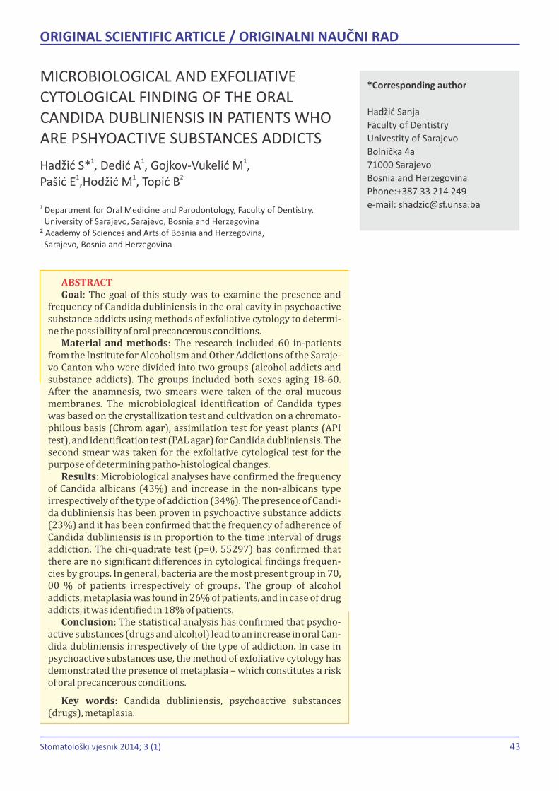

45Stomatološki vjesnik 2014; 3 (1)
MICROBIOLOGICAL AND EXFOLIATIVE CYTOLOGICAL FINDING OF THE ORAL CANDIDA DUBLINIENSIS IN PATIENTS WHO ARE PSHYOACTIVE SUBSTANCES ADDICTS Hadžić S, Dedić A, Gojkov-Vukelić M, Pašić E,Hodžić M, Topić B
Candida dubliniensis has been mentioned. As oppo-
sed to Candida albicans, it is a rare type of endo-
genous micro-flora of immune-suppressed patients
and has reduced ability of colonization and infection
(Sullivan et al. 2004). Candida dubliniensis is mostly
taken from the oral cavity (buccal mucous mem-
brane, saliva, tongue) of immune-deficient patients,
primarily those that are HIV positive [6]. The colonies
of Saboraud agar are soft, white and very similar to
Candida albicans colonies (Larone 2002). Today, the
most modern cultivation base Pal agar is used for the
creation of clamidospora colonies of Candida dubli-
niensis with a thin membrane and higher volume
(Candida albicans is a blastoconidium in a single-cell
form) [7, 8, 9].
Psychoactive substance addiction is a leading
social and health problem worldwide. The World
Health Organization defined a drug or psychoactive
substance as any natural or synthetic drug intro-
duced into the body that may change one or more
different functions of the body. Drugs are those natu-
ral or synthetic substances that may cause changes or
disturbances of one or more somatic or psychological
functions and are used for medical or non-medical
purposes. Drugs are classified as hard and soft drugs.
Hard drugs cause a strong psychological and physical
addiction (heroine, cocaine). Soft drugs have medium
potential to cause psychological addiction, and low
level of physical addiction. They include: alcohol,
amphetamines, nicotine, hallucinogens, caffeine,
marijuana (hashish), organic dissolvent, etc. [10, 11,
12]. A psychoactive substance may be introduced
into the body in several ways: orally (alcohol, pills),
by smoking, inhaling (marijuana, cocaine, opium),
snorting (cocaine, heroin), subcutaneously and intra-
venously (heroin, cocaine). Psychoactive substances
(drugs) have great impact on oral health. Oral chan-
ges depend on the type of drugs and length of use.
Changes in oral mucous membranes are not patho-
genic in psychoactive substance addicts, but the follo-
wing ones may be perceived: Candidiasa mucosae
oris; Cheilitis angularis; Glossitis rhombica mediana;
Leukoplakia; Hyperceratisis mucousae oris; Gingivo-
stomatitis ulceromembranacea; Lingua nigra villosa;
Stomatitis aphthosis recurrens (SAR); Erosiae muco-
sae oris; Herpes simplex; Morsicatio mucosae oris
[13,14]. In psychoactive substance addicts (alcohol,
drug addicts) an immune-suppression of the immune
system occurs, which leads to an increase in opportu-
nistic infections.
Introduction
Oral candidiasis is an opportunistic infection cau-
sed by candida yeasts, out of which Candida albicans
is the most pathogenic one and it has the capacity of
pathological adherence to oral mucous membranes
and causing oral diseases [1]. Other non-albicans
types of high frequency in clinically manifested can-
didiasis are: candida dubliniensis, candida glabrata,
candida tropicalis, candida crusei, candida parapsi-
losis, candida stelatoidea, candida kefur, candida
guillermondi [2, 3]. The mentioned types are oppor-
tunists, pathogenic yeasts that may cause a whole
array of lesions under certain circumstances. Candi-
da yeasts may cause superficial candidiasis (oro-
pharyngeal candidiasis, esophagitis, vulvovaginitis,
chronic muco-coutaneous candidiasis, intertrigo and
onycomicosis). Generalized candidiasis occurs as a
septic disease by means of dissemination through
blood and lymph and it frequently has a lethal
outcome.
Virulence factors that enable yeasts to become
patogenous are: the ability of yeasts to attach to the
host tissue (Fonzi 2001), production of proteolytic
enzymes that include highly molecular metabolites
from the group of aspartyl proteases (Naglik 2003),
phospholipases (Ghannoum 2000) and lipases
(Stehr 2003), yeast hyphae – a morpho-genetic trans-
formation that renders possible penetration (From-
tiling R A 2005), different immune and modular
effects of yeast determinants may contribute to
reduced activities of the defense system.
The authors Hazen, 1995, Hobson 2003, have
divided the pre-disposition factors for the occurren-
ce of a yeast infection in the following ones: immune-
suppression caused by the use of drugs in malignant
diseases treatment (cytostatics), application of anti-
inflammatory drugs (corticosteroids), use of drugs
against transplant rejection, idiopathic lymphocyto-
penia, and similar use of antibiotics and impairment
of the endogenous commensal flora, through a surge-
ry or other impairment of the protection barriers
integrity of the host (e.g. burns, catheters, etc.), HIV
infection, use of psychoactive substances (drugs).
A large number of epidemiological studies show
an increased incidence of infections (of the mucous
membrane, skin, hematogenous and disseminated
infections) caused by non-candida albicans yeasts
[4,5]. As of 1995, a new interesting type called
Researches of many authors point to the intercon-
nectedness between the intake of psychoactive sub-
stances (drugs), length of addiction and type of drugs
and the presence of oral Candida dubliniensis. Rocchi
et al. 2007 proved in their work that the presence of
Candida dubliniensis was related to the length of
heroin use. Rooban et al. 2009 examined the oral
health in addicts addicted to various drugs (opiates,
cocaine, marijuana) and confirmed highly significant
presence of oral Candida dubliniensis and the length
of drug intake, as well as the type of drug [15,16].
Psychoactive substance addicts are risk patients for
AIDS and Candida infections, the incidence of which
is on continuous rise. These patients demonstrate
frequent resistance to therapeutic protocol, so the
imperative of the research is to isolate and confirm
the importance of Candida dubliniensis by means of
microbiological procedures and to establish the pos-
sibility of occurrence of precancerous conditions by
means of exfoliative cytological tests.
Material and Methods
For the purpose of achieving the set goals, we have
selected a sample. 60 inpatients from the Institute for
Alcoholism and Other Addictions of the Sarajevo
Canton and they were divided into two groups: group
A – alcohol addicts, and group B – opiates addicts. The
groups included both sexes aging from 18 to 60 years.
After a comprehensive anamnesis, an extra-oral and
intra-oral examination of oral mucous membranes
was conducted. Smears were taken from the mucous
membranes for microbiological identification. Two
confirmatory methods were used for the identifica-
tion of Candida types: the crystallization test and
cultivation on a chromatophilous basis (Chrom agar).
The assimilation test for yeast plants (API test) was
used for the identification of non-albicans candida.
By planting a positive culture on a chromatophilous
basis (Chrom agar) and 24-48 hour incubation at
37°C, four types of Candida were identified based on
the different color (Candida albicans, Candida tropi-
calis, Candida glabrata, Candida crusei).
All cultures of Candida albicans (that have been
proven by the mentioned methods) were planted on
a PAL agar basis for the purpose of identifying
Candida dubliniensis. The culture had an incubation 0
period of 24-48 hours at 30 C. If yeast plants grew, a
preparation was made and it was observed under the
microscope with an enlargement of 40 x. The presen-
ce of clamidospora has confirmed that the identified
yeast plants were Candida dubliniensis.
The second smear was related to the Institute for
Pathology and Cytology where the exfoliative cytolo-
gical test was conducted.
Results
Microbiological analysis findings
Table 1 contains the frequency (percentage) of
patients in case of which the Candida types were
found by examination groups and in total.
Candida albicans was present at 43% of the total
patients. Non-albicans was identified at 34,3 % of the
patients. Candida dubliniensis was identified at 23%
of the total patients addicted to psychoactive
substances (Table 1).
Table 1 Frequency of Candida types by groups
Group A
Group B
Total
6
9
15
37,50
47,37
42,86
6,25
5,26
5,71
18,75
5,26
11,43
6,25
26,32
17,14
31,25
15,79
22,86
1
1
2
3
1
4
1
5
6
5
3
8
No.
ALBICANSTROPICALIS GLABRATE CRUSEI
DUBLINIENSISNON-ALBICANS
No. No. No. No.% % % % %
44 Stomatološki vjesnik 2014; 3 (1)

46 47Stomatološki vjesnik 2014; 3 (1) Stomatološki vjesnik 2014; 3 (1)
MICROBIOLOGICAL AND EXFOLIATIVE CYTOLOGICAL FINDING OF THE ORAL CANDIDA DUBLINIENSIS IN PATIENTS WHO ARE PSHYOACTIVE SUBSTANCES ADDICTS Hadžić S, Dedić A, Gojkov-Vukelić M, Pašić E,Hodžić M, Topić B
dida glabrata 1 (5.3) and Candida crusei 5 (26.3%).
Candida dubliniensis was identified in 8 patients,
namely in group A in 5 patients (31.25%) and in
group B in 3 patients (15.8%) (Table 2).
The chi-square test (p=0,55297) has confirmed
that there are no significant differences in the fre-
quencies of occurrence of Candida by groups. Candi-
In the case of group A (alcoholics), the following
Candida types were identified: Candida albicans in 6
patients (37.5 %), Candida tropicalis 1 patient
(6.25%), Candida glabrata 3 (19%) and Candida
crusei 1 (6.2 %). In the case of group B (addicts),
Candida albicans was identified in 9 patients
(47.4%), Candida tropicalis 1 patient (5.3%), Can-
da albicans is the most frequent one in total, present
in 42.86 % of the patients. In the group A, it amounts
to 37.5 %, and in group B to B 47.5%.
In group B, there were 3 patients (15.8 %) that had
Candida dubliniensis, whereas in the case of group A,
there were 5 such patients (31.25 %) (Figure 1).
Candida dubliniensis was found in 8 patients, i.e.
in case of 13.33 % of all patients. In case of group A,
there were 5 patients (62.50%), whereas in group B,
there were 9 of them (37.50%). In group A, all pati-
ents that had Candida dubliniensis had been drinking
for more than 15 years, whereas in group B, the
occurrence of Candida dubliniensis happened in the
first two observed time intervals. The presence of
Candida dubliniensis has been proven in case of psy-
choactive substance addicts (23%) and it has been
confirmed that the frequency of adherence of Can-
dida dubliniensis is in proportion to the time interval
of drug addiction (Table 3).
Results of the exfoliative cytological analysis
Table 4 contains the frequency (percentage) of
patients at whom bacteria, yeast and metaplasia
were identified, both by examination groups and in
total (Table 4).
The chi-square test (p=0, 55297) has confirmed
that there are no significant differences in frequen-
cies of cytological findings by groups. Bacteria are the
largest group in general, namely in 70.00% of the
patients. In group A, 6 patients (26.09%) had meta-
plasia, whereas in group B there were 2 such patients
(11.76%) (Figure 2).
Table 2Frequency of Candida types (consolidated non-albicans) by groups
Figure 1Presence of Candida (consolidated non-albicans) by groups
Group A
Group B
Total
6
9
15
37,50
47,37
42,86
31,25
36,84
34,29
6,25
26,32
17,14
12
7
12
5
3
8
16
19
35
No.
ALBICANS TOTALDUBLINIENSISNON-ALBICANS
No. No. No.% % %
Table 4Frequency of cytological findings
Group A
Group B
Total
15
13
28
65,22
76,47
70,00
8,70
11,76
10,00
26,09
11,76
20,00
0,00
0,00
0,00
2
2
4
6
2
8
0
0
0
123
17
40
No.
BACTERIA TOTALMETAPLASIA DYSKERATOSISYEAST
No. No. No. No.% % % %
0
2
4
6
8
10
12
14
16
Group A Group B
ALBICANS
Total
Figure 2Presence of cytological findings by groupss
0
5
10
15
20
25
30
35
40
45
Group A Group B
BACTERIA YEAST METAPLASIA TOTAL
Total
Group A
Group B
Total
5 - 10
0
1
1
10 - 15
0
2
2
More than 15
5
0
5
Total
5
3
8
Table 3Frequency of Candida dubliniensis according to the length of drug intake

48 49Stomatološki vjesnik 2014; 3 (1) Stomatološki vjesnik 2014; 3 (1)
MICROBIOLOGICAL AND EXFOLIATIVE CYTOLOGICAL FINDING OF THE ORAL CANDIDA DUBLINIENSIS IN PATIENTS WHO ARE PSHYOACTIVE SUBSTANCES ADDICTS Hadžić S, Dedić A, Gojkov-Vukelić M, Pašić E,Hodžić M, Topić B
Candida dubliniensis is a yeast species which increase was connected to the HIV infection for a long time, but its presence has been proven in case of other diseases, too [22].
Polacheck et al. (2000) have isolated Candida dubliniensis in case of inpatients in Jerusalem. None of the patients was HIV positive, but they all were receiving a broad spectrum of antibiotics. The high occurrence of Candida in case of psychoactive sub-stance addicts is a result of the activity of alpha-adrenergic receptors in the blood vessels of the sali-vary glands that lead to vasoconstriction thus redu-cing the saliva production. [23, 24]. Suhail 2004 has confirmed the incidence of Candida dubliniensis in case of immune-suppressed patients. Out of 7 isola-ted Candida dubliniensis, 5 of them were isolated from saliva, one from a vaginal smear and one from urine. Five patients had a tumor, one patient had sy-stematic lupus erythematodes, and one had diabetes mellitus [25]. Our research confirmed the presence of Candida dubliniensis in patients that are addicted to psychoactive substances and are not HIV positive, which is in compliance with the research of the men-tioned authors. In group A (alcoholics), there were 31.25% Candida dubliniensis (in 5 patients) and 16% in group B (in case of 3 patients), which con-firms that alcohol addiction with all immunological misbalances, including the local status of oral health, are an ideal medium for the occurrence of Candida dubliniensis.
According to results of both mentioned authors and our results, it is evident that Candida dublinien-sis is increasing and becoming a pathogen that has the potential to colonize and cause superficial, but invasive diseases in HIV negative patients.
The impact of psychoactive substances on the occurrence of a malignant alteration has been the subject of numerous researches. Harris (2004) used a sample of 388 alcoholics and 305 alcoholics that were also drug addicts, being 40 years old on average, during a period 1994-1999, and identified frictional keratosis in 8.8% of patients, changes on the lips in 4.8% of patients, candidiasis in 38% of patients, an-gular cheilitis in 3% of patients, three white lesions, out of which one turned out to be a carcinoma identi-fied during a cytological analysis. The author confir-med that the use of alcohol and drugs does constitute a risk factor for oral mucous lesions. (26) The type of drug and the length of addiction may have an impact to the occurrence of lesions and malignant changes. Rober (2008) used a sample of 100 patients who were addicts using different types of drugs to exami-
Discussion
The research over the past years has been related
to Candida species, especially to the escalation of
yeast infections in immune-suppressed patients,
AIDS patients and patients using an immuno-
suppressive therapy. The Candida species frequently
occurs in the oral cavity. The Candida species is an
opportunistic type and will lead to an infection if
there are factors of predisposition. The virulence of
Candida depends on the ability to adhere to the mu-
cous membrane of the host, production of filamen-
tous forms of growth and release of hydrolytical en-
zymes. A continuous increase in yeast infection inci-
dences, new yeast species and resistance to anti-
fungal therapy are a challenge for research in that
segment [17, 18].
Baradak (2009) examined in his work the clinical
and microbiological state of oral mucous membranes
in intravenous addicts. 60 patients participated in the
study. Candida species were isolated in 50 patients.
The most frequent type was Candida albicans with
70%, Candida parapsilosis 15%, Candida glabrata
7.5%, Candida tropicalis 5% and Candida dublinien-
sis in the case of one patient. This study has shown a
prevalence of oral candidiasis in intravenous addicts
[19]. The most frequent type is Candida albicans, but
the non-albicans types are also on the increase, 30%
[20]. Immunosuppressive conditions and anti-virus
therapy are significantly related to an increase in
candidiasis (Cerqueira 2010) [21]. In case of our re-
search on a sample including 60 patients that are
alcohol and drug addicts, we identified Candida albi-
cans in 43% of patients. In group A, there were 38%,
and in group B 48% respectively. With regards to the
non-albicans types, the most frequent type was
Candida crusei, namely in case of group B, amounting
to 26%, and in group A to 6%. Candida glabrata was
the most frequent type in group A, amounting to 10%,
and in group B it amounted to 5.2%. Candida tropi-
calis was also identified being equally present in both
group A and B group. The chi-square test (p=0,
55297) has confirmed that there is no statistically
important difference in the occurrence of Candida by
groups. The most frequent type is Candida albicans.
Our results are in compliance with the results of the
mentioned authors. New studies show that the occur-
rence of non-albicans types and Candida dubliniensis
is increasing (Gery 2011).
ne oral health. The patients mostly used heroin, am-phetamines and marijuana. The lesions have been diagnosed clinically and processed by means of a mi-crobiological analysis, and a statistical analysis con-firmed the importance of oral mucous lesions with malign alteration and length of drug use. Laks-man (2009) also confirmed that the length of addiction and type of drugs influence the occurrence of Candi-da dubliniensis and precancerous conditions [27, 28].
Metaplasia is a reversible replacement of one type of mature differentiated cells in organ tissue by ano-ther type of mature cells occurring due to the abnor-mal position or pathological stimulus. The importan-ce of metaplasia lies in the fact that dysplasia may occur more frequently in some areas of metaplasia (due to chronic stimulus).
Our results have shown the presence of meta-plasia in 8 patients. In group A (alcoholics), 6 patients (26%) had metaplasia, and in group B (drug addicts) 2 patients (12%).
Canković (2010) confirmed the prevalence of Can-dida in patients with oral carcinoma. In case of 30 patients with oral carcinoma, 17% of non-albicans types were identified, 13% of Candida albicans. Sta-tistical data have confirmed a significant degree of interrelatedness between candidiasis and age, smo-king, use of drugs, alcohol and precancerous conditi-ons [29]. The prevalence of oral mucous lesions at the Center for Addiction Rehabilitation is described by O'Sullivan (2011). The prevalence of abnormality amounted to 29% in a sample of 210 patients. The most frequent types were candidiasis 38%, aphtae 3%, lymphadenopathia 2.9%, 4 erythroplakia, 2 leu-koplakia and 2 malignant lesions. The author points out the need of the obligatory cytological analysis or biopsy for the purpose of early detection of precan-cerous conditions [30]. Our findings have not shown the presence of dyskeratosis and carcinoma, but the presence of metaplasia has warned us that alcohol and drug addicts constitute the riskiest group of pa-tients for the occurrence of carcinoma, and that a screening of oral tumors should be a part of the obli-gatory medical and dental examinations.
Conclusion
The use of psychoactive substances impacts the
frequency of occurrence of Candida species (Candida
albicans and non-albicans types). The presence of
Candida dubliniensis in psychoactive substance
addicts, irrespectively of the type of addiction, has
been proven and it has been confirmed that the
frequency of adherence of Candida dubliniensis is in
proportion with the time interval of drug addiction.
An early detection of Candida dubliniensis in patients
resistant to therapy indicates a misbalance or
suppression of the immune status.
In case of psychoactive substances use, the
method of exfoliative cytology has shown the pre-
sence of metaplasia. Alcohol and drug addicts consti-
tute groups of risk for the occurrence of malignant
transformation of oral mucous membranes.
References
1. Nucci MK, Marr A. Emerging fungal disease. Clin Infect Dis 2005; 41:521-526.
2. Mavor AL, Thewes S. Systemic fungal infections caused by Candida species: epidemiology, infection process and virulence attributes. Curr Drug Targets 2005;6: 863-74.
3. Arjuna N, Ellepola B, Zia U. Prevalenca of Candide dubliniensis among Oral Candida isolates in Pa-tients Attending the Kuwait University Dental Clinic. Med Princ Pract 2011; 20:271-276.
4. Hazen KC, Howell ISA. Candida and other yeast of medical importance. U: Manual of clinical micro-biology. Murray PR, Baron EJ, Jorgensen JH, Pfaller MA, Yolken RH. ASM Press. Washington DC 2005; 1693-1712
5. Hobson RP. The global epidemiology of invasive Candida infections-is the tide turning? Hosp Infect 2003; 55:159-168.
6. Sullivan DJ, Moran GP, Coleman DC. Candida dubliniensis: Ten years on. FEMS Microbiol Lett 2005; 253:9-17.
7. Larone DH. Medically important fungi. A guide to identification. ASM Press 2002
8. Jabra-Rizk MA, Baqaui AMA, Identification of Candida dubliniensis in a prospective study of patients in United States. J Clin Microbiol 1999; 37:321- 326.
9. Mosaid A, Sullivan DJ, Coleman DC, Differentia-tion of Candida dubliniensis from Candida al-bicans on Pal's agar. J Clin Microbio .2003; 41:4787-4789.

50 Stomatološki vjesnik 2014; 3 (1)
MICROBIOLOGICAL AND EXFOLIATIVE CYTOLOGICAL FINDING OF THE ORAL CANDIDA DUBLINIENSIS IN PATIENTS WHO ARE PSHYOACTIVE SUBSTANCES ADDICTS
dubliniensis isolated from the oral cavity of im-mune-compromised patients. Clinical Micro-biology and infection 2006;vol. 12 supplement 4.
21. Cerqueira DF, Portela MB, Ponarico L et al. Oral Candida colonization and its relation with predisporing factors in HIV-infected children and their urinfected. Siblings in Brazil: the era of highly active antretroviral therapy. J Oral Pathol Med 2010; 39 (2):188-94.99.
22. Gery P, David M, Coleman C, Derek J. Candida albi-cans versus candida dubliniensis:Wy is Candida albicans Pathogenik? Int J Microbiol 2011.
23. Polacheck I, Strahilevitz J, Sullivan D and all : Recovery of Candida dubliniensis from non-hu-man immunodeficiency virus-infected patients in Israel. J Clin Microbiol 2000 ; 8(1):170-4.
24. Jewtuchowicz VM, Mijuca MF et al. Genetic rela-tednes of subgingival and buccal candide dubli-niensis isolates in immunocompetent subject assessed by RAPD-PCR. Journal of Oral Micro-biology 2009,doi:10-3402/j om.v.1 i o.2009
25. Suhail A, Zaiba K, Eiman M, Khan UZ. Isolation and molecular identification of Candida dubliniensis from non –human immunodeficiency virus –infected patients in Kuwait. Journal of Medical Microbiology 2004;53: 633-637.
26. Harris CK, Warnakulasurya KA et al. Prevalence of oral mucosal lesions in alcohol misusers in South London. J Oral Pathl Med. 2004;33(59):253-9.
27. Rooban T, Rao A, Joshua E, Dental and health sta-tus in drug abusers in Chennai, India: A cross- sec-tional study, Department of Oral and Maxillofacial Pathology. Ragas Dental College and Hospital, Uthandi,Chennai 2008;600-119.
28. Laksman P, Samanarayake W et al. Oral mucosal fungal infections. J Periodontology 2000. 2009; vol 49:39-59.
29. Canković M, Bokor-Bratić M. Candida albicans in-fection in patients with oral squamous cell carci-noma. Vojno sanit Pregl 2010;67(9):766 – 70.
30. O'Sullivan EM. Prevalence of oral mucousae ab-normalitues in addiction treatment centre resi-dent in Southern Ireland. Oral Oncol 2011;25. (Epub ahead of print).
10. Cerić I, Mehić Basara N and ass. Misuse of psycho active substances and drugs. Medical Faculty University in Sarajevo 2007
11. Mehić Basara N, Cerić I, Hasečić H. Plan and Pro-gram of fighting drug abuse in Federation of Bos-nia and Herzegovina;13 International Confe-rence of Reduction of Drug Related Harm. Ljubljana 3-7 March, 2002;
12. Araujo MW, Dermen K, Connors G et al. Oral and health among patients in treatment for alcohol use disorder: pilot study. J Int. Acad Periodontol. 2004; 6(4):125-30.
13. Hamamoto DT, Rhodus NL. Methamphetamine abuse and dentistry. Journal of Oral Disease. School of Dentistry University of Minnesota USA 2009;15:27 37.
14. Arjuna N, Ellepola B, Zia U. Prevalenca of Candide dubliniensis among Oral Candida isolates in Patients Attending the Kuwait University Dental Clinic. Med Princ Pract 2011;20:271-276.
15. Rocchi L, D' Alessandro D, Fabiani M. Association between oral candidiasis and immunity level in a population of drug addicts in a centre for pre-vention and treatment of drug addictions in Roma, Italy. 17th. European Congress of Clinical Microbiology and Infectious Disease, Germany. 31 Mar - 04 Apr 2007: 73
16. Rooban T, Rao A, Joshua E, Dental and health sta-tus in drug abusers in Chennai, India: A cross- sectional study, Department of Oral and Maxillo-facial Pathology. Ragas Dental College and Hospital, Uthandi, Chennai 2008; 600-119. 100.
17. William D, Lewis M. Pathogenesis and treatment of oral candidiosis. Journal of Oral Microbiology 2011.
18. Kempa H, Sadlk-Nowicka J et al. Candida infec-tions of the oral mucosa – not only a dental problem. Przgel Lek 2006; 63 (59):257-60.
19. Baradkar VP, Kumar S. Species identification of Candida isolates obtained from oral lesions of HIV infected patients. Indian J Dermatol 2009; 54(4):385-386. 102.
20. Kosalec I, Matica B, Jarža-Davila N, i ostali. Hae-molytic activity of Candida albicans and candida
51Stomatološki vjesnik 2014; 3 (1)
ORIGINAL SCIENTIFIC ARTICLE / ORIGINALNI NAUČNI RAD
PERIODONTAL STATUS IN THE PERMANENT DENTITION OF CHILDREN WITH CEREBRAL PALSY
1 2Ihtijarević-Trtak S* , Kobašlija S , 2 2Hadžagić-Ćatibušić F , Huseinbegović A
1 Community Health Centre of Canton Sarajevo, Dental Department, Sarajevo, Bosnia and Herzegovina2 Department of Pediatric Dentistry, Faculty of Dentistry, University of Sarajevo, Sarajevo, Bosnia and Herzegovina
ABSTRACT
Introduction: Cerebral palsy is a group of permanent dis-orders of movements and posture resulting in physical disability caused by non-progressive disorder occurred in the developing fetal or infant brain. Children with cerebral palsy have the difficulties in maintaining oral hygiene. Periodontal diseases are common in the cerebral-palsied individuals as the result of poor hygiene, physical abilities and presence of malocclusions.
The aim: The aim of this study is to assess the periodontal sta-tus in the permanent dentition of children with cerebral palsy.
Material and Methods: A total of 44 patients aged 12 to 18 years with permanent dentition were included in this study and distributed in experimental (children with cerebral palsy, n=18), and control group (healthy individuals, n=26). The examinees were examined by a single examiner according to the WHO recommendations (Oral Health Surveys: Basic Methods). The periodontal status was assessed by using the following indexes: Plaque Index (Silness and Löe, 1964.), Gingival Index (Silness and Löe, 1964.) and Calculus Index.
Results: The mean value of Plaque Index ± SD is 4, 5 ± 3, 20 (experimental group) and 6,5 ± 3,35 (control group) (2=6,762; p=0,0799). The mean value of Gingival Index ± SD is 4, 5 ± 2 29 (experimental group) and 6, 5 ± 4, 92 (control group) (2=7,204; p=0, 0657). The mean value of Calculcus Index ± SD is 4, 5 ± 7, 37 (experimental group) (2=4,682; p=0, 1966).
Conclusion: The periodontal status (Plaque Index, Gingival Index, Calculus Index) of the children with cerebral palsy is poorer, compared with healthy individuals.
Key-words: periodontal status, children, cerebral palsy, permanent dentition.
*Corresponding author
Samra Ihtijarević-Trtak, DDS, MSC
Community Health Centre
of Canton Sarajevo
Dental Department
71000 Sarajevo
Bosnia and Herzegovina
e-mail: [email protected]

52 53
PERIODONTAL STATUS IN THE PERMANENT DENTITION OF CHILDREN WITH CEREBRAL PALSY Ihtijarević-Trtak S, Kobašlija S, Hadžagić-Ćatibušić F, Huseinbegović A
Stomatološki vjesnik 2014; 3 (1) Stomatološki vjesnik 2014; 3 (1)
tries, neglecting of oral health by a caregiver is consi-
dered as dental neglect, which is a form of child
abuse. This kind of child abuse is punishable and can
lead to the loss of child's custody.
As stated so far, the maintenance of oral health is
in particularly important in children with physical
and mental disabilities. This is difficult due to poor
manual coordination, intellectual disability, and less
importance given to oral health compared to major
health problems. The timely diagnosis, dental resto-
rations and oral rehabilitation is very important for
their specific growth and development [6].
The oral health of children in Bosnia and Herzego-
vina is less favorable compared to economically deve-
loped countries with health care system characte-
rized by more preventive programs and better health
care for disabled children. The developed counties
pay more attention to every aspect of social life for
children with disabilities (their rehabilitation, edu-
cation, professional training, employment and inte-
gration in society in general). The special attention is
also given to medical and dental health care, respec-
ting the saying that the disabled children also have
right to smile. In future, efforts should be undertaken
to improve dental care of this population: the regular
check-up appointments every 3 or 6 months and the
formation of specialized dental teams that will
exclusively treat this vulnerable population.
Aim
The aim of this study is to assess the periodontal
status in the permanent dentition of children with
cerebral palsy.
Material and Methods
A total of 44 patients aged 12 to 18 years with
permanent dentition were included in this study. The
study sample was distributed in two clinical groups:
the experimental that consisted of children with
cerebral palsy (n=18), and the control group with
healthy individuals (n=26). The healthy patients
seeking dental care in one of the Dental Health Care
Introduction
Cerebral palsy is a group of permanent disorders
of movements and posture resulting in physical disa-
bility caused by non-progressive disorder occurred
in developing fetal or infant brain. The motor dys-
function is often associated with sensory, perceptive
and cognitive impairment, communication difficul-
ties, behavioral disorders, epilepsy and secondary
musculoskeletal problems [1]. Cerebral palsy is the
most common cause for severe neuro-motor distur-
bance in children, and thus represents the significant
burden for the affected child, its family, health-care
and educational system and the society in general [2].
According to the American Academy of Neurology,
cerebral palsy has a highest treatment cost per per-
son [3]. The incidence of cerebral palsy is 2-2.5 per
1000 live births, making this disease the most com-
mon cause of physical disability in childhood [4].
Cerebral palsy does not cause the specific oral
manifestations. However, certain conditions are mo-
re frequent or more prominent in patients with cere-
bral palsy, compared with general population. Cha-
racteristics of oral pathology of patients with cere-
bral palsy are: poor oral hygiene, pronounced perio-
dontal diseases, more unrestored carious teeth, more
extracted teeth, less dental restorations and more
prominent dentofacial abnormalities compared with
healthy children of the same age [5]. Children with ce-
rebral palsy have difficulties with the maintenance of
oral hygiene. Periodontal diseases are common in the
cerebral-palsied individuals as the result of poor hy-
giene, physical abilities and presence of malocclu-
sions. Gingival hyperplasia resulting from medica-
tion therapy contributes to the occurrence of perio-
dontal diseases.
Numerous studies suggest that oral health is influ-
enced by socio-economic status and cultural values
in society. In physically disabled individuals the oral
health care is complex. Neglecting this segment of ge-
neral health may have more serious consequences in
relation to healthy individuals.
The problem of oral health of children with physi-
cally and mentally disabilities in recent years is beco-
ming more actual. Besides health reasons for oral
health care, the certain formal reasons are gaining
importance. In fact, in USA and many European coun-
Centers in Canton of Sarajevo were randomly assig-
ned to control group.
The examinees were examined by a single exa-
miner, using periodontal probe and plain mouth
mirror with artificial source of light at dental ward or
the natural source of light in patients who were
unable to be examined at dental ward. The patients
were positioned towards source of natural light,
avoiding direct sunlight [7].
The periodontal status is examined using perio-
dontal probe with 0, 05 mm diameter sphere at probe
tip. A controlled light pressure of 25 grams was
applied during examination, which refers to pressure
of WHO probe under the nail of thumb until white cir-
cle appearance. The pain during probing suggests the
applied pressure is too high. It is important to keep
the periodontal probe parallel to the contours of
tooth. For examination of sub gingival calculus the le-
ast possible pressure should be applied, which enab-
les the movement of probe tip along the surface of the
tooth. When the probe is applied on tooth, the anato-
mical configuration of tooth must be followed [8].
The periodontal status was assessed using the
following indexes: Plaque Index (Silness and Löe,
1964.), Gingival Index (Silness and Löe, 1964.) and
Calculus Index.
Plaque Index (Silness and Löe, 1964.)
The measurement of the oral hygiene status is
based on the Plaque Index, which records both soft
debris and the mineralized deposits on the teeth.
Cervical region of tooth is dried with three-way air
dental syringe. Inspection is performed visually and
by periodontal probe. The following score and cri-
teria were applied:
0 - No plaque; plaque is not visible, and by using
the probe, there is no presence on the probe tip
1 - Plaque is not visible; the probe use on tooth
reveals the plaque on the probe tip
2 - Plaque is visible by naked eye
3 - Abundance of plaque by naked eye is visible on
tooth and gingival margin [9].
Gingival Index (Silness and Löe, 1964.)
Gingival index assesses the condition of gingival
tissue. It records and quantifies the gingival inflam-
mation through the changesin color, bleeding inten-
sity and the edema. The following score and criteria
were applied:
0 - Normal gingiva without inflammation, there is
no change in color and no edema
1 - Mild inflammation, slight change in color and
slight edema but no bleeding on probing
2 - Moderate inflammation, redness, edema and
bleeding on probing
3 - Severe inflammation, marked redness and
edema or hypertrophy, tendency to spontane-
ous bleeding, ulcerations
Calculus Index
Calculus index refers to the amount of calculus on
teeth and is used for assessment of oral hygiene. The
following score and criteria were applied:
0 - No calculus present
1 - Presence of supra-gingival calculus
2 - Presence of sub-gingival calculus and small
amount of supra-gingival calculus
3 - Presence of sub-gingival calculus and lot of
supra-gingival calculus.
Results
The control group presented with Plaque Index
score 0 in 42,31% of cases, score 1 in 30,77%, score 2
in 19,12% and score 3 in 7,69% of cases. The
experimental group had the score 0 in 11,11%, score
1 in 33,33 %, score 2 in 50% and score 3 in 5,56% of
cases. The values of Plaque Index are lower in control
group compared with the experimental one. Results
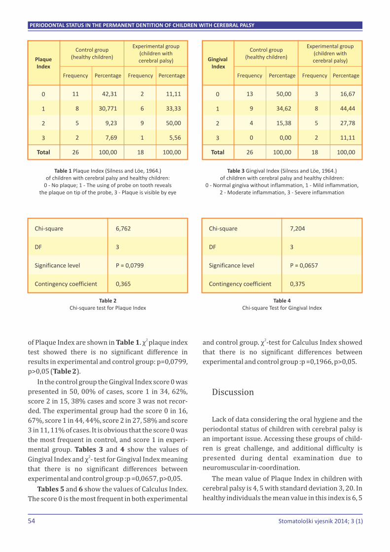
54
Frequency Percentage Frequency Percentage
11
8
5
2
26
2
6
9
1
18
42,31
30,771
9,23
7,69
100,00
11,11
33,33
50,00
5,56
100,00
0
1
2
3
Plaque Index
Control group(healthy children)
Experimental group(children with cerebral palsy)
Total
Table 1 Plaque Index (Silness and Löe, 1964.) of children with cerebral palsy and healthy children:
0 - No plaque; 1 - The using of probe on tooth reveals the plaque on tip of the probe, 3 - Plaque is visible by eye
Frequency Percentage Frequency Percentage
13
9
4
0
26
3
8
5
2
18
50,00
34,62
15,38
0,00
100,00
16,67
44,44
27,78
11,11
100,00
0
1
2
3
Gingival Index
Control group(healthy children)
Experimental group(children with cerebral palsy)
Total
Table 3 Gingival Index (Silness and Löe, 1964.) of children with cerebral palsy and healthy children:
0 - Normal gingiva without inflammation, 1 - Mild inflammation,2 - Moderate inflammation, 3 - Severe inflammation
55Stomatološki vjesnik 2014; 3 (1) Stomatološki vjesnik 2014; 3 (1)
PERIODONTAL STATUS IN THE PERMANENT DENTITION OF CHILDREN WITH CEREBRAL PALSY Ihtijarević-Trtak S, Kobašlija S, Hadžagić-Ćatibušić F, Huseinbegović A
2and control group. χ -test for Calculus Index showed
that there is no significant differences between
experimental and control group :p =0,1966, p>0,05.
Discussion
Lack of data considering the oral hygiene and the
periodontal status of children with cerebral palsy is
an important issue. Accessing these groups of child-
ren is great challenge, and additional difficulty is
presented during dental examination due to
neuromuscular in-coordination.
The mean value of Plaque Index in children with
cerebral palsy is 4, 5 with standard deviation 3, 20. In
healthy individuals the mean value in this index is 6, 5
2 of Plaque Index are shown in Table 1. χ plaque index
test showed there is no significant difference in
results in experimental and control group: p=0,0799,
p>0,05 (Table 2).
In the control group the Gingival Index score 0 was
presented in 50, 00% of cases, score 1 in 34, 62%,
score 2 in 15, 38% cases and score 3 was not recor-
ded. The experimental group had the score 0 in 16,
67%, score 1 in 44, 44%, score 2 in 27, 58% and score
3 in 11, 11% of cases. It is obvious that the score 0 was
the most frequent in control, and score 1 in experi-
mental group. Tables 3 and 4 show the values of 2
Gingival Index and χ - test for Gingival Index meaning
that there is no significant differences between
experimental and control group :p =0,0657, p>0,05.
Tables 5 and 6 show the values of Calculus Index.
The score 0 is the most frequent in both experimental
2with standard deviation 3, 35. Test for Plaque Index
was 6,762 (significance level p=0, 0799; contingency
coefficient 0,365).
Ivančić and Majstorović reported the 3, 8-4,53
values of OHI-S index in disabled children and 2,73-
2,84 in healthy children in Croatia [10]. Rodrigues
dos Santos et al (2003) in their study showed the
significantly higher DMFI (Decayed-Missing-Filled
teeth Index) and Plaque Index of children with per-
manent dentition with cerebral palsy in both gen-
ders, compared with healthy individuals [11].
The values of Gingival Index are lower in healthy
children, compared with children with cerebral palsy.
These results also support the findings that children
with cerebral palsy have the poorer oral hygiene. The
mean value of Gingival Index for children with cere-
bral palsy is 4, 5 with standard deviation 2, 29. The
index for healthy children is 6, 5 with standard devia-2tion 4, 92. The Test for Gingival Index in permanent
dentition is 7,204 (significance level p=0, 0657,
contingency coefficient 0,375).
The study conducted in Riyadhevaluated the Pla-
que Index and Gingival Index in children with prima-
ry, mixed and permanent dentition. The higher values
of Plaque Index were recorded in primary dentition
and the higher values of Gingival Index in children
with permanent dentition (level of significance
p=0,001). The gender did not significantly alter the
periodontal indexes in children [12]. These results
are in accordance with numerous results of similar
studies, as well as the results of our study.
The Calculus index in control group is 0 in 73,
08%, 1 in 11, 54% and 2 in 15, 38% of cases. Score 3
has not been recorded. This index in control group
was present in 44, 44% of cases for score 0, 27, 78%
of cases for score 1, 22, 22% of cases for score 2 and 5,
56% of cases for score 3. The mean value of Calculus
Index for children with cerebral palsy is 4, 5 with 2
standard deviation 7, 37. The Test for Calculus Index
is 4,682 (significance level p=0, 1966, contingency
coefficient 0,310).
Stevanović and Jovičić evaluated the Calculus
Index of children in age group 15-18 years [13]. The
children with cerebral palsy had statistically more
dental calculus, compared with the healthy ones
(p<0,001). Statistically significant periodontal treat-
ment needs (CPITN=2 and CPITN=3) are recorded in
the patients with cerebral palsy (82, 4%) compared
with healthy children (42, 6%; p<0,001). The mean of
CPITN index in children with cerebral palsy is 1,752
compared with 0,308 for healthy children (p<0,001).
The percentage of children with calculus in this age
group was very low and amounted for 2, 86%. The
mean value of Calculus Index for all subjects in this
study was 0, 6508 ± 0, 4661 [13]. The results of our
study correlate with previous studies. We can conclu-
de that the differences in level of oral hygiene and
periodontal status in children with cerebral palsy are
getting more prominent with years. This study has
focused on oral hygiene maintenance and its impact
on oral health in children.
Santos et al examined the oral health condition,
salivary and the microbiological parameters associa-
ted with caries in 62 children with cerebral palsy
Chi-square
DF
Significance level
Contingency coefficient
6,762
3
P = 0,0799
0,365
Table 2Chi-square test for Plaque Index
Chi-square
DF
Significance level
Contingency coefficient
7,204
3
P = 0,0657
0,375
Table 4Chi-square Test for Gingival Index
Frequency Percentage Frequency Percentage
19
3
4
0
26
8
5
4
1
18
73,08
11,54
15,38
0,00
100,00
44,44
27,78
22,22
5,56
100,00
0
1
2
3
Calculus Index
Control group(healthy children)
Experimental group(children with cerebral palsy)
Total
Table 5 Calculus Index of children with cerebral palsy and healthy children: 0 - No calculus present, 1 - Presence of supragingival
calculus, 2 - Presence of subgingival calculus and a little amount of supragingival calculus, 3 - Presence of subgingival calculus and
a lot of supragingival calculus.
Chi-square
DF
Significance level
Contingency coefficient
4,682
3
P = 0,1966
0,310
Table 6Chi-square Test for Calculus Index
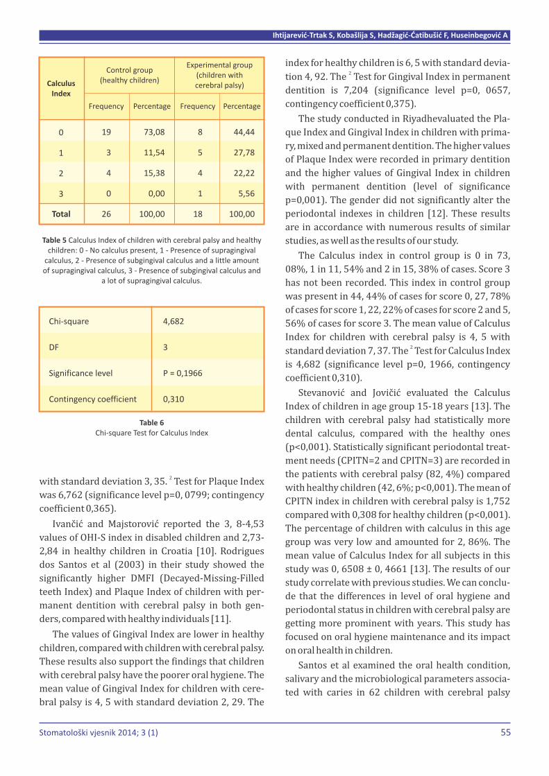

56 57Stomatološki vjesnik 2014; 3 (1) Stomatološki vjesnik 2014; 3 (1)
PERIODONTAL STATUS IN THE PERMANENT DENTITION OF CHILDREN WITH CEREBRAL PALSY Ihtijarević-Trtak S, Kobašlija S, Hadžagić-Ćatibušić F, Huseinbegović A
the oral health and the periodontal indexes are
poorer in these children, compared with healthy indi-
viduals. This may be related to their medical status,
poor manual coordination and intellectual disability.
The present study highlighted the importance of oral
hygiene maintenance in children with cerebral palsy.
Oral health programs should be created and con-
centrated towards these children concerning their
specific needs.
References
1. Rosenbaum P, Paneth N, Leviton A, Goldstein M,
Bax M.Definition and classification of cerebral
palsy.Dev Med Child Neurol2007; 49(2)(Suppl.
109):8–14.
2. Mejaški-Bošnjak V. Neurological syndromes of
infancy. Paediatr Croat 2007;51(Supl 1): 120-
129.
3. Ashwal S, Russman BS, Blasco PA, Miller G, Sand-
ler A. Practice Parameter: Diagnostic assessment
of the child with cerebral palsy. Neurology 2004;
62: 851-63.
4. Silver HK, Kempe CH, Bruyn HB. The Handbook of
Pediatrics. Beograd: Savremena administracija;
1989
5. Gajić M, Stevanović R, Jovičić O. Dental care for
children with developmental disorders. In:
Beloica D. The pediatric Dentitry: Handbook.
Zemun: Kuća štampe; 2006
6. Palmer CA. Nutrition, Diet and Oral Conditions. 4
th ed. In:Harris O. (ed). Primary preventive
Dentistry. San Antonio: University of Texas; 1994
7. World Health Organization. Oral Health Surveys:
Basic Methods. 4th ed. Geneva: OMS; 1998
8. Rajić A, Džingalašević G. The Children Autism and
Dental Care. Acta Stomatol Croat 1989; 2:175-83.
9. Ainamo J, Nordblad A, Kallio P. Use of CPITN in
population under 20 years of age. Int Dent J 1984
Dec; 34(4):285-91.
10. Ivančić- Jokić N, Majstorović M, Bakarčić D, Katali-
nić A, Szirovicza L.Dental Caries in Disabled
Children. Coll Antropol. 2007; 31 (1):321–324.
coming from households with low socio-economic
status in two dentitions: mixed (6-11 years) and
permanent (11-16 years). The dental examinations
were performed to measure dental caries, Plaque
Index, salivary levels of Streptococcus mutants and
Lactobacillus, salivary flow rate, pH of stimulated
saliva, and buffer capacity of saliva. The control group
comprised of 67 subjects with similar socio-econo-
mic status. The data were statistically analyzed. The
results of this study showed that children with cere-
bral palsy had more carious teeth and the higher
scores of Plaque Indexes for both genders. Microbio-
logical examination revealed the higher level of
Streptococcus mutants among children with cerebral
palsy, compared with healthy children. The higher
level of Lactobacilli is also recorded regardless to
gender or age. The lower mean values for salivary
flow rate, pH and buffer capacity of saliva were
obtained in experimental group [14].
Ivančić Jokić et al in Croatia obtained value of OHI-
s index 3, 8-4, 53 for disabled children and 2,73 for
healthy children [15].
Martinović et al revealed that Calculus Index is
significantly lower among children aged 12 and 15 in
rural than in urban area, and it accounts up to 40% (0,
26 in rural compared with 0, 44 in urban) [16].
Szoke and Pedersen presented the results of their
study in which the gingival bleeding score 1 was
presented in 12% of 12-year old children [17]. Sulej-
managić et al (2000) examined the periodontal sta-
tus of 12-year old children in Bosnia and Herzegovi-
na: 54 % of healthy sextants, 23,87% with gingival
bleeding and 17,95% of sextants with calculus [18].
Muratbegović et al (2008) in their research conduc-
ted in Bosnia and Herzegovina revealed that 43% of
subjects had healthy periodontal tissues, 43%
bleeding on probing and 12% with calculus [19]. The
examinees from Sarajevo had healthy periodontal
tissues in 54, 83%, 27,66% with gingival bleeding
and calculus in 6% of subjects [20].
Conclusion
The present study observed the periodontal in-
dexes (Plaque Index, Gingival Index, Calculus Index)
of children with cerebral palsy. The results showed
11. Rodrigues dos Santos MT, Masiero D, Novo NF,
Simionato MR. Oral conditions in children with
cerebral palsy. J Dent Child (Chic). 2003 Jan-
Apr;70(1):40-6.
12. Alhammad NS, Wyne AH.Caries experience and
oral hygiene status of cerebral palsy children in
Riyadh. Odontostomatol Trop. 2010 Jun; 33 (130)
:5-9.
13. Stevanović R, Jovičić O. The oral health of children
with cerebral palsy. Serbian archive for whole
medicine 2004;132(7-8): 214-218.
14. Dos Santos MT, Masiero D, Simionato MR. Risk
factors for dental caries in children with cerebral
palsy.Spec Care Dentist. 2002 May-Jun;22
(3):103-7.
15. Ivančić- Jokić N, Majstorović M, Bakarčić D, Katali-
nić A, Szirovicza L.Dental Caries in Disabled
Children. Coll Antropol. 2007;31(1): 321–324.
16. Martinović B, Milojković Z, Cvetković A, Stoja-
nović B. Periodontal health of school children
from Kosovo and Metohija. Praxis medica 2009;
37(1-2):61-64.
17. Szoke J, Petersen P. Evidence for dental caries
decline among children in an East European
Country (Hungary). Community Dent Oral
Epidemiol 2000; 28:155-60.
18. Sulejmanagić H, Kobašlija S, Sadiković M, Husein-
begović A, Selimović-Dragaš M, et al. Dentistry in
Bosnia and Herzegovina. Bilten ljekarske komore
Zeničkodobojskog Kantona 2001; 2:41-6.
19. Muratbegović A, Marković N, Kobašlija S, Zuka-
nović A. Oral Health Indices and Molar Incisor
Hypomineralization in 12 Year Old Bosnian. Acta
Stomatol Croatica 2008;42(2):155-163.
20. Selimović-Dragaš M. The influence of environ-
ment on the state of oral health in displaced
children. MA thesis, University of Sarajevo,
Faculty of Dentistry, Sarajevo, 2002

59Stomatološki vjesnik 2014; 3 (1)
REVIEW ARTICLE / PREGLEDNI ČLANAK
THE CLINICAL APPLICATION OF PLATELET-RICH FIBRIN (PRF) IN ORAL SURGERY: REVIEW
1 1Šečić S* , Prohić S
1 Department of Oral Surgery, Faculty of Dentistry, University of Sarajevo, Sarajevo, Bosnia and Herzegovina
ABSTRACT
Current trends in oral surgery are directed towards enhancing
healing process in surgical wound through manipulating the
wound microenvironment. PRF is a complex regenerative scaffold
that consists of fibrin matrix network with incorporation of pla-
telets, leucocytes, cytokines, glycosaminoglycans and circulating
stem cells. PRF can be considered as sn immune concentrate with
multitude growth factors that are slowly released from platelets
among which the most important are TGFβ-1, PDGF and IGF. At
Department of Oral surgery at Faculty of Dentistry Sarajevo, PRF
is used in treatment of peri-radicular defects after apicoectomy
and in osseous defects after enucleating of cysts and the removal
of impacted teeth. It is been used as filling material of surgical
cavities with or without bone grafts. The PRF proved as simple,
inexpensive and good substitute used as complement for bone
grafts. The beneficial effects of therapeutic potential of clinical use
of PRF at our Department are satisfactory. PRF accelerates wound
closure, synchronizes healing of soft and hard tissue and acts as
fibrin bandage supplemented with growth factors. PRF should be
considered as surgical adjuvant that provides good mechanical
support and acts as stabilizing sheath in large osseous defects af-
ter cyst enucleating. In cases of wound dehiscence due to insuffi-
ciency of soft tissues, layers of PRF provide protection for under-
lying tissues and enables epitelization through accelerating mi-
gration of epithelial cells. Preliminary observations of clinical
effects of PRF at our Department are promising and satisfactory
and could open a new era of regenerative therapy in oral surgery.
Key-words: Platelet rich fibrin (PRF), oral-surgery.
*Corresponding author
Sadeta Šečić, DDS, PhD
Department of Oral Surgery
Faculty of Dentistry
University of Sarajevo
Bolnička 4a
71000 Sarajevo
Bosnia and Herzegovina
Phone: +387 33 214 249
Email address: [email protected]

60 61
THE CLINICAL APPLICATION OF PLATELET-RICH FIBRIN (PRF) IN ORAL SURGERY: REVIEW Šečić S, Prohić S
Stomatološki vjesnik 2014; 3 (1) Stomatološki vjesnik 2014; 3 (1)
Studies have shown that it releases growth factors
quickly, just before the outgrowth of surrounding tis-
sue [3].
Developed second generation PRF is characteri-
zed by simplified handling with no biochemical pro-
cessing. PRF (platelet-rich fibrin) is considered as
healing biomaterial with high regenerative potentia-
lity to soft and hard oral tissues. PRF is a completely
autonomous, biocompatible regenerative inductive
scaffold. PRF consists of concentrated platelets trap-
ped within fibrin meshes, which release the growth
factors in a certain period of time (during seven
days). High concentration of platelets is responsible
for its clinical action, since platelets are autonomous
source of growth factors which are responsible for
immunity and healing. The PRF clot contains up to
95% of platelets, unlike natural human blood clot
where they account for 5% [4].
The protocol for PRF preparation is very simple. It
includes collection of whole venous blood (around 5
ml) in sterile vacutainer tubes (6 ml) without anti-
coagulant. The vacutainer tubes are then placed in a
centrifugal machine at 3,000 revolutions per minute
(rpm) for 10 min (Figure 1), after which it settles into
the following three layers: upper straw-colored acce-
llular plasma, red-colored lower fraction containing
red blood cells, and the middle fraction containing
the fibrin clot. The upper straw-colored layer is then
removed and middle fraction is collected, 2 mm be-
low to the lower dividing line, which is the PRF. The
mechanism involved in this is the fibrinogen concen-
trated in the upper part of the tube, combined with
circulating thrombin due to centrifugation to form
Introduction
Current trends in oral surgery are directed to-
wards enhancing healing process in surgical wound
through manipulating the wound microenviron-
ment. Regulation of inflammation and the improving
of the healing capacities of oral tissues is one of the
greatest challenges of clinical research. The process
of healing and its regulating and mediating factors
are not fully understood and many clinical trials are
performed in order to reveal optimal environment
for this process. The clinical application of different
bioactive surgical additives that manipulate surgical
wound environment is the important step in
developing new therapeutic approaches in order to
enhance the wound healing [1].
Platelet-rich fibrin (PRF) was introduced in 200,
in France, by Choukroun as the second generation of
platelet concentrate, four years after originally deve-
loped first generation - platelet-rich plasma (PRP)
was presented by Whitmen. PRF was presented as an
improvement of previously used PRP. Marx et al were
the first to evaluate the effects of PRP (fibrin glue
enriched with cytokines) in clinical conditions. They
used the PRP in mandible reconstruction along with
cancerous bone grafts and their findings suggested
that PRP addition accelerated both degree and rate of
bone formation [2]. Some of disadvantages of PRP are
associated with its preparation: PRP requires quick
and careful handling with use of anticoagulants and
addition of bovine thrombin. However, the main dis-
advantage is in its massive and uncontrollable effect.
fibrin. A fibrin clot is then formed in the middle bet-
ween the red corpuscles at bottom and acellular pla-
sma at the top. The middle part is platelets trapped
massively in fibrin meshes (Figure 2) [5].
The PRF can be considered as reabsorbing mem-
brane in which growth factors are released after the
activation of the platelets. Fibrin mesh is a supportive
matrix that releases the growth factors which have
regulatory role in a tissue regeneration process. Once
when they are released during wound healing, they
repair the bone through stimulating the mitogenic
response in the bone periosteum. The mitogenic
activity of periostaeum is responsible for bone repair
during wound healing [6].
PRF is a complex regenerative scaffold that con-
sists of a homogenous 3-dimenzional organization of
fibrin matrix polymerized in a tetra-molecular struc-
ture, with incorporation of platelets, leucocytes, cyto-
kines, glycosaminoglycans and circulating stem cells
[7, 8]. Fibrin is activated form of fibrinogen present in
blood plasma and α-granules of platelets. Soluble
fibrinogen is transformed in insoluble polymerized
fibrin matrix during hemostasis [9]. The fibrin matrix
present in PRF is flexible, elastic, stable and very
strong [10].
Since the blood clot is the essential and fundamen-
tal for healing of oral wounds, PRF can be considered
as the optimized blood clot with all significant consti-
tuents important for healing and regeneration. The
biologic activity of the fibrin molecule highlights its
significant cicatricle capacity since the matrix that
constitutes the incorporated elements is the respon-
sible for the real therapeutic potential of PRF. Fibrin
network is slowly polymerized which makes it more
coherent and better organized than natural fibrin
clots. Natural and progressive polymerization ena-
bles the intimate contact of intrinsic bioactive com-
ponents of PRF. Thus, PRF creates synergetic effect of
its elements which efficiently directs the cells in
healing process. For example, fibronectin potentates
the stimulation effects from PDGF. It is considered
that fibrin matrix is responsible for therapeutic ac-
tion of PRF, since it is supporting element that
determines its effects [11].
Fibrin matrix, as final substrate of all coagulation
reactions, plays a crucial role in hemostasis since it
forms cicatricial matrix at injured site. But fibrin
matrix has not only healing properties, it provides
supporting scaffold for structures enrolled in angio-
genesis. The changes in fibrin structure markedly
Figure 1Centrifugal machine for the Platelet Rich Fibrin (PRF) at Department of Oral Surgery, Faculty of Dentistry University of Sarajevo
a) three layers in vacutainer tubes centrifugation (schematic)
b) three layers in vacutainer tubes after centrifugation (in vitro)
c) platelet rich fibrin (PRF) (in vivo)
Figure 2The blood in the vacutainer tubes after centrifugation divided in three layers: upper straw-colored acellular plasma,
red-colored lower fraction containing red blood cells, and the middle fraction containing the fibrin clot
62 63Stomatološki vjesnik 2014; 3 (1) Stomatološki vjesnik 2014; 3 (1)
THE CLINICAL APPLICATION OF PLATELET-RICH FIBRIN (PRF) IN ORAL SURGERY: REVIEW Šečić S, Prohić S
Glycosaminoglicans (heparin and hyaluronic
acid) are enmeshed in fibrin polymer matrix, whose
glycanic links show very strong affinity to circulating
cytokines thus incorporating them in scaffold and
supporting cell migrations [11]. The PRF acts as sup-
portive matrix that enables slow release of growth
factors, migration of stem cells as well as the bone
morphogenesis protein in fibrin scaffold [5, 11].
Presence of natural fibrin network protects the
growth factors from proteolysis [14]. This persistent
release of growth factors (PDGF, TGF-β and IGF-1)
improves the process of healing thought self-
regulation of inflammatory process. Besides growth
factors, angiogenesis is crucial for wound healing. In
addition, PRF also promotes neo-angiogenesis and
vascularization thus demonstrating its effect in
multiple phases of wound healing. These effects are
synergistic on healing process [15].
PRF induces proliferation of various cells in vitro,
and shows the strongest effect on osteoblasts which
are significant for regeneration of osseal defects.
Because of this, PRF can be used as viable medium for
affect the micro-vascular formation and in-growth
thus influencing the process of angiogenesis. Fibrin
acts as natural guide to angiogenesis [12].
PRF can be considered as an immune concentrate
with multitude growth factors. Activated platelets re-
lease significant quantities of cytokines extrinsically
retained in the fibrin architecture. Fibrin clot acts as
reservoir of growth factors. The fundamental pro-
cesses needed for repair of injured tissue by platelets,
are their activation and de-granulation, through
which they release the granule which content is im-
portant for initial phase in hemostatis and healing
process.
The platelet cytokines include various cytokines
among which the most important are TGFβ-1, PDGF
and IGF (Table 1) [13]. TGFβ-1 (fibrosis agent) by
stimulating osteoblasts induces massive synthesis of
collagens and fibronectin. PDGF as the regulator of
mesenchymal lineages migration and proliferation
play essential role in physiologic cicatrisation. IGF is a
cell multiplication mediator and protects cells from
various apoptotic stimuli [11].
cultivation of periostal cells [6]. Studies show that
PRF enhances alveolar bone formation through
several mechanisms: increasing RUNX2 expression,
osteoblast differentiation, stimulating the produc-
tion of osteoprotegerin and matrix mineralization
and also by the stimulating alkaline phosphatase
activity [16-19]. In vitro study of Li suggests that PRF
can be used as successful bone augmentation mate-
rial in addition to soft tissue healing on implant site
[18]. PRF also promotes surrounding periodontal
soft tissue regeneration through increased collagen
synthesis in periodontal cells and osteoblasts, as well
as proliferation and migration of periodontal proge-
nitor cells [20]. Through regulating the interactions
in healing tissue it stimulates the healing and acts
protective on residual bone structure. Because the
fibrin matrix is better organized with strong archi-
tecture, it can more efficiently direct stem cell mi-
gration and the healing process. Release of growth
factors from PRF through in vitro studies and good
results from in vivo studies leads to the optimization
of the clinical application of PRF.
PRF enhances wound healing and sealing, bone
maturation and regeneration and hemostasis. It can
be used in order to improve graft stabilization
through its mechanical properties since it acts as bio-
logical connector between bone graft particles. Its
gelatinous consistency favors clot stability and crea-
tes natural membrane in surgical area. It is easy to
handle with and to be placed in surgical wound.
Cavities filled with PRF post oral and maxillofacial
surgical procedures showed faster healing taking
half time in comparison to physiologic healing [3, 21].
Studies also suggest that combination of bone grafts
and PRF enhance bone density [22]. The combination
of PRF with bone grafts (beta-tricalcium phosphate)
provides faster healing than using grafts alone. The
significant and predictable bone regeneration ater
enucleating of periapical cysts is observed in these
clinical cases, which was radiographic demonstrated
[23, 25]. PRF acts as membrane at surgical site which
promotes coherent healing and stimulate the more
rapid healing. The release of growth factors and
directing stem cell migration is responsible for
healing properties of PRF, which has been shown in
various clinical in vivo studies. In addition, PRF pre-
vents early invagination of undesired cells thus ac-
ting like viable barrier between desired and unde-
sired cells [13].
In oral surgery PRF can be used in:
1. wound sealing thought its effects on soft tissue
healing [22]
2. treating the residual extraction sockets in order
to achieve preservation of hard tissue and obtain
bone volume (regeneration of bone tissue archi-
tecture) where it stimulates the new bone forma-
tion [25, 26]
3. prevention of localized osteitis after removal of
mandible third molar [27]
4. enhancing palatal wound healing after free graft
[28]
5. treating mandible furcating defects in patients
with chronic periodontitis [29]
6. treatment of simple and multiple gingival reces-
sions, where PRF supports gingival tissues in
qualitative and quantitative sense[30]
7. infra-bony periodontal defects [31] as guided tis-
sue regeneration membrane to affect periodontal
healing,
8. sinus lift procedure and simultaneous implant
placement, where PRF secures bone grafts, pro-
tects the Schneider membrane from tearing or
protects it during healing when it has been torn
during sinus-lift procedure, stimulate perioste-
um-like regenerative potential of membrane and
maintain the implant on its position [32],
9. impairing better handling properties of bone
grafts in bone augmentation in combination with
BioOss or autologous bone [33, 34] and positive
impact in the addition of PRF to bone grafts,
10. treatment of periimplant defects, where PRF has
a strong regenerative potential on bone and gin-
giva (although is not efficient alone) [35]
11. preventing of hemorrhagic complications after
tooth extractions and treating the patients on
anticoagulant therapy [36]
12. as alternative to collagen membrane, where PRF
shows superior effects compared with BioGide
[6]
13. cleft palate repair [37].
The clinical application of PRF reveals promising
new therapeutic strategies in regenerative dentistry.
So far, the intrinsic properties of PRF has proven its Table 1Growth factors in PRF [13]
Factor
Interlukin-1 (IL-1)
Interlukin-6 (IL-6)
Interlukin-4 (IL-4)
Tumor necrosis factor alpha(TNF alpha)
Cytokine vascular endotheilialgrowth factor (VEGF)
Plateled derived growth factor(PDGF)
Insulin like growth factors(IGFs) 1 and 2
Transforminf growth factor β1(TGF β1)
Action
• key mediator of inflammation control• stimulates T-helper lymphocytes
• differentiation factor for B-• activator for T-lymphocytes• stimulates the secretion of antibodies• supports the chain reaction leading to inflammation, destruction and remodeling
lymphocytes
• activates monocytes• stimulates the remodeling capacities of fibroblasts• increases phagocytosis and neurophil cytotoxicity• modulates the expression of IL-1 and IL-6
• supports proliferation and differentiation of activated B cells• supports healing by moderating inflammation• increases fibrillary collagen synthesis by fibroblast
• functions to start angiogenesis
• can induce a massive synthesis of collagen and fibronectin
• regulates migration, proliferation and survival of mesenchymal cell lineages• plays an essential role in physiologic cicatrisation and pathogenesis of atherosclerosis and other fibroproliferative diseases
• cell multiplication mediator in apoptosis• exerts chemotactic effects towards human osteoblasts
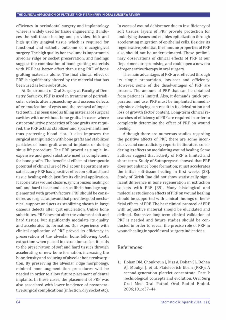
64 65Stomatološki vjesnik 2014; 3 (1) Stomatološki vjesnik 2014; 3 (1)
THE CLINICAL APPLICATION OF PLATELET-RICH FIBRIN (PRF) IN ORAL SURGERY: REVIEW Šečić S, Prohić S
In cases of wound dehiscence due to insufficiency of
soft tissues, layers of PRF provide protection for
underlying tissues and enables epitelization through
accelerating migration of epithelial cells. Besides its
regenerative potential, the immune properties of PRF
also should not be underestimated. These prelimi-
nary observations of clinical effects of PRF at our
Department are promising and could open a new era
of regenerative therapy in oral surgery.
The main advantages of PRF are reflected through
its simple preparation, low-cost and efficiency.
However, some of the disadvantages of PRF are
present. The amount of PRF that can be obtained
from patient is limited. Also, it demands quick pre-
paration and use. PRF must be implanted immedia-
tely since delaying can result in its dehydration and
loss of growth factor content. Long-term clinical re-
searches of efficiency of PRF are required in order to
completely determine the effect of PRF on wound
heeling.
Although there are numerous studies regarding
the positive affects of PRF, there are some incon-
clusive and contradictory reports in literature consi-
dering its effects on modulating wound healing. Some
authors suggest that activity of PRF is limited and
short-term. Study of Suttapreyasri showed that PRF
does not enhance bone formation; it just accelerates
the initial soft-tissue healing in first weeks [38].
Study of Girish Rao did not show statistically signi-
ficant difference in bone regeneration in extraction
sockets with PRF [39]. Many histological and
molecular studies on effects of PRF on wound healing
should be supported with clinical findings of bene-
ficial effects of PRF. The best clinical protocol of PRF
with adjunctive material should be elucidated and
defined. Extensive long-term clinical validation of
PRF is needed and future studies should be con-
ducted in order to reveal the precise role of PRF in
wound healing in specific oral-surgery indications.
References
1. Dohan DM, Choukroun J, Diss A, Dohan SL, Dohan
AJ, Mouhyi J, et al. Platelet-rich fibrin (PRF): A
second-generation platelet concentrate. Part I:
Technological concepts and evolution. Oral Surg
Oral Med Oral Pathol Oral Radiol Endod.
2006;101:e37–44.
efficiency in periodontal surgery and implantology
where is widely used for tissue engineering. It indu-
ces the soft-tissue healing and provides thick and
high quality gingival tissue which is required for
functional and esthetic outcome of mucogingival
surgery. The high quality bone volume is important in
alveolar ridge or socket preservation, and findings
suggest the combination of bone grafting materials
with PRF has better effect than using PRF of bone
grafting materials alone. The final clinical effect of
PRF is significantly altered by the material that has
been used as bone substitute.
At Department of Oral Surgery at Faculty of Den-
tistry Sarajevo, PRF is used in treatment of periradi-
cular defects after apicoectomy and osseous defects
after enucleation of cysts and the removal of impac-
ted teeth. It is been used as filling material of surgical
cavities with or without bone grafts. In cases where
osteoconductive properties of bone grafts are requi-
red, the PRF acts as stabilizer and space-maintainer
thus protecting blood clot. It also improves the
surgical manipulation with bone grafts and stabilizes
particles of bone graft around implants or during
sinus lift procedure. The PRF proved as simple, in-
expensive and good substitute used as complement
for bone grafts. The beneficial effects of therapeutic
potential of clinical use of PRF at our Department are
satisfactory. PRF has a positive effect on soft and hard
tissue healing which justifies its clinical application.
It accelerates wound closure, synchronizes healing of
soft and hard tissue and acts as fibrin bandage sup-
plemented with growth factors. PRF should be consi-
dered as surgical adjuvant that provides good mecha-
nical support and acts as stabilizing sheath in large
osseous defects after cyst enucleation. Unlike bone
substitutes, PRF does not alter the volume of soft and
hard tissues, but significantly modulate its quality
and accelerates its formation. Our experience with
clinical application of PRF proved its efficiency in
preservation of the alveolar bone following tooth
extraction: when placed in extraction socket it leads
to the preservation of soft and hard tissues through
accelerating of new bone formation, increasing the
bone density and reducing of alveolar bone reabsorp-
tion. By preserving the alveolar ridge morphology,
minimal bone augmentation procedures will be
needed in order to allow future placement of dental
implants. In these cases, the placement of PRF was
also associated with lower incidence of postopera-
tive surgical complications (infection, dry socket etc).
2. R. E. Marx, E. R. Carlson, R. M. Eichstaedt, S. R. Schimmele, J. E. Strauss, K. R. Georgeff. Platelet-rich plasma: growth factor enhancement for bone grafts, Oral Surgery, Oral Medicine, Oral Patho-logy, Oral Radiology, and Endodontics, vol. 85, no. 6, pp. 638–646, 1998
3. Saluja H, Dehane V, Mahindra U. Platelet-Rich fibrin: A second generation platelet concentrate and a new friend of oral and maxillofacial surge-ons. Ann Maxillofac Surg. 2011 Jan-Jun;1(1):53-57.
4. Nevins M, Giannobile WV, McGuire MK, Kao RT, Mellonig JT, Hinrichs JE, et al. Platelet-derived growth factor stimulates bone fill and rate of attachment level gain: Results of a large multi-center randomized controlled trial. J Periodontal. 2005;76:2205–15
5. Naik B, Karunakar P, Jayadev M, Rahul Marshal V. Role of Platelet Rich Fibrin in wound Healing: A critical review. J Conservat Dent. 2013 Jul;16(4): 284-93.
6. Gassling V, Douglas T, Warnke PH, Açil Y, Wiltfang J, Becker ST. Platelet-rich fibrin membranes as scaffolds for periosteal tissue engineering. Clin Oral Implants Res. 2010;21:543–9
7. Choukroun J, Diss A, Simonpieri A, Girard MO, Schoeffler C, Dohan SL, et al. Platelet-rich fibrin (PRF):A second-generation platelet concentrate, Part IV: Clinical effects on tissue healing. Oral Surg Oral Med Oral Pathol Oral Radiol Endod. 2006; 101:E56–60.
8. Choukroun J, Diss A, Simonpieri A, Girard MO, Schoeffler C, Dohan SL, et al. Platelet-rich fibrin (PRF): A second-generation platelet concentrate. Part V: Histologic evaluations of PRF effects on bone allograft maturation in sinus lift. Oral Surg Oral Med Oral Pathol Oral Radiol Endod. 2006; 101:299–303.
9. D. M. Dohan, J. Choukroun, A. Diss et al., Platelet-rich fibrin (PRF): a second-generation platelet concentrate. Part III: leucocyte activation: a new feature for platelet concentrates? Oral Surgery, Oral Medicine, Oral Pathology, Oral Radiology and Endodontology, vol. 101, no. 3, pp. E51–E55, 2006
10. M. W. Mosesson, K. R. Siebenlist, and D. A. Meh, The structure and biological features of fibrino-gen and fibrin, Annals of the New York Academy of Sciences, vol. 936, pp. 11–30, 2001
11. Dohan DM, Choukroun J, Diss A, Dohan SL, Dohan AJJ, Mouhyi J, Gogly B. Platelet-rich fibrin (PRF): A second-generation platelet concentrate. Part II:
Platelet-related biologic features. Oral Surg Oral Med Oral Pathol Oral Radiol Endod. 2006;101: E45-50.
12. Van Hinsbergh VW, Collen A, Koolwijk P. Role of fibrin matrix in angiogenesis. Ann N Y Acad Sci 2001;936:426-37.
13. Hotwani K, Sharma K. PRF- a novel acumen into regenerative endodontic therapy. Restorative Dentistry & Endodontics; 2013: 1-5.
14. R. Lundquist, M.H. Dziegiel, M.S. Agren. Bioac-tivity and stability of endogenous fibrogenic fac-tors in platelet-rich fibrin. Wound Repair Regen, 16 (2008), p. 356e63
15. Mammoto T, Jiang A, Jiang E, Mammoto A. Platelet rich plasma extract promotes angiogenesis through the angiopoetin1-Tie2 pathway. Micro-vasc Res. 2013 Sep;89:15-24.
16. D. M. D. Ehrenfest, P. Doglioli, G. M. de Peppo, M. Del Corso, and J. B. Charrier, "Choukroun's plate-let-rich fibrin (PRF) stimulates in vitro prolife-ration and differentiation of human oral bone mesenchymal stem cell in a dose-dependent way", Archives of Oral Biology, vol. 55, no. 3, pp. 185–194, 2010
17. L. He, Y. Lin, X. Hu, Y. Zhang, and H. Wu, “A com-parative study of platelet-rich fibrin (PRF) and platelet-rich plasma (PRP) on the effect of proli-feration and differentiation of rat osteoblasts in vitro,”Oral Surgery, Oral Medicine, Oral Pathology, Oral Radiology and Endodontology, vol. 108, no. 5, pp. 707–713, 2009
18. Li Q, Pan S, Dangaria SJ, Gopinathan G, Kolokythas A, Chu S, Geng Y, Zhou Y, Luan X. Platelet-rich fibrin promotes periodontal regeneration and enhances alveolar bone augmentation. Biomed Res Int. 2013;2013:638043.
19. I.C. Chang, C.H. Tsai, Y.C. Chang. Platelet-rich fibrin modulates the expression of extracellular signal-regulated protein kinase and osteoprotegerin in human osteoblasts. J Biomed Mater Res A, 95 (2010), pp. 327–33
20. Kawase T, Okuda K, Wolff LF, Yoshie H. Platelet-rich plasma-derived fibrin clot formation stimulates collagen synthesis in periodontal ligament andosteoblastic cells in vitro. J Periodontol. 2003 Jun;74(6):858-64.
21. Bambal D, Manwar NU, Chandak M, Rudagi K. A comparative evaluation of the healing ability of bilateral periapical lesions treated with and without the use of platelet-rich fibrin. Todays FDA. 2012 Sep-Oct;24(6):54-7.

66 67Stomatološki vjesnik 2014; 3 (1) Stomatološki vjesnik 2014; 3 (1)
CASE REPORT / PRIKAZ SLUČAJA
DELAYED TOOTH REPLANTATION AFTER TRAUMATIC AVULSION
1Jovanovska M*
1 PHO University Dental Clinical Centre "St. Pantelejmon", Department of Pediatric and Preventive Dentistry - Skopje, R. Macedonia
ABSTRACT
Replantation refers to the insertion and temporary fixation of
completely or partially avulsed teeth resulting from traumatic
injury. Replantation of an avulsed tooth depends on certain
clinical conditions like physiological status of periodontal liga-
ment, the stage of root development and the length of extra-oral
time. Depending on the patient's age, retention of the permanent
incisor can maintain the aesthetic appearance, occlusal function
and alveolar ridge height. Though the risk of progressive repla-
cement resorption and subsequent tooth loss is high after a long
dry storage, replantation provides an aesthetically acceptable
permanent prosthesis at a later age. This article presents manage-
ment of a case with two avulsed permanent incisors that were
stored in a physiological solution for ten hours.
Key words: dental trauma, delayed replantation, tooth
avulsion.
*Corresponding author
Dr Jovanovska Maja
PHO University Dental Clinical
Centre "St.Pantelejmon"
Department of Pediatric
and Preventive Dentistry
Vodnjanska 17
1000 Skopje, Macedonia
e-mail: [email protected]
THE CLINICAL APPLICATION OF PLATELET-RICH FIBRIN (PRF) IN ORAL SURGERY: REVIEW
22. Sunitha R, Munirathnam N. Platelet-rich fibrin: Evolution of a secondgeneration platelet concen-trate. Indian J Dent Res. 2008;19:42–6.
23. Jayalakshmi KB, Agarwal S, Singh MP, Vishwanath BT, Krishna A, Agrawal R. Platelet-rich fibrin with beta-tricalcium phosphate-a noval approach for bone augmentation in chronic periapical lesion: a case report. Case Rep Dent. 2012;2012:902858.
24. Kim BJ, Kwon TK, Baek HS, Hwang DS, Kim CH, Chung IK, et al. A comparative study of the effec-tiveness of sinus bone grafting with recombinant human bone morphogenetic protein 2-coated tricalcium phosphate and platelet-rich fibrin-mixed tricalcium phosphate in rabbits. Oral Surg Oral Med Oral Pathol Oral Radiol Endod. 2011
25. Hauser F, Gaydarov N, Badoud I, Vazquez L, Ber-nard JP, Ammann P. Clinical and histological eva-luation of postextraction platelet-rich fibrin socket filling: a prospective randomized control-led study. Implant Dent. 2013 Jun;22(3):295-303.
26. Vivek GK, Sripathi Rao BH. Potential for osseous regeneration of platelet rich plasma: a compara-tive study in mandible third molar sockets. J maxillofac Oral Surg. 2009 Dec;8(4):308-11. doi: 10.1007/s12663-009-0075-y. Epub 2010 Apr 24.
27. Hoaglin DR, Lines GK. Prevention of localized osteitis in mandibular third-molar sites using platelet-rich fibrin. Int J Dent. 2013;2013: 875380.
28. Jain V, Triveni MG, Kumar AB, Mahta DS. Role of platelet-rich-fibrin in enhancing palatal wound healing after free graft. Contemp Clin Dent. 2012 Sep;3(Suppl 2):S240-3.
29. Bajaj P, Pradeep AR, Agarwal E, Rao NS, Naik SB, Priyanka N, Kalra N. Comparative evaluation of autologous platelet-rich fibrin and platelet-rich plasma in the treatment of mandibular degree II furcation defects: a randomized controlled clini-cal trial. J periodontal Res. 2013 Oct;48(5):573-81.
30. Fan WJ, Yang M, Zhang C, Xue R, Zhang W, Qin HX. Effects of Choukroun's platelet-rich fibrin on hu-man gingival fibroblasts proliferation, migration and type I collagen secretion. Zhonghua Kou Qiang Yi Xue Za Zhi. 2013 Feb;48(2):72-6.
31. Rosamma Joseph V, Raghunath A, Sharma N. Clinical effectiveness of autologous platelet rich
fibrin in the management of infrabony periodon-tal defects. Singapore Dent J. 2012 Dec;33(1):5-12.
32. Mazor Z, Horowitz RA, Del Corso M, Prasad HS, Rohrer MD, Dohan Ehrenfest DM. Sinus floor augmentation with simultaneous implant place-ment using Choukroun's platelet-rich fibrin as the sole grafting material: A radiologic and histo-logic study at 6 months. J Periodontol. 2009;80: 2056–64
33. F. Inchingolo, M. Tatullo, M. Marrelli et al., Trial with platelet-rich fibrin and Bio-Oss used as grafting materials in the treatment of the severe maxillar bone atrophy: clinical and radiological evaluations,”European Review for Medical and Pharmacological Sciences, vol. 14, no. 12, pp. 1075–1084, 2010
34. Y. Zhang, S. Tangl, C. D. Huber, Y. Lin, L. Qiu, and X. Rausch-Fan, “Effects of Choukroun's platelet-rich fibrin on bone regeneration in combination with deproteinized bovine bone mineral in maxillary sinus augmentation: a histological and histomor-phometric study,” Journal of Cranio-Maxillofacial Surgery, vol. 40, no. 4, pp. 321–328, 2011
35. J.W. Lee, S.G. Kim, J.Y. Kim et al. Restoration of a peri-implant defect by platelet-rich fibrin. Oral Surg Oral Med Oral Pathol Oral Radiol, 113 (2012), pp. 459–463
36. Prevention of hemorrhagic complications after dental extractions into open heart surgery pati-ents under anticoagulant therapy: the use of leukocyte- and platelet-rich fibrin. J Oral Implantol, 37 (2011), pp. 681–690
37. Findik Y, Baykul T. Secondary closure of alveolar clefts with mandibular symphyseal bone grafts and with platelet-rich fibrin under local anesthesia: three case reports. J Contempor Dent Pract. 2013 Jul 1;14(4):751-3.
38. Suttapreyasri S, Leepong N. Influence of platelet-
rich fibrin on alveolar ridge preservation. J
Craniofac Surg. 2013 Jul;24(4):1088-94.
39. Girish Rao S, Bhat P, Nagesh KS, Rao GH,
Kharbhari L, Gangaprasad B. Bone regeneration
in extraction sockets with autologous platelet
rich fibrin gel. J maxillofac Oral Surg. 2013
Mar;12(1):11-6.

69
DELAYED TOOTH REPLANTATION AFTER TRAUMATIC AVULSION Jovanovska M
68 Stomatološki vjesnik 2014; 3 (1) Stomatološki vjesnik 2014; 3 (1)
tors, such as trauma, management of the root, extra-
alveolar period and storage medium [4]. The need of
performing endodontic treatment has been demon-
strated by several authors, indicating that the pulp
tissue may become necrotic and its toxins may reach
the periodontal ligament through the dentinal tubu-
les or root canal, definitely contributing to an increa-
se in the resorption process [5].
Nevertheless, if managed appropriately, avulsed
teeth with viable periodontal ligament when replan-
ted can remain functional for some years. This article
describes the management of a child with an avulsed
two central maxillary permanent incisors, kept in a
physiological solution for ten hours. The child has
also teeth's fractures.
Case report
A 15-year-old girl was referred to the Department
of Pediatric and Preventive Dentistry at the Univer-
sity Dental Clinical Center "St. Pantelejmon" Skopje,
for treatment of avulsed teeth. She had experienced a
trauma ten hours earlier and spat out her upper fron-
tal teeth, both central incisors. The intraoral exa-
mination showed that her both maxillar incisors had
been avulsed and their sockets were full with coagu-
lum (Figure 1).
The avulsed teeth were kept in 0,9% NaCl solution
after the injury. Radiological examination confirmed
empty sockets with no other hard-tissue injury
Introduction
Avulsion is defined as a complete separation of a
tooth from its alveolus following a traumatic injury,
which results in extensive damage to the pulp and
periodontal tissues. According to the World Health
Organisation's classification system, later modified
by Andreasen, avulsion is classified as an injury of
periodontal tissues, as well as extrusive, lateral, or
intrusive luxation [1]. As a treatment option, replan-
tation restores occlusal function and aesthetics
shortly after injury. In replantation complete reestab-
lishment of vitality of periodontal fibers is the prime
objective. The percentage of success of tooth replan-
tation has been presented to be low, ranging from 4 to
50% [2]. However, it should be emphasized that the
success of tooth replantation depends on the clinical
needs and patient expectations. The period of perma-
nence of the tooth in the dental arch may be enough to
meet some of the needs, such as prevention of atro-
phy of the alveolar ridge, allow the patients to better
accept the tooth loss, and even delay the need for
prosthetic solution. The success of tooth replantation
depends on the maintenance of vitality of the perio-
dontal ligament, allowing its remains adhered to the
avulsed tooth to survive and recover their function.
Resorption is the main cause of failure of replanta-
tions, and the prognosis of a replanted tooth is rela-
ted to the type of resorption that may ultimately lead
to complete destruction of the root [3]. Degeneration
of the periodontal ligament depends on several fac-
(Figure 2). The patient's medical history was unre-
markable. All of the adjacent teeth showed a positive
response to the test of vitality.
Replantation of the avulsed teeth was planned to
retain teeth in the mouth for as long as possible,
because of the patient's age, although the teeth were
fractured and one of them had also pulpal exposition.
This case was of delayed replantation and endodon-
tic treatment was started before replantation.
Teeth surfaces were cleaned with 0.9% NaCl
solution. Conventional extirpation, enlargement and
cleaning of root canals were performed (Figure 3).
Then, root canals were dried with sterile paper
points (Figure 4) and temporarily filled with calcium
hydroxide paste (Figure 5).
Under local anesthesia, the sockets were gently
curetted to remove any coagulum, granulation tissue
and pathological tissue (Figure 6) and irrigated with
physiologic saline solution. Teeth were then soaked
in 2% sodium fluoride solution for 20 minutes and
replanted into their respective sockets (Figure 7).
Once the teeth were properly seated, they were
checked for alignment or occlusion (Figure 8) and
were splinted to the adjacent teeth with a 0.5 mm
Figure 1Avulsed maxillary central incisors
Figure 2Pre-operative radiograph
Figure 3Extraoral root canal instrumentation
Figure 4Drying with sterile paper
Figure 5Temporary filling with calcium hydroxide
Figure 6Socket preparation with curette

orthodontic stainless steel wire and acid-etch com-
posite (Figure 9). Maxillary occlusal radiograph was
obtained to confirm proper positioning of replanted
teeth (Figure 10). Oral hygiene instructions were
given and chlorhexidine mouthwash was recommen-
ded. A 7-day course of systemic penicillin was pre-
scribed, and the patient was referred to the medical
practitioner for an antitetanus booster.
Six weeks after the injury, before the splint was
removed, the root canals were refilled with definite
sealer and gutta percha points (Figure 11). During
that period the teeth did not show any clinical
symptoms like mobility, periodontal pockets or any
type of root resorption.
A restoration of coronary fractures with acid-etch
composite followed in order to correct aesthetic,
phonetic and masticatory function, and to meet the
patient's and her parents expectation of this treat-
ment (Figure12). The patient came for regular con-
trols every three months (Figure 13) and the follow-
up period lasted for two years (Figure 14). During
this period, the patient was asymptomatic, with no
complications, and satisfied with the conservative
treatment.
DELAYED TOOTH REPLANTATION AFTER TRAUMATIC AVULSION Jovanovska M
Discussion
Tooth avulsion is a serious assault on the gingiva,
periodontal ligament and pulp. Young permanent
tooth loss leads to a severe arrest of alveolar bone
formation in a growing child. Alveolar ridge would be
narrow and difficult to restore in future with either a
bridge or implant. Most conservative approach for
managing the avulsed incisors is to replant them as
soon as possible. In clinical studies, teeth replanted
within 5 minutes after avulsion have the best progno-
sis and the chance of pulpal and periodontal healing
is inversely related to the stage of root development
and the extra-oral time, as well as the medium where
the tooth is stored [6]. A review of literature shows
that an extra-oral time of less than 15 minutes gives a
greater success rate of retention [7] of replanted
teeth, but if the time lapse is between 15-60 minutes,
some authors advise that the root must be stored in
suitable storage medium and transported to the cli-
nic [8]. Some of the storage media that have been
frequently used include Emergency Medical Tooth
saver, Euro-Collins solutions, saline, pasteurized
7170 Stomatološki vjesnik 2014; 3 (1) Stomatološki vjesnik 2014; 3 (1)
Figure 7Replantation
Figure 8Clinical aspects after replantation
Figure 11Extraoral root canal instrumentation
Figure 12Composite restoration
Figure 9Stabilization with composite-wire splint
Figure 10Radiograph after replantation
Figure 13Radiograph after six months
Figure 14Radiograph after 24 months
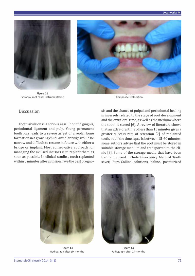

73
With the end of last year, from the press has come
an university textbook "Fundamentals of tooth mor-
phology and dental anthropology", published by the
Faculty of Dentistry, University of Sarajevo. The
authors Amra Vuković, Selma Zukić, Anita Bajsman
and Aida Selmanagić have made efforts to consolidate
and in a clear way present knowledge of the morpho-
logy of the teeth and the basics of dental anthropology.
Considering the course of development of tooth mor-
phology as a scientific discipline in Bosnia and Herze-
govina, the authors offered to us a book that is prima-
rily a textbook, but also will serve to all those who
want to understand the connection between tooth
morphology and function. This is certainly a basis for
quality clinical practice, and also for a scientific ap-
proach to understanding the biology of tooth in the
context of contemporary trends in biomedicine. The
material is processed through seven thematic sec-
tions and chapters. In the introduction the authors
write about stomatognahtic system and its compo-
nents. The next chapter deals with the nomenclature
in dental morphology, with the special emphasis on
the morphological details of the tooth. This is follo-
BOOK REVIEW / PRIKAZ KNJIGE
wed by a chapter on the permanent and after that by a
chapter on primary teeth. In the fifth thematic section
authors explain the morphological and anatomical
relations of the dental arches. Logical sequencing is
next thematic area that describes and explains the
anomalies of the teeth, dividing them into congenital
and acquired. The final chapter is about dental an-
thropology, where is in detail explained the classifi-
cation of morphological details by internationally
acclaimed system. All chapters are enriched with ori-
ginal photographs and illustrations, as many as 121,
which gives added value to this book. Reviewers of the
book were distinguished professors: Professor Eme-
ritus Hajrija Konjhodžić Raščić from Faculty of Denti-
stry, University of Sarajevo, Professor Hrvoje Brkić
from Faculty of Dentistry, University of Zagreb and
Professor Azijada Šuljak Loncarevic from Faculty of
Dentistry, University of Sarajevo. Giving their opinion
and a positive evaluation of the textbook reviewers
wrote this: "In this textbook, the content is processed
encyclopedicly ... It has a special significance for the
adoption and learning of terminology - anatomical
terminology of morphological details on the teeth and
TITLE:
AUTHORS:
PUBLISHER:
LANGUAGE:
FORMAT:
NUMBER OF PAGES:
DATE OF ISSUE:
ISBN:
Fundamentals of Tooth Morphology and Dental Anthropology
Amra Vuković, Selma Zukić, Anita Bajsman, Aida Selmanagić
Stomatološki fakultet Univerziteta u Sarajevu,Sarajevo
Bosnian
Softcover
199
2013
978-9958-9051-4-8
Stomatološki vjesnik 2014; 3 (1)Stomatološki vjesnik 2014; 3 (1)
DELAYED TOOTH REPLANTATION AFTER TRAUMATIC AVULSION
milk, saliva, or chicken egg white [9]. Literature data
shows that the average time range of replanted teeth
is between 1 to 4 hours, after which the success of
replantation is determined by the storage media. Suc-
cess has also been reported in a study where the
tooth was replanted after 8 hours, but was stored in
Euro-Collins15 solution. Other studies showed that
replantation after 60 minutes in no storage medium
with extra-oral endodontics may result in retention
of the tooth [10]. However, studies by Petrovic et al.
showed that replanted dry teeth in dry storage
between 15 minutes to 9 hours had low survival rates
and a reduced chance of retention. But, immediate
replantation is not always possible because of vario-
us unavoidable problems.
In our patient, the greatest benefit of successful
replantation of an avulsed tooth was preservation of
alveolar bone. Even if the replanted teeth must be
extracted later, the improved alveolar development
will provide better options for restoration of the site
later. We could successfully achieve objectives like
acceptable aesthetic appearance and occlusal func-
tion, prevention of root resorption and favorable hea-
ling for better permanent treatment in later life. Our
patient was asymptomatic during the follow-up
period of 24 months. This procedure gave invaluable
and timely contribution to their normal and physio-
logical growth.
References
1. Lin S, Zuckerman O, Askenazi M. American Asso-ciation of Endodontists; International Associaton of Dental Traumatology; Royal College of Sur-geons of England. New emphasis in the treatment of dental trauma. Dent Traumatol 2007; 23:297-303.
2. Andreasen JO, Borum MK, Jacobsen HL, Andrea-sen FM. Replantation of 400 avulsed permanent incisors. 4. Factors related to periodontal liga-ment healing. Endod Dent Traumatol 1995; 11:76-89.
3. Andreasen JO, Borum MK, Jacobsen HL, Andeasen FM. Replantation of 400 avulsed permanent incisors. 1. Diagnosis of healing comlications. Endod Dent Traumatol 1995; 11(2):51-8.
4. Askenazi M, Marouni M, Sarnat H. In vitro viabi-lity, mitodenicity and clonogenic capacites of periodontal ligament fibroblasts after storage in four media supplemented with growth factors. Dent Traumatol 2001; 17:27-35.
5. Flores MT, Andersson L, Andreasen JO, Bakland LK, Malmgren B, Barnett F, et al. Guidelines for the management of traumatic dental injuries. II. Avul-sion of permanent teeth. Dent Traumatol 2007; 23(3):130-6.
6. Hinkckfuss SE, Messer LB. An evidence-based assessment of the clinical guidelines for replan-ted avulsed teeth.Part I: Timing of pulp extir-pation. Dent Traumatol 2009; 25:32-42.
7. Petrovic B, Markovic D, Peric T, Blagojevic D. Factors related to treatment and outcomes of avulsed teeth. Dent Traumat 2010; 26:52-9.
8. Panzarini SR, GulineliJL, Poi WR, Sonoda CK, Pe-drini D, Brandini DA. Treatment of root surfacein delayed raplantation: A review of the literarure. Dent Traumat 2008; 24:277-82.
9. Sottovia AD, Sottovia Fiho D, Poi WR, Panzarini SR, Luize DS, Sonoda CK. Tooth replantation after use of Euro-Collins solution or bovine milk as storage medium: A histomorphometric analysis in dogs. J Oral Maxillofac Surg 2010; 68:111-9.
10. Day P, Duggal M. Interventions for treating trau-matized permanent front teeth. Avulsed (knocket out) and replanted. Cochrane Database Syst Rev 2010; 20:CD006542.
72

74 75
oral cavity in general". "This is an original work that is
based on scientific knowledge and achievements in
the field of dental morphology with dental anthro-
pology. In Bosnia and Herzegovina there is no
textbook with similar content and quality, etc. This
book will be intended for dentists, residents and
specialists from all fields of dentistry. It is of a special
importance for specialists of dental prosthetics,
where, without knowing the dental anatomical cha-
racteristics, in particular the characteristics of
occlusal tooth morphology and its expediency in all
functions of the stomatognathic system, one cannot
imagine a proper prosthetic therapy. The special
quality of this manuscript is provided with the
original photos and illustrations."
So in the end we can say that this book will serve
not only to those who are bound by their work with
universities, but also to clinicians and physicians
during the lifelong learning in all countries of the
same or similar language.
Sadeta Šečić
BOOK REVIEW / PRIKAZ KNJIGE INSTRUCTIONS FOR THE AUTHORS
Submissions of manuscripts are made through
the submission form available at web page of
the Journal (www.stomatoloskivjesnik.ba) or
by sending the email to Editorial office at
radovi@stomatoloski vjesnik.ba
E-mail must be composed of:
A) Covering letter, in which authors explain the
importance of their study (Explanation why we
should publish your manuscript ie. what is new
and what is important about your manuscript,
etc).
B) Title of the manuscript
C) Authors' names and email addresses (mark
corresponding author with *)
D) Abstract
E) Attached file of the Copyright assignment form
and
F) Manuscript.
Authors should NOT in addition post a hard copy
of the manuscript and submission letter, unless they
are supplying artwork, letters or files that cannot be
submitted electronically, or have been instructed to
do so by the editorial office.
Please read Instructions carefully to improve
yours paper's chances for acceptance for publi-
shing.
Thank you for your interest in submitting an
article to Stomatološki vjesnik.
Type of papers suitable for publishing in Sto-
matološki vijesnik (Journal in following text):
Original Articles, Case Reports, Letters to the Edi-
tors, Current Perspectives, Editorials, and Fast-Track
Articles are suitable for publishing in Stomatološki
vjesnik. Papers must be fully written in English with
at least title, abstract and key words bilingual in Bos-
nian/Croatian/Serbian language (B/C/S) and Eng-
lish language.
Editorial process:
All submitted manuscripts are initially evaluated
by at least two scientific and academic members of
editorial board. An initial decision is usually reached
within 3–7 days.
Submitted manuscripts may be rejected without
detailed comments after initial review by editorial
board if the manuscripts are considered inappro-
priate or of insufficient scientific priority for publi-
cation in Stomatološki vjesnik.
If sent for review, each manuscript is reviewed by
scientists in the relevant field. Decisions on reviewed
manuscripts are usually reached within one month.
When submission of a revised manuscript is invited
following review, the revision must be received in
short time of the decision date.
Criteria for acceptance:
Submitted manuscripts may be rejected without
detailed comments after initial review by editorial
board if the manuscripts are considered inappropria-
te or of insufficient scientific priority for publication
in the Journal. All other manuscripts undergo a com-
plete review by reviewers or other selected experts.
Criteria for acceptance include originality, validity of
data, clarity of writing, strength of the conclusions,
and potential importance of the work to the field of
dentistry and similar bio-medical sciences. Submit-
ted manuscripts will not be reviewed if they do not
meet the Instructions for authors, which are based on
"Uniform Requirements for Manuscripts Submitted
to Biomedical Journals" (http://www.icmje.org/).
INSTRUCTIONS FOR THE AUTHORSmade in accordance with the recommendations of the International Committee of Medical Journal based on "Uniform Requirements for Manuscripts Submitted to Biomedical Journals" (http://www.icmje.org/).
Stomatološki vjesnik 2014; 3 (1) Stomatološki vjesnik 2014; 3 (1)

7776
INSTRUCTIONS FOR THE AUTHORS
national bodies should be avoided. Use only standard
abbreviations. Avoid abbreviations in the title and
abstract. The full term for which an abbreviation
stands should precede its first use in the text unless it
is a standard unit of measure-ment.
The Title Page should carry the full title of the pa-
per and a short title to be used as a 'running head'
(and which should be so identified). The first name,
middle initial and last name of each author should
appear marked with superscript numbers or/and
symbols corresponding to their affiliation or/and
note. Affiliations of the authors should be written be-
low authors name list. Full name, address, phone and
fax number and e-mail of the author responsible for
correspondence should appear on the Title Page.
Please include the word count of the abstract and
word count of text on the title page.
The second page should carry a structured
abstract of no more than 250 words. The abstract
should state the Objective(s) of the study or investi-
gation, basic Methods (selection of study subjects or
laboratory animals; obser-vational and analytical
methods), main Results (giving specific data and
their statistical significance, if possible), and the
principal Conclusions. It should emphasize new and
important aspects of the study or observations.
The abstract should be followed by a list of 3–10
keywords or short phrases which will assist the
cross-indexing of the article and which may be publi-
shed. When possible, the terms used should be from
the Medical Subject Headings list of the National
Library of Medicine (http://www.nlm.nih.gov/
mesh/meshhome.html).
Body of the manuscript text of an experimental
or observational nature may be divided into sections
headed Introduction, Materials and Methods (inclu-
ding ethical and statistical information), Results,
Discussion and Conclusion, although reviews may
require a different format.
Original Articles: original experimental and clini-
cal studies should not exceed 4400 words (up to 15
pages) including tables and references.
With Case Reports: presentation of a clinical case
which may suggest novel working hypotheses, with a
short discussion on the pertinent literature. The text
should not exceed 2400 words (up to 8 pages).
Letters to the Editors should not exceed 500
words, should not be signed by more than three
authors and should not have more than 5 references.
Preferably, letters should be in reference to a Journal
article published within the last 3 months or to novel
hypotheses so as to stimulate comments on issues of
common interest. Authors of the letters accepted for
publication will receive the galley proofs. The Editors
will generally solicit replies. The Editors reserve the
right to modify the text.
Current Perspectives: invited articles by recogni-
zed authorities, to include position papers, reviews,
and special topics of general interest. Independent
submission will also be considered.
Editorials: invited articles or brief editorial com-
ments that represent opinions of recognized leaders
in biomedical research.
Fast-Track Articles: short articles on laboratory or
clinical findings, representing important new in-
sights or major advances, produced with established
methods or new applications of an established or
new method. The text should not exceed 2400 words.
Acknowledgements: One or more statements
should specify (a) contributions that need acknow-
ledging but do not justify authorship, such as general
support by a department chairman; (b) acknow-
ledgements of technical help; (c) acknowledgements
of financial and material support, specifying the
nature of the support. Persons who have contributed
intellectually to the paper but whose contributions
do not justify authorship may be named and their
function or contribution described – for example,
'scientific adviser', 'critical review of study proposal',
'data collection', 'participation in clinical trial'. Such
persons must have given their permission to be na-
med. Authors are responsible for obtaining written
permission from persons acknowledged by name
because readers may infer their endorsement of the
data and conclusions.
Declaration of Interest
A statement must be provided listing all financial
support received for the work and, for all authors, any
INSTRUCTIONS FOR THE AUTHORS
Authorship:
All authors have to sign the copyright assignment
form. We ask all authors to confirm that: they have
met the criteria for authorship as established by the
International Committee of Medical Journal Editors;
they believe that the paper represents honest work,
and are able to verify the validity of the results re-
ported.
Redundant or duplicate publication:
We ask the authors to confirm in the copyright
assignment form that the paper has not been publi-
shed in its current form or a substantially similar
form (in print or electronically, including on a web
site), that it has not been accepted for publication
elsewhere, and that it is not under consideration by
another publication. The International Committee of
Medical Journal Editors has provided details of what
is and what is not duplicate or redundant publi-
cation (http://www.icmje.org). In the submission
letter to the editors, authors are asked to draw
attention to any published work that concerns the
same patients or subjects as the present paper.
Author Contributions:
Authors are required to include a statement
to verify the contributions of each co-author in the
copyright assignment form (avai lable at
www.stomatoloskivjes nik.ba ).
Patient consent forms:
The protection of a patient's right to privacy is
essential. The authors may be asked by the editorial
board to send copy of patient consent forms on which
patients or other subjects of the experiments clearly
grant permission for the publication of photographs
or other material that might identify them. If the
consent form for the research did not specifically
include this, authors should obtain it or remove the
identifying material.
Ethics committee approval:
The authors must state clearly in the submission
letter and in the Methods section that the conducted
studies on human participants are with the approval
of an appro-priate named ethics committee. Please
also look at the latest version of the Declaration of
Helsinki (http:// www.wma.net/e/policy/b3.htm).
Similarly, the authors must confirm that experiments
involving animals adhered to ethical standards and
must state the care of animal and licensing guidelines
under which the study was perfor-med. The editorial
board may ask author(s) for copy of ethical
committee approval.
Declaration of Interest:
We ask the authors to state all potential financial
support received for the work. This applies to all
papers including editorials and letters to the editor
(see below). If you are sure that there is no conflict of
interest, please state that.
Copyright:
Copyright assignment form contains authors' sta-
tement that all the copyrights are transferred to the
publisher if and when the manuscript is accepted for
publishing.
Subscribers may reproduce tables of contents or
prepare lists of articles including abstracts for inter-
nal circulation within their institutions. Permission
of the Publisher is required for resale or distribution
outside the institution and for all other derivative
works, including compilations and translations. If
excerpts from other copyrighted works are included,
the author(s) must obtain written permission from
the publisher and credit the source(s) in the article.
Formatting requirements:
Manuscript should be written in Times New Ro-
man 12, Normal, double spacing.
Include the following sections, each starting on a
separate page: Title Page, Abstract and Keywords,
Text, Acknowledgements, Declaration of interest, Re-
ferences, Individual tables and figures with captions.
Margins should be not less than 3 cm. Pages
should be numbered consecutively, beginning with
the Title Page, and the page number should be placed
in the bottom right hand corner of each page.
Abbreviations should be defined on their first
appearance in the text; those not accepted by inter-
Stomatološki vjesnik 2014; 3 (1) Stomatološki vjesnik 2014; 3 (1)
7978
INSTRUCTIONS FOR THE AUTHORS
financial involvement (including employment, fees,
share ownership) or affiliation with any organization
whose financial interests may be affected by material
in the manuscript, or which might potentially bias it.
This applies to all papers including editorials and
letters to the editor. If you are sure that there is no
conflict of interest, please state this.
References
References should be numbered consecutively in
the order in which they first appear in the text. They
should be assigned Arabic numerals, which should be
given in brackets, e.g. [17]. References should include
the names of all authors when six or fewer; when
seven or more, list only the first six names and add et
al. References should also include full title and source
information (Vancouver style).
Journal names should be abbreviated as in
MEDLINE (http://www.medscape.com/Home/
Search/ IndexMedicus/IndexMedicus.html).
Examples of citation:
Standard journal article:
Tashiro H, Shimokawa H, Sadamatu K, Yamamoto
K. Prognostic significance of plasma concentra-
tions of transforming growth factor-ß. Coron
Artery Dis 2002; 13(3):139-143.
More than six authors:
Yetkin E, Senen K, Ileri M, Atak R, Tandogan I,
Yetkin Ö, et al. Comparison of low-dose dobuta-
mine stress echocardiography and echocardio-
graphy during glucose-insulin-potassium infu-
sion for detection of myocardial viability after
anterior myocardial infarction. Coron Artery Dis
2002; 13(3):145-149.
Books:
Heger JW, Niemann JT, Criley JM. Cardiology, 5th
ed. Philadelphia: Lippincott, Williams & Wilkins;
2003.
Chapter in a book:
Braunwald E, Perloff JK. Physical examination of
the heart and circulation. In; Braunwald E, Zipes
DP, Libby P (eds). Heart disease; a textbook of
cardiovascular medicine, 6th edn. Philadelphia:
WB Saunders; 2001, pp. 45-81.
Personal communications and unpublished work
should not feature in the reference list but should
appear in parentheses in the text. Unpublished work
accepted for publication but not yet released should
be included in the reference list with the words 'in
press' in parentheses beside the name of the journal
concerned. References must be verified by the
author(s) against the original documents.
Tables
• Provide each table on a separate page of the
manuscript after the references.
• Each table should be typed on a separate sheet in
double spacing.
• Number the table according to their sequence in
the text. The text should include references to all
tables. Each table should be assigned an Arabic
numeral, e.g. (Table 3).
• Include a brief and self-explanatory title with
explanations essential to the understanding of the
table at the bottom of the table.
• Identify statistical measures of variations, such as
standard deviation and standard error of the
mean or other where appropriate.
Figures
• Provide each figure on a separate page of the
manuscript after the references. Number the
figures according to their sequence in the text. The
text should include references to all figures.
• Graphs and figures should be in black/white or
greyscale format. Colour illustrations are accep-
table but not guaranteed. Minimal quality 300dpi,
figures should be filed in suitable format (*.JPG,
*.PNG, .*TIFF)
• If figures are not original provide source and
permition.
• All figures should be 100% of a suitable final size
and have the printing resolution of 300dpi and be
cropped to include the figure only (no blank
space).
INSTRUCTIONS FOR THE AUTHORS
Units of measurement
Measurements of length, height, weight, and volu-
me should be reported in metric units (meter, kilo-
gram, or liter) or their decimal multiples. All
hematologic and clinical chemistry measurements
should be reported in the metric system in terms of
the International System of Units (SI).
Post acceptance
All correspondence concerning the copy, editing
and production of accepted manuscripts should be
addressed to Stomatološki vjesnik.
Charges
Authors will not be charged fees in a first year of
publishing Stomatološki vjesnik. Optional charges
for colour reproduction of figures may apply. Authors
will be informed about this when the poof is supplied
to them. The charges must be completed before the
article is released.
Stomatološki vjesnik 2014; 3 (1) Stomatološki vjesnik 2014; 3 (1)