Embed Size (px)
Citation preview
Histol Histopathol (2001 ) 16: 827-832
001: 10.14670/HH-16.827
http://www.hh.um.es
Histology and Histopathology
Cellular and Molecular Biology
Scanning electron microscopy and calcification in amelogenesis imperfecta in anterior and posterior human teeth M.C. Sanchez-Quevedol, G. Ceballos2, J.M. Garcial, I.A. Rodriguez3, M.E. Gomez de Ferraris3 and A. Campos1 1 Departamento de Histologia y Biologia Celular, Facultad de Medicina y Odontologia, Universidad de Granada, Granada, Espana,
2Servicio de Estomatologia, Hospital Cllnico Universitario, Universidad de Granada, Granada, Espana and
3Catedra B de Histologia y Embriologia, Facultad de Odontologia, Universidad Nacional de Cordoba, Argentina
Summary. Teeth fragments from members of a fam ity' clinically and genetically diagnosed as having amelogenesis imperfecta were st udi ed by scanning electron microscopy and X-ray microprobe analysis to establish the morphological patterns and the quantitative concentration of calci um in the e namel of anterior (canine, incisor) and posterior (premolar and molar) teeth. The prism patterns in the enamel of teeth from both regions were parallel or irregularly decussate, with occasional filamentous prisms accompanied by small, irregularly rounded formations. Prism less enamel showed the R- and P-type patterns. Calcium levels in enamel of amelogenesis imperfecta and control teeth differed significantly between anterior and posterior teeth, indicating that the factors that influence normal mineralization in different regions of the dental arch are not altered in the process of amelogenesis imperfecta.
Key words: Amelogenesis imperfecta, Prism patterns, Calcification, Scanning microscopy, X-ray microprobe analysis, Anterior teeth, Posterior teeth
Introduction
Amelogenesis imperfecta (AI) is a term that identifies a group of hereditary alterations that affect the formation of enamel extracellular matrix (Dong et aI., 2000) . This disorder has been classified into 14 subgroups according to the characteristics of the enamel, clinical appearance and Mendelian mode of inheritance (autosoma l dominant or recessive, and sex-linked dominant or recessive). The difficulties in establ ishing an acceptable correlat ion between the clinical classification and microscopic findings (Backmann et
Offprint requests to: Prof. M.C. Sanchez-Quevedo, Departamento de Histologia y Biologia Celular, Facultad de Medicina, Universidad de Granada, E·18071 Granada, Spain. Fax: 34 958 244034. e-mail : [email protected]
aI., 1993) have brought to light the need to search for correlations between the mineral component of enamel in different teeth and microscopic observations of the prism and prism less structure of the enamel. To this end some studies have tried to measure the mineral content and distribution in enamel with quantitative microradiography and chemical analysis (Wright et aI., 1993; Backmann and Angmar-Mansson, 1994; Wright et aI., 1995) or with quantitative X-ray microanalytical histochemistry, a productive tool recently applied to mineralized tissues (Wright et aI., 1991; LOpez-Escamez et aI., 1992, 1993; L6pez-Esdimez and Campos, 1994; Campos et aI., 1994, 2000; Sanchez-Quevedo et aI. , 1998). The development of the tooth germ is influenced by the position of the tooth in the anterior or posterior part of the dental arch , where genetic and molecular factors along with interdental spacing, tooth inclination, arch growth and tooth size also come in to play (Thomas et a I. , 1998; Avery and Stelle, 1999; Ferraris and Campos, 2000).
This study was designed to use quantitative x-ray microprobe analysis (EPMA) with scanning electron microscopy (SEM) as a histological approach to determine the morphological patterns of the enamel in different types of anterior (canine, incisor) and posterior (premolar, molar) teeth in the dental arch in teeth from persons who were clinically and genetically diagnosed as having AI. In addition, we measured the concentration of calcium, as the influence of the anterior or posterior location of the tooth on the morphological pattern and degree of enamel calcification have not been analyzed in patients with AI.
Materials and methods
The material consisted of 20 specimens from the coronal part of permanent teeth obtained in the course of preparation for crown placement from patients with autosomal dominant clinically and genet ically diagnosed AI (5 incisors, 5 canines, 5 premolars and 5 molars). All

Scanning microscopy and calcification in amelogenesis imperfecta
patients were members of the same family (Ceballos and Ceballos, 1988). As the control material we used corona1 specimens from 20 normal teeth (5 incisors, 5 canines, 5 premolar and 5 molars) extracted during orthodontic or periodontal treatment.
Sample preparation for EPMA
The tooth fragments were plunge-frozen in liquid nitrogen-cooled Freon 22. The samples were transferred to Polaron E 5350 the freeze-drying apparatus and dried at -80 "C for 48 h. Al1 specimens were mounted on a carbon stub and sputter-coated with a thin layer of carbon in an argon atmosphere at 0.1 Torr for 30 s.
Electron probe X-ray microanalysis
Specimens prepared as described above were studied in a Phillips XL-30 scanning electron microscope (SEM) (operating voltage = 15 k v , spot size = 500 nm; tilt angle = 35"; take-off angle = 61.34"). An energy dispersive spectrometer (EDAX DX-4) was used for quantitative analyses (count rate = 1200 cps; live time = 50 S). Spectra were collected by pin-point electron beam at x40 000, and a total 200 spectra (10 for each of the 20 Ai and control specimens) were obtained. The peak-to- background (P/B) ratio method (Statham and Pawley, 1978; Small et al., 1979) was used to measure the concentration of calcium.
Microcrystalline salt standards were used to quantify calcium as described in previous publications (Campos et al., 1992, 2000; López-Escámez and Campos, 1994; Sánchez-Quevedo et al., 1998). The standards were cryofixed in liquid Na freeze-dried and sputter-coated as described above, and analyzed in the microscope immediately after preparation to avoid contamination or chemical modification. Micro-crystalline standards were
mounted on 200-mesh nickel grids fixed with adhesive graphite lamina to SEM holders. The elemental weight percent (WP) of each salt standard was calculated as reported in previous publications (Warley and Gupta, 1991; Warley, 1993, 1997).
Morphological study
Carbon-coated specimens were gold-coated after EPMA analysis, and were examined in a Phillip XL-30 SEM and photographed.
Statistical study
The data were analyzed with one-way ANOVA and paired comparisons with the Bonferroni test, using the BMDP statistical package.
Results
Scanning electron microscopic images of enamel in teeth with Ai showed a number of different patterns in both the anterior and posterior parts of the dental arch. In specimens with A1 the outer enamel showed a prismless structure of variable size, and an irregular array of fissures. The P-type (prism-dependent) and R-type pattern (Retzius-dependent) of prismless enamel were observed (Figs. 1,2).
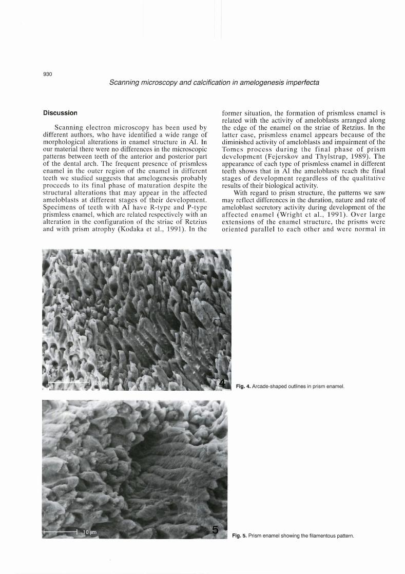
The underlying prismatic enamel was arranged in two patterns: parallel prisms were more common, and a decussate pattern was seen only in certain areas. Parallel prisms showed a regular diameter and a keyhole outline. Decussate prisms were varied in their orientation and diameter, and showed a variable arcade-like outline delimiting an irregular pit structure (Figs. 1 , 3, 4). Regardless of their architectural pattern, some prisms showed a filamentous configuration (Fig. 5); these were
Fig. 1. P-type patiern in prismless enamel, and parallel patiern in prism enamel.

Scanning microscopy and calcification in amelogenesis imperfecta
accompanied by irregularly rounded elements of variable size arranged, in some cases, in a beaded or ribbon-like pattern seen mainly in the region underlying the prismless enamel (Fig. 1).
Tables 1, 2 summarize the microanalytical data for calcium content in control and A1 specimens in the anterior and posterior parts of the dental arch. Statistical analysis revealed significant differences (Levene's: F test: = 4.35, p = 0.0384; Welch approximation: df
(1,193), F = 6.25, p = 0.0134; Bonferroni test: n = 100; p < 0.05) in calcium weight fraction between teeth with A1 from the anterior (canines and incisors, x = 32.16) and posterior part (premolars and molars, ?t = 33.46) of the dental arch. The difference between the anterior and posterior regions was also significant in control teeth (Levene's: F test: = 29.07, p < 0.0001; Welch approximation: df (1,154), F = 56.05, p < 0.0001; Bonferroni test: n = 100; p < 0.001).
Table 1. Percent weight fraction and mean weight fraction of calcium in Table 2. Percent weight fraction and mean weight fraction of calcium in teeth from the anterior region of the dental arch. teeth from the posterior region of the dental arch.
INCISORS CANINES RISD PREMOLARS MOLARS R+SD
Arnelogenesis imperfecta 32.6313.81 31.6813.31 32.1 623.36 Amelogenesis imperfecta 32.6623.31 34.2524.42 33.4613.97 Control 29.9321.85 30.1 921.45 30.0611.65 Control 31.16d.24 34.0413.00 32.61 13.00
Fig. 2. R-lype pattern in prismless enamel.
Flg. 3. Area showing the decussate pattern in pnsm enamel.
Scanning microscopy and calcification in amelogenesis imperfecta
Discussion
Scanning electron microscopy has been used by different authors, who have identified a wide range of morphological alterations in enamel structure in AI. In our material there were no differences in the microscopic patterns between teeth of the anterior and posterior part of the dental arch. The frequent presence of prismless enamel in the outer region of the enamel in different teeth we studied suggests that amelogenesis probably proceeds to its final phase of maturation despite the structural alterations that may appear in the affected ameloblasts at different stages of their development. Specimens of teeth with A1 have R-type and P-type prismless enamel, which are related respectively with an alteration in the configuration of the striae of Retzius and with prism atrophy (Kodaka et al., 1991). In the
former situation, the formation of prismless enamel is related with the activity of ameloblasts arranged along the edge of the enamel on the striae of Retzius. In the latter case, prismless enamel appears because of the diminished activity of ameloblasts and impairment of the Tomes process during the final phase of prism development (Fejerskov and Thylstrup, 1989). The appearance of each type of prismless enamel in different teeth shows that in A1 the ameloblasts reach the final stages of development regardless of the qualitative results of their biological activity.
With regard to prism structure, the patterns we saw may reflect differences in the duration, nature and rate of ameloblast secretory activity during development of the affected enamel (Wright et al., 1991). Over large extensions of the enamel structure, the prisms were oriented parallel to each other and were normal in
Flg. 4. Arcade-shaped outlines in prisrn enarnel.
m Flg. 5. Prisrn enarnel showing the filamentous paltern.

Scanning microscopy and calcification in amelogenesis imperfecta
appearance. However, in some areas we observed anomalous patterns, i.e., a decussate pattern, a superimposed filamentous pattern or an arcade-shaped outline. Because these features have been described in SEM studies of normal developing human enamel, immature morphological patterns can be said to persist in the enamel of persons with AI (Risnes et al., 1998).
Selective alterations may affect some ameloblasts during enamel formation. An alteration in the expression of the genes that control enamelin secretion or the phenomena of apoptosis during ameloblast differentiation are probably involved in the etiology (Hu et al., 2000) and in the origin of these morphological patterns (Vaahtokari et al., 1996). In this regard, the differences in ameloblast apoptosis in different types of tooth during the prolonged modulation phase, when the enamel is hardening, may contribute to the microscopic heterogeneity between teeth seen in the present study, and to the differences between our observations and earlier descriptions of the microscopic features of enamel (Smith and Warshawsky, 1977; Joseph et al., 1 994).
One of the most characteristic features of A1 that SEM detects is the presence of noncrystalline-appearing, irregular rounded formations located in interprismatic areas. Different authors have related these formations with organic matter that is not removed during maturation, and which might be normal unprocessed or structurally abnormal enamel proteins (Wright and Butler, 1989). In our tooth specimens these formations were seen mainly in the region underlying the prismless enamel, a location that appears to support this hypothesis.
Severa1 authors have investigated the correlation between morphological alterations and calcification with qualitative microprobe analysis. These studies have failed to find significant differences in mean calcium levels between normal and Ai teeth, and suggested that the mineral in both cases is probably similar, at least in its primary composition (Wright et al., 1991). However, interna1 variations in calcium levels through the thickness of the enamel have been reported, and the nature of qualitative X-ray microprobe analysis is such that it causes different degrees of tissue excitation depending on tissue density. These technical limitations complicate determinations of the real concentration of calcium in the enamel, and make it necessary to use an appropriate quantitative approach.
With our quantitative method of X-ray microprobe analysis for mineralized tissues, which have been validated in severa1 studies (Mpez-Escámez et al., 1992, 1993; Campos et al., 1994; Mpez-Escámez and Campos, 1994; Warley, 1997; Sánchez-Quevedo et al., 1998), we found that the degree of calcification differed significantly between the anterior and posterior parts of the dental arch in AI. Moreover, calcification also differed significantly between the anterior and posterior regions in normal teeth. Arnelogenesis imperfecta causes morphological alterations in both regions, with evident
signs of immaturity in the process of differentiation. However, these morphological changes were not accompanied by alterations in the foreseeable process of mineralization in anterior and posterior teeth. Although enamel calcium levels are higher in A1 as previously shown (Sánchez-Quevedo et al., 1999) the finding that calcium levels in the enamel of both AI and control teeth differed significantly between anterior and posterior teeth indicates that the factors that influence normal mineralization in different regions of the dental arch are not altered in the process of amelogenesis imperfecta.
Acknowledgements. This work was supported by the Ministerio de Educaci6n y Cultura through Project PB97-0804, and by the Agencia Española de Cooperación Internacional through Project AEC1/98/01. We thank M. Ángeles Robles for her wmpetent technical assistance, and K. Shashok for translatlng parts of the manuscript into English.
References
Avery J.K. and Stelle P.F. (1999). Essentials of oral histology and embryology: a clinical approach. Mosby. New York.
Blckman B. and Angmar-Mansson B. (1994). Mineral distribution in the enamel of teeth with amelogenesis imperfecta as determined by quantitat'we microradiography. Scand. J. Dent. Res. 102, 193-197.
Backman B., Lundgren T., Engstrom E.U., Falk L.K.L., Chabala J.M., Levi-Setti R. and Nor6n J.G. (1993). The absence of correlations between a clinical classification and ultrastructural findings in amelogenesis imperfecta. Acta Odontol. Scand. 51, 79-89.
Campos A., López-Escámez J.A., Cañizares F.J. and Crespo P.V. (1992). Electron probe X-ray microanalysis of Ca and K distributions in the otholithic membrane. Micron Microsc. Acta 23, 349-350.
Campos A., López-Escámez J.A., Crespo P.V., Cañizares F.J. and Baeyens J.M. (1994). Gentamicin ototoxicity in otownia: quantitative electron probe X-ray microanalysis. Acta Otolaryngol. (Stockh.) 1 14, 8-23.
Campos A., Rodriguez I.A., Sánchez-Quevedo M.C., Garcia J.M., Nieto- Albano O.H. and Gómez de Ferraris M.E. (2000). Mineralization of human premolar occlusal fissures. A quantitative histochemical microanalysis. Histol. Histopathol. 15, 499-502.
Ceballos A. and Ceballos G. (1988). Estudio de una familia con amelogenesis imperfecta. Avances Odontoestomatologia 4, 125- 128.
Dong J., Gu T.T., Simmons D. and MacDougall M. (2000). Enamelin maps to human chromosome 4q21 within the autosomal dominant amelogenesis imperfecta locus. Eur. J. Oral Sci. 108.353358.
Fejerskov 0. and Thylstrup A. (1989). Esmalte dentario. In: Embriologia e histología oral humana. Mjdr I.A. and Fejerskov O. (eds). Salvat- Munksgaard. Copenhagen. pp 43-74.
Ferraris M.E.G. and Campos A. (2000). Histología y Embriologia Bucodental. Panamericana. Madrid.
Hu C.C., Hart T.C., Dupont B.R., Chen J.J., Sun X., Qian Q., Zhang C.H., Jiang H., Mattern V.L., Wright J.T. and Simmer J.P. (2000). Cloning human enamelin cDNA, chromosomal localization, and analysis of expression during tooth development. J. Dent. Res. 79, 91 2-91 9.
Joseph B.K., Gob6 G.C., Savage N.W. and Young W.G. (1994). Expression and localization of sulphated glywprotein-2 mRNA in the

Scanning microscopy and calcification in amelogenesis imperfecta
rat incisor tooth ameloblasts: Relationships with apoptosis. int. J. Exp. Pathol. 75,313-320.
Kodaka T., Kuroiwa M. and Higashi S. (1991). Structural and distribution pattems of surface "prismless" enamel in human permanent teeth. Caries Res. 25, 7-20.
López-Escámez J.A. and Campos A. (1994). Standards for X-ray microanalysis of calcified structures. Scanning Electron Microsc. 8, 171-185.
López-Escámez J.A., Cañizares P.V., Crespo P.V. and Campos A. (1992). Electron probe microanalysis of otolithic membrane. A methodological and quantitative study. Scann. Microsc. 6, 765-772.
Lbpez-Esdmez J.A., Crespo P.V., Cañizares F. and Campos A (1993). Standards for quantification of element in the otolithic membrane by electron probe x-ray microanalysis: calibration curves and electron beam sensitivity. J. Microsc. 171,215-222.
Risnes S., Radlanski R.J. and Renz H. (1998). A scanning electron- microscopic study of developing human deciduous enamel on the dependence of the outline of surface pits on the angle of observation. Arch. Oral Biol. 43, 11 1-1 15.
Shnchez-Quevedo M.C., Nieto-Albano O.H., García J.M., Gómez de Ferraris M.E. and Campos A. (1998). Electron probe microanalysis of permanent human enamel and dentine. A methodological and quantitative study. Histol. Histopathol. 13, 109-1 13.
Sánchez-Quevedo M.C., Ceballos, G., Rodriguez I.A., García J.M., Gómez de Ferraris M.E. and Campos A. (1999). Quantitative X-ray microanalytical histochemistry of enamel in hypomineralized amelogenesis imperfecta. 35th Annual Meeting of the Continental European Division of the lnternational Association for Dental Research. Montpellier, 23-25 September (1999), p. 131.
Small J.A., Heinrich K.F.J., Newbury D.E. and Myklebust R.L. (1979). Progress in the development of the peak-to-background method for the quantitative analysis of single particles with the electron probe. Scanning Electron Microsc. 11, 807-816.
Smith C.E. and Warshawsky H. (1977). Quantitative analysis of cell turnover in the enamel organ of the rat incisor, evidence for ameloblast death immediately after enamel matrix secretion. Anat. Rec. 187, 63-98.
Statham P.J. and Pawley J.B. (1978). A new method for particle X-ray rnicroanalysis on peak to background measurements. Scanning Electron Microsc. 1,469-478.
Thomas B.L., Tucker AS., Ferguson C., Qiu M., Rubenstein J.L.R. and Sharpe P.T. (1998). Molecular control of odontogenic patterning: positional dependent initiation and morphogenesis. Eur. J. Oral Sci. 106,44-47.
Vaahtokari A., Aberg T. and Thesleff 1. (1996). Apoptosis in the developing tooth: association with an embryonic signalin center and suppression by EGF and FGF-4. Development 122, 121 -1 29.
Warley A. (1993). Quantitative X-ray microanalysis of thin sections in biology: appraisal and interpretation of results. In: X-ray microanalysis in biology. Experimental techniques and applications. Sigee D.C., Morgan A.J., Sumner A.T. and Warley A. (eds). Cambridge University Press. Cambridge. pp 47-57.
Warley A. (1997). X-ray microanalysis for biologists. Portland Press, London.
Warley A. and Gupta B.L. (1991). Quantitative biological X-ray microanalysis. In: Electron microscopy of tissues, cells and organelles: A practica1 approach. Harris J.R (ed.). IRL Oxford University Press. Oxford. pp 243-281.
Wright J.T. and Butler W.T. (1989). Alteration of enamel proteins in hypomaturation amelogenesis imperfecta. J. Dent. Res. 68, 1328- 1330.
Wnght J.T., Robinson C. and Shore R. (1991). Characterization of the enamel ultrastructure and mineral content in hypoplastic amelogenesis imperfecta. Oral Surg. Oral Med. Oral Pathol. 72, 594- 601.
Wright J.T., Duggal M.S., Robinson C., Kirkham J. and Shore R. (1993). The mineral composition and enamel ultrastructure of hypocalcified amelogenesis imperfecta. J. Craniofac. Genet. Dev. Biol. 13, 117- 126.
Wright J.T., Deaton T.G., Hall K.I. and Yamauchi M. (1995). The mineral and protein content of enamel in amelogenesis imperfecta. Conn. Tiss. Res. 32,247-252.
Accepted April30, 2001


![Ultrafast transmission electron microscopy using a laser ...transmission electron microscopy [4], scanning electron microscopy [5], x-ray diffraction [6], scanning tunneling and atomic](https://img.dokumen.tips/doc/110x75/607eb1335ce8082131294459/ultrafast-transmission-electron-microscopy-using-a-laser-transmission-electron.jpg)