Embed Size (px)
DESCRIPTION
LCWS12 @Arlington 10252012. Recent Surface Studies in KEK-STF. S. Kato, T. Kubo, T. Saeki, M. Sawabe, H. Monjushiro , H . Hayano KEK, Tsukuba, Ibaraki, Japan Y. Iwashita Kyoto Univ., Uji , Kyoto, Japan. Studies on Nb surface contaminations in BCP, EP process. - PowerPoint PPT Presentation
Citation preview
1
Recent Surface Studies in KEK-STF
S. Kato, T. Kubo, T. Saeki, M. Sawabe, H. Monjushiro, H. HayanoKEK, Tsukuba, Ibaraki, Japan
Y. IwashitaKyoto Univ., Uji, Kyoto, Japan
LCWS12@Arlington 10252012
2
(1) Motivation of the study
(2) Newly identified contamination on BCP sample coupon.
(3) Newly identified contamination on Lab-EP sample coupon.
(4) Effort to remove the contamination
Studies on Nb surface contaminations in BCP, EP process
3
Motivation of study on BCP Nb surface
Why Pits are formed on EBW seam?
BCP treatment just before EBW is the established recipe.Is it enough clean after BCP?Why spark is happened during EBW, sometime?
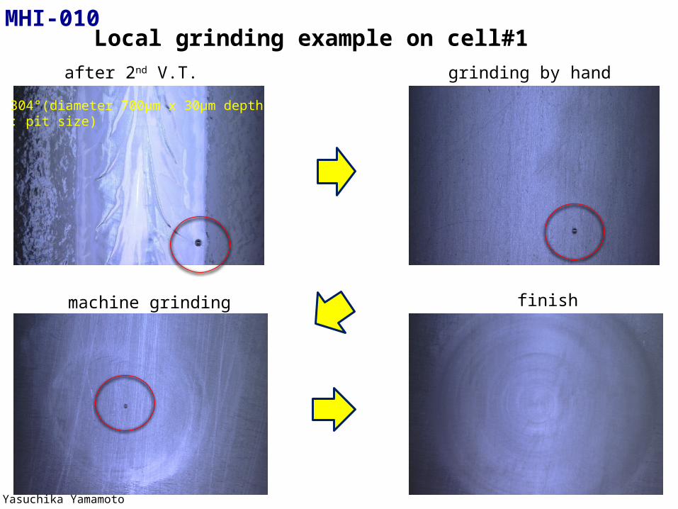
Local grinding example on cell#1
4
after 2nd V.T. grinding by hand
machine grinding finish
Yasuchika Yamamoto
304°(diameter 700μm x 30μm depth : pit size)
MHI-010
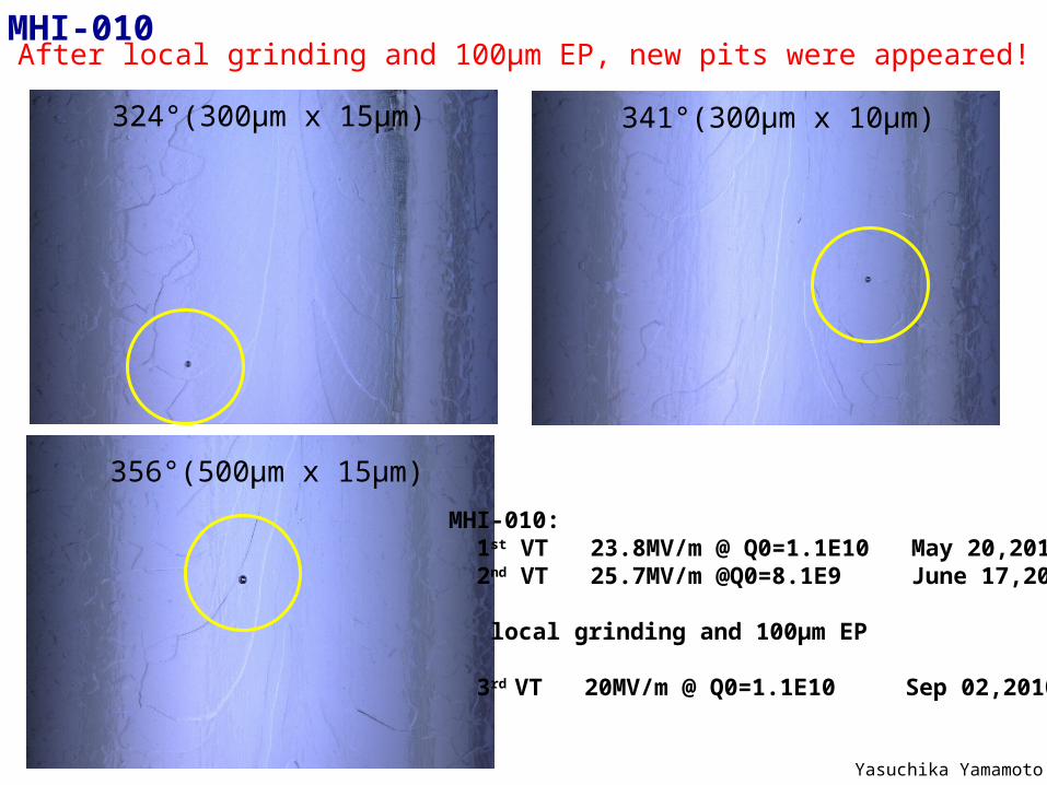
After local grinding and 100µm EP, new pits were appeared!
341°(300μm x 10μm)324°(300μm x 15μm)
356°(500μm x 15μm)
MHI-010
MHI-010: 1st VT 23.8MV/m @ Q0=1.1E10 May 20,2010 2nd VT 25.7MV/m @Q0=8.1E9 June 17,2010 local grinding and 100µm EP
3rd VT 20MV/m @ Q0=1.1E10 Sep 02,2010
Yasuchika Yamamoto
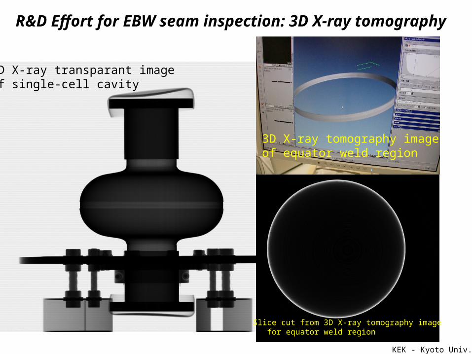
2D X-ray transparant image of single-cell cavity
Slice cut from 3D X-ray tomography image for equator weld region
3D X-ray tomography image of equator weld region
KEK - Kyoto Univ.
R&D Effort for EBW seam inspection: 3D X-ray tomography
KEK - Kyoto Univ.
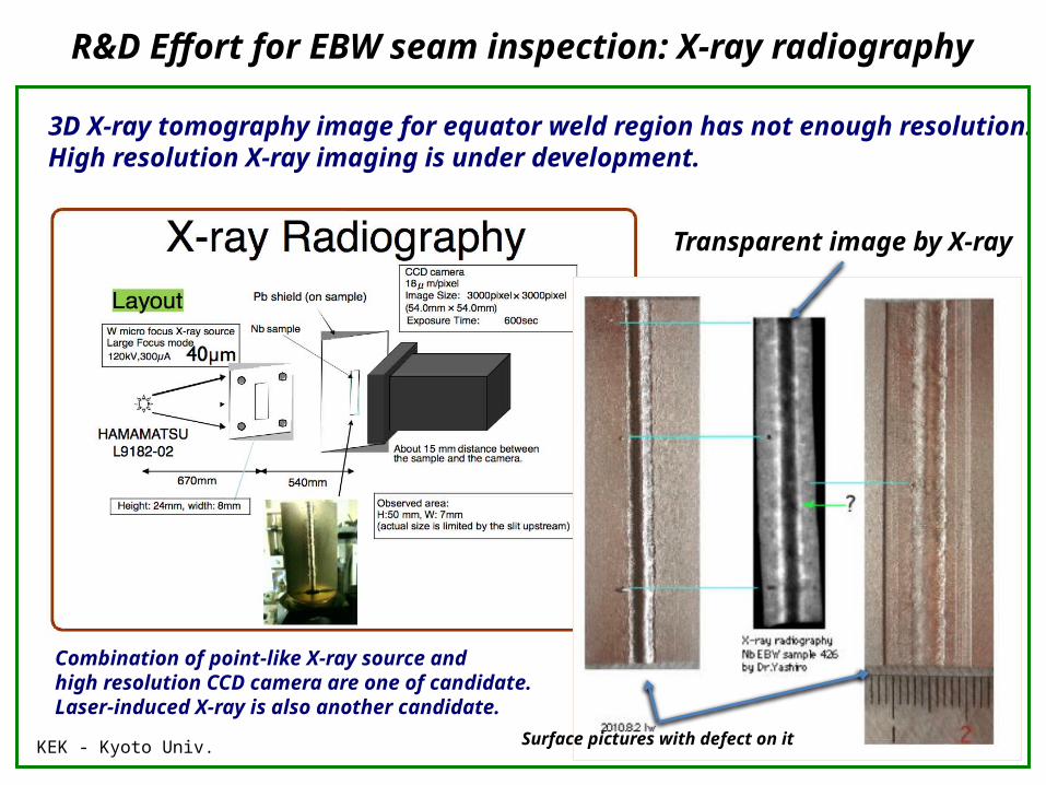
R&D Effort for EBW seam inspection: X-ray radiography
Transparent image by X-ray
3D X-ray tomography image for equator weld region has not enough resolution.High resolution X-ray imaging is under development.
Surface pictures with defect on it
Combination of point-like X-ray source and high resolution CCD camera are one of candidate.Laser-induced X-ray is also another candidate.
Candidate detector
DXI-11000
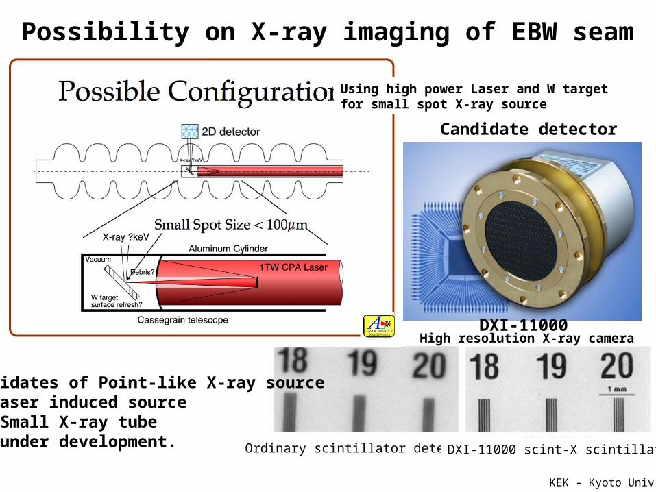
Ordinary scintillator detector DXI-11000 scint-X scintillator
Possibility on X-ray imaging of EBW seam
KEK - Kyoto Univ.
Using high power Laser and W target for small spot X-ray source
High resolution X-ray camera
Candidates of Point-like X-ray source(1) Laser induced source(2) Small X-ray tubeare under development.
9
Contamination found on BCP treated Nb surface
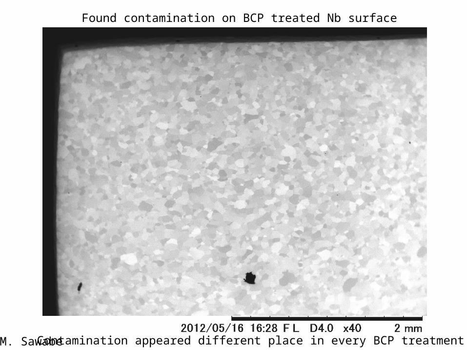
M. Sawabe
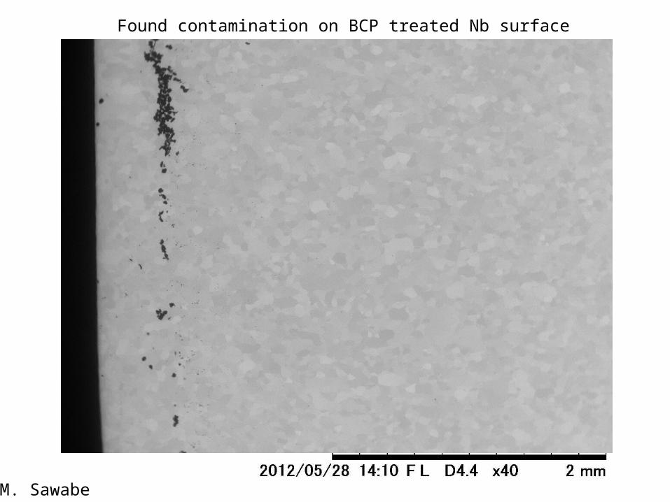
Found contamination on BCP treated Nb surface
Contamination appeared different place in every BCP treatment
M. Sawabe
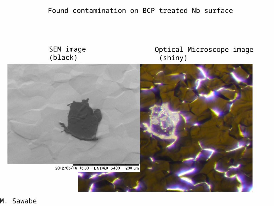
Found contamination on BCP treated Nb surface
SEM image(black)
Optical Microscope image (shiny)
M. Sawabe
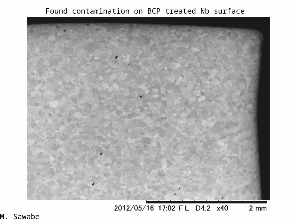
Found contamination on BCP treated Nb surface
M. Sawabe
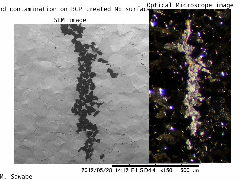
Found contamination on BCP treated Nb surface
SEM image
Optical Microscope image
M. Sawabe
Found contamination on BCP treated Nb surface
M. Sawabe
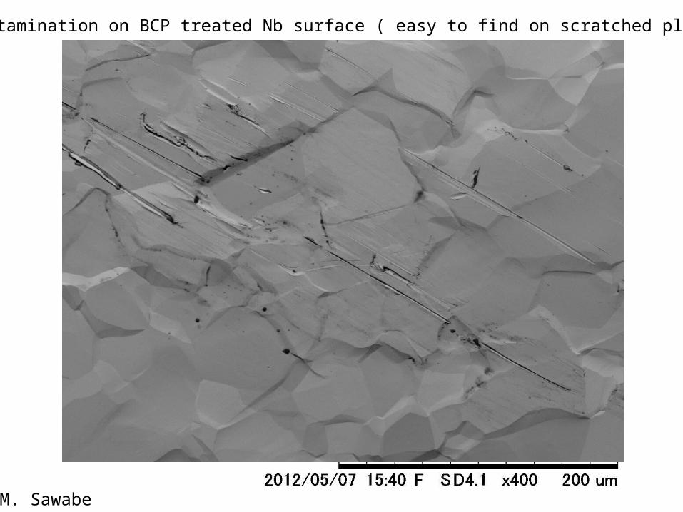
Found contamination on BCP treated Nb surface ( easy to find on scratched place)
M. Sawabe
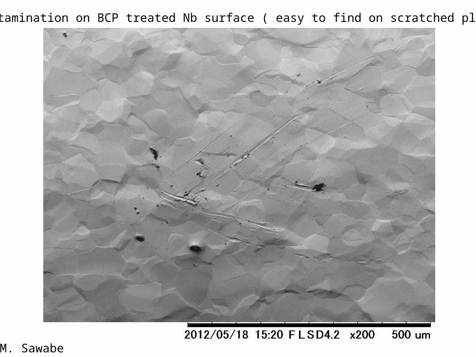
Found contamination on BCP treated Nb surface ( easy to find on scratched place)
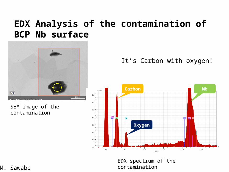
EDX Analysis of the contamination of BCP Nb surface
0.0 0.5 1.0 1.5 2.0 2.5keV
0.0
0.5
1.0
1.5
2.0
2.5
3.0
3.5
cps/eV
Tl Tl Tl C Zr Zr Zr O Nb Nb Nb
SEM image of the contamination
EDX spectrum of the contamination
Carbon
Oxygen
Nb
M. Sawabe
It’s Carbon with oxygen!
Raman Shift / cm-11000 2000 3000
400000
600000
800000
Raman Shift / cm-11000 2000 3000
400000
600000
Raman Shift / cm-11000 2000 3000
150000
200000
Raman Shift / cm-11000 2000 3000
400000
600000
800000
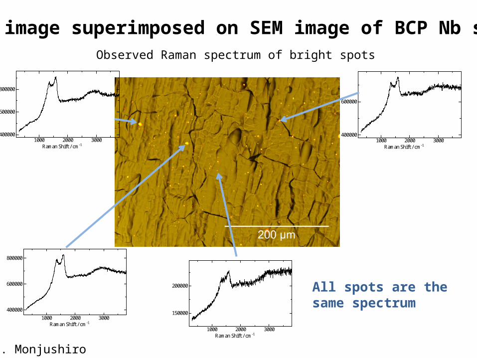
All spots are the same spectrum
Raman image superimposed on SEM image of BCP Nb surfaceObserved Raman spectrum of bright spots
H. Monjushiro
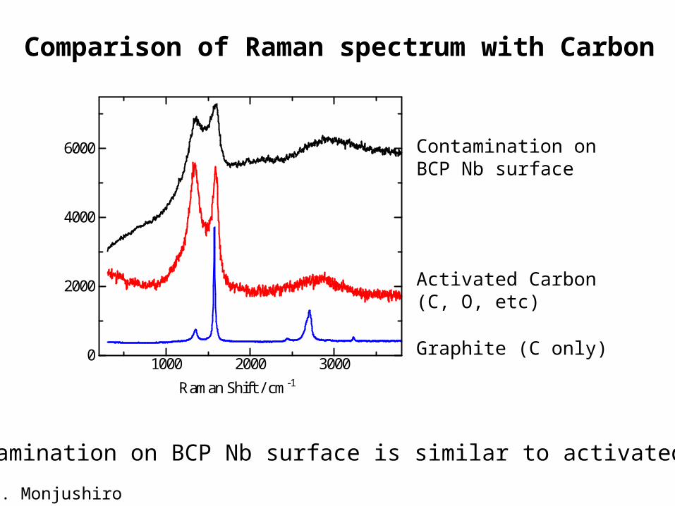
Contamination on BCP Nb surface
Activated Carbon (C, O, etc)
Raman Shift / cm-1
1000 2000 30000
2000
4000
6000
Graphite (C only)
Comparison of Raman spectrum with Carbon
The contamination on BCP Nb surface is similar to activated Carbon!
H. Monjushiro
20
Contamination found on EP treated Nb surface
M. Sawabe
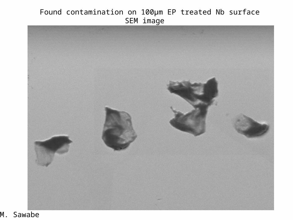
Found contamination on 100µm EP treated Nb surfaceSEM image
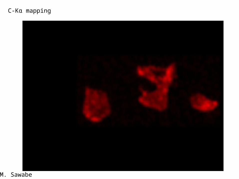
C-Kα mapping
M. Sawabe
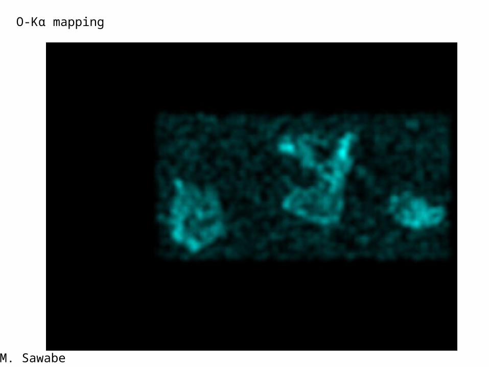
O-Kα mapping
M. Sawabe
0.0 0.5 1.0 1.5 2.0 2.5 3.0 3.5 4.0keV
0
5
10
15
20
25
30 cps/eV
C O
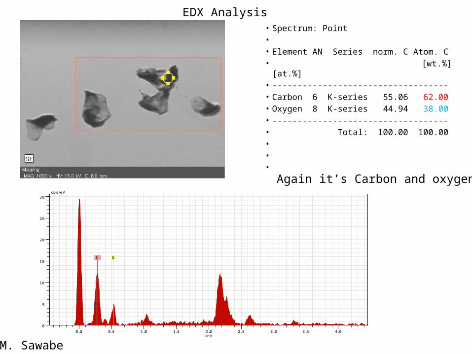
• Spectrum: Point• • Element AN Series norm. C Atom. C• [wt.%] [at.%]• -----------------------------------• Carbon 6 K-series 55.06 62.00• Oxygen 8 K-series 44.94 38.00• -----------------------------------• Total: 100.00 100.00• • •
M. Sawabe
EDX Analysis
Again it’s Carbon and oxygen!
25
Speculation of the contamination effect on the cavity performance
(1) Possible source of field emission at high electric field region(2) Possible heat source at high magnetic filed region(3) Possible source of pit-like defect formation at EBW seam
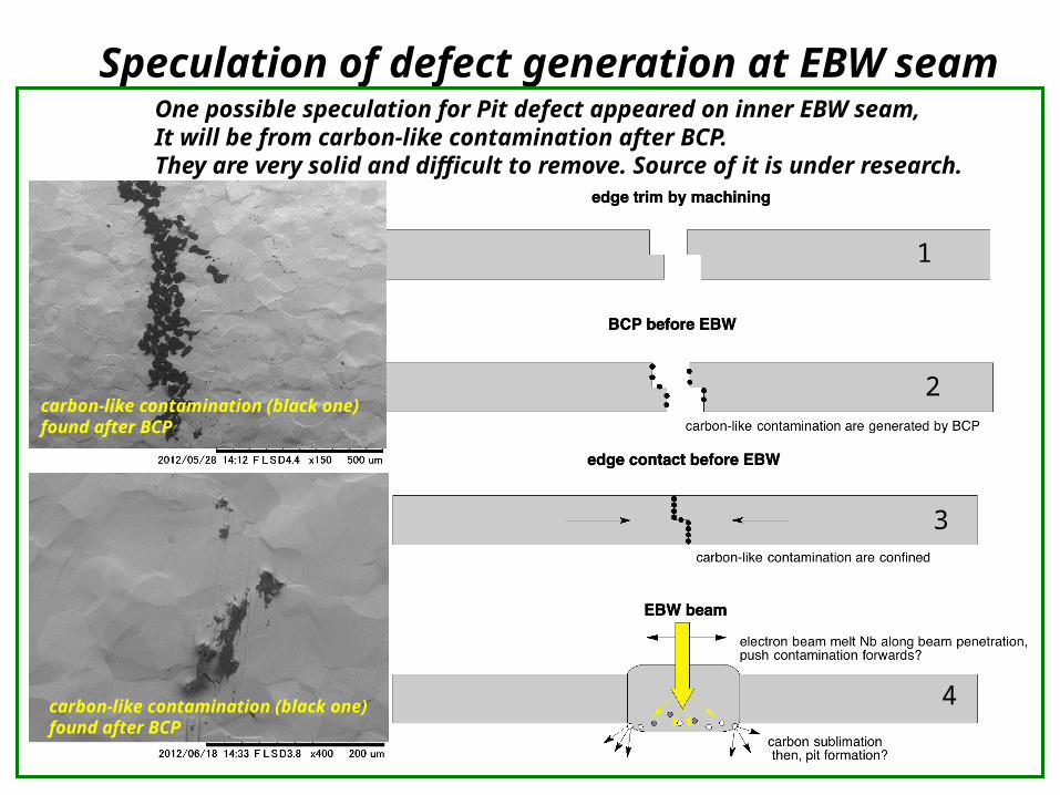
Speculation of defect generation at EBW seamOne possible speculation for Pit defect appeared on inner EBW seam,It will be from carbon-like contamination after BCP. They are very solid and difficult to remove. Source of it is under research.
1
2
3
4
carbon-like contamination (black one) found after BCP
carbon-like contamination (black one) found after BCP
27
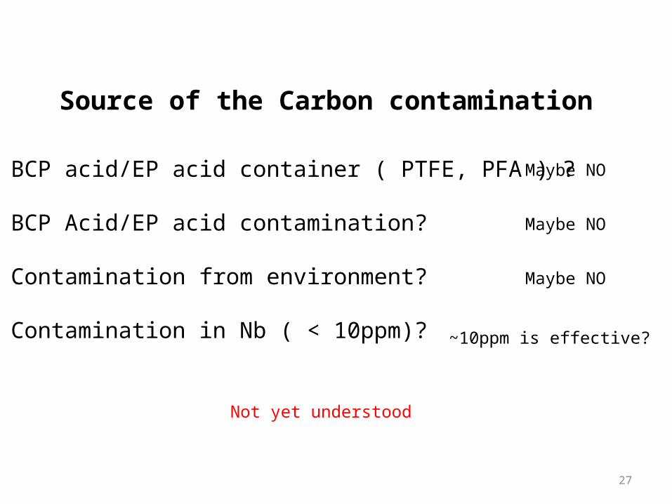
Source of the Carbon contamination
1) BCP acid/EP acid container ( PTFE, PFA ) ?
2) BCP Acid/EP acid contamination?
3) Contamination from environment?
4) Contamination in Nb ( < 10ppm)?
Not yet understood
Maybe NO
Maybe NO
Maybe NO
~10ppm is effective?
28
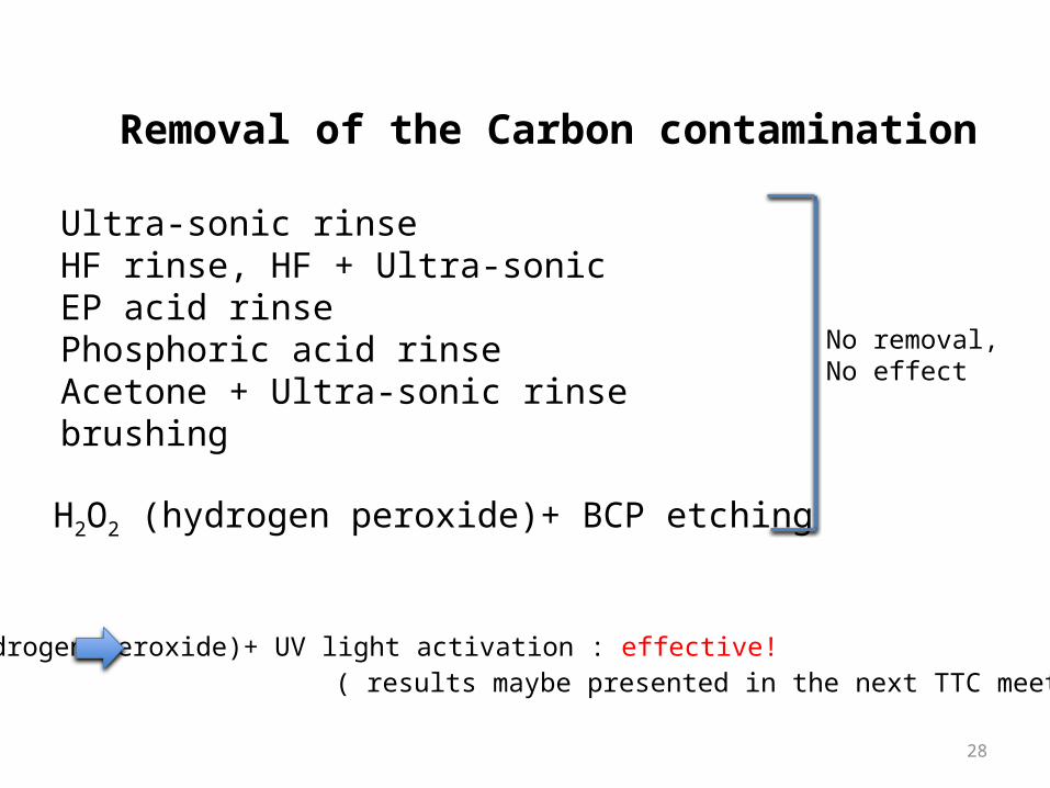
Removal of the Carbon contamination
Ultra-sonic rinseHF rinse, HF + Ultra-sonicEP acid rinsePhosphoric acid rinseAcetone + Ultra-sonic rinsebrushing
H2O2 (hydrogen peroxide)+ BCP etching
No removal,No effect
H2O2 (hydrogen peroxide)+ UV light activation : effective! ( results maybe presented in the next TTC meeting at JLAB )
29
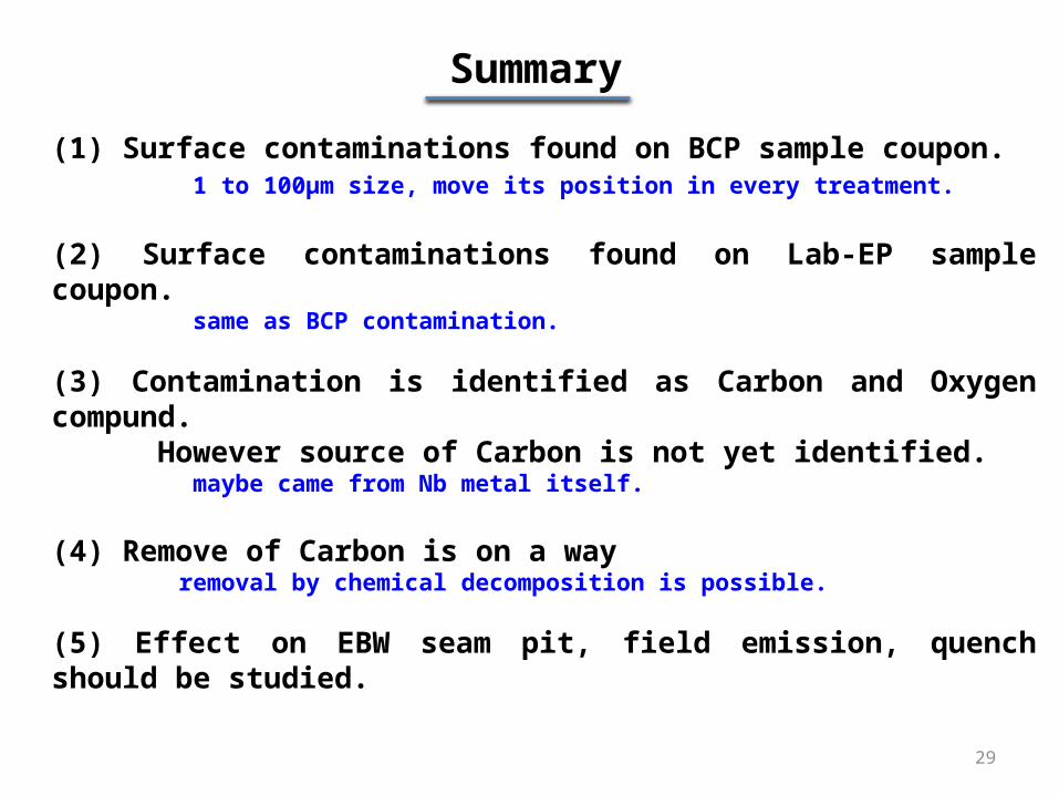
(1) Surface contaminations found on BCP sample coupon. 1 to 100µm size, move its position in every treatment.
(2) Surface contaminations found on Lab-EP sample coupon. same as BCP contamination.
(3) Contamination is identified as Carbon and Oxygen compund. However source of Carbon is not yet identified. maybe came from Nb metal itself.
(4) Remove of Carbon is on a way removal by chemical decomposition is possible.
(5) Effect on EBW seam pit, field emission, quench should be studied.
Summary
30
END of Slide