Embed Size (px)
Citation preview

78 ENDOVASCULAR TODAY OCTOBER 2015
INTER VENTIONAL O N C O L O G Y
An overview of the interventional oncologist’s arsenal.
BY JEFFREY R. RAMKARANSINGH, MD, AND MATTHEW S. JOHNSON, MD, FSIR
Hepatoma: Image-Guided Treatment Options in 2016
Globally, primary liver cancer is the sixth most common cancer and is the second highest cause of cancer mortality.1 Hepatoma, or hepatocellular carcinoma (HCC), accounts for
approximately 80% of primary liver tumors. Major risk factors for the development of HCC include hepatitis B infection, hepatitis C infection, cirrhosis, heavy alcohol consumption, and nonalcoholic steatohepatitis.2
PATIENT EVALUATIONPatients with HCC should be evaluated by a mul-
tidisciplinary group of specialists consisting of inter-ventional oncology, hepatology, medical oncology, radiation oncology, and surgical oncology. The purpose of this group is to confirm tumor diagnosis, evaluate liver function, evaluate patient performance status, determine tumor stage, and ultimately propose the most appropriate therapeutic option available for each unique patient.
The diagnosis of HCC is made primarily with imag-ing characteristics of arterial-phase enhancement and venous-phase washout on contrast-enhanced CT or MRI (Figure 1). Biopsy is seldom necessary, but may be useful in difficult diagnostic cases.3 Cross-sectional imaging can also define tumor size and number, delin-eate vascular invasion, and determine nodal or distant metastatic disease.
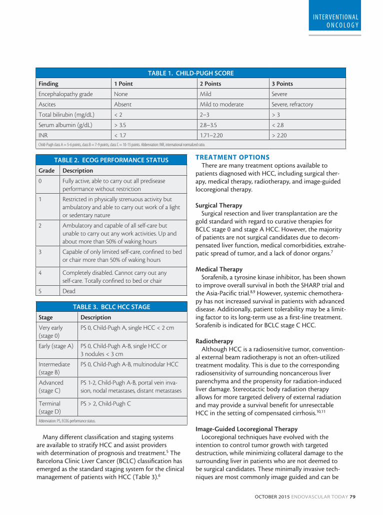
Liver function can be ascertained using the model for end-stage liver disease (MELD) score (www.mayoclinic.org/gi-rst/mayomodel5.html), as well as the Child-Pugh class (Table 1).4 Additionally, a patient’s overall func-tional ability to carry on activities of daily living is rep-resented by the Eastern Cooperative Oncology Group (ECOG) performance status (Table 2).
Figure 1. Arterial (A) and portal venous (B) phase images
from a dynamic, contrast-enhanced MRI demonstrate a
hypervascular mass with washout in segment VI of the right
hepatic lobe, characteristic of HCC.
A
B
OCTOBER 2015 ENDOVASCULAR TODAY 79
INTER VENTIONAL O N C O L O G Y
Many different classification and staging systems are available to stratify HCC and assist providers with determination of prognosis and treatment.5 The Barcelona Clinic Liver Cancer (BCLC) classification has emerged as the standard staging system for the clinical management of patients with HCC (Table 3).6
TREATMENT OPTIONSThere are many treatment options available to
patients diagnosed with HCC, including surgical ther-apy, medical therapy, radiotherapy, and image-guided locoregional therapy.
Surgical TherapySurgical resection and liver transplantation are the
gold standard with regard to curative therapies for BCLC stage 0 and stage A HCC. However, the majority of patients are not surgical candidates due to decom-pensated liver function, medical comorbidities, extrahe-patic spread of tumor, and a lack of donor organs.7
Medical TherapySorafenib, a tyrosine kinase inhibitor, has been shown
to improve overall survival in both the SHARP trial and the Asia-Pacific trial.8,9 However, systemic chemothera-py has not increased survival in patients with advanced disease. Additionally, patient tolerability may be a limit-ing factor to its long-term use as a first-line treatment. Sorafenib is indicated for BCLC stage C HCC.
RadiotherapyAlthough HCC is a radiosensitive tumor, convention-
al external beam radiotherapy is not an often-utilized treatment modality. This is due to the corresponding radiosensitivity of surrounding noncancerous liver parenchyma and the propensity for radiation-induced liver damage. Stereotactic body radiation therapy allows for more targeted delivery of external radiation and may provide a survival benefit for unresectable HCC in the setting of compensated cirrhosis.10,11
Image-Guided Locoregional TherapyLocoregional techniques have evolved with the
intention to control tumor growth with targeted destruction, while minimizing collateral damage to the surrounding liver in patients who are not deemed to be surgical candidates. These minimally invasive tech-niques are most commonly image guided and can be
TABLE 1. CHILD-PUGH SCORE
Finding 1 Point 2 Points 3 Points
Encephalopathy grade None Mild Severe
Ascites Absent Mild to moderate Severe, refractory
Total bilirubin (mg/dL) < 2 2–3 > 3
Serum albumin (g/dL) > 3.5 2.8–3.5 < 2.8
INR < 1.7 1.71–2.20 > 2.20Child-Pugh class A = 5-6 points, class B = 7-9 points, class C = 10-15 points. Abbreviation: INR, international normalized ratio.
TABLE 2. ECOG PERFORMANCE STATUS
Grade Description
0 Fully active, able to carry out all predisease performance without restriction
1 Restricted in physically strenuous activity but ambulatory and able to carry out work of a light or sedentary nature
2 Ambulatory and capable of all self-care but unable to carry out any work activities. Up and about more than 50% of waking hours
3 Capable of only limited self-care, confined to bed or chair more than 50% of waking hours
4 Completely disabled. Cannot carry out any self-care. Totally confined to bed or chair
5 Dead
TABLE 3. BCLC HCC STAGE
Stage Description
Very early (stage 0)
PS 0, Child-Pugh A, single HCC < 2 cm
Early (stage A) PS 0, Child-Pugh A-B, single HCC or 3 nodules < 3 cm
Intermediate (stage B)
PS 0, Child-Pugh A-B, multinodular HCC
Advanced (stage C)
PS 1-2, Child-Pugh A-B, portal vein inva-sion, nodal metastases, distant metastases
Terminal (stage D)
PS > 2, Child-Pugh C
Abbreviation: PS, ECOG performance status.

80 ENDOVASCULAR TODAY OCTOBER 2015
INTER VENTIONAL O N C O L O G Y
divided into percutaneous ablative therapies and intra-arterial, catheter-based embolic therapies.
AblationImage-guided, percutaneous ablation is an approach
designed to treat focal tumors by inducing irreversible cellular damage with the administration of thermal ener-gy, nonthermal energy, or denaturing chemicals. The goal is to achieve uniform ablation of the visible tumor in addition to a 1-cm ablative margin of normal liver tissue. Ablative therapy is indicated for BCLC stage A HCC.
Radiofrequency AblationRadiofrequency ablation (RFA) delivers high-
frequency alternating currents at the active portion of an ablation probe, which causes local ionic agitation and generates frictional heat.12 As tissue temperatures increase between 60ºC to 100ºC there is instantaneous coagulation necrosis of the exposed tissue (Figure 2).13 Temperatures > 100ºC can cause tissue charring, which may decrease the size of the ablation zone.14 The effi-cacy of RFA may be limited in subcapsular tumors adjacent to nearby structures due to the risk of thermal injury to these structures. Also, tumors in close proxim-ity to high-flow blood vessels > 3 mm in diameter are susceptible to a heat sink effect, whereby flowing blood dissipates thermal energy.
Microwave AblationMicrowave thermal ablation (MWA) involves insert-
ing antennae for an externally applied energy source into a tumor. There is a resultant oscillation of polar
molecules that produces frictional heat.15 MWA can reach higher temperatures in shorter periods of time compared to RFA and is not limited by tissue charring. MWA is not hampered by perivascular heat sink.
Irreversible ElectroporationIrreversible electroporation (IRE), a nonthermal
technique, uses an electrical field to induce apoptosis through irreversible cell membrane damage.16 IRE is not affected by heat sink from nearby blood vessels and also appears to limit damage to surrounding tissue. IRE requires general anesthesia and induced paralysis.
CryoablationCryoablation is a thermal ablative technique that
uses argon gas, traveling through a percutaneous probe, to cool the target lesion. Temperatures of –20ºC to –40ºC are reached, and cell death is induced by intracellular and extracellular ice crystal formation.17 Cryoablation may be associated with disseminated intravascular coagulation and cryoshock syndrome, a life-threatening multiorgan failure.18
Chemical AblationChemical ablation, percutaneous ethanol injection,
and percutaneous acetic acid injection, is mostly of his-torical value in the current landscape of interventional oncology. These procedures involve the direct, image-guided injection of a chemical into a hepatoma.19
Catheter-Based TherapyThe normal liver receives 75% of its blood supply
from the portal vein and 25% from the hepatic artery. Conversely, HCC is supplied predominantly by the hepatic artery.20 Intra-arterial embolotherapies exploit this nuance and can selectively deliver embolic mate-rials to tumor tissue while relatively sparing normal surrounding parenchyma. Endovascular techniques are recommended for BCLC stage B HCC.
Figure 2. A hypervascular HCC in segment II of the left hepatic lobe (A). An intraprocedural ultrasound image demonstrates
an RFA probe entering the hypoechoic tumor (B). An area of necrosis at the treatment site 24 months after ablation (C).
A B C
The efficacy of RFA may be limited . . . due to the risk of thermal injury . . .

82 ENDOVASCULAR TODAY OCTOBER 2015
INTER VENTIONAL O N C O L O G Y
Transarterial ChemoembolizationConventional transarterial chemoembolization
(cTACE) involves selective hepatic arterial delivery of a mixture of lipiodol with a cytotoxic agent(s) followed by bland particle embolization.21 Lipiodol is a poppy seed oil that accumulates in tumor cells due to a lack of Kupffer cells found in a normal liver. The most com-monly used chemotherapeutic agents are doxorubicin, mitomycin C, and cisplatin. The exact combinations are highly variable between institutions. Some may use only one agent, whereas others combine two or three. After chemoinfusion, bland permanent or temporary particles ranging from 100 to 500 µm in diameter are infused into the same hepatic arterial distribution to decrease washout and prolong tissue dwell of the chemotherapeutic. A meta-analysis of randomized con-trolled trials comparing cTACE to conservative treat-ment over a 24-year period found a significant 2-year survival benefit with cTACE.22
Drug-eluting bead chemoembolization (DEB-TACE) is a derivative of cTACE in which microspheres are loaded
with a chemotherapeutic agent, most commonly doxo-rubicin (Figure 3). DEB-TACE may result in prolonged chemotherapy exposure of the target tumor compared to cTACE.23 The PRECISION V study showed improved tolerability of DEB-TACE in patients with BCLC stage B HCC compared to cTACE, but did not demonstrate an improved therapeutic response.24
RadioembolizationRadioembolization uses glass or resin microspheres
loaded with beta-emitting yttrium-90 (Y-90) for selec-
Figure 3. Hypervascular HCC in segment VIII of the right hepatic lobe (A). Hypervascular tumor blush in the region of the
hepatic dome (B). Subselective catheterization of the segmental VIII right hepatic arterial branch (C). Area of necrosis at the
treatment site 12 months after DEB-TACE(D).
A B
C D
Intra-arterial embolotherapies . . . can selectively deliver embolic materials
to tumor tissue while relatively sparing normal surrounding
parenchyma.

84 ENDOVASCULAR TODAY OCTOBER 2015
INTER VENTIONAL O N C O L O G Y
tive injection into the hepatic arteries feeding an HCC. Treatment can be lobar or selective. Unlike other tran-sarterial therapies, radioembolization requires a pre-treatment planning arteriogram to determine precise vascular supply to the tumor and to detect possible extrahepatic shunting. Prophylactic embolization of various arteries, such as the gastroduodenal artery and
right gastric artery, may be necessary to prevent non-target radioembolization. Technetium-99m macroag-gregated albumin (Tc-99m MAA) is administered into the target hepatic artery during planning arteriography to assess for potential Y-90 shunting to the lungs.25 After obtaining the planning arteriogram, a single-pho-ton emission computed tomography (SPECT) scan is
Figure 4. Hypervascular HCC in segment II of the left hepatic lobe (A). Selective catheterization of the left hepatic artery
shows the hypervascular mass (B). Fused SPECT and CT shows uptake of Tc-99m MAA into the mass (C). Shrunken, necrotic
mass 12 months after radioembolization (D).
A B
C D
Figure 5. Same patient as in Figure 1. A hepatic arteriogram demonstrates the hypervascular HCC (A). Planar SPECT imaging
shows accumulation of Tc-99m MAA at the expected location of the mass (B). Hypovascular mass 12 months after radioem-
bolization (C).
A B C

86 ENDOVASCULAR TODAY OCTOBER 2015
INTER VENTIONAL O N C O L O G Y
obtained to demonstrate the degree of tumor uptake, degree of pulmonary shunting, and possible sites of extrahepatic radiotracer uptake. The patient returns for therapeutic Y-90 administration after appropriate dose calculation (Figures 4 and 5).26 Radioembolization has been shown to improve survival for intermediate-stage HCC at a rate similar to cTACE and DEB-TACE.
Tumor ResponseThe assessment of HCC response to locoregional
therapies has evolved during the past decade. The modified Response Evaluation Criteria in Solid Tumors (mRECIST) is currently accepted and takes into account intratumoral arterial enhancement on follow-up imag-ing (Table 4).27
CONCLUSIONHCC is a unique tumor in that it often develops on a
backdrop of underlying liver disease. As a result, most patients are not candidates for curative surgical thera-pies. Image-guided locoregional interventions have con-tinued to progress and provide both a survival benefit and an encouraging safety profile. n
Jeffrey R. Ramkaransingh, MD, is an Assistant Professor of Vascular and Interventional Radiology at Indiana University School of Medicine. He has stated that he has no financial interests related to this article. Dr. Ramkaransingh may be reached at [email protected].
Matthew S. Johnson, MD, FSIR, is a Professor of Radiology, Medicine and Surgery at Indiana University School of Medicine. He has disclosed that he is a paid con-sultant for BTG International, Boston Scientific Corporation, CeloNova BioSciences, and Cook Medical.
1. Ferlay J, Parkin DM, Curado MP, et al. Cancer incidence in five continents, volumes I to X: IARC CANCERBase No. 10 [Internet]. 2014. Available at: http://ci5. iarc.fr. Accessed August 28, 2015. 2. Shariff MI, Cox IJ, Gomaa AI, et al. Hepatocellular carcinoma: current trends in worldwide epidemiology, risk factors, diagnosis and therapeutics. Expert Rev Gastroenterol Hepatol. 2009;3:353-367.3. Gomaa AI, Khan SA, Leen EL, et al. Diagnosis of hepatocellular carcinoma. World J Gastroenterol. 2009;15:1301-1314.4. Kamath PS, Kim WR. The model for end-stage liver disease (MELD). Hepatology. 2007;45:797-805.5. Marrero JA, Fontana RJ, Barrat A, et al. Prognosis of hepatocellular carcinoma: comparison of 7 staging systems in an American cohort. Hepatology. 2005;41:707-715.6. Llovet JM, Brú C, Bruix J. Prognosis of hepatocellular carcinoma: the BCLC staging classification. Semin Liver Dis. 1999;19:329-338. 7. Llovet JM, Burroughs A, Bruix J. Hepatocellular carcinoma. Lancet. 2003;362:1907-1917. 8. Llovet JM, Ricci S, Mazzaferro V, et al. Sorafenib in advanced hepatocellular carcinoma. N Engl J Med. 2008;359:378-390.9. Cheng AL, Kang YK, Chen Z, et al. Efficacy and safety of sorafenib in patients in the Asia-Pacific region with advanced hepatocellular carcinoma: a phase III randomised, double-blind, placebo-controlled trial. Lancet Oncol. 2009;10:25-34. 10. Seong J, Park HC, Han KH, et al. Clinical results and prognostic factors in radiotherapy for unresectable hepato-cellular carcinoma: a retrospective study of 158 patients. Int J Radiat Oncol Biol Phys. 2003;55:329-336.11. Mornex F, Girard N, Beziat C, et al. Feasibility and efficacy of high-dose three- dimensional-conformal radio-therapy in cirrhotic patients with small-size hepatocellular carcinoma non-eligible for curative therapies: mature results of the French phase II RTF-1 trial. Int J Radiat Oncol Biol Phys. 2006;66:1152-1158. 12. Wood BJ, Ramkaransingh JR, Fojo T, et al. Percutaneous tumor ablation with radiofrequency. Cancer. 2002;94:443-451.13. Goldberg SN, Gazelle GS, Mueller PR. Thermal ablation therapy for focal malignancy: a unified approach to underlying principles, techniques, and diagnostic imaging guidance. Am J Roentgenol. 2000;174:323.14. Hong K, Georgiades C. Radiofrequency ablation: mechanism of action and devices. J Vasc Interv Radiol. 2010;21:S179-S186.15. Lubner MG, Brace CL, Hinshaw JL, Lee FT. Microwave tumor ablation: mechanism of action, clinical results and devices. J Vasc Interv Radiol. 2010;21:S192-S203.16. Golberg A, Yarmush ML. Nonthermal irreversible electroporation: fundamentals, applications, and challenges. Biomed Engineer IEEE Trans. 2013;60:707-714.17. Erinjeri JP, Clark TWI. Cryoablation: mechanism of action and devices. J Vasc Interv Radiol. 2010;21:S187-S191.18. Seifert JK, Morris DL. World survey on the complications of hepatic and prostate cryotherapy. World J Surg. 1999;23:109-114.19. Clark TWI. Chemical ablation of liver cancer. Tech Vasc Interv Radiol. 2007;10:58-63.20. Breedis C, Young G. The blood supply of neoplasms in the liver. Am J Pathol. 1954;30:969.21. Shin SW. The current practice of transarterial chemoembolization for the treatment of hepatocellular carci-noma. Korean J Radiol. 2009;10:425-434.22. Llovet JM, Bruix J. Systematic review of randomized trials for unresectable hepatocellular carcinoma: chemo-embolization improves survival. Hepatology. 2003;37:429-442.23. Namur J, Citron SJ, Sellers MT, et al. Embolization of hepatocellular carcinoma with drug-eluting beads: doxorubicin tissue concentration and distribution in patient liver explants. J Hepatol. 2011;55:1332-1338.24. Lammer J, Malagari K, Vogl T, et al. Prospective randomized study of doxorubicin-eluting-bead embolization in the treatment of hepatocellular carcinoma: results of the PRECISION V study. Cardiovasc Intervent Radiol. 2010;33:41-52.25. Salem R, Lewandowski RJ, Sato KT, et al. Technical aspects of radioembolization with 90 Y microspheres. Tech Vasc Interv Radiol. 2007;10:12-29.26. Salem R, Thurston KG. Radioembolization with 90yttrium microspheres: a state-of-the-art brachytherapy treatment for primary and secondary liver malignancies. J Vasc Interv Radiol. 2006;17:1251-1278.27. Lencioni R, Llovet JM. Modified RECIST (mRECIST) assessment for hepatocellular carcinoma. Semin Liver Dis. 2010;30:52-60.
TABLE 4. mRECIST CLASSIFICATION OF TUMOR RESPONSE
Response Description
Complete response Disappearance of any intratumoral arterial enhancement in all target lesions
Partial response At least a 30% decrease in the sum of diameters of viable (enhancement in the arterial phase) tar-get lesions, taking as reference the baseline sum of the diameters of target lesions
Stable disease Any cases that do not qualify for either partial response or progressive disease
Progressive disease An increase of at least 20% of the sum of the diameters of viable (enhancing) target lesions, taking as reference the smallest sum of the diameters of viable (enhancing) target lesions recorded since treatment started