Embed Size (px)
Citation preview

HED SESSION – ANTWERP 2019
In cooperation with the Belgium panel
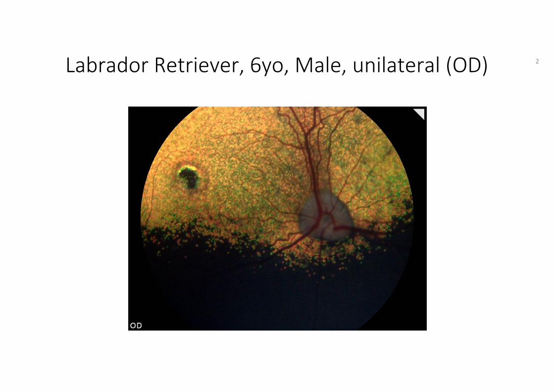
Labrador Retriever, 6yo, Male, unilateral (OD) 2
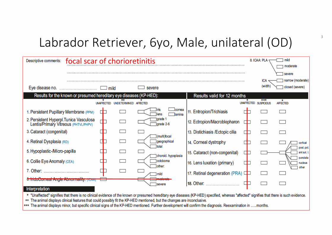
Labrador Retriever, 6yo, Male, unilateral (OD)focal scar of chorioretinitis
3
HED Manual Ch. 5 Definitions
• Chorioretinitis: an inflammatory process of the choroidal and outer retinal structures, observed in the acute phase as blurring, swollen, edematous areas and later as chorioretinalscaring shown as pigmented spots with hyperreflective borders
4

HED Manual Ch. 6 Guidelines
• Section Examination, part: ‘Descriptive comments’: In this section, the examiner should describe any findings in the eye and adnexae, either KP-HED or other.
5

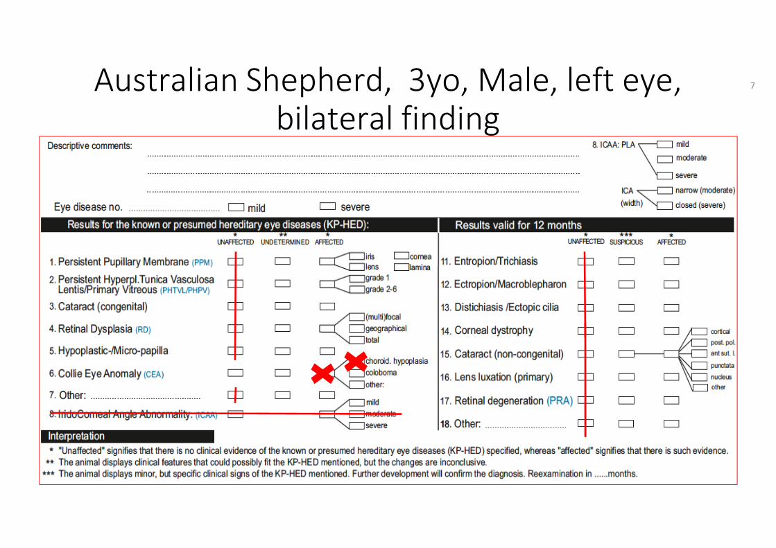
Australian Shepherd, 3yo, Male, left eye, bilateral finding
6
Australian Shepherd, 3yo, Male, left eye,bilateral finding
7
HED Manual Ch. 5 Definitions
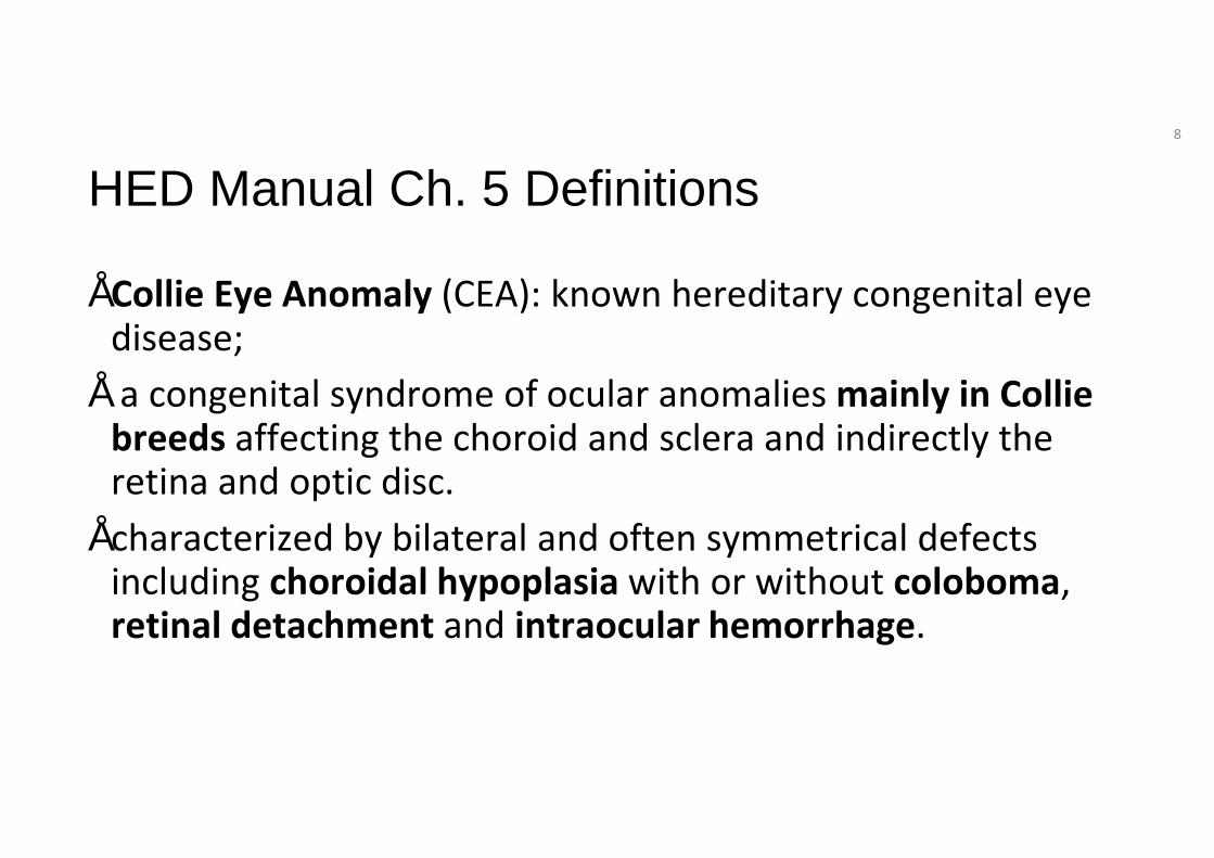
• Collie Eye Anomaly (CEA): known hereditary congenital eye disease;
• a congenital syndrome of ocular anomalies mainly in Collie breeds affecting the choroid and sclera and indirectly the retina and optic disc.
• characterized by bilateral and often symmetrical defects including choroidal hypoplasia with or without coloboma, retinal detachment and intraocular hemorrhage.
8
HED Manual Ch. 6 Guidelines
• Tick no 6. Collie eye anomaly (CEA)
9
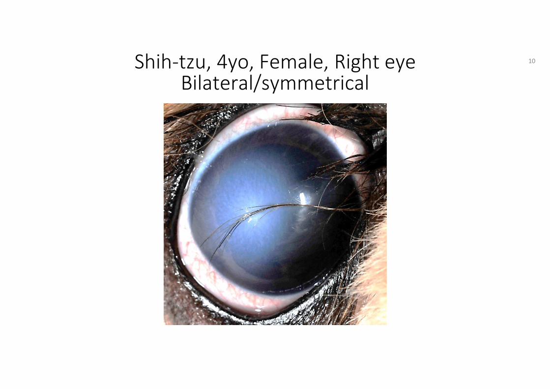
Shih-tzu, 4yo, Female, Right eyeBilateral/symmetrical
10
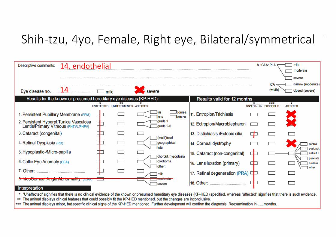
Shih-tzu, 4yo, Female, Right eye, Bilateral/symmetrical
14. endothelial
14
11

HED Manual Ch. 5 Definitions
• Trichiasis: presumed hereditary eye disease or acquired abnormality of deviated hairs on a normal place around the lid fissure, irritatingthe conjunctiva, the free lid margin of the opposite lid and/or the globe.
• Macroblepharon: Fissure length (stretched) in dog over 40 mm
12
HED Manual Ch. 5 Definitions
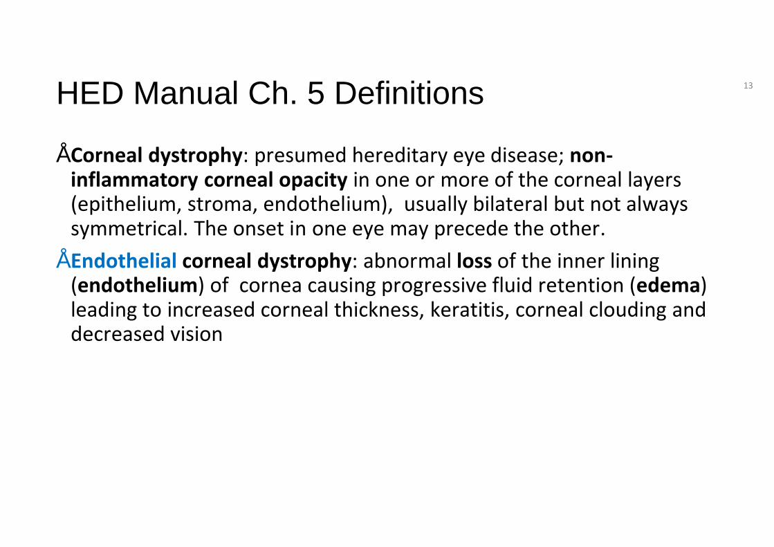
• Corneal dystrophy: presumed hereditary eye disease; non-inflammatory corneal opacity in one or more of the corneal layers (epithelium, stroma, endothelium), usually bilateral but not always symmetrical. The onset in one eye may precede the other.
• Endothelial corneal dystrophy: abnormal loss of the inner lining (endothelium) of cornea causing progressive fluid retention (edema) leading to increased corneal thickness, keratitis, corneal clouding and decreased vision
13
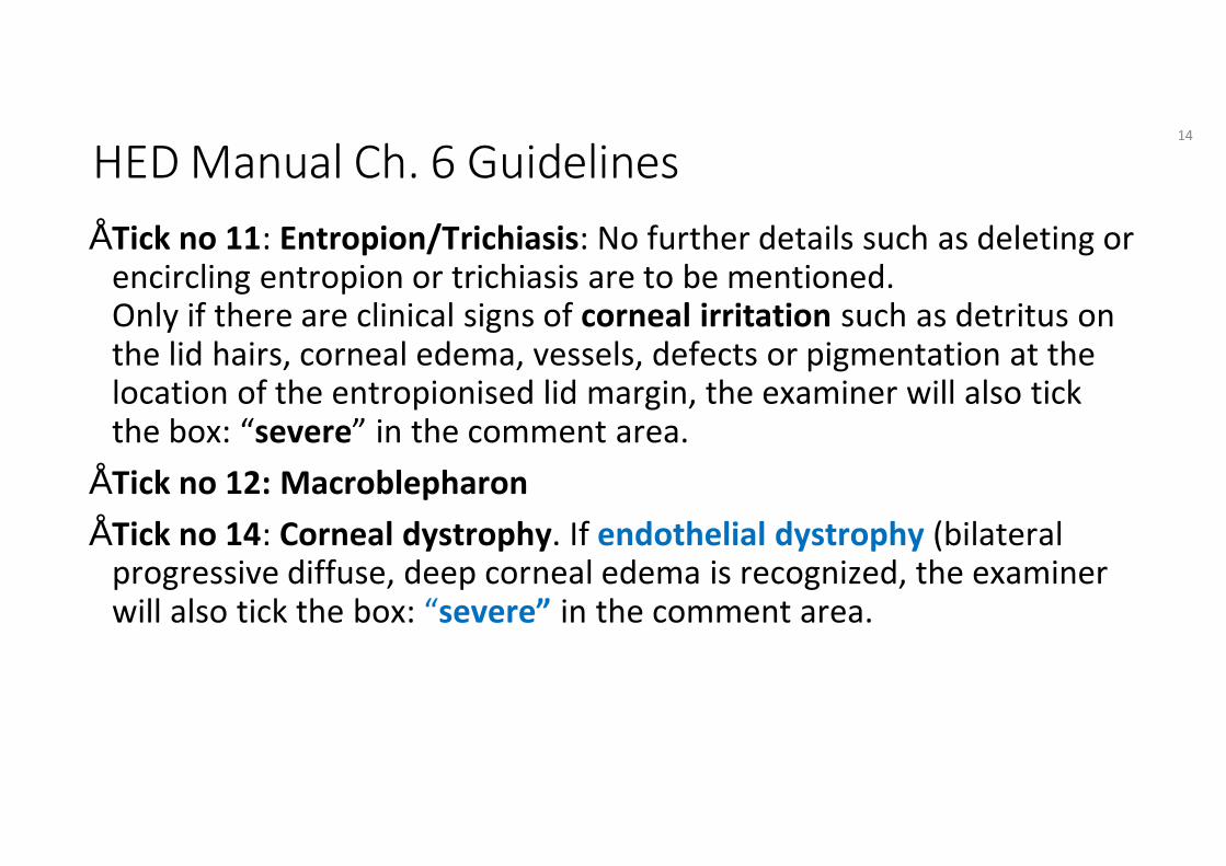
HED Manual Ch. 6 Guidelines• Tick no 11: Entropion/Trichiasis: No further details such as deleting or
encircling entropion or trichiasis are to be mentioned.Only if there are clinical signs of corneal irritation such as detritus on the lid hairs, corneal edema, vessels, defects or pigmentation at the location of the entropionised lid margin, the examiner will also tick the box: “severe” in the comment area.
• Tick no 12: Macroblepharon• Tick no 14: Corneal dystrophy. If endothelial dystrophy (bilateral
progressive diffuse, deep corneal edema is recognized, the examiner will also tick the box: “severe” in the comment area.
14
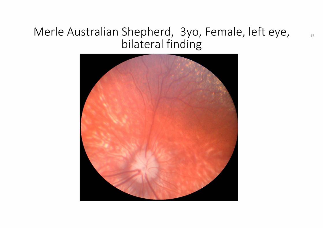
Merle Australian Shepherd, 3yo, Female, left eye,bilateral finding
15
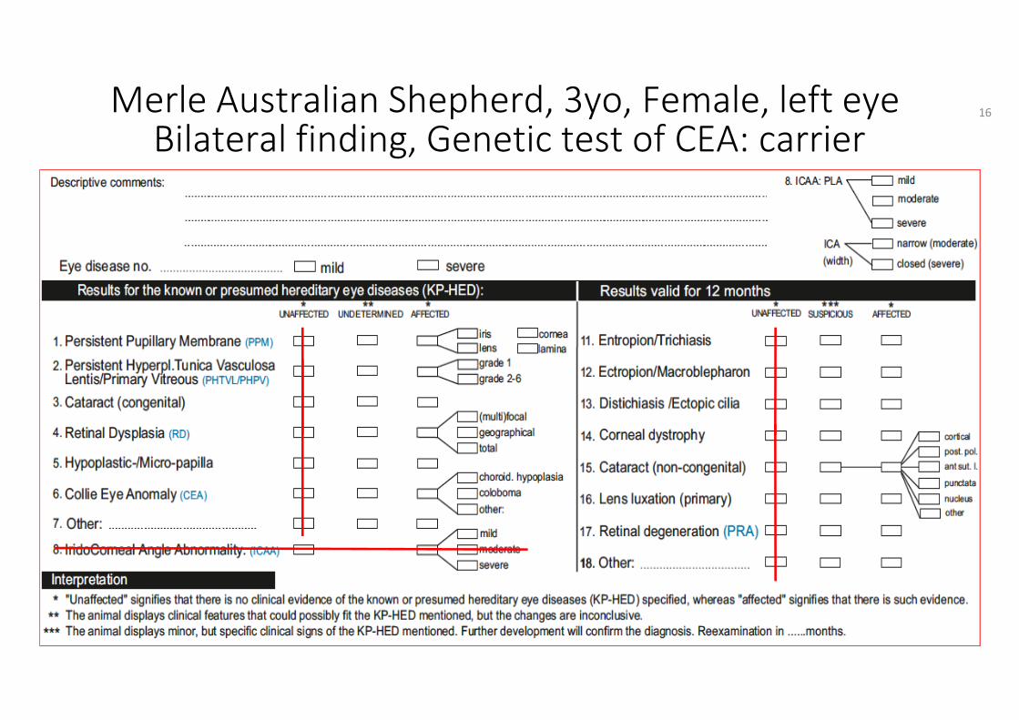
Merle Australian Shepherd, 3yo, Female, left eyeBilateral finding, Genetic test of CEA: carrier
16
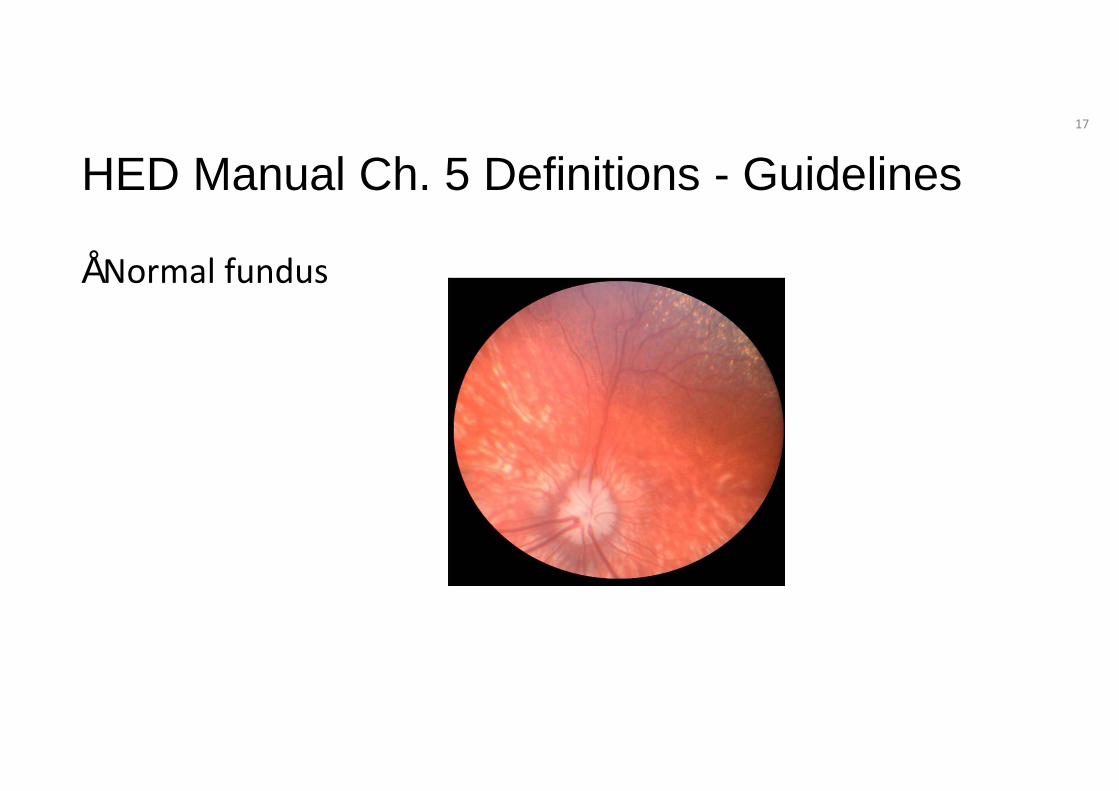
HED Manual Ch. 5 Definitions - Guidelines
• Normal fundus
17
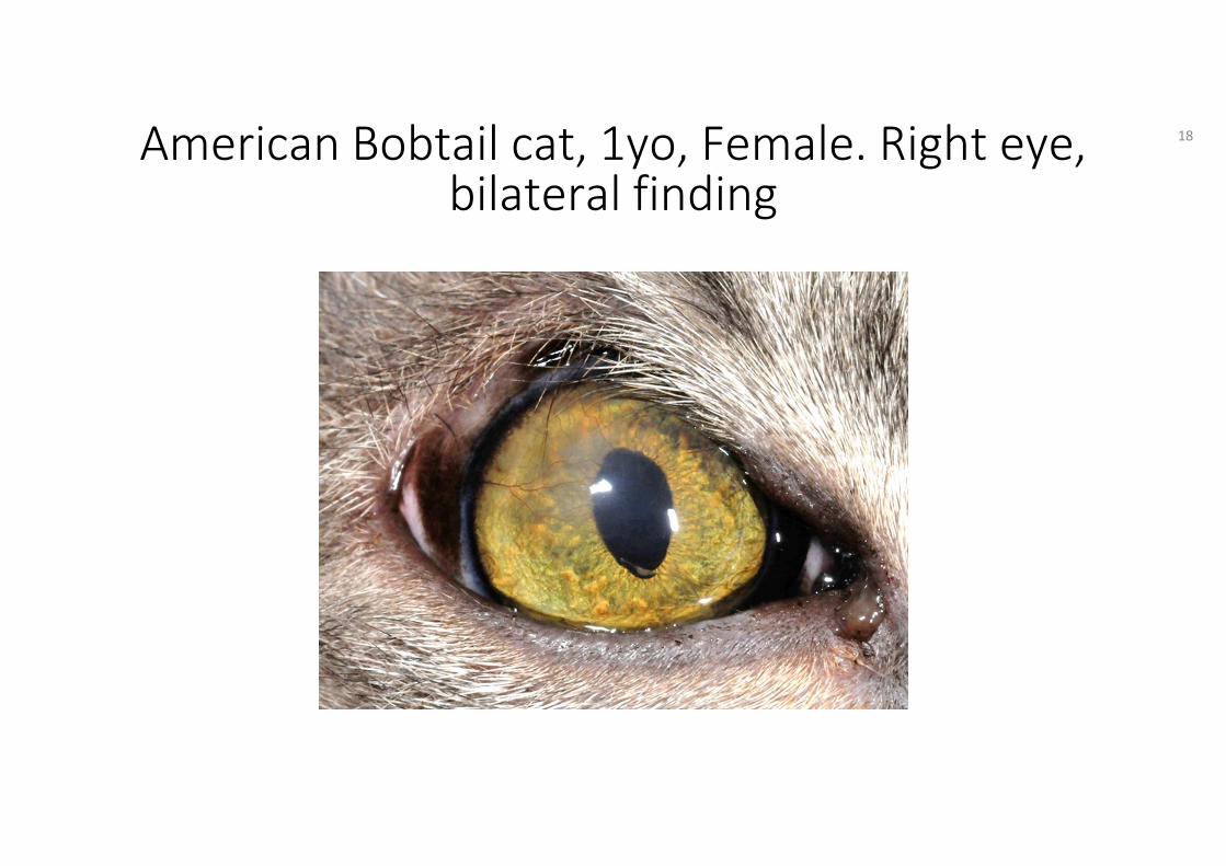
American Bobtail cat, 1yo, Female. Right eye,bilateral finding
18
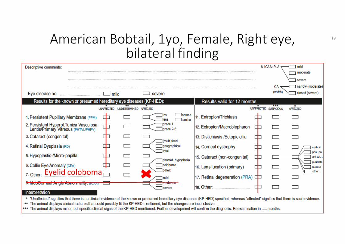
American Bobtail, 1yo, Female, Right eye,bilateral finding
Eyelid coloboma
19
HED Manual Ch. 5 Definitions
• Coloboma: congenital defect of a portion of the eye due to a failure in closure of the body halves; most frequently affecting the iris or the optic nerve at the 6 o’clock position.
• For coloboma in eyelid, retina, choroid, sclera or optic nerve/papilla use the anatomical name first then the anomaly, e.g. eyelid coloboma.
20
HED Manual Ch. 6 Guidelines
Tick no 7. Other: known and presumed hereditary eye anomalies (congenital/developmental, non-progressive) are mentioned here. • The terminology for the diseases is given in chapter 5.
Definitions of this manual are to be used: eyelid coloboma.
21
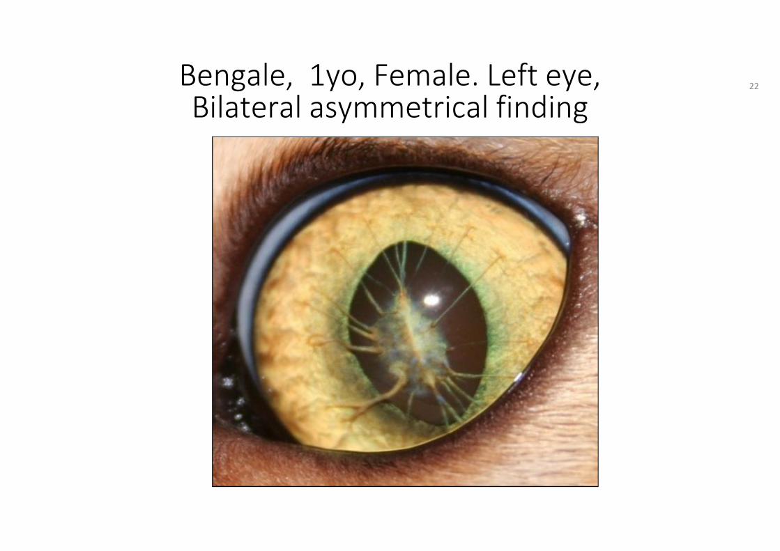
Bengale, 1yo, Female. Left eye,Bilateral asymmetrical finding
22

Bengale, 1yo, Female, Left eye,Bilateral asymmetrical finding
23
HED Manual Ch. 5 Definitions
• Persistent pupillary membrane (PPM): blood vessel remnants of the embryological vascular network in the anterior chamber fail to regress which normally occurs during the first 4 to 5 weeks of life.
• may be found on the surface of the iris at the collarette, on the lens capsule or against the corneal endothelium or strands may bridge from iris to iris, iris to cornea, iris to lens, with or without sheets of tissue (lamina) in the anterior chamber.
24
HED Manual Ch. 6 GuidelinesTick no 1 “affected” : Persistent pupillary membrane (PPM) and tick also the respective box of other parts involved.
Areas which can be involved:• retrocorneal: boxes PPM and cornea• strands from cornea to iris: boxes: PPM, cornea and iris• strands iris to iris: boxes PPM and iris• Strands iris to lens: boxes: PPM, iris and lens, • connected to areas of cataract: tick also no 3 for congenital cataract• strands connected to a sheet/”spider web” of tissue in the anterior
chamber: boxes PPM, lamina and other parts involved are ticked
25
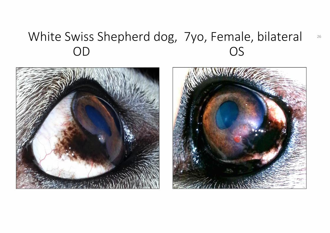
White Swiss Shepherd dog, 7yo, Female, bilateralOD OS
26
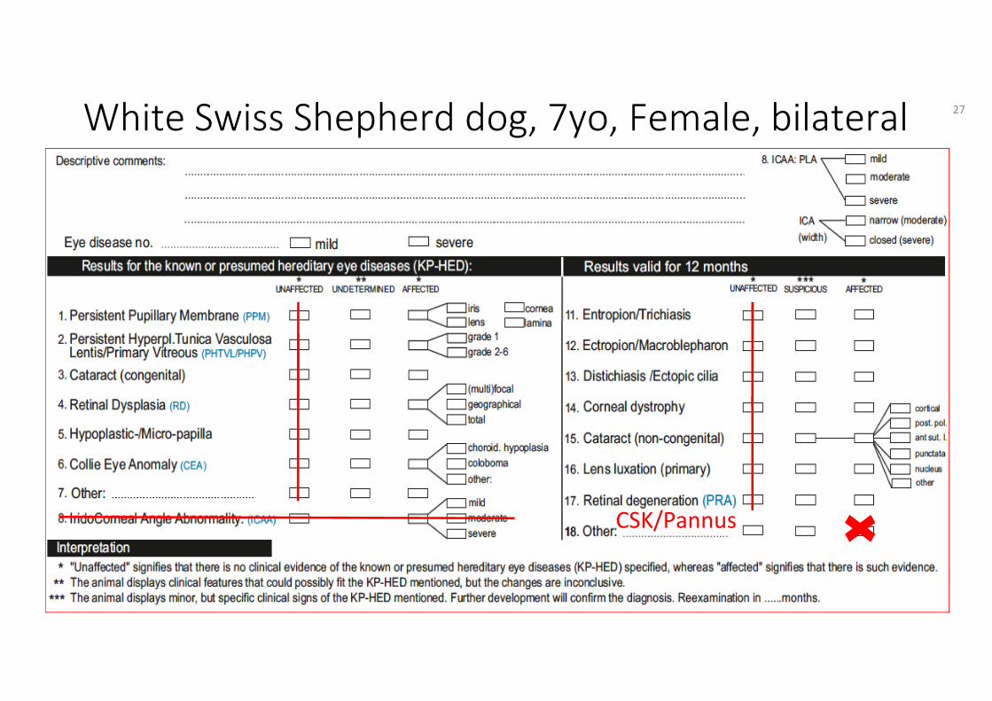
White Swiss Shepherd dog, 7yo, Female, bilateral
CSK/Pannus
27
HED Manual Ch. 5 Definitions
• Chronic superficial keratitis(CSK)/Pannus: Presumed hereditary eye disease; bilateral inflammatory disease of the cornea usually starting as a greyish haze at the inferior or inferiotemporal cornea, followed by the formation of a vascularized subepithelial opacity spreading towards the central cornea; pigmentation follows the vascularization.
• The disease can be seen with concurrent plasmoma(=plasmocytic infiltration of the external conjunctiva of the 3rd eyelid) and/or medial canthus erosion.
28
HED Manual Ch. 6 Guidelines
Tick no 18. Other: Chronic superf. Keratitis/Pannus
for abnormalities, which are considered not to be congenital/developmental or which are progressive, and not yet named on the form
29
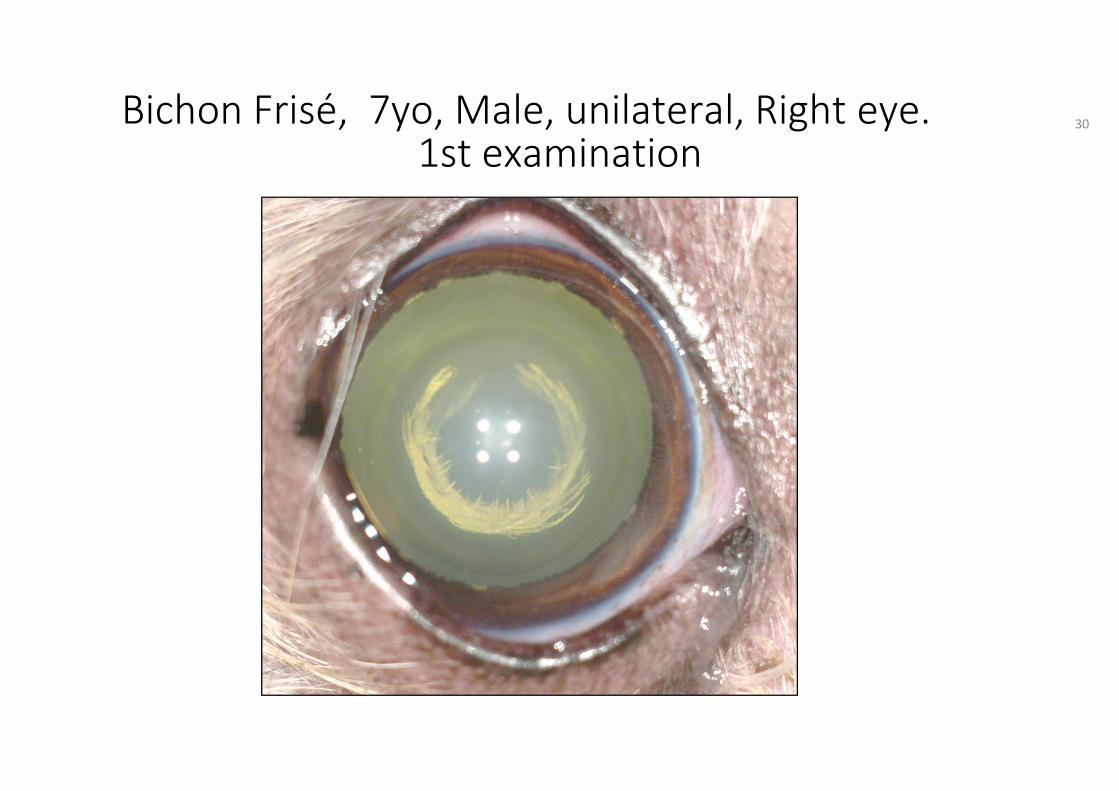
Bichon Frisé, 7yo, Male, unilateral, Right eye.1st examination
30
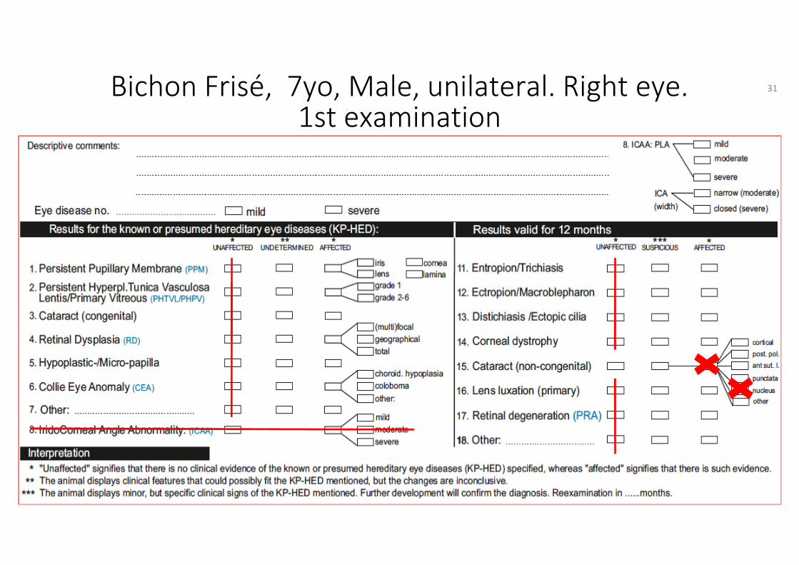
Bichon Frisé, 7yo, Male, unilateral. Right eye.1st examination
31
HED Manual Ch. 5 Definitions
• Cataract: any hereditary or non-hereditary, congenital or acquired, non-physiological opacity of the lens and/or its capsule.
• All bilateral or unilateral cataracts and especially cortical cataracts are known and presumed hereditary eye diseases
32
HED Manual Ch. 6 Guidelines
Tick no 15: Cataract: To describe the type of cataract, the general box for cataract and, if available, the specifying box for the type of cataract should be ticked.
• If cataracts are observed in the period between birth and the 8th week of age the entity is ticked as congenital (no 3)
• Cataracts diagnosed at older age are ticked as non-congenital (acquired), no 15
• It is strongly recommended to draw the cataract in the "pre-drawings" on the certificate.
33
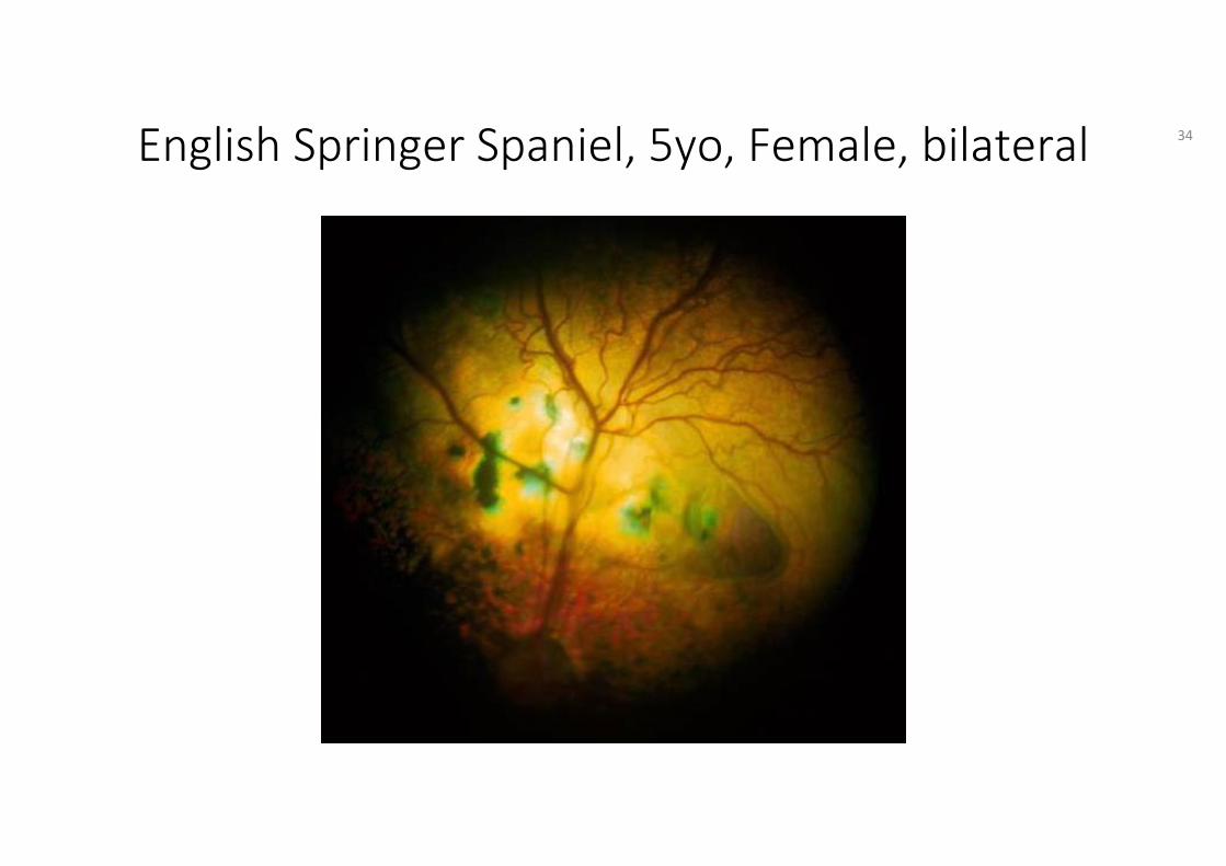
English Springer Spaniel, 5yo, Female, bilateral 34
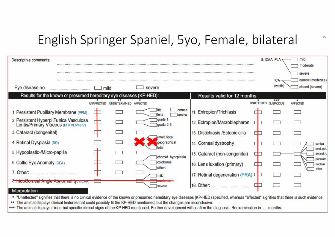
English Springer Spaniel, 5yo, Female, bilateral 35
HED Manual Ch. 5 Definitions• Retinal dysplasia: presumed hereditary eye disease;
abnormal development of the retina observed early in life: neuroretinal folding(s), rosettes and partial or total retinal detachment; non-progressive and generally recognized to have three forms: (multi)focal, geographic and total.
• Retinal dysplasia - geographical: any irregularly, horseshoe-or bladder-like shaped area of abnormal retinal development, most often in the central part of the tapetalarea, in close association with the dorsal retinal vasculature, containing both areas of thinning and areas of elevationrepresenting focal retinal detachment and areas of retinal disorganization. This form may be associated with vision impairment.
36
HED Manual Ch. 6 Guidelines
Tick no 4: Retinal dysplasia and geographical
37
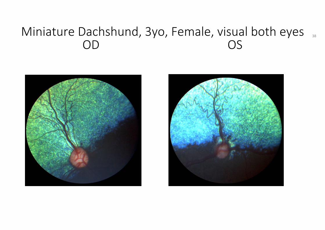
Miniature Dachshund, 3yo, Female, visual both eyesOD OS
38
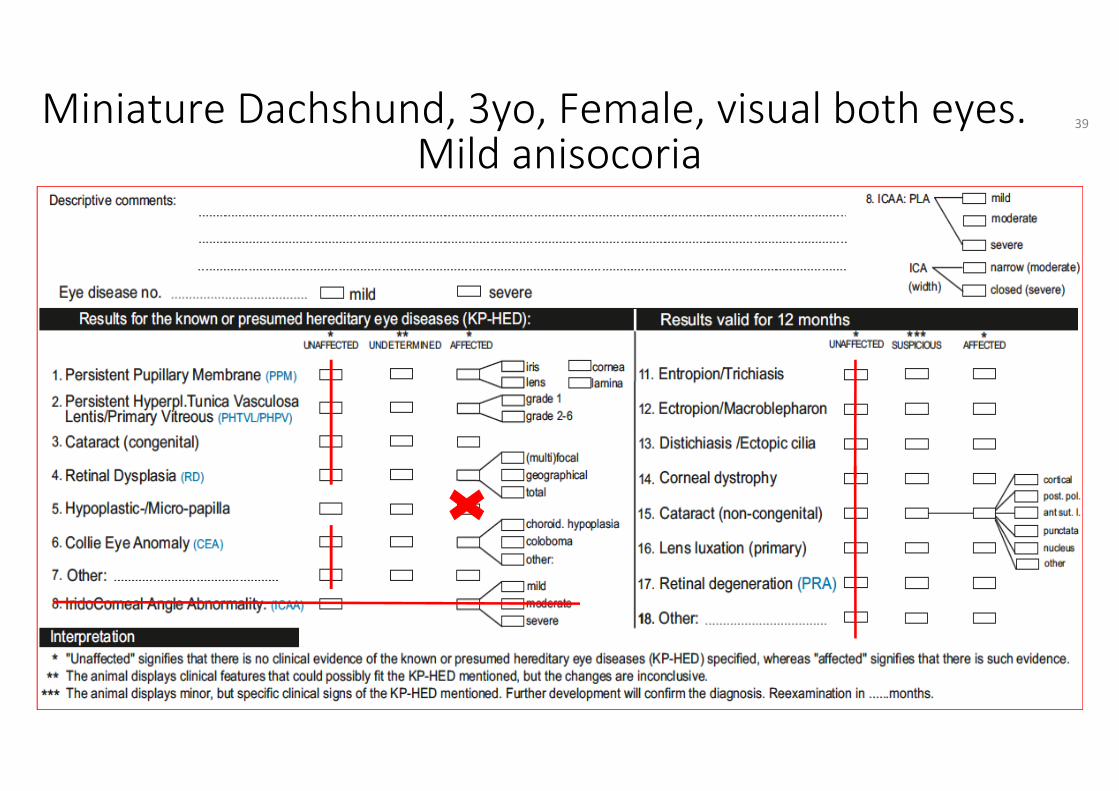
Miniature Dachshund, 3yo, Female, visual both eyes.Mild anisocoria
39
HED Manual Ch. 5 Definitions
• Hypoplasia: defective development of an organ or part resulting in a smaller than normal size or immature state
• Optic disc hypoplasia: presumed hereditary eye disease; congenital failure of development of the optic nerve which causes visual deficiency and abnormal pupil response in the affected eye.
• Micropapilla: small optic disc which is not associated with vision impairment.
40

HED Manual Ch. 6 Guidelines
Tick no 5: Hypoplastic-/Micropapilla
41
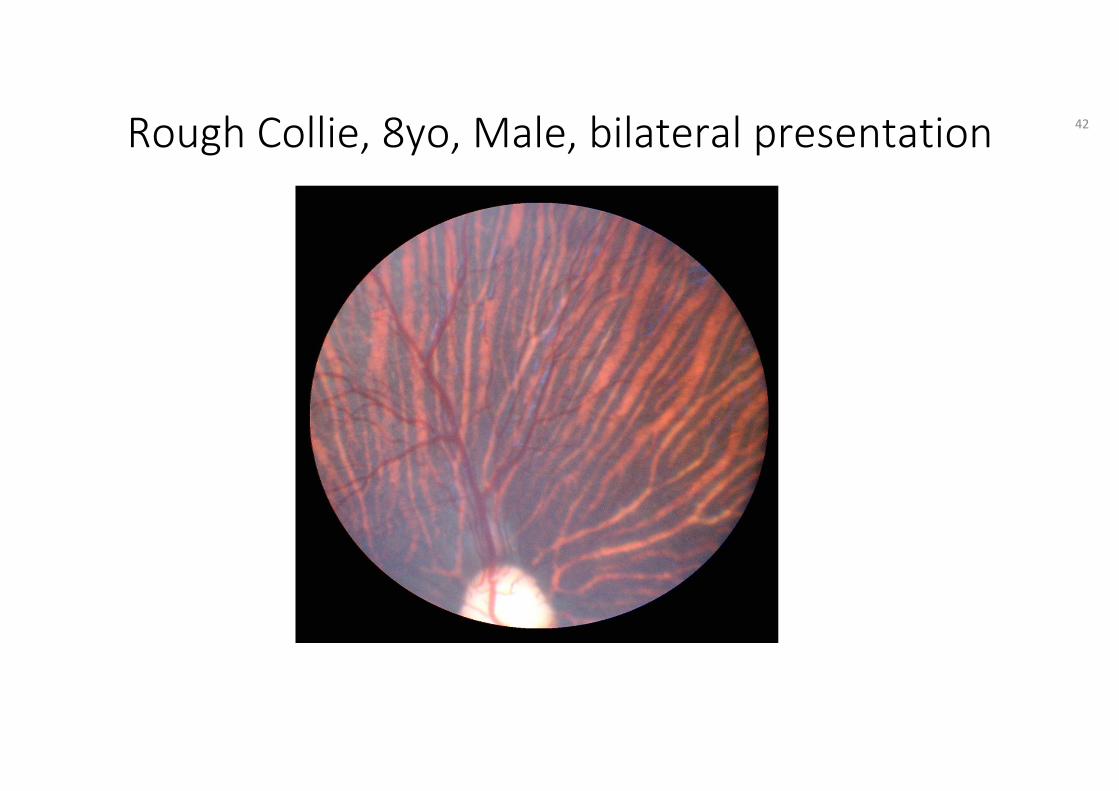
Rough Collie, 8yo, Male, bilateral presentation 42
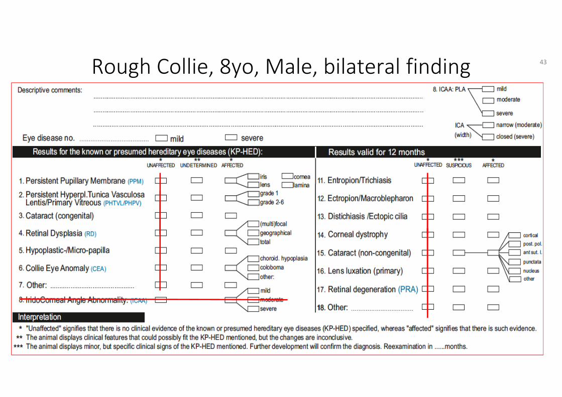
Rough Collie, 8yo, Male, bilateral finding 43

HED Manual Ch. 5 Definitions - Guidelines
• Normal fundus
44
Siberian Husky, 11month old, Female,bilateral/symmetrical
45
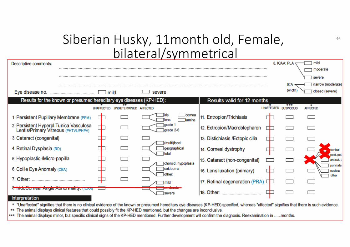
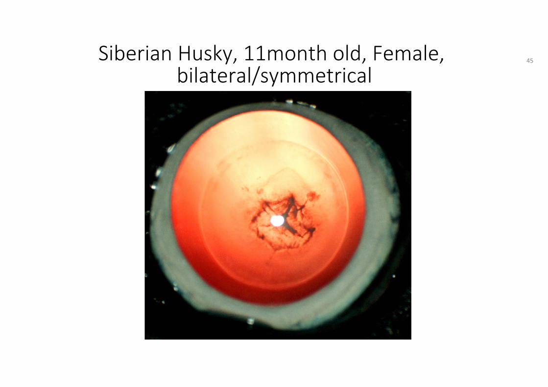
Siberian Husky, 11month old, Female,bilateral/symmetrical
46

HED Manual Ch. 5 Definitions
• Cataract: any hereditary or non-hereditary, congenital or acquired, non-physiological opacity of the lens and/or its capsule.
• All bilateral or unilateral cataracts and especially cortical cataracts are known and presumed hereditary eye diseases except in cases known to be associated with trauma, other causes of ocular inflammation, metabolic disease, nutritional deficiencies, persistent pupillary membrane, persistent hyaloid artery or old age.
47
HED Manual Ch. 6 Guidelines
Tick no 15: Cataract: post. polar & cortical
• To describe the type of cataract, if available, the specifying box for the type of cataract should be ticked.
• It is strongly recommended to draw the cataract in the "pre-drawings" on the certificate.
48

Australian Shepherd, 1yo, FemaleOnly right eye affected
49
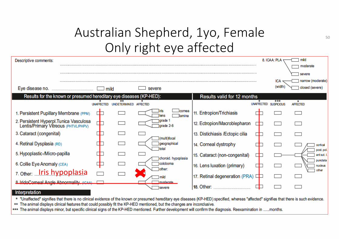
Australian Shepherd, 1yo, FemaleOnly right eye affected
Iris hypoplasia
50
HED Manual Ch. 5 Definitions
• Hypoplasia: defective development of an organ or part of it resulting in a smaller than normal size or immature state
• Hypoplasia iris: presumed hereditary eye disease characterized by congenital absence of iris (sphincter) tissue. It may be a separate disorder or associated with other ocular malformations.
51
HED Manual Ch. 6 Guidelines
Tick no 7: Iris hypoplasia
52
Labrador Retriever, 6yo, Female, right eye,unilateral finding
53
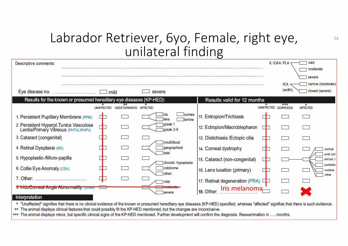
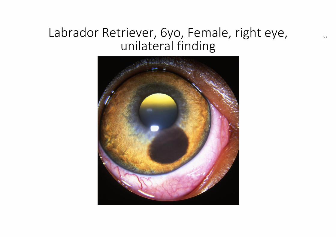
Iris melanoma
Labrador Retriever, 6yo, Female, right eye,unilateral finding
54
HED Manual Ch. 5 Definitions
Melanoma iris: presumed hereditary eye disease,• a locally invasive cancer of melanocyte (pigment) cell origin
within the iris. • Occurs with a higher than normal incidence in the Labrador
retriever. • Left untreated it may result in secondary glaucoma.
55

HED Manual Ch. 6 Guidelines
Tick no 18. Other:Iris melanoma
56
Shetland sheepdog, 3mo, Male, right&left eye
57
Severe
57
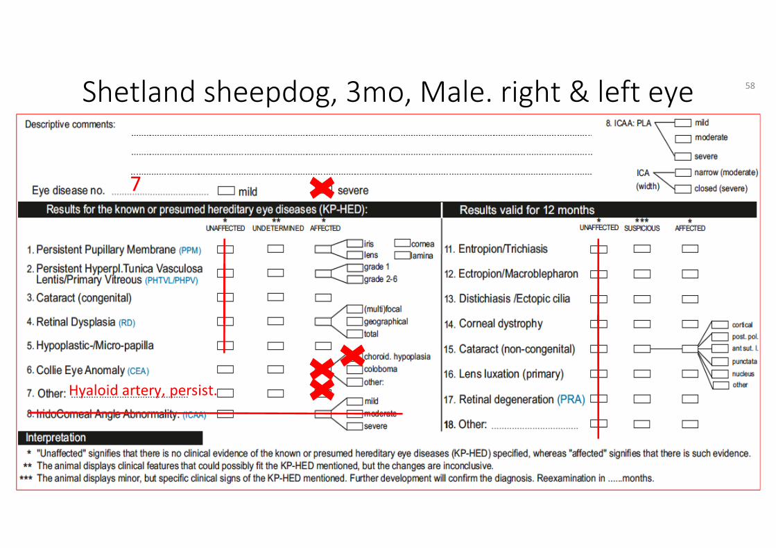
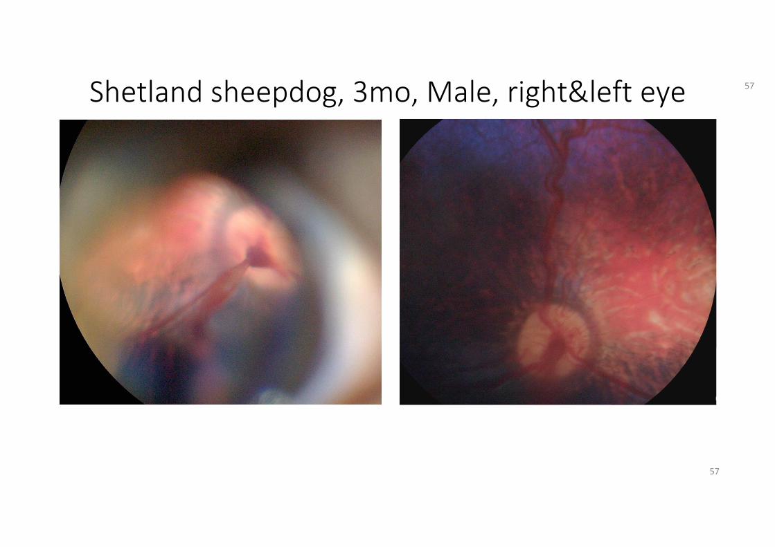
Shetland sheepdog, 3mo, Male. right & left eye
Hyaloid artery, persist.
7
58
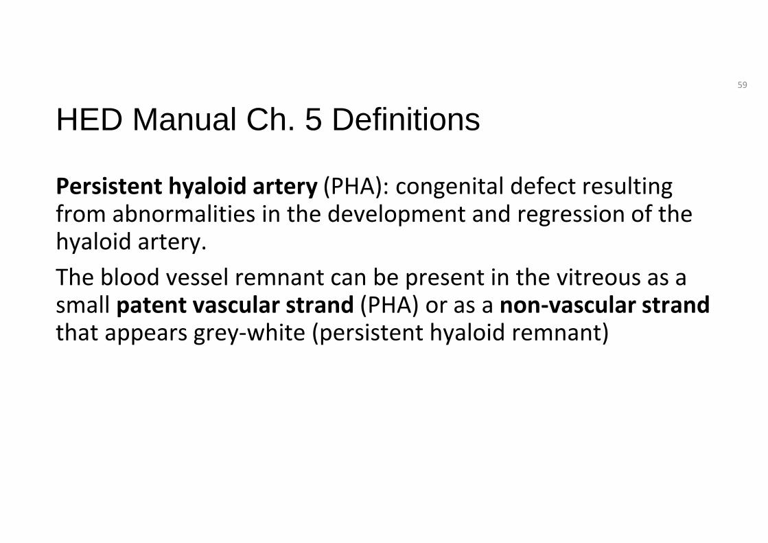
HED Manual Ch. 5 Definitions
Persistent hyaloid artery (PHA): congenital defect resulting from abnormalities in the development and regression of the hyaloid artery. The blood vessel remnant can be present in the vitreous as a small patent vascular strand (PHA) or as a non-vascular strand that appears grey-white (persistent hyaloid remnant)
59
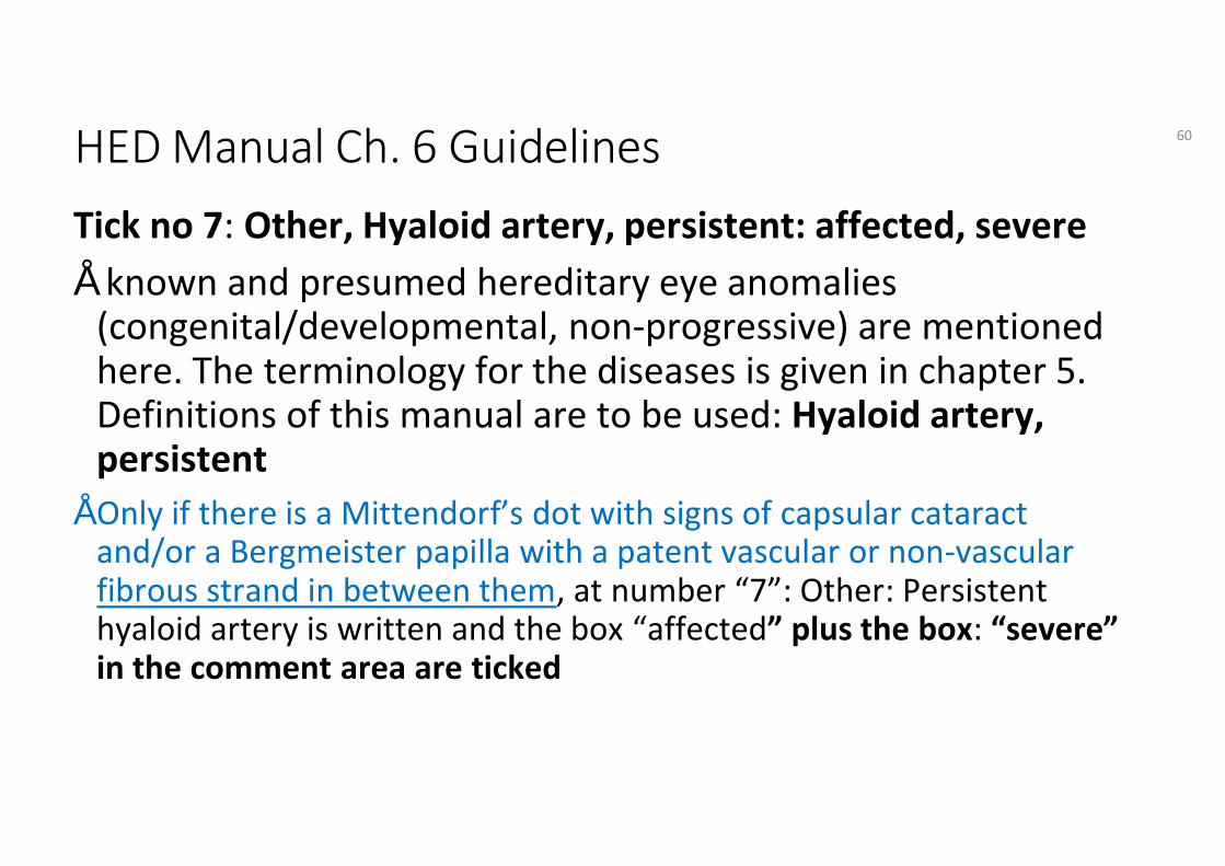
HED Manual Ch. 6 GuidelinesTick no 7: Other, Hyaloid artery, persistent: affected, severe• known and presumed hereditary eye anomalies
(congenital/developmental, non-progressive) are mentioned here. The terminology for the diseases is given in chapter 5. Definitions of this manual are to be used: Hyaloid artery, persistent
• Only if there is a Mittendorf’s dot with signs of capsular cataract and/or a Bergmeister papilla with a patent vascular or non-vascular fibrous strand in between them, at number “7”: Other: Persistent hyaloid artery is written and the box “affected” plus the box: “severe” in the comment area are ticked
60
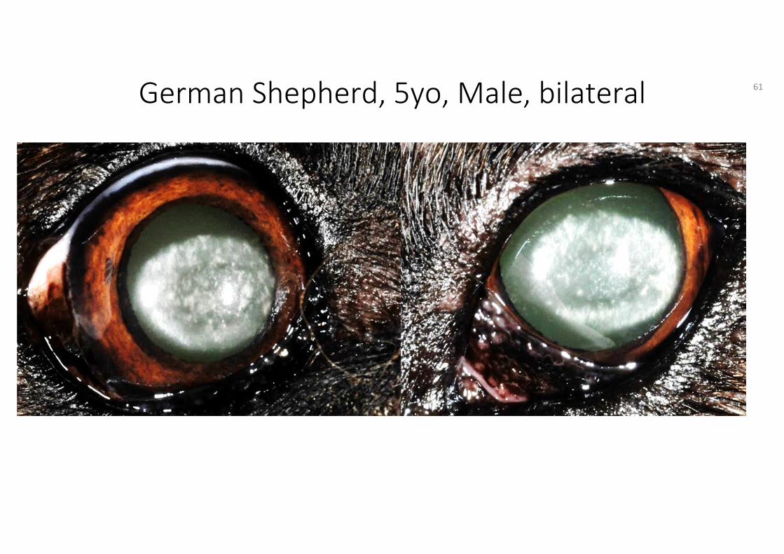
German Shepherd, 5yo, Male, bilateral 61
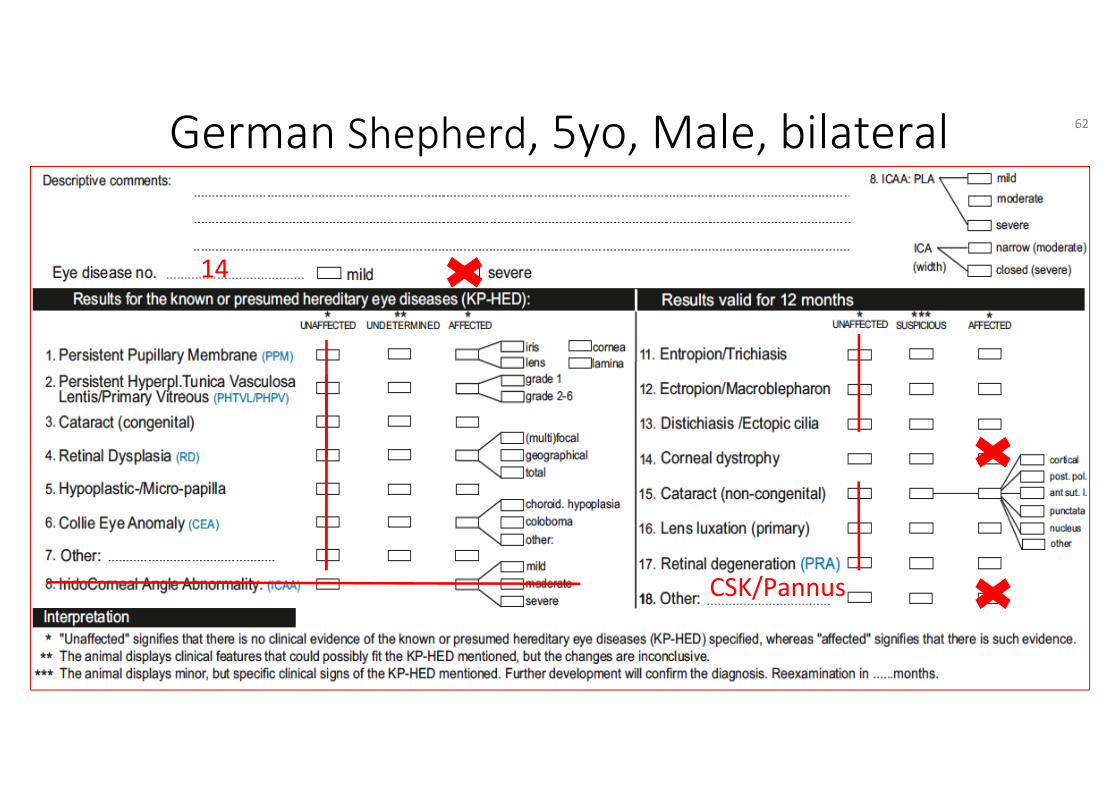
German Shepherd, 5yo, Male, bilateral
14
CSK/Pannus
62
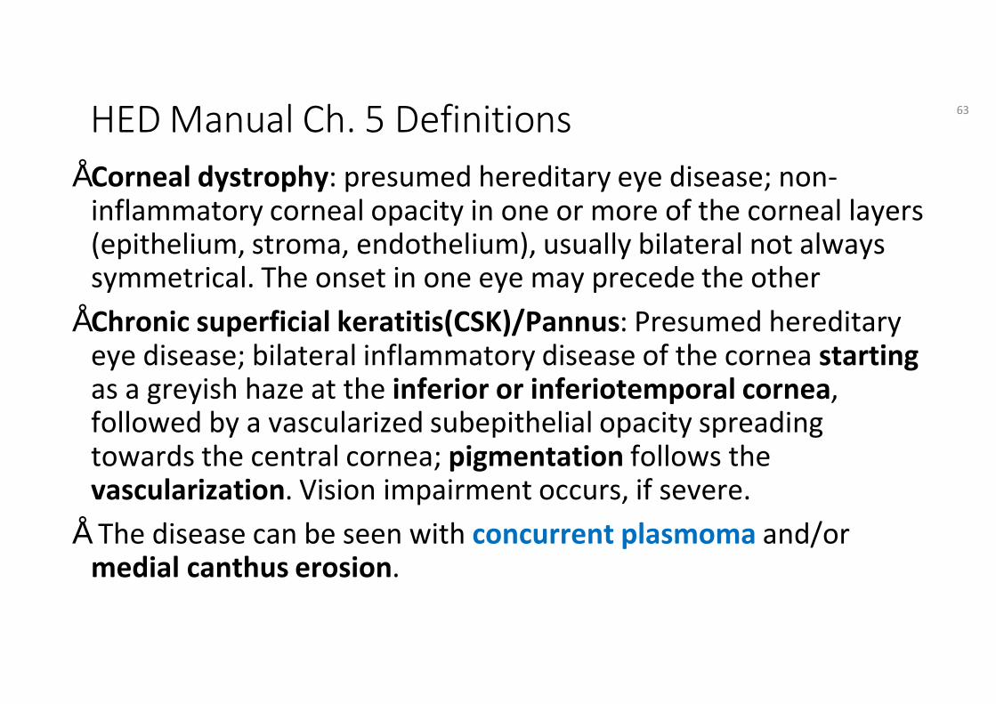
HED Manual Ch. 5 Definitions• Corneal dystrophy: presumed hereditary eye disease; non-
inflammatory corneal opacity in one or more of the corneal layers (epithelium, stroma, endothelium), usually bilateral not always symmetrical. The onset in one eye may precede the other
• Chronic superficial keratitis(CSK)/Pannus: Presumed hereditary eye disease; bilateral inflammatory disease of the cornea startingas a greyish haze at the inferior or inferiotemporal cornea, followed by a vascularized subepithelial opacity spreading towards the central cornea; pigmentation follows the vascularization. Vision impairment occurs, if severe.
• The disease can be seen with concurrent plasmoma and/or medial canthus erosion.
63
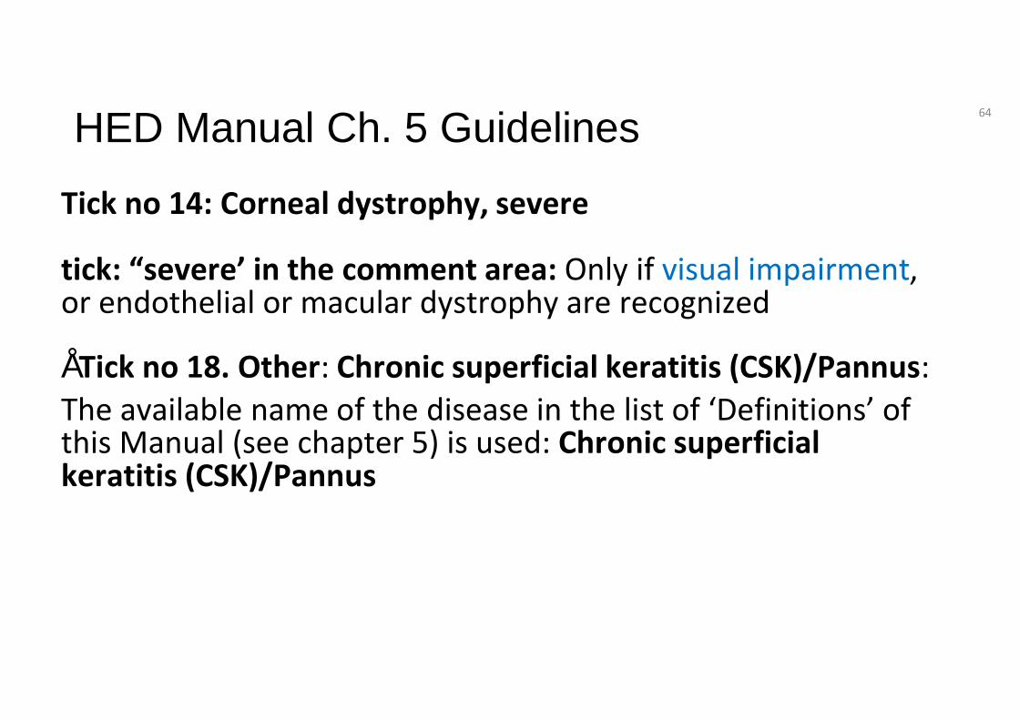
HED Manual Ch. 5 Guidelines
Tick no 14: Corneal dystrophy, severe
tick: “severe’ in the comment area: Only if visual impairment, or endothelial or macular dystrophy are recognized
• Tick no 18. Other: Chronic superficial keratitis (CSK)/Pannus:The available name of the disease in the list of ‘Definitions’ of this Manual (see chapter 5) is used: Chronic superficial keratitis (CSK)/Pannus
64
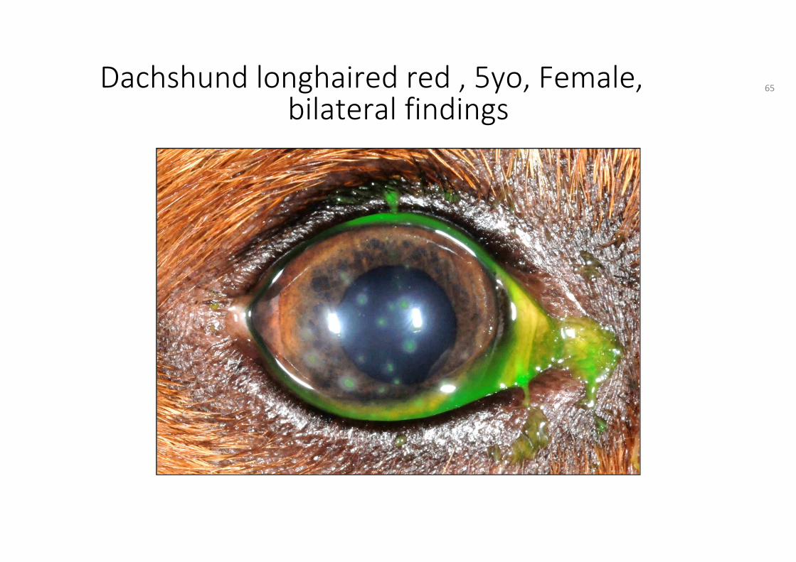
Dachshund longhaired red , 5yo, Female,bilateral findings
65
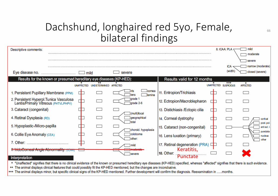
Keratitis, Punctate
Dachshund, longhaired red 5yo, Female,bilateral findings
66
HED Manual Ch. 5 Definitions
• Keratitis, punctate: presumed hereditary eye disease; inflammation of the cornea accompanied by multiple small areas of corneal ulceration
67
HED Manual Ch. 6 Guidelines
Tick no 18. Other: and Keratitis, Punctate
The available name of the disease can be found in the list of ‘Definitions’ of this Manual (see chapter 5) and is used: Keratitis, Punctate
68
Labrador Retriever, 1yo, Female, right eyeunilateral finding
69
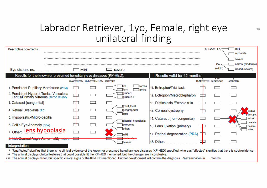
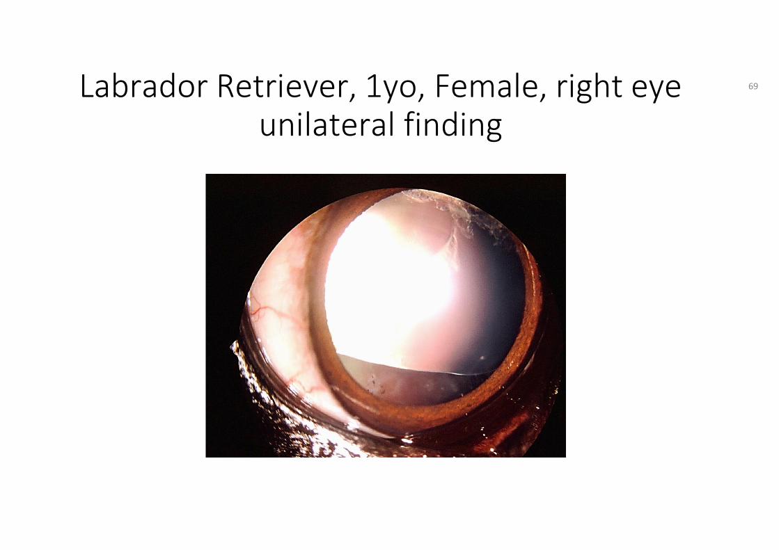
Labrador Retriever, 1yo, Female, right eyeunilateral finding
lens hypoplasia
70
HED Manual Ch. 5 Definitions
• Hypoplasia lens: presumed hereditary eye disease characterized by congenital incomplete formation of the lens equator, sometimes called lens coloboma. See and use lens hypoplasia
71
HED Manual Ch. 6 Guidelines
Tick no 7: Other: lens hypoplasia & no 15: cataract, cortical
The terminology for the diseases is given in chapter 5. Definitions of this manual and are to be used: lens hypoplasia and cataract
72

HED session:Gonioscopy
Antwerp 2019
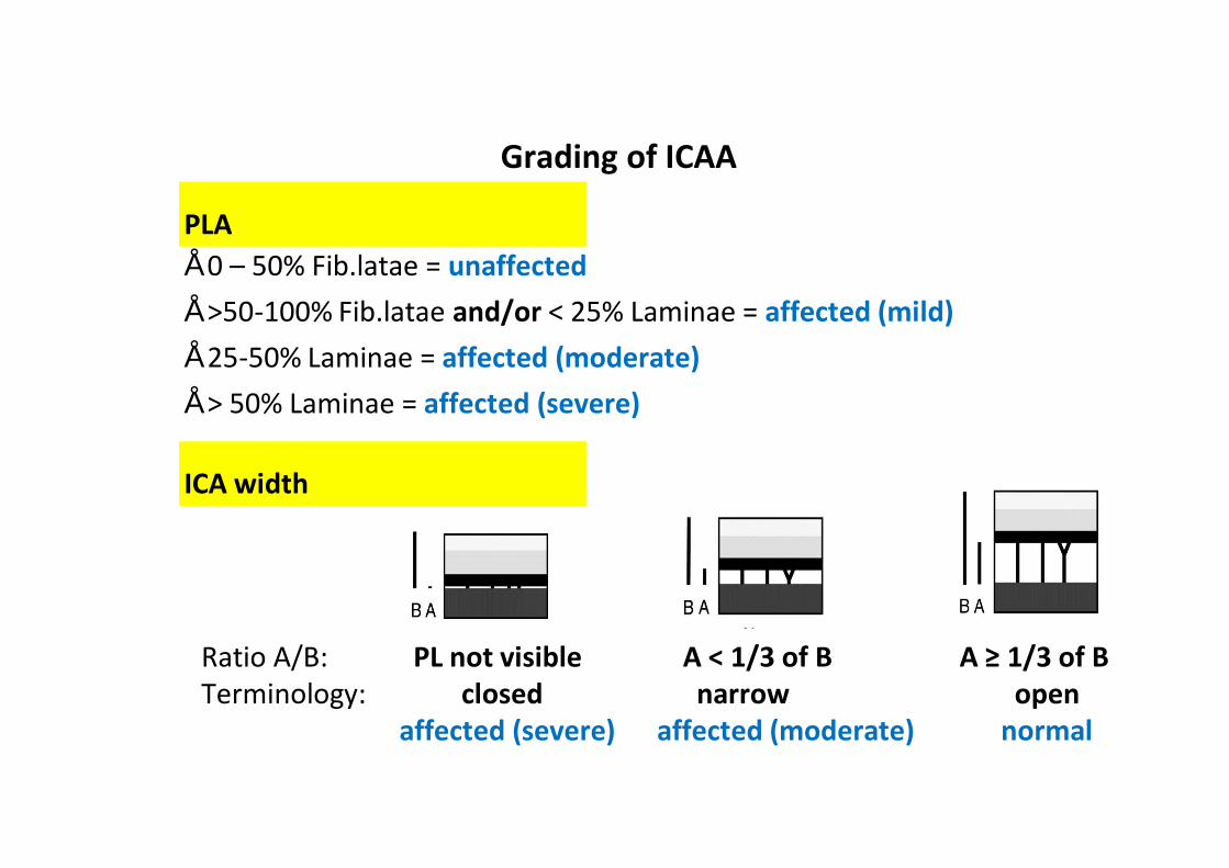
PLA• 0 – 50% Fib.latae = unaffected• >50-100% Fib.latae and/or < 25% Laminae = affected (mild)• 25-50% Laminae = affected (moderate)• > 50% Laminae = affected (severe)
Grading of ICAA
ICA width
Ratio A/B: PL not visible A < 1/3 of B A ≥ 1/3 of BTerminology: closed narrow open
affected (severe) affected (moderate) normal
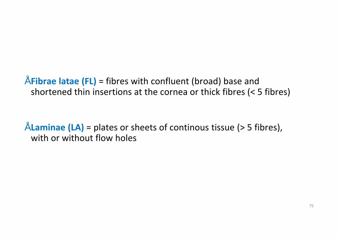
• Fibrae latae (FL) = fibres with confluent (broad) base and shortened thin insertions at the cornea or thick fibres (< 5 fibres)
• Laminae (LA) = plates or sheets of continous tissue (> 5 fibres), with or without flow holes
75
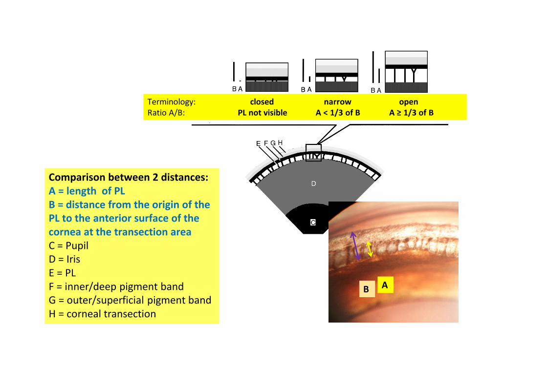
Comparison between 2 distances:A = length of PLB = distance from the origin of thePL to the anterior surface of thecornea at the transection areaC = PupilD = IrisE = PL F = inner/deep pigment bandG = outer/superficial pigment bandH = corneal transection
Terminology: closed narrow openRatio A/B: PL not visible A < 1/3 of B A ≥ 1/3 of B
B A
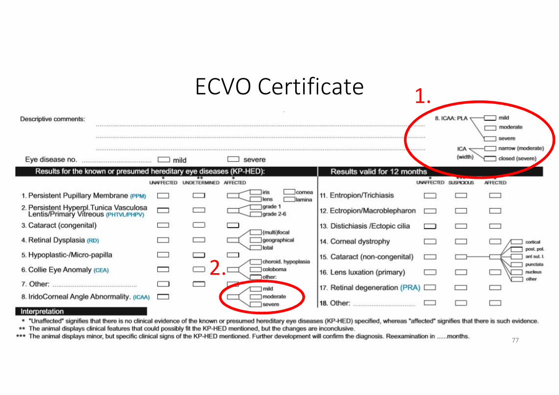
ECVO Certificate
77
1.
2.
Each picture represents one quarter of the iridocornealangle. The rest of the iridocorneal angle is similar to thepicture shown.
78
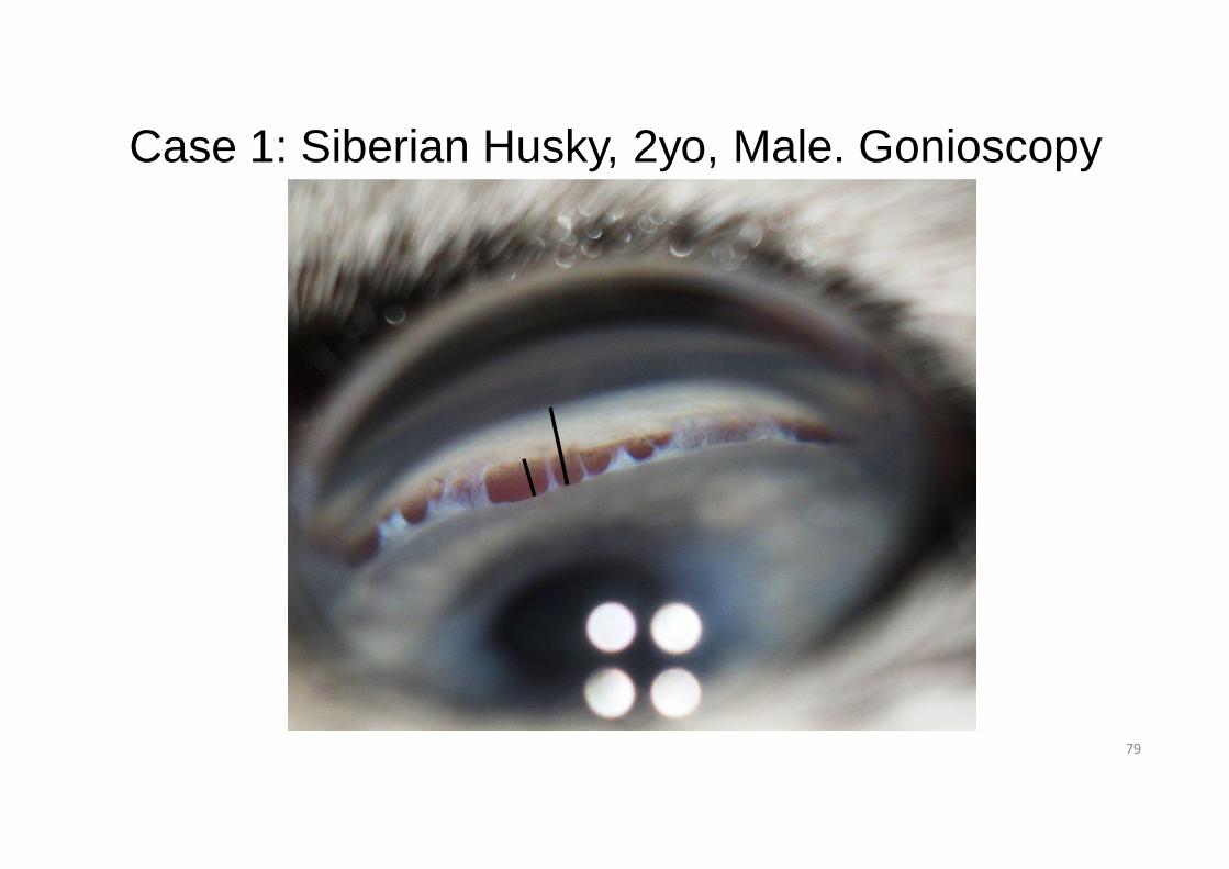
Case 1: Siberian Husky, 2yo, Male. Gonioscopy
79
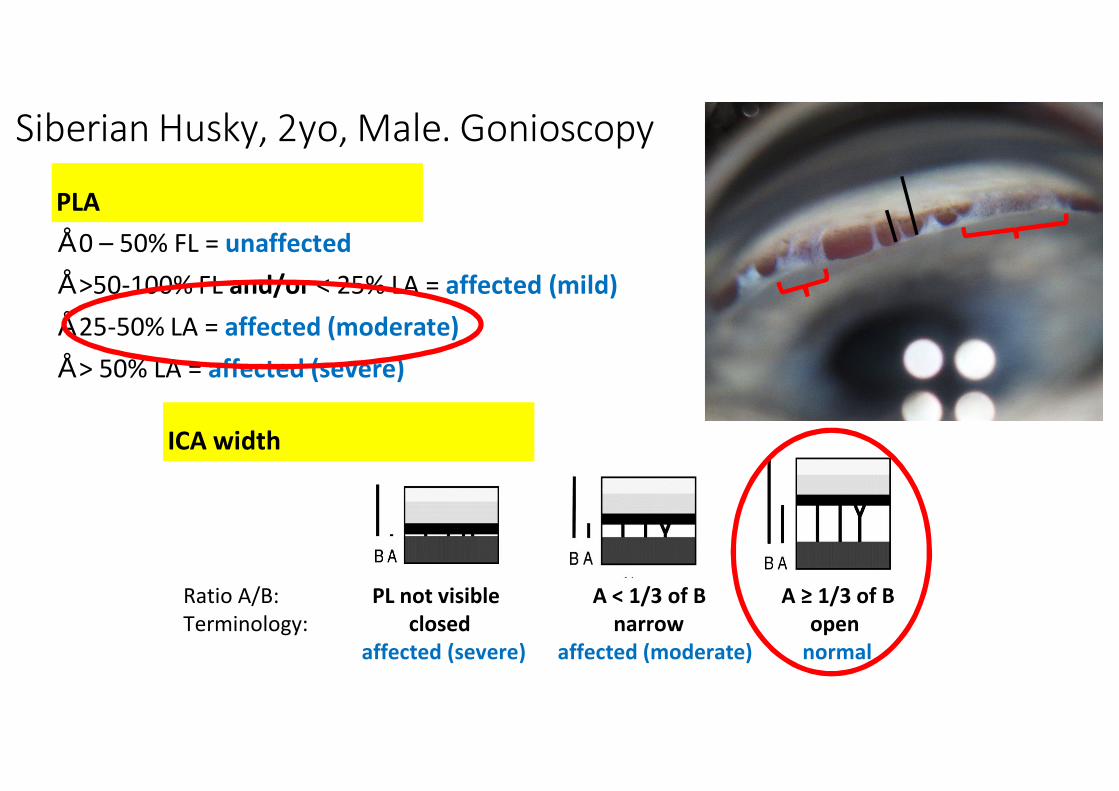
PLA• 0 – 50% FL = unaffected• >50-100% FL and/or < 25% LA = affected (mild)• 25-50% LA = affected (moderate)• > 50% LA = affected (severe)
ICA width
Ratio A/B: PL not visible A < 1/3 of B A ≥ 1/3 of BTerminology: closed narrow open
affected (severe) affected (moderate) normal
Siberian Husky, 2yo, Male. Gonioscopy
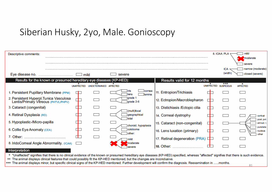
Siberian Husky, 2yo, Male. Gonioscopy
81
Case 1.
Vet advice: breeding is optional
• Mild-moderate forms: OPTIONAL (according to present scientific information available: if these dogs are used, it is recommended to breed these dogs to unaffected graded dogs).
• Iridocorneal angle formation may progressively change with age from normal/unaffected to abnormal/affected (mild/moderate/severe) regarding PLA and ICA-width. Therefore, gonioscopy should be started before breeding and repeated every 3 years
82
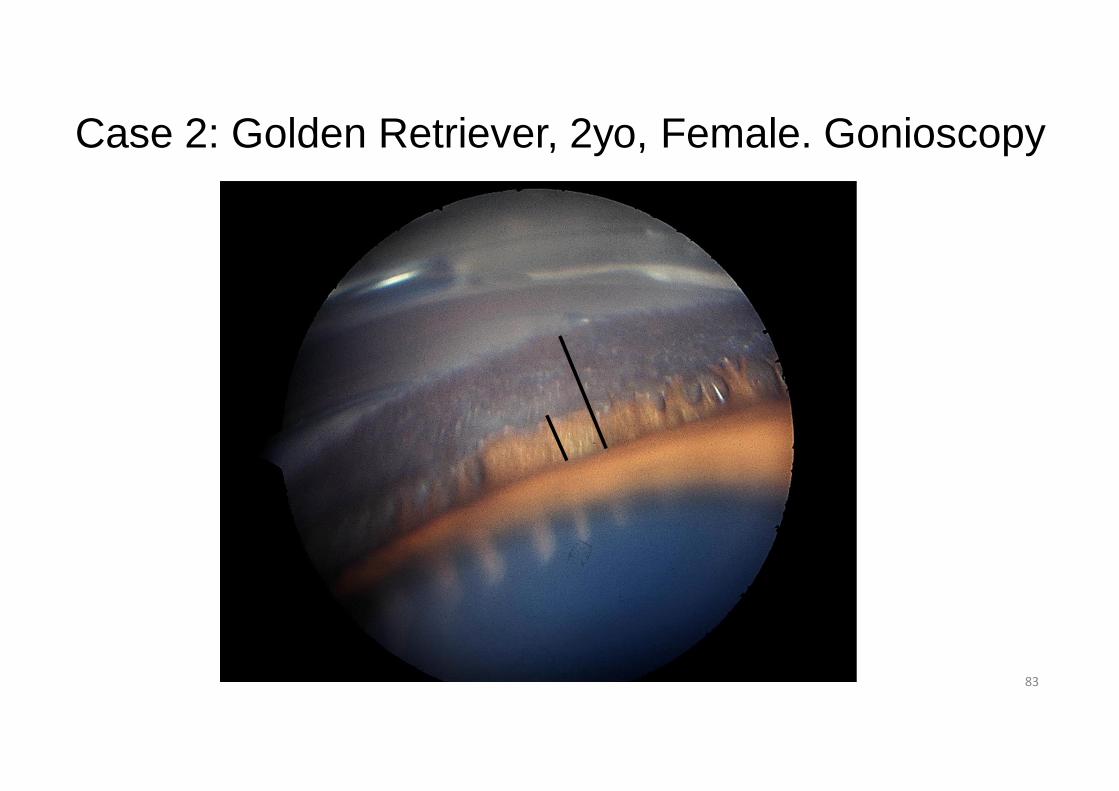
Case 2: Golden Retriever, 2yo, Female. Gonioscopy
83
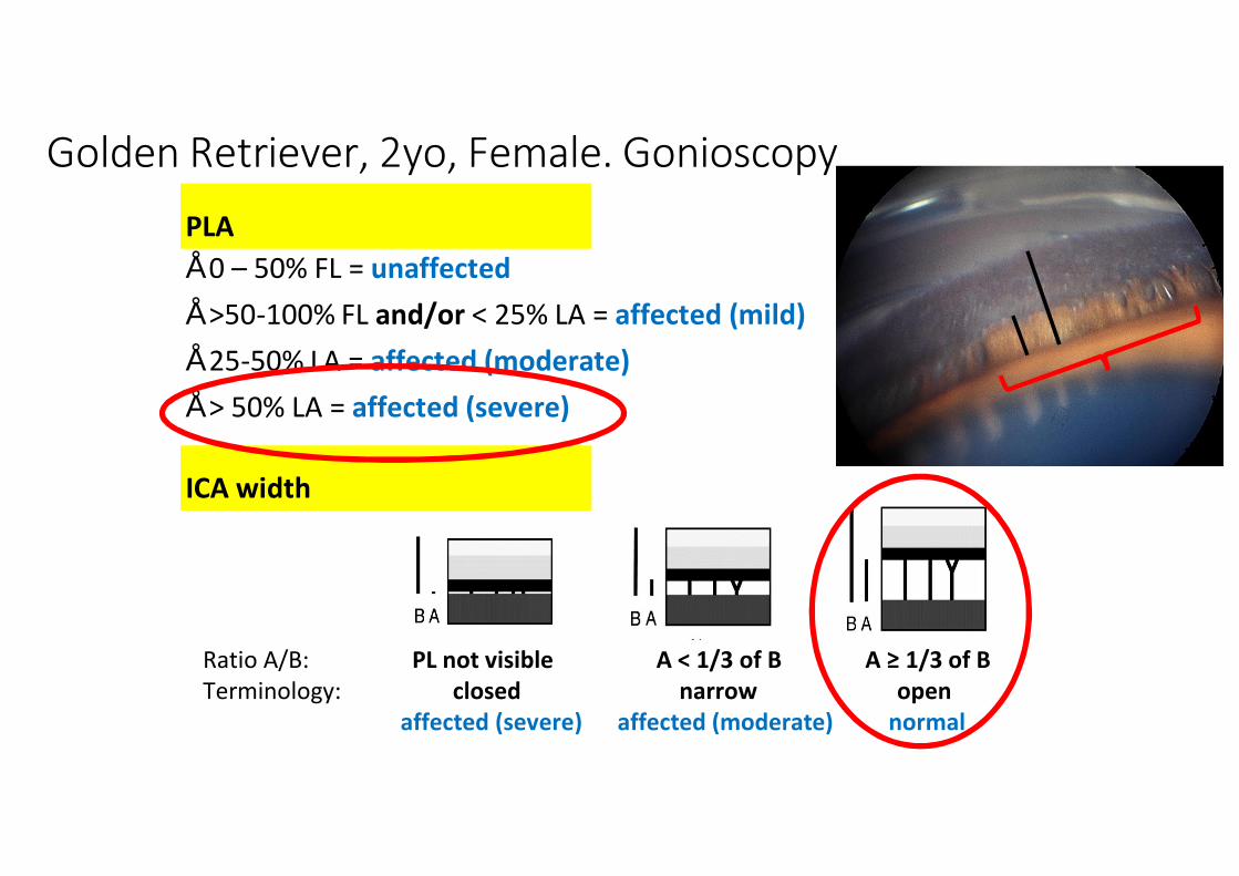
PLA• 0 – 50% FL = unaffected• >50-100% FL and/or < 25% LA = affected (mild)• 25-50% LA = affected (moderate)• > 50% LA = affected (severe)
ICA width
Ratio A/B: PL not visible A < 1/3 of B A ≥ 1/3 of BTerminology: closed narrow open
affected (severe) affected (moderate) normal
Golden Retriever, 2yo, Female. Gonioscopy
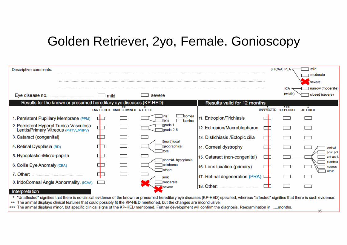
Golden Retriever, 2yo, Female. Gonioscopy
85
Case 2.
• Vet advice: no breeding from the affected animal
86
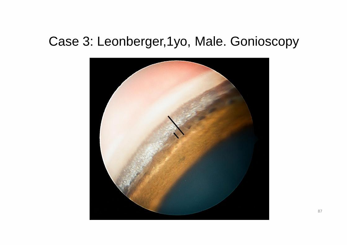
Case 3: Leonberger,1yo, Male. Gonioscopy
87
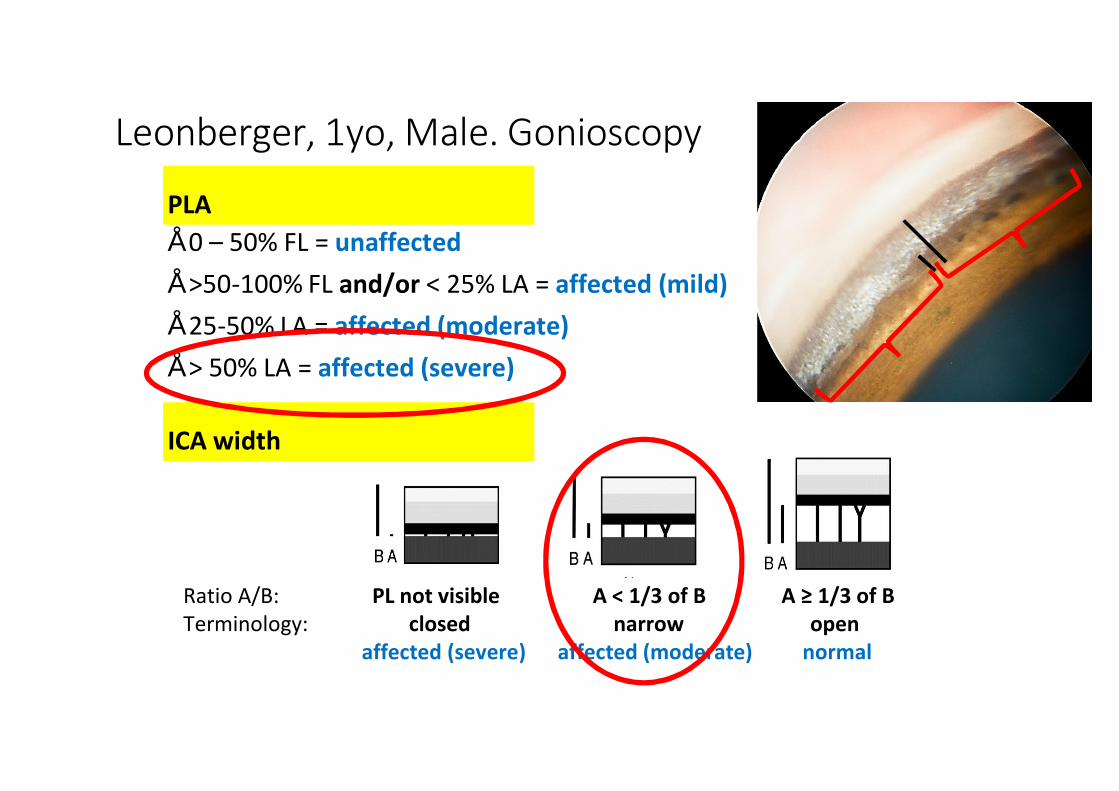
PLA• 0 – 50% FL = unaffected• >50-100% FL and/or < 25% LA = affected (mild)• 25-50% LA = affected (moderate)• > 50% LA = affected (severe)
ICA width
Ratio A/B: PL not visible A < 1/3 of B A ≥ 1/3 of BTerminology: closed narrow open
affected (severe) affected (moderate) normal
Leonberger, 1yo, Male. Gonioscopy
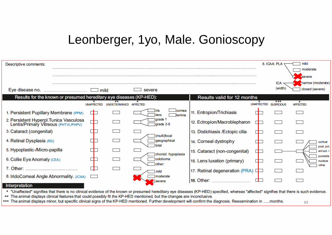
Leonberger, 1yo, Male. Gonioscopy
89

Case 3.
• Vet advice: no breeding from the affected animal
90
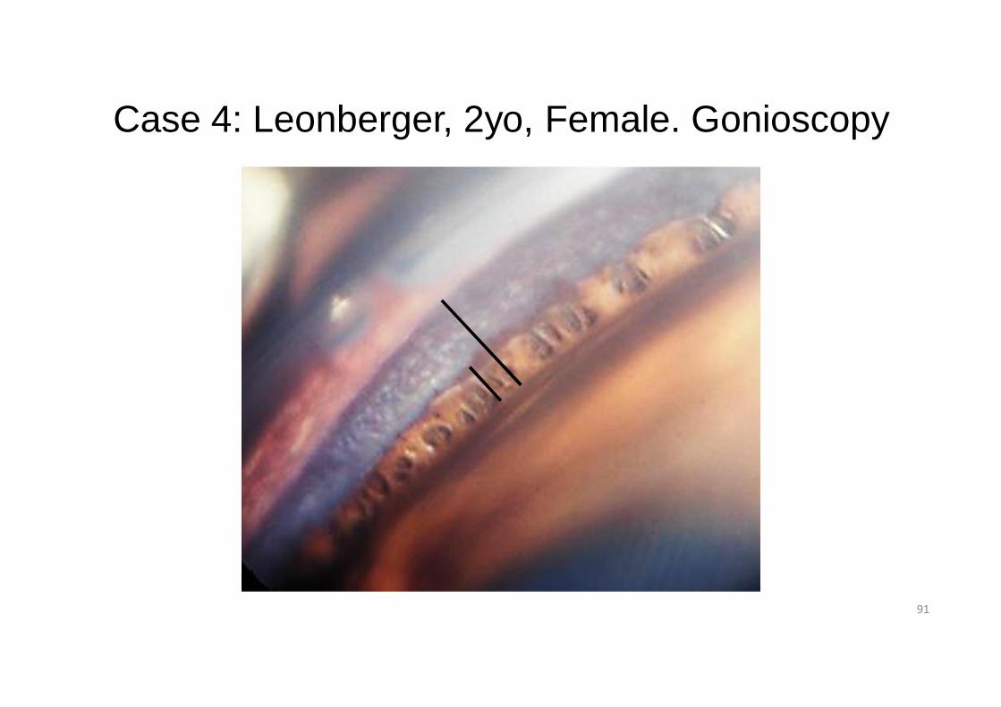
Case 4: Leonberger, 2yo, Female. Gonioscopy
91
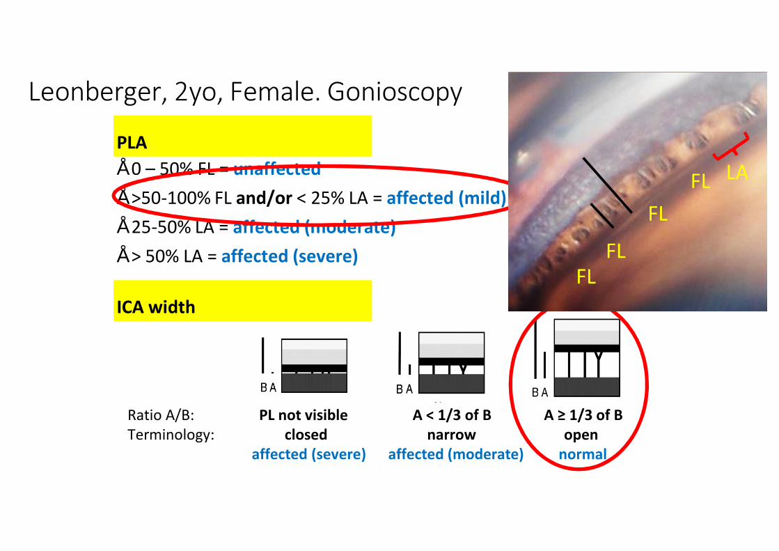
PLA• 0 – 50% FL = unaffected• >50-100% FL and/or < 25% LA = affected (mild)• 25-50% LA = affected (moderate)• > 50% LA = affected (severe)
ICA width
Ratio A/B: PL not visible A < 1/3 of B A ≥ 1/3 of BTerminology: closed narrow open
affected (severe) affected (moderate) normal
Leonberger, 2yo, Female. Gonioscopy
LA
FL
FLFL
FL
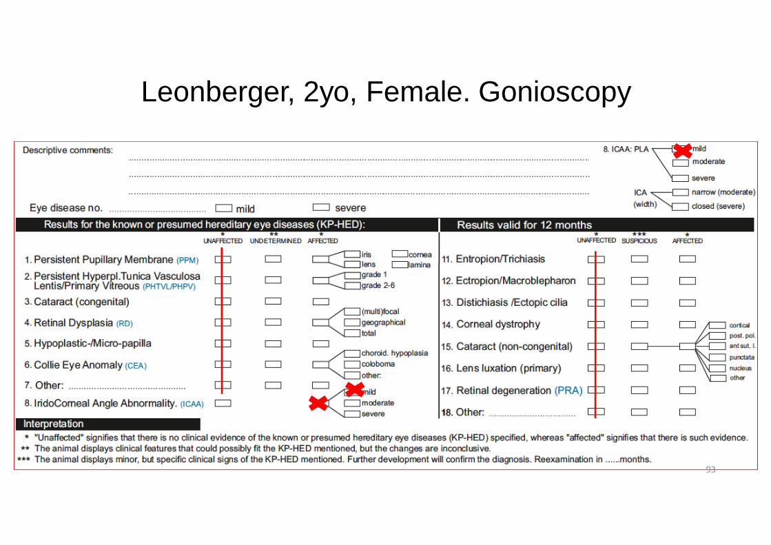
Leonberger, 2yo, Female. Gonioscopy
93
Case 4.
Vet advice: optional
• Mild-moderate forms: OPTIONAL (according to present scientific information available: if these dogs are used, it is recommended to breed these dogs to unaffected graded dogs).
• Iridocorneal angle formation may progressively change with age from normal/unaffected to abnormal/affected (mild/moderate/severe) regarding PLA and ICA-width. Therefore, gonioscopy should be started before breeding and repeated every 3 years
94

Case 5: Labrador Retriever,1yo, Male. Gonioscopy
95
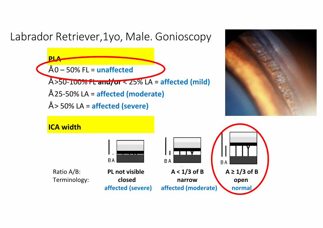
PLA• 0 – 50% FL = unaffected• >50-100% FL and/or < 25% LA = affected (mild)• 25-50% LA = affected (moderate)• > 50% LA = affected (severe)
ICA width
Ratio A/B: PL not visible A < 1/3 of B A ≥ 1/3 of BTerminology: closed narrow open
affected (severe) affected (moderate) normal
Labrador Retriever,1yo, Male. Gonioscopy
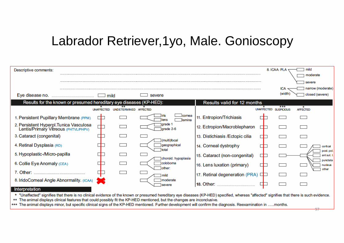
Labrador Retriever,1yo, Male. Gonioscopy
97
Case 5.
Tick: unaffected
• Iridocorneal angle formation may progressively change with age from normal/unaffected to abnormal/affected (mild/moderate/severe) regarding PLA and ICA-width. Therefore, gonioscopy should be started before breeding and repeated every 3 years
98
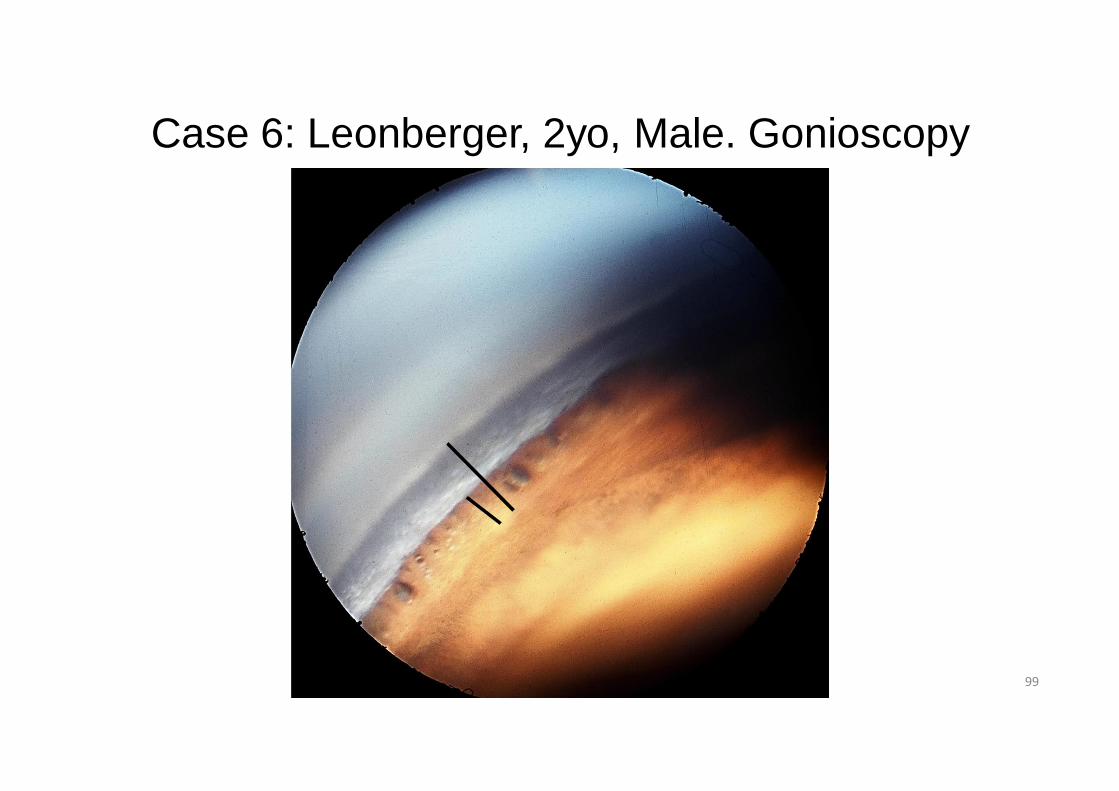
Case 6: Leonberger, 2yo, Male. Gonioscopy
99
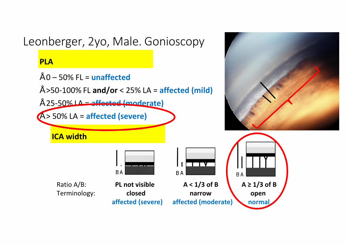
PLA
• 0 – 50% FL = unaffected• >50-100% FL and/or < 25% LA = affected (mild)• 25-50% LA = affected (moderate)• > 50% LA = affected (severe)
ICA width
Ratio A/B: PL not visible A < 1/3 of B A ≥ 1/3 of BTerminology: closed narrow open
affected (severe) affected (moderate) normal
Leonberger, 2yo, Male. Gonioscopy

Leonberger, 2yo, Male. Gonioscopy
101

Case 6.
• Vet advice: no breeding from the affected animal
102