Embed Size (px)
Citation preview

ACEM 2007.2 PRIMARY VIVA EXAMINATION
SUBJECT: ANATOMY 7 September 2007 am.
TOPIC: X-ray: Lateral C spine NUMBER: JL
OPENINGQUESTION
POINTSREQUIRED
PROMPTS
SECONDQUESTION(if needed)POINTSREQUIRED
PROMPTS
PROMPTS
Demonstrate the bony features of Cl and C2 vertebrae evident on thisXray
1 Odontoid peg (dens)
2 Bodies of Cl andC2
3 anterior and posterior arches of atlas
4 laminae and inferior articular process
5 spinous process
What are the components of the soft tissue shadow located anterior tothe upper cervical vertebrae?
1 Anterior longitudinal ligament
2 Longus colli muscle
3 Prevertebral fascia
4 Retropharyngeal space
5 Buccopharyngeal fascia
6 Pharyngeal muscle
COMMENTS
5/8 including dens topass
3/6 to pass includingretropharyngeal space
COMMENTS
JDEOSa Anatomy Viva workshop.rtf

ACEM 2007.2 PRIMARY VWA EXAMINATION
SUBJECT: ANATOMY FRIDAY SEPTEMBER 7 2007
TOPIC: Bone: Humerus and Scapula NUMBER: 2
OPENINGQUESTIONPOINTSREQUIRED
PROMPTS
SECONDQUESTION(if needed)POINTSREQUIRED
PROMPTS
Describe the bony features of the proximal humerus.
1 anatomical neck
Head
Surgical neck
2 greater and lesser tubercles
3 intertubercular groove
What factors contribute to the stability of the glenohumeraljoint?
1 Muscular- rotator cuff- TM,SS,SSp,Infsp
2 Bony structures and labrura- poor
3 Ligaments- coracoacromial ligament
4 capsule
COMMENTS
5/6 to pass
3 /4 to pass
DEOSa Anatomy Viva workshop.rtf

ACEM 2007.1 PRIMARY VIVA EXAMINATION
SUBJECT: ANATOMY Friday 7 September 2007 am
TOPIC: Ankle NUMBER: 3
OPENINGQUESTION(if needed)POINTSREQUIRED
PROMPTS
SECONDQUESTION
POINTSREQUIRED
PROMPTS
Demonstrate the structures passing behind the medialmalleolus
1)TP
2)FDL
3) Posterior tibial artery
4) Tibial nerve
5) Flexor hallucis
5
6
What is the cutaneous innervation of the tibial nerve?
1) Medial plantar nerve. . .medial sole of foot
2) lateral plantar... lateral sole of foot
3) also calcaneal branches
4
5
6
3 / 5 to pass
l/2 to pass
COMMENTS
DEOSa Anatomy Viva workshop.rtf

ACEM 2007.2 PRIMARY VIVA EXAMINATION
SUBJECT: ANATOMY 7 September 2007 am
TOPIC: Photo- PELVIS NUMBER: 4
OPENINGQUESTIONPOINTSREQUIRED
PROMPTS
SECONDQUESTION(if needed)POINTSREQUIRED
PROMPTS
PROMPTS
Identify the structures in this photo (prompt if needed)
1 Rectum No. 27
2 Uterus No. 6/12
3 Bladder No. 5
4 Sacrum Not numbered
5 Pubic symphysis No. 25
6 Anal canal No. 1
7 Cervix and vagina No. 7 and 24
Please show the potential spaces where free fluid canaccumulate in the pelvis
1 rectouterine pouch (of Douglas) No. 26
2 vesicouterine pouch No. 34
3
4
COMMENTS
Need 5/8 to pass
Need 1 to pass
COMMENTS
DEOSa Anatomy Viva workshop.rtf

ACEM 2007.2 PRIMARY VIVA EXAMINATION
SUBJECT: ANATOMY 6 September 2007 pm
TOPIC: PICTURE: CT SCAN BRAIN NUMBER: 1
OPENINGQUESTIONPOINTSREQUIRED
PROMPTS
SECONDQUESTION(if needed)POINTSREQUIRED
PROMPTS
THIRDQUESTION(if needed)POINTSREQUIRED
PROMPTS
Name the visible intracranial structures on this non contrast CT scan
I Lobes (frontal.parietal/temporal.occipital)
2 lateral ventricles ( anterior and posterior horns)
S.choroid plexus
4 pineal gland
5 thalamus
6 internal capsule, basa! ganglia, caudate, globus pallidus, putamen,sylvian fissure or lateral sulcus
What structures does CSF pass through to reach the base of thebrain?
1 Lateral venticles — Mntervenrricular formen
— » 3rd ventricle
— > aqueduct
— > 4 ventricle (posterior to pons/medulla)
Start at the lateral ventricle
COMMENTS
6 structures from 1st 5
Plus 1 of these forpass
3rt and 4th ventricles insequence to pass
COMMENTS
DEOSa Anatomy Viva workshop.rtf

ACEM 2007.2 PRIMARY VIVA EXAMINATION
SUBJECT: ANATOMY 6 September 2007 pm.
TOPIC: Bones: Cl and C2 NUMBER: _2
OPENINGQUESTION
POINTSREQUIRED
PROMPTS
SECONDQUESTION(if needed)POINTSREQUIRED
4
PROMPTS
THIRDQUESTION(if needed)POINTSREQUIRED
PROMPTS
Describe the features of this bone (C2)
1 odontoid peg (dens) and body
2 vertebral foramen (s.c.) transverse foramen (v.a.)
3 inferior/superior articular surfaces
4 pedicle
5 transverse process
6 lamina
7 spinous process
What ligaments stabilise the atlantoaxial joint?
1 Cruciate ligament which has two components
Transverse- between lateral masses of Cl- strong
Longitudinal- occiput to body C2- weak
2 Alar ligament- side of peg to foramen magnum
; 3 tectorial membrane- continuation of PtL from body ofC2 to internal occiput
What movement occurs at the C1/C2 joint?
Rotation of Cl onC2
COMMENTS
6/10 to pass
Identify it as C2
Cruciate and tectorialto pass i.e. 2/3
COMMENTS
DEOSa Anatomy Viva workshop.rtf

ACEM 2007.2 PRIMARY VIVA EXAMINATION
SUBJECT: ANATOMY 6 September 2007 pm.
TOPIC: MODEL: ANKLE NUMBER: 3
OPENINGQUESTION(if needed)POINTSREQUIRED
PROMPTS
SECONDQUESTION(if needed)POINTSREQUIRED
PROMPTS
Demonstrate the attachments of the inferior extensor retinaculum
1 upper anterior surface of calcaneus
2 Y-shaped 1. medial malleolus 2. blends with plantar aponeurosis
3
Identify the structures passing beneath the IER
1 tibialis anterior
2 extensor ballads longus
3 ant tibial artery dorsalis pedis artery
4 deep fibular nerve
5 extensor digitorum longus
6 fibularis tertius
4/6 to pass
Level of ankle joint
Deep peroneal nerve
Peroneus tertius
THIRD QUESTION What is the function of the inferior extensor retinaculum?
Prevent bowstringing of extensor tendons when ankle dorsiflexed.
DEOSa Anatomy Viva workshop.rtf

ACEM 2007.2 PRIMARY VIVA EXAMINATION
SUBJECT: ANATOMY 6 September 2007 om.
TOPIC: DISCUSSION: BRACHIAL PLEXUS NUMBER: _4
OPENINGQUESTIONPOINTSREQUIRED
Optional
PROMPTS
SECONDQUESTION(if needed)POINTSREQUIRED
PROMPTS
THIRDQUESTION(if needed)POINTSREQUIRED
PROMPTS
What is the brachial plexus and how is it formed?
1 Major nerve network supplying the upper limb, extends from neckto axilla.2 Results in the formation of multisegmental peripheral nerves.
3 Initially formed by the union of the anterior rami (roots) lowercervical (C5-8) and first thoracic nerves.
4 These roots unite to form 3 trunks; superior, middle and inferior
5 Each trunk divides into anterior and posterior divisions
6 These division then form 3 cords: Anterior divisions of superiorand middle trunks for the lateral cord, anterior division of inferiortrunk forms medial cord, posterior divisions of all 3 trunks unite toform posterior cord
7 Major peripheral nerves supplying upper limb form from thesecords. Lateral cord gives rise to lateral pectoral n, musculocutaneousn and lateral root of median n. Medial cord gives rise to medial rootmedian n, medial pectoral n, medial cutaneous n of arm, medialcutaneous n of forearm and ulnar n. Posterior cord gives rise toupper subscapular n, lower subscapular n, thoracodorsal n, axillary n,and radial n.
1 . No need to describe supraclavicular branches of brachialplexus.
2. Would vou prefer to describe the formation of the brachial
COMMENTS
Start proximally todistal
Need to knowrootSjtrunkSjdivisions,cords to pass
Name at least medial andlateral roots median n,axillary n, radial n andulnar n.
COMMENTS
DE08a Anatomy Viva workshop.rtf
ACEM 2007.2 PRIMARY VIVA EXAMINATION
SUBJECT: ANATOMY 6 September 2007 pm
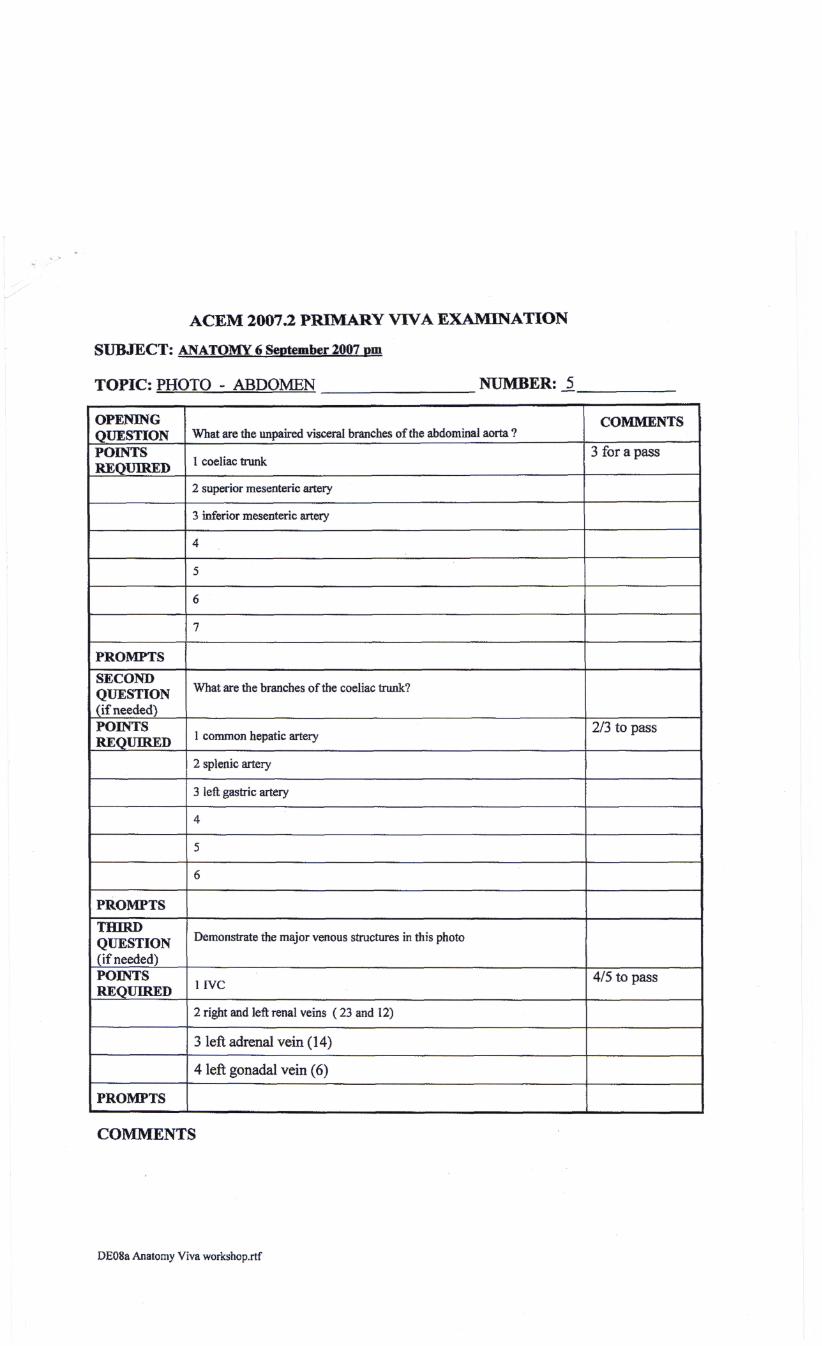
TOPIC: PHOTO - ABDOMEN NUMBER: _5
OPENINGQUESTIONPOINTSREQUIRED
PROMPTS
SECONDQUESTION(if needed)POINTSREQUIRED
PROMPTS
THIRDQUESTION(if needed)POINTSREQUIRED
PROMPTS
What are the unpaired visceral branches of the abdominal aorta ?
I coeliac trunk
2 superior mesenteric artery
3 inferior mesenteric artery
4
5
6
7
What are the branches of the coeliac trunk?
1 common hepatic artery
2 splenic artery
3 left gastric artery
4
5
6
Demonstrate the major venous structures in this photo
1IVC
2 right and left renal veins ( 23 and 12)
3 left adrenal vein (14)
4 left gonadal vein (6)
COMMENTS
3 for a pass
2/3 to pass
4/5 to pass
COMMENTS
DEOSa Anatomy Viva workshop.rtf