Embed Size (px)
Citation preview

A NEW ETHANOLAMINE-CONTAINING LIPIDE FROM EGG YOLK*
BY HERBERT E. CARTER, DONALD B. SMITH,t AND D. N. JONES1
(From the Division of Biochemistry, Noyes Laboratory of Chemistry, University of Illinois, Urbana, Illinois)
(Received for publication, November 27, 1957)
Recent studies of egg yolk lipides and lipoproteins, and in particular the results of the chromatographic procedures of Lea and coworkers (l-5), have caused renewed interest in egg yolk, which many years ago was considered the most important source of phosphatides for both scien- tific and commercial purposes. Lea and Rhodes have extensively studied the phospholipide content of egg yolk (l-3), whereas Lea and Hawke (4, 5) have worked mainly on the lipoproteins. Despite the attention given to egg yolk lipides, very few data are available on the sphingolipide constituents. In undertaking a study of this fraction, we prepared a considerable quantity of crude egg yolk phosphatide’ and subjected this material to the mild alkaline hydrolysis procedure of Schmidt et al. (6). The resulting crude sphingolipide fraction contained both cerebroside and sphingomyelin as determined by paper chromatography of the intact lipide mixture and of its hydrolysis products. Glycerol, inositol, and their phosphates were not detected in the hydrolysates but the intact crude sphingolipide mixture gave a positive ninhydrin test and an acid hydrolysate contained ethanolamine and ethanolamine phosphate. Since none of the known phospholipides (except sphingomyelin) is stable to mild alkaline and acid hydrolysis (6), these unexpected observations could reasonably be interpreted as indicating the presence in egg yolk lipides of the ethanolamine analogue of sphingomyelin (I) (tentatively designated as sphingoethanolamine) .
This substance has not been isolated from natural sources, although there are numerous indications in the literature of its possible existence.
* This work was supported by a generous grant from the Procter and Gamble Company and in part by a research grant (No. B574-C3) from the National Institutes of Health, United States Public Health Service. A preliminary report of this work was presented (Federation Proc., 16, 817 (1957)).
t Postdoctorate research associate in chemistry, 1955-57; present address, 13 Corby Avenue, Swindon, Wiltshire, England.
$ Postdoctorate research associate in chemistry, 195658. i The starting material for these studies was 50 pounds of “egg oil” obtained
through the courtesy of the VioBin Corporation, Monticello, Illinois. The authors wish to express their appreciation to Mr. Ezra Levin for providing this material.
681
by guest on August 29, 2018
http://ww
w.jbc.org/
Dow
nloaded from

682 NEW ETHANOLAMINE-LIPIDE FROM EGG YOLK
CH~(CH~)~~CH=CH-CH-CH-CH~-O-P-O-P-O-CH~~CHI~NH~ I I I
OH NH OH I
co I
R
(I)
Thus Dawson (7) found that the “alkali-stable” cerebral lipides gave a choline to phosphorus ratio of less than 1, indicating the presence of sphingolipides other than sphingomyelin. Brante (8) and Edgar (9, 10) have reported that certain cephalin fractions isolated from cerebral tissue contained alkali- and acid-stable lipide phosphorus although no choline was present. More recently Weiss (11) has observed a ninhydrin-positive component of the sphingolipide fraction obtained by column chromatog- raphy of brain lipides. And finally, a novel, alkali-stable lipide of a different type has been reported by Mallov et al. (12), who obtained sphingomyelin phosphorus values in excess of the total sphingosine content on sphingolipide fractions prepared from heart lipides by the Schmidt procedure (6). In view of these indications of the possible existence of a novel “alkali-stable” lipide, it seemed of interest to attempt the isolation of the ninhydrin-positive constituent.
The crude lipide fraction obtained from the mild alkaline hydrolysis of egg phosphatide was fractionated on a silicic acid column by eluting with increasing concentrations of methanol in chloroform. Four main fractions containing ceramide, cerebroside, ethanolamine-lipide plus cerebroside, and sphingomyelin were eluted by 5,15,55, and 70 to 90 per cent methanol, respectively, in chloroform.z Further fractionation of the mixture of ethanolamine-lipide and cerebroside by column chromatography could not be achieved either on silicic acid or on alumina columns. The two com- ponents were separated by chromatography on silicic acid-impregnated paper (13) with chloroform-methanol as solvent, but at this time a more promising method for the purification of the crude ethanolamine-lipide was obtained by a combination of solvent and column procedures. Treat- ment of the crude sphingolipide mixture with pyridine removed the bulk of the cerebroside as the soluble fraction, which was ninhydrin-negative. Treatment of the pyridine-insoluble residue with cold acetic acid left an insoluble material which contained mainly cerebroside. Addition of acetone to the acetic acid solution precipitated a ninhydrin-positive frac-
* These results closely parallel those reported by Weiss (11) in the fractionation of brain sphingolipides.
by guest on August 29, 2018
http://ww
w.jbc.org/
Dow
nloaded from

H. E. CARTER, D. B. SMITH, AND D. N. JONES 683
tion which still contained a little cerebroside. However, further additions of acetone gave three more fractions, all cerebroside-free, the first two being strongly ninhydrin-positive. Application of the silicic acid column procedure to these fractions gave 235 mg. of the ethanolamine-lipide uncontaminated with cerebroside.
To our surprise, this cerebroside-free material showed no long chain base nitrogen by the McKibbin-Taylor procedure (14) ; this result ruled out the sphingoethanolamine structure (I). No glycerol or glycerol phosphate was detected by paper chromatography of acid or alkaline hydrolysates, but the chloroform-soluble extract from the McKibbin- Taylor hydrolysis contained a ninhydrin-negative phosphatidic acid-like component. The marked stability of this material excluded the possible presence of an ester, acetal, or vinyl ether group. Several types of struc- ture were considered but the most interesting possibility was that the ethanolamine-lipide contained an ether linkage and was analogous in structure to the saturated ether (III) obtained by Rapport et al. (15) by reduction of lysophosphatidal ethanolamine (II). The phosphatidic acid would then have Structure IV.
CHzOH
R-CH=CH-0--CH +
I ii CHz-0-P-0-CHzCHzNHs
I OH
CHzOH CHzOH
I I RCHzCHzO-CH RCHzCHzO-CH
I iI I CHzO-P-OCHzCHzNHz CH2-O-P-OH
I I OH OH
am (IV)
This view was supported by the following evidence: (1) Glycerol de- terminations and elementary analytical data were in satisfactory agree- ment with the postulated structure. (2) Hydrolysis with acid or alkali gave as the main products ethanolamine and a chloroform-soluble phos- phatidic acid. Little or no water-soluble phosphate was produced. (3) The infrared spectrum of the ethanolamine-lipide showed a strong poly- methylene peak (1470 cm.-I) but was devoid of amide or ester peaks. Furthermore, the spectrum contained a definite peak at 1118 cm.-’ which is characteristic of a number of long chain ether derivatives of glycerol
by guest on August 29, 2018
http://ww
w.jbc.org/
Dow
nloaded from
684 NEW ETHANOLAMINE-LIPIDE FROM EGG YOLK
(range 1114 to 1119 cm.-‘), including batyl and chimyl alcohols and their diacetates.
To substantiate further the postulated structure, advantage was taken of the observation of Malkin and coworkers (16) that phosphate esters are cleaved by a refluxing acetic anhydride-acetic acid mixture. Ap- plication of this procedure to the ethanolamine-lipide readily yielded a phosphorus-free material, the solubility and melting point behavior of which were closely similar to those of the diacetates of glycerol LY- and P-octadecyl ethers3 (17, 18). The infrared spectra of the three substances proved to be identical and the presence of a strong ether band at 1118 cm.-’ in each affords conclusive proof of the presence of the glycerol ether structure.
These data provided no evidence on the position of attachment of the ether moiety to glycerol, nor does consideration of the possible biosynthetic relationships between the ethanolamine-lipide and the known naturally occurring long chain ethers of glycerol provide any indication of the position of attachment. Naturally occurring long chain ethers of glycerol fall into two categories: the saturated ethers, batyl and chimyl alcohols (octadecyl and hexadecyl glycerol ether, respectively), in which the point of attachment is at the a! position, and the vinyl ethers which are present in the plasmalogens, in which the point of attachment is in contention. Rapport and Franz1 (19) have provided strong enzymatic evidence for the /3 attachment of the vinyl ether group, but recently chemical evidence for the cu-vinyl ether structure also has been presented (20). Since there was no basis for preference of either structure, it was desirable to investigate this point further. The glycerol a-ether structure was conclusively es- tablished by periodate oxidation and by proton magnetic resonance studies. The glycerol ether diacetate, obtained by degradation of the ethanolamine- lipide, and batyl alcohol diacetate upon hydrolysis and treatment with periodate reduced 1 mole of reagent, whereas a synthetic ,ðer did not reduce periodate. This result was confirmed by comparison of the proton magnetic resonance spectra of the degradation product (diacetate) with those of the following compounds: the diacetates of cy- and @-octadecyl glycerol ethers, CY ,/3-diacetyl-cr’-stearin,3 a,~~‘-diacetyl-/3-stearin,~ and n-pro- pyl hexadecyl ether. The chemical shifts obtained, expressed in cycles per second relative to water, are shown in Table I and Fig. 1. The assignments made for the characteristic shifts are given in Fig. 1. The validity of these assignments is substantiated by the correspondence of the type (singlet, doublet, triplet, etc.) and relative intensities of the bands with those pre- dicted (Table II) from the number and location of like hydrogen atoms.
3 The authors gratefully acknowledge samples of various (Y- and p-glycerol ethers from Dr. Erich Baer, Dr. T. Malkin, and Dr. P. E. Verkade, and of the stearyl dia- cetins from Dr. William Griebstein of the Procter and Gamble Company.
by guest on August 29, 2018
http://ww
w.jbc.org/
Dow
nloaded from

H. E. CARTER, D. B. SMITH, ASD D. N. JONES 685
The identity of the spectra of synthetic a-octadecyl glycerol ether di- acetate and of the degradation product, together with the assignment of the chemical shifts (Fig. l), showed beyond all doubt that the two com- pounds have identical structures.
In view of these results, the ethanolamine-lipide and the derived phos- phatidic acid are assigned Structures V and VI, respectively. (It is as- sumed that the phosphorylethanolamine group is attached in the cx position by analogy with the known phosphatides of similar composition. How-
TABLE I
Proton Magnetic Resonance Spectra of Glycerol Derivatives
Resonance bands in cycles per second relative to wat,er.
Compound
n-Propyl hexa- decyl ether.
O-Stearyl ol,a’-di- acetate.........
ol’-Stearyl a,@-di- acetate.........
wOctadecy1 glyc- erol ether diace- tate............
@-Octadecyl glyc- erol ether diace- tate............
Degradation prod- uct (diacetate)
Band 1
-20 (qt)
-23 “
-19 “
-21 (qt)
Band 2
+I6 Cd)
+lS “
f20 Cd*)
+24 Cd)
+16 Cd*)
Band 3
+56 (t)
+51 Cd)
+46 (t + qt)
f50 (t)
Band 4
+104 (s)
f105 “
f108 “
$107 “
$106 “
Band 5
+140
f137
+13F3
$141
+138
+140
Band 6
f155
+151
+154
$156
+153
$155
Resonance bands marked s, d, t, qt appeared as singlets, doublets, triplets, and quintuples, respectively. d* signified two superimposed doublets. Bands not marked appeared as single unresolved bands, or multiplets.
ever, this point mill be confirmed when larger amounts of material become available.)
CH*OCHtR CHzOCH.zR
I I CH-OH CHOH
if i CHsO-P-0-CHzCHzNH2 CHxO-P-OH
I I OH OH
R = C,,H,s
(V) (VI)
by guest on August 29, 2018
http://ww
w.jbc.org/
Dow
nloaded from

686 NEW ETHANOLAMINE-LIPIDE FROM EGG YOLK
One property of the ethanolamine-lipide, namely its stability to hy- drolysis in the Schmidt procedure (6), deserves further comment. A
FIG. 1. Proton magnetic resonance spectra of glycerol derivatives. The cycles per second shifts, calculated against water as the standard, are given in Table I and the assignment of the numbered peaks is given below. Band 1, a quintuplet arising from the lone &hydrogen atom. Of the compounds studied, only those with a p-ester group showed this proton resonance. This result is anticipated by the greater inductive unshielding effect of the ester group than of that of the ether oxygen; the proton resonance of the p-hydrogen thus occurs at a lower field in the compounds with a o-ester than with a p-ether group; Band 2, a doublet in ol,ol’-diacetyl-p-stearin, or,@diacetyl-ol’-stearin, and fl-octadecyl glycerol ether diacetate arising from the four equivalent ol-CH2 hydrogens. In or-octadecyl glycerol ether diacetate, it is composed of two doublets arising from the two non-equivalent hydrogens on the &-carbon atom; Band 3, this arises from the hydrogens adjacent to the ether oxygen. It occurs as a triplet in n-propyl octadecyl ether and p-octadecyl glycerol ether diacetate, and a superimposed doublet and triplet in or-octadecyl ether diacetate; Band 4, the shift (singlet) due to the hydrogens of the ester methyl groups. The shoulders on the left side of this band in the second and third spectra are due to the methylene group cy to the ester carbonyl in the long chain; Band 5, proton resonance of the hydrogen atoms in the polymethylene chain; Band 6, the shift due to the hydrogens of the methyl group in the polymethylene chain.
number of compounds containing the phosphate diester group with a free hydroxyl group on the adjacent carbon are readily cleaved by mild alkaline reagents and this is true in the case of glycerylphosphorylcholine. How- ever, Schmidt et al. (21) have shown that glycerylphosphorylethanolamine
by guest on August 29, 2018
http://ww
w.jbc.org/
Dow
nloaded from

H. E. CARTER, D. B. SMITH, AND D. N. JONES 687
is moderately stable toward alkali (treatment with 1 N sodium hydroxide for 30 minutes at 100” gave incomplete hydrolysis), so that the stability of the ethanolamine-lipide in 1 N alkali at 37” may not be surprising. For- tunately, in Malkin’s laboratory an excellent model compound (VII) had been prepared synthetically and we are grateful to Dr. Malkin for making a sample available to us. This substance under the Schmidt (6) conditions
TABLE II
Predicted Relative Zntensities of Proton Magnetic Resonance Bands of Glycerol Derivatives
@Cktadecyl glycerol ether diacetate a-Octadecyl glycerol ether diacetate
Resonance band No. . . 5 4 3 2 1 5 4 3 2 1
Predicted relative intensity. 30 6 3 4 30 6 4 2 1
Predicted band type.. 9 t+qt d S d d* qt
The symbols s, t, d, qt, and d* are explained in Table I.
lost the ester group but only a small fraction of the ethanolamine was cleaved off.
CH-0-CO(CH&eCH3
1 CH2-0-P-OCH&HzNH2
I OH
(VII)
Thus the behavior of the ether phosphatide is similar to that of glyceryl- phosphorylethanolamine.
It seems entirely possible that the anomalous behavior of lipides reported by others may result from the general occurrence of similar ether phospha- tides. Such compounds would behave in the same way as “cephalin B” of Brante (8), and would account for the high phosphorus values of Mallov et al. (12) and the low choline to phosphorus ratios of Dawson (7). In our laboratories, brain sphingolipides (purified by the Schmidt procedure) have given ninhydrin and glycerol determinations, indicating the presence of 1 to 2 per cent of a glycerophosphatide capable of withstanding the alkaline and acidic hydrolysis procedure. The sphingolipides (sphingomyelin, cere-
by guest on August 29, 2018
http://ww
w.jbc.org/
Dow
nloaded from

688 NEW ETHANOLAMINE-LIPIDE FROM EGG YOLK
broside, ceramide, etc.) are stable toward mild alkaline treatment and the material obtained by the alkaline saponification procedure of Schmidt et al. (6) has been designated, usually, as the sphingolipide fraction. The dis- covery of a glycerol ether phosphatide in this fraction makes it undesirable to continue this practice. The term “alkali-stable” lipides might be a more appropriate designation for this fraction.
The discovery of an ether glycerophosphatide in egg yolk raises several interesting questions. What is the substance originally present in egg yolk lipide? In view of the hydrolysis procedure employed, the ethanolamine- lipide actually isolated might well have arisen by degradation of an esteri- fied derivative (VII). We are now attempting to isolate the original lipide by non-hydrolytic procedures in order to answer this question. The distribution of ether phosphatides in animal and plant tissues and their metabolic origin and possible physiological effects (including thromboplastic activity) pose interesting problems. Karnovsky et al. (22,23) have investi- gated the distribution and metabolism of glycerol ethers in the starfish and Karnovsky (24) has reported evidence for the possible existence of glycerol ether phospholipides in this marine animal. Bergstrom and Blomstrand (25) have reported studies on the absorption, distribution, and metabolism of chimyl alcohol in the rat. Extension of such experiments should clarify possible metabolic interrelationships of the glycerol ethers, the corre- sponding phospholipides, and the acetal phospholipides.
EXPERIMENTAL
Preparation of Crude Sphingolipide from Egg Yolk-Egg yolk phosphatide was obtained by exhaustive extraction of egg oil (10 kilos) with acetone. The dried insoluble phosphatide (about 1947 gm.) gave the following ana- lytical figures: total N 2.17, total P 3.13, LCB N4 0.078.
This phosphatide was hydrolyzed by the mild alkaline procedure of Schmidt et al. (6). 100 gm. of phosphatide were suspended in 2500 ml. of 1 N KOH and the mixture was shaken for 24 hours at 37”. The emulsion was neutralized with 500 ml. of 5 N HCl and brought to pH 1 with hydro- chloric and formic acids. After standing for 1 hour in order to destroy acetal lipides, 5 liters of acetone were added and the solution was centri- fuged after the fatty acids had dissolved. The residue was washed with 2 per cent aqueous hydrochloric acid-acetone (2:4), then with acetone, and was filtered and dried over PzO6 in a vacuum desiccator. From a total of 1215 gm. of phosphatide, 20.9 gm. (1.7 per cent) of crude sphingolipide fraction were obtained. This material, upon analysis, was as follows: total N 2.73, total P 1.8, LCB N4 1.06. It is obvious from these data that a considerable proportion of sphingolipide was lost in the supernatant solu- tions.
4 Long chain base nitrogen as determined by the McKibbin-Taylor procedure.
by guest on August 29, 2018
http://ww
w.jbc.org/
Dow
nloaded from
H. E. CARTER, D. B. SMITH, AND D. N. JONES 689
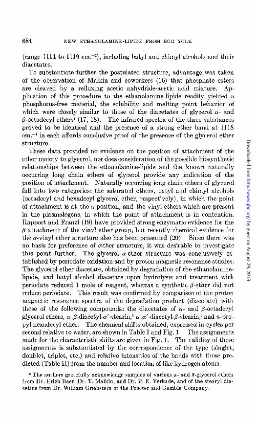
Paper Chromatography-Lipide samples (10 to 50 mg.) were hydrolyzed in 1 to 5 ml. of acid or base. The insoluble residue was extracted into chloroform and the water-soluble phase was neutralized and lyophilized. The water-soluble hydrolysis products were chromatographed on Whatman No. 540 acid-washed paper in isopropanol-acetic acid-water (3: 1: 1). In- tact lipides and some chloroform-soluble materials were chromatographed on Whatman No. 3 paper impregnated with silicic acid according to the method of Lea, Rhodes, and Stoll (13).
Reagents-Papers, after development and removal of solvent, were sprayed with ninhydrin (0.5 per cent w/v) in pyridine and heated for 5 minutes at 95” to detect substances containing free amino groups. Cho- line was detected by spraying with 2 per cent aqueous phosphomolybdic acid, followed by washing and reduction to molybdenum blue with acid stannous chloride (13). This spray can be used after the ninhydrin reagent with good results. Phosphorus was detected by spraying with the Hanes and Isherwood reagent (26), followed by exposure to bright sunlight or ultraviolet light. Bright blue spots were obtained, quite different from the greenish blue inorganic phosphate spots.
Column Chromatography-Silicic acid columns were prepared essentially as described by Lea, Rhodes, and Stoll (13) by mixing a slurry of silicic acid (Mallinckrodt’s silicic acid, reagent 100 mesh suitable for chromato- graphic analyses) in chloroform-methanol (4: 1), pouring it into a column of 2.2 cm. diameter, and washing the column, when settled, with the chloro- form-methanol mixture. Finally the column was washed with chloroform until transparent. The crude sphingolipide was added in chloroform as a thin band and eluted first with chloroform and then with mixtures of 5, 15, 35, 55, 70, and 90 per cent methanol-chloroform, at least two “hold-up” volumes of each solvent mixture being used. Generally about 350 mg. of sample were used per 40 gm. of silicic acid. However, much more material can be applied, depending on the content of the various sphingolipides present and the degree of separation required. Fractions of about 5 ml. were collected with an automatic fraction collector and appropriate frac- tions were combined and evaporated. 3 to 5 mg. portions of each solid fraction were dissolved in 10 ml. of chloroform or chloroform-methanol and 1 and 3 ml. aliquots were evaporated for the ninhydrin (1,2) and anthrone (galactose) (27) determinations, respectively. The infrared spectra of the various fractions proved helpful in determining the type of lipide structure. Typical column results are given later in the description of the isolation of the ethanolamine-lipide.
Preparation of Ethanolamine-Lipide from Crude Sphingolipide-Extrac- tion of the crude sphingolipide (20 gm.) with pyridine at room temperature removed most of the cerebroside as a soluble fraction (3.65 gm.), which gave an anthrone value of 15 per cent (cerebroside = galactose per cent X
by guest on August 29, 2018
http://ww
w.jbc.org/
Dow
nloaded from

690 NEW ETHANOLAMINE-LIPIDE FROM EGG YOLK
4.55) but was ninhydrin-negative. The pyridine-insoluble fraction was suspended in acetic acid at room temperature to remove further cerebroside as the insoluble residue (2.32 gm.). The acetic acid solution was evapora- ted to 50 ml. and acetone was added to precipitate Fraction A1 (3.45 gm.). This material still contained some cerebroside (anthrone 7 per cent), but was ninhydrin-positive. Further additions of acetone yielded three more fractions, AZ, AS, and Ad, all cerebroside-free. Fraction Az, 2.05 gm., strongly ninhydrin-positive (glycerol 2.86 per cent); Fraction AS, 1.41 gm.,
TABLE III Fractionation of Crude Ethanolamine-Lipide on Silicic Acid
395 mg. of Fraction As were chromatographed over a 35 gm. silicic acid column. The eluate was collected in 5 to 6 ml. fractions.
Tube No. Solvent’ Fraction No. Weight AminoNt
w. per cent
6-14 15-17 18-26 27-28 29-47 48-56 59-77 79-96 97-101
104-110 111-135
0 8 2.0
10.7 0.27 20
35 55 70
90
7.2 2.4
41.3 113.7 101.7 42.4 55.2
0.52 0.97 3.00 0.10 0.10 0.10
Total. . . . . --
- 376.6
AIltbIOIlet
fiel cent
3.5 0.0
1.0 3.7 0.0 0.9 0.0 0.4
* The numbers give the percentage of methanol in chloroform. t Calculated against a phosphatidylethanolamine standard. t: Calculated as galactose.
strongly ninhydrin-positive; Fraction Ad, 4.32 gm., ninhydrin-negative. Passage of Fractions AZ and A3 over silicic acid columns yielded the cere- broside-free ethanolamine-lipide. Typical column results are given in Table III.
From Fractions AZ (1.02 gm.) and A3 (1.4 gm.), 235 mg. (about 10 per cent) of the ethanolamine-lipide were obtained by column chromatography. The analyses of the combined fractions were in fair agreement with a batyl, or chimyl, alcohol phosphorylethanolamine.
CzsHroOaNP. Calculated. (batyl). C 59.10, H 10.71, N 3.00, P 6.64, glycerol 19.7
“ (chimyl). “ 57.4, “ 10.48, “ 3.19, “ 6.06, “ 20.9
by guest on August 29, 2018
http://ww
w.jbc.org/
Dow
nloaded from
H. E. CARTER, D. B. SMITH, AND D. N. JONES 691
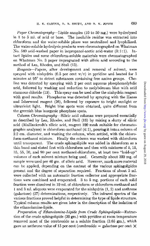
CzJLaOeNP. Found. C 57.37, H 10.08, N 3.24, P 6.24, glycerol 18.2
LCB N4 0.11; N:P = 1.15; anthrone (as galactose) 0.0
The melting behavior of this material was unusual. Slight sintering began at 140-150” and a mass of tiny liquid crystals appeared at 175”. These melted between 180-190” but almost immediately new larger crystals appeared which did not melt until 206-210”.
Hydrolysis of Ethanolamine-Lipide with Aqueous Sulfuric Acid-50 mg. of the ethanolamine-lipide were refluxed in 5 ml. of 1 N HzS04 for 40 min- utes. The aqueous phase was decanted, and the residue extracted into chloroform. The aqueous solution contained mainly ethanolamine plus a slight amount of ethanolamine phosphate. The chloroform-soluble ma- terial was chromatographed as a line on silicic acid-impregnated paper in chloroform-methanol (4: 1) and small strips were sprayed with the ninhy- drin and phosphate reagents. The main spot (RF 0.91) was strongly phosphate-positive and ninhydrin-negative. Unfortunately, unchanged starting material (R, 0.80, ninhydrin-positive) was also present and con- taminated the phosphatidic acid eluted from the paper. However, the solubility properties and general behavior are consistent with those de- scribed for synthetic batyl alcohol phosphates (28).
Hydrolysis of Ethanolamine-Lipides with Barium Hydroxide-The eth- anolamine-lipide (15 mg.) was treated with 10 ml. of saturated Ba(OH)z solution under reflux. After 6 hours the cooled reaction mixture was acidified and treated with chloroform. The chloroform extract was washed with water, dried over anhydrous NazSOI, and evaporated. The residue (10 mg.) of glycerol ether phosphate was characterized by paper chroma- tography and by an infrared spectrum. In chloroform solution, absorption maxima appeared at 2930 and 2860 cm.-i (CH,, CHZ, and CH stretch), 1730 cm.-’ (broad d an weak, acid phosphorus compound?), 1470 cm.-* (-CHz--- deformation), 1384 cm.-’ (C-CH, deformation), 1265 cm.-’ (very strong and sharp, P=O (free) stretch). There were also very broad, strong bands at 1090 cm.?, 1015 cm.-‘, 865 cm.-‘, and 810 cm.-‘, and very broad but shallow absorption in the region 2600 cm.-’ to 3400 cm.-’
No
! 1
P \
OH? . Acetolysis of Ethanolamine-Lipide-The procedure of Malkin and co-
workers (16) was slightly modified. The ethanolamine-lipide (20 mg.) was refluxed with a mixture of acetic anhydride and acetic acid (5 ml. 2:3) for 8.5 hours. The cooled reaction mixture was lyophilized and the residue shaken with a mixture of water and ether (1: 4). The ether layer was
by guest on August 29, 2018
http://ww
w.jbc.org/
Dow
nloaded from

692 NEW ETHANOLAMINE-LIPIDE FROM EGG YOLK
washed twice with small quantites of water, dried over anhydrous sodium sulfate, and evaporated to a waxy solid. The infrared and n-m-r spectra of this crude diacetate were identical with those of a-octadecyl glycerol ether diacetate (see below).
The aqueous layer and washings were’ combined and lyophilized. Paper chromatography of the residue showed the presence of ethanolamine, a little ethanolamine phosphate, and much inorganic phosphate.
Periodate Oxidation-The acetolysis product (15 mg.) was dissolved in 0.5 ml. of 1 N alcoholic KOH and the solution was allowed to stand at room temperature overnight. Water was added and the emulsion was treated twice with ether. The ether extracts were combined, washed with water, and dried. Removal of the ether and crystallization of the residue from acetone gave 10 mg. of crystalline material which melted at 66-68”. L-Q-
Octadecyl glycerol ether melts at 71-72’; the hexadecyl ether melts at 64” (18). 9.7 mg. of the hydrolysis product were dissolved in 2 ml. of absolute alcohol in a 5 ml. volumetric flask. 0.85 ml. of 0.2 M periodic acid was added and the solution was diluted to 5 ml. with absolute alcohol. The reaction mixture and a similarly prepared blank were allowed to stand in the dark at room temperature for 30 minutes. Then 1.0 ml. aliquots were removed and treated with excess arsenite solution and titrated with iodine in saturated bicarbonate in the usual way. The 1.0 ml. aliquot reduced 0.0052 mmole of periodate (theory = 0.0056 mmole). The degradation product thus reduced 0.93 mole of periodic acid per mole. In similar ti- trations cy- and p-octadecyl glycerol ethers reduced, respectively, 1.02 and 0.03 moles of periodic acid per mole.
p-Octadecyl Glycerol Ether--This compound was prepared according to the directions of Davies, Heilbron, and Jones (17). 1,3-Benzylidene glyc- erol (1.6 gm.) was refluxed in 50 ml. of benzene with 207 mg. of potassium for 1 hour. Octadecyl iodide (2.6 gm.) was added, and refluxing was con- tinued for a further 8 hours. The cooled mixture was poured into water and extracted with ether and the ether layer was washed three times with water, dried, and evaporated. The excess octadecyl alcohol was distilled off under reduced pressure. Crystallization of the residual 1,3-benzyli- dene glycerol-2-octadecyl ether from aqueous alcohol gave a waxy solid, m.p. 4244’. This product was refluxed for 70 minutes in 35 ml. of 75 per cent aqueous alcohol containing 1 ml. of concentrated HCl. The solution was diluted with water, and the precipitated solid was crystallized several times from alcohol to furnish p-octadecyl glycerol ether, m.p. 60-61”. The material showed no melting point depression when admixed with an authentic sample of p-octadecyl glycerol ether, m.p. 60-61°, kindly supplied by Dr. P. E. Verkade.
@-Octadecyl Glycerol Ether cx ,a’-Diacetate-+Octadecyl glycerol (52.5 mg.)
by guest on August 29, 2018
http://ww
w.jbc.org/
Dow
nloaded from

H. E. CARTER, D. B. SMITH, AND D. N. JONES 693
in 0.2 ml. of pyridine was heated with 1 ml. of acetic anhydride at N-85” for 1 hour. The solution was allowed to stand for 12 hours at room tem- perature and then was evaporated under reduced pressure. The residual oil was dissolved in chloroform and the solution was washed with water, dried over anhydrous sodium sulfate, and evaporated to a waxy solid (68.4 mg.). After repeated digestion with hot hexane and removal of the in- soluble residue (6.6 mg., m.p. 100°) the filtrate was evaporated to a waxy solid. The infrared spectrum was consistent with the ether diacetate structure, the following bands being present: 2920 cm.-’ and 3860 cm.-’ (CHB, CHZ, and CH stretch), 1740 cm.-’ (ester C==O), 1470 cm.-’ (-CHY- deformation), 1375 (C-CH, deformation), 1238 cm.? (C-G- stretch of acetate), and 1119 cm.-l (-CH-0-CH-). There was an additional very strong band at 1050 cm.-‘. Analysis of P-octadecyl glycerol ether a! ,a’-diacetate
GJLsOa. Calculated, C 70.0, H 11.2; found, C 69.4, H 11.2
wOctadecy1 Glycerol Ether a ,p-Diacetate-ar-Octadecyl glycerol ether (batyl alcohol) (59.5 mg.) was acetylated in the above manner to furnish a waxy solid (63.7 mg.). Repeated crystallization from alcohol furnished soft crystals, m.p. 34-35” (Baer and Fischer (18) report m.p. 34-35”). The infrared absorption spectrum in chloroform solution displayed maxima at 2935 and 2870 cm.-’ (CHB, CHZ, and CH stretch), 1739 cm.-’ (ester C=O), 1465 cm.-* (-CH2- deformation), 1375 cm.-’ (C-CHI deformation), 1230 cm.-1 (C-O- stretch of acetate), and at 1116 cm.-l (-CHz-O-CHZ). Additional bands were observed at 1048 cm.-’ (strong) and 960 cm.-r (weak). There was no hydroxyl absorption at approximately 3500 cm.-l.
Proton Magnetic Resonance Spectra-The proton magnetic resonance spectra were determined at 40 mc. rf. with a Varian model V4300B high resolution spectrometer fitted with a field-sensing stabilizer (Super Stabil- izer) . The compounds were measured as about 25 per cent solutions in chloroform or deuterochloroform. The chemical shifts expressed relative to water were actually determined relative to methylene chloride with a concentric tube cell (29) with methylene chloride in the outer compartment. Subtraction of 26 c.p.s. from shifts relative to methylene chloride was used in determining shifts in cycles per second relative to water. For the sake of clarity the spectra illustrated were run in the absence of methylene chloride. The actual shifts were measured on separate spectra in which the methylene chloride band was included.
SUMMARY
Alkaline hydrolysis of egg yolk phosphatides gives a crude sphingolipide fraction containing a ninhydrin-positive substance (ethanolamine-lipide).
by guest on August 29, 2018
http://ww
w.jbc.org/
Dow
nloaded from

694 NEW ETHANOLAMINE-LIPIDE FROM EGG YOLK
The purified ethanolamine-lipide contains no sphingosine and has been shown to consist mainly of a phosphorylethanolamine derivative of batyl alcohol. The presence of similar glycerol ether phosphatides in other lipides is indicated by analytical data. Studies of the distribution of ether phosphatides are now in progress.
BIBLIOGRAPHY
1. Lea, C. H., and Rhodes, D. N., &o&em. J., 66, 613 (1954). 2. Lea, C. H., and Rhodes, D. N., Biochim. et biophys. ucta, 17, 416 (1955). 3. Rhodes, D. N., and Lea, C. H., Biochem. J., 66, 526 (1957). 4. Lea, C. H., and Hawke, J. C., Biochem. J., 62, 105 (1952). 5. Hawke, J. C., and Lea, C. H., Biochem. J., 64, 475, 479 (1953). 6. Schmidt, G., Benotti, J., Hershman, B., and Thannhauser, S. J., J. Biol. Chem.,
166, 505 (1946). 7. Dawson, R. M. C., Biochem. J., 66, 621 (1954). 8. Brante, G., Acta physiol. &and., 18, Suppl. 63 (1949). 9. Edgar, G. W. F., Monatsschr. Psych&. U. Neural., 131, 274 (1956).
10. Edgar, G. W. F., Actu an&., 27, 240 (1956). 11. Weiss, B., J. Biol. Chem., 233, 523 (1956). 12. Mallov, S., McKibbin, J. M., and Robb, J. S., J. Biol. Chem., 201, 825 (1953). 13. Lea, C. H., Rhodes, D. N., and Stoll, R. D., Biochem. J., 60,353 (1955). 14. McKibbin, J. M., and Taylor, W. E., J. BioZ. Chem., 178, 29 (1949). 15. Rapport, M. M., Lerner, B., Alonzo, N., and Franal, R. E., J. BioZ. Chem., 226,
859 (1957). 16. Bevan, T. H., Brown, D. A., Gregory, G. I., and Malkin, T., J. Chem. Sot., 127
(1953). 17. Davies, W. H., Heilbron, I. M., and Jones, W. E., J. Chem. Sot., 1232 (1934). 18. Baer, E., and Fischer, H. 0. L., J. BioZ. Chem., 140, 397 (1941). 19. Rapport, M. M., and Franzl, R. E., J. BioZ. Chem., 226, 851 (1957). 20. Marinetti, G. V., and Erbland, J., Biochim. et biophys. acta, 26. 429 (1957). 21. Schmidt, G., Bessman, M. J., and Thannhauser, S. J., J. BioZ. Chem., 203, 849
(1953). 22. Karnovsky, M. L., and Brumm, A. F., J. BioZ. Chem., 216, 689 (1955). 23. Karnovsky, M. L., Jeffrey, S. S., Thompson, M. S., and Deane, H. W., J. Biophys.
and Biochem. Cytol., 1, 173 (1955). 24. Karnovsky, M. L., BioZ. Bull., 107, 238 (1951). 25. Bergstrijm, S., and Blomstrand, R., Actu physiol. &and., 38, 166 (1956). 26. Hanes, C. S., and Isherwood, F. A., Nature, 164, 1107 (1949). 27. Radin, N. S., Lavin, F. B., and Brown, J. R., J. BioZ. Chem., 217, 789 (1955). 28. Stegerhoek, L. J., and Verkade, P. E., Rec. &au. chim. Pays-Bus, 76, 467 (1956). 29. Zimmerman, J. R., and Foster, M. R., J. Phys. Chem., 61, 282 (1957).
by guest on August 29, 2018
http://ww
w.jbc.org/
Dow
nloaded from

JonesHerbert E. Carter, Donald B. Smith and D. N.
LIPIDE FROM EGG YOLKETHANOLAMINE-CONTAINING
A NEW
1958, 232:681-694.J. Biol. Chem.
http://www.jbc.org/content/232/2/681.citation
Access the most updated version of this article at
Alerts:
When a correction for this article is posted•
When this article is cited•
alerts to choose from all of JBC's e-mailClick here
tml#ref-list-1
http://www.jbc.org/content/232/2/681.citation.full.haccessed free atThis article cites 0 references, 0 of which can be by guest on A
ugust 29, 2018http://w
ww
.jbc.org/D
ownloaded from