Embed Size (px)
Citation preview

In: Aneuploidy: Etiology, Disorders and Risk Factors ISBN: 978-1-62100-070-9
Editors: Salvatore de Rossi and Filippo Bianchi ©2012 Nova Science Publishers, Inc.
Chapter III
The Spindle Assembly
Checkpoint and Aneuploidy
Juliana Faria1, Joana Barbosa
1, Inês M. B. Moura
1,
Rui M. Reis2,3
and Hassan Bousbaa*,1,4
1Centro de Investigação em Ciências da Saúde (CICS), Instituto Superior de Ciências da
Saúde – Norte, CESPU, Gandra PRD, Portugal 2Life and Health Sciences Research Institute (ICVS), Health Sciences School,
University of Minho, Braga, Portugal 3Molecular Oncology Research Center, Barretos Cancer Hospital,
Barretos, São Paulo, Brazil 4Centro de Química Medicinal da Universidade do Porto (CEQUIMED-UP),
Porto, Portugal
Abstract
Abnormal chromosome number, or aneuploidy, is commonly observed in most solid
tumors, and results from mis-segregation of whole chromosomes in a phenomenon
referred to as chromosome instability (CIN). Dysregulation of the spindle assembly
checkpoint (SAC) is thought as one of the mechanisms underlying CIN. The SAC is a
signaling pathway that prevents precocious chromosome segregation until all
chromosomes of a dividing cell are aligned at the metaphase plate. While complete loss of
the SAC activity is lethal due to massive mis-segregation, partial loss of the SAC is a
common feature of many aneuploid tumor cells allowing them to gain or lose a small
number of chromosomes. We review our current knowledge on the molecular
mechanisms of SAC and discuss its contribution to CIN as well as its potential as a
suitable target in cancer therapy.
* Corresponding author: Prof. Hassan Bousbaa, Centro de Investigação em Ciências da Saúde (CICS), Instituto
Superior de Ciências da Saúde - Norte, CESPU, Rua Central de Gandra, 1317, 4585-116 Gandra PRD,
Portugal. Phone: +351 – 224157186. Fax: +351 – 224157102, [email protected].
The exclusive license for this PDF is limited to personal website use only. No part of this digital document may be reproduced, stored in a retrieval system or transmitted commercially in any form or by any means. The publisher has taken reasonable care in the preparation of this digital document, but makes no expressed or implied warranty of any kind and assumes no responsibility for any errors or omissions. No liability is assumed for incidental or consequential damages in connection with or arising out of information contained herein. This digital document is sold with the clear understanding that the publisher is not engaged in rendering legal, medical or any other professional services.

Juliana Faria, Joana Barbosa, Inês M. B. Moura et al. 60
Introduction
The cell cycle is a ubiquitous and complex process that is required for cell growth,
proliferation, genetic material transmission and tissue regeneration (Schafer, 1998). It consists
of temporally coordinated events that ensure proper embryogenesis and subsequent cell
differentiation. Four checkpoint mechanisms are responsible for its tight control: DNA
damage checkpoints at G1/S, S and G2/M, and the spindle assembly checkpoint (SAC)
during mitosis (Tyson and Novak, 2008). These checkpoint mechanisms consist of complex
signaling cascades that only allow cells to progress through the cell cycle if their requirements
are met (Rieder, 2011; Tyson and Novak, 2008).
The spindle assembly checkpoint (SAC) is a surveillance mechanism that is constitutively
expressed during the transition from prometaphase to metaphase in eukaryotic dividing cells
(Kops et al., 2005). It detects improper kinetochore-microtubule attachments, imposing a
mitotic delay by preventing anaphase onset, in order to allow cells to correct them. This ‗wait
anaphase‘ mechanism is sustained until all chromosomes are correctly connected to the
microtubule network, bi-oriented and aligned at the metaphase plate (Logarinho and Bousbaa,
2008; May and Hardwick, 2006; Rieder et al., 1994; Zich and Hardwick, 2010). Therefore,
the SAC activity accounts for equal chromosome segregation to cell progeny and, hence, for
an effective reduction in mitotic error rates. Not surprisingly, weakened SAC activity has
been reported in many aneuploid tumors (Bannon and Mc Gee, 2009; Chi and Jeang, 2007;
Dalton and Yang, 2009; Suijkerbuijk and Kops, 2008). Given its importance in genomic
stability and cancer prevention, we will focus on the molecular mechanism of SAC activity,
its relation to cancer, and its use in current anti-cancer strategies.
The Molecular Mechanism of SAC
In order to be accurately segregated at the onset of anaphase, chromosomes must attach,
through their sister kinetochores, to the microtubules emanating from the opposite poles of
the mitotic spindle. This bi-orientation ensures their alignment at the metaphase equator so
that each chromatid is transported toward the corresponding pole to be delivered to the future
daughter cell. However, attachment of chromosomes to microtubules is a stochastic and
asynchronous event and, upon nuclear envelope breakdown at prometaphase, many
chromosomes experience improper attachments before successful bi-orientation. Such mis-
attachments include monotelic attachment (with one kinetochore of a chromosome attached to
microtubules from one pole and its sister unattached), syntelic attachment (with two sister
kinetochores attached to microtubules from the same pole), and merotelic attachment (with a
sister kinetochore attached to microtubules from both poles). These erroneous attachments, if
left undetected and uncorrected, can lead to chromosome mis-segregation and genomic
instability. Fortunately, they are detected by the SAC, which delays anaphase onset until all
mis-attachments are corrected. Next, we will address the mechanism by which the SAC halts
mitosis to prevent precocious sister chromatid separation. The components involved in the

The Spindle Assembly Checkpoint and Aneuploidy 61
SAC molecular pathway have been reviewed elsewhere (Cheeseman and Desai, 2008;
Musacchio and Salmon, 2007).
Sister chromatids are held together at the centromere by a ring-like structure consisting of
a complex of cohesin proteins synthesized in S phase (Marangos and Carroll, 2008;
Suijkerbuijk and Kops, 2008; Zhou et al., 2002). Their separation at the onset of anaphase
requires degradation of one of cohesin subunits, Scc1, which is promoted by the proteolytic
activity of separase (Bannon and Mc Gee, 2009; Bolanos-Garcia and Blundell, 2010;
Nasmyth, 2005; Przewloka and Glover, 2009). This caspase-like protein is normally kept
inactive by Securin. When all chromosomes are aligned at the metaphase plate with correct
bipolar attachment to spindle microtubules, Securin becomes ubiquitinated by the anaphase
promoting complex/cyclosome (APC/C), a multi-subunit E3 ubiquitin ligase (Logarinho and
Bousbaa, 2008; Morgan, 1999; Reddy et al., 2007; Stegmeier et al., 2007). Ubiquitination
targets securin for degradation by the 26S proteasome (Decordier et al., 2008; Suijkerbuijk
and Kops, 2008). Separase is then activated and cleaves Scc1, no longer holding sister
chromatids together and, thus, anaphase begins (Bannon and Mc Gee, 2009; Bharadwaj and
Yu, 2004; Bolanos-Garcia and Blundell, 2010; Morgan, 1999; Nasmyth, 2005). Degradation
of cyclin B is also accomplished through APC/C-mediated ubiquitination, leading to the
inactivation of cyclin-dependent kinase 1 (Cdk1) and subsequent mitotic exit (Logarinho and
Bousbaa, 2008; May and Hardwick, 2006; Schmidt and Medema, 2006; Suijkerbuijk and
Kops, 2008).
The main downstream target of the spindle assembly checkpoint is Cdc20, a protein
required for APC/C activation. Once the nuclear envelope is broken down, SAC proteins,
namely Mad2, Bub3, and BubR1, are recruited to the outer kinetochore surface of all
unattached chromosomes. These proteins use the kinetochore as a platform to generate, in
near equal stoichiometry, the so called mitotic checkpoint complex (MCC) that diffuses
through the cytosol to prevent Cdc20 from activating the APC/C (Figure 1) (Sudakin et al.,
2001). This diffusible inhibitory signal is generated as long as unattached or mis-attached
kinetochores are present, hence preventing anaphase onset until all chromosomes achieve
correct attachment to bipolar spindle and align at the metaphase equator (Bharadwaj and Yu,
2004; Kops et al., 2005; May and Hardwick, 2006; Zich and Hardwick, 2010). Additionally,
according to the Mad2-template model, a closed conformation of Mad2 (C-Mad2) in complex
with Mad1 resides at unattached kinetochores and serves as receptor to convert cytosolic open
conformation of Mad2 (O-Mad2) into C-Mad2 bound to Cdc20. This latter leaves the
kinetochore and promotes inhibitory signal amplification by converting more O-Mad2 into C-
Mad2 in the cytosol (De Antoni et al., 2005). The C-Mad2 form is a more potent inhibitor of
APC/C in vitro given its higher affinity for Cdc20 (Chan et al., 2005; Musacchio and Salmon,
2007; Suijkerbuijk and Kops, 2008).
The nature of the signal that triggers SAC response is still controversial. It is more likely
to be the result of a redundant combination of both absence of kinetochore-microtubule
attachment and of the lack of physical tension between sister kinetochores (Bharadwaj and
Yu, 2004; May and Hardwick, 2006; Pinsky and Biggins, 2005).
When all chromosomes undergo bipolar attachments to spindle microtubules, tension
between sister kinetochores promotes SAC silencing and anaphase onset (Schmidt and
Medema, 2006; Zhou et al., 2002). Anaphase inhibitory complexes are then disassembled and

Juliana Faria, Joana Barbosa, Inês M. B. Moura et al. 62
SAC proteins withdrawn from the kinetochores, both through free diffusion into the cytosol
and through motor protein-mediated transport along microtubules to the spindle poles (Lu et
al., 2009). Mad1 and Mad2 become undetectable at the kinetochores, while Bub1 and BubR1
levels are diminished three to four-fold (Chan and Yen, 2003; Zhou et al., 2002).
When cells are not capable of satisfying the SAC after a long mitotic arrest, they may
have different fates: some undergo apoptotic death during mitosis, others exit mitosis but die
via apoptosis in G1 phase, and others exit mitosis but are tetraploid and reproductively dead
(Niikura et al., 2007; Suijkerbuijk and Kops, 2008). In this context, Bub1 and BubR1 have
been shown to play an important role in eliminating cells that adapt to prolonged mitosis and
undergo defective mitotic events (Suijkerbuijk and Kops, 2008).
Figure 1. Molecular basis of spindle assembly checkpoint. Unattached kinetochore activates the SAC
(Checkpoint On) by recruiting the Mad2, BubR1, and Bub3. These proteins form the Mitotic
Checkpoint Complex (MCC), the diffusible inhibitory signal that sequesters Cdc20, keeping the APC/C
inactive thereby preventing it from targeting Securin and Cyclin B for degradation. As a consequence,
sister-chromatid cohesion is maintained and the cell cycle is arrested. The MCC disassembles
(Checkpoint Off) once all kinetochores become properly attached and aligned. Cdc20 is the free to
activate the APC/C which results in Securin and Cyclin B ubiquitination (U) and degradation. Securin
degradation leads to the activation of the protease Separase, which cleaves cohesin, leading to sister-
chromatid separation. Cyclin B degradation decreases the cyclin-dependent kinase (Cdk) 1 activity,
which results in mitotic exit.
The Spindle Assembly Checkpoint and Aneuploidy 63
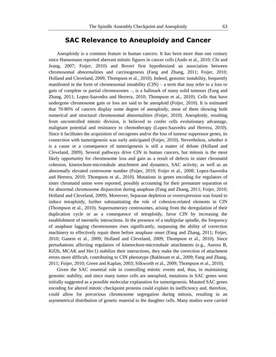
SAC Relevance to Aneuploidy and Cancer
Aneuploidy is a common feature in human cancers. It has been more than one century
since Hansemann reported aberrant mitotic figures in cancer cells (Ando et al., 2010; Chi and
Jeang, 2007; Foijer, 2010) and Boveri first hypothesized an association between
chromosomal abnormalities and carcinogenesis (Fang and Zhang, 2011; Foijer, 2010;
Holland and Cleveland, 2009; Thompson et al., 2010). Indeed, genomic instability, frequently
manifested in the form of chromosomal instability (CIN) – a term that may refer to a loss or
gain of complete or partial chromosomes –, is a hallmark of many solid tumours (Fang and
Zhang, 2011; Lopez-Saavedra and Herrera, 2010; Thompson et al., 2010). Cells that have
undergone chromosome gain or loss are said to be aneuploid (Foijer, 2010). It is estimated
that 70-80% of cancers display some degree of aneuploidy, most of them showing both
numerical and structural chromosomal abnormalities (Foijer, 2010). Aneuploidy, resulting
from uncontrolled mitotic division, is believed to confer cells evolutionary advantage,
malignant potential and resistance to chemotherapy (Lopez-Saavedra and Herrera, 2010).
Since it facilitates the acquisition of oncogenes and/or the loss of tumour suppressor genes, its
connection with tumorigenesis was early anticipated (Foijer, 2010). Nevertheless, whether it
is a cause or a consequence of tumorigenesis is still a matter of debate (Holland and
Cleveland, 2009). Several pathways drive CIN in human cancers, but mitosis is the most
likely opportunity for chromosome loss and gain as a result of defects in sister chromatid
cohesion, kinetochore-microtubule attachment and dynamics, SAC activity, as well as an
abnormally elevated centrosome number (Foijer, 2010; Foijer et al., 2008; Lopez-Saavedra
and Herrera, 2010; Thompson et al., 2010). Mutations in genes encoding for regulators of
sister chromatid union were reported, possibly accounting for their premature separation or
for abnormal chromosome disjunction during anaphase (Fang and Zhang, 2011; Foijer, 2010;
Holland and Cleveland, 2009). Moreover, Separase depletion or overexpression was found to
induce tetraploidy, further substantiating the role of cohesion-related elements in CIN
(Thompson et al., 2010). Supernumerary centrosomes, arising from the deregulation of their
duplication cycle or as a consequence of tetraploidy, favor CIN by increasing the
establishment of merotelic interactions. In the presence of a multipolar spindle, the frequency
of anaphase lagging chromosomes rises significantly, surpassing the ability of correction
machinery to effectively repair them before anaphase onset (Fang and Zhang, 2011; Foijer,
2010; Ganem et al., 2009; Holland and Cleveland, 2009; Thompson et al., 2010). Since
perturbations affecting regulators of kinetochore-microtubule attachments (e.g., Aurora B,
Kif2b, MCAK and Hec1) stabilize their interactions, they make the correction of attachment
errors more difficult, contributing to CIN phenotype (Bakhoum et al., 2009; Fang and Zhang,
2011; Foijer, 2010; Green and Kaplan, 2003; Silkworth et al., 2009; Thompson et al., 2010).
Given the SAC essential role in controlling mitotic events and, thus, in maintaining
genomic stability, and since many tumor cells are aneuploid, mutations in SAC genes were
initially suggested as a possible molecular explanation for tumorigenesis. Mutated SAC genes
encoding for altered mitotic checkpoint proteins could explain its inefficiency and, therefore,
could allow for precocious chromosome segregation during mitosis, resulting in an
asymmetrical distribution of genetic material to the daughter cells. Many studies were carried

Juliana Faria, Joana Barbosa, Inês M. B. Moura et al. 64
out in order to establish a causal connection between mutations in genes encoding for SAC
proteins and tumor development (Marchetti and Venkatachalam, 2010).
One of the most illustrative results is the one that links biallelic mutations in the SAC
BubR1-encoding gene, Bub1B, with mosaic variegated aneuploidy (MVA) (Chi and Jeang,
2007; Hanks et al., 2004; Lopez-Saavedra and Herrera, 2010; Rio Frio et al., 2010;
Suijkerbuijk and Kops, 2008; Suijkerbuijk et al., 2010; Thompson et al., 2010). MVA is a
rare autosomal recessive disease that is characterized by a high degree of aneuploidy, mild to
severe physical and mental limitations and a strong predisposition to cancer (Hanks et al.,
2004; Suijkerbuijk and Kops, 2008; Suijkerbuijk et al., 2010; Thompson et al., 2010; Yen and
Kao, 2005). Other studies have identified heterozygous mutations in Bub1 and BubR1 in a
panel of 19 aneuploid colorectal cancer cell lines (Cahill et al., 1999). Mutations have also
been found in Mad2-encoding gene, both in breast cancer (Percy et al., 2000) and gastric
cancer (Kim et al., 2005) cell lines.
However, many other attempts surprisingly failed to find SAC mutations, both in human
cancer cell lines and in tissue samples from oncologic patients (Yen and Kao, 2005). For
instance, none or few mutations were detected in Bub3, BubR1 and Bub1 genes in
glioblastoma, breast, lung, bladder and thyroid cancers (Fagin, 2002; Haruki et al., 2001;
Myrie et al., 2000; Olesen et al., 2001; Ouyang et al., 2002; Reis et al., 2001; Sato et al.,
2000). Bub1 gene mutations were shown to be a rare event in a study using 92 acute myeloid
leukemia specimens and 5 hematopoietic cell lines (Lin et al., 2002), in head and neck
squamous cell carcinoma and lung cell lines (Yamaguchi et al., 1999), in a series of
colorectal, hepatocellular and renal tumors (Shichiri et al., 2002) and in breast and gastric
carcinomas (Langerod et al., 2003; Shigeishi et al., 2001). Also, Mad1 gene was found to
carry few or no mutations in lymphomas, bladder, breast, gliomas (Tsukasaki et al., 2001) and
lung carcinomas (Nomoto et al., 1999), as does Mad2 gene in transitional-cell carcinomas of
the bladder, soft-tissue sarcomas, hepatocellular carcinomas (Hernando et al., 2001), breast,
lung (Gemma et al., 2001; Percy et al., 2000; Takahashi et al., 1999) and digestive tract (Imai
et al., 1999) cancer cells. In addition, no mutations were found in the coding sequences of
Mad2 gene in 11 hepatoma cell lines (Sze et al., 2004). In a study of 8 hepatocellular
carcinoma cell lines and 50 hepatocellular carcinoma specimens, although some polymorphic
base changes were noticed in Bub1, BubR1 and Cdc20, no mutations accountable for SAC
impairment were detected neither in these genes nor in Bub3 or Mad2B (Saeki et al., 2002). It
should be noted that these studies did not cover all possible known SAC genes, meaning that
some of their mutations might be yet to unveil. Furthermore, in the vast majority of cases, the
effect of the gene mutation was not studied at the protein level. Further quantitative and
subcellular localization assays are thus needed to clarify the actual impact of these mutations
(Yen and Kao, 2005). Nevertheless, the low frequency of mutations affecting SAC genes
indicates that they are not the main mechanism through which cells may become aneuploid
(Lopez-Saavedra and Herrera, 2010; Schvartzman et al., 2010; Thompson et al., 2010).
Subsequently, studies concerning expression of SAC components, both at the gene and
protein level, have gained attention, suggesting a correlation between altered expression
levels, compromised SAC activities and tumorigenesis (Chi and Jeang, 2007; Fang and
Zhang, 2011; Holland and Cleveland, 2009; Kops et al., 2005; Suijkerbuijk and Kops, 2008).
The SAC efficiency can be easily evaluated as the ability of a given cell population to sustain

The Spindle Assembly Checkpoint and Aneuploidy 65
a prolonged mitotic arrest upon exposure to chemical compounds that interfere with
microtubule polymerization and dynamics. Such evaluation has been performed in large
panels of tumor cell lines, as well as in histological samples collected from numerous
patients. SAC impairment was implicated in the resistance to anti-microtubule agents-induced
apoptosis in human lung cancers (Masuda et al., 2003), as well as in breast cancer (Yoon et
al., 2002) and in head and neck squamous cell lines, in which it may contribute to
chromosomal instability (Minhas et al., 2003). Most studies have sought for a molecular
explanation for the SAC weakening. Overexpression of SAC components seems to be more
frequent (Foijer, 2010; Holland and Cleveland, 2009; Liu et al., 2009). Even subtle deviations
in SAC mRNA and protein levels were shown to drive tumorigenesis (Bharadwaj and Yu,
2004). Low BubR1 levels, resulting from biallelic mutations in the Bub1B gene, were
associated with chromosome alignment and segregation defects (Suijkerbuijk et al., 2010); a
significantly reduced Bub1B expression was ascertained as the causative event of aneuploidy
in colorectal adenocarcinomas (Burum-Auensen et al., 2008). On the other hand, Bub1B
expression was shown to be high in 25.9% of 27 salivary duct carcinomas, although it had no
prognostic significance (Ko et al., 2010). Additionally, BubR1 overexpression was
documented in oesophageal squamous cell (Tanaka et al., 2008), thyroid (Wada et al., 2008),
hepatocellular (Liu et al., 2009) and squamous cell carcinomas (Hsieh et al., 2010), as well as
in lung cancers (Seike et al., 2002), where it has been associated with worse prognosis,
carcinogenesis progression and suggested as a possible compensatory mechanism that could
represent a potential tumor biomarker (Hsieh et al., 2010). BubR1 overexpression was
reported in 50.3% of 181 gastric cancer samples, correlating significantly with aneuploidy,
tumor invasiveness, metastasis likelihood and poor prognosis (Ando et al., 2010), and in 68%
of 43 gastric carcinomas (Grabsch et al., 2003). BubR1 overexpression was found to be
closely related to chromosomal instability in bladder cancer (Yamamoto et al., 2007) and in
clear cell kidney carcinomas (Pinto et al., 2008). Although some studies point to diminished
expression and aberrant transcription of the hBub1 gene (Lin et al., 2002), its expression was
found to be up-regulated in follicular thyroid adenomas when compared to adjacent normal
tissues (Wada et al., 2008). Along with that of Bub1B, Bub1 overexpression was also found
in a large panel of breast tumor samples and proposed to be implied in the transition of breast
tissues from normal to benign tumors (Bieche et al., 2011). Bub1 overexpression was also
demonstrated, both at mRNA and protein levels, in salivary gland tumors, where it
contributes to abnormal cell proliferation (Shigeishi et al., 2006). Gastric cancers also
overexpress Bub1 in 84% of 43 samples under analysis (Grabsch et al., 2004). Similar
alterations were detected in Mad2 expression. Mad2l1 gene up-regulation was reported in
Familial Adenomatous Polyposis colorectal adenomas, suggesting that this up-regulation is
associated with adenomatous polyposis coli (APC) gene mutation and may constitute an early
event in colorectal carcinogenesis (Abal et al., 2007). Pronounced Mad2 overexpression has
also been documented in advanced differentiated thyroid carcinomas (Wada et al., 2008),
salivary duct carcinomas (Ko et al., 2010) and lung cancers, in which it has been correlated
with enhanced aggressiveness, shorter survival and identified as a prognostic factor (Kato et
al., 2011). Up-regulated Mad2l2 expression, both at gene and protein levels, was found in
21% of 118 colorectal tumor samples, in which it has been suggested to promote mitotic
aberrancies, chromosomal instability and reduced patient survival (Rimkus et al., 2007). Also,

Juliana Faria, Joana Barbosa, Inês M. B. Moura et al. 66
Mad2 protein overexpression, concomitant with that of Aurora A and Aurora B, was
observed in aneuploid colorectal adenocarcinomas (Burum-Auensen et al., 2008), as well as
in a series of 6 oesophageal squamous cell carcinoma (ESCC) cell lines and 21 ESCC
patients, along with that of BubR1 (Tanaka et al., 2008). Inversely, Mad2 protein levels were
found to be lower in colon cancer cell lines that had been exposed to deoxycholate, a
hydrophobic bile acid associated with cancer risk (Payne et al., 2010). Mad2 decreased levels
were suggested to contribute to an escape from cell death, thus leading to tumorigenesis
(Payne et al., 2010). Mad2 protein was also shown to be underexpressed in 75% of 8
testicular germ cell tumour (Fung et al., 2007) and in 54.5% of 11 aneuploid hepatoma tumor
cell lines that failed to arrest in mitosis (Sze et al., 2004).
Mouse Models Link SAC Dysfunction,
Aneuploidy and Cancer
To further investigate the role of the SAC components in checkpoint signaling and in the
prevention of chromosomal imbalance, mouse models lacking SAC genes were created
(Foijer, 2010; Foijer et al., 2008; Li et al., 2009; Schvartzman et al., 2010; Suijkerbuijk and
Kops, 2008; Thompson et al., 2010; Yen and Kao, 2005). Conventional gene knockouts have
been constructed for almost all SAC known genes, including Mad1, Mad2, Bub1, BubR1,
Bub3 and CENP-E, and hypomorphic alleles for Bub1 and BubR1 have been generated
(Holland and Cleveland, 2009). In spite of being compatible with viability and fertility, their
downregulation increased cancer susceptibility and caused aneuploidy in mouse embryonic
fibroblasts and tissues, in a degree that was dependent on the knocked-out gene and on the
extension to which its expression had been decreased (Holland and Cleveland, 2009; Lopez-
Saavedra and Herrera, 2010). Mouse embryonic cells lacking Mad2 displayed a dysfunctional
SAC, leading to chromosome missegregation and apoptosis (Dobles et al., 2000). BubR1 (+/-
) mouse embryonic fibroblasts were proven to defectively activate SAC and to have reduced
amounts of Securin and Cdc20, predisposing mice to rapid development of lung and intestinal
adenocarcinomas and supporting BubR1 role as a tumor suppressor (Dai et al., 2004). In turn,
although there was not a higher susceptibility to tumor formation, perhaps because of the
presence of a partially functional SAC, increased aneuploidy and premature sister chromatid
segregation were reported in Bub3-haploinsufficient mouse embryonic fibroblasts (Kalitsis et
al., 2005). Analysis of mutant mice has shown Bub1 to be essential in preventing malignant
cell transformation and in mediating cell death upon chromosome missegregation (Jeganathan
et al., 2007).
SAC Components and Perspectives
in Anticancer Therapy
The developments in the knowledge in cell cycle regulation and control have allowed the
conception of several pharmacological approaches aiming at stopping tumor cell

The Spindle Assembly Checkpoint and Aneuploidy 67
proliferation. Abnormal cell proliferation is frequently associated with altered expression
levels of cell cycle-regulating proteins or with alterations in checkpoint mechanisms. These
alterations constitute an advantage not only for tumor cell progression, but also for the
acquisition of increasingly aggressive phenotypes (De Falco and De Luca, 2010).
Given the role of microtubules and SAC proteins on chromosome segregation and cell
division accuracy, these have become the main targets of anti-cancer treatment strategies.
Current chemotherapy approaches use microtubule-targeting agents (MTAs). MTAs affect
the dynamic equilibrium between microtubule polymers and tubulin heterodimers, and have
been used with a considerable degree of success in a wide range of tumors (Bannon and Mc
Gee, 2009; Fojo and Menefee, 2007; Zhou and Giannakakou, 2005). Vinca alkaloids and
taxanes, like paclitaxel and docetaxel, are amongst the most used drugs in cancer treatment.
The first have an inhibitory action in microtubule polymerization, while the latter stabilize
microtubules. Their effects are more pronounced in mitotic cells, which explains their
classification as anti-mitotic drugs (Yamada and Rao, 2010).
Vinca alkaloids were extracted from Vinca rosea (also known as Catharanthus roseus),
and promote microtubule depolymerization, which causes a prolonged mitotic arrest and
subsequently results in cell death. They are used in solid and hematological tumor treatment
(Jordan and Wilson, 2004; Perez, 2009). Taxanes paclitaxel and docetaxel were isolated from
Taxus brevifolia and Taxus baccata, respectively. They stabilize microtubule polymerization
by interfering with their dynamics, which blocks cell cycle at G2/M phase, leading to cell
death. Taxanes are highly efficient, for instance, in lung, breast and head and neck squamous
carcinomas. MTAs are used either alone or combined with other cytotoxic agents (Perez,
2009). They act by interfering with mitotic spindle dynamics, thereby inducing SAC
activation and mitotic arrest (Zhou and Giannakakou, 2005). However, even though mitotic
arrest avoids cell proliferation, it does not necessarily end in cell death (Riffell et al., 2009).
Some cells are capable of escaping mitosis in the absence of chromosomal segregation or cell
division, in a process termed SAC adaptation or mitotic slippage. These cells exhibit multiple
nuclei and polyploidy, both potential tumorigenesis stages. Accordingly, the fates of mitosis-
arrested cells are still unknown (Riffell et al., 2009). In this respect, small-molecule inhibitors
of the APC/C produce a more efficient retention in mitosis than MTAs do, since proteolysis is
APC/C-dependent and required for mitotic slippage. For this reason, APC inhibitors may
constitute a more powerful strategy to induce and sustain mitotic arrest (Zeng et al., 2010).
Since many tumor cells present defective SAC activity, they sometimes do not respond to
mitotic errors, which affects MTAs efficacy (Bolanos-Garcia, 2009). Hence, the development
and optimization of new therapeutic strategies that target SAC proteins may contribute to a
more successful treatment. While a defective SAC may contribute to aneuploidy and CIN
(Figure 2), a more expressive suppression of its activity leads to cell death as a result of
massive chromosome mis-segregation. In this regard, from all protein kinases validated for
this purpose, those that act in the SAC, in particular Bub1, BubR1 and Mps1, constitute main
drug targets (Bolanos-Garcia, 2009). Therapeutic approaches could thus make use of the
specific targeting of SAC-defective tumor cells.
RNAi-mediated Mps1 depletion has allowed for a decrease in tumor cell viability
(Janssen et al., 2009; Janssen et al., 2011). Mps1 is a crucial SAC component that monitors
chromosome alignment; as such, it influences the stability of kinetochore-microtubule

Juliana Faria, Joana Barbosa, Inês M. B. Moura et al. 68
interactions (Colombo and Moll, 2010). Its inactivation compromises the SAC, originating
alignment errors and decreasing cell viability. In vitro assays have unveiled an orally
bioavailable Mps1 small-molecule inhibitor that selectively reduces tumor cell progression
(Colombo et al., 2010). When compared to cancer cells, normal cells were significantly less
sensitive to Mps1 inhibition, whether through siRNA or by treatment with specific small-
molecule-based inhibitors. If proven to occur in vivo, this difference could illustrate the
potential of SAC selective inhibitors as a therapeutic approach (Colombo and Moll, 2010).
These results corroborate that Mps1 may constitute a suitable target to preferentially eliminate
cancer cells (Colombo et al., 2010; Janssen et al., 2009; Janssen et al., 2011).
Inhibitors that specifically interfere with Aurora kinase activity significantly decrease
viability of cells in rapid division, resulting in a 98% reduction in tumor volume in nude mice
injected with human leukemia cells (Bolanos-Garcia, 2009). In fact, several studies have
demonstrated that cells with abnormal Aurora A or Aurora B expression have mitotic spindle
defects and do not undergo cytokinesis. For that reason, interfering with Aurora kinase
activity has been suggested as a possible strategy for cancer treatment (De Falco and De
Luca, 2010; Deep and Agarwal, 2008). Diverse small-molecule Aurora kinase inhibitors are
under development in clinical trials; pan-Aurora kinase inhibitor Danusertib (PHA-739358)
was the first to be tested in humans (Colombo and Moll, 2010).
Apoptotic cell death was observed when BubR1 or Mad2 protein levels were decreased
or when BubR1 kinase activity was blocked in human cancer cells. Unless when cytokinesis
was also inhibited, apoptotic cell death took place within six cell divisions (Kops et al., 2004;
Michel et al., 2004). This can also be explored in order to inhibit tumor cell proliferation
(Kops et al., 2004; Michel et al., 2004).
Figure 2. Contribution of SAC impairment to aneuploidy. A fully functional SAC ensures accurate
chromosome segregation and genomic stability. In cells with weakened SAC, however, occasional mis-
segregations may escape SAC control and lead to aneuploidy.

The Spindle Assembly Checkpoint and Aneuploidy 69
A possible strategy consists in Mad2 silencing. siRNA nanoparticle-based strategies were
already tested, leading to an unequal division of genetic material and, consequently, to
apoptosis in colon carcinoma cells (Kaestner et al., 2011). Assays using nude mice to which
Mad2 siRNA containing nanoparticles were systemically administered showed a decrease in
tumor growth, suggesting SAC inhibition as a promising concept for anticancer research
(Kaestner et al., 2011). However, therapeutic strategies regarding SAC inhibition through
RNAi have not been well explored to date (Kaestner et al., 2011).
Conclusion
In spite of their satisfactory pre-clinical effectiveness, the clinical efficacy of the
compounds that interfere with cell cycle falls short of expectations. SAC inhibitors are not
completely efficient by themselves, so they must be tested in combination with standard
chemotherapeutic drugs because of their vast clinical application. Therefore, a reasonable
anti-cancer approach would be the combination of cell cycle-based agents with conventional
chemotherapy, so that the resistance to the drug could be minimized (De Falco and De Luca,
2010; Zhou and Giannakakou, 2005).
Acknowledgments
This work was supported by grant 02-GCQF-CICS-2011N, from Cooperativa de Ensino
Superior Politécnico e Universitário (CESPU), and by grant PTDC/SAU-FCF/100930/2008
from Fundação para a Ciência e Tecnologia (FCT).
References
Abal, M. et al., 2007. APC inactivation associates with abnormal mitosis completion and
concomitant BUB1B/MAD2L1 up-regulation. Gastroenterology, 132(7): 2448-58.
Ando, K. et al., 2010. High expression of BUBR1 is one of the factors for inducing DNA
aneuploidy and progression in gastric cancer. Cancer Sci, 101(3): 639-45.
Bakhoum, S.F., Genovese, G., Compton, D.A., 2009. Deviant kinetochore microtubule
dynamics underlie chromosomal instability. Curr Biol, 19(22): 1937-42.
Bannon, J.H., Mc Gee, M.M., 2009. Understanding the role of aneuploidy in tumorigenesis.
Biochem Soc Trans, 37(Pt 4): 910-3.
Bharadwaj, R., Yu, H., 2004. The spindle checkpoint, aneuploidy, and cancer. Oncogene,
23(11): 2016-27.
Bieche, I. et al., 2011. Expression analysis of mitotic spindle checkpoint genes in breast
carcinoma: role of NDC80/HEC1 in early breast tumorigenicity, and a two-gene
signature for aneuploidy. Mol Cancer, 10: 23.

Juliana Faria, Joana Barbosa, Inês M. B. Moura et al. 70
Bolanos-Garcia, V.M., 2009. Assessment of the mitotic spindle assembly checkpoint (SAC)
as the target of anticancer therapies. Curr Cancer Drug Targets, 9(2): 131-41.
Bolanos-Garcia, V.M., Blundell, T.L., 2010. BUB1 and BUBR1: multifaceted kinases of the
cell cycle. Trends Biochem Sci.
Burum-Auensen, E. et al., 2008. Reduced level of the spindle checkpoint protein BUB1B is
associated with aneuploidy in colorectal cancers. Cell Prolif, 41(4): 645-59.
Cahill, D.P. et al., 1999. Characterization of MAD2B and other mitotic spindle checkpoint
genes. Genomics, 58(2): 181-7.
Chan, G.K., Liu, S.T., Yen, T.J., 2005. Kinetochore structure and function. Trends Cell Biol,
15(11): 589-98.
Chan, G.K., Yen, T.J., 2003. The mitotic checkpoint: a signaling pathway that allows a single
unattached kinetochore to inhibit mitotic exit. Prog Cell Cycle Res, 5: 431-9.
Cheeseman, I.M., Desai, A., 2008. Molecular architecture of the kinetochore-microtubule
interface. Nat Rev Mol Cell Biol, 9(1): 33-46.
Chi, Y.H., Jeang, K.T., 2007. Aneuploidy and cancer. J Cell Biochem, 102(3): 531-8.
Colombo, R. et al., 2010. Targeting the mitotic checkpoint for cancer therapy with NMS-
P715, an inhibitor of MPS1 kinase. Cancer Res, 70(24): 10255-64.
Colombo, R., Moll, J., 2010. Destabilizing aneuploidy by targeting cell cycle and mitotic
checkpoint proteins in cancer cells. Curr Drug Targets, 11(10): 1325-35.
Dai, W. et al., 2004. Slippage of mitotic arrest and enhanced tumor development in mice with
BubR1 haploinsufficiency. Cancer Res, 64(2): 440-5.
Dalton, W.B., Yang, V.W., 2009. Role of prolonged mitotic checkpoint activation in the
formation and treatment of cancer. Future Oncol, 5(9): 1363-70.
De Antoni, A. et al., 2005. The Mad1/Mad2 complex as a template for Mad2 activation in the
spindle assembly checkpoint. Curr Biol, 15(3): 214-25.
De Falco, M., De Luca, A., 2010. Cell cycle as a target of antineoplastic drugs. Curr Pharm
Des, 16(12): 1417-26.
Decordier, I., Cundari, E., Kirsch-Volders, M., 2008. Mitotic checkpoints and the
maintenance of the chromosome karyotype. Mutat Res, 651(1-2): 3-13.
Deep, G., Agarwal, R., 2008. New combination therapies with cell-cycle agents. Curr Opin
Investig Drugs, 9(6): 591-604.
Dobles, M., Liberal, V., Scott, M.L., Benezra, R., Sorger, P.K., 2000. Chromosome
missegregation and apoptosis in mice lacking the mitotic checkpoint protein Mad2. Cell,
101(6): 635-45.
Fagin, J.A., 2002. Minireview: branded from the start-distinct oncogenic initiating events may
determine tumor fate in the thyroid. Mol Endocrinol, 16(5): 903-11.
Fang, X., Zhang, P., 2011. Aneuploidy and tumorigenesis. Semin Cell Dev Biol.
Foijer, F., 2010. CINister thoughts. Biochem Soc Trans, 38(6): 1715-21.
Foijer, F., Draviam, V.M., Sorger, P.K., 2008. Studying chromosome instability in the mouse.
Biochim Biophys Acta, 1786(1): 73-82.
Fojo, T., Menefee, M., 2007. Mechanisms of multidrug resistance: the potential role of
microtubule-stabilizing agents. Ann Oncol, 18 Suppl 5: v3-8.
Fung, M.K. et al., 2007. MAD2 expression and its significance in mitotic checkpoint control
in testicular germ cell tumour. Biochim Biophys Acta, 1773(6): 821-32.

The Spindle Assembly Checkpoint and Aneuploidy 71
Ganem, N.J., Godinho, S.A., Pellman, D., 2009. A mechanism linking extra centrosomes to
chromosomal instability. Nature, 460(7252): 278-82.
Gemma, A. et al., 2001. Genomic structure of the human MAD2 gene and mutation analysis
in human lung and breast cancers. Lung Cancer, 32(3): 289-95.
Grabsch, H. et al., 2003. Overexpression of the mitotic checkpoint genes BUB1, BUBR1, and
BUB3 in gastric cancer--association with tumour cell proliferation. J Pathol, 200(1): 16-
22.
Grabsch, H.I. et al., 2004. Expression of BUB1 protein in gastric cancer correlates with the
histological subtype, but not with DNA ploidy or microsatellite instability. J Pathol,
202(2): 208-14.
Green, R.A., Kaplan, K.B., 2003. Chromosome instability in colorectal tumor cells is
associated with defects in microtubule plus-end attachments caused by a dominant
mutation in APC. J Cell Biol, 163(5): 949-61.
Hanks, S. et al., 2004. Constitutional aneuploidy and cancer predisposition caused by biallelic
mutations in BUB1B. Nat Genet, 36(11): 1159-61.
Haruki, N. et al., 2001. Molecular analysis of the mitotic checkpoint genes BUB1, BUBR1
and BUB3 in human lung cancers. Cancer Lett, 162(2): 201-5.
Hernando, E. et al., 2001. Molecular analyses of the mitotic checkpoint components
hsMAD2, hBUB1 and hBUB3 in human cancer. Int J Cancer, 95(4): 223-7.
Holland, A.J., Cleveland, D.W., 2009. Boveri revisited: chromosomal instability, aneuploidy
and tumorigenesis. Nat Rev Mol Cell Biol, 10(7): 478-87.
Hsieh, P.C. et al., 2010. Expression of BUBR1 in human oral potentially malignant disorders
and squamous cell carcinoma. Oral Surg Oral Med Oral Pathol Oral Radiol Endod,
109(2): 257-67.
Imai, Y., Shiratori, Y., Kato, N., Inoue, T., Omata, M., 1999. Mutational inactivation of
mitotic checkpoint genes, hsMAD2 and hBUB1, is rare in sporadic digestive tract
cancers. Jpn J Cancer Res, 90(8): 837-40.
Janssen, A., Kops, G.J., Medema, R.H., 2009. Elevating the frequency of chromosome mis-
segregation as a strategy to kill tumor cells. Proc Natl Acad Sci U S A, 106(45): 19108-
13.
Janssen, A., Kops, G.J., Medema, R.H., 2011. Targeting the Mitotic Checkpoint to Kill
Tumor Cells. Horm Cancer, 2(2): 113-116.
Jeganathan, K., Malureanu, L., Baker, D.J., Abraham, S.C., van Deursen, J.M., 2007. Bub1
mediates cell death in response to chromosome missegregation and acts to suppress
spontaneous tumorigenesis. J Cell Biol, 179(2): 255-67.
Jordan, M.A., Wilson, L., 2004. Microtubules as a target for anticancer drugs. Nat Rev
Cancer, 4(4): 253-65.
Kaestner, P., Aigner, A., Bastians, H., 2011. Therapeutic targeting of the mitotic spindle
checkpoint through nanoparticle-mediated siRNA delivery inhibits tumor growth in vivo.
Cancer Lett, 304(2): 128-36.
Kalitsis, P. et al., 2005. Increased chromosome instability but not cancer predisposition in
haploinsufficient Bub3 mice. Genes, Chromosomes and Cancer, 44(1): 29-36.
Kato, T. et al., 2011. Overexpression of MAD2 predicts clinical outcome in primary lung
cancer patients. Lung Cancer.

Juliana Faria, Joana Barbosa, Inês M. B. Moura et al. 72
Kim, H.S. et al., 2005. Frequent mutations of human Mad2, but not Bub1, in gastric cancers
cause defective mitotic spindle checkpoint. Mutat Res, 578(1-2): 187-201.
Ko, Y.H. et al., 2010. Expression of mitotic checkpoint proteins BUB1B and MAD2L1 in
salivary duct carcinomas. J Oral Pathol Med, 39(4): 349-55.
Kops, G.J., Foltz, D.R., Cleveland, D.W., 2004. Lethality to human cancer cells through
massive chromosome loss by inhibition of the mitotic checkpoint. Proc Natl Acad Sci U
S A, 101(23): 8699-704.
Kops, G.J., Weaver, B.A., Cleveland, D.W., 2005. On the road to cancer: aneuploidy and the
mitotic checkpoint. Nat Rev Cancer, 5(10): 773-85.
Langerod, A., Stromberg, M., Chin, K., Kristensen, V.N., Borresen-Dale, A.L., 2003. BUB1
infrequently mutated in human breast carcinomas. Hum Mutat, 22(5): 420.
Li, M., Fang, X., Wei, Z., York, J.P., Zhang, P., 2009. Loss of spindle assembly checkpoint-
mediated inhibition of Cdc20 promotes tumorigenesis in mice. J Cell Biol, 185(6): 983-
94.
Lin, S.F. et al., 2002. Expression of hBUB1 in acute myeloid leukemia. Leuk Lymphoma,
43(2): 385-91.
Liu, A.W. et al., 2009. The clinicopathological significance of BUBR1 overexpression in
hepatocellular carcinoma. J Clin Pathol, 62(11): 1003-8.
Logarinho, E., Bousbaa, H., 2008. Kinetochore-microtubule interactions "in check" by Bub1,
Bub3 and BubR1: The dual task of attaching and signalling. Cell Cycle, 7(12): 1763-8.
Lopez-Saavedra, A., Herrera, L.A., 2010. The role of alternative mRNA splicing in
chromosome instability. Mutat Res, 705(3): 246-51.
Lu, Y., Wang, Z., Ge, L., Chen, N., Liu, H., 2009. The RZZ complex and the spindle
assembly checkpoint. Cell Struct Funct, 34(1): 31-45.
Marangos, P., Carroll, J., 2008. Securin regulates entry into M-phase by modulating the
stability of cyclin B. Nat Cell Biol, 10(4): 445-51.
Marchetti, F., Venkatachalam, S., 2010. The multiple roles of Bub1 in chromosome
segregation during mitosis and meiosis. Cell Cycle, 9(1): 58-63.
Masuda, A., Maeno, K., Nakagawa, T., Saito, H., Takahashi, T., 2003. Association between
Mitotic Spindle Checkpoint Impairment and Susceptibility to the Induction of Apoptosis
by Anti-Microtubule Agents in Human Lung Cancers. The American Journal of
Pathology, 163(3): 1109-1116.
May, K.M., Hardwick, K.G., 2006. The spindle checkpoint. J Cell Sci, 119(Pt 20): 4139-42.
Michel, L. et al., 2004. Complete loss of the tumor suppressor MAD2 causes premature
cyclin B degradation and mitotic failure in human somatic cells. Proc Natl Acad Sci U S
A, 101(13): 4459-64.
Minhas, K.M., Singh, B., Jiang, W.W., Sidransky, D., Califano, J.A., 2003. Spindle assembly
checkpoint defects and chromosomal instability in head and neck squamous cell
carcinoma. Int J Cancer, 107(1): 46-52.
Morgan, D.O., 1999. Regulation of the APC and the exit from mitosis. Nat Cell Biol, 1(2):
E47-53.
Musacchio, A., Salmon, E.D., 2007. The spindle-assembly checkpoint in space and time. Nat
Rev Mol Cell Biol, 8(5): 379-93.

The Spindle Assembly Checkpoint and Aneuploidy 73
Myrie, K.A., Percy, M.J., Azim, J.N., Neeley, C.K., Petty, E.M., 2000. Mutation and
expression analysis of human BUB1 and BUB1B in aneuploid breast cancer cell lines.
Cancer Lett, 152(2): 193-9.
Nasmyth, K., 2005. How do so few control so many? Cell, 120(6): 739-46.
Niikura, Y., Dixit, A., Scott, R., Perkins, G., Kitagawa, K., 2007. BUB1 mediation of
caspase-independent mitotic death determines cell fate. J Cell Biol, 178(2): 283-96.
Nomoto, S. et al., 1999. Search for in vivo somatic mutations in the mitotic checkpoint gene,
hMAD1, in human lung cancers. Oncogene, 18(50): 7180-3.
Olesen, S.H., Thykjaer, T., Orntoft, T.F., 2001. Mitotic checkpoint genes hBUB1, hBUB1B,
hBUB3 and TTK in human bladder cancer, screening for mutations and loss of
heterozygosity. Carcinogenesis, 22(5): 813-5.
Ouyang, B., Knauf, J.A., Ain, K., Nacev, B., Fagin, J.A., 2002. Mechanisms of aneuploidy in
thyroid cancer cell lines and tissues: evidence for mitotic checkpoint dysfunction without
mutations in BUB1 and BUBR1. Clin Endocrinol (Oxf), 56(3): 341-50.
Payne, C.M. et al., 2010. Hydrophobic bile acid-induced micronuclei formation, mitotic
perturbations, and decreases in spindle checkpoint proteins: relevance to genomic
instability in colon carcinogenesis. Nutr Cancer, 62(6): 825-40.
Percy, M.J. et al., 2000. Expression and mutational analyses of the human MAD2L1 gene in
breast cancer cells. Genes Chromosomes Cancer, 29(4): 356-62.
Perez, E.A., 2009. Microtubule inhibitors: Differentiating tubulin-inhibiting agents based on
mechanisms of action, clinical activity, and resistance. Mol Cancer Ther, 8(8): 2086-95.
Pinsky, B.A., Biggins, S., 2005. The spindle checkpoint: tension versus attachment. Trends
Cell Biol, 15(9): 486-93.
Pinto, M. et al., 2008. Overexpression of the mitotic checkpoint genes BUB1 and BUBR1 is
associated with genomic complexity in clear cell kidney carcinomas. Cell Oncol, 30(5):
389-95.
Przewloka, M.R., Glover, D.M., 2009. The kinetochore and the centromere: a working long
distance relationship. Annu Rev Genet, 43: 439-65.
Reddy, S.K., Rape, M., Margansky, W.A., Kirschner, M.W., 2007. Ubiquitination by the
anaphase-promoting complex drives spindle checkpoint inactivation. Nature, 446(7138):
921-5.
Reis, R.M. et al., 2001. Mutation analysis of hBUB1, hBUBR1 and hBUB3 genes in
glioblastomas. Acta Neuropathol, 101(4): 297-304.
Rieder, C.L., 2011. Mitosis in vertebrates: the G2/M and M/A transitions and their associated
checkpoints. Chromosome Res, 19(3): 291-306.
Rieder, C.L., Schultz, A., Cole, R., Sluder, G., 1994. Anaphase onset in vertebrate somatic
cells is controlled by a checkpoint that monitors sister kinetochore attachment to the
spindle. J Cell Biol, 127(5): 1301-10.
Riffell, J.L., Zimmerman, C., Khong, A., McHardy, L.M., Roberge, M., 2009. Effects of
chemical manipulation of mitotic arrest and slippage on cancer cell survival and
proliferation. Cell Cycle, 8(18): 3025-38.
Rimkus, C. et al., 2007. Expression of the mitotic checkpoint gene MAD2L2 has prognostic
significance in colon cancer. Int J Cancer, 120(1): 207-11.

Juliana Faria, Joana Barbosa, Inês M. B. Moura et al. 74
Rio Frio, T. et al., 2010. Homozygous BUB1B mutation and susceptibility to gastrointestinal
neoplasia. N Engl J Med, 363(27): 2628-37.
Saeki, A. et al., 2002. Frequent impairment of the spindle assembly checkpoint in
hepatocellular carcinoma. Cancer, 94(7): 2047-54.
Sato, M. et al., 2000. Infrequent mutation of the hBUB1 and hBUBR1 genes in human lung
cancer. Jpn J Cancer Res, 91(5): 504-9.
Schafer, K.A., 1998. The cell cycle: a review. Vet Pathol, 35(6): 461-78.
Schmidt, M., Medema, R.H., 2006. Exploiting the compromised spindle assembly checkpoint
function of tumor cells: dawn on the horizon? Cell Cycle, 5(2): 159-63.
Schvartzman, J.M., Sotillo, R., Benezra, R., 2010. Mitotic chromosomal instability and
cancer: mouse modelling of the human disease. Nat Rev Cancer, 10(2): 102-15.
Seike, M. et al., 2002. The promoter region of the human BUBR1 gene and its expression
analysis in lung cancer. Lung Cancer, 38(3): 229-34.
Shichiri, M., Yoshinaga, K., Hisatomi, H., Sugihara, K., Hirata, Y., 2002. Genetic and
epigenetic inactivation of mitotic checkpoint genes hBUB1 and hBUBR1 and their
relationship to survival. Cancer Res, 62(1): 13-7.
Shigeishi, H. et al., 2001. No mutations of the Bub1 gene in human gastric carcinomas. Oncol
Rep, 8(4): 791-4.
Shigeishi, H. et al., 2006. Correlation of human Bub1 expression with tumor-proliferating
activity in salivary gland tumors. Oncol Rep, 15(4): 933-8.
Silkworth, W.T., Nardi, I.K., Scholl, L.M., Cimini, D., 2009. Multipolar spindle pole
coalescence is a major source of kinetochore mis-attachment and chromosome mis-
segregation in cancer cells. PLoS One, 4(8): e6564.
Stegmeier, F. et al., 2007. Anaphase initiation is regulated by antagonistic ubiquitination and
deubiquitination activities. Nature, 446(7138): 876-81.
Sudakin, V., Chan, G.K., Yen, T.J., 2001. Checkpoint inhibition of the APC/C in HeLa cells
is mediated by a complex of BUBR1, BUB3, CDC20, and MAD2. J Cell Biol, 154(5):
925-36.
Suijkerbuijk, S.J., Kops, G.J., 2008. Preventing aneuploidy: the contribution of mitotic
checkpoint proteins. Biochim Biophys Acta, 1786(1): 24-31.
Suijkerbuijk, S.J. et al., 2010. Molecular causes for BUBR1 dysfunction in the human cancer
predisposition syndrome mosaic variegated aneuploidy. Cancer Res, 70(12): 4891-900.
Sze, K.M., Ching, Y.P., Jin, D.Y., Ng, I.O., 2004. Association of MAD2 expression with
mitotic checkpoint competence in hepatoma cells. J Biomed Sci, 11(6): 920-7.
Takahashi, T. et al., 1999. Identification of frequent impairment of the mitotic checkpoint and
molecular analysis of the mitotic checkpoint genes, hsMAD2 and p55CDC, in human
lung cancers. Oncogene, 18(30): 4295-300.
Tanaka, K. et al., 2008. Mitotic checkpoint genes, hsMAD2 and BubR1, in oesophageal
squamous cancer cells and their association with 5-fluorouracil and cisplatin-based
radiochemotherapy. Clin Oncol (R Coll Radiol), 20(8): 639-46.
Thompson, S.L., Bakhoum, S.F., Compton, D.A., 2010. Mechanisms of chromosomal
instability. Curr Biol, 20(6): R285-95.
Tsukasaki, K. et al., 2001. Mutations in the mitotic check point gene, MAD1L1, in human
cancers. Oncogene, 20(25): 3301-5.

The Spindle Assembly Checkpoint and Aneuploidy 75
Tyson, J.J., Novak, B., 2008. Temporal organization of the cell cycle. Curr Biol, 18(17):
R759-R768.
Wada, N. et al., 2008. Overexpression of the mitotic spindle assembly checkpoint genes
hBUB1, hBUBR1 and hMAD2 in thyroid carcinomas with aggressive nature. Anticancer
Res, 28(1A): 139-44.
Yamada, H.Y., Rao, C.V., 2010. Genes that modulate the sensitivity for anti-microtubule
drug-mediated chemotherapy. Curr Cancer Drug Targets, 10(6): 623-33.
Yamaguchi, K. et al., 1999. Mutation analysis of hBUB1 in aneuploid HNSCC and lung
cancer cell lines. Cancer Lett, 139(2): 183-7.
Yamamoto, Y. et al., 2007. Overexpression of BUBR1 is associated with chromosomal
instability in bladder cancer. Cancer Genet Cytogenet, 174(1): 42-7.
Yen, T.J., Kao, G.D., 2005. Mitotic checkpoint, aneuploidy and cancer. Adv Exp Med Biol,
570: 477-99.
Yoon, D.S. et al., 2002. Variable levels of chromosomal instability and mitotic spindle
checkpoint defects in breast cancer. Am J Pathol, 161(2): 391-7.
Zeng, X. et al., 2010. Pharmacologic inhibition of the anaphase-promoting complex induces a
spindle checkpoint-dependent mitotic arrest in the absence of spindle damage. Cancer
Cell, 18(4): 382-95.
Zhou, J., Giannakakou, P., 2005. Targeting microtubules for cancer chemotherapy. Curr Med
Chem Anticancer Agents, 5(1): 65-71.
Zhou, J., Panda, D., Landen, J.W., Wilson, L., Joshi, H.C., 2002. Minor alteration of
microtubule dynamics causes loss of tension across kinetochore pairs and activates the
spindle checkpoint. J Biol Chem, 277(19): 17200-8.
Zich, J., Hardwick, K.G., 2010. Getting down to the phosphorylated 'nuts and bolts' of spindle
checkpoint signalling. Trends Biochem Sci, 35(1): 18-27.