Embed Size (px)
Citation preview
Linking DNA replication checkpoint to MBFcell-cycle transcription reveals a distinct classof G1/S genes
Francisco M Bastos de Oliveira1,4,Michael R Harris2,4, Pijus Brazauskas2,Robertus AM de Bruin2,3,*and Marcus B Smolka1,*1Department of Molecular Biology and Genetics, Weill Institute for Celland Molecular Biology, Cornell University, Ithaca, NY, USA, 2MRCLaboratory for Molecular Cell Biology, University College London,London, UK and 3The UCL Cancer Institute, University College London,London, UK
Reprogramming gene expression is crucial for DNA repli-
cation stress response. We used quantitative proteomics to
establish how the transcriptional response results in
changes in protein levels. We found that expression of
G1/S cell-cycle targets are strongly up-regulated upon
replication stress, and show that MBF, but not SBF
genes, are up-regulated via Rad53-dependent inactivation
of the MBF co-repressor Nrm1. A subset of G1/S genes was
found to undergo an SBF-to-MBF switch at the G1/S
transition, enabling replication stress-induced transcrip-
tion of genes targeted by SBF during G1. This subset of
G1/S genes is characterized by an overlapping Swi4/
Mbp1-binding site and is enriched for genes that cause a
cell cycle and/or growth defect when overexpressed.
Analysis of the prototypical switch gene TOS4 (Target Of
SBF 4) reveals its role as a checkpoint effector supporting
the importance of this distinct class of G1/S genes for the
DNA replication checkpoint response. Our results reveal
that replication stress induces expression of G1/S genes
via the Rad53-MBF pathway and that an SBF-to-MBF
switch characterizes a new class of genes that can be
induced by replication stress.
The EMBO Journal (2012) 31, 1798–1810. doi:10.1038/
emboj.2012.27; Published online 14 February 2012
Subject Categories: chromatin & transcription; cell cycle;
genome stability & dynamics
Keywords: G1–S transcription; Rad53; replication checkpoint;
replication stress; SBF and MBF
Introduction
A cell division cycle is initiated during the G1 phase of the cell
cycle by the transcriptional induction of genes that drive
transition into S phase. In human cells, G1/S transcriptional
regulation depends on the E2F family of transcription factors
and their co-regulators, the transcriptional co-repressor pRB
family members (Frolov and Dyson, 2004; Cobrinik, 2005;
Chen et al, 2009). The importance of G1/S transcriptional
regulation for controlled cell-cycle progression is illustrated
by the fact that E2F-dependent cell-cycle transcription is
misregulated in nearly all tumour types (Stevens and La
Thangue, 2003).
In budding yeast, G1/S transcription depends on two
transcription factor complexes, SBF and MBF (Breeden,
2003; Wittenberg and Reed, 2005). The SBF and MBF com-
plexes comprise a shared regulatory subunit, Swi6, and the
DNA-binding subunits Swi4 and Mbp1, respectively. Swi4
and Mbp1 recognize distinct DNA sequence elements, SCB
(Swi4 Cell-cycle Box) and MCB (MluI Cell-cycle Box), respec-
tively, which define SBF and MBF target promoters. SBF is a
potent transcriptional activator, while MBF behaves primarily
as a transcriptional repressor (Amon et al, 1993; Bean et al,
2005; de Bruin et al, 2006). During the cell cycle, SBF and
MBF are bound and regulated by the transcriptional
co-repressors, Whi5 and Nrm1, respectively (Costanzo et al,
2004; de Bruin et al, 2004, 2006).
Once G1/S transcription is activated, cells progress into
S phase and initiate DNA replication. During DNA replica-
tion, cells are particularly vulnerable to accumulate genomic
instability, as replication forks are prone to stall and collapse
when encountering replication blocks or damaged DNA
templates (Lopes et al, 2001; Tercero and Diffley, 2001). To
properly replicate the genome and avoid DNA damage, cells
rely on the DNA replication checkpoint. In budding yeast, the
DNA replication checkpoint is coordinated mostly by the Pl3-
like sensor kinase Mec1 and by the effector kinase Rad53,
orthologues of human ATR and CHK2/CHK1, respectively
(Segurado and Tercero, 2009). The replication checkpoint
protects cells from irreversible DNA damage by delaying
mitotic entry and, among other responses, initiating a tran-
scriptional programme to deal with stresses in the replication
process (Zhou and Elledge, 1993; Allen et al, 1994; Gasch
et al, 2001; Zhao et al, 2001).
Genome-wide expression analysis in budding yeast has
revealed that hundreds of genes are regulated in response
to genotoxic stress (Fu et al, 2008). The best understood
mechanism by which the DNA replication checkpoint regu-
lates replication stress-induced transcription is via inactivation
of the transcriptional repressor Crt1 (Huang et al, 1998). Upon
replication stress, Rad53 phosphorylates and activates the
downstream checkpoint kinase Dun1, which in turn phosphor-
ylates the transcriptional repressor Crt1. Phosphorylated Crt1Received: 14 December 2011; accepted: 20 January 2012; publishedonline: 14 February 2012
*Corresponding authors. RAM de Bruin, MRC Laboratory for MolecularCell Biology, University College London, Gower St, London, WC1E 6BT,UK. Tel.: þ 44 020 7679 7255; Fax.: þ 44 020 7679 7805;E-mail: [email protected] or MB Smolka, Department of MolecularBiology and Genetics, Weill Institute for Cell and Molecular Biology,Cornell University, 339 Weill Hall, Ithaca, NY 14853-7202, USA.Tel.: þ 1 607 255 0274; Fax.: þ 1 607 255 5961;E-mail: [email protected] two authors contributed equally to this work
The EMBO Journal (2012) 31, 1798–1810 | & 2012 European Molecular Biology Organization |All Rights Reserved 0261-4189/12
www.embojournal.org
The EMBO Journal VOL 31 | NO 7 | 2012 &2012 European Molecular Biology Organization
EMBO
THE
EMBOJOURNAL
THE
EMBOJOURNAL
1798
becomes inactive and no longer binds its target promoters
leading to the transcription activation of DNA damage response
(DDR) genes. However, the Rad53–Dun1–Crt1 regulatory cir-
cuit represents only a small part of the checkpoint transcrip-
tional response and is not conserved in higher eukaryotes.
In order to establish how the transcriptional response
results in the accumulation of proteins during replication
stress, we utilized a quantitative proteomic approach. From
this study we found that upon genotoxic stress, apart from
Crt1 targets, expression of several G1/S cell-cycle genes are
strongly up-regulated. We show that MBF transcripts are
up-regulated in response to replication stress via Rad53-
dependent, but Dun1-independent, inhibition of the MBF
co-repressor Nrm1. Detailed analysis of the putative SBF-
only target TOS4 (Target Of SBF 4) revealed an SBF-to-MBF
switch at its promoter that allows its Rad53-dependent tran-
scriptional induction upon replication stress. This led to the
identification of a specific subset of G1/S genes that are likely
targeted by SBF during G1 and undergo an SBF to MBF switch
at the G1/S transition. These genes are characterized by an
overlapping SBF and MBF-binding motif (SCB and MCB,
respectively), which likely results in a competition for SBF/
MBF binding and might be the basis of the SBF-to-MBF
switch. This new set of G1/S genes is regulated differently
in many ways as compared with G1/S target genes containing
non-overlapping SCB and MCB motifs in their promoter.
These switch G1/S genes are enriched for genes that cause
a cell cycle and/or growth defect when overexpressed and
include genes important for the checkpoint response. These
findings define the mechanism of the regulation of cell-cycle
genes in response to DNA replication stress and reveal a
novel mode of transcriptional regulation via the coordinated
action of SBF and MBF at distinct stages of the cell cycle.
Results
Expression of G1/S genes is induced during replication
stress
Treating cells with hydroxyurea (HU), a specific inhibitor of
ribonucleotide reductase, effectively decreases dNTP levels,
resulting in the stalling of DNA replication forks and activa-
tion of the DNA replication checkpoint response. In order to
establish which proteins accumulate in response to DNA
replication stress, we carried out quantitative proteomics
analysis comparing the protein levels of cells before and
after HU-induced replication stress (Dataset 1 in Supplement-
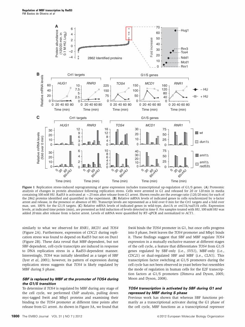
ary data). As expected, we detected an increase in the
abundance of the well-established replication stress-induced
proteins Rnr3 and Hug1, which are under the control of the
Crt1 transcription repressor (Figure 1A). In addition, we
found increased abundance of the MBF-only target Rnr1 as
well as of other G1/S proteins, including Mcd1 (putative
MBF-only target), Tos4 (putative SBF-only target) and Ndd1
(SBF-only target), in response to replication stress. Increase
on Tos4 protein levels during replication stress was also
validated by western blot analysis (Supplementary
Figure S1). To establish whether the accumulation of these
proteins during DNA replication stress is the result of tran-
scriptional induction, we carried out a gene expression
analysis during the cell cycle in untreated and HU-treated
cells (Figure 1B). Treatment with other genotoxic agents
(MMS and CPT) has been shown to give similar results
(Wittenberg laboratory, personal communication). This ana-
lysis confirms previous observations showing transcriptional
induction of RNR3 and HUG1 strictly in response to DNA
replication stress. It also shows that expression of the G1/S
genes TOS4, RNR1 and MCD1 is periodic during an unper-
turbed cell cycle, peaking at the G1/S transition and up-
regulated during S phase in response to replication stress.
Transcription of NDD1 was not increased during replication
stress (Supplementary Figure S2), revealing that the
abundance of Ndd1 protein in response to replication stress
is under an alternative mode of regulation.
Together, these data show that both the transcript and
protein levels of the G1/S cell cycle regulated genes TOS4,
MCD1 and RNR1 are induced in response to DNA replication
stress. The fold induction observed for these proteins in our
proteomic analysis is among the most drastic changes
observed in the proteome, supporting that G1/S transcription
is strongly impacted upon replication stress. Moreover, the
transcriptional regulation of G1/S genes is different from that
of Crt1 targets, whose transcription is not induced in a
normal cell cycle but strongly up-regulated in response to
DNA replication stress.
Replication stress induces expression of G1/S genes via
a Rad53-dependent but Dun1-independent pathway
In response to DNA replication stress, Dun1-dependent phos-
phorylation inactivates the transcriptional repressor Crt1
leading to induction of its targets, such as RNR3 and HUG1.
To establish whether the induction of the G1/S genes TOS4,
MCD1 and RNR1 during replication stress depends on Dun1,
we measured their transcript levels during the cell cycle and
in response to HU treatment in wt and dun1D cells. As
previously shown, the induction of the Crt1 targets RNR3
and HUG1 in response to replication stress depends on Dun1
(Figure 1C, upper panels). We also show that G1/S targets
remain cell cycle regulated and replication stress induced in
dun1D cells, revealing that their expression is Dun1 indepen-
dent (Figure 1C, upper panels). Upstream of Dun1 functions
the Rad53 protein kinase. To determine if the expression of
TOS4, MCD1 and RNR1 is dependent on Rad53, the experi-
ment above was carried out in rad53D cells. Since Rad53 is an
essential gene, the experiment was done in an sml1D back-
ground, which suppresses the lethality of Rad53 inactivation.
The expression analysis establishes that, as expected, the
induction of Crt1 targets is Rad53 dependent (Figure 1C,
lower panels). It also shows that the induction of TOS4,
MCD1 and RNR1 depends on Rad53. Overall, these data
show that the induction of the G1/S cell cycle regulated
genes TOS4, MCD1 and RNR1, in response to replication
stress is Rad53 dependent, but is independent of Dun1.
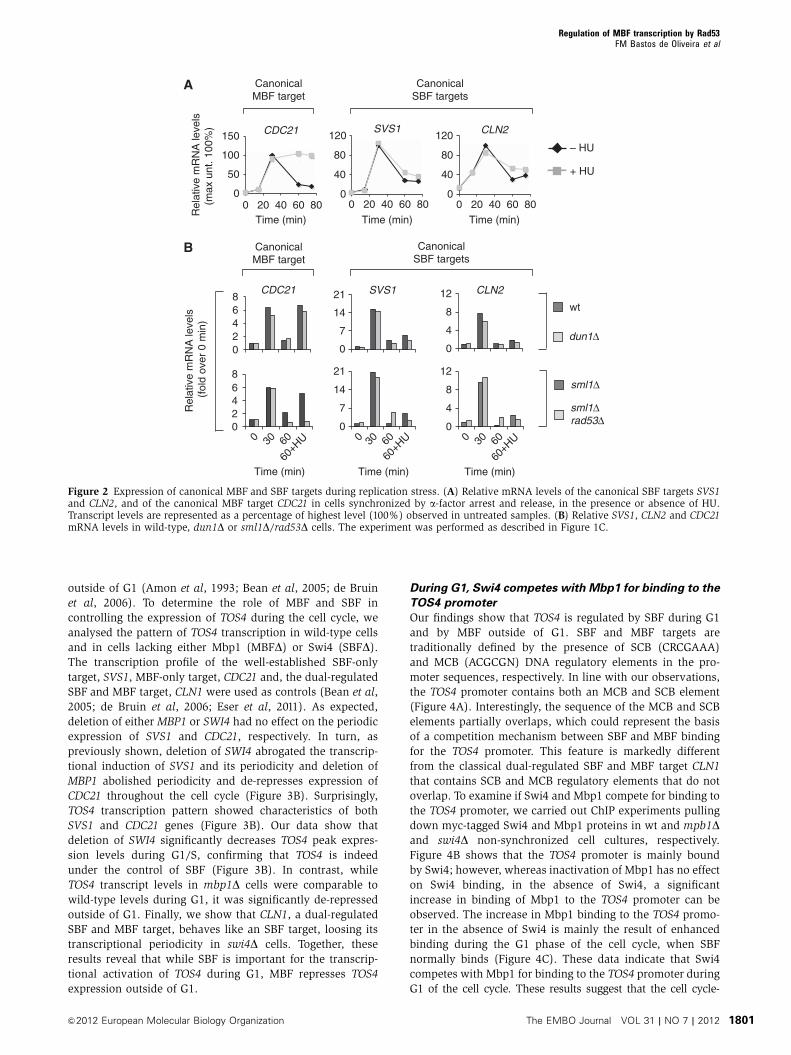
Targets of MBF, but not SBF, are up-regulated in
response to replication stress
To determine if transcriptional induction of G1/S genes upon
replication stress is a general feature of G1/S genes or is
specific for MBF and/or SBF-only targets, we determined the
cell-cycle expression levels of other well-established G1/S
transcripts, two SBF targets (SVS1 and CLN2) and one MBF
target (CDC21), in untreated and HU-treated conditions. Our
analysis revealed that whereas the SBF targets SVS1 and
CLN2 are repressed in a timely manner, the MBF target
CDC21 is induced in response to DNA replication stress
Regulation of MBF transcription by Rad53FM Bastos de Oliveira et al
&2012 European Molecular Biology Organization The EMBO Journal VOL 31 | NO 7 | 2012 1799
similarly to what we observed for RNR1, MCD1 and TOS4
(Figure 2A). Furthermore, expression of CDC21 during repli-
cation stress was found to depend on Rad53 but not on Dun1
(Figure 2B). These data reveal that MBF-dependent, but not
SBF-dependent, cell-cycle transcripts are induced in response
to DNA replication stress in a Rad53-dependent manner.
Interestingly, TOS4 was initially identified as a target of SBF
(Iyer et al, 2001); however, its pattern of expression during
replication stress suggests that TOS4 is likely regulated by
MBF during S phase.
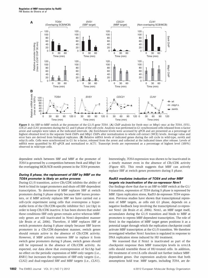
SBF is replaced by MBF at the promoter of TOS4 during
the G1/S transition
To determine if TOS4 is regulated by MBF during any stage of
the cell cycle, we performed ChIP analysis, pulling down
myc-tagged Swi4 and Mbp1 proteins and examining their
binding to the TOS4 promoter at different time points after
release from G1 arrest. As shown in Figure 3A, we found that
Swi4 binds the TOS4 promoter in G1, but once cells progress
into S phase, Swi4 leaves the TOS4 promoter and Mbp1 binds
it. These findings suggest that SBF and MBF regulate TOS4
expression in a mutually exclusive manner at different stages
of the cell cycle, a feature that differentiates TOS4 from G1/S
genes regulated by SBF-only (i.e., SVS1), MBF-only (i.e.,
CDC21) or dual-regulated SBF and MBF (i.e., CLN1). This
transcription factor switching at G1/S promoters during the
cell cycle has not been observed in yeast before but resembles
the mode of regulation in human cells for the E2F transcrip-
tion factors at G1/S promoters (Dimova and Dyson, 2005;
Moon and Dyson, 2008).
TOS4 transcription is activated by SBF during G1 and
repressed by MBF during S phase
Previous work has shown that whereas SBF functions pri-
marily as a transcriptional activator during the G1 phase of
the cell cycle, MBF functions as a transcriptional repressor
A
C
0
20
40
60
0 20 40 60 800
75
150
225
0 20 40 60 800
50
100
150
0 20 40 60 800
2.55
7.510
0 20 40 60 800
4080
120 160
0 20 40 60 80Rel
ativ
e m
RN
A le
vels
Time (min)
RNR3HUG1 TOS4 RNR1
B
MCD1
Time (min) Time (min) Time (min) Time (min)
Crt1 targets
– HU
+ HU
G1/S genes
10
20
30
40
50
60
Fol
d in
crea
se
Hug1
Rnr3Tos4
Ndd1Mcd1Rnr10
70
–4
–2
0
2
4
6
2862 Identified proteins
-
-
-
-
-
-
-
-
Pro
tein
abu
ndan
ce(1
20/2
0 m
in. i
n0.
1 M
HU
; Log
2)
wt
dun1Δ
sml1Δ
sml1Δrad53Δ
RNR3 TOS4HUG1 RNR1MCD1
Rel
ativ
e m
RN
A le
vels
(fo
ld o
ver
0 m
in)
Time (min) Time (min) Time (min) Time (min) Time (min)
Crt1 targets
0102030
0
2
4
6
02468
10
010203040
0
25
50
75
0
25
50
75
0
10
20
30
048
1216
0369
12
0102030
G1/S genes
0 30 60
60+H
U 0 30 60
60+H
U 0 30 60
60+H
U 0 30 60
60+H
U 0 30 60
60+H
U
Figure 1 Replication stress-induced reprogramming of gene expression includes transcriptional up-regulation of G1/S genes. (A) Proteomicanalysis of changes in protein abundance following replication stress. Cells were arrested in G1 and released for 20 or 120min in mediacontaining 100mM HU. Rad53 is activated atB25min after release from G1 arrest. Shown results are the average ratio (120/20min) for each ofthe 2862 proteins identified and quantified in the experiment. (B) Relative mRNA levels of indicated genes in cells synchronized by a-factorarrest and release, in the presence or absence of HU. Transcript levels are represented as a fold over 0 min for the Crt1 targets and a fold overmax. unt. 100% for the G1/S targets. (C) Relative mRNA levels of indicated genes in wild-type, dun1D or sml1D/rad53D cells. Expressionlevels, at indicated time points (min), are presented as fold induction of levels detected in time 0. For samples treated with HU, 100mM HU wasadded 20min after release from a-factor arrest. Levels of mRNA were quantified by RT–qPCR and normalized to ACT1.
Regulation of MBF transcription by Rad53FM Bastos de Oliveira et al
The EMBO Journal VOL 31 | NO 7 | 2012 &2012 European Molecular Biology Organization1800
outside of G1 (Amon et al, 1993; Bean et al, 2005; de Bruin
et al, 2006). To determine the role of MBF and SBF in
controlling the expression of TOS4 during the cell cycle, we
analysed the pattern of TOS4 transcription in wild-type cells
and in cells lacking either Mbp1 (MBFD) or Swi4 (SBFD).The transcription profile of the well-established SBF-only
target, SVS1, MBF-only target, CDC21 and, the dual-regulated
SBF and MBF target, CLN1 were used as controls (Bean et al,
2005; de Bruin et al, 2006; Eser et al, 2011). As expected,
deletion of either MBP1 or SWI4 had no effect on the periodic
expression of SVS1 and CDC21, respectively. In turn, as
previously shown, deletion of SWI4 abrogated the transcrip-
tional induction of SVS1 and its periodicity and deletion of
MBP1 abolished periodicity and de-represses expression of
CDC21 throughout the cell cycle (Figure 3B). Surprisingly,
TOS4 transcription pattern showed characteristics of both
SVS1 and CDC21 genes (Figure 3B). Our data show that
deletion of SWI4 significantly decreases TOS4 peak expres-
sion levels during G1/S, confirming that TOS4 is indeed
under the control of SBF (Figure 3B). In contrast, while
TOS4 transcript levels in mbp1D cells were comparable to
wild-type levels during G1, it was significantly de-repressed
outside of G1. Finally, we show that CLN1, a dual-regulated
SBF and MBF target, behaves like an SBF target, loosing its
transcriptional periodicity in swi4D cells. Together, these
results reveal that while SBF is important for the transcrip-
tional activation of TOS4 during G1, MBF represses TOS4
expression outside of G1.
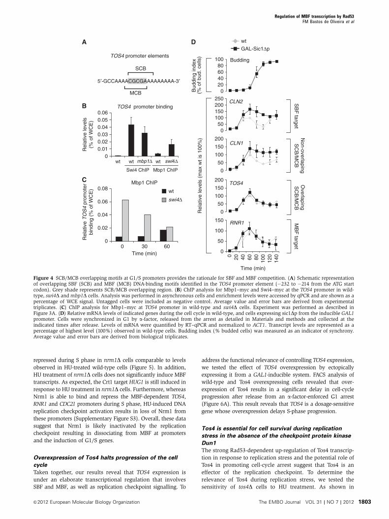
During G1, Swi4 competes with Mbp1 for binding to the
TOS4 promoter
Our findings show that TOS4 is regulated by SBF during G1
and by MBF outside of G1. SBF and MBF targets are
traditionally defined by the presence of SCB (CRCGAAA)
and MCB (ACGCGN) DNA regulatory elements in the pro-
moter sequences, respectively. In line with our observations,
the TOS4 promoter contains both an MCB and SCB element
(Figure 4A). Interestingly, the sequence of the MCB and SCB
elements partially overlaps, which could represent the basis
of a competition mechanism between SBF and MBF binding
for the TOS4 promoter. This feature is markedly different
from the classical dual-regulated SBF and MBF target CLN1
that contains SCB and MCB regulatory elements that do not
overlap. To examine if Swi4 and Mbp1 compete for binding to
the TOS4 promoter, we carried out ChIP experiments pulling
down myc-tagged Swi4 and Mbp1 proteins in wt and mpb1Dand swi4D non-synchronized cell cultures, respectively.
Figure 4B shows that the TOS4 promoter is mainly bound
by Swi4; however, whereas inactivation of Mbp1 has no effect
on Swi4 binding, in the absence of Swi4, a significant
increase in binding of Mbp1 to the TOS4 promoter can be
observed. The increase in Mbp1 binding to the TOS4 promo-
ter in the absence of Swi4 is mainly the result of enhanced
binding during the G1 phase of the cell cycle, when SBF
normally binds (Figure 4C). These data indicate that Swi4
competes with Mbp1 for binding to the TOS4 promoter during
G1 of the cell cycle. These results suggest that the cell cycle-
0
40
80
120
0 20 40 60 800
50
100
150
0 20 40 60 80
0
4
8
12
0
7
14
21
0
7
14
21
0
4
8"
12
02468
Rel
ativ
e m
RN
A le
vels
(max
unt
. 100
%)
Time (min)
SVS1 CLN2CDC21
A
– HU
+ HU
Time (min)Time (min)
SVS1 CLN2CDC21
B
Rel
ativ
e m
RN
A le
vels
(fo
ld o
ver
0 m
in)
02468
wt
dun1Δ
sml1Δ
sml1Δrad53Δ
Time (min)
0 30 60
60+H
U
Time (min)
0 30 60
60+H
U
Time (min)
0 30 60
60+H
U
0
40
80
120
0 20 40 60 80
CanonicalSBF targets
CanonicalMBF target
CanonicalMBF target
CanonicalSBF targets
Figure 2 Expression of canonical MBF and SBF targets during replication stress. (A) Relative mRNA levels of the canonical SBF targets SVS1and CLN2, and of the canonical MBF target CDC21 in cells synchronized by a-factor arrest and release, in the presence or absence of HU.Transcript levels are represented as a percentage of highest level (100%) observed in untreated samples. (B) Relative SVS1, CLN2 and CDC21mRNA levels in wild-type, dun1D or sml1D/rad53D cells. The experiment was performed as described in Figure 1C.
Regulation of MBF transcription by Rad53FM Bastos de Oliveira et al
&2012 European Molecular Biology Organization The EMBO Journal VOL 31 | NO 7 | 2012 1801
dependent switch between SBF and MBF at the promoter of
TOS4 is governed by a competition between Swi4 and Mbp1 for
the overlapping MCB/SCB motifs present in the TOS4 promoter.
During S phase, the replacement of SBF by MBF on the
TOS4 promoter is likely an active process
During G1/S transition, active Clb/CDK inhibits the ability of
Swi4 to bind its target promoters and shuts off SBF-dependent
transcription. To determine if MBF replaces SBF at switch
promoters during S phase simply because SBF leaves promo-
ters, or if MBF actively replaces SBF we have carried out a
cell-cycle experiment using cells that overexpress a hyper-
stable form of the Clb/CDK-specific inhibitor Sic1 (sic1Dp) inorder to keep Clb/CDK inactive. It has been shown that under
these conditions SBF-only genes remain active whereas MBF-
only genes are still inactivated in Nrm1-dependent manner
(de Bruin et al, 2006). Therefore, if MBF replaces SBF at
switch promoters during S phase simply because SBF leaves
promoters in a Clb/CDK-dependent manner, switch genes
should remain active in the absence of Clb/CDK activity.
However, if MBF actively removes SBF to replace it from
switch gene promoters during S phase, switch genes should
still be repressed in the absence of Clb/CDK activity. As
expected, our data show that overexpression of Sic1Dp had
no effect on the periodic expression of MBF-only targets (i.e.,
RNR1) but increases the expression of SBF-only targets (i.e.,
CLN2) and dual-regulated SBF and MBF targets (i.e., CLN1).
Interestingly, TOS4 expression was shown to be inactivated in
a timely manner even in the absence of Clb/CDK activity
(Figure 4D). This result suggests that MBF can actively
replace SBF at switch genes promoters during S phase.
Rad53 mediates induction of TOS4 and other MBF
targets via inactivation of the co-repressor Nrm1
Our findings show that due to an SBF-to-MBF switch at the G1/
S transition, expression of TOS4 during S phase is repressed by
MBF. Upon replication stress, Rad53 de-represses TOS4 expres-
sion. Previous studies have shown that transcriptional repres-
sion of MBF targets, as cells exit G1 phase, depends on a
negative feedback loop involving the transcriptional co-repres-
sor Nrm1 (de Bruin et al, 2006). Nrm1, an MBF target itself,
accumulates during the G1/S transition and binds to MBF at
promoters to repress MBF-dependent transcription. The role of
Nrm1 in the regulation of MBF target genes positions it as a
potential target through which the replication checkpoint could
activate MBF transcription at the G1/S transition. We therefore
investigated whether Nrm1 function is regulated in response to
DNA replication stress induced by HU treatment.
We reasoned that if Nrm1 is inactivated as part of the
checkpoint response then MBF transcripts levels in nrm1Dcells should resemble those of HU-treated cells. In addition,
HU treatment of nrm1D cells should not further induce MBF-
dependent genes. Our expression analysis shows that both
assumptions hold true. MBF targets, including TOS4, are de-
B
020406080
100120
Rel
ativ
e m
RN
A le
vels
(max
wt i
s 10
0%) SVS1 CDC21
wt
mbp1Δswi4Δ
120
TOS4
0 30 60 90 120Time (min)
0 30 60 90 120Time (min)
0 30 60 90 120Time (min)
0 30 60 90 120Time (min)
CLN1
A
020406080
100120
020406080
100120
Rel
ativ
e pr
omot
er b
indi
ng(m
ax is
CLN
1 C
hIP
30
min
)
0 30 60Time (min)
Sw
i4 ChIP
Mbp1 C
hIP
0 30 60Time (min)
020406080
100120
020406080
100120
0 30 60Time (min)
0 30 60Time (min)
020406080
100120
0 30 60Time (min)
020406080
100120
0 30 60Time (min)
020406080
100120
0 30 60Time (min)
020406080
100120
0 30 60Time (min)
TOS4(Overlaping SCB/MCB)
SVS1(SBF target)
CDC21(MBF target)
CLN1(Non-overlaping SCB/MCB)
Figure 3 An SBF-to-MBF switch at the promoter of the G1/S gene TOS4. (A) ChIP analysis for Swi4–myc or Mbp1–myc at the TOS4, SVS1,CDC21 and CLN1 promoters during the G1 and S phase of the cell cycle. Analysis was performed in G1 synchronized cells released from a-factorarrest and samples were taken at the indicated intervals. (A) Enrichment levels were accessed by qPCR and are presented as a percentage ofhighest obtained level in the separate Swi4 ChIPs and Mbp1 ChIPs after normalization to whole cell extract (WCE) levels. Average value anderror bars are derived from biological replicates. (B) Relative mRNA levels of indicated genes during the cell cycle in wild-type, swi4D andmbp1D cells. Cells were synchronized in G1 by a-factor, released from the arrest and collected at the indicated times after release. Levels ofmRNA were quantified by RT–qPCR and normalized to ACT1. Transcript levels are represented as a percentage of highest level (100%)observed in wild-type cells.
Regulation of MBF transcription by Rad53FM Bastos de Oliveira et al
The EMBO Journal VOL 31 | NO 7 | 2012 &2012 European Molecular Biology Organization1802
repressed during S phase in nrm1D cells comparable to levels
observed in HU-treated wild-type cells (Figure 5). In addition,
HU treatment of nrm1D cells does not significantly induce MBF
transcripts. As expected, the Crt1 target HUG1 is still induced in
response to HU treatment in nrm1D cells. Furthermore, whereas
Nrm1 is able to bind and repress the MBF-dependent TOS4,
RNR1 and CDC21 promoters during S phase, HU-induced DNA
replication checkpoint activation results in loss of Nrm1 from
these promoters (Supplementary Figure S3). Overall, these data
suggest that Nrm1 is likely inactivated by the replication
checkpoint resulting in dissociating from MBF at promoters
and the induction of G1/S genes.
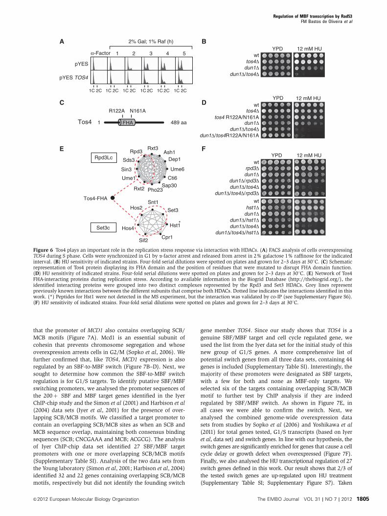
Overexpression of Tos4 halts progression of the cell
cycle
Taken together, our results reveal that TOS4 expression is
under an elaborate transcriptional regulation that involves
SBF and MBF, as well as replication checkpoint signalling. To
address the functional relevance of controlling TOS4 expression,
we tested the effect of TOS4 overexpression by ectopically
expressing it from a GAL1-inducible system. FACS analysis of
wild-type and Tos4 overexpressing cells revealed that over-
expression of Tos4 results in a significant delay in cell-cycle
progression after release from an a-factor-enforced G1 arrest
(Figure 6A). This result reveals that TOS4 is a dosage-sensitive
gene whose overexpression delays S-phase progression.
Tos4 is essential for cell survival during replication
stress in the absence of the checkpoint protein kinase
Dun1
The strong Rad53-dependent up-regulation of Tos4 transcrip-
tion in response to replication stress and the potential role of
Tos4 in promoting cell-cycle arrest suggest that Tos4 is an
effector of the replication checkpoint. To determine the
relevance of Tos4 during replication stress, we tested the
sensitivity of tos4D cells to HU treatment. As shown in
00.010.020.030.040.05
0
0.02
0.04
0.06
0.08
B
Rel
ativ
e le
vels
(% o
f WC
E)
TOS4 promoter binding
wt wt wtmbp1Δ swi4Δ
Swi4 ChIP Mbp1 ChIP
5′-GCCAAAACGCGAAAAAAAAA-3′
MCB
SCB
TOS4 promoter elements
Rel
ativ
e T
OS
4 pr
omot
erbi
ndin
g (%
of W
CE
)
Mbp1 ChIP
0 30 60Time (min)
C
wt
swi4Δ
0.06
A D
020406080
100
050
100150200250
0
50
100
150
200
0
50
100
150
200
0
50
100
150
0 40 80 120
Time (min)
CLN2
CLN1
TOS4
RNR1
Budding
Rel
ativ
e le
vels
(m
ax w
t is
100%
)B
uddi
ng in
dex
(% o
f bud
. cel
ls)
SB
F target
Non-overlapingS
CB
/MC
BO
verlapingS
CB
/MC
BM
BF
target
wt
GAL-Sic1Δp
20 60 100
140
Figure 4 SCB/MCB overlapping motifs at G1/S promoters provides the rationale for SBF and MBF competition. (A) Schematic representationof overlapping SBF (SCB) and MBF (MCB) DNA-binding motifs identified in the TOS4 promoter element (�232 to �214 from the ATG startcodon). Grey shade represents SCB/MCB overlapping region. (B) ChIP analysis for Mbp1–myc and Swi4–myc at the TOS4 promoter in wild-type, swi4D andmbp1D cells. Analysis was performed in asynchronous cells and enrichment levels were accessed by qPCR and are shown as apercentage of WCE signal. Untagged cells were included as negative control. Average value and error bars are derived from experimentaltriplicates. (C) ChIP analysis for Mbp1–myc at TOS4 promoter in wild-type and swi4D cells. Experiment was performed as described inFigure 3A. (D) Relative mRNA levels of indicated genes during the cell cycle in wild-type, and cells expressing sic1Dp from the inducible GAL1promoter. Cells were synchronized in G1 by a-factor, released from the arrest as detailed in Materials and methods and collected at theindicated times after release. Levels of mRNA were quantified by RT–qPCR and normalized to ACT1. Transcript levels are represented as apercentage of highest level (100%) observed in wild-type cells. Budding index (% budded cells) was measured as an indicator of synchrony.Average value and error bars are derived from biological triplicates.
Regulation of MBF transcription by Rad53FM Bastos de Oliveira et al
&2012 European Molecular Biology Organization The EMBO Journal VOL 31 | NO 7 | 2012 1803
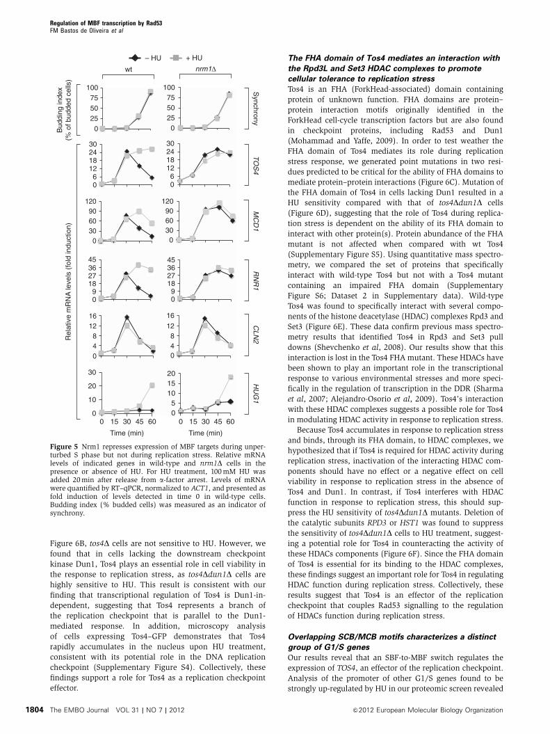
Figure 6B, tos4D cells are not sensitive to HU. However, we
found that in cells lacking the downstream checkpoint
kinase Dun1, Tos4 plays an essential role in cell viability in
the response to replication stress, as tos4Ddun1D cells are
highly sensitive to HU. This result is consistent with our
finding that transcriptional regulation of Tos4 is Dun1-in-
dependent, suggesting that Tos4 represents a branch of
the replication checkpoint that is parallel to the Dun1-
mediated response. In addition, microscopy analysis
of cells expressing Tos4–GFP demonstrates that Tos4
rapidly accumulates in the nucleus upon HU treatment,
consistent with its potential role in the DNA replication
checkpoint (Supplementary Figure S4). Collectively, these
findings support a role for Tos4 as a replication checkpoint
effector.
The FHA domain of Tos4 mediates an interaction with
the Rpd3L and Set3 HDAC complexes to promote
cellular tolerance to replication stress
Tos4 is an FHA (ForkHead-associated) domain containing
protein of unknown function. FHA domains are protein–
protein interaction motifs originally identified in the
ForkHead cell-cycle transcription factors but are also found
in checkpoint proteins, including Rad53 and Dun1
(Mohammad and Yaffe, 2009). In order to test weather the
FHA domain of Tos4 mediates its role during replication
stress response, we generated point mutations in two resi-
dues predicted to be critical for the ability of FHA domains to
mediate protein–protein interactions (Figure 6C). Mutation of
the FHA domain of Tos4 in cells lacking Dun1 resulted in a
HU sensitivity compared with that of tos4Ddun1D cells
(Figure 6D), suggesting that the role of Tos4 during replica-
tion stress is dependent on the ability of its FHA domain to
interact with other protein(s). Protein abundance of the FHA
mutant is not affected when compared with wt Tos4
(Supplementary Figure S5). Using quantitative mass spectro-
metry, we compared the set of proteins that specifically
interact with wild-type Tos4 but not with a Tos4 mutant
containing an impaired FHA domain (Supplementary
Figure S6; Dataset 2 in Supplementary data). Wild-type
Tos4 was found to specifically interact with several compo-
nents of the histone deacetylase (HDAC) complexes Rpd3 and
Set3 (Figure 6E). These data confirm previous mass spectro-
metry results that identified Tos4 in Rpd3 and Set3 pull
downs (Shevchenko et al, 2008). Our results show that this
interaction is lost in the Tos4 FHA mutant. These HDACs have
been shown to play an important role in the transcriptional
response to various environmental stresses and more speci-
fically in the regulation of transcription in the DDR (Sharma
et al, 2007; Alejandro-Osorio et al, 2009). Tos4’s interaction
with these HDAC complexes suggests a possible role for Tos4
in modulating HDAC activity in response to replication stress.
Because Tos4 accumulates in response to replication stress
and binds, through its FHA domain, to HDAC complexes, we
hypothesized that if Tos4 is required for HDAC activity during
replication stress, inactivation of the interacting HDAC com-
ponents should have no effect or a negative effect on cell
viability in response to replication stress in the absence of
Tos4 and Dun1. In contrast, if Tos4 interferes with HDAC
function in response to replication stress, this should sup-
press the HU sensitivity of tos4Ddun1D mutants. Deletion of
the catalytic subunits RPD3 or HST1 was found to suppress
the sensitivity of tos4Ddun1D cells to HU treatment, suggest-
ing a potential role for Tos4 in counteracting the activity of
these HDACs components (Figure 6F). Since the FHA domain
of Tos4 is essential for its binding to the HDAC complexes,
these findings suggest an important role for Tos4 in regulating
HDAC function during replication stress. Collectively, these
results suggest that Tos4 is an effector of the replication
checkpoint that couples Rad53 signalling to the regulation
of HDACs function during replication stress.
Overlapping SCB/MCB motifs characterizes a distinct
group of G1/S genes
Our results reveal that an SBF-to-MBF switch regulates the
expression of TOS4, an effector of the replication checkpoint.
Analysis of the promoter of other G1/S genes found to be
strongly up-regulated by HU in our proteomic screen revealed
0
10
20
30
0 15 30 45 6005
101520
0 15 30 45 60
0255075
100
0255075
100
06
12182430
06
12182430
09
18273645
048
1216
09
18273645
048
12
16
Bud
ding
inde
x(%
of b
udde
d ce
lls)
Rel
ativ
e m
RN
A le
vels
(fo
ld in
duct
ion)
TO
S4
RN
R1
CLN
2
wt nrm1Δ
Time (min) Time (min)
– HU + HU
Synchrony
0306090
120
0306090
120
MC
D1
HU
G1
Figure 5 Nrm1 represses expression of MBF targets during unper-turbed S phase but not during replication stress. Relative mRNAlevels of indicated genes in wild-type and nrm1D cells in thepresence or absence of HU. For HU treatment, 100mM HU wasadded 20min after release from a-factor arrest. Levels of mRNAwere quantified by RT–qPCR, normalized to ACT1, and presented asfold induction of levels detected in time 0 in wild-type cells.Budding index (% budded cells) was measured as an indicator ofsynchrony.
Regulation of MBF transcription by Rad53FM Bastos de Oliveira et al
The EMBO Journal VOL 31 | NO 7 | 2012 &2012 European Molecular Biology Organization1804
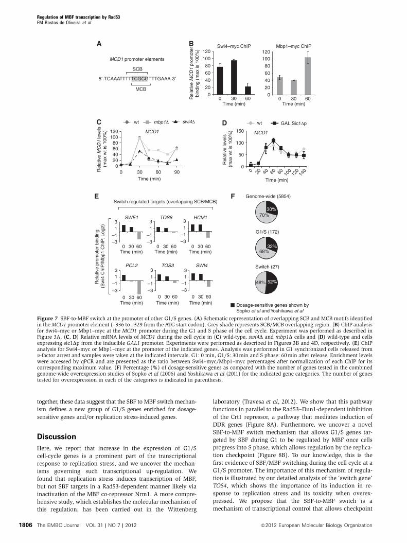
that the promoter of MCD1 also contains overlapping SCB/
MCB motifs (Figure 7A). Mcd1 is an essential subunit of
cohesin that prevents chromosome segregation and whose
overexpression arrests cells in G2/M (Sopko et al, 2006). We
further confirmed that, like TOS4, MCD1 expression is also
regulated by an SBF-to-MBF switch (Figure 7B–D). Next, we
sought to determine how common the SBF-to-MBF switch
regulation is for G1/S targets. To identify putative SBF/MBF
switching promoters, we analysed the promoter sequences of
the 200þ SBF and MBF target genes identified in the Iyer
ChIP-chip study and the Simon et al (2001) and Harbison et al
(2004) data sets (Iyer et al, 2001) for the presence of over-
lapping SCB/MCB motifs. We classified a target promoter to
contain an overlapping SCB/MCB sites as when an SCB and
MCB sequence overlap, maintaining both consensus binding
sequences (SCB; CNCGAAA and MCB; ACGCG). The analysis
of Iyer ChIP-chip data set identified 27 SBF/MBF target
promoters with one or more overlapping SCB/MCB motifs
(Supplementary Table SI). Analysis of the two data sets from
the Young laboratory (Simon et al, 2001; Harbison et al, 2004)
identified 32 and 22 genes containing overlapping SCB/MCB
motifs, respectively but did not identify the founding switch
gene member TOS4. Since our study shows that TOS4 is a
genuine SBF/MBF target and cell cycle regulated gene, we
used the list from the Iyer data set for the initial study of this
new group of G1/S genes. A more comprehensive list of
potential switch genes from all three data sets, containing 44
genes is included (Supplementary Table SI). Interestingly, the
majority of these promoters were designated as SBF targets,
with a few for both and none as MBF-only targets. We
selected six of the targets containing overlapping SCB/MCB
motif to further test by ChIP analysis if they are indeed
regulated by SBF/MBF switch. As shown in Figure 7E, in
all cases we were able to confirm the switch. Next, we
analysed the combined genome-wide overexpression data
sets from studies by Sopko et al (2006) and Yoshikawa et al
(2011) for total genes tested, G1/S transcripts (based on Iyer
et al, data set) and switch genes. In line with our hypothesis, the
switch genes are significantly enriched for genes that cause a cell
cycle delay or growth defect when overexpressed (Figure 7F).
Finally, we also analysed the HU transcriptional regulation of 27
switch genes defined in this work. Our result shows that 2/3 of
the tested switch genes are up-regulated upon HU treatment
(Supplementary Table SI; Supplementary Figure S7). Taken
A B
wttos4Δ
dun1Δdun1Δ/tos4Δ
YPD 12 mM HU
D
489 aaFHA
R122A N161A
1Tos4
wttos4Δ
dun1Δdun1Δ/tos4Δ
dun1Δ/tos4R122A/N161A
tos4 R122A/N161A
12 mM HUYPD
E
C
F12 mM HU
wt
dun1Δ
dun1Δ/tos4Δ/rpd3Δ
rpd3Δ
dun1Δ/tos4Δdun1Δ/rpd3Δ
YPD
wt
dun1Δ
dun1Δ/tos4Δ/hst1Δ
hst1Δ
dun1Δ/tos4Δdun1Δ/hst1Δ
1C 2C
pYES
pYES TOS4
α-Factor 1 2 3 4 5
2% Gal; 1% Raf (h)
1C 2C 1C 2C 1C 2C 1C 2C 1C 2C
Hos2
Hos4
Sif2
Snt1
Set3
Hst1
Cpr1
Tos4-FHA
Rpd3
Sds3
Sin3
Ume1
Rxt2 Pho23Sap30
Cti6
Ume6
Dep1Ash1
Rxt3
*
Rpd3Lc
Set3c
Figure 6 Tos4 plays an important role in the replication stress response via interaction with HDACs. (A) FACS analysis of cells overexpressingTOS4 during S phase. Cells were synchronized in G1 by a-factor arrest and released from arrest in 2% galactose 1% raffinose for the indicatedinterval. (B) HU sensitivity of indicated strains. Four-fold serial dilutions were spotted on plates and grown for 2–3 days at 301C. (C) Schematicrepresentation of Tos4 protein displaying its FHA domain and the position of residues that were mutated to disrupt FHA domain function.(D) HU sensitivity of indicated strains. Four-fold serial dilutions were spotted on plates and grown for 2–3 days at 301C. (E) Network of Tos4FHA-interacting proteins during replication stress. According to available information in the Biogrid Database (http://thebiogrid.org/), theidentified interacting proteins were grouped into two distinct complexes represented by the Rpd3 and Set3 HDACs. Grey lines representpreviously known interactions between the different subunits that comprise both HDACs. Dotted line indicates the interactions identified in thiswork. (*) Peptides for Hst1 were not detected in the MS experiment, but the interaction was validated by co-IP (see Supplementary Figure S6).(F) HU sensitivity of indicated strains. Four-fold serial dilutions were spotted on plates and grown for 2–3 days at 301C.
Regulation of MBF transcription by Rad53FM Bastos de Oliveira et al
&2012 European Molecular Biology Organization The EMBO Journal VOL 31 | NO 7 | 2012 1805
together, these data suggest that the SBF to MBF switch mechan-
ism defines a new group of G1/S genes enriched for dosage-
sensitive genes and/or replication stress-induced genes.
Discussion
Here, we report that increase in the expression of G1/S
cell-cycle genes is a prominent part of the transcriptional
response to replication stress, and we uncover the mechan-
isms governing such transcriptional up-regulation. We
found that replication stress induces transcription of MBF,
but not SBF targets in a Rad53-dependent manner likely via
inactivation of the MBF co-repressor Nrm1. A more compre-
hensive study, which establishes the molecular mechanism of
this regulation, has been carried out in the Wittenberg
laboratory (Travesa et al, 2012). We show that this pathway
functions in parallel to the Rad53–Dun1-dependent inhibition
of the Crt1 repressor, a pathway that mediates induction of
DDR genes (Figure 8A). Furthermore, we uncover a novel
SBF-to-MBF switch mechanism that allows G1/S genes tar-
geted by SBF during G1 to be regulated by MBF once cells
progress into S phase, which allows regulation by the replica-
tion checkpoint (Figure 8B). To our knowledge, this is the
first evidence of SBF/MBF switching during the cell cycle at a
G1/S promoter. The importance of this mechanism of regula-
tion is illustrated by our detailed analysis of the ‘switch gene’
TOS4, which shows the importance of its induction in re-
sponse to replication stress and its toxicity when overex-
pressed. We propose that the SBF-to-MBF switch is a
mechanism of transcriptional control that allows checkpoint
0
20
40
60
80
0
50
100
150
Time (min)
5′-TCAAATTTTTCGCGTTTGAAA-3′
MCB
SCB
MCD1 promoter elements
Rel
ativ
e M
CD
1 pr
omot
erbi
ndin
g (m
ax is
100
%)
Mbp1–myc ChIPSwi4–myc ChIP
Time (min)
020406080
100120
0 30 60 90 Rel
ativ
e M
CD
1 le
vels
(m
ax w
t is
100%
) MCD1
wt mbp1Δ swi4Δ
Genome-wide (5854)
G1/S (172)
Switch (27)
30%70%
32%68%
52%48%
Dosage-sensitive genes shown by Sopko et al and Yoshikawa et al
0
20
40
60
80
0 30 60Time (min)
Time (min)
0
0 30 60
100 100
120 120
MCD1
wt GAL Sic1Δp
Rel
ativ
e le
vels
(m
ax w
t is
100%
)
A B
C D
F
–3–113
–3–113
–3–113
–3–113
TOS8
PCL2
HCM1
TOS3
SWE1
0 30 60
0 30 60 0 30 60
Time (min) Time (min)
Time (min) Time (min)
Rel
ativ
e pr
omot
er b
indi
ng
(Sw
i4 C
hIP
/Mbp
1 C
hIP
; Log
2)
0 30 60Time (min)
–3–113
SWI4
0 30 600 30 60Time (min)
Switch regulated targets (overlapping SCB/MCB)
–3
–113
E
20 40 60 80 100
120
140
Figure 7 SBF-to-MBF switch at the promoter of other G1/S genes. (A) Schematic representation of overlapping SCB and MCB motifs identifiedin theMCD1 promoter element (–336 to –329 from the ATG start codon). Grey shade represents SCB/MCB overlapping region. (B) ChIP analysisfor Swi4–myc or Mbp1–myc at the MCD1 promoter during the G1 and S phase of the cell cycle. Experiment was performed as described inFigure 3A. (C, D) Relative mRNA levels of MCD1 during the cell cycle in (C) wild-type, swi4D and mbp1D cells and (D) wild-type and cellsexpressing sic1Dp from the inducible GAL1 promoter. Experiments were performed as described in Figures 3B and 4D, respectively. (E) ChIPanalysis for Swi4–myc or Mbp1–myc at the promoter of the indicated genes. Analysis was performed in G1 synchronized cells released froma-factor arrest and samples were taken at the indicated intervals. G1: 0 min, G1/S: 30min and S phase: 60min after release. Enrichment levelswere accessed by qPCR and are presented as the ratio between Swi4–myc/Mbp1–myc percentages after normalization of each ChIP for itscorresponding maximum value. (F) Percentage (%) of dosage-sensitive genes as compared with the number of genes tested in the combinedgenome-wide overexpression studies of Sopko et al (2006) and Yoshikawa et al (2011) for the indicated gene categories. The number of genestested for overexpression in each of the categories is indicated in parenthesis.
Regulation of MBF transcription by Rad53FM Bastos de Oliveira et al
The EMBO Journal VOL 31 | NO 7 | 2012 &2012 European Molecular Biology Organization1806
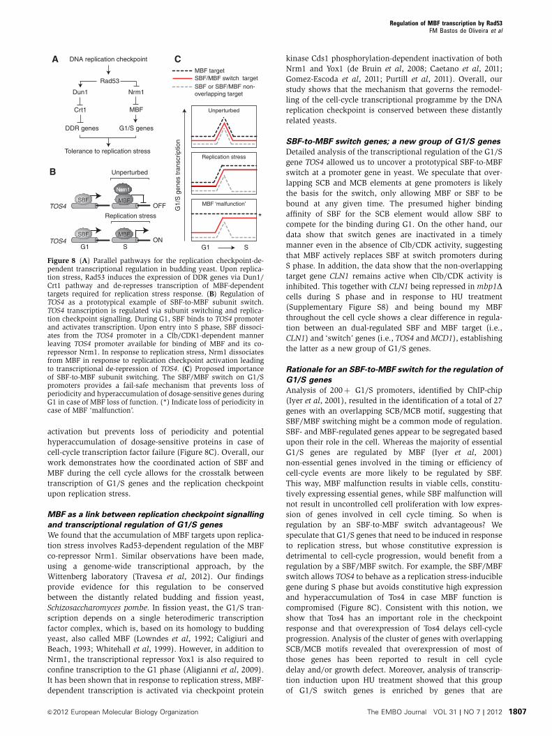
activation but prevents loss of periodicity and potential
hyperaccumulation of dosage-sensitive proteins in case of
cell-cycle transcription factor failure (Figure 8C). Overall, our
work demonstrates how the coordinated action of SBF and
MBF during the cell cycle allows for the crosstalk between
transcription of G1/S genes and the replication checkpoint
upon replication stress.
MBF as a link between replication checkpoint signalling
and transcriptional regulation of G1/S genes
We found that the accumulation of MBF targets upon replica-
tion stress involves Rad53-dependent regulation of the MBF
co-repressor Nrm1. Similar observations have been made,
using a genome-wide transcriptional approach, by the
Wittenberg laboratory (Travesa et al, 2012). Our findings
provide evidence for this regulation to be conserved
between the distantly related budding and fission yeast,
Schizosaccharomyces pombe. In fission yeast, the G1/S tran-
scription depends on a single heterodimeric transcription
factor complex, which is, based on its homology to budding
yeast, also called MBF (Lowndes et al, 1992; Caligiuri and
Beach, 1993; Whitehall et al, 1999). However, in addition to
Nrm1, the transcriptional repressor Yox1 is also required to
confine transcription to the G1 phase (Aligianni et al, 2009).
It has been shown that in response to replication stress, MBF-
dependent transcription is activated via checkpoint protein
kinase Cds1 phosphorylation-dependent inactivation of both
Nrm1 and Yox1 (de Bruin et al, 2008; Caetano et al, 2011;
Gomez-Escoda et al, 2011; Purtill et al, 2011). Overall, our
study shows that the mechanism that governs the remodel-
ling of the cell-cycle transcriptional programme by the DNA
replication checkpoint is conserved between these distantly
related yeasts.
SBF-to-MBF switch genes; a new group of G1/S genes
Detailed analysis of the transcriptional regulation of the G1/S
gene TOS4 allowed us to uncover a prototypical SBF-to-MBF
switch at a promoter gene in yeast. We speculate that over-
lapping SCB and MCB elements at gene promoters is likely
the basis for the switch, only allowing MBF or SBF to be
bound at any given time. The presumed higher binding
affinity of SBF for the SCB element would allow SBF to
compete for the binding during G1. On the other hand, our
data show that switch genes are inactivated in a timely
manner even in the absence of Clb/CDK activity, suggesting
that MBF actively replaces SBF at switch promoters during
S phase. In addition, the data show that the non-overlapping
target gene CLN1 remains active when Clb/CDK activity is
inhibited. This together with CLN1 being repressed in mbp1Dcells during S phase and in response to HU treatment
(Supplementary Figure S8) and being bound my MBF
throughout the cell cycle shows a clear difference in regula-
tion between an dual-regulated SBF and MBF target (i.e.,
CLN1) and ‘switch’ genes (i.e., TOS4 andMCD1), establishing
the latter as a new group of G1/S genes.
Rationale for an SBF-to-MBF switch for the regulation of
G1/S genes
Analysis of 200þ G1/S promoters, identified by ChIP-chip
(Iyer et al, 2001), resulted in the identification of a total of 27
genes with an overlapping SCB/MCB motif, suggesting that
SBF/MBF switching might be a common mode of regulation.
SBF- and MBF-regulated genes appear to be segregated based
upon their role in the cell. Whereas the majority of essential
G1/S genes are regulated by MBF (Iyer et al, 2001)
non-essential genes involved in the timing or efficiency of
cell-cycle events are more likely to be regulated by SBF.
This way, MBF malfunction results in viable cells, constitu-
tively expressing essential genes, while SBF malfunction will
not result in uncontrolled cell proliferation with low expres-
sion of genes involved in cell cycle timing. So when is
regulation by an SBF-to-MBF switch advantageous? We
speculate that G1/S genes that need to be induced in response
to replication stress, but whose constitutive expression is
detrimental to cell-cycle progression, would benefit from a
regulation by a SBF/MBF switch. For example, the SBF/MBF
switch allows TOS4 to behave as a replication stress-inducible
gene during S phase but avoids constitutive high expression
and hyperaccumulation of Tos4 in case MBF function is
compromised (Figure 8C). Consistent with this notion, we
show that Tos4 has an important role in the checkpoint
response and that overexpression of Tos4 delays cell-cycle
progression. Analysis of the cluster of genes with overlapping
SCB/MCB motifs revealed that overexpression of most of
those genes has been reported to result in cell cycle
delay and/or growth defect. Moreover, analysis of transcrip-
tion induction upon HU treatment showed that this group
of G1/S switch genes is enriched by genes that are
G1 S
MBF target
MBF ‘malfunction’
G1/
S g
enes
tran
scrip
tion
SBF/MBF switch targetSBF or SBF/MBF non- overlapping target
Unperturbed
Replication stress
DNA replication checkpoint
Rad53
Dun1
Crt1
DDR genes
Nrm1
MBF
G1/S genes
Tolerance to replication stress
*
A C
B
Nrm1
OFF
Unperturbed
ON
TOS4
S
Replication stress
TOS4G1
SBF
SBF MBF
MBF
Nrm1
Figure 8 (A) Parallel pathways for the replication checkpoint-de-pendent transcriptional regulation in budding yeast. Upon replica-tion stress, Rad53 induces the expression of DDR genes via Dun1/Crt1 pathway and de-represses transcription of MBF-dependenttargets required for replication stress response. (B) Regulation ofTOS4 as a prototypical example of SBF-to-MBF subunit switch.TOS4 transcription is regulated via subunit switching and replica-tion checkpoint signalling. During G1, SBF binds to TOS4 promoterand activates transcription. Upon entry into S phase, SBF dissoci-ates from the TOS4 promoter in a Clb/CDK1-dependent mannerleaving TOS4 promoter available for binding of MBF and its co-repressor Nrm1. In response to replication stress, Nrm1 dissociatesfrom MBF in response to replication checkpoint activation leadingto transcriptional de-repression of TOS4. (C) Proposed importanceof SBF-to-MBF subunit switching. The SBF/MBF switch on G1/Spromoters provides a fail-safe mechanism that prevents loss ofperiodicity and hyperaccumulation of dosage-sensitive genes duringG1 in case of MBF loss of function. (*) Indicate loss of periodicity incase of MBF ‘malfunction’.
Regulation of MBF transcription by Rad53FM Bastos de Oliveira et al
&2012 European Molecular Biology Organization The EMBO Journal VOL 31 | NO 7 | 2012 1807
up-regulated during replication stress conditions. Taken
together, the results reveal that SBF-to-MBF switch preferen-
tially regulates dosage-sensitive genes, and we propose
that most of the genes controlled by the switch play impor-
tant roles during replication stress response, as shown here
for Tos4, and previously for Mcd1 and Swe1 (Strom et al,
2004; Enserink et al, 2006; Liu and Wang, 2006; Covo
et al, 2010). Further functional analysis of the other genes
regulated by the SBF/MBF switch, most of which are
currently annotated as putative genes, may reveal other
dosage-sensitive genes and/or yet undescribed checkpoint
effectors.
Rad53-MBF-Tos4 pathway; a novel branch of the
replication checkpoint
Tos4 was originally identified as an SBF target and a putative
transcription factor suggested to repress pheromone response
genes during the cell cycle and in the presence of a-factor(Horak et al, 2002). Several lines of evidence presented here
support that Tos4 is an effector of the replication checkpoint.
First, the expression of Tos4 is strongly up-regulated during
replication stress in a Rad53-dependent manner. Second,
following replication stress, Tos4 rapidly accumulates in the
nucleus. Third, overexpression of Tos4 causes a cell cycle
delay. Fourth, Tos4 contains an FHA domain, commonly
found in checkpoint proteins, that is required for its function.
Furthermore, we found that Tos4 plays a parallel role to Dun1
in the replication checkpoint. This is supported by the find-
ings that transcription of TOS4 is regulated in a Dun1-
independent manner and that tos4Ddun1D cells exhibit a
much stronger HU sensitivity as compared with the single
deletes. Together, our findings establish that the Rad53-MBF-
Tos4 pathway represents a novel branch of the replication
checkpoint (Figure 8A). While the exact role of Tos4 remains
unclear, the ability of Tos4 to interact with the Rpd3 and Set3
HDAC complexes and the importance of this interaction for
the survival of cells in response to replication stress suggests
that Tos4 functions to couple replication checkpoint signal-
ling to the regulation of HDAC function.
Comparisons with mammalian G1/S transcription
Our work suggests that G1/S transcription factor switching
assures that malfunction of any one transcription factor
does not result in complete loss of periodicity (Figure 8C).
In mammalian cells, switching of the E2F transcription
factors at G1/S promoters during the cell cycle seems to be
the standard (Dimova and Dyson, 2005; Moon and Dyson,
2008), suggesting that loss of periodicity is detrimental.
In line with this, the high frequency of genetic alterations
in genes involved in E2F-dependent G1/S transcriptional
regulation found in human tumour cells indicates an impor-
tant role for proper regulation of the E2F system in the
prevention of tumour development (Stevens and La
Thangue, 2003).
In addition, we uncovered in yeast a simple but elegant
mechanism by which checkpoint activation can override the
G1/S transcriptional programme by directly regulating a
transcriptional repressor. In human cells, the main regulator
of DNA damage-inducible genes in G1 is the transcription
factor p53, a target of both ATM and Chk2 (Carr, 2000).
However, the transcriptional response induced by the DNA
replication checkpoint in S phase is regulated largely through
a p53-independent mechanism that has yet to be fully eluci-
dated. Interestingly, recent data suggest that, in response to
DNA replication stress, like in yeast, G1/S transcription,
regulated by the E2F family of transcription factors, is main-
tained at high levels in a Chk1-dependent manner (Cosetta
Bertoli and RAM de Bruin, unpublished data).
If, as preliminary data suggest, remodelling of the G1/S
cell-cycle transcriptional programme by the DNA replication
checkpoint proves to be conserved from yeast to man, this
would represent the largest group of co-regulated genes
among DNA replication stress-induced targets and, conse-
quently, is likely to comprise an important mechanism for the
avoidance of genomic instability in human cells. It will be
important to establish the precise mechanism of this control
in human cells and the importance of this regulation for
genome integrity in eukaryotes.
Materials and methods
Yeast strains and plasmidsStrains used in this work were generated by standards geneticsmethods and derived from S288C (MATa, ura3-52, trp1-63, his3-200), MBS164-YPH499, congenic to S288C (MATa, ura3-52, leu2D1,trp1-63, his3-200, lys2DBgl, hom3-10, ade2D1, ade8, arg4D,sml1HTRP bar1HHIS3) or 15Daub (MATa, ade1, leu2-3, 112 his2,trp1-1, ura3Dns, bar1D) unless otherwise stated. All plasmids andyeast strains used in this study are described on Supplementarydata (Supplementary Tables SII and SIII, respectively). Strains andplasmids are available upon request.
Cell synchronizationMating pheromone arrest synchrony experiments were also carriedout as described (Stuart and Wittenberg, 1995). In the experimentsinvolving strains carrying GAL-sic1Dp, an overnight culture grownin 2% raffinose 0.2% glucose was used to inoculate a 2% raffinoseculture. This culture was grown for 2 h before mating pheromonewas added. Two hours after mating pheromone addition, 2%galactose was added and 2 h later cells were released from the G1arrest in 2% galactose medium and samples were taken.
SILAC labelling of yeast and mass spectrometry analysisFor mass spectrometry experiments, cells were grown in (–) Arg (–)Lys dropout media (‘light’ version complemented with normalarginine and lysine; ‘heavy’ version complemented with lysine13C6, 15N2 and arginine 13C6, 15N4) (see Supplementary data fordetails). Proteins were digested with trypsin, peptides weredesalted, dried, reconstituted in 80% acetonitrile and 1% formicacid and fractionated by hydrophilic interaction chromatography(HILIC) as previously described in Albuquerque et al (2008).Fractions were dried, reconstituted in 0.1% trifluoroacetic acid andanalysed by LC-MS/MS using 125 mM ID capillary C18 column andan Orbitrap XL mass spectrometer coupled with and Eksigent nano-flow system. Database search and analysis was performed aspreviously described in Ohouo et al (2010).
Reverse transcriptase (RT) and quantitative (q)PCRTotal RNA was isolated using the Rneasy kit (Qiagen). TheQuantiTechTM SYBRs Green PCR kit (Qiagen) was used forquantitative PCR on ChIP samples and the QuantiTech SYBR GreenRT–PCR kit (Qiagen) was used for RT–PCR experiments. Reactionswere run on the Chromo-4 qPCR I system (MJ Research) usingstandard PCR and RT/PCR conditions. Data were analysed using MJOpticon Monitor Analysis Software 3.0.
ChIP analysisChromatin immunoprecipitation was performed as described (Flicket al, 2003).
Regulation of MBF transcription by Rad53FM Bastos de Oliveira et al
The EMBO Journal VOL 31 | NO 7 | 2012 &2012 European Molecular Biology Organization1808
Supplementary dataSupplementary data are available at The EMBO Journal Online(http://www.embojournal.org).
Acknowledgements
We thank Steffi Klier and Inesa Rozenman from the de Bruinlaboratory and Beatriz Almeida from the Smolka laboratory fortechnical assistance. We thank J Skotheim and J Bahler forcomments on the manuscript and members of the MRC LMCB forhelpful discussion. Thanks to the Wittenberg laboratory for sharingresults before publication. FMBdO was supported by a Cornell
Fleming Research Fellowship. RAMdB, MRH and PB were fundedby the MRC. This work was supported by Research Scholar Grant#RSG-11-146-01-DMC from the American Cancer Society to MBS.Author contributions: FMBdO, MRH, RAMdB and MBS designed
the research; FMBdO, MRH, RAMdB, PB and MBS performed theresearch; FMBdO, MRH, RAMdB and MBS analysed the data; andFMBdO, RAMdB and MBS wrote the paper.
Conflict of interest
The authors declare that they have no conflict of interest.
References
Albuquerque CP, Smolka MB, Payne SH, Bafna V, Eng J, Zhou H(2008) A multidimensional chromatography technology forin-depth phosphoproteome analysis. Mol Cell Proteomics 7:1389–1396
Alejandro-Osorio AL, Huebert DJ, Porcaro DT, Sonntag ME,Nillasithanukroh S, Will JL, Gasch AP (2009) The histonedeacetylase Rpd3p is required for transient changes in genomicexpression in response to stress. Genome Biol 10: R57
Aligianni S, Lackner DH, Klier S, Rustici G, Wilhelm BT,Marguerat S, Codlin S, Brazma A, de Bruin RA, Bahler J (2009)The fission yeast homeodomain protein Yox1p binds to MBF andconfines MBF-dependent cell-cycle transcription to G1-S vianegative feedback. PLoS Genet 5: e1000626
Allen JB, Zhou Z, Siede W, Friedberg EC, Elledge SJ (1994)The SAD1/RAD53 protein kinase controls multiple checkpointsand DNA damage-induced transcription in yeast. Genes Dev 8:2401–2415
Amon A, Tyers M, Futcher B, Nasmyth K (1993) Mechanismsthat help the yeast cell cycle clock tick: G2 cyclins transcription-ally activate G2 cyclins and repress G1 cyclins. Cell 74:993–1007
Bean JM, Siggia ED, Cross FR (2005) High functional overlapbetween MluI cell-cycle box binding factor and Swi4/6cell-cycle box binding factor in the G1/S transcriptional programin Saccharomyces cerevisiae. Genetics 171: 49–61
Breeden LL (2003) Periodic transcription: a cycle within a cycle.Curr Biol 13: R31–R38
Caetano C, Klier S, de Bruin RA (2011) Phosphorylation of the MBFrepressor Yox1p by the DNA replication checkpoint keeps theG1/S cell-cycle transcriptional program active. PLoS One 6: e17211
Caligiuri M, Beach D (1993) Sct1 functions in partnership withCdc10 in a transcription complex that activates cell cycle STARTand inhibits differentiation. Cell 72: 607–619
Carr AM (2000) Cell cycle piecing together the p53 puzzle. Science287: 1765–1766
Chen HZ, Tsai SY, Leone G (2009) Emerging roles of E2Fs in cancer:an exit from cell cycle control. Nat Rev Cancer 9: 785–797
Cobrinik D (2005) Pocket proteins and cell cycle control. Oncogene24: 2796–2809
Costanzo M, Nishikawa JL, Tang X, Millman JS, Schub O,Breitkreuz K, Dewar D, Rupes I, Andrews B, Tyers M (2004)CDK activity antagonizes Whi5, an inhibitor of G1/S transcriptionin yeast. Cell 117: 899–913
Covo S, Westmoreland JW, Gordenin DA, Resnick MA (2010)Cohesin is limiting for the suppression of DNA damage-inducedrecombination between homologous chromosomes. PLoS Genet6: e1001006
de Bruin RA, Kalashnikova TI, Chahwan C, McDonald WH,Wohlschlegel J, Yates 3rd J, Russell P, Wittenberg C (2006)Constraining G1-specific transcription to late G1 phase: theMBF-associated corepressor Nrm1 acts via negative feedback.Mol Cell 23: 483–496
de Bruin RA, Kalashnikova TI, Chahwan C, Wohlschlegel J,Yates 3rd JR, Russell P, Wittenberg C (2008) The DNA replicationcheckpoint regulates G1/S dependent transcription via phosphor-ylation of the MBF-bound repressor, Nrm1. Proc Nat Acad USA105: 11230–11235
de Bruin RA, McDonald WH, Kalashnikova TI, Yates 3rd J,Wittenberg C (2004) Cln3 activates G1-specific transcription
via phosphorylation of the SBF bound repressor Whi5. Cell 117:887–898
Dimova DK, Dyson NJ (2005) The E2F transcriptional network: oldacquaintances with new faces. Oncogene 24: 2810–2826
Enserink JM, Smolka MB, Zhou H, Kolodner RD (2006) Checkpointproteins control morphogenetic events during DNA replicationstress in Saccharomyces cerevisiae. J Cell Biol 175: 729–741
Eser U, Falleur-Fettig M, Johnson A, Skotheim JM (2011)Commitment to a cellular transition precedes genome-widetranscriptional change. Mol Cell 43: 515–527
Flick KM, Spielewoy N, Kalashnikova TI, Guaderrama M, Zhu Q,Chang HC, Wittenberg C (2003) Grr1-dependent inactivation ofMth1 mediates glucose-induced dissociation of Rgt1 from HXTgene promoters. Mol Biol Cell 14: 3230–3241
Frolov MV, Dyson NJ (2004) Molecular mechanisms of E2F-dependent activation and pRB-mediated repression. J Cell Sci117(Pt 11): 2173–2181
Fu Y, Pastushok L, Xiao W (2008) DNA damage-induced geneexpression in Saccharomyces cerevisiae. FEMS Microbiol Rev 32:908–926
Gasch AP, Huang M, Metzner S, Botstein D, Elledge SJ, Brown PO(2001) Genomic expression responses to DNA-damaging agentsand the regulatory role of the yeast ATR homolog Mec1p.Mol BiolCell 12: 2987–3003
Gomez-Escoda B, Ivanova T, Calvo IA, Alves-Rodrigues I, HidalgoE, Ayte J (2011) Yox1 links MBF-dependent transcription tocompletion of DNA synthesis. EMBO Rep 12: 84–89
Harbison CT, Gordon DB, Lee TI, Rinaldi NJ, Macisaac KD, Danford TW,Hannett NM, Tagne JB, Reynolds DB, Yoo J, Jennings EG, Zeitlinger J,Pokholok DK, Kellis M, Rolfe PA, Takusagawa KT, Lander ES, GiffordDK, Fraenkel E, Young RA (2004) Transcriptional regulatory code of aeukaryotic genome. Nature 431: 99–104
Horak CE, Luscombe NM, Qian J, Bertone P, Piccirrillo S, Gerstein M,Snyder M (2002) Complex transcriptional circuitry at the G1/Stransition in Saccharomyces cerevisiae. Genes Dev 16: 3017–3033
Huang M, Zhou Z, Elledge SJ (1998) The DNA replication anddamage checkpoint pathways induce transcription by inhibitionof the Crt1 repressor. Cell 94: 595–605
Iyer VR, Horak CE, Scafe CS, Botstein D, Snyder M, Brown PO(2001) Genomic binding sites of the yeast cell-cycle transcriptionfactors SBF and MBF. Nature 409: 533–538
Liu H, Wang Y (2006) The function and regulation of budding yeastSwe1 in response to interrupted DNA synthesis. Mol Biol Cell 17:2746–2756
Lopes M, Cotta-Ramusino C, Pellicioli A, Liberi G, Plevani P,Muzi-Falconi M, Newlon CS, Foiani M (2001) The DNA replica-tion checkpoint response stabilizes stalled replication forks.Nature 412: 557–561
Lowndes NF, McInerny CJ, Johnson AL, Fantes PA, Johnston LH(1992) Control of DNA synthesis genes in fission yeast by the cell-cycle gene cdc10+. Nature 355: 449–453
Mohammad DH, Yaffe MB (2009) 14-3-3 proteins, FHA domains andBRCT domains in the DNA damage response. DNA Repair 8:1009–1017
Moon NS, Dyson N (2008) E2F7 and E2F8 keep the E2F family inbalance. Dev Cell 14: 1–3
Ohouo PY, Bastos de Oliveira FM, Almeida BS, Smolka MB (2010)DNA damage signaling recruits the Rtt107-Slx4 scaffolds via Dpb11to mediate replication stress response. Mol Cell 39: 300–306
Regulation of MBF transcription by Rad53FM Bastos de Oliveira et al
&2012 European Molecular Biology Organization The EMBO Journal VOL 31 | NO 7 | 2012 1809
Purtill FS, Whitehall SK, Williams ES, McInerny CJ, Sharrocks AD,Morgan BA (2011) A homeodomain transcription factorregulates the DNA replication checkpoint in yeast. Cell Cycle 10:664–670
Segurado M, Tercero JA (2009) The S-phase checkpoint: targetingthe replication fork. Biol Cell 101: 617–627
Sharma VM, Tomar RS, Dempsey AE, Reese JC (2007) Histonedeacetylases RPD3 and HOS2 regulate the transcriptional activa-tion of DNA damage-inducible genes. Mol Cell Biol 27: 3199–3210
Shevchenko A, Roguev A, Schaft D, Buchanan L, Habermann B,Sakalar C, Thomas H, Krogan NJ, Stewart AF (2008)Chromatin central: towards the comparative proteome by accu-rate mapping of the yeast proteomic environment. Genome Biol 9:R167
Simon I, Barnett J, Hannett N, Harbison CT, Rinaldi NJ, Volkert TL,Wyrick JJ, Zeitlinger J, Gifford DK, Jaakkola TS, Young RA (2001)Serial regulation of transcriptional regulators in the yeast cellcycle. Cell 106: 697–708
Sopko R, Huang D, Preston N, Chua G, Papp B, Kafadar K,Snyder M, Oliver SG, Cyert M, Hughes TR, Boone C, AndrewsB (2006) Mapping pathways and phenotypes by systematic geneoverexpression. Mol Cell 21: 319–330
Stevens C, La Thangue NB (2003) E2F and cell cycle control: adouble-edged sword. Arch Biochem Biophys 412: 157–169
Strom L, Lindroos HB, Shirahige K, Sjogren C (2004) Postreplicativerecruitment of cohesin to double-strand breaks is required forDNA repair. Mol Cell 16: 1003–1015
Stuart D, Wittenberg C (1995) CLN3, not positive feedback,determines the timing of CLN2 transcription in cycling cells.Genes Dev 9: 2780–2794
Tercero JA, Diffley JF (2001) Regulation of DNA replication forkprogression through damaged DNA by the Mec1/Rad53checkpoint. Nature 412: 553–557
Travesa A, Kuo D, de Bruin RAM, Kalashnikova TI, Guaderrama M,Thai K, Aslanian A, Smolka MB, Yates III JR, Ideker T, Wittenberg C(2012) DNA replication stress differentially regulates G1/S genes viaRad53-dependent inactivation of Nrm1. EMBO J 31: 1811–1822
Whitehall S, Stacey P, Dawson K, Jones N (1999) Cell cycle-regulated transcription in fission yeast: Cdc10-Res protein inter-actions during the cell cycle and domains required for regulatedtranscription. Mol Biol Cell 10: 3705–3715
Wittenberg C, Reed SI (2005) Cell cycle-dependent transcription inyeast: promoters, transcription factors, and transcriptomes.Oncogene 24: 2746–2755
Yoshikawa K, Tanaka T, Ida Y, Furusawa C, Hirasawa T, Shimizu H(2011) Comprehensive phenotypic analysis of single-gene dele-tion and overexpression strains of Saccharomyces cerevisiae.Yeast 28: 349–361
Zhao X, Chabes A, Domkin V, Thelander L, Rothstein R (2001) Theribonucleotide reductase inhibitor Sml1 is a new target ofthe Mec1/Rad53 kinase cascade during growth and in responseto DNA damage. EMBO J 20: 3544–3553
Zhou Z, Elledge SJ (1993) DUN1 encodes a protein kinase thatcontrols the DNA damage response in yeast. Cell 75: 1119–1127
Regulation of MBF transcription by Rad53FM Bastos de Oliveira et al
The EMBO Journal VOL 31 | NO 7 | 2012 &2012 European Molecular Biology Organization1810