Embed Size (px)
Citation preview
International Journal of
Molecular Sciences
Review
Stereotactic Radiosurgery and Immune CheckpointInhibitors in the Management of Brain Metastases
Eric J. Lehrer 1 , Heather M. McGee 1 , Jennifer L. Peterson 2,3, Laura Vallow 2,Henry Ruiz-Garcia 2 , Nicholas G. Zaorsky 4, Sonam Sharma 1 and Daniel M. Trifiletti 2,3,*
1 Department of Radiation Oncology, The Icahn School of Medicine at Mount Sinai,New York, NY 10029, USA; [email protected] (E.J.L.); [email protected] (H.M.M.);[email protected] (S.S.)
2 Department of Radiation Oncology, Mayo Clinic, Jacksonville, FL 32224, USA;[email protected] (J.L.P.); [email protected] (L.V.);[email protected] (H.R.-G.)
3 Department of Neurosurgery, Mayo Clinic, Jacksonville, FL 32224, USA4 Department of Radiation Oncology, Penn State Cancer Institute, Hershey, PA 17033, USA;
[email protected]* Correspondence: [email protected]; Tel.: +1-904-953-1000
Received: 1 September 2018; Accepted: 4 October 2018; Published: 7 October 2018�����������������
Abstract: Brain metastases traditionally carried a poor prognosis with an overall survival of weeksto months in the absence of treatment. Radiation therapy modalities include whole brain radiationtherapy (WBRT) and stereotactic radiosurgery (SRS). WBRT delivers a relatively low dose of radiation,has neurocognitive sequelae, and has not been investigated for its immunostimulatory effects.Furthermore, WBRT exposes the entire intracranial tumor immune microenvironment to radiation.SRS delivers a high dose of conformal radiation with image guidance to minimize dose to surroundingnormal brain tissue, and appears to promote anti-tumor immunity. In parallel with many of thesediscoveries, immune checkpoint inhibitors (ICIs) have demonstrated a survival advantage in multiplemalignancies commonly associated with brain metastases (e.g., melanoma). Combination SRS andICI are theorized to be synergistic in anti-tumor immunity directed to brain metastases. The purposeof this review is to explore the synergy of SRS and ICIs, including pre-clinical data, existing clinicaldata, and ongoing prospective trials.
Keywords: brain metastases; immunotherapy; checkpoint inhibitors; radiation oncology; radiosurgery
1. Introduction
It is estimated that 200,000 patients are diagnosed with brain metastases each year in the UnitedStates, due to the fact that about 10–30% of cancer patients are diagnosed with brain metastasesover the course of their disease [1,2]. However, the true incidence is likely higher, which is due to acombination of factors, such as an increased incidence noted on autopsy and limitations in reportingwith national registries (e.g., surveillance, epidemiology, and end results), which tend to focus moreon the initial diagnosis and treatment of malignancies rather than events that occur later in the diseasecourse [3,4]. Additionally, ongoing advances in systemic therapies have resulted in improved overallsurvival with an associated increase in the incidence of brain metastases, largely due to an increase inthe number of patients who have systemic disease control with progression only in the central nervoussystem (CNS).
Currently, brain metastases (Figure 1) represent the most common intracranial neoplasm and areestimated to occur up to ten times more frequently than primary brain tumors [3]. While the most
Int. J. Mol. Sci. 2018, 19, 3054; doi:10.3390/ijms19103054 www.mdpi.com/journal/ijms
Int. J. Mol. Sci. 2018, 19, 3054 2 of 18
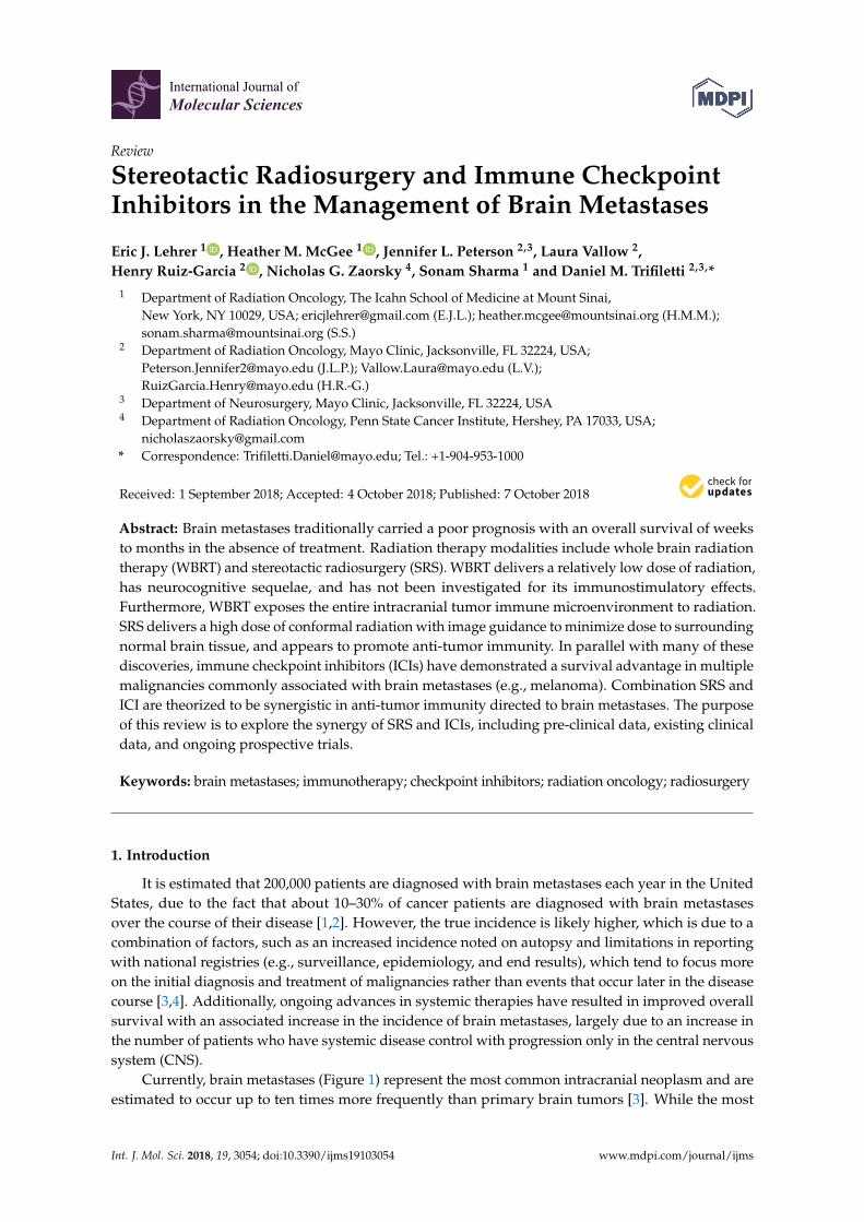
common primary cancers that metastasize to the brain are non-small cell carcinoma, melanoma, renalcell carcinoma, and breast cancer, there is now an increased number of patients with brain metastasesdue to less common histologies (e.g., pancreatic cancer), as survival improves for these types of cancersthat traditionally had a very poor prognosis [1,5,6]. Historically, the prognosis for brain metastaseswas poor with a median survival of 3–4 months in patients who were treated non-surgically [7].
Int. J. Mol. Sci. 2018, 19, x 2 of 19
common primary cancers that metastasize to the brain are non-small cell carcinoma, melanoma, renal cell carcinoma, and breast cancer, there is now an increased number of patients with brain metastases due to less common histologies (e.g., pancreatic cancer), as survival improves for these types of cancers that traditionally had a very poor prognosis [1,5,6]. Historically, the prognosis for brain metastases was poor with a median survival of 3–4 months in patients who were treated non-surgically [7].
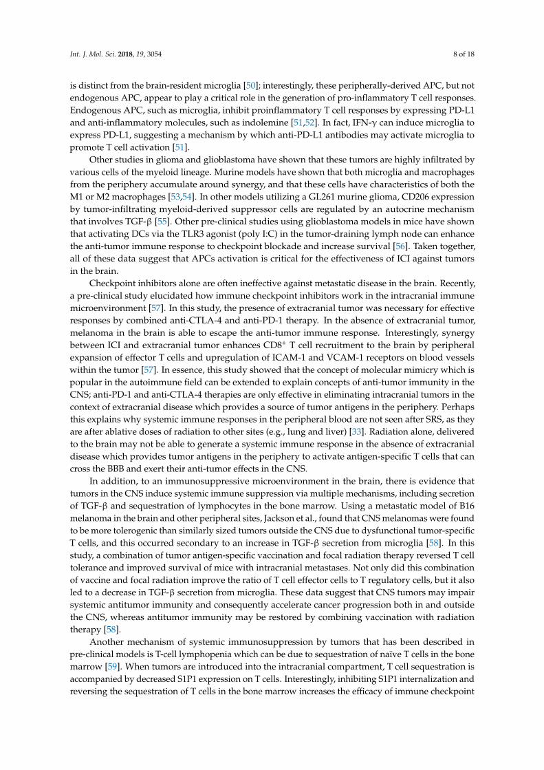
Figure 1. A T1 post-contrast axial magnetic resonance image of a contrast-enhancing tumor (circled in red). This patient is a 57-year-old female with metastatic breast carcinoma. The image and presentation are consistent with a brain metastasis.
For many years, brain metastases were considered a discrete condition with very little attention paid to the underlying biology of each patient’s disease. A paradigm shift occurred in 1997 when a recursive partitioning analysis (RPA) including 1200 patients from three Radiation Therapy Oncology Group studies was published [8]. Median survival time ranged from the best survival of 7.1 months, for patients with a Karnofsky Performance Status (KPS) > 70%, age < 65 years, and a controlled primary tumor with no other site of metastatic disease (RPA class I), to the worst survival of 2.3 months, for patients with a KPS < 70% (RPA class III). All other patients (RPA class II) had a median survival of 4.2 months. From this point forward, individual patient characteristics began to play more of a prominent role in the management of brain metastases. Today, the Diagnosis-Specific Graded Prognostic Assessment (DS-GPA) accounts for the primary tumor type and other unique features in order to take individual patient characteristics into account (e.g., age, KPS, and histology) [9]. However, the DS-GPA is not without limitations, such as it being based on retrospective data involving patients treated between 1985 and 2007. During (and since) that time period, there have been many new advances in treating and staging these patients. Therefore, it is important to use the DS-GPA in the context of individual patient factors.
2. Traditional Treatment of Brain Metastases
Figure 1. A T1 post-contrast axial magnetic resonance image of a contrast-enhancing tumor (circled in red).This patient is a 57-year-old female with metastatic breast carcinoma. The image and presentation areconsistent with a brain metastasis.
For many years, brain metastases were considered a discrete condition with very little attentionpaid to the underlying biology of each patient’s disease. A paradigm shift occurred in 1997 when arecursive partitioning analysis (RPA) including 1200 patients from three Radiation Therapy OncologyGroup studies was published [8]. Median survival time ranged from the best survival of 7.1 months,for patients with a Karnofsky Performance Status (KPS) > 70%, age < 65 years, and a controlledprimary tumor with no other site of metastatic disease (RPA class I), to the worst survival of 2.3 months,for patients with a KPS < 70% (RPA class III). All other patients (RPA class II) had a median survivalof 4.2 months. From this point forward, individual patient characteristics began to play more ofa prominent role in the management of brain metastases. Today, the Diagnosis-Specific GradedPrognostic Assessment (DS-GPA) accounts for the primary tumor type and other unique features inorder to take individual patient characteristics into account (e.g., age, KPS, and histology) [9]. However,the DS-GPA is not without limitations, such as it being based on retrospective data involving patientstreated between 1985 and 2007. During (and since) that time period, there have been many newadvances in treating and staging these patients. Therefore, it is important to use the DS-GPA in thecontext of individual patient factors.
Int. J. Mol. Sci. 2018, 19, 3054 3 of 18
2. Traditional Treatment of Brain Metastases
Traditional treatment strategies for brain metastases involves a complex multimodality regimen,consisting of a combination of surgery, SRS, WBRT, glucocorticoids, or systemic treatment alone.The choice of treatment is often guided by patient specific factors (e.g., DS-GPA and medicalcomorbidities); however, the optimal way to coordinate these therapies is unknown and prospectivedata are needed to ascertain the answer to this question. Whole brain radiation therapy had been themainstay of radiation treatment for decades in these patients. Most commonly, WBRT is deliveredover the course of two weeks, to a total dose of 30 Gy in 10 fractions [10,11]. While WBRT has beenshown to improve rates of local tumor control (LC), it is not associated with an improvement in overallsurvival (OS) [12–14]. Furthermore, its association with late toxicities and a decrease in quality oflife due to neurocognitive decline has led to the development of hippocampal sparing WBRT [15–22].WBRT remains an excellent treatment option for patients with a large burden of metastatic disease andhas the added benefit of targeting microscopic disease that has disseminated to other locations withinthe brain.
Patchell et al. authored the original study that demonstrated a benefit with the addition ofsurgery to WBRT, which included 48 patients with single brain metastases randomized to surgicalresection + WBRT versus biopsy only + WBRT [23]. The surgery arm had a decreased rate of localrecurrence (20% surgery + WBRT versus 52% WBRT alone); additionally, the surgery arm had improvedoverall survival (40 weeks versus 15 weeks). Most importantly, patients who received surgery hadan improved quality of life with a significantly increased time to remain functionally independent,38 weeks in the surgery arm versus eight weeks in the WBRT arm [23]. This study showed that surgerycombined with WBRT over WBRT alone was associated with a longer time to recurrence, improvedoverall survival, decreased risk of neurologic death, and increased functional independence.
Patchell et al. conducted a subsequent trial in 1998, exploring whether surgery alone withoutWBRT was sufficient [12]. This phase III trial enrolled 95 patients with single brain metastases andMRI-confirmed gross total resection with randomization to surgery +/−WBRT. Local failure at oneyear was 66% after surgery versus 20% after surgery + WBRT. The distant failure at one year was 50%for the surgery only arm versus 18% for surgery + WBRT. Additionally, there was a decreased riskof neurologic death (44% after surgery alone vs 15% after surgery + WBRT). However, there was nosignificant difference in median OS with or without the addition of WBRT following surgery [12].
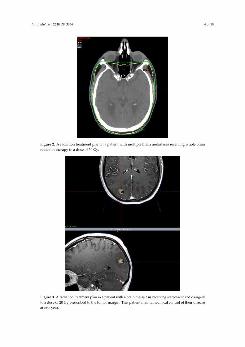
Since the 1990s, there have been multiple prospective randomized trials demonstrating localfailure rates of 55–65% with surgical resection alone, and a reduction in both locoregional failureand the incidence of new metastases with WBRT following resection [24,25]. These findings mayindicate synergy between surgical resection and WBRT. Figure 2 depicts a patient with brain metastaseswho was treated with WBRT to a dose of 30 Gy. As depicted, the entire intracranial tumor immunemicroenvironment is impacted by WBRT.
3. Stereotactic Radiosurgery
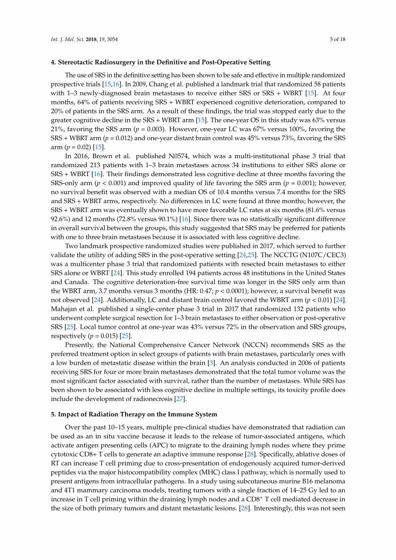
Stereotactic radiosurgery is the delivery of a large dose of highly conformal radiation therapy(RT) in a single session to a specified target while limiting dose to normal tissue, as shown in Figure 3.The original Gamma Knife® was developed in the 1960s by Swedish neurosurgeon Lars Leksell,which uses cobalt sources to deliver RT. Traditionally, delivery of SRS via this platform requires theplacement of a stereotactic frame; however, now linear accelerator (LINAC)-based SRS, Cyberknife®,and Gamma Knife ICON® technologies are all options to treat patients with “frameless” SRS. SRSoffers the option of increased LC without the neurocognitive side effects of WBRT; therefore, SRS hasemerged as one of the most effective treatments for brain metastases [16,24,26]. Additionally, it canoften be performed in a single session and may not require interruption or delay in initiating systemictherapies, such as immunotherapy.
Int. J. Mol. Sci. 2018, 19, 3054 4 of 18
Int. J. Mol. Sci. 2018, 19, x 4 of 19
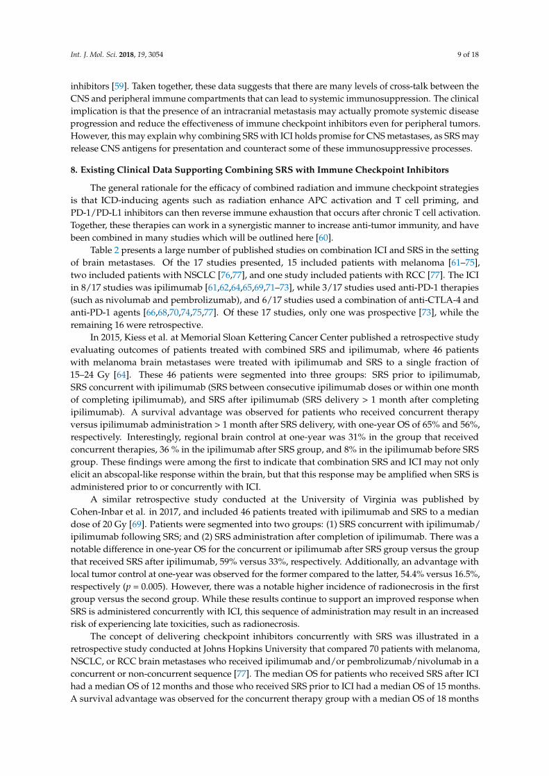
Figure 2. A radiation treatment plan in a patient with multiple brain metastases receiving whole brain radiation therapy to a dose of 30 Gy.
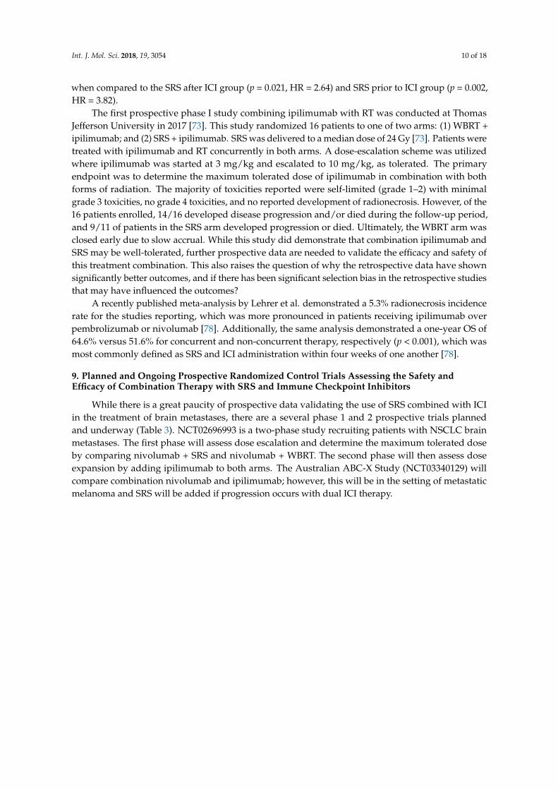
Figure 3. A radiation treatment plan in a patient with a brain metastasis receiving stereotactic radiosurgery to a dose of 20 Gy prescribed to the tumor margin. This patient maintained local control of their disease at one year.
Figure 2. A radiation treatment plan in a patient with multiple brain metastases receiving whole brainradiation therapy to a dose of 30 Gy.
Int. J. Mol. Sci. 2018, 19, x 4 of 19
Figure 2. A radiation treatment plan in a patient with multiple brain metastases receiving whole brain radiation therapy to a dose of 30 Gy.
Figure 3. A radiation treatment plan in a patient with a brain metastasis receiving stereotactic radiosurgery to a dose of 20 Gy prescribed to the tumor margin. This patient maintained local control of their disease at one year.
Figure 3. A radiation treatment plan in a patient with a brain metastasis receiving stereotactic radiosurgeryto a dose of 20 Gy prescribed to the tumor margin. This patient maintained local control of their diseaseat one year.
Int. J. Mol. Sci. 2018, 19, 3054 5 of 18
4. Stereotactic Radiosurgery in the Definitive and Post-Operative Setting
The use of SRS in the definitive setting has been shown to be safe and effective in multiple randomizedprospective trials [15,16]. In 2009, Chang et al. published a landmark trial that randomized 58 patientswith 1–3 newly-diagnosed brain metastases to receive either SRS or SRS + WBRT [15]. At fourmonths, 64% of patients receiving SRS + WBRT experienced cognitive deterioration, compared to20% of patients in the SRS arm. As a result of these findings, the trial was stopped early due to thegreater cognitive decline in the SRS + WBRT arm [15]. The one-year OS in this study was 63% versus21%, favoring the SRS arm (p = 0.003). However, one-year LC was 67% versus 100%, favoring theSRS + WBRT arm (p = 0.012) and one-year distant brain control was 45% versus 73%, favoring the SRSarm (p = 0.02) [15].
In 2016, Brown et al. published N0574, which was a multi-institutional phase 3 trial thatrandomized 213 patients with 1–3 brain metastases across 34 institutions to either SRS alone orSRS + WBRT [16]. Their findings demonstrated less cognitive decline at three months favoring theSRS-only arm (p < 0.001) and improved quality of life favoring the SRS arm (p = 0.001); however,no survival benefit was observed with a median OS of 10.4 months versus 7.4 months for the SRSand SRS + WBRT arms, respectively. No differences in LC were found at three months; however, theSRS + WBRT arm was eventually shown to have more favorable LC rates at six months (81.6% versus92.6%) and 12 months (72.8% versus 90.1%) [16]. Since there was no statistically significant differencein overall survival between the groups, this study suggested that SRS may be preferred for patientswith one to three brain metastases because it is associated with less cognitive decline.
Two landmark prospective randomized studies were published in 2017, which served to furthervalidate the utility of adding SRS in the post-operative setting [24,25]. The NCCTG (N107C/CEC3)was a multicenter phase 3 trial that randomized patients with resected brain metastases to eitherSRS alone or WBRT [24]. This study enrolled 194 patients across 48 institutions in the United Statesand Canada. The cognitive deterioration-free survival time was longer in the SRS only arm thanthe WBRT arm, 3.7 months versus 3 months (HR: 0.47; p < 0.0001); however, a survival benefit wasnot observed [24]. Additionally, LC and distant brain control favored the WBRT arm (p < 0.01) [24].Mahajan et al. published a single-center phase 3 trial in 2017 that randomized 132 patients whounderwent complete surgical resection for 1–3 brain metastases to either observation or post-operativeSRS [25]. Local tumor control at one-year was 43% versus 72% in the observation and SRS groups,respectively (p = 0.015) [25].
Presently, the National Comprehensive Cancer Network (NCCN) recommends SRS as thepreferred treatment option in select groups of patients with brain metastases, particularly ones witha low burden of metastatic disease within the brain [3]. An analysis conducted in 2006 of patientsreceiving SRS for four or more brain metastases demonstrated that the total tumor volume was themost significant factor associated with survival, rather than the number of metastases. While SRS hasbeen shown to be associated with less cognitive decline in multiple settings, its toxicity profile doesinclude the development of radionecrosis [27].
5. Impact of Radiation Therapy on the Immune System
Over the past 10–15 years, multiple pre-clinical studies have demonstrated that radiation canbe used as an in situ vaccine because it leads to the release of tumor-associated antigens, whichactivate antigen presenting cells (APC) to migrate to the draining lymph nodes where they primecytotoxic CD8+ T cells to generate an adaptive immune response [28]. Specifically, ablative doses ofRT can increase T cell priming due to cross-presentation of endogenously acquired tumor-derivedpeptides via the major histocompatibility complex (MHC) class I pathway, which is normally used topresent antigens from intracellular pathogens. In a study using subcutaneous murine B16 melanomaand 4T1 mammary carcinoma models, treating tumors with a single fraction of 14–25 Gy led to anincrease in T cell priming within the draining lymph nodes and a CD8+ T cell mediated decrease inthe size of both primary tumors and distant metastatic lesions. [28]. Interestingly, this was not seen
Int. J. Mol. Sci. 2018, 19, 3054 6 of 18
after treatment with conventionally fractionated RT [28]. However, other studies have emphasizedthat hypofractionated radiation regimens (e.g., 8 Gy × 3 fractions) result in a more robust CD8+ Tcell-mediated abscopal response and improved survival when compared to high-dose single fraction(e.g., 20 Gy × 1 fraction) in the context of cytotoxic T-lymphocyte-associated protein 4 (CTLA-4)blockade [29]. One explanation for this is that hypofractionated radiation causes double-strandedDNA (dsDNA) to accumulate in the cytoplasm, which stimulates the cGAS/STING (STimulator ofINterferon Genes)/IFN-beta pathway to recruit Batf3+ dendritic cells (DC) that can activate CD8+ Tcell-mediated abscopal responses. However, radiation doses > 12 Gy per fraction induce the expressionof the DNA exonuclease three prime repair exonuclease 1 (TREX1; 3’→ 5’), which degrades cytosolicdsDNA, abrogating radiation-induced c-GAS/STING induction of Type I interferon (IFN) responses,and leading to a decrease systemic anti-tumor immunity [30]. Immunogenic cell death (ICD), which canoccur after radiotherapy, is characterized by the exposure of calreticulin on the surface of dying cells, aswell as release of ATP (adenosine triphosphate) and secretion of high-mobility group box 1 protein intothe tumor microenvironment [31]. All of these markers of ICD can increase antigen-presentation andsubsequent CD8+ T cell activation. However, an important caveat is that all of the studies investigatingthese pathways used subcutaneous murine models.
Additional evidence suggests that radiation alone also activates many immunosuppressivemechanisms in the tumor microenvironment, such as release of transforming growth factor beta(TGF-β) which causes conversion of CD4+ T cells to T regulatory cells (Tregs), and polarization of tumorassociated macrophages (TAMs) into an immunosuppressive M2 phenotype. In addition, radiationleads to the release of ATP, which is rapidly catabolized into adenosine in the tumor microenvironment.Local accumulation of extracellular adenosine suppresses DC and effector T cells while promotingproliferation of Tregs [32]. Emerging data suggests that the site which is irradiated may play a role indetermining the balance between immune activating and immunosuppressive mechanisms [33].
6. Immune Checkpoint Inhibitors
The ability to enhance anti-tumor responses by the immune system offers great promise and haselicited a great deal of enthusiasm, particularly in the development of immune checkpoint inhibitors.In 2011, the U.S. Food and Drug Administration (FDA) approved ipilimumab, a monoclonal antibodyagainst CTLA-4 for use in refractory metastatic melanoma [34,35]. CTLA-4 is a receptor on the surfaceof T cells that binds to the B7-1 molecule in antigen presenting cells, resulting in the delivery of aninhibitory signal which downregulates T cell activity. In 2014, the FDA approved pembrolizumaband nivolumab, which are monoclonal antibodies against programmed death cell protein 1 (PD-1).PD-1 is a receptor that is present on the surface of T cells that is a member of the immunoglobulinsuperfamily and binds to programmed death ligand-1 (PD-L1) and PD-L2 to decrease T cell activity.PD-L1 is expressed on a wide repertoire of cells such as tumor cells, macrophages, dendritic cells, andB cells. In 2016, atezolizumab, an inhibitor of PD-L1 became FDA approved for bladder cancer andnon-small cell lung cancer (NSCLC).
Pembrolizumab has been shown to improve OS and progression free survival with fewerside-effects when compared to platinum-based chemotherapy in the treatment of advanced NSCLCwith at least 50% PD-L1 expression on tumor cells [36]. Presently, pembrolizumab is FDA approvedfor melanoma, NSCLC, head and neck cancer, urothelial carcinoma, gastric cancer, cervical cancer, andHodgkin lymphoma. Nivolumab has also shown promise in the clinical setting; two phase 3 trialspublished in 2015 demonstrated a survival advantage in patients with advanced NSCLC comparedto chemotherapy [37,38]. Nivolumab is presently FDA approved for melanoma, NSCLC, renal cellcarcinoma (RCC), Hodgkin lymphoma, head and neck cancer, urothelial carcinoma, colorectal cancer(CRC), hepatocellular carcinoma (HCC), and small cell lung cancer (SCLC) (Table 1).
Int. J. Mol. Sci. 2018, 19, 3054 7 of 18
Table 1. Immune checkpoint inhibitors and indications.
Drug Target FDA Approved Indications
Ipilimumab CTLA-4 Metastatic Melanoma
Pembrolizumab PD-1Metastatic Melanoma, NSCLC, Head and Neck Cancer,
Hodgkin Lymphoma, Urothelial Carcinoma, GastricCancer, Cervical Cancer
Nivolumab PD-1Metastatic Melanoma, NSCLC, RCC, Hodgkin Lymphoma,
Head and Neck Cancer, Urothelial Carcinoma, CRC,HCC, SCLC
Atezolizumab PD-L1 Bladder Cancer, NSCLC
Ipilimumab + Nivolumab CTLA-4 + PD-1 RCC, CRC
7. Rationale for Combining SRS and Immune Checkpoint Inhibitors in Treating Brain Metastases
The CNS had been viewed as immunologically isolated from the peripheral immune systembased on experiments conducted in the early-mid 20th century, where tumors and fetal tissuestransplanted into the brain parenchyma escaped rejection, unlike their counterparts transplanted inthe periphery [39–41]. The prevailing opinion for many years was that the CNS was “immunologicallyprivileged” due to: (1) the presence of the blood-brain barrier (BBB), a highly selective permeabilitybarrier formed by endothelial cells and astroglial cells which regulate the entry of blood-bornemetabolites and toxins into the brain [42]; (2) the absence of a readily draining lymphatic systemwhich prevented T cells in the cervical lymph nodes from being exposed to CNS antigens; and (3) thebelief that tissue resident macrophages in the CNS (microglia) were ineffective APC due to limitedexpression of MHC or costimulatory molecules in the CNS parenchyma [43]. Unfortunately, thisresulted in decreased efforts within the scientific community to develop systemic therapies targetingmetastatic disease in the brain. However, more recent work has suggested that if a robust immuneresponse is generated outside the CNS, the immune system can bypass all three of these barriers [44].
Data from multiple groups has challenged the view of immune privilege by revealing that theCNS is not isolated from the rest of the immune system. Neuroinflammation can alter the vasculatureof the BBB in order to allow peripheral immune cells to cross the BBB, microglia can play a key role inorchestrating interactions with other immune cells, and T cells activated in the periphery can detectantigens located in the CNS parenchyma [44]. Activated T cells express Very late antigen-4 (VLA-4)and leukocyte-function-associated antigen-1 (LFA-1), which aid T cell migration across the BBB [45,46].Additionally, recent evidence suggests there is an alternative route of T cell entry into the CNS thatis independent of the BBB, in which T cells can migrate from the blood to the CSF via the choroidplexuses within the ventricles [43]. In addition, the discovery of the lymphatic system draining fromthe CNS to the cervical lymph nodes has provided further evidence that T cells can be activated byCNS-derived antigens.
The first evidence of antigen-specific T cell responses in the CNS came from studies of the“molecular mimicry hypothesis” of autoimmunity. Immune responses against pathogenic epitopeswith similarity to molecules in the CNS can clear the pathogens and lead to T cell mediated responsesagainst similar antigens in the CNS. Original evidence of this hypothesis came from pre-clinical studiesin a murine model of multiple sclerosis called experimentally-induced autoimmune encephalitis,where activated T cells specific for components of CNS myelin can be transferred into geneticallysusceptible mice to trigger CNS inflammation and hindlimb paralysis [47,48].
In addition, activation of the peripheral immune response can lead to systemic productionof tumor necrosis factor and nitric oxide, which promote transmigration of macrophages and DCacross the BBB in order to serve as APC in the brain [49]. In fact, recent evidence has emergedsuggesting that peripherally derived macrophages can engraft the brain and maintain an identity that
Int. J. Mol. Sci. 2018, 19, 3054 8 of 18
is distinct from the brain-resident microglia [50]; interestingly, these peripherally-derived APC, but notendogenous APC, appear to play a critical role in the generation of pro-inflammatory T cell responses.Endogenous APC, such as microglia, inhibit proinflammatory T cell responses by expressing PD-L1and anti-inflammatory molecules, such as indolemine [51,52]. In fact, IFN-γ can induce microglia toexpress PD-L1, suggesting a mechanism by which anti-PD-L1 antibodies may activate microglia topromote T cell activation [51].
Other studies in glioma and glioblastoma have shown that these tumors are highly infiltrated byvarious cells of the myeloid lineage. Murine models have shown that both microglia and macrophagesfrom the periphery accumulate around synergy, and that these cells have characteristics of both theM1 or M2 macrophages [53,54]. In other models utilizing a GL261 murine glioma, CD206 expressionby tumor-infiltrating myeloid-derived suppressor cells are regulated by an autocrine mechanismthat involves TGF-β [55]. Other pre-clinical studies using glioblastoma models in mice have shownthat activating DCs via the TLR3 agonist (poly I:C) in the tumor-draining lymph node can enhancethe anti-tumor immune response to checkpoint blockade and increase survival [56]. Taken together,all of these data suggest that APCs activation is critical for the effectiveness of ICI against tumorsin the brain.
Checkpoint inhibitors alone are often ineffective against metastatic disease in the brain. Recently,a pre-clinical study elucidated how immune checkpoint inhibitors work in the intracranial immunemicroenvironment [57]. In this study, the presence of extracranial tumor was necessary for effectiveresponses by combined anti-CTLA-4 and anti-PD-1 therapy. In the absence of extracranial tumor,melanoma in the brain is able to escape the anti-tumor immune response. Interestingly, synergybetween ICI and extracranial tumor enhances CD8+ T cell recruitment to the brain by peripheralexpansion of effector T cells and upregulation of ICAM-1 and VCAM-1 receptors on blood vesselswithin the tumor [57]. In essence, this study showed that the concept of molecular mimicry which ispopular in the autoimmune field can be extended to explain concepts of anti-tumor immunity in theCNS; anti-PD-1 and anti-CTLA-4 therapies are only effective in eliminating intracranial tumors in thecontext of extracranial disease which provides a source of tumor antigens in the periphery. Perhapsthis explains why systemic immune responses in the peripheral blood are not seen after SRS, as theyare after ablative doses of radiation to other sites (e.g., lung and liver) [33]. Radiation alone, deliveredto the brain may not be able to generate a systemic immune response in the absence of extracranialdisease which provides tumor antigens in the periphery to activate antigen-specific T cells that cancross the BBB and exert their anti-tumor effects in the CNS.
In addition, to an immunosuppressive microenvironment in the brain, there is evidence thattumors in the CNS induce systemic immune suppression via multiple mechanisms, including secretionof TGF-β and sequestration of lymphocytes in the bone marrow. Using a metastatic model of B16melanoma in the brain and other peripheral sites, Jackson et al., found that CNS melanomas were foundto be more tolerogenic than similarly sized tumors outside the CNS due to dysfunctional tumor-specificT cells, and this occurred secondary to an increase in TGF-β secretion from microglia [58]. In thisstudy, a combination of tumor antigen-specific vaccination and focal radiation therapy reversed T celltolerance and improved survival of mice with intracranial metastases. Not only did this combinationof vaccine and focal radiation improve the ratio of T cell effector cells to T regulatory cells, but it alsoled to a decrease in TGF-β secretion from microglia. These data suggest that CNS tumors may impairsystemic antitumor immunity and consequently accelerate cancer progression both in and outsidethe CNS, whereas antitumor immunity may be restored by combining vaccination with radiationtherapy [58].
Another mechanism of systemic immunosuppression by tumors that has been described inpre-clinical models is T-cell lymphopenia which can be due to sequestration of naïve T cells in the bonemarrow [59]. When tumors are introduced into the intracranial compartment, T cell sequestration isaccompanied by decreased S1P1 expression on T cells. Interestingly, inhibiting S1P1 internalization andreversing the sequestration of T cells in the bone marrow increases the efficacy of immune checkpoint
Int. J. Mol. Sci. 2018, 19, 3054 9 of 18
inhibitors [59]. Taken together, these data suggests that there are many levels of cross-talk between theCNS and peripheral immune compartments that can lead to systemic immunosuppression. The clinicalimplication is that the presence of an intracranial metastasis may actually promote systemic diseaseprogression and reduce the effectiveness of immune checkpoint inhibitors even for peripheral tumors.However, this may explain why combining SRS with ICI holds promise for CNS metastases, as SRS mayrelease CNS antigens for presentation and counteract some of these immunosuppressive processes.
8. Existing Clinical Data Supporting Combining SRS with Immune Checkpoint Inhibitors
The general rationale for the efficacy of combined radiation and immune checkpoint strategiesis that ICD-inducing agents such as radiation enhance APC activation and T cell priming, andPD-1/PD-L1 inhibitors can then reverse immune exhaustion that occurs after chronic T cell activation.Together, these therapies can work in a synergistic manner to increase anti-tumor immunity, and havebeen combined in many studies which will be outlined here [60].
Table 2 presents a large number of published studies on combination ICI and SRS in the settingof brain metastases. Of the 17 studies presented, 15 included patients with melanoma [61–75],two included patients with NSCLC [76,77], and one study included patients with RCC [77]. The ICIin 8/17 studies was ipilimumab [61,62,64,65,69,71–73], while 3/17 studies used anti-PD-1 therapies(such as nivolumab and pembrolizumab), and 6/17 studies used a combination of anti-CTLA-4 andanti-PD-1 agents [66,68,70,74,75,77]. Of these 17 studies, only one was prospective [73], while theremaining 16 were retrospective.
In 2015, Kiess et al. at Memorial Sloan Kettering Cancer Center published a retrospective studyevaluating outcomes of patients treated with combined SRS and ipilimumab, where 46 patientswith melanoma brain metastases were treated with ipilimumab and SRS to a single fraction of15–24 Gy [64]. These 46 patients were segmented into three groups: SRS prior to ipilimumab,SRS concurrent with ipilimumab (SRS between consecutive ipilimumab doses or within one monthof completing ipilimumab), and SRS after ipilimumab (SRS delivery > 1 month after completingipilimumab). A survival advantage was observed for patients who received concurrent therapyversus ipilimumab administration > 1 month after SRS delivery, with one-year OS of 65% and 56%,respectively. Interestingly, regional brain control at one-year was 31% in the group that receivedconcurrent therapies, 36 % in the ipilimumab after SRS group, and 8% in the ipilimumab before SRSgroup. These findings were among the first to indicate that combination SRS and ICI may not onlyelicit an abscopal-like response within the brain, but that this response may be amplified when SRS isadministered prior to or concurrently with ICI.
A similar retrospective study conducted at the University of Virginia was published byCohen-Inbar et al. in 2017, and included 46 patients treated with ipilimumab and SRS to a mediandose of 20 Gy [69]. Patients were segmented into two groups: (1) SRS concurrent with ipilimumab/ipilimumab following SRS; and (2) SRS administration after completion of ipilimumab. There was anotable difference in one-year OS for the concurrent or ipilimumab after SRS group versus the groupthat received SRS after ipilimumab, 59% versus 33%, respectively. Additionally, an advantage withlocal tumor control at one-year was observed for the former compared to the latter, 54.4% versus 16.5%,respectively (p = 0.005). However, there was a notable higher incidence of radionecrosis in the firstgroup versus the second group. While these results continue to support an improved response whenSRS is administered concurrently with ICI, this sequence of administration may result in an increasedrisk of experiencing late toxicities, such as radionecrosis.
The concept of delivering checkpoint inhibitors concurrently with SRS was illustrated in aretrospective study conducted at Johns Hopkins University that compared 70 patients with melanoma,NSCLC, or RCC brain metastases who received ipilimumab and/or pembrolizumab/nivolumab in aconcurrent or non-concurrent sequence [77]. The median OS for patients who received SRS after ICIhad a median OS of 12 months and those who received SRS prior to ICI had a median OS of 15 months.A survival advantage was observed for the concurrent therapy group with a median OS of 18 months
Int. J. Mol. Sci. 2018, 19, 3054 10 of 18
when compared to the SRS after ICI group (p = 0.021, HR = 2.64) and SRS prior to ICI group (p = 0.002,HR = 3.82).
The first prospective phase I study combining ipilimumab with RT was conducted at ThomasJefferson University in 2017 [73]. This study randomized 16 patients to one of two arms: (1) WBRT +ipilimumab; and (2) SRS + ipilimumab. SRS was delivered to a median dose of 24 Gy [73]. Patients weretreated with ipilimumab and RT concurrently in both arms. A dose-escalation scheme was utilizedwhere ipilimumab was started at 3 mg/kg and escalated to 10 mg/kg, as tolerated. The primaryendpoint was to determine the maximum tolerated dose of ipilimumab in combination with bothforms of radiation. The majority of toxicities reported were self-limited (grade 1–2) with minimalgrade 3 toxicities, no grade 4 toxicities, and no reported development of radionecrosis. However, of the16 patients enrolled, 14/16 developed disease progression and/or died during the follow-up period,and 9/11 of patients in the SRS arm developed progression or died. Ultimately, the WBRT arm wasclosed early due to slow accrual. While this study did demonstrate that combination ipilimumab andSRS may be well-tolerated, further prospective data are needed to validate the efficacy and safety ofthis treatment combination. This also raises the question of why the retrospective data have shownsignificantly better outcomes, and if there has been significant selection bias in the retrospective studiesthat may have influenced the outcomes?
A recently published meta-analysis by Lehrer et al. demonstrated a 5.3% radionecrosis incidencerate for the studies reporting, which was more pronounced in patients receiving ipilimumab overpembrolizumab or nivolumab [78]. Additionally, the same analysis demonstrated a one-year OS of64.6% versus 51.6% for concurrent and non-concurrent therapy, respectively (p < 0.001), which wasmost commonly defined as SRS and ICI administration within four weeks of one another [78].
9. Planned and Ongoing Prospective Randomized Control Trials Assessing the Safety andEfficacy of Combination Therapy with SRS and Immune Checkpoint Inhibitors
While there is a great paucity of prospective data validating the use of SRS combined with ICIin the treatment of brain metastases, there are a several phase 1 and 2 prospective trials plannedand underway (Table 3). NCT02696993 is a two-phase study recruiting patients with NSCLC brainmetastases. The first phase will assess dose escalation and determine the maximum tolerated doseby comparing nivolumab + SRS and nivolumab + WBRT. The second phase will then assess doseexpansion by adding ipilimumab to both arms. The Australian ABC-X Study (NCT03340129) willcompare combination nivolumab and ipilimumab; however, this will be in the setting of metastaticmelanoma and SRS will be added if progression occurs with dual ICI therapy.

Int. J. Mol. Sci. 2018, 19, 3054 11 of 18
Table 2. Selected studies combining SRS with immune checkpoint inhibitors in brain metastases.
Study H Arm N ICI Target DS-GPA 1-Year OS (%) 1-Year LC (%) 1-Year RBC (%) RN
Mathew et al., 2013 [61]. M NR 25 CTLA-4 NR 32.6 40 16 NR
Silk et al., 2013 [62]. M NR 17 CTLA-4 25% (0-1); 39.3% (2); 25% (3); 10.7% (4) 82.3 NR NR NR
Ahmed et al., 2016 [63]. M NR 26 PD-1 27% (1-2); 19% (3-4) 74.7 82 45.9 NR
Kiess et al., 2015 [64]. MSRS→ ICI 19
CTLA-4 3 (median)56 87 36 NR
SRS = ICI 15 65 100 31 NRICI→ SRS 12 50 89 8 NR
Schoenfeld et al., 2015 [65]. MSRS→ ICI 5
CTLA-4 NRNR NR NR NR
SRS = ICI 4 NR NR NR NRICI→ SRS 7 NR NR NR NR
Qian et al., 2015 [66]. MSRS 6= ICI 22
CTLA-4 or PD-13 (median) 44.4 NR NR NR
SRS = ICI 33 2 (median) 62.5 NR NR NR
Ahmed et al., 2017 [76]. NSCLC NR 17 PD-1 59% (0-1.5); 41% (2-3) 40.0 96.0 0.0 NR
Anderson et al., 2017 [67]. M NR 11 PD-1 3 (median) NR NR NR 0
Choong et al., 2017 [68]. M NR 39 CTLA-4 or PD-1 NR 54.9 NR NR 5
Cohen-Inbar et al., 2017 [69]. MSRS = ICI;SRS→ ICI 32
CTLA-42.5% (0-1); 53% (2); 18.8% (3); 15.6% (4) 59.0 54.4 25.8 31
ICI→ SRS 14 14.3% (0-1); 64.3% (2); 0% (3); 21.4% (4) 33.0 16.5 26.8 7
Gaudy-Marqueste et al.,2017 [70]. M SRS→ ICI 43 CTLA-4 or PD-1 NR 52.4 NR NR NR
Patel et al., 2017 [71]. M NR 20 CTLA-4 10% (1); 35% (2); 30% (3); 25% (4) 37.1 71.4 12.7 NR
Skrepnik et al., 2017 [72]. M NR 25 CTLA-4 NR 83.0 94.8 72.0 12
Williams et al., 2017 [73]. M NR 11 CTLA-4 NR 60.0 NR NR 0
Yusuf et al., 2017 [75]. MSRS = ICI 12
CTLA-4 or PD-1 NR45.0 87.6 46.4 2
SRS 6= ICI 6 21.5 NR 0.0 0
Acharya et al., 2017 [74]. M NR 18 CTLA-4 and/orPD-1 6% (1); 28% (2); 39% (3); 0% (4) 58.5 85.0 60.0 NR
Chen et al., 2018 [77].M,
NSCLC,RCC
SRS→ ICI 30CTLA-4 and/or
PD-1NR
63.6 NR NR NRSRS = ICI 28 77.9 88.0 NR NRICI→ SRS 23 50.7 NR NR NR
Note: ICI→ SRS indicates ICI was administered prior to SRS; SRS = ICI indicates that ICI was administered concurrently with SRS; SRS→ ICI indicates that ICI was administered afterSRS; SRS 6= ICI indicates that SRS was not administered concurrently with SRS but the relative timing of each treatment was not provided; Radionecrosis (RN) expressed as number oflesions affected

Int. J. Mol. Sci. 2018, 19, 3054 12 of 18
Table 3. Active and planned randomized control trials assessing the safety and efficacy of SRS and immune checkpoint inhibitors in the treatment of brain metastases.
Study Phase Country Histology SRS Dose ICI Target Primary Outcome
NCT02886585 2 USA Melanoma NR PD-1Overall Response Rate; OverallSurvival; Extracranial Overall
Response Rate
NCT02858869 1 USA Melanoma andNSCLC
30 Gy/5 fractions27 Gy/3 fractions
18-21 Gy/1 fractionPD-1 Dose-Limiting Toxicities
NCT02978404 2 Canada NSCLC and RCC 15-20 Gy/1 fraction PD-1 Progression-Free Survival
NCT02696993 1 & 2 USA NSCLC NR CTLA-4 and PD-1 Maximum Tolerated Dose;Dose-Limiting Toxicities
NCT03340129 2 Australia Melanoma 16-22 Gy/1 fraction CTLA-4 and PD-1 Intracranial Response to ImmuneCheckpoint Inhibitor
NCT02716948 1 USA Melanoma NR PD-1 Incidence of Severe Adverse Effects
NCT02097732 2 USA Melanoma NR CTLA-4 Local Control at 6 months
NCT01703507 1 USA Melanoma NR CTLA-4 Maximum Tolerated Dose
Int. J. Mol. Sci. 2018, 19, 3054 13 of 18
10. Conclusions
Patients with brain metastases have greater outcomes than ever before due to the evolution ofsurgical and SRS techniques alongside improved systemic treatment. Just as the use of SRS involvescareful consideration of size, location, mass effect, edema, symptomatic burden, and the extent ofvisceral disease, there is a need to investigate how all of these factors also influence the clinicalresponse to ICI. While ablative doses of RT, such as those administered with SRS appear to enhance theanti-tumor activity of the immune system, there is less evidence for this in the brain as compared toother organs. Recent discoveries challenging the belief that the brain is “immunologically privileged”have led to increased enthusiasm in combining SRS with immune checkpoint inhibitors. Combinationtherapy appears to enhance survival and abscopal-like responses within the brain, which may beamplified when administered concurrently rather than sequentially. Ongoing and planned prospectivetrials are needed to further explore and validate these findings. Future studies will explore elucidatingpredictive biomarkers that can be utilized to stratify patients based on the likelihood of the responseto such combined treatment approaches, but this will require collaboration between immunologists,oncologists, neurosurgeons, and radiation oncologists.
Author Contributions: All authors drafted and approved the final manuscript.
Conflicts of Interest: The authors declare no conflict of interest. H.M.M. was a former advisory board memberfor AstraZeneca; D.M.T. received clinical research funding from Novocure. The founding sponsors had no rolein the design of the study; in the collection, analyses, or interpretation of data; in the writing of the manuscript,and in the decision to publish the results.
Abbreviations
MDPI Multidisciplinary Digital Publishing InstituteDOAJ Directory of open access journalsWBRT Whole brain radiation therapySRS Stereotactic radiosurgeryICI Immune checkpoint inhibitorsCNS Central nervous systemRPA Recursive partitioning analysisKPS Karnofsky Performance StatusDS-GPA Diagnosis Specific–Graded Prognostic AssessmentGy GrayLC Local controlOS Overall survivalMRI Magnetic resonance imagingRT Radiation therapyLINAC Linear acceleratorNCCN National Comprehensive Cancer NetworkAPC Antigen presenting cellsMHC Major histocompatibility complexCTLA-4 Cytotoxic T-lymphocyte-associated protein 4dsDNA Double-stranded deoxyribonucleic acidSTING STimulator of INterferon GenesDC Dendritic cellsIFN InterferonICD Immunogenic cell deathATP Adenosine triphosphateTGF-β Transforming growth factor βTregs T regulatory cellsTAMs Tumor associated macrophagesDNA Deoxyribonucleic acidFDA Food and Drug Administration
Int. J. Mol. Sci. 2018, 19, 3054 14 of 18
PD-1 Programmed death cell protein 1PD-L1 Programmed death ligand-1NSCLC Non-small cell lung cancerRCC Renal cell carcinomaCRC Colorectal cancerHCC Hepatocellular carcinomaSCLC Small cell lung cancerBBB Blood brain barrierVLA-4 Very late antigen-4LFA-1 Leukocyte-function-associated antigen-1H HistologyM MelanomaN Total number of patientsNR Not reportedUSA United States of America
References
1. Barnholtz-Sloan, J.S.; Sloan, A.E.; Davis, F.G.; Vigneau, F.D.; Lai, P.; Sawaya, R.E. Incidence proportions ofbrain metastases in patients diagnosed (1973 to 2001) in the metropolitan detroit cancer surveillance system.J. Clin. Oncol. 2004, 22, 2865–2872. [CrossRef] [PubMed]
2. Kohler, B.A.; Ward, E.; McCarthy, B.J.; Schymura, M.J.; Ries, L.A.; Eheman, C.; Jemal, A.; Anderson, R.N.;Ajani, U.A.; Edwards, B.K. Annual report to the nation on the status of cancer, 1975–2007, featuring tumorsof the brain and other nervous system. J. Natl. Cancer Inst. 2011, 103, 714–736. [CrossRef] [PubMed]
3. Nabors, L.B.; Portnow, J.; Ammirati, M.; Baehring, J.; Brem, H.; Butowski, N.; Fenstermaker, R.A.; Forsyth, P.;Hattangadi-Gluth, J.; Holdhoff, M.; et al. Nccn practice guidelines in oncology—Central nervous systemcancers, version 1.2017. J. Natl. Compr. Canc. Netw. 2017, 15, 9–20. [CrossRef] [PubMed]
4. Arvold, N.D.; Lee, E.Q.; Mehta, M.P.; Margolin, K.; Alexander, B.M.; Lin, N.U.; Anders, C.K.; Soffietti, R.;Camidge, D.R.; Vogelbaum, M.A.; et al. Updates in the management of brain metastases. Neuro. Oncol. 2016,18, 1043–1065. [CrossRef] [PubMed]
5. Sanghvi, S.M.; Lischalk, J.W.; Cai, L.; Collins, S.; Nair, M.; Collins, B.; Unger, K. Clinical outcomes ofgastrointestinal brain metastases treated with radiotherapy. Radiat. Oncol. 2017, 12, 43. [CrossRef] [PubMed]
6. Trifiletti, D.M.; Patel, N.; Lee, C.C.; Romano, A.M.; Sheehan, J.P. Stereotactic radiosurgery in the treatment ofbrain metastases from gastrointestinal primaries. J. Neurooncol. 2015, 124, 439–446. [CrossRef] [PubMed]
7. Nieder, C.; Spanne, O.; Mehta, M.P.; Grosu, A.L.; Geinitz, H. Presentation, patterns of care, and survivalin patients with brain metastases: What has changed in the last 20 years? Cancer 2011, 117, 2505–2512.[CrossRef] [PubMed]
8. Gaspar, L.; Scott, C.; Rotman, M.; Asbell, S.; Phillips, T.; Wasserman, T.; McKenna, W.G.; Byhardt, R.Recursive partitioning analysis (rpa) of prognostic factors in three radiation therapy oncology group (rtog)brain metastases trials. Int. J. Radiat. Oncol. Biol. Phys. 1997, 37, 745–751. [CrossRef]
9. Sperduto, P.W.; Kased, N.; Roberge, D.; Xu, Z.; Shanley, R.; Luo, X.; Sneed, P.K.; Chao, S.T.; Weil, R.J.;Suh, J.; et al. Summary report on the graded prognostic assessment: An accurate and facile diagnosis-specifictool to estimate survival for patients with brain metastases. J. Clin. Oncol. 2012, 30, 419–425. [CrossRef][PubMed]
10. Borgelt, B.; Gelber, R.; Kramer, S.; Brady, L.W.; Chang, C.H.; Davis, L.W.; Perez, C.A.; Hendrickson, F.R.The palliation of brain metastases: Final results of the first two studies by the radiation therapy oncologygroup. Int. J. Radiat. Oncol. Biol. Phys. 1980, 6, 1–9. [CrossRef]
11. Sneed, P.K.; Larson, D.A.; Wara, W.M. Radiotherapy for cerebral metastases. Neurosurg. Clin. N. Am. 1996, 7,505–515. [CrossRef]
12. Patchell, R.A.; Tibbs, P.A.; Regine, W.F.; Dempsey, R.J.; Mohiuddin, M.; Kryscio, R.J.; Markesbery, W.R.;Foon, K.A.; Young, B. Postoperative radiotherapy in the treatment of single metastases to the brain:A randomized trial. JAMA 1998, 280, 1485–1489. [CrossRef] [PubMed]
Int. J. Mol. Sci. 2018, 19, 3054 15 of 18
13. McPherson, C.M.; Suki, D.; Feiz-Erfan, I.; Mahajan, A.; Chang, E.; Sawaya, R.; Lang, F.F. Adjuvant whole-brainradiation therapy after surgical resection of single brain metastases. Neuro. Oncol. 2010, 12, 711–719.[CrossRef] [PubMed]
14. Kocher, M.; Soffietti, R.; Abacioglu, U.; Villa, S.; Fauchon, F.; Baumert, B.G.; Fariselli, L.; Tzuk-Shina, T.;Kortmann, R.D.; Carrie, C.; et al. Adjuvant whole-brain radiotherapy versus observation after radiosurgeryor surgical resection of one to three cerebral metastases: Results of the eortc 22952–26001 study. J. Clin. Oncol.2011, 29, 134–141. [CrossRef] [PubMed]
15. Chang, E.L.; Wefel, J.S.; Hess, K.R.; Allen, P.K.; Lang, F.F.; Kornguth, D.G.; Arbuckle, R.B.; Swint, J.M.;Shiu, A.S.; Maor, M.H.; et al. Neurocognition in patients with brain metastases treated with radiosurgery orradiosurgery plus whole-brain irradiation: A randomised controlled trial. Lancet Oncol. 2009, 10, 1037–1044.[CrossRef]
16. Brown, P.D.; Jaeckle, K.; Ballman, K.V.; Farace, E.; Cerhan, J.H.; Anderson, S.K.; Carrero, X.W.;Barker, F.G., 2nd; Deming, R.; Burri, S.H.; et al. Effect of radiosurgery alone vs radiosurgery with wholebrain radiation therapy on cognitive function in patients with 1 to 3 brain metastases: A randomized clinicaltrial. JAMA 2016, 316, 401–409. [CrossRef] [PubMed]
17. Brown, P.D.; Pugh, S.; Laack, N.N.; Wefel, J.S.; Khuntia, D.; Meyers, C.; Choucair, A.; Fox, S.; Suh, J.H.;Roberge, D.; et al. Memantine for the prevention of cognitive dysfunction in patients receiving whole-brainradiotherapy: A randomized, double-blind, placebo-controlled trial. Neuro. Oncol. 2013, 15, 1429–1437.[CrossRef] [PubMed]
18. Cohen-Inbar, O.; Melmer, P.; Lee, C.C.; Xu, Z.; Schlesinger, D.; Sheehan, J.P. Leukoencephalopathy in longterm brain metastases survivors treated with radiosurgery. J. Neurooncol. 2016, 126, 289–298. [CrossRef][PubMed]
19. McTyre, E.; Scott, J.; Chinnaiyan, P. Whole brain radiotherapy for brain metastasis. Surg. Neurol. Int. 2013, 4,S236–244. [PubMed]
20. Abe, E.; Aoyama, H. The role of whole brain radiation therapy for the management of brain metastases inthe era of stereotactic radiosurgery. Curr. Oncol. Rep. 2012, 14, 79–84. [CrossRef] [PubMed]
21. Duan, L.; Zeng, R.; Yang, K.H.; Tian, J.H.; Wu, X.L.; Dai, Q.; Niu, X.D.; Ma, D.W. Whole brain radiotherapycombined with stereotactic radiotherapy versus stereotactic radiotherapy alone for brain metastases:A meta-analysis. Asian Pac. J. Cancer Prev. 2014, 15, 911–915. [CrossRef] [PubMed]
22. Gondi, V.; Pugh, S.L.; Tome, W.A.; Caine, C.; Corn, B.; Kanner, A.; Rowley, H.; Kundapur, V.; DeNittis, A.;Greenspoon, J.N.; et al. Preservation of memory with conformal avoidance of the hippocampal neuralstem-cell compartment during whole-brain radiotherapy for brain metastases (rtog 0933): A phase iimulti-institutional trial. J. Clin. Oncol. 2014, 32, 3810–3816. [CrossRef] [PubMed]
23. Patchell, R.A.; Tibbs, P.A.; Walsh, J.W.; Dempsey, R.J.; Maruyama, Y.; Kryscio, R.J.; Markesbery, W.R.;Macdonald, J.S.; Young, B. A randomized trial of surgery in the treatment of single metastases to the brain.N. Engl. J. Med. 1990, 322, 494–500. [CrossRef] [PubMed]
24. Brown, P.D.; Ballman, K.V.; Cerhan, J.H.; Anderson, S.K.; Carrero, X.W.; Whitton, A.C.; Greenspoon, J.;Parney, I.F.; Laack, N.N.I.; Ashman, J.B.; et al. Postoperative stereotactic radiosurgery compared with wholebrain radiotherapy for resected metastatic brain disease (ncctg n107c/cec.3): A multicentre, randomised,controlled, phase 3 trial. Lancet Oncol. 2017, 18, 1049–1060. [CrossRef]
25. Mahajan, A.; Ahmed, S.; McAleer, M.F.; Weinberg, J.S.; Li, J.; Brown, P.; Settle, S.; Prabhu, S.S.; Lang, F.F.;Levine, N.; et al. Post-operative stereotactic radiosurgery versus observation for completely resected brainmetastases: A single-centre, randomised, controlled, phase 3 trial. Lancet Oncol. 2017, 18, 1040–1048.[CrossRef]
26. Andrews, D.W.; Scott, C.B.; Sperduto, P.W.; Flanders, A.E.; Gaspar, L.E.; Schell, M.C.; Werner-Wasik, M.;Demas, W.; Ryu, J.; Bahary, J.P.; et al. Whole brain radiation therapy with or without stereotactic radiosurgeryboost for patients with one to three brain metastases: Phase iii results of the rtog 9508 randomised trial.Lancet 2004, 363, 1665–1672. [CrossRef]
27. Chao, S.T.; Ahluwalia, M.S.; Barnett, G.H.; Stevens, G.H.; Murphy, E.S.; Stockham, A.L.; Shiue, K.; Suh, J.H.Challenges with the diagnosis and treatment of cerebral radiation necrosis. Int. J. Radiat. Oncol. Biol. Phys.2013, 87, 449–457. [CrossRef] [PubMed]
Int. J. Mol. Sci. 2018, 19, 3054 16 of 18
28. Lee, Y.; Auh, S.L.; Wang, Y.; Burnette, B.; Wang, Y.; Meng, Y.; Beckett, M.; Sharma, R.; Chin, R.; Tu, T.; et al.Therapeutic effects of ablative radiation on local tumor require cd8+ T cells: Changing strategies for cancertreatment. Blood 2009, 114, 589–595. [CrossRef] [PubMed]
29. Dewan, M.Z.; Galloway, A.E.; Kawashima, N.; Dewyngaert, J.K.; Babb, J.S.; Formenti, S.C.; Demaria, S.Fractionated but not single-dose radiotherapy induces an immune-mediated abscopal effect when combinedwith anti-ctla-4 antibody. Clin. Cancer Res. 2009, 15, 5379–5388. [CrossRef] [PubMed]
30. Vanpouille-Box, C.; Alard, A.; Aryankalayil, M.J.; Sarfraz, Y.; Diamond, J.M.; Schneider, R.J.; Inghirami, G.;Coleman, C.N.; Formenti, S.C.; Demaria, S. DNA exonuclease trex1 regulates radiotherapy-induced tumourimmunogenicity. Nat. Commun. 2017, 8, 15618. [CrossRef] [PubMed]
31. Golden, E.B.; Pellicciotta, I.; Demaria, S.; Barcellos-Hoff, M.H.; Formenti, S.C. The convergence of radiationand immunogenic cell death signaling pathways. Front Oncol. 2012, 2, 88. [CrossRef] [PubMed]
32. Wennerberg, E.; Lhuillier, C.; Vanpouille-Box, C.; Pilones, K.A.; Garcia-Martinez, E.; Rudqvist, N.P.; Formenti, S.C.;Demaria, S. Barriers to radiation-induced in situ tumor vaccination. Front Immunol. 2017, 8, 229. [CrossRef][PubMed]
33. McGee, H.M.; Daly, M.E.; Azghadi, S.; Stewart, S.L.; Oesterich, L.; Schlom, J.; Donahue, R.; Schoenfeld, J.D.;Chen, Q.; Rao, S.; et al. Stereotactic ablative radiation therapy induces systemic differences in peripheralblood immunophenotype dependent on irradiated site. Int. J. Radiat. Oncol. Biol. Phys. 2018, 101, 1259–1270.[CrossRef] [PubMed]
34. Hodi, F.S.; O’Day, S.J.; McDermott, D.F.; Weber, R.W.; Sosman, J.A.; Haanen, J.B.; Gonzalez, R.; Robert, C.;Schadendorf, D.; Hassel, J.C.; et al. Improved survival with ipilimumab in patients with metastatic melanoma.N. Engl. J. Med. 2010, 363, 711–723. [CrossRef] [PubMed]
35. Robert, C.; Thomas, L.; Bondarenko, I.; O’Day, S.; Weber, J.; Garbe, C.; Lebbe, C.; Baurain, J.F.; Testori, A.;Grob, J.J.; et al. Ipilimumab plus dacarbazine for previously untreated metastatic melanoma. N. Engl. J. Med.2011, 364, 2517–2526. [CrossRef] [PubMed]
36. Reck, M.; Rodriguez-Abreu, D.; Robinson, A.G.; Hui, R.; Csoszi, T.; Fulop, A.; Gottfried, M.; Peled, N.;Tafreshi, A.; Cuffe, S.; et al. Pembrolizumab versus chemotherapy for pd-l1-positive non-small-cell lungcancer. N. Engl. J. Med. 2016, 375, 1823–1833. [CrossRef] [PubMed]
37. Brahmer, J.; Reckamp, K.L.; Baas, P.; Crino, L.; Eberhardt, W.E.; Poddubskaya, E.; Antonia, S.; Pluzanski, A.;Vokes, E.E.; Holgado, E.; et al. Nivolumab versus docetaxel in advanced squamous-cell non-small-cell lungcancer. N. Engl. J. Med. 2015, 373, 123–135. [CrossRef] [PubMed]
38. Borghaei, H.; Paz-Ares, L.; Horn, L.; Spigel, D.R.; Steins, M.; Ready, N.E.; Chow, L.Q.; Vokes, E.E.; Felip, E.;Holgado, E.; et al. Nivolumab versus docetaxel in advanced nonsquamous non-small-cell lung cancer.N. Engl. J. Med. 2015, 373, 1627–1639. [CrossRef] [PubMed]
39. Murphy, J.B.; Sturm, E. Conditions determining the transplantability of tissues in the brain. J. Exp. Med.1923, 38, 183–197. [CrossRef] [PubMed]
40. Widner, H.; Brundin, P. Immunological aspects of grafting in the mammalian central nervous system.A review and speculative synthesis. Brain Res. 1988, 472, 287–324. [CrossRef]
41. Medawar, P.B. Immunity to homologous grafted skin; the fate of skin homografts transplanted to the brain,to subcutaneous tissue, and to the anterior chamber of the eye. Br. J. Exp. Pathol. 1948, 29, 58–69. [PubMed]
42. Han, H.S.; Suk, K. The function and integrity of the neurovascular unit rests upon the integration of thevascular and inflammatory cell systems. Curr. Neurovasc. Res. 2005, 2, 409–423. [CrossRef] [PubMed]
43. Ransohoff, R.M.; Kivisakk, P.; Kidd, G. Three or more routes for leukocyte migration into the central nervoussystem. Nat. Rev. Immunol. 2003, 3, 569–581. [CrossRef] [PubMed]
44. Carson, M.J.; Doose, J.M.; Melchior, B.; Schmid, C.D.; Ploix, C.C. Cns immune privilege: Hiding in plainsight. Immunol. Rev. 2006, 213, 48–65. [CrossRef] [PubMed]
45. Stoll, G.; Jander, S.; Schroeter, M. Detrimental and beneficial effects of injury-induced inflammation andcytokine expression in the nervous system. Adv. Exp. Med. Biol. 2002, 513, 87–113. [PubMed]
46. Carman, C.V.; Springer, T.A. A transmigratory cup in leukocyte diapedesis both through individual vascularendothelial cells and between them. J. Cell. Biol. 2004, 167, 377–388. [CrossRef] [PubMed]
47. Fujinami, R.S.; Oldstone, M.B. Amino acid homology between the encephalitogenic site of myelin basicprotein and virus: Mechanism for autoimmunity. Science 1985, 230, 1043–1045. [CrossRef] [PubMed]
Int. J. Mol. Sci. 2018, 19, 3054 17 of 18
48. Levin, M.C.; Lee, S.M.; Kalume, F.; Morcos, Y.; Dohan, F.C., Jr.; Hasty, K.A.; Callaway, J.C.; Zunt, J.;Desiderio, D.; Stuart, J.M. Autoimmunity due to molecular mimicry as a cause of neurological disease.Nat. Med. 2002, 8, 509–513. [CrossRef] [PubMed]
49. Deli, M.A.; Abraham, C.S.; Kataoka, Y.; Niwa, M. Permeability studies on in vitro blood-brain barrier models:Physiology, pathology, and pharmacology. Cell. Mol. Neurobiol. 2005, 25, 59–127. [CrossRef] [PubMed]
50. Cronk, J.C.; Filiano, A.J.; Louveau, A.; Marin, I.; Marsh, R.; Ji, E.; Goldman, D.H.; Smirnov, I.; Geraci, N.;Acton, S.; et al. Peripherally derived macrophages can engraft the brain independent of irradiation andmaintain an identity distinct from microglia. J. Exp. Med. 2018, 215, 1627–1647. [CrossRef] [PubMed]
51. Magnus, T.; Schreiner, B.; Korn, T.; Jack, C.; Guo, H.; Antel, J.; Ifergan, I.; Chen, L.; Bischof, F.; Bar-Or, A.; et al.Microglial expression of the b7 family member b7 homolog 1 confers strong immune inhibition: Implicationsfor immune responses and autoimmunity in the cns. J. Neurosci. 2005, 25, 2537–2546. [CrossRef] [PubMed]
52. Kwidzinski, E.; Bunse, J.; Aktas, O.; Richter, D.; Mutlu, L.; Zipp, F.; Nitsch, R.; Bechmann, I. Indolamine2,3-dioxygenase is expressed in the cns and down-regulates autoimmune inflammation. FASEB J. 2005, 19,1347–1349. [CrossRef] [PubMed]
53. Szulzewsky, F.; Pelz, A.; Feng, X.; Synowitz, M.; Markovic, D.; Langmann, T.; Holtman, I.R.; Wang, X.;Eggen, B.J.; Boddeke, H.W.; et al. Glioma-associated microglia/macrophages display an expression profiledifferent from m1 and m2 polarization and highly express gpnmb and spp1. PLoS ONE 2015, 10, e0116644.[CrossRef] [PubMed]
54. Gabrusiewicz, K.; Rodriguez, B.; Wei, J.; Hashimoto, Y.; Healy, L.M.; Maiti, S.N.; Thomas, G.; Zhou, S.;Wang, Q.; Elakkad, A.; et al. Glioblastoma-infiltrated innate immune cells resemble m0 macrophagephenotype. JCI Insight 2016, 1, e85841. [CrossRef] [PubMed]
55. Umemura, N.; Saio, M.; Suwa, T.; Kitoh, Y.; Bai, J.; Nonaka, K.; Ouyang, G.F.; Okada, M.;Balazs, M.; Adany, R.; et al. Tumor-infiltrating myeloid-derived suppressor cells are pleiotropic-inflamedmonocytes/macrophages that bear m1- and m2-type characteristics. J. Leukoc. Biol. 2008, 83, 1136–1144.[CrossRef] [PubMed]
56. Garzon-Muvdi, T.; Theodros, D.; Luksik, A.S.; Maxwell, R.; Kim, E.; Jackson, C.M.; Belcaid, Z.; Ganguly, S.;Tyler, B.; Brem, H.; et al. Dendritic cell activation enhances anti-pd-1 mediated immunotherapy againstglioblastoma. Oncotarget 2018, 9, 20681–20697. [CrossRef] [PubMed]
57. Taggart, D.; Andreou, T.; Scott, K.J.; Williams, J.; Rippaus, N.; Brownlie, R.J.; Ilett, E.J.; Salmond, R.J.;Melcher, A.; Lorger, M. Anti-pd-1/anti-ctla-4 efficacy in melanoma brain metastases depends on extracranialdisease and augmentation of cd8(+) t cell trafficking. Proc. Natl. Acad. Sci. USA 2018, 115, E1540–E1549.[CrossRef] [PubMed]
58. Jackson, C.M.; Kochel, C.M.; Nirschl, C.J.; Durham, N.M.; Ruzevick, J.; Alme, A.; Francica, B.J.; Elias, J.;Daniels, A.; Dubensky, T.W., Jr.; et al. Systemic tolerance mediated by melanoma brain tumors is reversibleby radiotherapy and vaccination. Clin. Cancer Res. 2016, 22, 1161–1172. [CrossRef] [PubMed]
59. Chongsathidkiet, P.; Jackson, C.; Koyama, S.; Loebel, F.; Cui, X.; Farber, S.H.; Woroniecka, K.;Elsamadicy, A.A.; Dechant, C.A.; Kemeny, H.R.; et al. Sequestration of t cells in bone marrow in thesetting of glioblastoma and other intracranial tumors. Nat. Med. 2018, 24, 1459–1468. [CrossRef] [PubMed]
60. Formenti, S.C.; Demaria, S. Understanding responses to stereotactic body radiotherapy and pembrolizumab.J. Clin. Oncol. 2018, 36, 2661–2662. [CrossRef] [PubMed]
61. Mathew, M.; Tam, M.; Ott, P.A.; Pavlick, A.C.; Rush, S.C.; Donahue, B.R.; Golfinos, J.G.; Parker, E.C.;Huang, P.P.; Narayana, A. Ipilimumab in melanoma with limited brain metastases treated with stereotacticradiosurgery. Melanoma Res. 2013, 23, 191–195. [CrossRef] [PubMed]
62. Silk, A.W.; Bassetti, M.F.; West, B.T.; Tsien, C.I.; Lao, C.D. Ipilimumab and radiation therapy for melanomabrain metastases. Cancer Med. 2013, 2, 899–906. [CrossRef] [PubMed]
63. Ahmed, K.A.; Stallworth, D.G.; Kim, Y.; Johnstone, P.A.; Harrison, L.B.; Caudell, J.J.; Yu, H.H.; Etame, A.B.;Weber, J.S.; Gibney, G.T. Clinical outcomes of melanoma brain metastases treated with stereotactic radiationand anti-pd-1 therapy. Ann. Oncol. 2016, 27, 434–441. [CrossRef] [PubMed]
64. Kiess, A.P.; Wolchok, J.D.; Barker, C.A.; Postow, M.A.; Tabar, V.; Huse, J.T.; Chan, T.A.; Yamada, Y.; Beal, K.Stereotactic radiosurgery for melanoma brain metastases in patients receiving ipilimumab: Safety profileand efficacy of combined treatment. Int. J. Radiat. Oncol. Biol. Phys. 2015, 92, 368–375. [CrossRef] [PubMed]

Int. J. Mol. Sci. 2018, 19, 3054 18 of 18
65. Schoenfeld, J.D.; Mahadevan, A.; Floyd, S.R.; Dyer, M.A.; Catalano, P.J.; Alexander, B.M.; McDermott, D.F.;Kaplan, I.D. Ipilmumab and cranial radiation in metastatic melanoma patients: A case series and review.J. Immunother. Cancer 2015, 3, 50. [CrossRef] [PubMed]
66. Qian, J.M.; Yu, J.B.; Kluger, H.M.; Chiang, V.L. Timing and type of immune checkpoint therapy affect theearly radiographic response of melanoma brain metastases to stereotactic radiosurgery. Cancer 2016, 122,3051–3058. [CrossRef] [PubMed]
67. Anderson, E.S.; Postow, M.A.; Wolchok, J.D.; Young, R.J.; Ballangrud, A.; Chan, T.A.; Yamada, Y.; Beal, K.Melanoma brain metastases treated with stereotactic radiosurgery and concurrent pembrolizumab displaymarked regression; efficacy and safety of combined treatment. J. Immunother. Cancer 2017, 5, 76. [CrossRef][PubMed]
68. Choong, E.S.; Lo, S.; Drummond, M.; Fogarty, G.B.; Menzies, A.M.; Guminski, A.; Shivalingam, B.; Clarke, K.;Long, G.V.; Hong, A.M. Survival of patients with melanoma brain metastasis treated with stereotacticradiosurgery and active systemic drug therapies. Eur. J. Cancer 2017, 75, 169–178. [CrossRef] [PubMed]
69. Cohen-Inbar, O.; Shih, H.H.; Xu, Z.; Schlesinger, D.; Sheehan, J.P. The effect of timing of stereotacticradiosurgery treatment of melanoma brain metastases treated with ipilimumab. J. Neurosurg. 2017, 127,1007–1014. [CrossRef] [PubMed]
70. Gaudy-Marqueste, C.; Dussouil, A.S.; Carron, R.; Troin, L.; Malissen, N.; Loundou, A.; Monestier, S.;Mallet, S.; Richard, M.A.; Regis, J.M.; et al. Survival of melanoma patients treated with targeted therapy andimmunotherapy after systematic upfront control of brain metastases by radiosurgery. Eur. J. Cancer 2017, 84,44–54. [CrossRef] [PubMed]
71. Patel, K.R.; Shoukat, S.; Oliver, D.E.; Chowdhary, M.; Rizzo, M.; Lawson, D.H.; Khosa, F.; Liu, Y.; Khan, M.K.Ipilimumab and stereotactic radiosurgery versus stereotactic radiosurgery alone for newly diagnosedmelanoma brain metastases. Am. J. Clin. Oncol. 2017, 40, 444–450. [CrossRef] [PubMed]
72. Skrepnik, T.; Sundararajan, S.; Cui, H.; Stea, B. Improved time to disease progression in the brain inpatients with melanoma brain metastases treated with concurrent delivery of radiosurgery and ipilimumab.Oncoimmunology 2017, 6, e1283461. [CrossRef] [PubMed]
73. Williams, N.L.; Wuthrick, E.J.; Kim, H.; Palmer, J.D.; Garg, S.; Eldredge-Hindy, H.; Daskalakis, C.; Feeney, K.J.;Mastrangelo, M.J.; Kim, L.J.; et al. Phase 1 study of ipilimumab combined with whole brain radiation therapyor radiosurgery for melanoma patients with brain metastases. Int. J. Radiat. Oncol. Biol. Phys. 2017, 99, 22–30.[CrossRef] [PubMed]
74. Acharya, S.; Mahmood, M.; Mullen, D.; Yang, D.; Tsien, C.I.; Huang, J.; Perkins, S.M.; Rich, K.; Chicoine, M.;Leuthardt, E.; et al. Distant intracranial failure in melanoma brain metastases treated with stereotacticradiosurgery in the era of immunotherapy and targeted agents. Adv. Radiat. Oncol. 2017, 2, 572–580.[CrossRef] [PubMed]
75. Yusuf, M.B.; Amsbaugh, M.J.; Burton, E.; Chesney, J.; Woo, S. Peri-srs administration of immune checkpointtherapy for melanoma metastatic to the brain: Investigating efficacy and the effects of relative treatmenttiming on lesion response. World Neurosurg. 2017, 100, 632–640.e4. [CrossRef] [PubMed]
76. Ahmed, K.A.; Kim, S.; Arrington, J.; Naghavi, A.O.; Dilling, T.J.; Creelan, B.C.; Antonia, S.J.; Caudell, J.J.;Harrison, L.B.; Sahebjam, S.; et al. Outcomes targeting the pd-1/pd-l1 axis in conjunction with stereotacticradiation for patients with non-small cell lung cancer brain metastases. J. Neurooncol. 2017, 133, 331–338.[CrossRef] [PubMed]
77. Chen, L.; Douglass, J.; Kleinberg, L.; Ye, X.; Marciscano, A.E.; Forde, P.M.; Brahmer, J.; Lipson, E.;Sharfman, W.; Hammers, H.; et al. Concurrent immune checkpoint inhibitors and stereotactic radiosurgeryfor brain metastases in non-small cell lung cancer, melanoma, and renal cell carcinoma. Int. J. Radiat. Oncol.Biol. Phys. 2018, in press. [CrossRef] [PubMed]
78. Lehrer, E.J.; Peterson, J.; Brown, P.D.; Sheehan, J.P.; Quinones-Hinojosa, A.; Zaorsky, N.G.; Trifiletti, D.M.Treatment of brain metastases with stereotactic radiosurgery and immune checkpoint inhibitors:An international meta-analysis of individual patient data. Radiother. Oncol. 2018. [CrossRef] [PubMed]
© 2018 by the authors. Licensee MDPI, Basel, Switzerland. This article is an open accessarticle distributed under the terms and conditions of the Creative Commons Attribution(CC BY) license (http://creativecommons.org/licenses/by/4.0/).