Embed Size (px)
Citation preview
Active Core in a Triazole Peptide Dual Site Antagonist of HIV-1gp120
Dr. Muddegowda Umashankara,Department of Biochemistry and Molecular Biology, Drexel University College of Medicine,Philadelphia, Pennsylvania 19102 (USA)
Karyn McFadden,Department of Biochemistry and Molecular Biology, Drexel University College of Medicine,Philadelphia, Pennsylvania 19102 (USA)
Isaac Zentner,Department of Biochemistry and Molecular Biology, Drexel University College of Medicine,Philadelphia, Pennsylvania 19102 (USA)
Dr. Arne Schön,Department of Biology, The Johns Hopkins University, Baltimore, Maryland 21218 (USA)
Dr. Srivats Rajagopal,Department of Biochemistry and Molecular Biology, Drexel University College of Medicine,Philadelphia, Pennsylvania 19102 (USA)
Ferit Tuzer,Department of Biochemistry and Molecular Biology, Drexel University College of Medicine,Philadelphia, Pennsylvania 19102 (USA)
Syna A Kuriakose,Department of Biochemistry and Molecular Biology, Drexel University College of Medicine,Philadelphia, Pennsylvania 19102 (USA)
Dr. Mark Contarino,Department of Biochemistry and Molecular Biology, Drexel University College of Medicine,Philadelphia, Pennsylvania 19102 (USA)
Dr. Judith LaLonde,Department of Chemistry, Bryn Mawr College, Bryn Mawr, PA 19010 (USA)
Prof. Ernesto Freire, andDepartment of Biology, The Johns Hopkins University, Baltimore, Maryland 21218 (USA)
Prof. Irwin Chaiken*
Department of Biochemistry and Molecular Biology, Drexel University College of Medicine,Philadelphia, Pennsylvania 19102 (USA)
AbstractIn an effort to identify broadly active inhibitors of HIV-1 entry into host cells, we had previouslyreported a family of dodecamer triazole-peptide conjugates with nanomolar affinity for viral
Fax: 215-762-4452, [email protected] Information AvailableI. MALDI-TOF spectrometry and RP-HPLC analytical chromatograms for all reported peptides.II. ELISA competition assays, antiviral assays, and SPR sensorgrams for all reported peptides.
NIH Public AccessAuthor ManuscriptChemMedChem. Author manuscript; available in PMC 2011 November 8.
Published in final edited form as:ChemMedChem. 2010 November 8; 5(11): 1871–1879. doi:10.1002/cmdc.201000222.
NIH
-PA Author Manuscript
NIH
-PA Author Manuscript
NIH
-PA Author Manuscript
surface protein gp120. This class of peptides exhibits potent antiviral activity and the capacity tosimultaneously inhibit interaction of viral envelope protein with both CD4 and co-receptor. In thecurrent investigation, we used minimization of structural complexity of the lead triazole inhibitorHNG-156 (peptide 1) in order to explore the limits of the pharmacophore that enables dualantagonism and to improve opportunities for peptidomimetic design. Truncations of bothcarboxyl- and amino-terminal residues of the initial 12 amino acid residues of peptide 1 werefound to have minimal effect on both affinity and antiviral activity. In contrast, the central triazolePro-Trp cluster at residues 6 and 7 with ferrocenyl-triazole-Pro (Ftp) was found to be critical forbioactivity. Amino terminal residues distal to the central triazole Pro-Trp sequence tolerateddecreasing degrees of side chain variation upon approaching the central cluster. A peptidefragment containing residues 3-7 (Asn-Asn-Ile-Ftp-Trp) exhibited substantial direct bindingaffinity, antiviral potency, dual receptor site antagonism and induction of gp120 structuring, allproperties defining the functional signature of the parent compound 1. This active core contains astereochemically specific hydrophobic triazole-Pro-Trp cluster, with a short N-terminal peptideextension providing groups for potential main chain and side chain hydrogen binding. The resultsof this work argue that the pharmacophore for dual antagonism is structurally limited, enhancingthe potential to develop minimized peptidomimetic HIV-1 entry inhibitors that simultaneouslysuppress binding of envelope protein to both of its host cell receptors. The results also argue thatthe target epitope on gp120 is relatively small, pointing to a localized allosteric inhibition site inthe HIV-1 envelope that could be targeted for small-molecule inhibitor discovery.
KeywordsHIV-1; surface plasmon resonance; entry inhibitors; click chemistry; peptide triazole
IntroductionHuman immunodeficiency virus-1 (HIV-1) is the etiological agent of acquiredimmunodeficiency syndrome (AIDS).[1] As of 2009 the virus had infected 33.4 millionpeople worldwide, with 2.7 million new infections each year leading to the persistence ofthe global AIDS pandemic.[2] HIV-1 entry is mediated by the viral envelope glycoproteinspike, which is derived from the proteolytic cleavage of gp160 into gp120 and gp41subunits.[3] The first step in productive viral entry into the host cell involves the high-affinity attachment of gp120 to the N-terminal domain of CD4.[4] This binding interactioninitiates conformational changes in gp120 that stabilize a binding domain for subsequentinteraction with a host cell chemokine receptor (often denoted ‘co-receptor’, most frequentlyCCR5 or CXCR4).[5] Co-receptor binding allows for further envelope/receptorrearrangement that leads to fusion of virus and cellular membranes and delivery of viralreplication proteins and RNA into the cells.[6]
The proteins involved in the entry process, on both the host cell and the virus, providemultiple targets for functional intervention.[7] Fusion inhibitors have begun to emerge as apromising new class of HIV therapeutics, by blocking conformational changes within gp41.[8] In addition, several small molecules[9] and peptidomimetics[10] have been reported thatcompete directly for CD4 binding to its binding pocket in gp120. CD4 mimetics have thebenefit of targeting the most conserved binding site identified so far in the gp120 protein,with the caveat that these molecules often induce conformational changes that can lead toenhanced co-receptor binding. It has recently been shown, however, that the activated stateof the gp120/gp41 complex caused by the binding of CD4 and CD4 mimetics is short-livedand is followed by complete loss of function of the virion.[11]
Umashankara et al. Page 2
ChemMedChem. Author manuscript; available in PMC 2011 November 8.
NIH
-PA Author Manuscript
NIH
-PA Author Manuscript
NIH
-PA Author Manuscript
Novel agents that can simultaneously inhibit both gp120-CD4 and gp120-co-receptorinteractions have substantial benefit for developing entry inhibitor drugs. We havepreviously reported a family of peptide conjugates that exhibit high affinity binding togp120 and inhibit interactions of gp120 with both CD4 and the co-receptor surrogate mAb17b.[12] These peptides were generated by click chemistry with alkynes[13] on anazidoproline at position 6 of the parent 12mer peptide (12p1, RINNIPWSEAMM).[14]Structure-activity relationship analysis led to identification of 1, the ferrocene triazoleconjugated peptide HNG-156,[15] which has low nM affinity for HIV-1 gp120 and strongpotency for inhibiting cell infection. In the present study, we investigated whether smalleractive fragments of 1 could be formed by truncating both the C- and N-terminal regions of 1,as well as by specific residue replacements, in order to explore a pharmacophore core whichcould lead to the design of smaller peptidomimetic inhibitors. Through this work, activefragments were found to have substantial binding and antiviral activity, retained the dualreceptor site antagonism signature of the 12-residue starting compound and have structure-inducing effects on gp120 consistent with stabilization of a conformationally inactivatedEnv protein state. This indicates the presence of a localized allosteric inhibition site in gp120and the potential to target this site with structure-minimized peptidomimetic ligands in thesearch for entry inhibitor leads.
ResultsRetention of function of an N-terminal fragment containing the central triazole (Pro)-Trpcluster
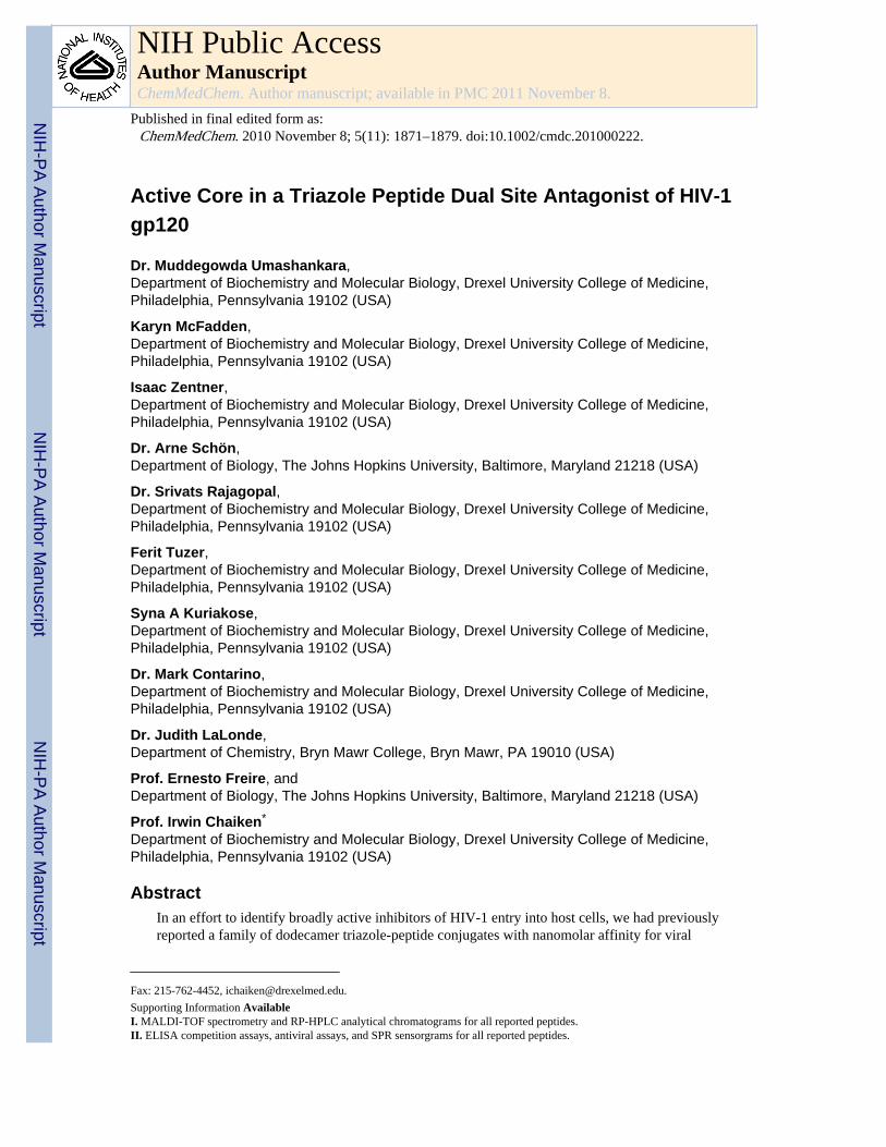
The high affinity and antiviral potency of the 12mer metallocene-conjugated peptideHNG-156 (Peptide 1, Table 1) led us to the current work aimed at identifying sequence-minimized peptides that retain the dual antagonist inhibition function characteristic of 1. Theessential triazole-indole side chain cluster in the middle of the peptide provided a focal pointfor initial truncation analysis. We truncated parent full-length peptide 1 into C and N-terminal heptapeptides 2 (residues 6-12) and 3 (residues 1-7), such that both truncatedpeptides retained the central triazole-indole moiety. The two truncates were compared fortheir ability to bind gp120 using SPR. Increasing concentrations of 2 and 3 were passedseparately over a high-density (3500 RU) HIV-1YU-2 gp120 surface. The binding data werefit to a bimolecular binding process. The N-terminal fragment peptide 3 binds to gp120 (KD= 12.9 nM) with comparable affinity to 1 while the C-terminal fragment peptide 2 binds togp120 with 809 nM KD, which is 100 fold lower affinity versus the 12mer peptide (Figure1A, B; Table 2).
Since the parent peptide 1 has been found to inhibit gp120 interactions at both its CD4 andco-receptor binding sites, we examined the inhibition of HIV-1YU-2 gp120 interactions withimmobilized sCD4 and mAb 17b by fragment peptides 2 and 3 using both ELISA and SPRanalysis. As shown in Figure 1B and C, peptide 3 maintained the dual antagonist signature.Mean inhibitory concentration (IC50) for 3 by SPR was 500 nM and 250 nM for sCD4 andmAb 17b, respectively (Table 2). Peptide 2 was unable to inhibit either sCD4 or mAb 17bbinding to gp120 at concentrations up to 10 μM, in contrast to its detectable binding togp120. In control experiments, neither 2 nor 3 exhibited direct binding to either a sCD4 ormAb 17b SPR chip surface (data not shown).
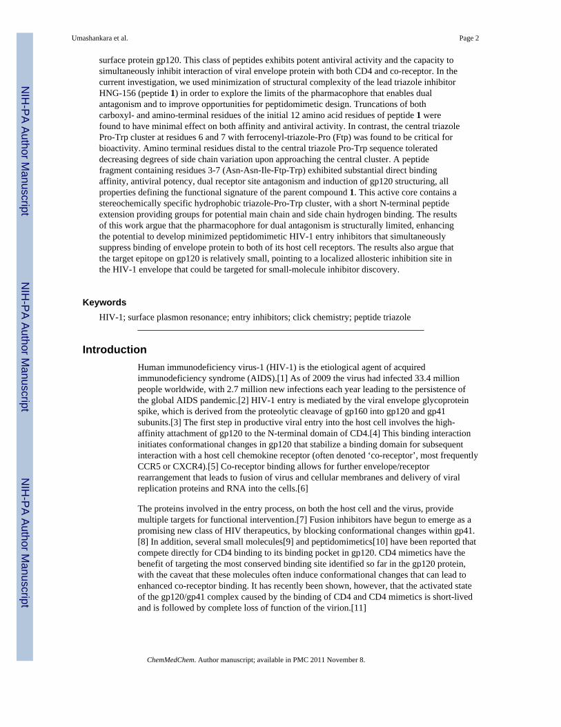
We examined the antiviral activities of 2 and 3 using single round infection of target cells byHIV-1BaL. The antiviral potency of 3 is only slightly diminished compared to the parentpeptide 1, consistent with the direct binding data. The C-terminal peptide 2 had a significantreduction in potency with an IC50 value of 35 μM as compared to 0.8 μM for 1 (Figure 2,Table 2). Neither of these compounds inhibited negative control viruses pseudotyped with
Umashankara et al. Page 3
ChemMedChem. Author manuscript; available in PMC 2011 November 8.
NIH
-PA Author Manuscript
NIH
-PA Author Manuscript
NIH
-PA Author Manuscript
the VSV-G envelope (data not shown), indicating that the inhibition is specific to the HIV-1envelope glycoprotein.
To assess the importance of Ser 7 in the C-terminal region of 1, we evaluated the bindingand dual antagonist property of peptide 4, in which the additional serine residue was presentat the C-terminus. Incorporation of this Ser led to a relatively small and variable effect onthe direct binding and antiviral properties versus those for peptide 3 (Table 2) arguingagainst a major role for this residue.
Progressive truncation and sequence variation of the N-terminal residues to identifyminimum length active dual antagonist sequences
To assess the functional importance of N-terminal amino acids, we first examined thesignificance of Arg residue 1 for direct binding to gp120 and antiviral activity of peptide 4by replacing this positively charged residue with the negatively charged residue Glu. Theresulting peptide 5 enhances the direct binding affinity for gp120 (KD = 5.5 nM), as well asantiviral potency (IC50=1.2 μM) (Table 2). Although peptide 5 showed somewhat increasedefficacy versus 4, both Arg and Glu at position 1 were acceptable. These findings argue thatthe positively charged side chain of Arg residue at the N-terminus is relatively unimportant.
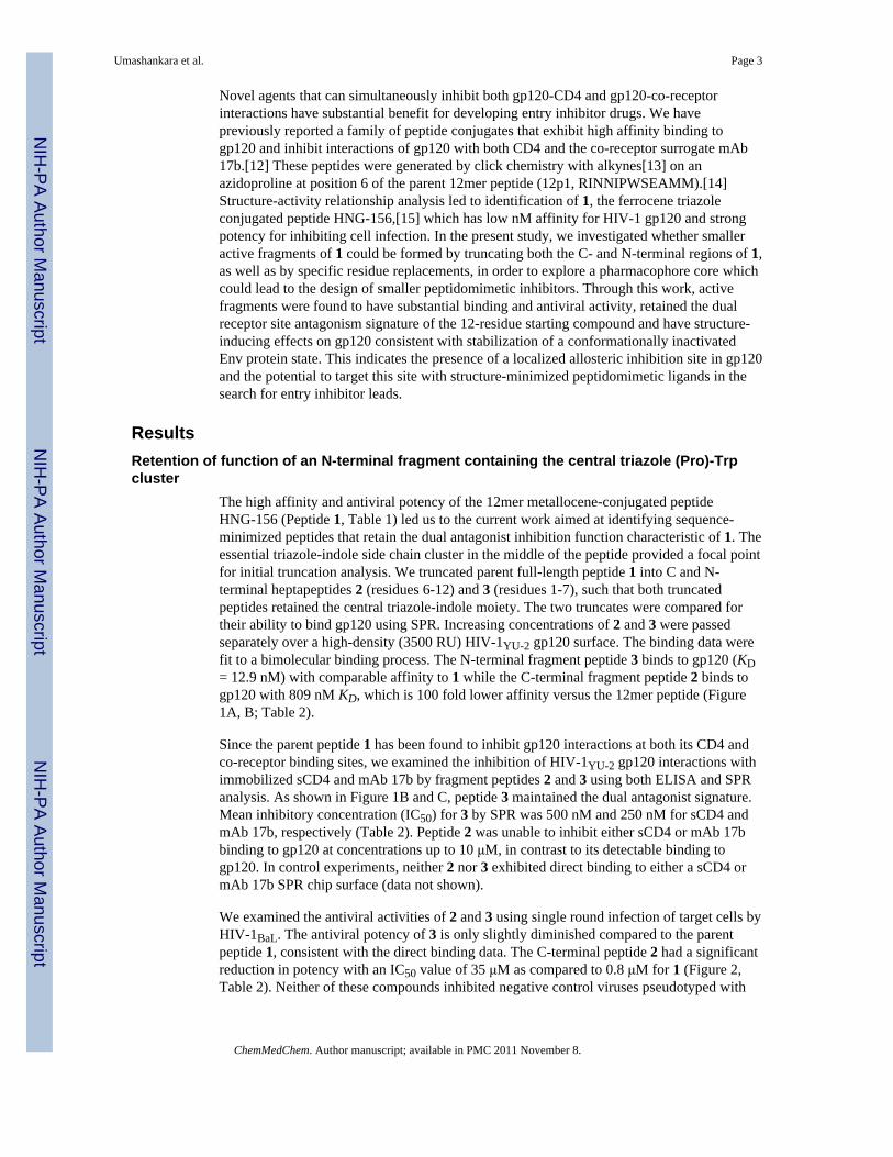
In peptide 6, we truncated the N-terminus further to remove the Arg residue and found thatthis deletion only partially reduced the direct binding activity (KD = 169 nM), mainly due toincreased off-rate of the peptide in SPR analysis (Figure 3B). In addition, peptide 6 retainedsignificant antiviral potency in single round infection assays. We went on to evaluate sidechain variations with different functionalities of the N-terminal Ile residue in peptide 6. Aseries of hepta-peptides (Y-Asn-Asn-Ile-X-Trp-Ser), where Y=Arginine (7), Glutamic acid(8), Citrulline (9), Lysine (10) and Phenylalanine (11) and X = ferrocene conjugatedtriazolePro, were evaluated for their direct binding and inhibition of viral infection activities.In general, these replacements led to peptide fragments with similar antiviral potencies andbinding activities (Table 2). Interestingly, replacement with the unnatural amino acidcitrulline in 9 significantly increased the direct binding affinity to KD = 4.1 nM (Figure 3C)and antiviral potency to IC50 = 2.6 μM (Table 2). Variation of amino acid side chain doesnot affect either the direct binding or antiviral potency of the 7mer peptide, indicating thatspecific interaction of the side chain of the amino acid in the Ile position with gp120 is notessential for the viral inhibition function. This was further supported by the substantial,though somewhat reduced, activities observed with the hexa-peptide 12, in which the Ileresidue of peptide 6 was truncated.
In contrast to the relatively small effects from sequence replacements at N-terminal residuesof 1 and 2 in the parent peptide 1, the Asn residue in position 3 is substantially moresensitive. We investigated replacement of the Asn residue in peptide 12 with Arg (13), Glu(14), Cit (15) and Ile (16) by ELISA, SPR and antiviral assays. Side chain variations of Asnin position 3 led to substantial suppression of antiviral activity and direct binding affinity togp120 (Table 2). This suggests that the Asn side chain at position 3 is important forfunction. This conclusion is further supported by the complete loss of activity of penta-peptide 20, which was derived from the N-terminal truncation of the Asn residue in peptide12.
Active core sequence within the peptide triazole inhibitorsBased on the above findings that several residues in the N- and C-terminal regions hadlimited importance for gp120 binding and inhibition, we explored the limits of truncation.Deletion of the C-terminal Trp residue led to complete loss of activity in 18. In contrast, twofragments, 17 and 12, were found to retain substantial binding, dual site antagonism and
Umashankara et al. Page 4
ChemMedChem. Author manuscript; available in PMC 2011 November 8.
NIH
-PA Author Manuscript
NIH
-PA Author Manuscript
NIH
-PA Author Manuscript
antiviral activity (Table 2). We tested the five-residue peptide 19, containing residues 3-7 ofparent peptide 1. This peptide was found by SPR to bind to gp120 with weakened thoughstill finite affinity with KD = 333 nM (Figure 3D) and exhibited significant antiviral activity(IC50 = 33 μM). An alternative penta-peptide 20, containing residues 4-8, was found to beinactive.
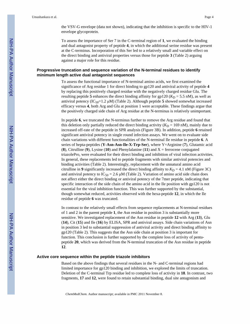
In addition to its dual antagonism of CD4 and mAb 17b binding, the core peptide 19 wasfound to suppress the interaction with other ligands of gp120 that are dependent on theconformation of the Env protein. Previous work [15] showed that compound 1 inhibitedbinding to monoclonal antibodies mAb b12 and mAb F105 to the viral glycoprotein gp120.ELISA data presented in Figure 4 show that the active pharmacophore pentapeptide 19retains similar functionalities.
Importance of stereochemistry in the triazolePro-Trp clusterThe active core sequences found in this work embody a C-terminal hydrophobic cluster.Previous work has shown that the stereochemistry of the triazolePro residue in this cluster incompound 1 was critical, in that the S configuration of triazole was active while Rconfiguration was not.[13] Here, we examined the stereochemical requirements in the Trpposition of the cluster. Compared to 9 (all-L amino acids), 21 with D-Trp substituted wasvirtually inactive (Table 2). Taking these new and previous observations together, it is clearthat the function of peptide triazole inhibitors is highly dependent on the stereochemistry ofthe “triazolePro-Trp” cluster.
Preservation of conformational effects on gp120 by truncated peptide triazolesPrior ITC analysis with analogues of compound 1 showed that binding of the parent peptidetriazole had the ability to structurally constrain gp120.[13] The change in conformationinduced upon binding would explain the ability of the peptide to inhibit both CD4 andcoreceptor binding. Here, we examined whether the property of conformational structuringwas retained in the peptide triazole truncates. The results of Figure 5 compare thecalorimetric titration data for parent peptide 1 with those for truncated peptides 9, 19 and 20.The thermodynamic parameters obtained by ITC for these and other peptides aresummarized in Table 3. Except for the low affinity peptide 20, all of the peptide truncatestested were similar to compound 1 in showing large favorable enthalpy and unfavorableentropy changes. CD4 binding to gp120 is also characterized by a large change in favorableenthalpy (ΔH = − 34.5 kcal mol−1) that is coupled to a large unfavorable change in entropy(ΔS = − 79 cal K−1 mol−1; −TΔS = 23.6 kcal mol−1) that together with a large negative heatcapacity change (ΔCp = −1800 cal K−1 mol −1) make up the thermodynamic signature for abinding event that is associated with a large conformational structuring of gp120[9c,16]Although the thermodynamic changes are much larger for CD4 binding to gp120, someconformational structuring is likely to be induced also upon binding of the peptides truncatespresented here.
DiscussionIn this work, we explored structural minimization of a peptide triazole class of potent HIV-1entry inhibitor candidates. The starting point was the 12-residue peptide 1, which has a lownanomolar affinity for Env protein, can inhibit the binding of Env gp120 to both CD4 andco-receptor binding sites and neutralizes HIV-1 infection. Through examination of a rangeof chemically synthesized peptide derivatives, we found that truncated fragments of 1retained significant binding and inhibition activities. Truncations at the C-terminal residueswere most tolerated, while those for the first two N-terminal residues Arg and Ile werelargely tolerated. The internal sequence cluster of X-Trp, in which X was the
Umashankara et al. Page 5
ChemMedChem. Author manuscript; available in PMC 2011 November 8.
NIH
-PA Author Manuscript
NIH
-PA Author Manuscript
NIH
-PA Author Manuscript
ferrocenyltriazole on Pro at position 6 of 1, was required for function. Amino-terminalextensions of 3 residues from this cluster provided a minimized functional core. The shortestsequences observed so far with the dual antagonist signature, namely compounds 12, 17 and19, retained substantial antiviral and binding activities.
The ability to produce size-minimized fragments that retain potent antiviral activity arguesfor the potential to derive smaller inhibitors by focusing on the crucial triazolePro-Trpcluster and short N-terminal extensions from this cluster. The presence of a hydrophobiccluster infers the importance of interactions of its hydrophobic side chains withcomplementary epitopes in Env protein, with the N-terminal extension providing other typesof stabilizing contacts with the protein. The stereochemical nature of the triazolePro-Trpclearly is critical. Previous work has shown that greater activity of peptides is obtained withthe S configuration versus R in the triazole side chain.[13] The current study shows thatswitching the main chain configuration at Trp from L to D also leads to a large decrease ofinhibitory function. Likely, the specific stereochemistry provides an arrangement ofstructural elements in the triazolePro-Trp cluster enabling productive complementarycontacts with gp120. While we cannot define from current data what the arrangement ofstructural elements are at the peptide triazole-gp120 interface, one may speculate that bothpeptide-protein contacts and intra-peptide interactions can occur. In any case, we mayconclude that the triazolePro-Trp sequence provides a spatially-arranged aromatic cluster,while the N-terminal extension of residues may make hydrogen bonding interactions thathelp stabilize productive Env protein binding. The sequence starting point of greatlyshortened peptides, such as 12, 17 and 19, implies the potential to form smallerpeptidomimetic compounds that retain the essential structural elements embodied in theminimized peptides.
The observation in the current work of active core peptide fragments implies that the targetsite for inhibition is substantially localized within the gp120 protein. Twelve residuepeptides such as 1, especially in an elongated conformation, have at least the potential tobind to a relatively large surface area of gp120, leaving open exactly how large and well-defined the inhibitor binding site could be. In contrast, observing functionality in muchsmaller fragments makes it more likely that the inhibitor binding site of Env gp120 involvesa relatively small footprint on the Env surface. That the putative allosteric site of the HNG/UM classes of inhibitors exhibits strong stereo-specificity of the small hydrophobictriazolePro-Trp cluster further argues that this site can be a viable target for small moleculeinhibitors. This suggests the potential to screen for small molecule HIV-1 entry inhibitorleads that block HNG/UM peptide binding, exhibit dual antagonist activity and retainantiviral activity.
Experimental sectionReagents
The following reagents were obtained from the NIH AIDS Reference and Reagent Program,Division of AIDS, NIAID: HIV-1 gp120 Monoclonal Antibody (b12) from Dr. DennisBurton and Dr. Carlos Barbas; HOS.CD4.CCR5 cells and pNL4-3.Luc. R-E- from Dr.Nathaniel Landau; pHEF-VSV-G from Dr. Lung-Ji Chang; CHO-ST4.2 cells from Dr. DanLittman; Monoclonal Antibody 17b was obtained from Strategic Biosolutions. The plasmidfor monomeric HIV-1YU-2 gp120 was a generous gift from Dr. Joseph Sodroski. Theplasmid for HIV-1BaL gp160 was a generous gift from Dr. Julio Martin-Garcia.
Peptide Synthesis and PurificationThe sequences and denotations of all peptides reported are given in Table 1. Peptides weresynthesized manually by stepwise solid-phase peptide synthesis on a Rink Amide Resin with
Umashankara et al. Page 6
ChemMedChem. Author manuscript; available in PMC 2011 November 8.
NIH
-PA Author Manuscript
NIH
-PA Author Manuscript
NIH
-PA Author Manuscript
a substitution value of 0.7mmole gm−1 (Novabiochem). All Fmoc-amino acid derivativesand coupling reagents were purchased from Chem-Impex International, Inc. Synthesis gradesolvents were used in all procedures. 9-Fluorenylmethoxycarbonyl (Fmoc) group wasemployed for protection of the α-amino group during coupling steps. Side chain protectinggroups were triphenylmethyl (Trt) for Asn, tert-butyl (tBu) for Ser and tert-butyloxycarbonyl (Boc) for Trp. Coupling of each residue was carried out using HBTU/HOBt in Dimethylformamide (DMF). Four equivalents of each Fmoc protected amino acidwere used for coupling. Cleavage of the peptide from the resin was performed using acleavage cocktail comprising 90:2.5:2.5:5(v/v) trifluoroaceticacid (TFA)/thioanisole/ethanedithiol/H2O at room temperature for 2 hours. After the filtration and washing of resinby TFA, excess of solvent was removed under vacuum. Crude peptides were treated withice-cold diethyl ether and then centrifuged at 3000 rpm for 10 minutes at 4 °C. Syntheticpeptides were purified by HPLC (Beckman Coulter System Gold) using a semiPrep VydacC-18 column with a 5 to 60% acetonitrile/water (0.1% TFA) gradient over 75 minutes at 3mL min−1 flow rate. All peptides were purified to ≥ 98% homogeneity as judged byanalytical reverse phase HPLC on C-18. The integrity of purified peptides was confirmed byMALDI-TOF spectra. Analytical chromatograms and mass spectra for all peptides reportedare given in the Supporting Material.
Protein ProductionCHO-ST4.2 cells, which secrete the full extracellular domain of the CD4 protein (solubleCD4, sCD4), were grown in a hollow fiber bioreactor (FiberCell Systems, Inc.) in HiQCDM4CHO media (Hyclone) supplemented with 4 mM L-glutamine, 300 nM methotrexateand 1% antibiotic/antimycotic. Supernatant was purified over a sulfopropyl-substituted ionexchange column (GE Healthcare) on an AKTA Fast Liquid Chromatography (FPLC)machine (GE Healthcare). Fractions were collected using a gradient buffer system consistingof 50 mM MES/50 mM NaCl and 50 mM MES/500 mM NaCl at pH 6.0. Fractions weredialyzed into 50 mM bis-tris propane at pH 6.0 and loaded onto a quaternary ammonium-substituted ion exchange column (GE Healthcare) using a gradient buffer system consistingof 50 mM bis-tris propane and 50 mM bis-tris propane/1 M NaCl at pH 9.0. The columnflow-through containing purified CD4 was pooled and dialyzed into 1× PBS, pH 7.4overnight at 4 °C. The CD4 protein was then concentrated using a 10 kDa Amiconcentrifugal filter unit (Fisher Scientific). All proteins were analyzed by SDS-PAGE/coomassie stain (Invitrogen) and found to be of greater than 95% purity.
Full length HIV-1YU-2 gp120 with a C-terminal hexa-histidine epitope tag was produced bytransient transfection of 293F cells according to manufacturer's protocol (Invitrogen). Cellswere harvested, spun down and the supernatant filtered using 0.2 μm filters. Supernatant waspurified over an F105-antibody coupled NHS-activated Sepharose column according to themanufacturer's instructions (GE Healthcare). HIV-1YU-2 gp120 was eluted from the columnwith 100 mM glycine buffer, pH 2.4. The samples were neutralized with 1:10 v/v addition of1M Tris, pH 8.0 buffer immediately after elution. Peak fractions were collected andanalyzed by SDS-PAGE. Single band fractions corresponding to the correct protein sizewere collected and concentrated, dialyzed against PBS and stored at −80 °C.
Preparation of Recombinant Luciferase Expressing Virus and Cell Infection AssaysSingle round recombinant, luciferase-reporter viruses were produced in a HEK293T cellsusing Fugene transfection reagent (Qiagen) according to the manufacturer's protocol. Cellswere seeded in T75 flasks (approximately 3 × 10⁶ cells per flask) and transfected thefollowing day with 4 μg of plasmid encoding the envelope (HIV-1BaL or VSV-G) togetherwith 8 μg of the envelope-deficient pNL4-3-Fluc+env− provirus developed by N. Landau.[17] Culture supernatants containing viral particles were collected 48-72 hours after
Umashankara et al. Page 7
ChemMedChem. Author manuscript; available in PMC 2011 November 8.
NIH
-PA Author Manuscript
NIH
-PA Author Manuscript
NIH
-PA Author Manuscript
transfection, clarified by centrifugation, filtered, aliquoted and stored at -80 °C until use. Forinhibition experiments, the viral stocks were first incubated with serial dilutions of theinhibitor at 37 °C for 30 minutes. The mixture was added to human osteosarcoma cells thatstably express CD4 and CCR5 (HOS.CD4.CCR5) for 48 hours. The cells were then lysedwith passive lysis buffer (Promega) followed by freeze-thaw cycles. Luciferase assays wereperformed using 1 mM D-luciferin salt (Anaspec) as substrate and detected on a 1450Microbeta Liquid Scintillation and Luminescence Counter (Wallac and Jet). IC50 valueswere estimated using non-linear regression analysis with Origin V.8.1 (Origin Lab). Allexperiments were performed at least in triplicate and results were expressed as relativeinfection with respect to cell infected with virus in the absence of inhibitor (100% infected).
Competition ELISAThe ability of peptides to inhibit the binding of sCD4, mAb F105, mAb b12 and mAb 17b togp120 was screened by competition ELISA. 100 ng HIV-1YU-2 gp120 was adsorbed to a 96-well microtiter plate overnight at 4 °C. After washing the plate three times with PBST buffer(1× PBS with 0.1% Tween-20 v/v), the plate was blocked with 3% BSA in 1× PBS for 2hours at 25 °C, followed by washing of the plate 3 times with PBST. For the CD4competition experiments, 100 μL of sCD4 (0.1 μg ml−1) was added to each well in thepresence of increasing concentrations of peptide (0.01, 0.02, 0.04, 0.08, 0.16, ----100 μM).This and all subsequent incubation steps until detection were done in 0.5% BSA in PBS.After 1hr incubation, the plate was washed 3 times with PBST followed by addition of 100μL/well of biotinylated anti-CD4 antibody (eBioscience) at a 1:5000 dilution and the plateincubated for 1hr at 25 °C. After washing the plate, streptavidin-bound HRP (AnaSpec) wasadded at a 1:5000 dilution and again incubated for 1 hour at room temperature. To determineeffectiveness of peptides to inhibit antibody binding to gp120, 100 μL of mAb 17b, mAbF105 and mAb b12 (0.1 μg ml−1 of each antibody) was added to the immobilized gp120 inthe presence of increasing concentrations of peptide. After 1hr incubation at RT followed bywashing three times, HRP-conjugated goat-antihuman antibody (CHEMICON) was addedand incubated for 1 hour at RT. The extent of HRP conjugate binding was detected in bothassays by adding 200 μL of o-phenylenediamine dihydrochloride (Sigma-Aldrich) reagentfor 30 min followed by measuring optical density (OD) at 450 nm using a microplate reader(Molecular Devices).
Optical Biosensor Binding AssaysSurface plasmon resonance (SPR) interaction analyses were performed on a Biacore® 3000optical biosensor (GE Healthcare). All experiments were carried out at 25 °C using standard1× PBS, pH 7.4, with 0.005% Surfactant P-20. A CM5 sensor chip was derivatized by aminecoupling by using N-ethyl-N-(3-dimethylaminopropyl)carbodiimide/N-hydroxy-succininide[18] with HIV-1YU-2 gp120 or, as a control surface, mAb 2E3 (monoclonalantibody to human Interleukin-5). For direct binding experiments, HIV-1YU-2 gp120 wasimmobilized on the sensor surface (∼3500 RU); peptide analyte in PBS buffer(concentration range of 10 μM - 0.61 nM) was passed over the surface at a flow rate of 50 μlmin−1 with a 5 minute association phase and a 5 minute dissociation phase. Regeneration ofthe surface was achieved by a single 5 second pulse of 10 mM glycine, pH 1.5. Analysis ofpeptide-mediated sCD4 and mAb 17b inhibition was achieved by injecting a fixedconcentration of HIV-1YU-2 gp120 (100 nM), with increasing peptide concentrations, oversCD4 (∼2000 RU) and mAb 17b (∼900 RU) surfaces for 5 minute association and 5 minutedissociation at a flow rate of 50 μl min−1 in PBS. Regeneration of the surface was achievedby single 10 second pulse of 1.3 M NaCl / 35 mM NaOH and single 5-second pulse of 10mM glycine, pH 1.5, for sCD4 and mAb 17b, respectively. All analyses were performed intriplicate.
Umashankara et al. Page 8
ChemMedChem. Author manuscript; available in PMC 2011 November 8.
NIH
-PA Author Manuscript
NIH
-PA Author Manuscript
NIH
-PA Author Manuscript
SPR Data analyses were performed using BIAEvaluation 4.0 software (GE Healthcare). Theresponses of buffer injection and of signals observed in a control flow cell were subtractedto account for nonspecific binding. Experimental data were fitted to a simple 1:1 bindingmodel with a parameter included for mass transport. The average kinetic parameters(association {ka} and dissociation {kd} rates) generated from a minimum of 3 data sets wereused to define equilibrium dissociation (KD) constants. The evaluation method for SPRinhibition data included a calculation of the inhibitor concentration at 50% of the maximalresponse (IC50). The inhibition curve was converted into a calibration curve by the use of afitting function. The fitting was done using the 4-parameter equation in BIAevaluationsoftware:
(1)
where Rhigh is the response value at high inhibitor concentrations and Rlow is response atlow inhibitor concentrations. Conc is the concentration of inhibitor, and A1 and A2 arefitting parameters. At the IC50 the following is true:
(2)
Under this condition, A1 = Conc and is therefore taken as the desired IC50 parameter.
Isothermal Titration CalorimetryIsothermal titration calorimetric experiments were performed using high-precisioncalorimetric systems of model VP-ITC or ITC20 (MicroCal Inc). The titrations described inthis paper were performed by stepwise addition of peptide to gp120 contained in thecalorimetric cell at a constant temperature of 25 °C. The VT-ITC has a cell volume of ∼1.4mL and the volume per injection of peptide is 10 μL, whereas ITC20 has a cell volume of∼200 μL and the peptide is injected in steps of 1.4 μL. For the experiments carried out in theVP-ITC the concentration of gp120 was ∼2 μM and the syringe contained the peptide at aconcentration of ∼30 μM. For the experiment carried out using ITC20 gp120 was prepared atabout 4 μM and the peptide at 50-100 μM. The solutions contained within the calorimetriccells and injector syringes were prepared in 1× PBS, pH 7.4 and thoroughly degassed toavoid bubble formation in the calorimetric cell. The heat evolved upon each injection of thepeptide solution was obtained from the integral of the calorimetric signal. The heatassociated with binding of a ligand to the protein in the cell was obtained by subtracting theheat of dilution from the heat of reaction. Heats of dilution due to mismatch between thesyringe and cell solutions were negligible in all experiments. The individual heats wereplotted as a function of the molar ratio, and nonlinear regression of the data provided theenthalpy change (ΔH) and the association constant (KA = KD
−1).
Supplementary MaterialRefer to Web version on PubMed Central for supplementary material.
Umashankara et al. Page 9
ChemMedChem. Author manuscript; available in PMC 2011 November 8.
NIH
-PA Author Manuscript
NIH
-PA Author Manuscript
NIH
-PA Author Manuscript
AcknowledgmentsThis research was supported by National Institute of Health grants P01 GM 56550 and R21 AI071965, as well as agrant from International Partnership for Microbicides/USAID. EF also acknowledges support from the NationalInstitutes of Health GM 57144 and the National Science Foundation MCB 0641252.
References1. a) Barre-Sinoussi F, Chermann JC, Rey F, Nugeyre MT, Chamaret S, Gruest J, Dauguet C, Axler-
Blin C, Vezinet-Brun F, Rouzioux C, Rozenbaum W, Montagnier L. Science. 1983; 220:868.[PubMed: 6189183] b) Gallo RC, Salahuddin SZ, Popovic M, Shearer GM, Kaplan M, Haynes BF,Palker TJ, Redfield R, Oleske J, Safai B, et al. Science. 1984; 224:500. [PubMed: 6200936]
2. UNAIDS. AIDS Epidemic Update. 20093. a) Allan JS, Coligan JE, Barin F, McLane MF, Sodroski JG, Rosen CA, Haseltine WA, Lee TH,
Essex M. Science. 1985; 228:1091. [PubMed: 2986290] b) Robey WG, Safai B, Oroszlan S, ArthurLO, Gonda MA, Gallo RC, Fischinger PJ. Science. 1985; 228:593. [PubMed: 2984774] c) KowalskiM, Potz J, Basiripour L, Dorfman T, Goh WC, Terwilliger E, Dayton A, Rosen C, Haseltine W,Sodroski J. Science. 1987; 237:1351. [PubMed: 3629244] d) Helseth E, Olshevsky U, Furman C,Sodroski J. J Virol. 1991; 65:2119. [PubMed: 2002555]
4. a) Klatzmann D, Champagne E, Chamaret S, Gruest J, Guetard D, Hercend T, Gluckman JC,Montagnier L. Nature. 1984; 312:767. [PubMed: 6083454] b) Dalgleish AG, Beverley PC, ClaphamPR, Crawford DH, Greaves MF, Weiss RA. Nature. 1984; 312:763. [PubMed: 6096719]
5. a) Sattentau QJ, Moore JP, Vignaux F, Traincard F, Poignard P. J Virol. 1993; 67:7383. [PubMed:7693970] b) Wu L, Gerard NP, Wyatt R, Choe H, Parolin C, Ruffing N, Borsetti A, Cardoso AA,Desjardin E, Newman W, Gerard C, Sodroski J. Nature. 1996; 384:179. [PubMed: 8906795] c)Trkola A, Dragic T, Arthos J, Binley JM, Olson WC, Allaway GP, Cheng-Mayer C, Robinson J,Maddon PJ, Moore JP. Nature. 1996; 384:184. [PubMed: 8906796] d) Choe H, Farzan M, Sun Y,Sullivan N, Rollins B, Ponath PD, Wu L, Mackay CR, LaRosa G, Newman W, Gerard N, Gerard C,Sodroski J. Cell. 1996; 85:1135. [PubMed: 8674119] e) Deng H, Liu R, Ellmeier W, Choe S,Unutmaz D, Burkhart M, Di Marzio P, Marmon S, Sutton RE, Hill CM, Davis CB, Peiper SC,Schall TJ, Littman DR, Landau NR. Nature. 1996; 381:661. [PubMed: 8649511] f) Dragic T, LitwinV, Allaway GP, Martin SR, Huang Y, Nagashima KA, Cayanan C, Maddon PJ, Koup RA, MooreJP, Paxton WA. Nature. 1996; 381:667. [PubMed: 8649512]
6. a) Chan DC, Fass D, Berger JM, Kim PS. Cell. 1997; 89:263. [PubMed: 9108481] b) WeissenhornW, Dessen A, Harrison SC, Skehel JJ, Wiley DC. Nature. 1997; 387:426. [PubMed: 9163431]
7. Liu S, Lu H, Niu J, Xu Y, Wu S, Jiang S. J Biol Chem. 2005; 280:11259. [PubMed: 15640162]8. a) He Y, Cheng J, Lu H, Li J, Hu J, Qi Z, Liu Z, Jiang S, Dai Q. Proc Natl Acad Sci U S A. 2008;
105:16332. [PubMed: 18852475] b) Liu S, Zhao Q, Jiang S. Peptides. 2003; 24:1303. [PubMed:14706544] c) Liu D, Madani N, Li Y, Cao R, Choi WT, Kawatkar SP, Lim MY, Kumar S, DongCZ, Wang J, Russell JD, Lefebure CR, An J, Wilson S, Gao YG, Pallansch LA, Sodroski JG, HuangZ. J Virol. 2007; 81:11489. [PubMed: 17686848]
9. a) Zhao Q, Ma L, Jiang S, Lu H, Liu S, He Y, Strick N, Neamati N, Debnath AK. Virology. 2005;339:213. [PubMed: 15996703] b) Madani N, Schon A, Princiotto AM, Lalonde JM, Courter JR,Soeta T, Ng D, Wang L, Brower ET, Xiang SH, Kwon YD, Huang CC, Wyatt R, Kwong PD, FreireE, Smith AB 3rd, Sodroski J. Structure. 2008; 16:1689. [PubMed: 19000821] c) Schon A, MadaniN, Klein JC, Hubicki A, Ng D, Yang X, Smith AB 3rd, Sodroski J, Freire E. Biochemistry. 2006;45:10973. [PubMed: 16953583]
10. a) Neffe AT, Meyer B. Angew Chem Int Ed Engl. 2004; 43:2937. [PubMed: 15170309] b) NeffeAT, Bilang M, Meyer B. Org Biomol Chem. 2006; 4:3259. [PubMed: 17036114] c) Neffe AT,Bilang M, Gruneberg I, Meyer B. J Med Chem. 2007; 50:3482. [PubMed: 17602545]
11. Haim H, Si Z, Madani N, Wang L, Courter JR, Princiotto A, Kassa A, DeGrace M, McGee-EstradaK, Mefford M, Gabuzda D, Smith AB 3rd, Sodroski J. PLoS Pathog. 2009; 5:e1000360. [PubMed:19343205]
Umashankara et al. Page 10
ChemMedChem. Author manuscript; available in PMC 2011 November 8.
NIH
-PA Author Manuscript
NIH
-PA Author Manuscript
NIH
-PA Author Manuscript
12. a) Gopi HN, Tirupula KC, Baxter S, Ajith S, Chaiken IM. ChemMedChem. 2006; 1:54. [PubMed:16892335] b) Cocklin S, Gopi H, Querido B, Nimmagadda M, Kuriakose S, Cicala C, Ajith S,Baxter S, Arthos J, Martin-Garcia J, Chaiken IM. J Virol. 2007; 81:3645. [PubMed: 17251295]
13. Gopi H, Umashankara M, Pirrone V, LaLonde J, Madani N, Tuzer F, Baxter S, Zentner I, CocklinS, Jawanda N, Miller SR, Schon A, Klein JC, Freire E, Krebs FC, Smith AB, Sodroski J, ChaikenI. J Med Chem. 2008; 51:2638. [PubMed: 18402432]
14. Ferrer M, Harrison SC. J Virol. 1999; 73:5795. [PubMed: 10364331]15. Gopi H, Cocklin S, Pirrone V, McFadden K, Tuzer F, Zentner I, Ajith S, Baxter S, Jawanda N,
Krebs FC, Chaiken IM. J Mol Recognit. 2009; 22:169. [PubMed: 18498083]16. Leavitt SA, SchOn A, Klein JC, Manjappara U, Chaiken IM, Freire E. Curr Protein Pept Sci. 2004;
5:1. [PubMed: 14965316]17. Connor RI, Chen BK, Choe S, Landau NR. Virology. 1995; 206:935. [PubMed: 7531918]18. Ishino T, Pillalamarri U, Panarello D, Bhattacharya M, Urbina C, Horvat S, Sarkhel S, Jameson B,
Chaiken I. Biochemistry. 2006; 45:1106. [PubMed: 16430207]
Umashankara et al. Page 11
ChemMedChem. Author manuscript; available in PMC 2011 November 8.
NIH
-PA Author Manuscript
NIH
-PA Author Manuscript
NIH
-PA Author Manuscript
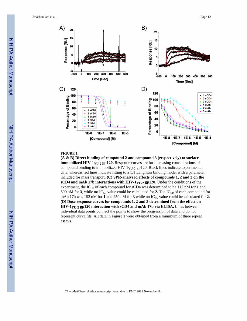
FIGURE 1.(A & B) Direct binding of compound 2 and compound 3 (respectively) to surface-immobilized HIV-1YU-2 gp120. Response curves are for increasing concentrations ofcompound binding to immobilized HIV-1YU-2 gp120. Black lines indicate experimentaldata, whereas red lines indicate fitting to a 1:1 Langmuir binding model with a parameterincluded for mass transport. (C) SPR-analyzed effects of compounds 1, 2 and 3 on thesCD4 and mAb 17b interactions with HIV-1YU-2 gp120. Under the conditions of theexperiment, the IC50 of each compound for sCD4 was determined to be 112 nM for 1 and500 nM for 3, while no IC50 value could be calculated for 2. The IC50 of each compound formAb 17b was 152 nM for 1 and 250 nM for 3 while no IC50 value could be calculated for 2.(D) Dose response curves for compounds 1, 2 and 3 determined from the effect onHIV-1YU-2 gp120 interaction with sCD4 and mAb 17b via ELISA. Lines betweenindividual data points connect the points to show the progression of data and do notrepresent curve fits. All data in Figure 1 were obtained from a minimum of three repeatassays.
Umashankara et al. Page 12
ChemMedChem. Author manuscript; available in PMC 2011 November 8.
NIH
-PA Author Manuscript
NIH
-PA Author Manuscript
NIH
-PA Author Manuscript

FIGURE 2. Analysis of antiviral potencies of peptides 1, 2 and 3 using single round viralinfection assaysRecombinant HIV-1BaL was pre-incubated with serial dilutions of 1, 2 or 3 for 30 minutes at37 °C. The virus-inhibitor mixture was then added to HOS.CD4.CCR5 for 48 hours.Infection was determined based on luciferase activity. Data points were fit to a simplesigmoidal inhibition model using Origin software to derive the best-fit lines. The IC50 valueswere 0.8 μM for 1, 35 μM for 2 and 6.8 μM for 3. Data represent a minimum of threerepeats.
Umashankara et al. Page 13
ChemMedChem. Author manuscript; available in PMC 2011 November 8.
NIH
-PA Author Manuscript
NIH
-PA Author Manuscript
NIH
-PA Author Manuscript
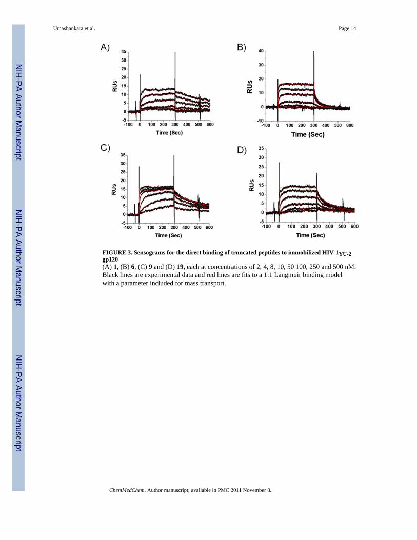
FIGURE 3. Sensograms for the direct binding of truncated peptides to immobilized HIV-1YU-2gp120(A) 1, (B) 6, (C) 9 and (D) 19, each at concentrations of 2, 4, 8, 10, 50 100, 250 and 500 nM.Black lines are experimental data and red lines are fits to a 1:1 Langmuir binding modelwith a parameter included for mass transport.
Umashankara et al. Page 14
ChemMedChem. Author manuscript; available in PMC 2011 November 8.
NIH
-PA Author Manuscript
NIH
-PA Author Manuscript
NIH
-PA Author Manuscript
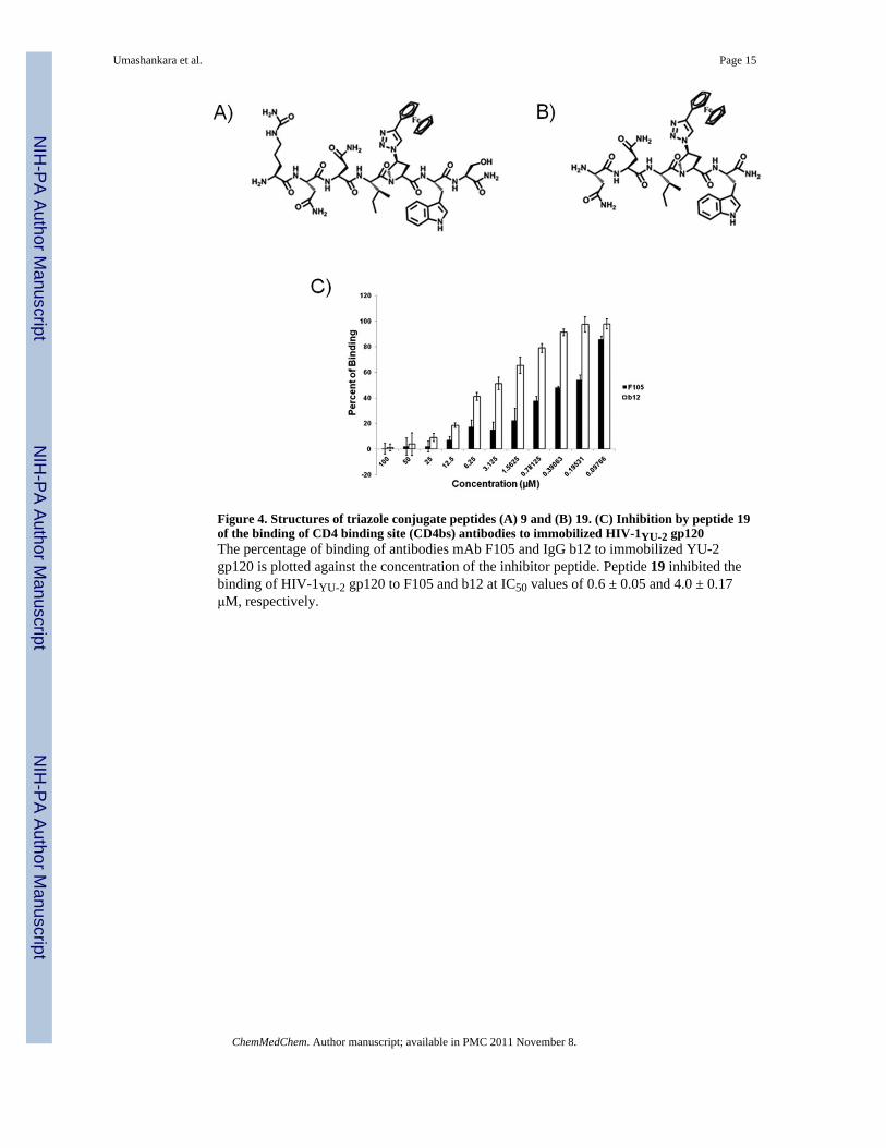
Figure 4. Structures of triazole conjugate peptides (A) 9 and (B) 19. (C) Inhibition by peptide 19of the binding of CD4 binding site (CD4bs) antibodies to immobilized HIV-1YU-2 gp120The percentage of binding of antibodies mAb F105 and IgG b12 to immobilized YU-2gp120 is plotted against the concentration of the inhibitor peptide. Peptide 19 inhibited thebinding of HIV-1YU-2 gp120 to F105 and b12 at IC50 values of 0.6 ± 0.05 and 4.0 ± 0.17μM, respectively.
Umashankara et al. Page 15
ChemMedChem. Author manuscript; available in PMC 2011 November 8.
NIH
-PA Author Manuscript
NIH
-PA Author Manuscript
NIH
-PA Author Manuscript
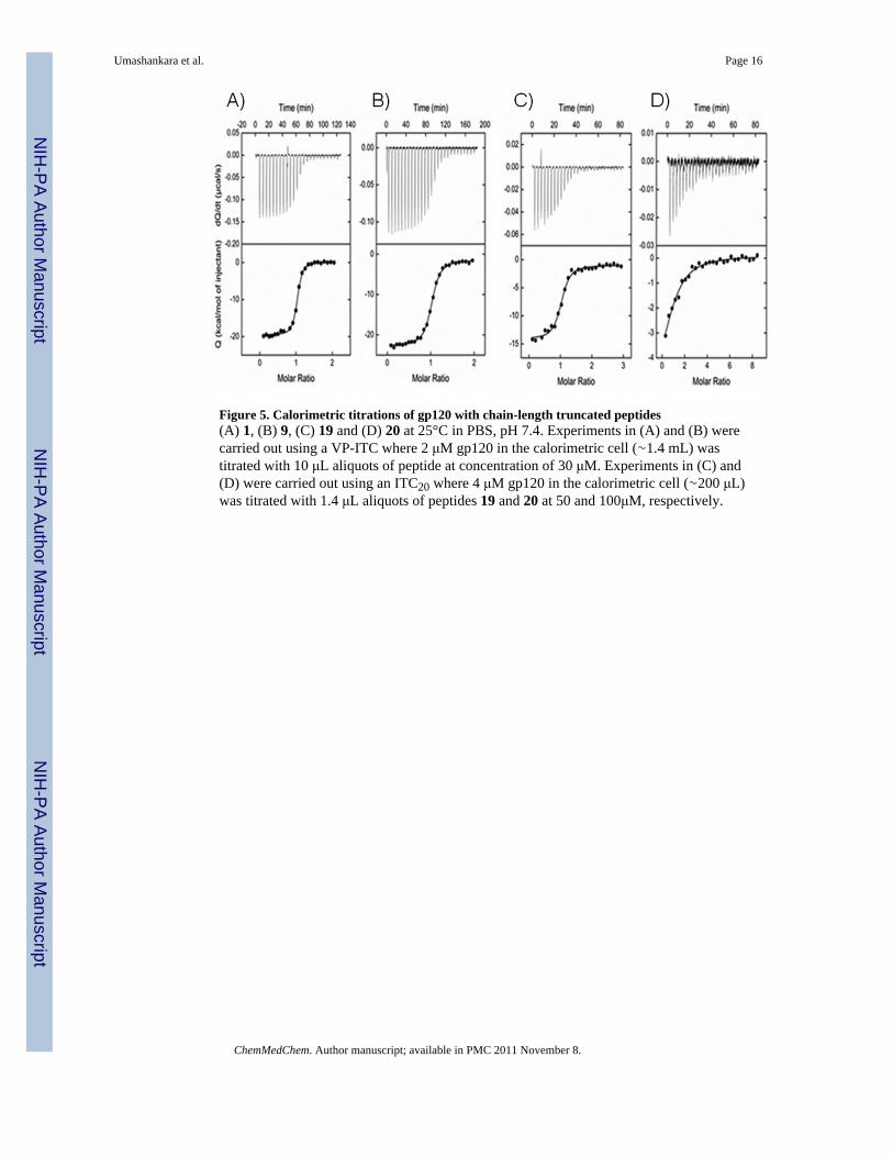
Figure 5. Calorimetric titrations of gp120 with chain-length truncated peptides(A) 1, (B) 9, (C) 19 and (D) 20 at 25°C in PBS, pH 7.4. Experiments in (A) and (B) werecarried out using a VP-ITC where 2 μM gp120 in the calorimetric cell (∼1.4 mL) wastitrated with 10 μL aliquots of peptide at concentration of 30 μM. Experiments in (C) and(D) were carried out using an ITC20 where 4 μM gp120 in the calorimetric cell (∼200 μL)was titrated with 1.4 μL aliquots of peptides 19 and 20 at 50 and 100μM, respectively.
Umashankara et al. Page 16
ChemMedChem. Author manuscript; available in PMC 2011 November 8.
NIH
-PA Author Manuscript
NIH
-PA Author Manuscript
NIH
-PA Author Manuscript
NIH
-PA Author Manuscript
NIH
-PA Author Manuscript
NIH
-PA Author Manuscript
Umashankara et al. Page 17
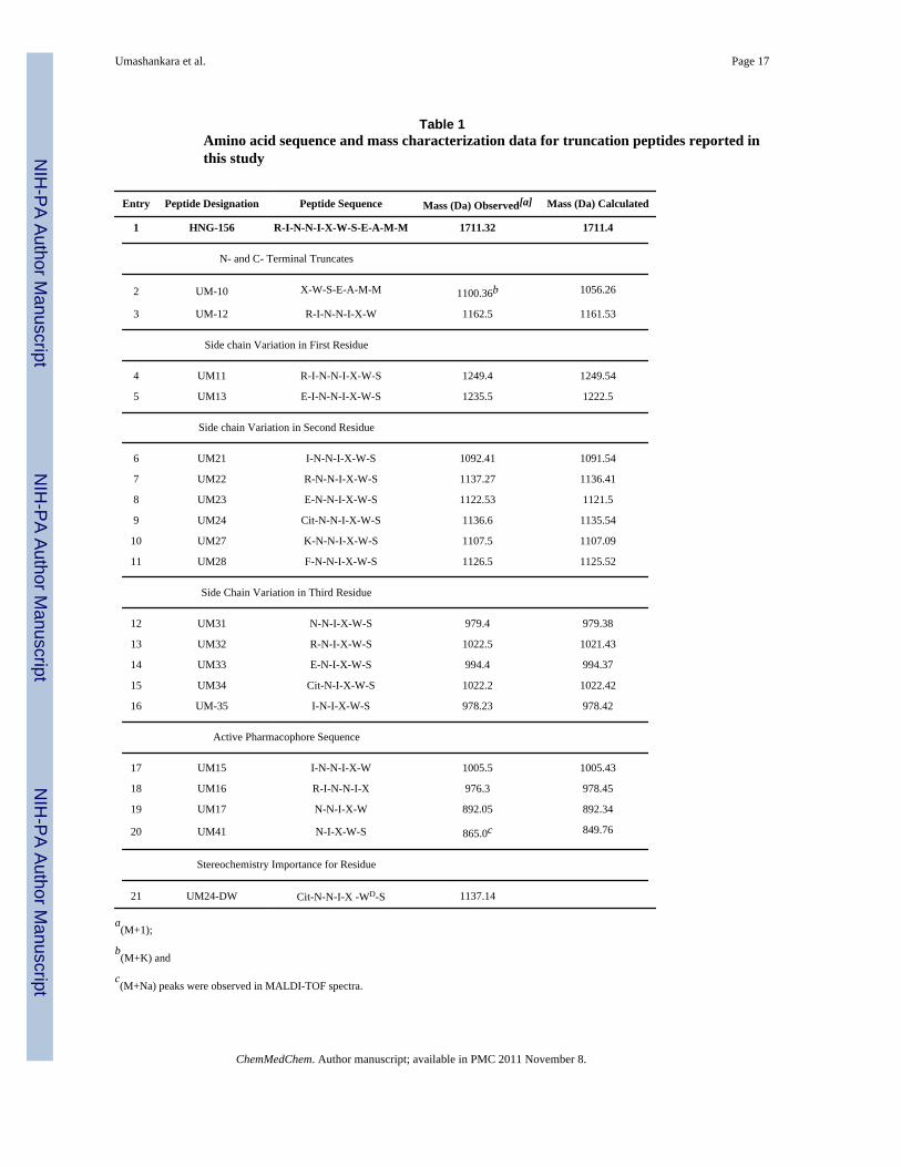
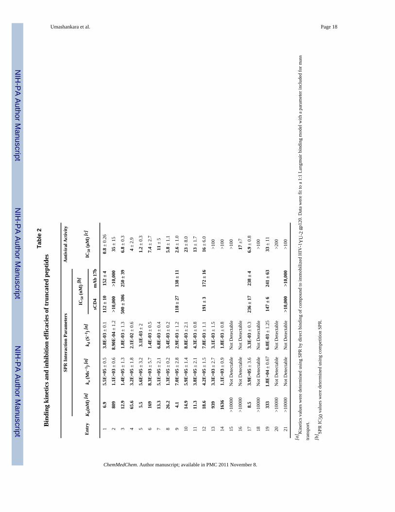
Table 1Amino acid sequence and mass characterization data for truncation peptides reported inthis study
Entry Peptide Designation Peptide Sequence Mass (Da) Observed[a] Mass (Da) Calculated
1 HNG-156 R-I-N-N-I-X-W-S-E-A-M-M 1711.32 1711.4
N- and C- Terminal Truncates
2 UM-10 X-W-S-E-A-M-M 1100.36b 1056.26
3 UM-12 R-I-N-N-I-X-W 1162.5 1161.53
Side chain Variation in First Residue
4 UM11 R-I-N-N-I-X-W-S 1249.4 1249.54
5 UM13 E-I-N-N-I-X-W-S 1235.5 1222.5
Side chain Variation in Second Residue
6 UM21 I-N-N-I-X-W-S 1092.41 1091.54
7 UM22 R-N-N-I-X-W-S 1137.27 1136.41
8 UM23 E-N-N-I-X-W-S 1122.53 1121.5
9 UM24 Cit-N-N-I-X-W-S 1136.6 1135.54
10 UM27 K-N-N-I-X-W-S 1107.5 1107.09
11 UM28 F-N-N-I-X-W-S 1126.5 1125.52
Side Chain Variation in Third Residue
12 UM31 N-N-I-X-W-S 979.4 979.38
13 UM32 R-N-I-X-W-S 1022.5 1021.43
14 UM33 E-N-I-X-W-S 994.4 994.37
15 UM34 Cit-N-I-X-W-S 1022.2 1022.42
16 UM-35 I-N-I-X-W-S 978.23 978.42
Active Pharmacophore Sequence
17 UM15 I-N-N-I-X-W 1005.5 1005.43
18 UM16 R-I-N-N-I-X 976.3 978.45
19 UM17 N-N-I-X-W 892.05 892.34
20 UM41 N-I-X-W-S 865.0c 849.76
Stereochemistry Importance for Residue
21 UM24-DW Cit-N-N-I-X -WD-S 1137.14
a(M+1);
b(M+K) and
c(M+Na) peaks were observed in MALDI-TOF spectra.
ChemMedChem. Author manuscript; available in PMC 2011 November 8.
NIH
-PA Author Manuscript
NIH
-PA Author Manuscript
NIH
-PA Author Manuscript
Umashankara et al. Page 18
Tabl
e 2
Bin
ding
kin
etic
s and
inhi
bitio
n ef
ficac
ies o
f tru
ncat
ed p
eptid
es
SPR
Inte
ract
ion
Para
met
ers
Ant
ivir
al A
ctiv
ity
Ent
ryK D
(nM
) [a]
k a (M
s−1 )
[a]
k d (S
−1 )
[a]
IC50
(nM
) [b]
IC50
(μM
) [c]
sCD
4m
Ab
17b
16.
95.
5E+0
5 ±
0.5
3.8E
-03
± 0.
111
2 ±
1015
2 ±
40.
8 ±
0.26
280
91.
1E+0
3 ±
0.6
8.90
E-0
4 ±
1.2
>10,
000
>10,
000
35 ±
15
312
.91.
4E+0
5 ±
1.3
1.8E
-03
± 1.
350
0 ±
306
250
± 39
6.8
± 0.
3
465
.63.
2E+0
5 ±
1.8
2.1E
-02
± 0.
64
± 2.
9
55.
55.
6E+0
5 ±
3.2
3.1E
-03
± 2
1.2
± 0.
3
616
98.
3E+0
3 ±
5.7
1.4E
-03
± 0.
57.
4 ±
2.7
713
.35.
1E+0
5 ±
2.1
6.8E
-03
± 0.
411
± 5
826
.21.
3E+0
5 ±
0.2
3.4E
-03
± 0.
25.
8 ±
1.1
94.
17.
0E+0
5 ±
2.8
2.9E
-03
± 1.
211
8 ±
2713
8 ±
112.
6 ±
1.0
1014
.95.
9E+0
5 ±
1.4
8.8E
-03
± 2.
123
± 8
.0
1111
.33.
8E+0
5 ±
2.1
4.3E
-03
± 0.
813
± 1
.7
1218
.64.
2E+0
5 ±
1.5
7.8E
-03
± 1.
119
1 ±
317
2 ±
1616
± 6
.0
1393
93.
3E+0
3 ±
2.7
3.1E
-03
± 1.
5>1
00
1416
361.
1E+0
3 ±
0.9
1.8E
-03
± 0.
8>1
00
15>1
0000
Not
Det
ecta
ble
Not
Det
ecta
ble
>100
16>1
0000
Not
Det
ecta
ble
Not
Det
ecta
ble
17 ±
7
178.
53.
9E+0
5 ±
3.6
3.3E
-03
± 0.
323
6 ±
1723
8 ±
46.
9 ±
0.8
18>1
0000
Not
Det
ecta
ble
Not
Det
ecta
ble
>100
1933
31.
8E+0
4 ±
0.67
6.0E
-03
± 1.
2514
7 ±
624
1 ±
6333
± 1
1
20>1
0000
Not
Det
ecta
ble
Not
Det
ecta
ble
>200
21>1
0000
Not
Det
ecta
ble
Not
Det
ecta
ble
>10,
000
>10,
000
>100
[a] K
inet
ics v
alue
s wer
e de
term
ined
usi
ng S
PR b
y di
rect
bin
ding
of c
ompo
und
to im
mob
ilize
d H
IV-1
YU
-2 g
p120
. Dat
a w
ere
fit to
a 1
:1 L
angm
uir b
indi
ng m
odel
with
a p
aram
eter
incl
uded
for m
ass
trans
port.
[b] SP
R IC
50 v
alue
s wer
e de
term
ined
usi
ng c
ompe
titio
n SP
R.
ChemMedChem. Author manuscript; available in PMC 2011 November 8.
NIH
-PA Author Manuscript
NIH
-PA Author Manuscript
NIH
-PA Author Manuscript
Umashankara et al. Page 19[c
] Ant
ivira
l IC
50 v
alue
s wer
e ob
tain
ed th
roug
h a
sing
le ro
und
infe
ctio
n as
say
and
expr
esse
d as
rela
tive
infe
ctio
n ve
rsus
unt
reat
ed (1
00%
). A
ll da
ta re
pres
ent a
min
imum
of t
hree
exp
erim
ents
.
ChemMedChem. Author manuscript; available in PMC 2011 November 8.
NIH
-PA Author Manuscript
NIH
-PA Author Manuscript
NIH
-PA Author Manuscript
Umashankara et al. Page 20
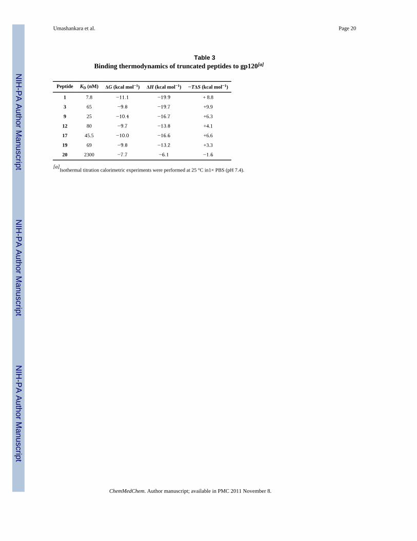
Table 3Binding thermodynamics of truncated peptides to gp120[a]
Peptide KD (nM) ΔG (kcal mol−1) ΔH (kcal mol−1) −TΔS (kcal mol−1)
1 7.8 −11.1 −19.9 + 8.8
3 65 −9.8 −19.7 +9.9
9 25 −10.4 −16.7 +6.3
12 80 −9.7 −13.8 +4.1
17 45.5 −10.0 −16.6 +6.6
19 69 −9.8 −13.2 +3.3
20 2300 −7.7 −6.1 −1.6
[a]Isothermal titration calorimetric experiments were performed at 25 °C in1× PBS (pH 7.4).
ChemMedChem. Author manuscript; available in PMC 2011 November 8.