Embed Size (px)
Citation preview

HIV-1 gp120 Mannoses InduceImmunosuppressiveResponses from Dendritic CellsMeimei Shan
1, Per Johan Klasse
1, Kaustuv Banerjee
1, Antu K. Dey
1, Sai Prasad N. Iyer
2, Robert Dionisio
1,
Dustin Charles1
, Lila Campbell-Gardener1
, William C. Olson2
, Rogier W. Sanders3
, John P. Moore1*
1 Department of Microbiology and Immunology, Weill Medical College of Cornell University, New York, New York, United States of America, 2 Progenics Pharmaceuticals
Incorporated, Tarrytown, New York, United States of America, 3 Laboratory of Experimental Virology, Department Medical Microbiology, Center of Infection and Immunity
Amsterdam (CINIMA), Academic Medical Center of the University of Amsterdam, Amsterdam, The Netherlands
The human immunodeficiency virus type 1 (HIV-1) envelope glycoprotein gp120 is a vaccine immunogen that cansignal via several cell surface receptors. To investigate whether receptor biology could influence immune responses togp120, we studied its interaction with human, monocyte-derived dendritic cells (MDDCs) in vitro. Gp120 from the HIV-1strain JR-FL induced IL-10 expression in MDDCs from 62% of donors, via a mannose C-type lectin receptor(s) (MCLR).Gp120 from the strain LAI was also an IL-10 inducer, but gp120 from the strain KNH1144 was not. The mannose-binding protein cyanovirin-N, the 2G12 mAb to a mannose-dependent gp120 epitope, and MCLR-specific mAbsinhibited IL-10 expression, as did enzymatic removal of gp120 mannose moieties, whereas inhibitors of signaling viaCD4, CCR5, or CXCR4 were ineffective. Gp120-stimulated IL-10 production correlated with DC-SIGN expression on thecells, and involved the ERK signaling pathway. Gp120-treated MDDCs also responded poorly to maturation stimuli byup-regulating activation markers inefficiently and stimulating allogeneic T cell proliferation only weakly. Theseadverse reactions to gp120 were MCLR-dependent but independent of IL-10 production. Since such mechanisms mightsuppress immune responses to Env-containing vaccines, demannosylation may be a way to improve theimmunogenicity of gp120 or gp140 proteins.
Citation: Shan M, Klasse PJ, Banerjee K, Key AK, Iyer SPN, et al. (2007) HIV-1 gp120 mannoses induce immunosuppressive responses from dendritic cells. PLoS Pathog 3(11):e169. doi: 10.1371/journal.ppat.0030169
Introduction
One approach to a vaccine against HIV-1 is the use of theviral envelope glycoproteins (Env) as immunogens to induceneutralizing antibodies (NAbs) [1–3]. Usually, the Env glyco-proteins are presented as adjuvanted, soluble proteins afterproduction in vitro as recombinant proteins, but they can alsobe expressed in vivo from delivery systems based on DNA orlive recombinant viruses (e.g., poxvirus or adenovirus vectors)[4]. Different configurations of Env glycoproteins have beenstudied as vaccine antigens, initially the surface glycoproteingp120; more recently, soluble oligomeric gp140 proteinsbased broadly on the native gp120-gp41 complex [1–3].
Irrespective of how HIV-1 Env glycoproteins have beenpresented and in whatever configuration, the induction ofbroadly active NAbs has proven problematic [1]. Onegenerally accepted problem is the evolution of the nativeEnv complex into a configuration that limits the exposure ofthe few neutralization sites that are present. The potentialsolution is to further understand the structure of thecomplex, then to engineer antigens that are better able topresent relevant NAb epitopes to the immune system;attempts to do this are in progress in many laboratoriesworldwide [1]. Here, however, we focus on what we considerto be another factor hindering NAb induction: the limitedimmunogenicity of HIV-1 Env proteins in general.
Although antibody responses to HIV-1 Env can clearly beinduced in infected or vaccinated humans and animals, theseproteins are not particularly immunogenic. Thus, gp120 or
gp140 proteins are typically administered at 100–500 lg perdose, and the binding antibody titers raised against them canbe highly variable; some humans and animals respond fairlywell, others only poorly [5–9]. Anti-Env antibody titers alsodecay rather rapidly (half-lives are typically in the range 30–50 d) and frequent boosting is required to maintain them.Few directly comparative studies have ever been performed,but the limited information available supports the contentionthat Env is an unusually problematic immunogen comparedto most other vaccine antigens [10] (S. Plotkin and B. Graham,personal communication).The immune responses to HIV-1 Env vaccine antigens are
TH2-polarized to an extent that is unusual even for a solubleprotein [11,12]. The same TH2 bias can also be observed during
Editor: Marianne Manchester, The Scripps Research Institute, United States ofAmerica
Received July 13, 2007; Accepted September 24, 2007; Published November 2,2007
Copyright: � 2007 Shan et al. This is an open-access article distributed under theterms of the Creative Commons Attribution License, which permits unrestricteduse, distribution, and reproduction in any medium, provided the original authorand source are credited.
Abbreviations: DC, dendritic cell; Env, envelope glycoprotein; iMDDC, immaturemonocyte-derived dendritic cell; LPS, lipopolysaccharide; MCLR, mannose C-typelectin receptor; MDDC, monocyte-derived dendritic cell; mMDDC, mature mono-cyte-derived dendritic cell; MR, mannose receptor, NAb, neutralizing antibody; RT,reverse transcriptase; TNIL, TNFa and IL-1b
* To whom correspondence should be addressed. E-mail: [email protected]
PLoS Pathogens | www.plospathogens.org November 2007 | Volume 3 | Issue 11 | e1691637

HIV-1 infection, although this is a much more complex andcontroversial situation [13–15]. The nature of the immuneresponse to gp120 may be attributable to the fundamentalproperties of this unusual protein. One feature that distin-guishes gp120 from many other vaccine immunogens is itsbiological activity; gp120 can bind to several cell surfacereceptors: CD4, CCR5, CXCR4, and several mannose C-typelectin receptors (MCLR) including but not limited to DC-SIGN[2]. In vitro, one consequence of gp120 binding to suchreceptors is the transduction of intracellular signals that canhave many different, but generally adverse, effects on thevarious target cells. Although the gp120 concentrations used toelicit such signals (lg/ml range) are usually grossly in excess ofwhat could be present in serum during HIV-1 infection [16],they are compatible with what is used for immunization (severalhundred lg of protein delivered in a few ml into a localizedtissue site) [5–9]. We therefore considered it possible that gp120immunization could trigger signals affecting how an immuneresponse develops. For example, one cellular response to gp120in vitro is the induction of IL-10, an anti-inflammatory cytokine[17–24]. Here, we have studied what happens when gp120interacts with human monocyte-derived dendritic cells(MDDCs) in vitro. We show that a consequence of JR-FLgp120 binding to these cells from ;50% of donors is theinduction of IL-10. Moreover, gp120-treated MDDCs impairthe proliferation of co-cultured CD4þ T cells and reduce theirexpression of IL-12. These responses are also a consequence ofthe mannose-dependent interaction of gp120 with an MCLR,although they are not obligatorily linked to IL-10 expression.The various outcomes of gp120-MCLR interactions areprevented by enzymatic removal of gp120 mannoses, a methodthat may improve the immunogenicity of HIV-1 Env proteinsand some other vaccine-relevant immunogens.
Results
HIV-1 gp120 Induces MDDCs to Produce IL-10We investigated how gp120 affected MDDC maturation
and cytokine secretion, and MDDC-T cell interactions in viewof the key role dendritic cells (DCs) play in antigen capture,
processing, and presentation. The preparation and proper-ties of the MDDCs are described under Supporting Informa-tion (Figure S1). We were particularly interested to ascertainwhether gp120 induced IL-10 expression in MDDCs, in viewof the immunosuppressive effects of IL-10 and its role inTH2-polarization of responses to gp120 in immunized mice[11], and the induction of IL-10 by gp120 in humanmonocyte/macrophages in vitro [17,18,20,22,24]. We there-fore used MDDCs that were immature at the start of theexperiment (iMDDCs), to enable us to monitor the subse-quent maturation process. However, in some studies, weinvestigated the effects of gp120 on MDDC that weresimultaneously induced to mature by other stimuli, notablylipopolysaccharide (LPS).iMDDCs from a day-6 culture were washed thoroughly to
prevent further stimulation with IL-4 and GM-CSF, thenincubated for two further days with or without CHO-cellexpressed, JR-FL (R5) gp120 (the 3 lg/ml; 25 nM concen-tration was based on titrations in pilot studies; see below). Inthe absence of any stimulus, the cells produced little IL-10(mean 11 6 2.5 pg/ml at 24 h, n¼ 71 and 28 6 3.8 pg/ml at 48h, n ¼ 52) and no detectable IL-12p70 over a 48-h periodstarting on day 6. The addition of JR-FL gp120 triggeredsignificant IL-10 secretion from MDDCs from a subset of the71 blood donors (Figure 1A). Thus, 24 h later, IL-10production was increased by .5-fold in MDDCs from 62%(44/71) donors, with the median increase being 8.5-fold(median control value: 7.5 pg/ml; median þ gp120, 64 pg/ml). Similar responses were observed at 48 h (median controlvalue: 17 pg/ml; þ gp120, 98 pg/ml). The IL-10 increasestriggered by gp120 were significant at both 24 h and 48 h(Mann-Whitney U test, one tail, p , 0.0001). However,MDDCs from 38% of the donors did not respond to gp120(IL-10 increases of ,5-fold).Although a subset was unresponsive to gp120, day-6
iMDDCs from every donor reacted to the classic TNIL þLPS (þCD40L when IL-12p70 was analyzed) maturationstimulus by producing high levels of both IL-10 (mean 1,6396 665 pg/ml, n ¼ 71) and IL-12p70 (mean 235 6 56 pg/ml, n¼ 12) over a 48-h period (Figure 1A and unpublished data).The median fold-increase in IL-10 production in response toTNIL þ LPS after 24 h was 149-fold, 17.5 times greater thanthe median response to gp120. The IL-10 responses to TNILþLPS and to gp120 did not correlate (at 24 h, n¼71, r2¼0.0007and at 48 h, n¼ 12, r2¼ 0.006, respectively). The time coursesof the IL-10 responses to JR-FL gp120, at both the mRNA andprotein levels, and to TNIL þ LPS at the mRNA level, areshown as Figure S2.The donor-dependent variation in the IL-10 response to
gp120 could be explained by genetic or epigenetic factors. As afirst step to determining which applied, we performed experi-ments onMDDCs from 11 repeat donors, at two time points, 1–3 months apart. An IL-10 response to gp120 was observed inMDDCs from four of the 11 donors at both time points, whereasthere was no response at either time point from cells of theother seven donors (Figure 1B). The consistency of the responsepattern is more suggestive of a genetic or a constant epigeneticdeterminant than of a variable epigenetic factor such as, forexample, an inter-current infection.IL-10 secretion by MDDCs from responsive donors was
dependent on the concentration and the identity of thegp120 protein used (Figure 1C). The optimal response to JR-
PLoS Pathogens | www.plospathogens.org November 2007 | Volume 3 | Issue 11 | e1691638
HIV-1 gp120 Gives Immunosuppressive MDDC
Author Summary
Dendritic cells (DCs) initiate immune responses to pathogens orvaccine antigens. The HIV-1 gp120 envelope glycoprotein is anantigen that is a focus of vaccine design strategies. We have studiedhow gp120 proteins interact with DCs in cell culture. Certain gp120sstimulate DCs from some, but not all, human donors to produce IL-10, a cytokine that is generally immunosuppressive. In addition,whether or not the DCs produce IL-10, their ability to matureproperly when activated is impaired by gp120—the gp120-treatedDCs have a reduced ability to stimulate T cell growth when the twocell types are cultured together. These various effects of gp120 arecaused by its binding to cell surface receptors of the mannose C-type lectin receptor family, including (but probably not exclusively)one called DC-SIGN. Gp120 binds to these receptors via mannoseresidues that are present on some of the glycan structures thatoverlay much of its protein surface. Removing the mannoses bydigesting gp120 with a suitable enzyme prevents IL-10 inductionand impairment of DC maturation, as does the use of inhibitors ofthe binding of gp120 to DC-SIGN and similar receptors. This workcould help with the design of better HIV-1 vaccines.
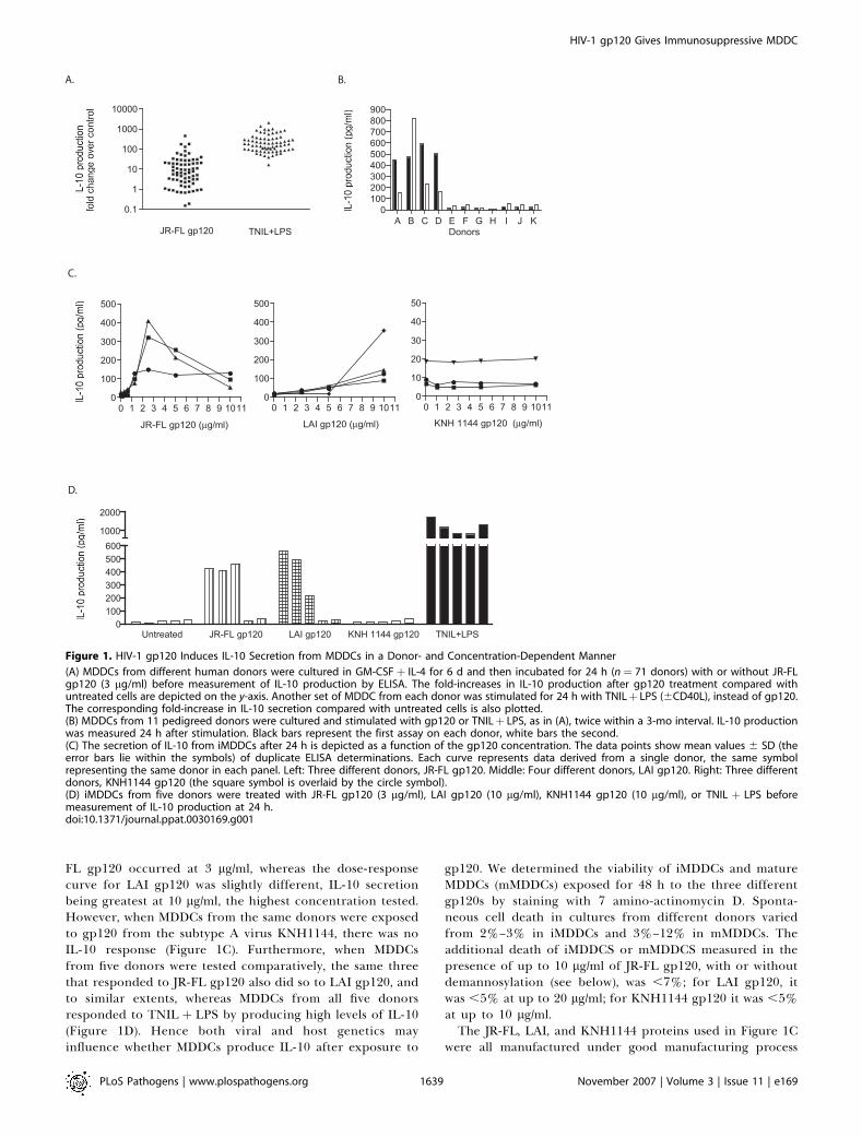
FL gp120 occurred at 3 lg/ml, whereas the dose-responsecurve for LAI gp120 was slightly different, IL-10 secretionbeing greatest at 10 lg/ml, the highest concentration tested.However, when MDDCs from the same donors were exposedto gp120 from the subtype A virus KNH1144, there was noIL-10 response (Figure 1C). Furthermore, when MDDCsfrom five donors were tested comparatively, the same threethat responded to JR-FL gp120 also did so to LAI gp120, andto similar extents, whereas MDDCs from all five donorsresponded to TNIL þ LPS by producing high levels of IL-10(Figure 1D). Hence both viral and host genetics mayinfluence whether MDDCs produce IL-10 after exposure to
gp120. We determined the viability of iMDDCs and matureMDDCs (mMDDCs) exposed for 48 h to the three differentgp120s by staining with 7 amino-actinomycin D. Sponta-neous cell death in cultures from different donors variedfrom 2%–3% in iMDDCs and 3%–12% in mMDDCs. Theadditional death of iMDDCS or mMDDCS measured in thepresence of up to 10 lg/ml of JR-FL gp120, with or withoutdemannosylation (see below), was ,7%; for LAI gp120, itwas ,5% at up to 20 lg/ml; for KNH1144 gp120 it was ,5%at up to 10 lg/ml.The JR-FL, LAI, and KNH1144 proteins used in Figure 1C
were all manufactured under good manufacturing process
Figure 1. HIV-1 gp120 Induces IL-10 Secretion from MDDCs in a Donor- and Concentration-Dependent Manner
(A) MDDCs from different human donors were cultured in GM-CSFþ IL-4 for 6 d and then incubated for 24 h (n¼ 71 donors) with or without JR-FLgp120 (3 lg/ml) before measurement of IL-10 production by ELISA. The fold-increases in IL-10 production after gp120 treatment compared withuntreated cells are depicted on the y-axis. Another set of MDDC from each donor was stimulated for 24 h with TNILþ LPS (6CD40L), instead of gp120.The corresponding fold-increase in IL-10 secretion compared with untreated cells is also plotted.(B) MDDCs from 11 pedigreed donors were cultured and stimulated with gp120 or TNILþ LPS, as in (A), twice within a 3-mo interval. IL-10 productionwas measured 24 h after stimulation. Black bars represent the first assay on each donor, white bars the second.(C) The secretion of IL-10 from iMDDCs after 24 h is depicted as a function of the gp120 concentration. The data points show mean values 6 SD (theerror bars lie within the symbols) of duplicate ELISA determinations. Each curve represents data derived from a single donor, the same symbolrepresenting the same donor in each panel. Left: Three different donors, JR-FL gp120. Middle: Four different donors, LAI gp120. Right: Three differentdonors, KNH1144 gp120 (the square symbol is overlaid by the circle symbol).(D) iMDDCs from five donors were treated with JR-FL gp120 (3 lg/ml), LAI gp120 (10 lg/ml), KNH1144 gp120 (10 lg/ml), or TNIL þ LPS beforemeasurement of IL-10 production at 24 h.doi:10.1371/journal.ppat.0030169.g001
PLoS Pathogens | www.plospathogens.org November 2007 | Volume 3 | Issue 11 | e1691639
HIV-1 gp120 Gives Immunosuppressive MDDC

conditions and were essentially LPS-free. We also tested severaladditional gp120 proteins of different genotypes and expressedin different cell types (including insect cells) that we obtainedfrom commercial sources and academic collaborators. Ingeneral, the degree of LPS contamination in these preparationswas too high for the results to be interpretable, since LPS isitself a highly efficient inducer of IL-10 fromMDDCs (Figure 1).
HIV-1 gp120 Stimulates IL-10 Production by MDDCs
through a Mannose-Dependent Interaction
To determine which gp120 receptors on iMDDCs wereresponsible for activating IL-10 expression, we incubatedeither gp120 or the cells with ligands that should block knowngp120-receptor interactions (Figures 2A, S3, and S4). Neither
Figure 2. The Induction of IL-10 Secretion by gp120 Is Mannose-Dependent
(A) The bars represent IL-10 production from MDDCs on day 6 after 24 h (black bars) of treatment with JR-FL gp120 (3 lg/ml). The reagents listed on thehorizontal axis were incubated with gp120 or iMDDCs for 1 h prior to addition of gp120 to the cells (see Materials and Methods for the inhibitorconcentrations tested). The bars represent the mean value 6 SEM for data derived from five different, gp120-responsive donors. The left and rightpanels show data derived from different experiments. The various reagents were also tested in the absence of gp120 and found not to stimulate IL-10production (,25 pg/ml, unpublished data), with the exception of mannan (see right panel).(B) The reactivities of mAbs b12 and 2G12, the CD4-IgG2 protein, or DC-SIGN-Fc with mock-treated JR-FL M-gp120 (squares) and a-(1,2,3,6)mannosidase-treated JR-FL D-gp120 (circles) were compared using ELISAs. In the fifth panel, the binding of DC-SIGN-Fc to JR-FL gp120 was inhibited bythe Ca2þ-chelator EGTA and the anti-DC-SIGN mAb, AZN-D1 (M-gp120 black bar; D-gp120 white bar).(C) Left panel: A reducing SDS-PAGE gel shows the reduction in JR-FL gp120 m.wt. caused by treatment with a-(1,2,3,6) mannosidase. Right panel:western blotting with anti-gp120 serum ARP3119 confirms the m.wt reduction, and blotting with mAb 2G12 shows that its mannose-dependentepitope has been removed from gp120. (M ¼m.wt markers; enzyme only ¼ no gp120 present).(D) The experimental design was the same as in (A). The gp120 proteins (or influenza HA or TNILþ LPSþCD40L) tested are listed on the x-axis. The barsrepresent the mean values 6 SEM for data derived from five different donors (black bars, IL-10 production after 24 h; white bars, after 48 h).doi:10.1371/journal.ppat.0030169.g002
PLoS Pathogens | www.plospathogens.org November 2007 | Volume 3 | Issue 11 | e1691640
HIV-1 gp120 Gives Immunosuppressive MDDC

the b12 mAb to the CD4-binding site on gp120 nor sCD4inhibited IL-10 production, implying that a gp120-CD4interaction was not responsible. The small-molecule CCR5antagonist AD101 was not inhibitory, ruling out signalstransduced via gp120-CCR5 binding (Figure S4). The CXCR4antagonist AMD3100 was inactive against IL-10 induction bygp120 from the X4 virus, LAI, so CXCR4 is also uninvolved(unpublished data). As expected, AMD3100 did not inhibitthe IL-10 response to JR-FL gp120, or AD101 the response toLAI gp120 (Figure S4 and unpublished data). In contrast,when gp120 was pre-treated with either mAb 2G12 or CV-N,IL-10 induction was strongly inhibited (Figure 2A). Both 2G12and CV-N bind to mannose moieties on gp120 N-linkedglycans [25–27], implicating an interaction between gp120and an MCLR(s) as the critical trigger for IL-10 induction. Wealso tested whether soluble mannans antagonized gp120-dependent IL-10 expression, but found that mannansthemselves strongly activated an IL-10 response (Figure 2A).However, combining gp120 with mannans did not furtherelevate IL-10 levels, suggesting that both of these mannose-containing ligands bind to, and saturate, the same MCLR(s).To explore which MCLR(s) might be involved, we used mAbsspecific to DC-SIGN and the mannose receptor (MR). Theanti-DC-SIGN mAb AZN-D1 completely blocks the binding ofmannosylated gp120 to DC-SIGN in an ELISA (Figure 2B).Two mAbs to DC-SIGN, including AZN-D1 and mAb Clone19 to the MR, can each reduce the binding of gp120 to asubset of tonsillar B cells [28]. When the anti-DC-SIGN andanti-MR mAbs AZN-D1 and Clone 15–2 were each pre-incubated with iMDDCs, AZN-D1 partially (;50%) reducedgp120-mediated IL-10 induction whereas Clone 15–2 was notinhibitory; adding the two mAbs together completelyabolished the IL-10 response (Figure 2A). An anti-CD4 mAbwas not inhibitory by itself at 24 h and did not affect theactions of the anti-MCLR mAbs when combined with them,although it did cause partial (45% 6 SD 29%) inhibition at 48h (Figure 2A and unpublished data). Other than mannan, thevarious mAbs and ligands described above did not induce IL-10 expression or block LPS-induced IL-10 expression (Figures2A, S4, and unpublished data). The same concentrations (40lg/ml) of different murine isotype control antibodies, aloneand in combination, were also without effect (Figure S4 andunpublished data).
The above experiments imply that MCLRs, particularly butprobably not only DC-SIGN, are the gp120 receptors thattrigger the IL-10 response. If so, the high mannose residueson gp120 glycans are likely to be the cognate ligands. Toprove this, the mannose moieties were removed from gp120by enzymatic digestion with a-(1,2,3,6)-mannosidase [25].Reducing SDS-PAGE gel analysis showed the demannosylatedJR-FL gp120 (D-gp120) was slightly smaller than mock-treatedgp120 (M-gp120; processed without the enzyme) and had lostits 2G12 epitope (Figure 2C; compare lanes markedþ and�).The successful removal of mannose was verified by showingthat D-gp120 failed to bind either 2G12 or DC-SIGN-Fc inELISAs, in contrast to M-gp120 (Figure 2B). However, bothmAb b12 and CD4-IgG2 bound efficiently to structuresassociated with the CD4-binding site on D-gp120 (Figure2B), showing that JR-FL gp120 was efficiently demannosylatedwithout impairing its overall conformation [25,27]. Hence wecould now directly assess the role of the high mannose glycansin the IL-10 response.
M-gp120 induced substantial IL-10 production (150–300pg/ml) from MDDCs from five different donors, whereas D-gp120 had no such effect. Influenza virus HA did notstimulate IL-10 production, whereas TNIL þ LPS activated astrong response (Figure 2D). An interaction between themannose moieties on gp120 and an MCLR(s) can thereforetrigger IL-10 production from MDDCs from a significantproportion of human donors. The lack of effect of HA, whichdoes not bind to DC-SIGN, compared to gp120 is consistentwith the outcome of comparative immunization studies withthese two viral receptor-binding glycoproteins in mice [11].The blocking effect of the anti-DC-SIGN and anti-MR mAb
combination implicated these MCLRs as likely mediators ofthe IL-10 response to gp120 (Figure 2A). Because thisresponse is donor-dependent (Figure 1A), we measured DC-SIGN and MR expression on day-6 iMDDCs (the time ofaddition of gp120) from nine donors, as well as the expressionof CD80, CD83, and CD86. DC-SIGN levels varied by 18-foldamong these nine donors, MR by 3.3-fold (in studies of othersets of donors, we have found that the expression levels ofboth these receptors can vary by about an order ofmagnitude; unpublished data). The IL-10 response to JR-FLgp120 correlated with the level of DC-SIGN expression onday 6 (n¼9, r2¼0.52, p¼0.028) but not with MR expression (n¼ 9, r2¼ 0.19, p¼ 0.25). Moreover, there were no correlationsbetween IL-10 production and the expression of CD80 (n¼ 9,r2¼0.052, p¼0.59), CD83 (n¼9, r2¼0.33, p¼0.14), or CD86 (n¼ 9, r2¼ 0.27, p¼ 0.19). Thus, of the five correlations with IL-10 production that we performed, the only substantial onewas with DC-SIGN expression.
HIV-1 gp120 Induces IL-10 Production via the ERKSignaling PathwayWe next sought to identify which signaling pathway(s) was
involved in the upregulation of IL-10 expression after thegp120-MCLR interaction. We focused on the ERK1/2 and p38MAP kinase pathways [29], because ERK1/2 phosphorylationand activation promotes IL-10 production and inhibits IL-12production by DC [30], whereas inhibition of ERK1/2activation has the opposite effects [31]. Conversely, p38mediates the induction of IL-12p70 expression by LPS [29].Furthermore, DC-SIGN ligation by antibodies can lead toERK1/2 phosphorylation [32]. We observed that TNIL þ LPSstrongly activated the phosphorylation of both ERK1/2 andp38 within 5 min, effects that persisted for 30–60 min (Figure3A and unpublished data). A lesser, but still significant, levelof ERK1/2 activation occurred after 5–10 min in MDDCstreated with M-gp120, ERK1/2 phosphorylation levels thendeclining back to baseline after 30 min. However, there wasno detectable phosphorylation of p38 in response to M-gp120at any time point (Figure 3A and unpublished data). D-gp120,in contrast, failed to activate the phosphorylation of eitherERK1/2 or p38 (Figure 3A), implying that the gp120-MCLRinteraction leads to the specific, albeit transient, activation ofthe ERK1/2 signaling pathway. In the same experiment, M-gp120, but not D-gp120, induced modest levels of IL-10production, whereas TNIL þ LPS þ CD40L activated a muchgreater IL-10 response (Figure 3B), observations in propor-tion to the levels of ERK1/2 activation induced by thedifferent stimuli (Figure 3A).We used signaling inhibitors to determine whether there is
a link between ERK1/2 activation and IL-10 production.
PLoS Pathogens | www.plospathogens.org November 2007 | Volume 3 | Issue 11 | e1691641
HIV-1 gp120 Gives Immunosuppressive MDDC

MDDCs were treated with 5 lM U0126 (an ERK1/2 inhibitor)or 10 lM SB 203580 (a p38 MAP kinase inhibitor) for 1–2 h,then IL-10 and IL-12p70 production in response to JR-FL M-gp120 or TNIL þ LPS þ CD40L were measured 24 h later.U0126 inhibited ERK1/2 phosphorylation by ;60% (Figure3A), and the same compound reduced the IL-10 responses toboth M-gp120 (by 70%) and TNILþLPSþCD40L (by ;90%)
(Figure 3B). SB 203580 had a negligible effect on M-gp120-stimulated IL-10 production but did inhibit the IL-10response to TNILþ LPS by 70% (Figure 3B).In view of the reciprocal effects of the ERK1/2 pathway on
IL-10 and IL-12 production by DCs [30,31], we also measuredthe IL-12p70 responses to M-gp120 and to TNIL þ LPS. M-gp120 triggered a very slight increase in IL-12p70 expression.In contrast, TNIL þ LPS activated a substantial IL-12p70response that was completely blocked by SB 203580 butpotentiated (4.7-fold) by U0126, a pattern consistent with aprevious report [31]. Neither inhibitor, by itself, activated IL-10 or IL-12p70 production (Figure 3B).Together, the use of signaling inhibitors implies that ERK1/
2 activation is required for IL-10 production by MDDCs inresponse to either M-gp120 or TNIL þ LPS.
HIV-1 gp120 Impairs iMDDC MaturationWe used immunophenotypic analyses to investigate
whether gp120 affects iMDDC maturation. Neither M-gp120nor D-gp120 induced iMDDCs to mature in the absence ofTNIL þ LPS þ CD40L; the cell-surface expression of nomaturation marker changed by more than 1.5-fold (unpub-lished data). However, expression of CD80 was reduced by 3-fold, CD83 by 7-fold, and CD86 by 2-fold when iMDDCs wereincubated with M-gp120 together with TNILþ LPSþCD40L,compared with when the cells were matured with TNILþLPSþ CD40L alone. DC-SIGN expression was 2- to 3-fold greateron MDDCs treated with TNIL þ LPS þ CD40L plus M-gp120than on cells receiving only TNIL þ LPS þ CD40L, but MRexpression was unchanged. D-gp120 did not mimic the effectsof M-gp120 on the expression of CD80, CD83, CD86, and DC-SIGN, implicating an MCLR(s) as a mediator of these effectsof gp120 (Figure 4). Furthermore, gp120 impaired thematuration of iMDDCs from both IL-10-responding andnon-responding donors. The reduced expression of CD80,CD83, CD86, and the increased expression of DC-SIGN didnot correlate with IL-10 secretion 48 h after gp120 additionamong the 15 donors tested, of which nine were IL-10responders, six non-responders. There were no correlations:r2 ¼ 0.05 for CD80 fold-decrease versus IL-10; r2 ¼ 0.02 forCD83 fold-decrease versus IL-10; r2 ¼ 0.00004 for the fold-increase in DC-SIGN expression versus IL-10. The reductionin CD86 expression also did not correlate with IL-10, r2 ¼0.00003.The interaction of gp120 with an MCLR(s) therefore
partially blocks the TNILþLPSþCD40L-induced maturationof iMDDCs that normally leads to increases in CD80, CD83,and CD86 expression and a reduction in DC-SIGN expres-sion. These events occur irrespective of whether the gp120-treated cells produce IL-10.
HIV-1 gp120 Inhibits the Ability of mMDDCs to Induce TCell ProliferationWe next explored whether the effects of gp120 on MDDC
maturation (and cytokine production, see Supporting In-formation) would affect their ability to stimulate theproliferation of allogeneic T cells. To do this, M-gp120 orD-gp120 (JR-FL) was added to iMDDCs simultaneously withTNIL þ LPS (i.e., on day 6 from the start of the MDDCculture). Influenza virus HA was used as a control antigen,also given simultaneously with TNILþLPS. After the iMDDCshad been cultured with the various stimuli for 48 h, the cells
Figure 3. Involvement of the ERK1/2 and p38 MAP Kinase Signaling
Pathways in the Induction of IL-10 and IL-12p70 by gp120 and TNILþLPS
(A) Day-6 MDDCs were incubated with or without JR-FL M-gp120 or D-gp120 (3 lg/ml), or with TNIL þ LPS, for 10 min before pERK1/2 (upperpanel) and p-p38 levels (lower panel) were measured by ELISA (blackbars). The white bars show the effects of adding the pERK1/2 inhibitorUO126 (5 lM) or the p38 inhibitor SB 203580 (10 lM) 1 h before thegp120s or TNIL þ LPS. The bars represent the mean values 6 SEM fordata derived from four different donors.(B) The experimental design was based on that used for (A), except thatthe iMDDCs were incubated with or without UO126 or SB 203580 for 1 hprior to the addition of M-gp120 or TNILþ LPSþ CD40L and continuedincubation for 24 h. IL-10 (upper panel) or IL-12p70 (lower panel)production was measured by ELISA after 24 h. The bars represent themean values 6 SEM for data derived from five different donors, whichwere not the same as the ones used in (A).doi:10.1371/journal.ppat.0030169.g003
PLoS Pathogens | www.plospathogens.org November 2007 | Volume 3 | Issue 11 | e1691642
HIV-1 gp120 Gives Immunosuppressive MDDC

were washed to remove any free gp120 or HA, then negativelyselected for CD8, CD14, CD16, CD19, CD36, CD56, CD123,TCRc/d, and CD235a. CFSE-labeled, allogeneic CD4þ T cellswere then added (the ratio of 1/10 was optimized fordetection of T cell proliferation) for a 5-d co-culture (i.e.,from days 8–13 from the start of the MDDC culture). T cellproliferation was measured as the proportion of CFSE-negative cells. Flow-cytometric histograms supporting thedata presented below are shown in Figure S5.
MDDCs treated with gp120 in the absence of TNIL þ LPSdid not stimulate T cell proliferation (Figure 5A). However,exposing the TNILþ LPS-stimulated MDDCs to M-gp120 for
24–48 h reduced their ability to stimulate T cell proliferationby ;65%. D-gp120 was less inhibitory, the ;30% decreasebeing little different from the ;20% decrease seen with theHA control antigen. M-gp120 depressed proliferation sig-nificantly more than did D-gp120 (one-tailed Mann-WhitneyU test, n¼ 15, p , 0.0001). MDDCs from all 15 donors tested(ten IL-10 responders, five non-responders) behaved similarlyin the T cell proliferation assay; the relative proliferation ofCD4þ T cells in co-cultures with M-gp120 þ TNIL þ LPS-treated MDDCs varied in a narrow range (60%–85%reduction in proliferation) over a broad range of IL-10responses (0–420 pg/ml) (Figure 5B). Overall, there was no
Figure 4. Gp120 Impairs iMDDC Maturation via Interaction with an MCLR(s)
The maturation status of MDDCs was evaluated after treatment for 48 h (days 6–8) with TNILþ LPSþ CD40L 6 3 lg/ml of JR-FL M-gp120 or D-gp120.The cell surface expression of CD80, CD83, CD86, DC-SIGN, and MR on CD11cþ cells was measured by flow cytometry as described in SupportingInformation.(A) The histograms show expression of the surface markers on MDDCs from one donor whose expression marker response to gp120 was of averagemagnitude. The grey shaded profiles depict the use of isotype control mAbs, the other profiles were derived using the various specific test mAbs. Theblack curves represent control MDDCs; red curves, MDDCs treated with TNILþ LPSþCD40L; blue curves, TNILþ LPSþCD40LþD-gp120; green curves,TNILþ LPSþ CD40LþM-gp120.(B) The average fold-changes (for 14 or 15 donors) in MFI values for MDDC cell-surface marker expression are depicted. The background MFIs obtainedwith the respective isotype controls were subtracted from MFIs for all conditions. Then the ratios of MFI for presence of gp120 over the MFI for absenceof gp120 were calculated. For each marker, the MFI value derived from MDDC matured with TNILþ LPSþCD40L alone is thus defined as 1.0 (log ratio¼0). The means of the 10-logarithms of the ratios for all donors were calculated. The mean log MFI ratio 6 SD for cells also treated with either M-gp120(black bars) or D-gp120 (white bars) are plotted relative to this baseline value corresponding to TNILþ LPS þ CD40L alone.doi:10.1371/journal.ppat.0030169.g004
PLoS Pathogens | www.plospathogens.org November 2007 | Volume 3 | Issue 11 | e1691643
HIV-1 gp120 Gives Immunosuppressive MDDC

correlation between IL-10 levels in the cultures of the gp120-treated, TNIL þ LPS-stimulated MDDCs on day 8 and theinhibition of subsequent T cell proliferation (% CFSEdilution versus IL-10, r2 ¼ 0.0008). The lack of effect of IL-10 is not surprising because both cytokines and any stimulusfor their continued secretion are washed out of the MDDCcultures before the T cells are added. However, the principalpoint is that, as with maturation marker expression, theMDDC phenotype is adversely affected by gp120 treatment,whether or not the cells have an immediate IL-10 response.We also found that JR-FL and KNH1144 gp120s each
caused a ;70% reduction in the capacity of LPS þ TNIL-stimulated MDDCs from four donors to induce T cellproliferation (similarly to what is shown for JR-FL gp120 inFigure 5A, unpublished data). Since KNH1144 gp120 does notinduce IL-10 secretion from MDDCs (Figure 1C), this experi-ment corroborates the finding that the anti-proliferativeeffect is independent of IL-10 from MDDCs (Figure 5B). Italso confirms that KNH1144 gp120 is biologically active in theMDDC system despite its inability to induce IL-10 expression.We measured the concentrations of both IL-10 and IL-
12p70 in the various MDDC-T-cell co-cultures on day 13(Figure 5C). IL-10 concentrations varied by ,5-fold overall,the co-cultures with MDDCs exposed to M-gp120 þ TNIL þLPS containing the highest level (280 6 45 pg/ml). The IL-10response to M-gp120 þ TNIL þ LPS was significantly higherthan to D-gp120 þ TNIL þ LPS (one-tailed Mann-Whitney Utest, n ¼ 5, p ¼ 0.028). IL-12p70 concentrations varied muchmore substantially. They were very low (,10 pg/ml) in co-cultures containing MDDCs treated with M-gp120, D-gp120,or HA in the absence of TNILþ LPS. When TNILþ LPS wasused to mature the MDDCs, IL-12p70 concentrations reached200 6 22 pg/ml. The inclusion of D-gp120 or HA caused amodest (;2-fold) reduction, but when M-gp120 was used onlybaseline levels of IL-12p70 were produced (7.2 6 1.7 pg/ml).The IL-12p70 response to M-gp120 þ TNIL þ LPS wassignificantly lower than to D-gp120þTNILþ LPS (one-tailedMann-Whitney U test, n ¼ 5, p ¼ 0.0040). Thus, compared tothe use of TNILþ LPS alone, exposure of the MDDCs also toM-gp120 caused a 76-fold increase in the IL-10/IL-12p70 ratioin the co-cultures, whereas the use of D-gp120 and HA causedonly 2.4- and 1.3-fold increases, respectively. Moreover, thepattern of IL-12p70 responses in the various co-cultures(Figure 5C, lower panel) was similar to the pattern of T cellproliferation in the same cultures (Figure 5A). IL-4 was alsomeasured, the concentrations ranging from 5–15 pg/ml in thedifferent co-cultures, with no obvious pattern of responsedetectable (unpublished data).In conclusion, MDDCs matured in the presence of gp120
are functionally impaired, irrespective of whether theysecrete IL-10 soon after gp120 binds to MCLRs.
Figure 5. Treatment with gp120 Inhibits MDDC-Induced T Cell
Proliferation
(A) Day-6 MDDCs were incubated for 48 h with or without TNIL þ LPSand/or JR-FL M-gp120, D-gp120, or influenza HA as specified on the x-axis. The MDDCs were then co-cultured for 5 d with CFSE-labeled CD4þTcells before determination of the extent of the allogeneic mixed Tlymphocyte reaction on day 13. Relative CD4þ T cell proliferation wascalculated by first subtracting the background value for CFSE-negativecells obtained using unstimulated iMDDCs (9.95% 6 0.70%, n¼ 15) fromthe value obtained when the MDDCs were stimulated with TNIL þ LPS(46.4% 6 1.54%, n¼15). The net value was defined as 100% and used fornormalization. The bars represent the mean values 6 SD for data derivedfrom 15 donors (except for influenza HA; ten donors) tested in 15independent experiments. The superantigen SEB served as a positivecontrol for CD4þ T cell stimulation in the absence of any MDDCs; CFSE
dilution in response to SEB was 130% 6 2.9% (n¼ 15) of that seen withTNILþ LPS (unpublished data).(B) The extent of T cell proliferation in co-cultures (as in A) containingiMDDCs exposed to M-gp120 þ TNIL þ LPS þ CD40L is plotted as afunction of the IL-10 response of the iMDDCs to M-gp120 after 24 h.There was no correlation for iMDDCs from 15 donors (r2¼ 0.0008).(C) In a subset of the experiments shown in (A), extracellular cytokinelevels were measured at the end of the MDDC-T cell co-culture (i.e., onday 13). The bars represent the mean values 6 SEM from five differentdonors. Upper panel, IL-10; lower panel, IL-12p70.doi:10.1371/journal.ppat.0030169.g005
PLoS Pathogens | www.plospathogens.org November 2007 | Volume 3 | Issue 11 | e1691644
HIV-1 gp120 Gives Immunosuppressive MDDC

Discussion
We show here that exposure to HIV-1 gp120s can impairthe maturation of human iMDDCs, triggering cells from somedonors to secrete IL-10, a cytokine generally associated withimmunosuppressive responses [23]. Irrespective of whetherthey secrete IL-10, the gp120-treated MDDCs mature in-efficiently in response to conventional stimuli, and theirabilities to stimulate the proliferation of T cells in co-culturesare impaired. The latter defect could be due to their reducedexpression of CD80, CD83, and CD86 and hence a weakeningof the co-stimulatory interactions with T cells that drive thelatter’s proliferation. The reduction in IL-12p70 levels (and asubstantial increase in the IL-10/IL-12p70 ratio) in the co-cultures may also be relevant [33].
These various effects are a consequence of an interactionbetween the mannose components of gp120 glycans and anMCLR(s), in that the enzymatic removal of mannoses fromgp120 reduced or prevented their occurrence. We alsoprobed the IL-10 response to gp120 using various blockingligands. Thus, CV-N and the 2G12 mAb bind to gp120mannoses, and each inhibited IL-10 induction, whereasinhibitors of gp120 binding to CD4, CCR5, or CXCR4 wereineffective. Furthermore, mannan, another MCLR ligand,activated IL-10 expression. Also relevant is that gp120induces IL-10 expression in immunized mice [11]: Gp120cannot bind to murine CD4, CCR5, or CXCR4, or to themurine MCLR with the greatest sequence similarity to humanDC-SIGN [34]. However, five murine DC-SIGN homologueshave been described [35], so it is possible that some of themdo bind gp120. The influenza HA Env protein does notinduce IL-10 expression either in the immunized mice or inour own in vitro experiments; HA binds the MR [36] but notDC-SIGN or DC-SIGNR [37].
Several different MCLRs are known or potential bindingsites for gp120 on DC, including DC-SIGN, langerin, and theMR [38]. We found that mAbs to DC-SIGN and the MRtogether completely ablated the IL-10 response to gp120,while the anti-DC-SIGN mAb was partially inhibitory by itself.Furthermore, there was a correlation between the extent ofIL-10 production and the level of DC-SIGN expression on theMDDCs. Together, these observations strongly suggest a rolefor DC-SIGN binding in the IL-10 response to gp120 butother MCLRs, particularly the MR, also seem likely to beinvolved.
M-gp120, but not its demannosylated derivative, activatedERK1/2 phosphorylation, and the ERK1/2 inhibitor U0126inhibited the IL-10 response to M-gp120. These findingsimply that the gp120-MCLR interaction triggers the ERK1/2signaling pathway and that this is necessary for activation ofIL-10 expression. Whether the same pathway mediates theother MDDC responses to gp120 remains to be determined.
This conclusion is consistent with earlier reports on therole of the ERK1/2/MAP kinase pathways in the IL-10response when DCs are activated by other stimuli, includingTLR ligands and DC-SIGN-specific antibodies [30–32]. Thebinding of pathogens, including HIV-1, to DC-SIGN has alsobeen shown to activate the Raf-1-acetylation-dependentsignaling pathway [39]. The gp120-treated MDDC from abouthalf the 71 donors we studied secreted elevated amounts ofIL-10, and the response pattern was consistent when 11donors were re-tested a month later. Hence, genetic or other
invariable factors and not, for example, an inter-currentinfection seem most likely to determine whether a donor’sMDDCs respond to gp120 in this way, or not. Complex hostgenetic factors influence IL-10 gene regulation [23,40–42],suggesting one area for further study. The genetics of MCLRexpression might also be relevant; different MCLRs might beinvolved to different extents on MDDCs from differentdonors. DC-SIGN expression varies considerably in rectaltissue samples from different individuals and has beenassociated with local increases in the IL-10/IL-12 ratio [33].Nonetheless, the IL-10 response to gp120 is only one markerfor the adverse effect of this ligand on MDDCs; whether ornot a donor’s cells secreted IL-10 in response to gp120, theywere functionally impaired, matured poorly, and were unableto efficiently stimulate T cell proliferation.We also observed that both the concentration and the
identity of the gp120 protein influenced the IL-10 response.Two of the three tested gp120s (JR-FL and LAI) triggered IL-10 release from MDDCs of responsive donors, whereas gp120from KNH1144 did not. We do not yet know why KNH1144differs from the other two gp120s in this regard. The mostlikely explanation is that there are subtle differences betweenthe gp120s in exactly how they interact with one or moreMCLR, and that the IL-10 response is particularly sensitive toa specific, but as yet uncharacterized, facet of theseinteractions. Differences in how diverse HIV-1 virions andgp120 proteins interact with DC-SIGN have been reported,although the variations in gp120 structure that affect theinteraction have still to be fully defined [43] (M. Jansson,personal communication). A sequence alignment of the JR-FL, LAI, and KNH1144 gp120 proteins, with emphasis on thepositions of N-linked glycans, suggests a number of poten-tially relevant differences (Figure S6). Since understandingthe molecular basis for the lack of IL-10 induction might helpin the design of new Env-based immunogens, mutagenesisstudies that focus on the N-linked glycans of JR-FL andKNH1144 could be informative, as might the use of addi-tional gp120s that vary in sequence and that are expressed incell types that lead to differences in glycosylation patterns. Itis important, however, that any such reagents be highlypurified free of the LPS contaminants that are common inmost commercial gp120 preparations and in some othersmade under non-GMP conditions. The same constraintsapply to the use of inactivated HIV-1 virions. We have notyet tested virions, as the focus of the present study is onsoluble Env proteins that are (or were) vaccine candidates.Moreover, virions contain several TLR activators that mightinduce different cytokine responses that complicate anyanalysis of the effects of the Env component [44–46].Several earlier studies have shown that gp120 and
inactivated HIV-1 virions can have complex effects onMDDCs and their interactions with T cells and on cytokinesecretion by both cell types in vitro. Thus, compared to LPS,R5, and X4 gp120s both stimulated much less IL-12production from MDDCs, but without IL-10 release [19]. Justas we have observed, gp120-treatment impaired MDDCmaturation in response to classical stimuli, reducing theirability to stimulate T cells, but unlike our results, CD80,CD83, and CD86 were up-regulated on the gp120-treatedcells [19]. In another study, exposure of MDDCs to X4 gp120up-regulated CD80 and CD86 and down-regulated MR, withincreased secretion of IL-10, IL-12, IL-18, and TNF-a [47].
PLoS Pathogens | www.plospathogens.org November 2007 | Volume 3 | Issue 11 | e1691645
HIV-1 gp120 Gives Immunosuppressive MDDC

Various surface markers were also up-regulated on HIV-1-infected MDDCs, associated with an inability of the cells tosecrete IL-12 in response to CD40L [48]. The receptorinteractions of gp120 most responsible for its variousbiological effects were not determined in these variousstudies. Gp120 is also known to stimulate IL-10 release frommonocyte/macrophages in vitro [17,18,20,22,24]. There is onereport that MDDCs undergoing continued stimulation withGM-CSF and IL-4 did not secrete IL-10 in response to gp120,although differences in the experimental conditions areprobably responsible, and the donor- and gp120-dependentvariation we now describe may also be relevant [49].
Our observations are consistent with a mounting body ofevidence on the biological effects of ligating MCLRs on DCs.Thus, HIV-1 BaL and a specific DC-SIGN mAb have recentlybeen shown to activate Rho-GTPase-dependent signals viaDC-SIGN that favor the formation of DC-T-cell synapses andHIV-1 infection of the T cells [50]. The same signaling eventsalso induced the ATF3 transcription factor that suppressedTLR-response genes, attenuating the LPS responses of thecells by reducing IL-12p70 secretion and down-modulatingCD86 and HLA-DR. Thus, as we observed with gp120, theanti-DC-SIGN mAb induced a semi-immature state in theMDDCs, which failed to stimulate T cell proliferationeffectively [50]. Indeed, the binding of an antibody to DC-SIGN was previously found to activate ERK-1/2 but not p38[32], similar to what we have observed with gp120. Cross-linking the MR via a specific mAb can have a broadly similareffect on the MDDC phenotype [51]. It will be worth studyingwhether the downstream signals activated by the MCLR MAbsare also triggered by gp120. Of further note is that anallergenic glycoprotein from peanuts also induces ERK1/2signaling in MDDCs via DC-SIGN, but up-regulates MHC andco-stimulatory molecules and thereby increases the ability ofthe MDDCs to activate T cell proliferation [52]. Thus, theremay be considerable subtleties to how different glycoproteinsand mAbs bind to DC-SIGN and other MCLRs on the MDDCsurface, and the intracellular consequences of these inter-actions. Most mAbs to DC-SIGN or the MR do not inducetransmembrane signals, but some do [50,51]; likewise, somegp120s induce IL-10 expression, others do not. One relevantpoint may relate to how an MCLR ligand is mannosylated:adding O-linked mannoses to ovalbumin increases lympho-proliferation in mixed BMDC-T-cell cultures while N-linkedmannoses have the opposite effect, and mannosylatedovalbumin impaired IL-12p70 secretion [53]. Most mannoseresidues on gp120 are N-linked [54], but the relative amountsof N- and O-linked moieties could vary between strains andinfluence the overall signaling patterns that are activated.Other pathogens also use mannose moieties to suppressimmune responses, again via binding to MCLRs. For example,the M.Tb cell wall component ManLAM binds to DC-SIGN ata similar site to gp1209s, induces IL-10 production, impairsDC maturation, and suppresses the host immune response tothis pathogen [55,56]. Some lactobacilli do much the same,although without the involvement of mannose residues [57].
Although DC-SIGN, and MCLRs in general, are importantsentinels for the presence of pathogens, some organisms maybe able to subvert at least some of the natural functions ofthese receptors for their own purposes [58]. DC-SIGN, inparticular, may be considered as an unconventional PRR(pattern recognition receptor) that drives TH2 and Treg
responses [32,58]. Silencing SOCS-1 in DC has been shownto reduce the suppressive effect of gp120 on the production ofpro-inflammatory cytokines in vitro [59]. Mice immunizedwith gp120-pulsed, SOCS-1-silenced DC produced higher andmore sustained titers of anti-gp120 antibodies, and TH1-polarized cellular responses to gp120 [59]. Conversely, over-expressing SOCS-3 in murine DC increased IL-10 expression,and SOCS-3-transduced DC primed a TH2-dominant re-sponse when co-cultured with CD4þ T cells in vitro [60].Perhaps these observations are linked mechanistically to ours?Caution must always be taken when extrapolating from cell
culture systems to the more complex environment of tissuesin vivo where the DC phenotype differs from the MDDCs wehave used here and where gp120 concentrations are hard toestimate [16,61]. We note, however, that DCs and T cellsisolated from HIV-1-infected persons can have aberrantphenotypes that are broadly similar to those of the gp120-exposed MDDCs that we have studied in vitro [62]. Inparticular, elevated numbers of tolerogenic semi-matureDCs, and FOXP3þ CD4þ regulatory T cells, have beenobserved in lymph nodes of HIV-1-infected people [63].Moreover, high levels of IL-10, accompanied by a reduction inIL-12, can be found in plasma during primary HIV-1infection [64]. IL-10 can have a substantial effect on thecourse of viral infections [65]. Thus, blocking IL-10 signalingby antibodies to its receptor promotes the clearance oflymphocytic choriomenigitis virus and prevents the establish-ment of a persistent infection [66,67]. Perhaps similar eventsare involved in persistent infection by HIV-1? Thus it ispossible that, during primary infection, env-gene productscould help suppress the development of anti-HIV-1 immuneresponses at this critical time, particularly as virion-associ-ated gp120 is more efficient than free gp120 at inducingvarious signaling events [68]. If so, the retention of highmannose moieties on the Env complex would be yet anotherdefense HIV-1 uses in its battle with host immunity. Wepreviously noted that the presence of mannoses on Env isparadoxical because they might facilitate virion clearancefrom the blood [25]: Counter-functions would justify theirretention.Our observations could help understand the outcome of
immunizing with Env-based antigens, and perhaps whydifferent individuals respond to these vaccines with Ab titersthat can vary over a several-log range [5–9]. When milligramamounts of gp120 are delivered in a bolus into tissues, localconcentrations are likely to be high enough to affect theperformance of various immune system cells, including DCs,during the earliest, formative stages of the immune response[16]. In a comparative DNA and protein immunization studyin mice, the antibody and cytokine responses to gp120 werestrongly TH2-polarized, whereas responses to HA were TH1-biased. Furthermore, the TH2 bias of the anti-gp120 responsedid not occur in IL-10 knock-out mice [11]. Although T-helper phenotypes are more complex in humans than mice,the responses to gp120, during infection and after vaccina-tion, do appear to be TH2-biased [12–15]. Including Env inmulti-component HIV/SIV vaccines can sometimes be dele-terious to protection [69,70]. Also, immunizing horses withinsect cell–expressed Env proteins (which are enriched forhigh-mannose moieties) from Equine Infectious AnemiaVirus (EIAV) enhanced post-immunization infection withEIAV, whereas EIAV Env proteins expressed in mammalian
PLoS Pathogens | www.plospathogens.org November 2007 | Volume 3 | Issue 11 | e1691646
HIV-1 gp120 Gives Immunosuppressive MDDC

cells induced protective responses [71–73]. Insect cell–ex-pressed gp120 proteins were also comparatively poorimmunogens in mice, because of a limited ability to induceT-helper responses [9].
Any vaccine-related, adverse influences of the high-mannose moieties on gp120 glycans could be overcome bytreating gp120 with a mannosidase enzyme. We are nowinvestigating whether this strategy improves the immunoge-nicity of HIV-1 Env proteins. Deleting a subset of N-linkedglycans altered the IgG isotype profile of the antibodyresponse to the HCV E1 protein in immunized mice andimproved its immunogenicity overall [74]. Of course, raisinghigher titers of antibodies and/or reducing the rate of decayof the antibody response to HIV-1 Env will achieve little ifthose antibodies are non-neutralizing. Our hope, however, isthat a general increase in the immunogenicity of Env proteinscould facilitate the development of otherwise sub-thresholdNAb responses, and/or enable lower amounts of Env trimersto be used. Combining the mannose-removal technique withother strategies intended to increase the immunogenicity ofNAb epitopes should also be possible. Several other vaccineantigens that are considered to be problematic from theimmunogenicity perspective, such as RSV F, RSV G, CMV gB,and Ebola GP, are also highly glycosylated and/or can bind toMCLRs (S. Plotkin and B. Graham, personal communication)[75–77]. Whether these proteins might also contain high-mannose moieties or other carbohydrate structures that caninteract with MCLRs and that could be removed enzymati-cally is worth considering.
Materials and Methods
Recombinant proteins and cytokines. Recombinant, CHO-cellexpressed monomeric gp120s from HIV-1 JR-FL, LAI, andKNH1144 were manufactured at Progenics, as previously described,under GMP conditions [78]. The concentration of the gp120 stockswas 1 mg/ml, with Endotoxin contamination ,3 EU/ml. Gp120 wasadded to target cells at 3 lg/ml (25 nM), except when otherwisespecified. Insect cell–expressed influenza hemagglutinin (HA) protein(100 lg/ml) was purchased from Protein Sciences Corporation andused at 3 lg/ml (Endotoxin ,10 EU/ml, no fungal or bacterialcontamination).
LPS from Salmonella Typhimurium (1 mg, Sigma) was used at 100 ng/ml. Recombinant soluble CD40L (50 lg, Bristol-Myers Squibb) with anEndotoxin level of , 0.1 ng per lg (1 EU/lg) was used at 1 ng/ml; TNF-a and IL-1b (R&D Systems) at 25 ng/ml and 10 ng/ml, respectively.
Inhibition of gp120-induced IL-10 production. iMDDCs wereincubated for 1 h at 37 8C with various agents before gp120 wasadded. The anti-DC-SIGN mAb AZN-D1 (Beckman Coulter), theisotype control mouse IgG1 (Clone 2T8-2F5, Beckman Coulter), theanti-mannose receptor (MR; CD206) mAb Clone 15–2 (Cell Sciences),and the isotype control mouse IgG1, j (Clone MOPC-21, BDPharmingen) were each used at 40 lg/ml, alone or in combination.The CCR5 inhibitor AD101 (from J. Strizki, Schering Plough ResearchInstitute)[79] and the CXCR4 inhibitor AMD3100 (from G. Bridger,AnorMed Incorporated) [80] were each used at 10 lM. Mannan(Sigma) was added at 30 lg/ml.
Alternatively, gp120 was mixed with sCD4 (Progenics) [81], mAbb12 (from D. Burton, Scripps) [82], mAb 2G12 (from H. Katinger,University of Vienna) [83], each at 25 lg/ml, or with cyanovirin-N(CV-N; from R. Shattock, St. George’s, London) [26] at 5 lg/ml for 1 hat room temperature on a roller before addition to the cells.
Mannosidase treatment of recombinant gp120. The mannoseresidues were removed from JR-FL gp120 to make demannosylatedgp120 (D-gp120) as follows [25]. Aliquots of gp120 (120 lg) wereincubated for 16–18 h at 37 8C with no enzyme (mock treatment; M-gp120) or with a-(1,2,3,6)-mannosidase (Jack Bean, GKX-5010; 25Units/mg, 0.14 Units/lg gp120; from ProZyme Incorporated) in a finalvolume of 1.2 ml, in the presence of protease inhibitors (Roche). Acontrol incubation of enzyme-only (no gp120) was also performed.The samples were desalted into half-strength PBS (1/2 PBS) using PD-
10 desalting columns (GE Healthcare) and concentrated to 1 ml usingVivaspin 30k MWCO 6 ml spin concentrators (Vivascience). Afteraddition of 1 volume of 1/2 PBS, each sample was processed using theEndofree Red 5/1 Endotoxin removal kit (Profos AG). The finalvolumes of the D-gp120 and M-gp120 preparations after endotoxinremoval were ;2 ml, with endotoxin levels ,8–20 EU/mg and gp120concentrations 60 lg/ml. SDS-PAGE and western blot analyses wereperformed using mAbs 2G12 and CA13 (ARP3119).
ELISA for gp120-binding ligands. gp120 proteins were capturedonto ELISA wells via sheep antibody D3724 to the gp120 C-terminus,and mAb or CD4-IgG2 binding was assessed essentially as describedpreviously [84]. For DC-SIGN binding to the captured gp120, thestandard procedure was adapted as follows: The plates were washedthree times with TSM (20 mM Tris, 150 mM NaCl, 1 mM CaCl2, 2 mMMgCl2), followed by incubation with TSM/1% BSA for 30 min. Afterthree washes with TSM, DC-SIGN-Fc (a gift from T. Geijtenbeek [85])in TSM was added for 2 h, with or without a prior incubation for 15min with EGTA (10 mM) or mAb AZN-D1 (10 lg/ml). The plates werewashed five times with TSM/0.05% Tween, then bound DC-SIGN-Fcwas detected with peroxidase-labeled goat anti-human Fc (1:3,000) inTSM/0.05% Tween using standard conditions.
Cell culture. Peripheral blood mononuclear cells (PBMC) wereisolated from buffy coats (New York Blood Center or Research BloodComponents) by Ficoll density gradient centrifugation. Monocyteswere isolated to high purity (.98%) by magnetic cell sorting withanti-CD14-coated beads according to the manufacturer’s recommen-dations (Miltenyi Biotec). The percentage of CD14þ monocytesamong the cells sorted from PBMC was determined by flow cytometryand always exceeded 98%. The CD14� fraction was frozen and used asthe source of T cells for MDDC-T cell co-cultures. The monocyteswere subsequently cultured for 6–8 d in complete culture medium(RPMI 1640, GIBCO/Invitrogen) containing 1 mM sodium pyruvate,0.1 mM nonessential amino acids, 2 mM L-glutamine, 25 mM HEPES,100 U/ml penicillin, 100 lg/ml Streptomycin (all obtained fromGIBCO/Invitrogen), and supplemented with 5% Human AB serum(Sigma) (R-5), 1,000 U/ml GM-CSF (Leukine, Sargramostim), and 1,000U/ml of recombinant human IL-4 (R&D Systems) at 37 8C in anatmosphere containing 5% CO2. Every 2 d, 400 ll of medium weregently removed from each well and replaced by 500 ll of freshmedium containing the appropriate cytokines.
MDDC maturation. iMDDCs were either used without maturationor were differentiated for 24 h or 48 h with TNILþ LPS 6 CD40L, amixture of inflammatory cytokines: 25 ng/ml of TNF-a and 10 ng/mlof IL-1b (TNIL), and LPS (10 ng/ml or 100 ng/ml) 6 CD40L (1 lg/ml).Because elevated CD83 expression on MDDCs (a response to TNF-a)is necessary but not sufficient for IL-12 responses [86], CD40L, astrong inducer of IL-12, was included in all experiments in which IL-12p70 was measured. The flow-cytometric analysis of maturationmarkers is described in Supporting Information.
Reverse transcriptase-PCR. iMDDC were incubated with andwithout gp120 (3 lg/ml) for various times at 37 8C, and analyzed forthe expression of IL-10 mRNA by reverse transcriptase (RT)-PCR.Total RNA was extracted from 1 3 106 iMDDCs by using theAbsolutely RNA Miniprep Kit (Stratagene) according to the manu-facturer’s manual. The isolated total RNA (2 ll) was used for synthesisof cDNA using the Super Script III First-Strand Synthesis System forRT-PCR ( Invitrogen). Human IL-10 and b-actin transcripts wereamplified using the following primers: IL-10 forward 59-ATGCCC-CAAGCTGAGAACCAAGACCCA - 3 9 a n d r e v e r s e 5 9-TCTCAAGGGGCTGG GTCAGCTATCCCA-39. The PCR product is352 bp and was verified by sequencing. The b-actin primers usedwere: forward 59- TCCTGTGGCATCCACGAAACT-39 and reverse 59-GAAGCATTTGCGGTGGACGA T-39. Their amplification product of315 bp was also verified by sequencing. The annealing temperaturefor gradient PCR detection of IL-10 transcripts was optimized so as toavoid cross-reaction with IL-4, IL-6, IL-12p35, and IL-12p40.
Cytokine or chemokine measurements. Purified monocytes werecultured in RPMI 1640 supplemented with 5% human AB serum,1,000 U/ml GM-CSF, and 1,000 U/ml IL-4 for 6 d in order to produceiMDDCs, then washed thoroughly. The cells were aliquoted at variousdensities from 53 105 to 13 106 cells/ml into 24-well plates, and thenstimulated as described in Results. Cytokine IL-10 and IL-12p70concentrations in cell-free culture supernatants were measured byELISA using OptEIA kits from BD Pharmingen, as per themanufacturer’s protocol. The detection sensitivity for each cytokinewas 4 pg/ml. Chemokine CCL17/TARC, CCL22/MDC, CCL19/MIP-3b,and CXCL10/IP10 were measured by ELISA assays using DuoSetELISA kits from R&D Systems.
MAPK assay. For analysis of MAPK signaling pathways, day-6iMDDCs were collected, washed three times with warm PBS, and then
PLoS Pathogens | www.plospathogens.org November 2007 | Volume 3 | Issue 11 | e1691647
HIV-1 gp120 Gives Immunosuppressive MDDC

cultured in a serum-free medium for at least 24 h before additionalstimuli. The cells were then incubated in the presence or absence ofgp120 or TNIL þ LPS for various times. Where indicated, an MEKinhibitor (U0126, 5 lM) or a p38 inhibitor (SB 203580, 10 lM) wasadded to the cultures 1–2 h before gp120 or TNIL þ LPS. The cellswere harvested and washed twice with cold PBS, then centrifuged intoa pellet, and resuspended in 300 ll of lysis buffer (1% Nonidet P-40,0.1% SDS, 0.5% sodium deoxycholate in PBS) containing PMSE (100lg/ml) and a protease inhibitor mixture (500 lg/ml) (RocheDiagnostics). In some experiments, the supernatants were alsocollected and stored at�80 8C for later analysis of cytokine content.The total protein concentration of the cell pellets was measured usingthe bicinchoninic acid assay (Pierce). Samples containing 30 lg oftotal protein were heated at 100 8C for 5 min in the presence of DTT,then the following assay kits were used according to the manufac-turer’s instructions (Calbiochem): p38[TOTAL] ELISA kit;P38[pTpY180/182] ELISA kit; ERK1/2[TOTAL] ELISA kit; ERK1/2[pTpY185/187] ELISA kit.
T cell proliferation assay. Allogeneic CD4þT cells were obtained bynegative selection with magnetic beads and washed twice with PBS(see Supporting Information); the cells were then incubated with 2.5lM carboxy-fluorescein diacetate, succinimidyl ester (CFSE) (derivedfrom a 5-mM CFSE stock; Molecular Probes) for 15 min at roomtemperature, with gentle agitation every 2–3 min [87]. The reactionwas quenched by the addition of an equal volume of RPMI 1640containing 10% human AB serum followed by incubation for 5 min.The cells were then washed with PBS three times and resuspended at2 3106 cells/ml in complete culture medium before use in experi-ments. For the mixed T lymphocyte reaction assay, CFSE-labeled orunlabeled allogeneic CD4þT cells were co-cultured with differentiallytreated MDDCs at a 1/10 ratio for 5 d. (In preliminary experiments,the DC:T cell ratio was varied over the range 1/10�2 to 1/102 in 10-foldincrements, for both iMDDCs and mMDDCs, the optimal ratio fordetecting T cell proliferation after 5 d of co-culture being 1/10.)Proliferation of the CFSE-labeled naı̈ve T cells was analyzed by flowcytometry [87]. Supernatants were collected from the co-cultures ofMDDCs with unlabeled allogeneic CD4þ T-cells on day 5, formeasurement of cytokine levels by ELISA.
Statistical analysis. IL-10 measurements were subjected to theD’Agostino and Spearman omnibus normality test. The data were notuniformly normal. Hence, differences between groups were analyzedby one-tailed Mann-Whitney U test. The a level was set to 0.05.Correlations rather than regression analyses were performed since weanalyzed measured variables (IL-10 secretion, cell surface antigenexpression, and cell proliferation).
Supporting information. The derivation and phenotypic charac-terization of the iMDDCs and mMDDCs, as well as the purification ofCD4þ T cells, are described in Text S1. We also show the time courseof IL-10 induction and Ab controls for the blocking of gp120-inducedIL-10 secretion. Furthermore, we describe the effects of mAbs to DC-SIGN and MR on the expression of MDDC maturation markers, andprovide examples of flow cytometric histograms illustrating inhib-ition of T cell proliferation. The cytokine and chemokine responsesof gp120-treated MDDC are discussed.
Supporting Information
Figure S1. Flow-Cytometric Analysis of Surface Markers on PBMC,Monocytes, and MDDCs
Found at doi:10.1371/journal.ppat.0030169.sg001 (488 KB PDF).
Figure S2. Time Course of IL-10 Production Induced by JR-FL gp120
Found at doi:10.1371/journal.ppat.0030169.sg002 (12.9 MB PDF).
Figure S3. Effect of Anti-MR and Anti-DC-SIGN mAbs on MDDCMaturation
Found at doi:10.1371/journal.ppat.0030169.sg003 (540 KB PDF).
Figure S4. Effect of Anti-MR and Anti-DC-SIGN mAbs and OtherLigands on IL-10 Production by MDDCs
Found at doi:10.1371/journal.ppat.0030169.sg004 (797 KB PDF).
Figure S5. Gp120 Impairs iMDDC Maturation via Interaction with anMCLR(s)
Found at doi:10.1371/journal.ppat.0030169.sg005 (257 KB PDF).
Figure S6. Alignment of gp120 Amino Acid Sequences
Found at doi:10.1371/journal.ppat.0030169.sg006 (318 KB PDF).
Text S1. Supporting Information
Found at doi:10.1371/journal.ppat.0030169.sd001 (163 KB PDF).
Acknowledgments
We are grateful to Ammie Krauchuk (Progenics) and Tom Ketas(Weill Medical College) for their excellent technical assistance. Wethank Shawn Kuhmann, Stuart Turville, Melissa Robbiani, MarloesNaarding, Ben Berkhout, Sofija Andjelic, Sunil Ahuja, Esther de Jong,and Andrea Cerutti for advice. We appreciate the gifts of reagentsfrom all the individuals named in the main text.
Author contributions. MS, PJK, RWS, and JPM conceived anddesigned the experiments. MS, KB, AKD, RD, DC, LCG, and RWSperformed the experiments. PJK analyzed the data. SPNI and WCOcontributed reagents/materials/analysis tools. PJK and JPM wrote thepaper.
Funding. RWS is a recipient of an Anton Meelmeijer fellowshipand a VENI fellowship from the Netherlands Organization forScientific Research (NWO), Chemical Sciences. The work at the WeillMedical College was supported by National Institutes of Healthgrants and contracts AI 36082 and AI 30030, and by the InternationalAIDS Vaccine Initiative. The work at Progenics was supported byNIH contract AI 30030. The Department of Microbiology andImmunology gratefully acknowledges the support of the WilliamRandolph Hearst Foundation.
Competing interests. The authors have declared that no competinginterests exist.
References1. Burton DR, Desrosiers RC, Doms RW, Koff WC, Kwong PD, et al. (2004)
HIV vaccine design and the neutralizing antibody problem. Nat Immunol 5:233–236.
2. Pantophlet R, Burton DR (2006) GP120: target for neutralizing HIV-1antibodies. Annu Rev Immunol 24: 739–769.
3. Zwick MB (2005) The membrane-proximal external region of HIV-1 gp41: avaccine target worth exploring. Aids 19: 1725–1737.
4. McMichael AJ (2006) HIV vaccines. Annu Rev Immunol 24: 227–255.5. Connor RI, Korber BT, Graham BS, Hahn BH, Ho DD, et al. (1998)
Immunological and virological analyses of persons infected by humanimmunodeficiency virus type 1 while participating in trials of recombinantgp120 subunit vaccines. J Virol 72: 1552–1576.
6. Flynn NM, Forthal DN, Harro CD, Judson FN, Mayer KH, et al. (2005)Placebo-controlled phase 3 trial of a recombinant glycoprotein 120 vaccineto prevent HIV-1 infection. J Infect Dis 191: 654–665.
7. Gilbert PB, Peterson ML, Follmann D, Hudgens MG, Francis DP, et al.(2005) Correlation between immunologic responses to a recombinantglycoprotein 120 vaccine and incidence of HIV-1 infection in a phase 3HIV-1 preventive vaccine trial. J Infect Dis 191: 666–677.
8. Graham BS, McElrath MJ, Connor RI, Schwartz DH, Gorse GJ, et al. (1998)Analysis of intercurrent human immunodeficiency virus type 1 infectionsin phase I and II trials of candidate AIDS vaccines. AIDS VaccineEvaluation Group and the Correlates of HIV Immune Protection Group.J Infect Dis 177: 310–319.
9. Grundner C, Pancera M, Kang JM, Koch M, Sodroski J, et al. (2004) Factorslimiting the immunogenicity of HIV-1 gp120 envelope glycoproteins.Virology 330: 233–248.
10. Wright PF, Mestecky J, McElrath MJ, Keefer MC, Gorse GJ, et al. (2004)Comparison of systemic and mucosal delivery of 2 canarypox virus vaccinesexpressing either HIV-1 genes or the gene for rabies virus G protein. JInfect Dis 189: 1221–1231.
11. Daly LM, Johnson PA, Donnelly G, Nicolson C, Robertson J, et al. (2005)Innate IL-10 promotes the induction of Th2 responses with plasmid DNAexpressing HIV gp120. Vaccine 23: 963–974.
12. Gorse GJ, Corey L, Patel GB, Mandava M, Hsieh RH, et al. (1999) HIV-1MNrecombinant glycoprotein 160 vaccine-induced cellular and humoralimmunity boosted by HIV-1MN recombinant glycoprotein 120 vaccine.National Institute of Allergy and Infectious Diseases AIDS VaccineEvaluation Group. AIDS Res Hum Retroviruses 15: 115–132.
13. Abbas A, Vasilescu A, Do H, Hendel H, Maachi M, et al. (2005) Analysis ofIGG and IGG4 in HIV-1 seropositive patients and correlation withbiological and genetic markers. Biomed Pharmacother 59: 38–46.
14. Martinez V, Costagliola D, Bonduelle O, N’go N, Schnuriger A, et al. (2005)Combination of HIV-1-specific CD4 Th1 cell responses and IgG2 anti-bodies is the best predictor for persistence of long-term nonprogression. JInfect Dis 191: 2053–2063.
15. Ngo-Giang-Huong N, Candotti D, Goubar A, Autran B, Maynart M, et al.(2001) HIV type 1-specific IgG2 antibodies: markers of helper T cell type 1
PLoS Pathogens | www.plospathogens.org November 2007 | Volume 3 | Issue 11 | e1691648
HIV-1 gp120 Gives Immunosuppressive MDDC

response and prognostic marker of long-term nonprogression. AIDS ResHum Retroviruses 17: 1435–1446.
16. Klasse PJ, Moore JP (2004) Is there enough gp120 in the body fluids of HIV-1-infected individuals to have biologically significant effects? Virology 323:1–8.
17. Ameglio F, Capobianchi MR, Castilletti C, Cordiali Fei P, Fais S, et al. (1994)Recombinant gp120 induces IL-10 in resting peripheral blood mono-nuclear cells; correlation with the induction of other cytokines. Clin ExpImmunol 95: 455–458.
18. Borghi P, Fantuzzi L, Varano B, Gessani S, Puddu P, et al. (1995) Inductionof interleukin-10 by human immunodeficiency virus type 1 and its gp120protein in human monocytes/macrophages. J Virol 69: 1284–1287.
19. Fantuzzi L, Purificato C, Donato K, Belardelli F, Gessani S (2004) Humanimmunodeficiency virus type 1 gp120 induces abnormal maturation andfunctional alterations of dendritic cells: a novel mechanism for AIDSpathogenesis. J Virol 78: 9763–9772.
20. Gessani S, Borghi P, Fantuzzi L, Varano B, Conti L, et al. (1997) Inductionof cytokines by HIV-1 and its gp120 protein in human peripheral bloodmonocyte/macrophages and modulation of cytokine response duringdifferentiation. J Leukoc Biol 62: 49–53.
21. Granelli-Piperno A, Golebiowska A, Trumpfheller C, Siegal FP, SteinmanRM (2004) HIV-1-infected monocyte-derived dendritic cells do not under-go maturation but can elicit IL-10 production and T cell regulation. ProcNatl Acad Sci U S A 101: 7669–7674.
22. Mellado M, Llorente M, Rodriguez-Frade JM, Lucas P, Martinez C, et al.(1998) HIV-1 envelope protein gp120 triggers a Th2 response in mice thatshifts to Th1 in the presence of human growth hormone. Vaccine 16: 1111–1115.
23. Moore KW, de Waal Malefyt R, Coffman RL, O’Garra A (2001) Interleukin-10 and the interleukin-10 receptor. Annu Rev Immunol 19: 683–765.
24. Taoufik Y, Lantz O, Wallon C, Charles A, Dussaix E, et al. (1997) Humanimmunodeficiency virus gp120 inhibits interleukin-12 secretion by humanmonocytes: an indirect interleukin-10-mediated effect. Blood 89: 2842–2848.
25. Sanders RW, Venturi M, Schiffner L, Kalyanaraman R, Katinger H, et al.(2002) The mannose-dependent epitope for neutralizing antibody 2G12 onhuman immunodeficiency virus type 1 glycoprotein gp120. J Virol 76:7293–7305.
26. Botos I, O’Keefe BR, Shenoy SR, Cartner LK, Ratner DM, et al. (2002)Structures of the complexes of a potent anti-HIV protein cyanovirin-N andhigh mannose oligosaccharides. J Biol Chem 277: 34336–34342.
27. Scanlan CN, Pantophlet R, Wormald MR, Ollmann Saphire E, Stanfield R,et al. (2002) The broadly neutralizing anti-human immunodeficiency virustype 1 antibody 2G12 recognizes a cluster of alpha1–.2 mannose residueson the outer face of gp120. J Virol 76: 7306–7321.
28. He B, Qiao X, Klasse PJ, Chiu A, Chadburn A, et al. (2006) HIV-1 envelopetriggers polyclonal Ig class switch recombination through a CD40-independent mechanism involving BAFF and C-type lectin receptors. JImmunol 176: 3931–3941.
29. Dong C, Davis RJ, Flavell RA (2002) MAP kinases in the immune response.Annu Rev Immunol 20: 55–72.
30. Yi AK, Yoon JG, Yeo SJ, Hong SC, English BK, et al. (2002) Role of mitogen-activated protein kinases in CpG DNA-mediated IL-10 and IL-12production: central role of extracellular signal-regulated kinase in thenegative feedback loop of the CpG DNA-mediated Th1 response. JImmunol 168: 4711–4720.
31. Puig-Kroger A, Relloso M, Fernandez-Capetillo O, Zubiaga A, Silva A, et al.(2001) Extracellular signal-regulated protein kinase signaling pathwaynegatively regulates the phenotypic and functional maturation of mono-cyte-derived human dendritic cells. Blood 98: 2175–2182.
32. Caparros E, Munoz P, Sierra-Filardi E, Serrano-Gomez D, Puig-Kroger A, etal. (2006) DC-SIGN ligation on dendritic cells results in ERK and PI3Kactivation and modulates cytokine production. Blood 107: 3950–3958.
33. Gurney KB, Elliott J, Nassanian H, Song C, Soilleux E, et al. (2005) Bindingand transfer of human immunodeficiency virus by DC-SIGNþ cells inhuman rectal mucosa. J Virol 79: 5762–5773.
34. Gramberg T, Caminschi I, Wegele A, Hofmann H, Pohlmann S (2006)Evidence that multiple defects in murine DC-SIGN inhibit a functionalinteraction with pathogens. Virology 345: 482–491.
35. Park CG, Takahara K, Umemoto E, Yashima Y, Matsubara K, et al. (2001)Five mouse homologues of the human dendritic cell C-type lectin, DC-SIGN. Int Immunol 13: 1283–1290.
36. Reading PC, Miller JL, Anders EM (2000) Involvement of the mannosereceptor in infection of macrophages by influenza virus. J Virol 74: 5190–5197.
37. Thielens NM, Tacnet-Delorme P, Arlaud GJ (2002) Interaction of C1q andmannan-binding lectin with viruses. Immunobiology 205: 563–574.
38. Turville S, Wilkinson J, Cameron P, Dable J, Cunningham AL (2003) Therole of dendritic cell C-type lectin receptors in HIV pathogenesis. J LeukocBiol 74: 710–718.
39. Gringhuis SI, den Dunnen J, Litjens M, van Het Hof B, van Kooyk Y, et al.(2007) C-type lectin DC-SIGN modulates Toll-like receptor signaling viaRaf-1 kinase-dependent acetylation of transcription factor NF-kappaB.Immunity 26: 605–616.
40. Kurzai O, Schmitt C, Claus H, Vogel U, Frosch M, et al. (2005) Carbohydrate
composition of meningococcal lipopolysaccharide modulates the inter-action of Neisseria meningitidis with human dendritic cells. Cell Microbiol7: 1319–1334.
41. Lin MT, Storer B, Martin PJ, Tseng LH, Grogan B, et al. (2005) Geneticvariation in the IL-10 pathway modulates severity of acute graft-versus-hostdisease following hematopoietic cell transplantation: synergism betweenIL-10 genotype of patient and IL-10 receptor beta genotype of donor.Blood 106: 3995–4001.
42. Wilson JN, Rockett K, Jallow M, Pinder M, Sisay-Joof F, et al. (2005) Analysisof IL10 haplotypic associations with severe malaria. Genes Immun 6: 462–466.
43. Nabatov AA, van Montfort T, Geijtenbeek TB, Pollakis G, Paxton WA(2006) Interaction of HIV-1 with dendritic cell-specific intercellularadhesion molecule-3-grabbing nonintegrin-expressing cells is influencedby gp120 envelope modifications associated with disease progression. Febs J273: 4944–4958.
44. Beignon AS, McKenna K, Skoberne M, Manches O, DaSilva I, et al. (2005)Endocytosis of HIV-1 activates plasmacytoid dendritic cells via Toll-likereceptor-viral RNA interactions. J Clin Invest 115: 3265–3275.
45. Meier A, Alter G, Frahm N, Sidhu H, Li B, et al. (2007) MyD88-dependentimmune activation mediated by human immunodeficiency virus type 1-encoded toll-like receptor ligands. J Virol 81: 8180–8191.
46. Kawai T, Akira S (2006) Innate immune recognition of viral infection. NatImmunol 7: 131–137.
47. Williams MA, Trout R, Spector SA (2002) HIV-1 gp120 modulates theimmunological function and expression of accessory and co-stimulatorymolecules of monocyte-derived dendritic cells. J Hematother Stem Cell Res11: 829–847.
48. Smed-Sorensen A, Lore K, Walther-Jallow L, Andersson J, Spetz AL (2004)HIV-1-infected dendritic cells up-regulate cell surface markers but fail toproduce IL-12 p70 in response to CD40 ligand stimulation. Blood 104:2810–2817.
49. Megiovanni AM, Sanchez F, Gluckman JC, Rosenzwajg M (2004) Double-stranded RNA stimulation or CD40 ligation of monocyte-derived dendriticcells as models to study their activation and maturation process. EurCytokine Netw 15: 126–134.
50. Hodges A, Sharrocks K, Edelmann M, Baban D, Moris A, et al. (2007)Activation of the lectin DC-SIGN induces an immature dendritic cellphenotype triggering Rho-GTPase activity required for HIV-1 replication.Nat Immunol 8: 569–577.
51. Chieppa M, Bianchi G, Doni A, Del Prete A, Sironi M, et al. (2003) Cross-linking of the mannose receptor on monocyte-derived dendritic cellsactivates an anti-inflammatory immunosuppressive program. J Immunol171: 4552–4560.
52. Shreffler WG, Castro RR, Kucuk ZY, Charlop-Powers Z, Grishina G, et al.(2006) The major glycoprotein allergen from Arachis hypogaea, Ara h 1, is aligand of dendritic cell-specific ICAM-grabbing nonintegrin and acts as aTh2 adjuvant in vitro. J Immunol 177: 3677–3685.
53. Luong M, Lam JS, Chen J, Levitz SM (2007) Effects of fungal N- and O-linked mannosylation on the immunogenicity of model vaccines. Vaccine25: 4340–4344.
54. Leonard CK, Spellman MW, Riddle L, Harris RJ, Thomas JN, et al. (1990)Assignment of intrachain disulfide bonds and characterization of potentialglycosylation sites of the type 1 recombinant human immunodeficiencyvirus envelope glycoprotein (gp120) expressed in Chinese hamster ovarycells. J Biol Chem 265: 10373–10382.
55. Geijtenbeek TB, Van Vliet SJ, Koppel EA, Sanchez-Hernandez M,Vandenbroucke-Grauls CM, et al. (2003) Mycobacteria target DC-SIGN tosuppress dendritic cell function. J Exp Med 197: 7–17.
56. Nigou J, Gilleron M, Rojas M, Garcia LF, Thurnher M, et al. (2002)Mycobacterial lipoarabinomannans: modulators of dendritic cell functionand the apoptotic response. Microbes Infect 4: 945–953.
57. Smits HH, Engering A, van der Kleij D, de Jong EC, Schipper K, et al. (2005)Selective probiotic bacteria induce IL-10-producing regulatory T cells invitro by modulating dendritic cell function through dendritic cell-specificintercellular adhesion molecule 3-grabbing nonintegrin. J Allergy ClinImmunol 115: 1260–1267.
58. Pulendran B (2004) Modulating vaccine responses with dendritic cells andToll-like receptors. Immunol Rev 199: 227–250.
59. Song XT, Evel-Kabler K, Rollins L, Aldrich M, Gao F, et al. (2006) Analternative and effective HIV vaccination approach based on inhibition ofantigen presentation attenuators in dendritic cells. PLoS Med 3: e11.doi:10.1371/journal.pmed.0030011
60. Li Y, Chu N, Rostami A, Zhang GX (2006) Dendritic cells transduced withSOCS-3 exhibit a tolerogenic/DC2 phenotype that directs type 2 Th celldifferentiation in vitro and in vivo. J Immunol 177: 1679–1688.
61. Popovic M, Tenner-Racz K, Pelser C, Stellbrink HJ, van Lunzen J, et al.(2005) Persistence of HIV-1 structural proteins and glycoproteins in lymphnodes of patients under highly active antiretroviral therapy. Proc Natl AcadSci U S A 102: 14807–14812.
62. Chougnet C, Gessani S (2006) Role of gp120 in dendritic cell dysfunction inHIV infection. J Leukoc Biol 80: 994–1000.
63. Krathwohl MD, Schacker TW, Anderson JL (2006) Abnormal presence ofsemimature dendritic cells that induce regulatory T cells in HIV-infectedsubjects. J Infect Dis 193: 494–504.
PLoS Pathogens | www.plospathogens.org November 2007 | Volume 3 | Issue 11 | e1691649
HIV-1 gp120 Gives Immunosuppressive MDDC

64. Norris PJ, Pappalardo BL, Custer B, Spotts G, Hecht FM, et al. (2006)Elevations in IL-10, TNF-alpha, and IFN-gammafrom the Earliest Point ofHIV Type 1 Infection. AIDS Res Hum Retroviruses 22: 757–762.
65. Klenerman P, Ludewig B (2006) Virus scores a perfect 10. Nat Med 12:1246–1248.
66. Ejrnaes M, Filippi CM, Martinic MM, Ling EM, Togher LM, et al. (2006)Resolution of a chronic viral infection after interleukin-10 receptorblockade. J Exp Med 203: 2461–2472.
67. Brooks DG, Trifilo MJ, Edelmann KH, Teyton L, McGavern DB, et al. (2006)Interleukin-10 determines viral clearance or persistence in vivo. Nat Med12: 1301–1309.
68. Melar M, Ott DE, Hope TJ (2007) Physiological levels of virion-associatedhuman immunodeficiency virus type 1 envelope induce coreceptor-dependent calcium flux. J Virol 81: 1773–1785.
69. Buge SL, Ma HL, Amara RR, Wyatt LS, Earl PL, et al. (2003) Gp120-alumboosting of a Gag-Pol-Env DNA/MVA AIDS vaccine: poorer control of apathogenic viral challenge. AIDS Res Hum Retroviruses 19: 891–900.
70. Staprans SI, Barry AP, Silvestri G, Safrit JT, Kozyr N, et al. (2004) EnhancedSIV replication and accelerated progression to AIDS in macaques primedto mount a CD4 T cell response to the SIV envelope protein. Proc NatlAcad Sci U S A 101: 13026–13031.
71. Hammond SA, Raabe ML, Issel CJ, Montelaro RC (1999) Evaluation ofantibody parameters as potential correlates of protection or enhancementby experimental vaccines to equine infectious anemia virus. Virology 262:416–430.
72. Raabe ML, Issel CJ, Cook SJ, Cook RF, Woodson B, et al. (1998)Immunization with a recombinant envelope protein (rgp90) of EIAVproduces a spectrum of vaccine efficacy ranging from lack of clinicaldisease to severe enhancement. Virology 245: 151–162.
73. Wang SZ, Rushlow KE, Issel CJ, Cook RF, Cook SJ, et al. (1994)Enhancement of EIAV replication and disease by immunization with abaculovirus-expressed recombinant envelope surface glycoprotein. Virol-ogy 199: 247–251.
74. Liu M, Chen H, Luo F, Li P, Pan Q, et al. (2007) Deletion of N-glycosylationsites of hepatitis C virus envelope protein E1 enhances specific cellular andhumoral immune responses. Vaccine 25: 6572–6580.
75. Halary F, Amara A, Lortat-Jacob H, Messerle M, Delaunay T, et al. (2002)Human cytomegalovirus binding to DC-SIGN is required for dendritic cellinfection and target cell trans-infection. Immunity 17: 653–664.
76. Lambert DM (1988) Role of oligosaccharides in the structure and functionof respiratory syncytial virus glycoproteins. Virology 164: 458–466.
77. Lin G, Simmons G, Pohlmann S, Baribaud F, Ni H, et al. (2003) DifferentialN-linked glycosylation of human immunodeficiency virus and Ebola virusenvelope glycoproteins modulates interactions with DC-SIGN and DC-SIGNR. J Virol 77: 1337–1346.
78. Beddows S, Kirschner M, Campbell-Gardener L, Franti M, Dey AK, et al.(2006) Construction and characterization of soluble, cleaved, and stabilizedtrimeric Env proteins based on HIV type 1 Env subtype A. AIDS Res HumRetroviruses 22: 569–579.
79. Trkola A, Kuhmann SE, Strizki JM, Maxwell E, Ketas T, et al. (2002) HIV-1escape from a small molecule, CCR5-specific entry inhibitor does notinvolve CXCR4 use. Proc Natl Acad Sci U S A 99: 395–400.
80. Donzella GA, Schols D, Lin SW, Este JA, Nagashima KA, et al. (1998)AMD3100, a small molecule inhibitor of HIV-1 entry via the CXCR4 co-receptor. Nat Med 4: 72–77.
81. Allaway GP, Davis-Bruno KL, Beaudry GA, Garcia EB, Wong EL, et al.(1995) Expression and characterization of CD4-IgG2, a novel heterote-tramer that neutralizes primary HIV type 1 isolates. AIDS Res HumRetroviruses 11: 533–539.
82. Roben P, Moore JP, Thali M, Sodroski J, Barbas CF III, et al. (1994)Recognition properties of a panel of human recombinant Fab fragments tothe CD4 binding site of gp120 that show differing abilities to neutralizehuman immunodeficiency virus type 1. J Virol 68: 4821–4828.
83. Trkola A, Purtscher M, Muster T, Ballaun C, Buchacher A, et al. (1996)Human monoclonal antibody 2G12 defines a distinctive neutralizationepitope on the gp120 glycoprotein of human immunodeficiency virus type1. J Virol 70: 1100–1108.
84. Moore JP, McCutchan FE, Poon SW, Mascola J, Liu J, et al. (1994)Exploration of antigenic variation in gp120 from clades A through F ofhuman immunodeficiency virus type 1 by using monoclonal antibodies. JVirol 68: 8350–8364.
85. Geijtenbeek TB, Torensma R, van Vliet SJ, van Duijnhoven GC, Adema GJ,et al. (2000) Identification of DC-SIGN, a novel dendritic cell-specificICAM-3 receptor that supports primary immune responses. Cell 100: 575–585.
86. Mosca PJ, Hobeika AC, Clay TM, Nair SK, Thomas EK, et al. (2000) A subsetof human monocyte-derived dendritic cells expresses high levels ofinterleukin-12 in response to combined CD40 ligand and interferon-gamma treatment. Blood 96: 3499–3504.
87. Lyons AB (2000) Analyzing cell division in vivo and in vitro using flowcytometric measurement of CFSE dye dilution. J Immunol Methods 243:147–154.
PLoS Pathogens | www.plospathogens.org November 2007 | Volume 3 | Issue 11 | e1691650
HIV-1 gp120 Gives Immunosuppressive MDDC


