Embed Size (px)
Citation preview
Fitoterapia 95 (2014) 220–228
Contents lists available at ScienceDirect
Fitoterapia
j ourna l homepage: www.e lsev ie r .com/ locate / f i to te
New flavonoids from Campylotropis hirtella withimmunosuppressive activity
Xiaoping Li a,d, Bixia Xuan a, Qingyao Shou b, Zhengwu Shen a,c,⁎a School of Pharmacy, Shanghai University of Traditional Chinese Medicine, Shanghai 201203, PR Chinab Department of Chemistry and Biochemistry, University of Oklahoma, Norman, OK 73019, United Statesc School of Medicine, Shanghai Jiao Tong University, Shanghai 200025, PR Chinad Suzhou Polytechnic Institute of Agriculture, Suzhou 215008, PR China
a r t i c l e i n f o
⁎ Corresponding author. Tel.: +86 21 63846590; faE-mail address: [email protected] (Z. Sh
http://dx.doi.org/10.1016/j.fitote.2014.03.0280367-326X/© 2014 Elsevier B.V. All rights reserved.
a b s t r a c t
Article history:Received 18 November 2013Accepted in revised form 26 March 2014Available online 5 April 2014
In an effort to identify natural compoundswith immunosuppressive activity, nine new flavonoids,including one isoflav-3-ene derivative (1), one coumaronochromone (2), two isoflavanones(3, 4), one isoflavone derivative (6), one isoflavone (7), three flavonols (8, 9, 10), as well as oneknown compound, hydroisoflavone C (5), were isolated from the roots of Campylotropis hirtella.The structures of these compounds were elucidated by extensive spectroscopic measurements.All of the compounds were assessed for immunosuppressive activity. Among the isolates,compound 2 showed good inhibitory activity against mitogen-induced splenocyte proliferationwith an IC50 of 0.28 μM and relatively low cytotoxicity.
© 2014 Elsevier B.V. All rights reserved.
Keywords:Campylotropis hirtellaCoumaronochromoneIsoflavanoneIsoflavoneFlavonolImmunosuppressive activity
1. Introduction
Campylotropis hirtella (Franch.) Schindl. (family Legumi-nosae) (Fig. 1) is an undershrub of approximately 1 m inheight distributed widely in the subtropical zones of China,such as the Yunnan, Sichuan and Guizhou provinces. Theroots of this species have been used in traditional Chinesemedicine for the treatment of irregular menstruation,dysmenorrhea, metrorrhagia, metrostaxis, as well as gastriculcers, either alone or in combinations [1]. The species haspreviously been reported to contain lignans, sesquilignans,dilignans and coumarins in its roots by Yao et al. and some ofthe compounds showed inhibitory activity on prostatespecific antigen secretion in LNCaP cells [2–4].Our group'searlier efforts have led to the isolation of a bunch of newflavonoids and their derivatives, most of which possessimmunosuppressive activity [5–9]. The diversity of flavo-noid structures and intriguing bioactivity of the flavonoids
x: +86 21 54650067en).
.
from this species attracted our attention and prompted usto perform a further chemical investigation; herein wereport the isolation and structure elucidation of nine newflavonoids including one isoflav-3-ene derivative (1), onecoumaronochromone (2), two isoflavanones (3, 4), one isofla-vone derivative (6), one isoflavone (7), three flavonols (8, 9, 10),as well as one known compound, hydroisoflavone C (5) (Fig. 2).All of the isolates were assessed for their cytotoxicity andinhibitory activities against mitogen-induced splenocyte prolif-eration. Compound 2was found to be themost active compoundwith an IC50 of 0.28 μM and relatively low cytotoxicity, whilecompounds 6, 7 and 8 showmoderate activity.
2. Experimental
2.1. General experimental procedures
UV spectra were acquired with a Shimadzu UV–Vis 2201spectrometer, IR spectra were acquired with a ShimadzuFTIR-8400S Spectrometer, and optical rotations were acquired
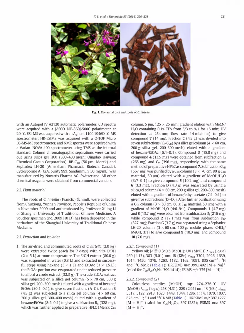
Fig. 1. The aerial part and roots of C. hirtella.
221X. Li et al. / Fitoterapia 95 (2014) 220–228
with an Autopol IV A2120 automatic polarimeter. CD spectrawere acquired with a JASCO DIP-360J-500C polarimeter at20 °C. ESI-MSwas acquiredwith an Agilent 1100 1946D LC-MSspectrometer, HR-ESIMS was acquired with a Q-TOF MicroLC-MS-MS spectrometer, and NMR spectra were acquired witha Varian INOVA 400 spectrometer using TMS as the internalstandard. Column chromatographic separations were carriedout using silica gel H60 (300–400 mesh; Qingdao HaiyangChemical Group Corporation), RP-C18 (50 μm; Merck) andSephadex LH-20 (Amersham Pharmacia Biotech, Canada).Cyclosporine A (CsA, purity 99%, Sandimmun, 50 mg/mL) wasmanufactured by Novartis Pharma AG, Switzerland. All otherchemical reagents were obtained from commercial vendors.
2.2. Plant material
The roots of C. hirtella (Franch.) Schindl. were collectedfrom Chuxiong, Yunnan Province, People's Republic of Chinain November 2009 and authenticated by Professor Xiling Liof Shanghai University of Traditional Chinese Medicine. Avoucher specimen (no. 200911013) has been deposited in theherbarium of the Shanghai University of Traditional ChineseMedicine.
2.3. Extraction and isolation
1. The air-dried and comminuted roots of C. hirtella (2.0 kg)were extracted twice (each for 7 days) with 95% EtOH(2 × 5 L) at room temperature. The EtOH extract (80.0 g)was suspended in water (0.8 L) and extracted in success-ful steps using hexane (3 × 1 L) and EtOAc (3 × 1.5 L);the EtOAc portion was evaporated under reduced pressureto afford a crude extract (32.3 g). The crude EtOAc extractwas subjected on a silica gel column (5 × 70 cm, 300 gsilica gel, 200–300 mesh) eluted with a gradient of hexane/EtOAc (30:1–0:1), to give seven fractions (A–G). Fraction B(4.8 g) was subjected to a silica gel column (4 × 60 cm,200 g silica gel, 300–400 mesh) eluted with a gradient ofhexane/EtOAc (8:2–0:1) to give a subfraction BII (328 mg),which was further applied to preparative HPLC (Merck C18
column, 5 μm, 125 × 25 mm; gradient elution with MeCN/H2O containing 0.1% TFA from 5/3 to 9/1 for 15 min; UVdetection at 254 nm; flow rate 14 mL/min;) to givecompound 7 (14 mg). Fraction C (4.3 g) was divided intoseven subfractions (CI–CVII) by a silica gel column (4 × 60 cm,200 g silica gel, 200–300 mesh) eluted with a gradientof hexane/EtOAc (6:1–0:1). Compound 3 (18.0 mg) andcompound 4 (13.5 mg) were obtained from subfraction CI(265 mg) and CV (396 mg), respectively, with the samemethod of preparativeHPLC as compound 7. Subfraction CVII(567 mg)waspurified by a C18 column (3 × 70 cm, 80 g C18material, 50 μm) eluted with a gradient of MeOH/H2O(3:7–9:1) to give compound 5 (10.2 mg) and compound6 (3.3 mg). Fraction D (4.0 g) was separated by using asilica gel column (4 × 60 cm, 200 g silica gel, 200–300 mesh)eluted with a gradient of hexane/ethyl acetate (7:1–0:1) togive five subfractions (DI–DV). After further purification usinga C18 column (3 × 50 cm, 60 g C18 material, 50 μm) with agradient of MeOH–H2O (4:6–9:1), Compounds 1 (4.7 mg)and 8 (13.7 mg)were obtained from subfraction DI (216 mg)while compound 2 (17.1 mg) was from subfraction DIV
(327 mg); Fraction G (3.7 g)was separated using a SephadexLH-20 column (3 × 60 cm, 100 g; mobile phase: CHCl3/MeOH, 3:1) to give compound 9 (10.0 mg) and compound10 (7.0 mg).
2.3.1. Compound (1)Yellow oil; [a]D25 0 (c 0.5, MeOH); UV (MeOH) λmax (log ε)
269 (4.13), 383 (5.01) nm; IR (KBr) νmax 3304, 2926, 1639,1614, 1450, 1379, 1263, 1182, 1163, 1091, 835 cm−1; 1Hand 13C NMR (Table 1); HRESIMS m/z 399.1402 [M + Na]+
(calcd for C20H24O7Na, 399.1414); ESIMS m/z 375 [M − H]−.
2.3.2. Compound (2)Colourless needles (MeOH), mp: 274–276 °C; UV
(MeOH) λmax (log ε) 258 (4.31), 289 (2.95) nm; IR (KBr) νmax
3517, 3122, 2918, 1623, 1448, 1344, 1286, 1114, 1070, 1037,823 cm−1; 1H and 13C NMR (Table 1); HRESIMS m/z 397.1277[M + H]+ (calcd for C22H21O7, 397.1282); ESIMS m/z 397[M + H]+.
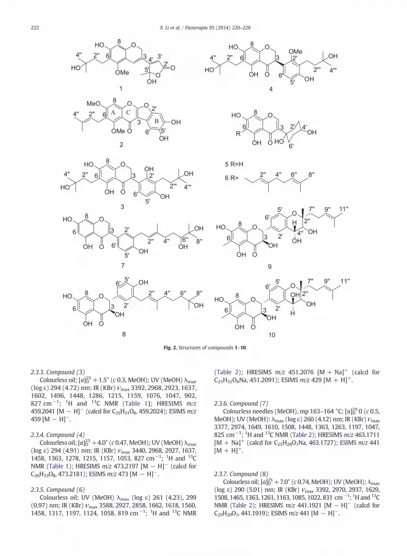
Fig. 2. Structures of compounds 1–10.
222 X. Li et al. / Fitoterapia 95 (2014) 220–228
2.3.3. Compound (3)Colourless oil; [a]D25 +1.5° (c 0.3, MeOH); UV (MeOH) λmax
(log ε) 294 (4.72) nm; IR (KBr) νmax 3392, 2968, 2923, 1637,1602, 1496, 1448, 1286, 1215, 1159, 1076, 1047, 902,827 cm−1; 1H and 13C NMR (Table 1); HRESIMS m/z459.2041 [M − H]− (calcd for C25H31O8, 459.2024); ESIMS m/z459 [M − H]−.
2.3.4. Compound (4)Colourless oil; [a]D25+4.0° (c 0.47,MeOH); UV (MeOH) λmax
(log ε) 294 (4.91) nm; IR (KBr) νmax 3440, 2968, 2927, 1637,1458, 1363, 1278, 1215, 1157, 1053, 827 cm−1; 1H and 13CNMR (Table 1); HRESIMS m/z 473.2197 [M − H]− (calcd forC26H33O8, 473.2181); ESIMS m/z 473 [M − H]−.
2.3.5. Compound (6)Colourless oil; UV (MeOH) λmax (log ε) 261 (4.23), 299
(0.97) nm; IR (KBr) νmax 3588, 2927, 2858, 1662, 1618, 1560,1458, 1317, 1197, 1124, 1058, 819 cm−1; 1H and 13C NMR
(Table 2); HRESIMS m/z 451.2076 [M + Na]+ (calcd forC25H32O6Na, 451.2091); ESIMS m/z 429 [M + H]+.
2.3.6. Compound (7)Colourless needles (MeOH), mp 163–164 °C; [a]D25 0 (c 0.5,
MeOH); UV (MeOH): λmax (log ε) 260 (4.12) nm; IR (KBr) νmax
3377, 2974, 1649, 1610, 1508, 1448, 1363, 1263, 1197, 1047,825 cm−1; 1H and 13C NMR (Table 2); HRESIMS m/z 463.1711[M + Na]+ (calcd for C25H28O7Na, 463.1727); ESIMS m/z 441[M + H]+.
2.3.7. Compound (8)Colourless oil; [a]D25+7.0° (c 0.74, MeOH); UV (MeOH): λmax
(log ε) 290 (5.01) nm; IR (KBr) νmax 3392, 2970, 2937, 1629,1508, 1465, 1363, 1261, 1163, 1085, 1022, 831 cm−1; 1H and 13CNMR (Table 2); HRESIMS m/z 441.1921 [M − H]− (calcd forC25H29O7, 441.1919); ESIMSm/z 441 [M − H]−.
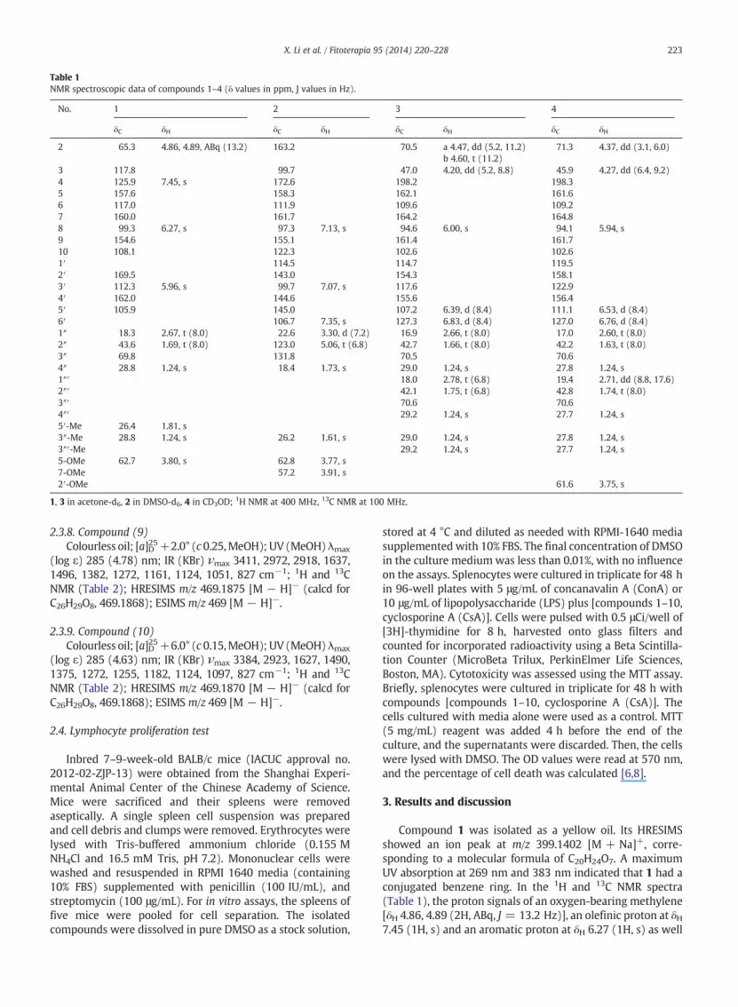
Table 1NMR spectroscopic data of compounds 1–4 (δ values in ppm, J values in Hz).
No. 1 2 3 4
δC δH δC δH δC δH δC δH
2 65.3 4.86, 4.89, ABq (13.2) 163.2 70.5 a 4.47, dd (5.2, 11.2)b 4.60, t (11.2)
71.3 4.37, dd (3.1, 6.0)
3 117.8 99.7 47.0 4.20, dd (5.2, 8.8) 45.9 4.27, dd (6.4, 9.2)4 125.9 7.45, s 172.6 198.2 198.35 157.6 158.3 162.1 161.66 117.0 111.9 109.6 109.27 160.0 161.7 164.2 164.88 99.3 6.27, s 97.3 7.13, s 94.6 6.00, s 94.1 5.94, s9 154.6 155.1 161.4 161.710 108.1 122.3 102.6 102.61′ 114.5 114.7 119.52′ 169.5 143.0 154.3 158.13′ 112.3 5.96, s 99.7 7.07, s 117.6 122.94′ 162.0 144.6 155.6 156.45′ 105.9 145.0 107.2 6.39, d (8.4) 111.1 6.53, d (8.4)6′ 106.7 7.35, s 127.3 6.83, d (8.4) 127.0 6.76, d (8.4)1″ 18.3 2.67, t (8.0) 22.6 3.30, d (7.2) 16.9 2.66, t (8.0) 17.0 2.60, t (8.0)2″ 43.6 1.69, t (8.0) 123.0 5.06, t (6.8) 42.7 1.66, t (8.0) 42.2 1.63, t (8.0)3″ 69.8 131.8 70.5 70.64″ 28.8 1.24, s 18.4 1.73, s 29.0 1.24, s 27.8 1.24, s1″′ 18.0 2.78, t (6.8) 19.4 2.71, dd (8.8, 17.6)2″′ 42.1 1.75, t (6.8) 42.8 1.74, t (8.0)3″′ 70.6 70.64″′ 29.2 1.24, s 27.7 1.24, s5′-Me 26.4 1.81, s3″-Me 28.8 1.24, s 26.2 1.61, s 29.0 1.24, s 27.8 1.24, s3″′-Me 29.2 1.24, s 27.7 1.24, s5-OMe 62.7 3.80, s 62.8 3.77, s7-OMe 57.2 3.91, s2′-OMe 61.6 3.75, s
1, 3 in acetone-d6, 2 in DMSO-d6, 4 in CD3OD; 1H NMR at 400 MHz, 13C NMR at 100 MHz.
223X. Li et al. / Fitoterapia 95 (2014) 220–228
2.3.8. Compound (9)Colourless oil; [a]D25+2.0° (c 0.25,MeOH); UV (MeOH) λmax
(log ε) 285 (4.78) nm; IR (KBr) νmax 3411, 2972, 2918, 1637,1496, 1382, 1272, 1161, 1124, 1051, 827 cm−1; 1H and 13CNMR (Table 2); HRESIMS m/z 469.1875 [M − H]− (calcd forC26H29O8, 469.1868); ESIMS m/z 469 [M − H]−.
2.3.9. Compound (10)Colourless oil; [a]D25+6.0° (c 0.15,MeOH); UV (MeOH) λmax
(log ε) 285 (4.63) nm; IR (KBr) νmax 3384, 2923, 1627, 1490,1375, 1272, 1255, 1182, 1124, 1097, 827 cm−1; 1H and 13CNMR (Table 2); HRESIMS m/z 469.1870 [M − H]− (calcd forC26H29O8, 469.1868); ESIMS m/z 469 [M − H]−.
2.4. Lymphocyte proliferation test
Inbred 7–9-week-old BALB/c mice (IACUC approval no.2012-02-ZJP-13) were obtained from the Shanghai Experi-mental Animal Center of the Chinese Academy of Science.Mice were sacrificed and their spleens were removedaseptically. A single spleen cell suspension was preparedand cell debris and clumps were removed. Erythrocytes werelysed with Tris-buffered ammonium chloride (0.155 MNH4Cl and 16.5 mM Tris, pH 7.2). Mononuclear cells werewashed and resuspended in RPMI 1640 media (containing10% FBS) supplemented with penicillin (100 IU/mL), andstreptomycin (100 μg/mL). For in vitro assays, the spleens offive mice were pooled for cell separation. The isolatedcompounds were dissolved in pure DMSO as a stock solution,
stored at 4 °C and diluted as needed with RPMI-1640 mediasupplemented with 10% FBS. The final concentration of DMSOin the culture medium was less than 0.01%, with no influenceon the assays. Splenocytes were cultured in triplicate for 48 hin 96-well plates with 5 μg/mL of concanavalin A (ConA) or10 μg/mL of lipopolysaccharide (LPS) plus [compounds 1–10,cyclosporine A (CsA)]. Cells were pulsed with 0.5 μCi/well of[3H]-thymidine for 8 h, harvested onto glass filters andcounted for incorporated radioactivity using a Beta Scintilla-tion Counter (MicroBeta Trilux, PerkinElmer Life Sciences,Boston, MA). Cytotoxicity was assessed using the MTT assay.Briefly, splenocytes were cultured in triplicate for 48 h withcompounds [compounds 1–10, cyclosporine A (CsA)]. Thecells cultured with media alone were used as a control. MTT(5 mg/mL) reagent was added 4 h before the end of theculture, and the supernatants were discarded. Then, the cellswere lysed with DMSO. The OD values were read at 570 nm,and the percentage of cell death was calculated [6,8].
3. Results and discussion
Compound 1 was isolated as a yellow oil. Its HRESIMSshowed an ion peak at m/z 399.1402 [M + Na]+, corre-sponding to a molecular formula of C20H24O7. A maximumUV absorption at 269 nm and 383 nm indicated that 1 had aconjugated benzene ring. In the 1H and 13C NMR spectra(Table 1), the proton signals of an oxygen-bearing methylene[δH 4.86, 4.89 (2H, ABq, J = 13.2 Hz)], an olefinic proton at δH7.45 (1H, s) and an aromatic proton at δH 6.27 (1H, s) as well

Table 2NMR spectroscopic data of compounds 6–10 (δ values in ppm, J values in Hz).
6 7 8 9 10
δC δH δC δH δC δH δC δH δC δH
2 153.6 8.15, s 153.5 8.11, s 83.9 5.06, d (11.6) 84.4 5.07, d (11.6) 83.8 5.07, d (11.6)3 128.3 123.6 72.4 4.64, d (11.2) 73.7 4.64, m 72.6 4.64, m4 182.3 181.0 197.7 198.3 197.75 159.8 162.9 164.3 162.0 161.46 111.9 99.1 6.28, d (1.6) 96.4 5.98, d (1.9) 105.0 104.47 162.5 164.4 167.2 165.5 164.98 93.0 6.46, s 93.8 6.41, d (1,6) 95.4 5.95, d (1.9) 95.3 6.03, s 94.7 6.03, s9 156.2 158.4 163.5 161.6 161.010 105.2 105.4 100.9 101.2 100.61′ 70.4 122.4 128.4 129.9 129.42′ 33.8 a 2.27, t (9.2)
b 1.71, m130.5 7.35, d (1.6) 129.6 7.33, d (1.9) 128.6 7.67, s 128.7 7.67, s
3′ 30.8 a 1.76, mb 1.60, m
127.9 127.9 125.3 125.3
4′ 69.3 3.61, m 155.3 155.7 153.9 153.15′ 30.8 a 1.76, m
b 1.60, m114.8 6.89, d (8.0) 114.8 6.89, d (8.0) 116.9 6.80, d (8.4) 116.5 6.80, d (8.4)
6′ 33.8 a 2.27, t (9.2)b 1.71, m
128.0 7.26, dd(1.6, 8.4)
126.9 7.24, brd (8.4) 129.9 7.36, brd (8.4) 128.4 7.35, brd (8.4)
1″ 21.3 3.34, m 28.3 3.37, d (7.2) 28.2 3.37, d (7.6)2″ 122.4 5.27, t (6.8) 122.7 5.43, t (7.2) 122.6 5.39, t (7.2) 80.9 80.63″ 134.7 136.2 136.2 73.8 3.71, d (8.4) 76.8 3.65, d (7.6)4″ 39.9 1.95, m 37.0 a 2.08, m
(overlapped)b 2.31, m
40.4 2.04, m 69.6 4.63, m 68.7 4.63, m
5″ 26.7 2.05, m(overlapped)
30.0 a 1.37, mb 1.70, m
22.7 1.52, m
6″ 124.5 5.06, t (6.0) 77.7 3.28,d (10.0) 43.6 1.44, m7″ 130.9 72.2 69.8 39.6 1.74, m
1.84, m31.6 1.76, m
1.54, m8″ 17.0 1.55, s 25.2 1.10, s 28.9 1.13, s 21.9 2.21, m 21.7 2.11, m9″ 125.3 5.19, t (7.2) 124.7 5.06, m10″ 131.8 131.111″ 17.6 1.63, s 16.9 1.53, s6-Me 7.0 1.99, s 6.4 1.98, s2″-Me 18.6 1.22, s 22.7 1.46, s3″-Me 15.6 1.77, s 15.7 1.74, s 15.4 1.71,s7″-Me 25.2 1.60, s 24.4 1.10, s 28.9 1.13,s10″-Me 25.8 1.68, s 25.1 1.61, s
6–10 in acetone-d6; 1H NMR at 400 MHz, 13C NMR at 100 MHz.
224 X. Li et al. / Fitoterapia 95 (2014) 220–228
as the carbon signals of a trisubstituted double bond (δC125.9, 117.8) and an oxygen-bearing carbon at δC 65.3suggested the presence of a 2H-chromene core [10,11]. Thesignals of a methoxy group at [δH 3.80 (3H, s), δC 62.7] and a3-hydroxy-3-methylbutyl group [δH 1.69 (2H, t, J = 8.0 Hz,H-2″), δC 43.6; δH 2.67 (2H, t, J = 8.0 Hz, H-1″), δC 18.3; δH1.24 (6H, s)] were also observed in the NMR spectra. Themethoxy group was unambiguously attached to C-5 by theHMBC correlations of H-4 (δH 7.45) and -OMe (δH 3.80)with C-5 (δC 157.6) and the assignment of the 3-hydroxy-3-methylbutyl group was achieved by the correlations ofH-1″ (δH 2.67) with C-5, C-6 and C-7. Thus, a moiety of7-hydroxyl-5-methoxy 6-(3-hydroxyl-3-methylbutyl)-2H-chromene was established. The remaining signals wereseen arising from a vinylic proton adjacent to a carbonylgroup at δH 5.96 (1H, s), a carbonyl group at δC 169.5,a trisubstituted double bond (δC 112.3, 162.0), a sp3 carbonbearing two oxygens at 105.9 as well as a tertiary methyl atδH 1.81. The above signals suggested a unit of γ-methyl,γ-hydroxyl-α,β-unsaturated γ-lactone [12,13] and alsosupported by the analysis of the HMBC spectrum. The
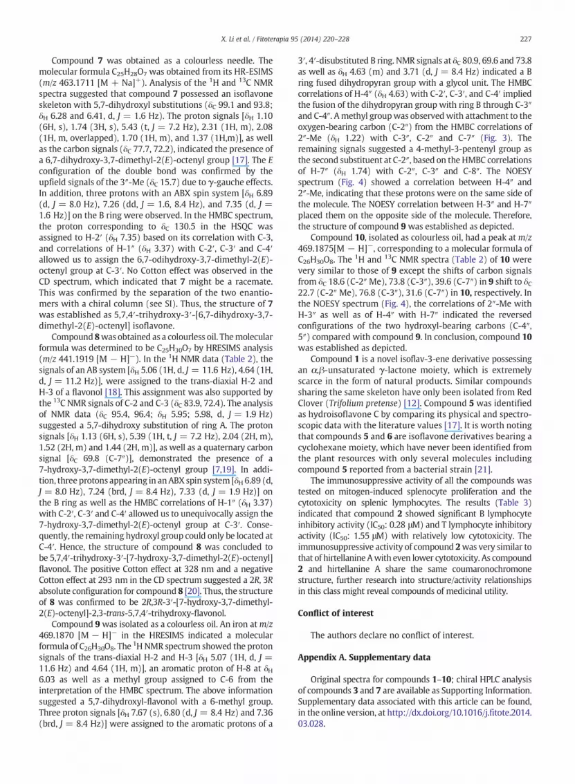
linkage of the unit of γ-methyl,γ-hydroxyl-α,β-unsaturatedγ-lactone with the 2H-chromene was achieved by the HMBCcorrelations of H-2 with C-4′ as well as H-3′ with C-3. Thus,the structure of 1was elucidated as depicted. Optical activityof this compound was absent, which indicated that com-pound 1 might be a racemate the same as pratenol A [12].The α,β-unsaturated γ-lactone of a B ring instead of abenzene ring is extremely rare in flavonoids and the origin ofthe α,β-unsaturated γ-lactone could be rationalized bioge-netically and traced back to a B ring of 2,4-dihydroxyl-benzene. After oxidation, decarboxylation, and cyclization,the moiety of γ-methyl,γ-hydroxyl-α,β-unsaturated γ-lactonecould be formed, finally. The proposed biosynthetic pathway ofcompound 1 was illustrated in Fig. 5.
Compound 2 was obtained as a colourless needle. TheHRESIMS showed a peak at m/z 397.1277 [M + H]+, corre-sponding to the molecular formula of C22H20O7. ThemaximumUV absorption at 258 nm and 289 nm aswell as the absence ofa characteristic proton signal for 2-H (usually appearing at δH8) (Table 1) indicated a coumaronochromone nature of 2similar to hirtellanine A [7]. Typical α,β-unsaturated carbonyl
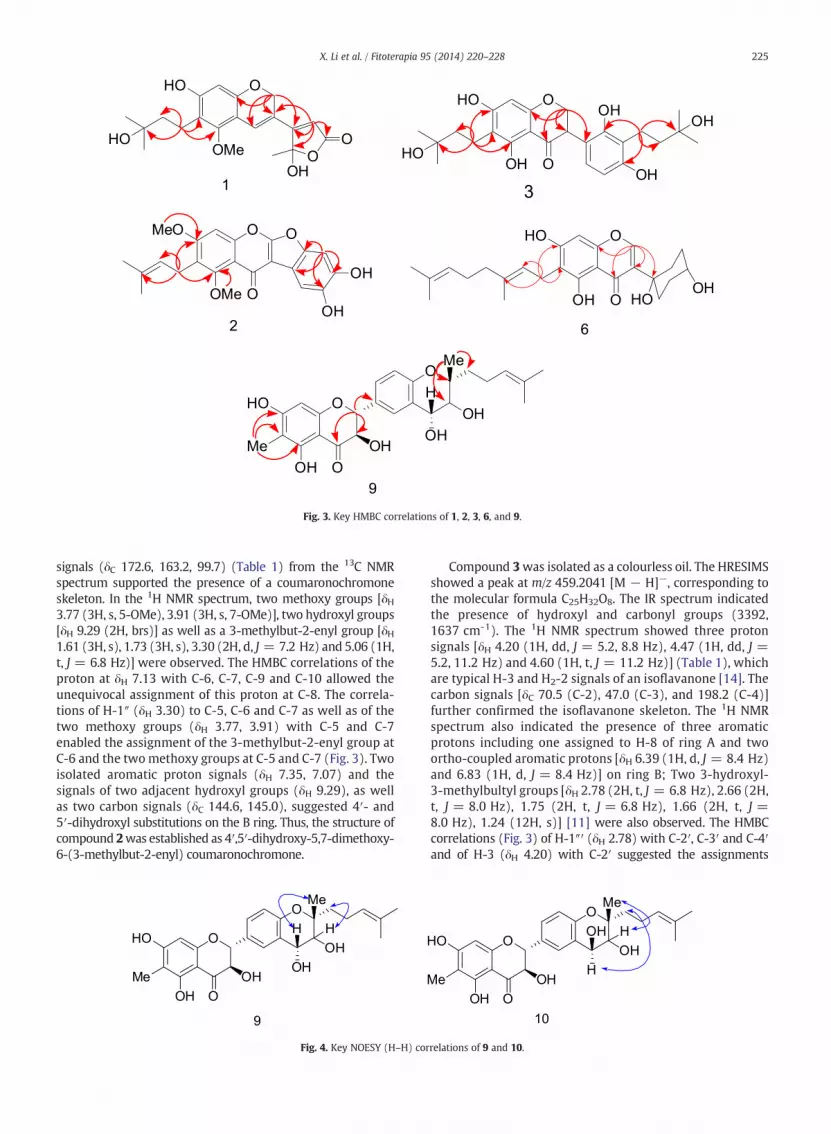
Fig. 3. Key HMBC correlations of 1, 2, 3, 6, and 9.
225X. Li et al. / Fitoterapia 95 (2014) 220–228
signals (δC 172.6, 163.2, 99.7) (Table 1) from the 13C NMRspectrum supported the presence of a coumaronochromoneskeleton. In the 1H NMR spectrum, two methoxy groups [δH3.77 (3H, s, 5-OMe), 3.91 (3H, s, 7-OMe)], two hydroxyl groups[δH 9.29 (2H, brs)] as well as a 3-methylbut-2-enyl group [δH1.61 (3H, s), 1.73 (3H, s), 3.30 (2H, d, J = 7.2 Hz) and 5.06 (1H,t, J = 6.8 Hz)] were observed. The HMBC correlations of theproton at δH 7.13 with C-6, C-7, C-9 and C-10 allowed theunequivocal assignment of this proton at C-8. The correla-tions of H-1″ (δH 3.30) to C-5, C-6 and C-7 as well as of thetwo methoxy groups (δH 3.77, 3.91) with C-5 and C-7enabled the assignment of the 3-methylbut-2-enyl group atC-6 and the twomethoxy groups at C-5 and C-7 (Fig. 3). Twoisolated aromatic proton signals (δH 7.35, 7.07) and thesignals of two adjacent hydroxyl groups (δH 9.29), as wellas two carbon signals (δC 144.6, 145.0), suggested 4′- and5′-dihydroxyl substitutions on the B ring. Thus, the structure ofcompound 2was established as 4′,5′-dihydroxy-5,7-dimethoxy-6-(3-methylbut-2-enyl) coumaronochromone.
Fig. 4. Key NOESY (H–H) cor
Compound 3was isolated as a colourless oil. The HRESIMSshowed a peak at m/z 459.2041 [M − H]−, corresponding tothe molecular formula C25H32O8. The IR spectrum indicatedthe presence of hydroxyl and carbonyl groups (3392,1637 cm-1). The 1H NMR spectrum showed three protonsignals [δH 4.20 (1H, dd, J = 5.2, 8.8 Hz), 4.47 (1H, dd, J =5.2, 11.2 Hz) and 4.60 (1H, t, J = 11.2 Hz)] (Table 1), whichare typical H-3 and H2-2 signals of an isoflavanone [14]. Thecarbon signals [δC 70.5 (C-2), 47.0 (C-3), and 198.2 (C-4)]further confirmed the isoflavanone skeleton. The 1H NMRspectrum also indicated the presence of three aromaticprotons including one assigned to H-8 of ring A and twoortho-coupled aromatic protons [δH 6.39 (1H, d, J = 8.4 Hz)and 6.83 (1H, d, J = 8.4 Hz)] on ring B; Two 3-hydroxyl-3-methylbultyl groups [δH 2.78 (2H, t, J = 6.8 Hz), 2.66 (2H,t, J = 8.0 Hz), 1.75 (2H, t, J = 6.8 Hz), 1.66 (2H, t, J =8.0 Hz), 1.24 (12H, s)] [11] were also observed. The HMBCcorrelations (Fig. 3) of H-1″′ (δH 2.78) with C-2′, C-3′ and C-4′and of H-3 (δH 4.20) with C-2′ suggested the assignments
relations of 9 and 10.
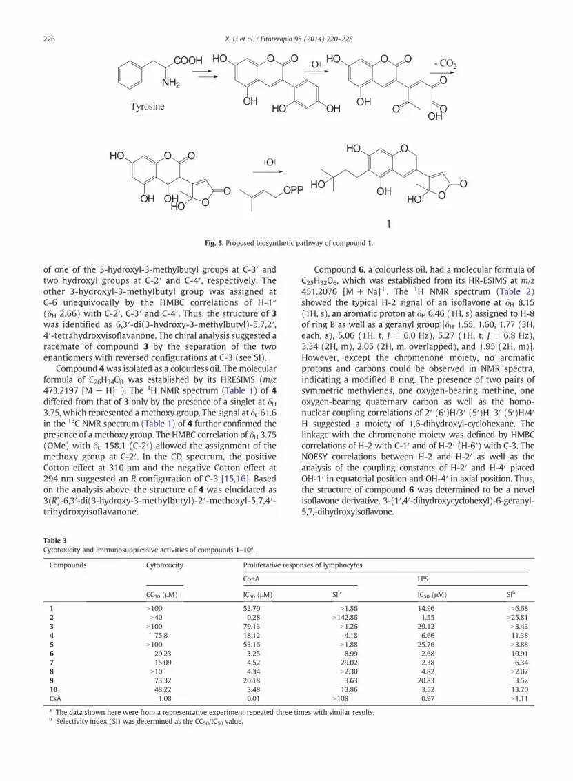
Fig. 5. Proposed biosynthetic pathway of compound 1.
226 X. Li et al. / Fitoterapia 95 (2014) 220–228
of one of the 3-hydroxyl-3-methylbutyl groups at C-3′ andtwo hydroxyl groups at C-2′ and C-4′, respectively. Theother 3-hydroxyl-3-methylbutyl group was assigned atC-6 unequivocally by the HMBC correlations of H-1″(δH 2.66) with C-2′, C-3′ and C-4′. Thus, the structure of 3was identified as 6,3′-di(3-hydroxy-3-methylbutyl)-5,7,2′,4′-tetrahydroxyisoflavanone. The chiral analysis suggested aracemate of compound 3 by the separation of the twoenantiomers with reversed configurations at C-3 (see SI).
Compound 4was isolated as a colourless oil. The molecularformula of C26H34O8 was established by its HRESIMS (m/z473.2197 [M − H]−). The 1H NMR spectrum (Table 1) of 4differed from that of 3 only by the presence of a singlet at δH3.75, which represented amethoxy group. The signal at δC 61.6in the 13C NMR spectrum (Table 1) of 4 further confirmed thepresence of a methoxy group. The HMBC correlation of δH 3.75(OMe) with δC 158.1 (C-2′) allowed the assignment of themethoxy group at C-2′. In the CD spectrum, the positiveCotton effect at 310 nm and the negative Cotton effect at294 nm suggested an R configuration of C-3 [15,16]. Basedon the analysis above, the structure of 4 was elucidated as3(R)-6,3′-di(3-hydroxy-3-methylbutyl)-2′-methoxyl-5,7,4′-trihydroxyisoflavanone.
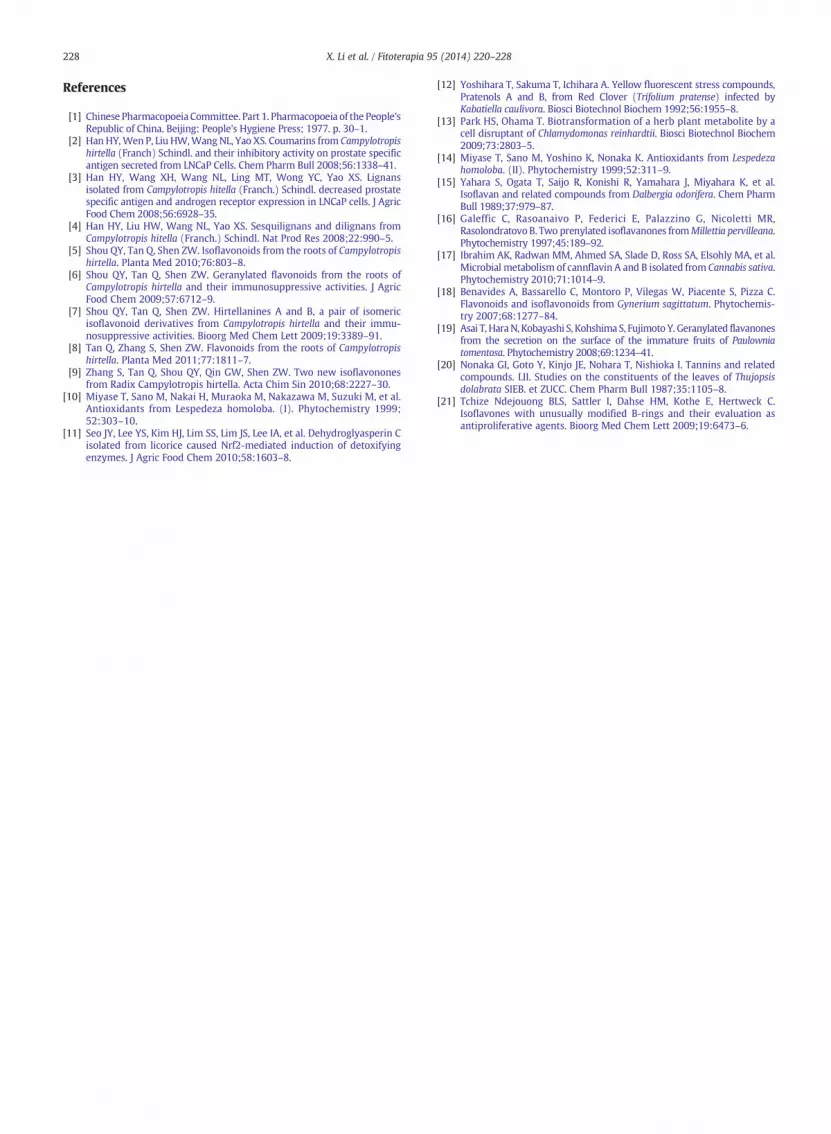
Table 3Cytotoxicity and immunosuppressive activities of compounds 1–10a.
Compounds Cytotoxicity Proliferative respo
ConA
CC50 (μM) IC50 (μM)
1 N100 53.702 N40 0.283 N100 79.134 75.8 18.125 N100 53.166 29.23 3.257 15.09 4.528 N10 4.349 73.32 20.1810 48.22 3.48CsA 1.08 0.01
a The data shown here were from a representative experiment repeated three tib Selectivity index (SI) was determined as the CC50/IC50 value.
Compound 6, a colourless oil, had a molecular formula ofC25H32O6, which was established from its HR-ESIMS at m/z451.2076 [M + Na]+. The 1H NMR spectrum (Table 2)showed the typical H-2 signal of an isoflavone at δH 8.15(1H, s), an aromatic proton at δH 6.46 (1H, s) assigned to H-8of ring B as well as a geranyl group [δH 1.55, 1.60, 1.77 (3H,each, s), 5.06 (1H, t, J = 6.0 Hz), 5.27 (1H, t, J = 6.8 Hz),3.34 (2H, m), 2.05 (2H, m, overlapped), and 1.95 (2H, m)].However, except the chromenone moiety, no aromaticprotons and carbons could be observed in NMR spectra,indicating a modified B ring. The presence of two pairs ofsymmetric methylenes, one oxygen-bearing methine, oneoxygen-bearing quaternary carbon as well as the homo-nuclear coupling correlations of 2′ (6′)H/3′ (5′)H, 3′ (5′)H/4′H suggested a moiety of 1,6-dihydroxyl-cyclohexane. Thelinkage with the chromenone moiety was defined by HMBCcorrelations of H-2 with C-1′ and of H-2′ (H-6′) with C-3. TheNOESY correlations between H-2 and H-2′ as well as theanalysis of the coupling constants of H-2′ and H-4′ placedOH-1′ in equatorial position and OH-4′ in axial position. Thus,the structure of compound 6 was determined to be a novelisoflavone derivative, 3-(1′,4′-dihydroxycyclohexyl)-6-geranyl-5,7,-dihydroxyisoflavone.
nses of lymphocytes
LPS
SIb IC50 (μM) SIb
N1.86 14.96 N6.68N142.86 1.55 N25.81
N1.26 29.12 N3.434.18 6.66 11.38
N1.88 25.76 N3.888.99 2.68 10.91
29.02 2.38 6.34N2.30 4.82 N2.073.63 20.83 3.52
13.86 3.52 13.70N108 0.97 N1.11
mes with similar results.
227X. Li et al. / Fitoterapia 95 (2014) 220–228
Compound 7 was obtained as a colourless needle. Themolecular formula C25H28O7 was obtained from its HR-ESIMS(m/z 463.1711 [M + Na]+). Analysis of the 1H and 13C NMRspectra suggested that compound 7 possessed an isoflavoneskeleton with 5,7-dihydroxyl substitutions (δC 99.1 and 93.8;δH 6.28 and 6.41, d, J = 1.6 Hz). The proton signals [δH 1.10(6H, s), 1.74 (3H, s), 5.43 (t, J = 7.2 Hz), 2.31 (1H, m), 2.08(1H, m, overlapped), 1.70 (1H, m), and 1.37 (1H,m)], as wellas the carbon signals (δC 77.7, 72.2), indicated the presence ofa 6,7-dihydroxy-3,7-dimethyl-2(E)-octenyl group [17]. The Econfiguration of the double bond was confirmed by theupfield signals of the 3″-Me (δC 15.7) due to γ-gauche effects.In addition, three protons with an ABX spin system [δH 6.89(d, J = 8.0 Hz), 7.26 (dd, J = 1.6, 8.4 Hz), and 7.35 (d, J =1.6 Hz)] on the B ring were observed. In the HMBC spectrum,the proton corresponding to δC 130.5 in the HSQC wasassigned to H-2′ (δH 7.35) based on its correlation with C-3,and correlations of H-1″ (δH 3.37) with C-2′, C-3′ and C-4′allowed us to assign the 6,7-odihydroxy-3,7-dimethyl-2(E)-octenyl group at C-3′. No Cotton effect was observed in theCD spectrum, which indicated that 7 might be a racemate.This was confirmed by the separation of the two enantio-mers with a chiral column (see SI). Thus, the structure of 7was established as 5,7,4′-trihydroxy-3′-[6,7-dihydroxy-3,7-dimethyl-2(E)-octenyl] isoflavone.
Compound 8was obtained as a colourless oil. Themolecularformula was determined to be C25H30O7 by HRESIMS analysis(m/z 441.1919 [M − H]−). In the 1H NMR data (Table 2), thesignals of an AB system [δH 5.06 (1H, d, J = 11.6 Hz), 4.64 (1H,d, J = 11.2 Hz)], were assigned to the trans-diaxial H-2 andH-3 of a flavonol [18]. This assignment was also supported bythe 13C NMR signals of C-2 and C-3 (δC 83.9, 72.4). The analysisof NMR data (δC 95.4, 96.4; δH 5.95; 5.98, d, J = 1.9 Hz)suggested a 5,7-dihydroxy substitution of ring A. The protonsignals [δH 1.13 (6H, s), 5.39 (1H, t, J = 7.2 Hz), 2.04 (2H, m),1.52 (2H, m) and 1.44 (2H, m)], as well as a quaternary carbonsignal [δC 69.8 (C-7″)], demonstrated the presence of a7-hydroxy-3,7-dimethyl-2(E)-octenyl group [7,19]. In addi-tion, three protons appearing in anABX spin system [δH 6.89 (d,J = 8.0 Hz), 7.24 (brd, J = 8.4 Hz), 7.33 (d, J = 1.9 Hz)] onthe B ring as well as the HMBC correlations of H-1″ (δH 3.37)with C-2′, C-3′ and C-4′ allowed us to unequivocally assign the7-hydroxy-3,7-dimethyl-2(E)-octenyl group at C-3′. Conse-quently, the remaining hydroxyl group could only be located atC-4′. Hence, the structure of compound 8 was concluded tobe 5,7,4′-trihydroxy-3′-[7-hydroxy-3,7-dimethyl-2(E)-octenyl]flavonol. The positive Cotton effect at 328 nm and a negativeCotton effect at 293 nm in the CD spectrum suggested a 2R, 3Rabsolute configuration for compound 8 [20]. Thus, the structureof 8 was confirmed to be 2R,3R-3′-[7-hydroxy-3,7-dimethyl-2(E)-octenyl]-2,3-trans-5,7,4′-trihydroxy-flavonol.
Compound 9 was isolated as a colourless oil. An iron atm/z469.1870 [M − H]− in the HRESIMS indicated a molecularformula of C26H30O8. The 1HNMR spectrum showed the protonsignals of the trans-diaxial H-2 and H-3 [δH 5.07 (1H, d, J =11.6 Hz) and 4.64 (1H, m)], an aromatic proton of H-8 at δH6.03 as well as a methyl group assigned to C-6 from theinterpretation of the HMBC spectrum. The above informationsuggested a 5,7-dihydroxyl-flavonol with a 6-methyl group.Three proton signals [δH 7.67 (s), 6.80 (d, J = 8.4 Hz) and 7.36(brd, J = 8.4 Hz)] were assigned to the aromatic protons of a
3′, 4′-disubstituted B ring. NMR signals at δC 80.9, 69.6 and 73.8as well as δH 4.63 (m) and 3.71 (d, J = 8.4 Hz) indicated a Bring fused dihydropyran group with a glycol unit. The HMBCcorrelations of H-4″ (δH 4.63) with C-2′, C-3′, and C-4′ impliedthe fusion of the dihydropyran group with ring B through C-3″and C-4″. Amethyl groupwas observedwith attachment to theoxygen-bearing carbon (C-2″) from the HMBC correlations of2″-Me (δH 1.22) with C-3″, C-2″ and C-7″ (Fig. 3). Theremaining signals suggested a 4-methyl-3-pentenyl group asthe second substituent at C-2″, based on the HMBC correlationsof H-7″ (δH 1.74) with C-2″, C-3″ and C-8″. The NOESYspectrum (Fig. 4) showed a correlation between H-4″ and2″-Me, indicating that these protons were on the same side ofthe molecule. The NOESY correlation between H-3″ and H-7″placed them on the opposite side of the molecule. Therefore,the structure of compound 9 was established as depicted.
Compound 10, isolated as colourless oil, had a peak at m/z469.1875[M − H]−, corresponding to a molecular formula ofC26H30O8. The 1H and 13C NMR spectra (Table 2) of 10 werevery similar to those of 9 except the shifts of carbon signalsfrom δC 18.6 (C-2″Me), 73.8 (C-3″), 39.6 (C-7″) in 9 shift to δC22.7 (C-2″Me), 76.8 (C-3″), 31.6 (C-7″) in 10, respectively. Inthe NOESY spectrum (Fig. 4), the correlations of 2″-Me withH-3″ as well as of H-4″ with H-7″ indicated the reversedconfigurations of the two hydroxyl-bearing carbons (C-4″,5″) compared with compound 9. In conclusion, compound 10was established as depicted.
Compound 1 is a novel isoflav-3-ene derivative possessingan α,β-unsaturated γ-lactone moiety, which is extremelyscarce in the form of natural products. Similar compoundssharing the same skeleton have only been isolated from RedClover (Trifolium pretense) [12]. Compound 5 was identifiedas hydroisoflavone C by comparing its physical and spectro-scopic data with the literature values [17]. It is worth notingthat compounds 5 and 6 are isoflavone derivatives bearing acyclohexane moiety, which have never been identified fromthe plant resources with only several molecules includingcompound 5 reported from a bacterial strain [21].
The immunosuppressive activity of all the compounds wastested on mitogen-induced splenocyte proliferation and thecytotoxicity on splenic lymphocytes. The results (Table 3)indicated that compound 2 showed significant B lymphocyteinhibitory activity (IC50: 0.28 μM) and T lymphocyte inhibitoryactivity (IC50: 1.55 μM) with relatively low cytotoxicity. Theimmunosuppressive activity of compound 2was very similar tothat of hirtellanine Awith even lower cytotoxicity. As compound2 and hirtellanine A share the same coumaronochromonestructure, further research into structure/activity relationshipsin this class might reveal compounds of medicinal utility.
Conflict of interest
The authors declare no conflict of interest.
Appendix A. Supplementary data
Original spectra for compounds 1–10; chiral HPLC analysisof compounds 3 and 7 are available as Supporting Information.Supplementary data associated with this article can be found,in the online version, at http://dx.doi.org/10.1016/j.fitote.2014.03.028.
228 X. Li et al. / Fitoterapia 95 (2014) 220–228
References
[1] Chinese Pharmacopoeia Committee. Part 1. Pharmacopoeia of the People'sRepublic of China. Beijing: People's Hygiene Press; 1977. p. 30–1.
[2] Han HY,Wen P, Liu HW,Wang NL, Yao XS. Coumarins from Campylotropishirtella (Franch) Schindl. and their inhibitory activity on prostate specificantigen secreted from LNCaP Cells. Chem Pharm Bull 2008;56:1338–41.
[3] Han HY, Wang XH, Wang NL, Ling MT, Wong YC, Yao XS. Lignansisolated from Campylotropis hitella (Franch.) Schindl. decreased prostatespecific antigen and androgen receptor expression in LNCaP cells. J AgricFood Chem 2008;56:6928–35.
[4] Han HY, Liu HW, Wang NL, Yao XS. Sesquilignans and dilignans fromCampylotropis hitella (Franch.) Schindl. Nat Prod Res 2008;22:990–5.
[5] Shou QY, Tan Q, Shen ZW. Isoflavonoids from the roots of Campylotropishirtella. Planta Med 2010;76:803–8.
[6] Shou QY, Tan Q, Shen ZW. Geranylated flavonoids from the roots ofCampylotropis hirtella and their immunosuppressive activities. J AgricFood Chem 2009;57:6712–9.
[7] Shou QY, Tan Q, Shen ZW. Hirtellanines A and B, a pair of isomericisoflavonoid derivatives from Campylotropis hirtella and their immu-nosuppressive activities. Bioorg Med Chem Lett 2009;19:3389–91.
[8] Tan Q, Zhang S, Shen ZW. Flavonoids from the roots of Campylotropishirtella. Planta Med 2011;77:1811–7.
[9] Zhang S, Tan Q, Shou QY, Qin GW, Shen ZW. Two new isoflavononesfrom Radix Campylotropis hirtella. Acta Chim Sin 2010;68:2227–30.
[10] Miyase T, Sano M, Nakai H, Muraoka M, Nakazawa M, Suzuki M, et al.Antioxidants from Lespedeza homoloba. (I). Phytochemistry 1999;52:303–10.
[11] Seo JY, Lee YS, Kim HJ, Lim SS, Lim JS, Lee IA, et al. Dehydroglyasperin Cisolated from licorice caused Nrf2-mediated induction of detoxifyingenzymes. J Agric Food Chem 2010;58:1603–8.
[12] Yoshihara T, Sakuma T, Ichihara A. Yellow fluorescent stress compounds,Pratenols A and B, from Red Clover (Trifolium pratense) infected byKabatiella caulivora. Biosci Biotechnol Biochem 1992;56:1955–8.
[13] Park HS, Ohama T. Biotransformation of a herb plant metabolite by acell disruptant of Chlamydomonas reinhardtii. Biosci Biotechnol Biochem2009;73:2803–5.
[14] Miyase T, Sano M, Yoshino K, Nonaka K. Antioxidants from Lespedezahomoloba. (II). Phytochemistry 1999;52:311–9.
[15] Yahara S, Ogata T, Saijo R, Konishi R, Yamahara J, Miyahara K, et al.Isoflavan and related compounds from Dalbergia odorifera. Chem PharmBull 1989;37:979–87.
[16] Galeffic C, Rasoanaivo P, Federici E, Palazzino G, Nicoletti MR,Rasolondratovo B. Two prenylated isoflavanones fromMillettia pervilleana.Phytochemistry 1997;45:189–92.
[17] Ibrahim AK, Radwan MM, Ahmed SA, Slade D, Ross SA, Elsohly MA, et al.Microbial metabolism of cannflavin A and B isolated from Cannabis sativa.Phytochemistry 2010;71:1014–9.
[18] Benavides A, Bassarello C, Montoro P, Vilegas W, Piacente S, Pizza C.Flavonoids and isoflavonoids from Gynerium sagittatum. Phytochemis-try 2007;68:1277–84.
[19] Asai T, HaraN, Kobayashi S, KohshimaS, FujimotoY. Geranylated flavanonesfrom the secretion on the surface of the immature fruits of Paulowniatomentosa. Phytochemistry 2008;69:1234–41.
[20] Nonaka GI, Goto Y, Kinjo JE, Nohara T, Nishioka I. Tannins and relatedcompounds. LII. Studies on the constituents of the leaves of Thujopsisdolabrata SIEB. et ZUCC. Chem Pharm Bull 1987;35:1105–8.
[21] Tchize Ndejouong BLS, Sattler I, Dahse HM, Kothe E, Hertweck C.Isoflavones with unusually modified B-rings and their evaluation asantiproliferative agents. Bioorg Med Chem Lett 2009;19:6473–6.


