Embed Size (px)
Citation preview
�����������������
Citation: Kazberuk, A.; Chalecka, M.;
Palka, J.; Bielawska, K.; Surazynski,
A. NSAIDs Induce Proline
Dehydrogenase/Proline
Oxidase-Dependent and
Independent Apoptosis in MCF7
Breast Cancer Cells. Int. J. Mol. Sci.
2022, 23, 3813. https://doi.org/
10.3390/ijms23073813
Academic Editor: Ahmad R. Safa
Received: 28 January 2022
Accepted: 28 March 2022
Published: 30 March 2022
Publisher’s Note: MDPI stays neutral
with regard to jurisdictional claims in
published maps and institutional affil-
iations.
Copyright: © 2022 by the authors.
Licensee MDPI, Basel, Switzerland.
This article is an open access article
distributed under the terms and
conditions of the Creative Commons
Attribution (CC BY) license (https://
creativecommons.org/licenses/by/
4.0/).
International Journal of
Molecular Sciences
Article
NSAIDs Induce Proline Dehydrogenase/ProlineOxidase-Dependent and Independent Apoptosis in MCF7Breast Cancer CellsAdam Kazberuk 1 , Magda Chalecka 1, Jerzy Palka 1 , Katarzyna Bielawska 2 and Arkadiusz Surazynski 1,*
1 Department of Medicinal Chemistry, Medical University of Bialystok, Mickiewicza 2D, 15-222 Bialystok,Poland; [email protected] (A.K.); [email protected] (M.C.); [email protected] (J.P.)
2 Department of Analysis and Bioanalysis of Medicines, Medical University of Bialystok, Mickiewicza 2D,15-222 Bialystok, Poland; [email protected]
* Correspondence: [email protected]
Abstract: Non-steroidal anti-inflammatory drugs (NSAIDs) are considered in cancer therapy for theirinhibitory effect on cyclooxygenase-2 (COX-2), which is overexpressed in most cancers. However,we found that NSAIDs as ligands of peroxisome proliferator-activated receptor-γ (PPARγ)-inducedapoptosis independent of the COX-2 inhibition, and the process was mediated through activation ofproline dehydrogenase/proline oxidase (PRODH/POX)-dependent generation of reactive oxygenspecies (ROS). This mitochondrial enzyme converts proline to ∆1-pyrroline-5-carboxylate (P5C)during which ATP or ROS is generated. To confirm the role of PRODH/POX in the mechanismof NSAID-induced apoptosis we obtained an MCF7 CRISPR/Cas9 PRODH/POX knockout breastcancer cell model (MCF7POK-KO). Interestingly, the studied NSAIDs (indomethacin and diclofenac)in MCF7POK-KO cells contributed to a more pronounced pro-apoptotic phenotype of the cells thanin PRODH/POX-expressing MCF7 cells. The observed effect was independent of ROS generation,but it was related to the energetic disturbances in the cells as shown by an increase in the expressionof AMPKα (sensor of cell energy status), GLUD1/2 (proline producing enzyme from glutamate),prolidase (proline releasing enzyme), PPARδ (growth supporting transcription factor) and a decreasein the expression of proline cycle enzymes (PYCR1, PYCRL), mammalian target of rapamycin(mTOR), and collagen biosynthesis (the main proline utilizing process). The data provide evidencethat the studied NSAIDs induce PRODH/POX-dependent and independent apoptosis in MCF7breast cancer cells.
Keywords: mitochondria; proline metabolism; proline oxidase; proline dehydrogenase; NSAIDs;PPAR; COX; apoptosis; breast cancer; oxidative stress
1. Introduction
Cancer treatment strategies include different classes of drugs that are not typicallyantineoplastic. One class is the non-steroidal anti-inflammatory drugs (NSAIDs) knownto decrease the risk of various cancers such as prostate, colorectal, breast, and lung [1–3].However, NSAID-induced antineoplastic pathways are not well recognized. It has beenconsidered that a potent target of NSAIDs in cancer therapy is inflammation [4]. Numerousstudies have shown that an inflammatory environment facilitates cancer development.Cyclooxygenases (COXs) are key inflammatory regulators responsible for prostaglandinsynthesis from fatty acids (such as linoleic and arachidonic acid). Two isoforms of thisenzyme are known: constitutively expressed COX1 and conditionally expressed COX2in response to inflammation. An important fact is that in most cancers COX2 is over-expressed [5], which is linked to regulation of invasiveness, angiogenesis, proliferation,and migration [6,7]. Although the hypothesis of the anticancer properties of NSAIDs wasproven in vitro and in vivo (as they are COX2 inhibitors), other research has showed that in
Int. J. Mol. Sci. 2022, 23, 3813. https://doi.org/10.3390/ijms23073813 https://www.mdpi.com/journal/ijms
Int. J. Mol. Sci. 2022, 23, 3813 2 of 18
cancer cells lacking COX2 or in the cells with knocked-down expression of COX2, NSAIDshad similar anticancer activity. This suggests other COX2 independent anticancer activitiesof NSAIDs [8–10].
Indomethacin and diclofenac are representative NSAIDs and well-documented syn-thetic ligands of peroxisome proliferator-activated receptor-γ (PPARγ) [11]. It is knownthat three isoforms of PPAR exist: PPARα, PPARβ/δ, and PPARγ, and all of them havetranscriptional activity as they belong to the nuclear receptor family. The main role ofthese receptors is to regulate adipogenesis, lipid metabolism, and glucose homeostasis,but they are also involved in the processes such as inflammation, proliferation, and cancermetabolism [12,13]. Ligands of these receptors are mostly synthetic drugs, as mentionedearlier, but also include natural ligands such as fatty acids or prostaglandins [14–16].Thiazolidinediones (i.e., troglitazone, pioglitazone), the class of drugs used for type 2diabetes mellitus treatment, are the most potent inducers of PPARγ. Cancer cells treatedwith thiazolidinediones evoke pro-apoptotic phenotypes, suggesting that the process isPPARγ-dependent [17–19].
Activation of PPARγ induces proline dehydrogenase/proline oxidase (PRODH/POX);(PRODH<, GenBankTM NM_016335), the inner mitochondrial membrane flavin-dependentenzyme [20–22]. PRODH/POX catalyzes conversion of proline to ∆1-pyrroline-5-carboxylate(P5C). During this reaction free electrons are transported to the electron transport chainto produce pro-survival ATP or they are accepted by oxygen to generate reactive oxygenspecies (ROS) inducing apoptosis/autophagy [21,23–25]. However, the mechanism for driv-ing PRODH/POX into a pro-survival or pro-apoptotic pathway is not clear. Conversion ofproline into P5C is a part of proline turnover coupled with a reverse reaction in which P5Cis turned into proline by three isoenzymes of P5C reductase (PYCR). Two of them are mito-chondrial (PYCR1 and PYCR2) and PYCRL is localized in the cytoplasm. P5C reduction toproline and proline shuttling between mitochondria and cytosol are coupled to the glucosemetabolism by the pentose phosphate pathway [26,27]. P5C is also a substrate for P5Cdehydrogenase (P5CDH)-producing glutamate, which is a precursor of α-ketoglutaric acid,an important compound of the TCA cycle [21]. Thus, the proline–P5C cycle is an importantmetabolic regulator of the PRODH/POX-dependent pathway. Interestingly, COX-2, MAPK,EGFR, and Wnt/β-catenin signaling pathways are also related to PRODH/POX [28–30]. Apotent inducer of PRODH/POX is tumor suppressor p53 [31]. Transcriptional regulation ofPRODH/POX by p53 was found in the PRODH/POX promoter, containing a p53-responseelement [32].
PRODH/POX-induced functions are dependent on proline availability. The aminoacid can come from protein degradation products, mainly derived from collagen. The finalcollagen degradation is catalyzed by prolidase.
Prolidase [E.C.3.4.13.9], known also as peptidase D or iminopeptidase, is a cyto-plasmic imido-dipeptidase or imido-tripeptidase [33,34] that cleaves imido-peptides withC-terminal proline or hydroxyproline [35]. The substrate for prolidase is derived mainlyfrom collagen degradation (the most abundant protein containing imino bonds) and alsofrom other degraded proline-containing proteins [36,37]. Prolidase activity was found torecycle proline for collagen re-synthesis, and therefore, the enzyme may play an importantrole in the regulation of collagen biosynthesis. It has been well documented in fibroblaststreated with anti-inflammatory drugs [38], P5C [39], during experimental fibroblast ag-ing [40], experimental chondrocyte inflammation [41], activation of integrin receptor fortype I collagen [42], in fibroblast derived from Osteogenesis Imperfecta patients [43], andin several cancer tissues [44–46].
Other factors that contribute to cytoplasmic proline levels are proline utilizing pro-cesses. The most effective is collagen synthesis [47] that, apart from important biologicalfunctions, may work as a “sink” for proline. From the perspective of redox balance, col-lagen can also be a sink for reducing the potential of proline and removing it from themetabolic pool. In fact, metabolic abnormalities concomitant to inflammatory processes arefrequently associated with collagen formation, i.e., fibrosis [48,49]. During the inhibition of
Int. J. Mol. Sci. 2022, 23, 3813 3 of 18
collagen biosynthesis, reducing the potential of proline is carried out in mitochondria byPRODH/POX, yielding P5C. The intensity of this process is regulated by the energetic sta-tus of the cells, which is controlled by mTOR, AMPK (77–79), as well as certain oncogenes,e.g., c-Myc [50].
The aim of the study was to establish the role of PRODH/POX in NSAID-inducedapoptosis in breast cancer cells. CRISPR/Cas9 technology [51–54] was used to preparethe MCF7 cell line with PRODH/POX knockout. In this article we provide evidencethat indomethacin (IND) and diclofenac (DCF) induce apoptosis in both a PRODH/POX-dependent and a PRODH/POX-independent manner through different mechanisms.
2. Results
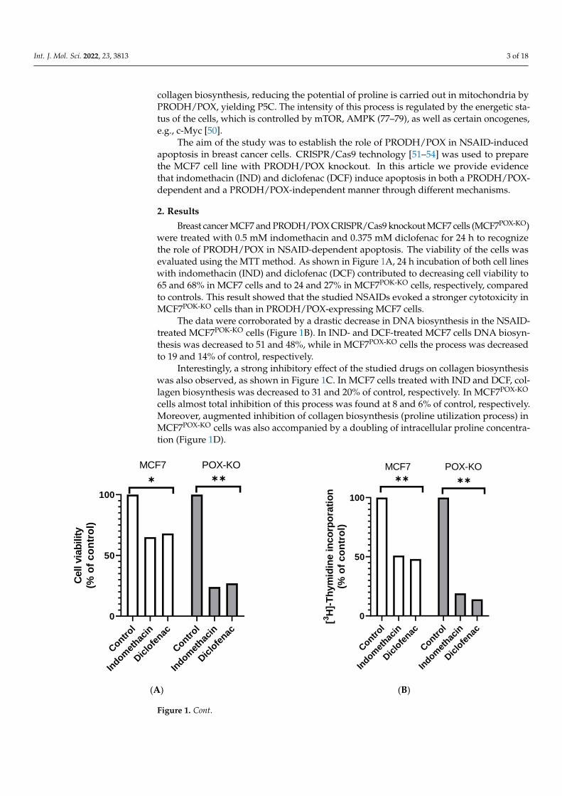
Breast cancer MCF7 and PRODH/POX CRISPR/Cas9 knockout MCF7 cells (MCF7POX-KO)were treated with 0.5 mM indomethacin and 0.375 mM diclofenac for 24 h to recognizethe role of PRODH/POX in NSAID-dependent apoptosis. The viability of the cells wasevaluated using the MTT method. As shown in Figure 1A, 24 h incubation of both cell lineswith indomethacin (IND) and diclofenac (DCF) contributed to decreasing cell viability to65 and 68% in MCF7 cells and to 24 and 27% in MCF7POK-KO cells, respectively, comparedto controls. This result showed that the studied NSAIDs evoked a stronger cytotoxicity inMCF7POK-KO cells than in PRODH/POX-expressing MCF7 cells.
The data were corroborated by a drastic decrease in DNA biosynthesis in the NSAID-treated MCF7POK-KO cells (Figure 1B). In IND- and DCF-treated MCF7 cells DNA biosyn-thesis was decreased to 51 and 48%, while in MCF7POX-KO cells the process was decreasedto 19 and 14% of control, respectively.
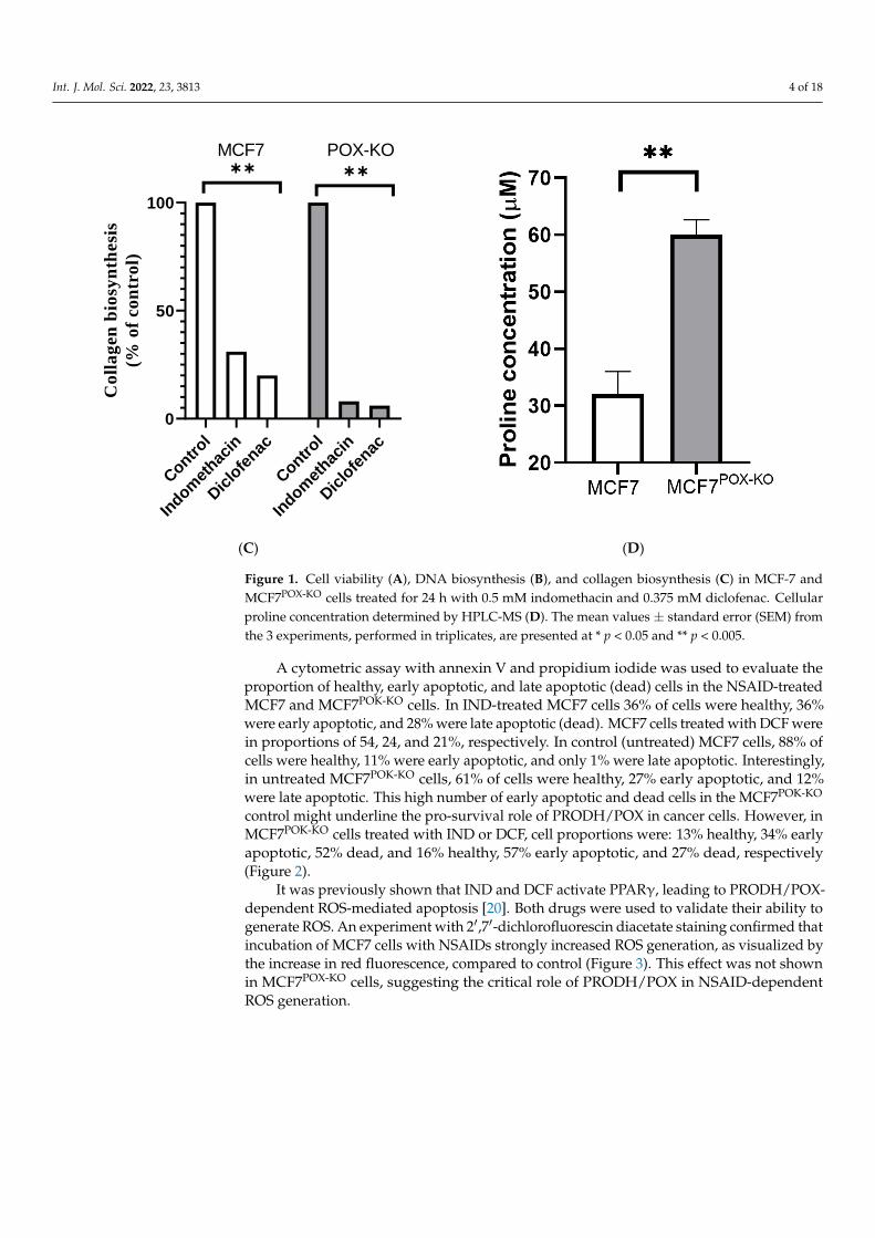
Interestingly, a strong inhibitory effect of the studied drugs on collagen biosynthesiswas also observed, as shown in Figure 1C. In MCF7 cells treated with IND and DCF, col-lagen biosynthesis was decreased to 31 and 20% of control, respectively. In MCF7POX-KO
cells almost total inhibition of this process was found at 8 and 6% of control, respectively.Moreover, augmented inhibition of collagen biosynthesis (proline utilization process) inMCF7POX-KO cells was also accompanied by a doubling of intracellular proline concentra-tion (Figure 1D).
Int. J. Mol. Sci. 2022, 23, x FOR PEER REVIEW 3 of 18
frequently associated with collagen formation, i.e., fibrosis [48,49]. During the inhibition
of collagen biosynthesis, reducing the potential of proline is carried out in mitochondria
by PRODH/POX, yielding P5C. The intensity of this process is regulated by the energetic
status of the cells, which is controlled by mTOR, AMPK (77–79), as well as certain onco-
genes, e.g., c-Myc [50].
The aim of the study was to establish the role of PRODH/POX in NSAID-induced
apoptosis in breast cancer cells. CRISPR/Cas9 technology [51–54] was used to prepare the
MCF7 cell line with PRODH/POX knockout. In this article we provide evidence that in-
domethacin (IND) and diclofenac (DCF) induce apoptosis in both a PRODH/POX-de-
pendent and a PRODH/POX-independent manner through different mechanisms.
2. Results
Breast cancer MCF7 and PRODH/POX CRISPR/Cas9 knockout MCF7 cells (MCF7POX-
KO) were treated with 0.5 mM indomethacin and 0.375 mM diclofenac for 24 h to recognize
the role of PRODH/POX in NSAID-dependent apoptosis. The viability of the cells was
evaluated using the MTT method. As shown in Figure 1A, 24 h incubation of both cell
lines with indomethacin (IND) and diclofenac (DCF) contributed to decreasing cell viabil-
ity to 65 and 68% in MCF7 cells and to 24 and 27% in MCF7POK-KO cells, respectively, com-
pared to controls. This result showed that the studied NSAIDs evoked a stronger cytotox-
icity in MCF7POK-KO cells than in PRODH/POX-expressing MCF7 cells.
Contr
ol
Indom
ethac
in
Dic
lofe
nac
Contr
ol
Indom
ethac
in
Dic
lofe
nac
0
50
100
Cell v
iab
ilit
y
(% o
f c
on
tro
l)
✱ ✱✱
MCF7 POX-KO
Contr
ol
Indom
ethac
in
Dic
lofe
nac
Contr
ol
Indom
ethac
in
Dic
lofe
nac
0
50
100
[3H
]-T
hym
idin
e in
co
rpo
rati
on
(% o
f c
on
tro
l)
MCF7 POX-KO✱✱ ✱✱
(A) (B)
Figure 1. Cont.
Int. J. Mol. Sci. 2022, 23, 3813 4 of 18Int. J. Mol. Sci. 2022, 23, x FOR PEER REVIEW 4 of 18
Contr
ol
Indom
ethac
in
Dic
lofe
nac
Contr
ol
Indom
ethac
in
Dic
lofe
nac
0
50
100
Co
lla
gen
bio
syn
thesi
s
(% o
f co
ntr
ol)
MCF7 POX-KO✱✱ ✱✱
(C) (D)
Figure 1. Cell viability (A), DNA biosynthesis (B), and collagen biosynthesis (C) in MCF-7 and
MCF7POX-KO cells treated for 24 h with 0.5 mM indomethacin and 0.375 mM diclofenac. Cellular pro-
line concentration determined by HPLC-MS (D). The mean values ± standard error (SEM) from the
3 experiments, performed in triplicates, are presented at * p < 0.05 and ** p < 0.005.
The data were corroborated by a drastic decrease in DNA biosynthesis in the NSAID-
treated MCF7POK-KO cells (Figure 1B). In IND- and DCF-treated MCF7 cells DNA biosyn-
thesis was decreased to 51 and 48%, while in MCF7POX-KO cells the process was decreased
to 19 and 14% of control, respectively.
Interestingly, a strong inhibitory effect of the studied drugs on collagen biosynthesis
was also observed, as shown in Figure 1C. In MCF7 cells treated with IND and DCF, col-
lagen biosynthesis was decreased to 31 and 20% of control, respectively. In MCF7POX-KO
cells almost total inhibition of this process was found at 8 and 6% of control, respectively.
Moreover, augmented inhibition of collagen biosynthesis (proline utilization process) in
MCF7POX-KO cells was also accompanied by a doubling of intracellular proline concentra-
tion (Figure 1D).
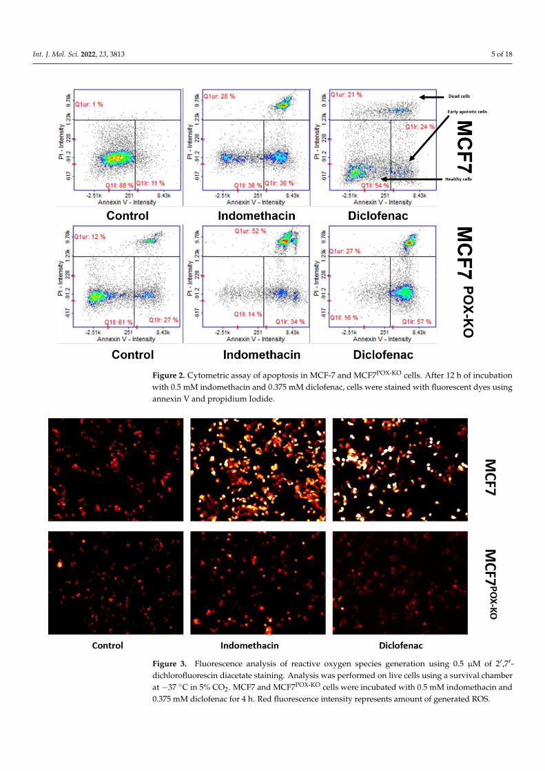
A cytometric assay with annexin V and propidium iodide was used to evaluate the
proportion of healthy, early apoptotic, and late apoptotic (dead) cells in the NSAID-
treated MCF7 and MCF7POK-KO cells. In IND-treated MCF7 cells 36% of cells were healthy,
36% were early apoptotic, and 28% were late apoptotic (dead). MCF7 cells treated with
DCF were in proportions of 54, 24, and 21%, respectively. In control (untreated) MCF7
cells, 88% of cells were healthy, 11% were early apoptotic, and only 1% were late apop-
totic. Interestingly, in untreated MCF7POK-KO cells, 61% of cells were healthy, 27% early
apoptotic, and 12% were late apoptotic. This high number of early apoptotic and dead
cells in the MCF7POK-KO control might underline the pro-survival role of PRODH/POX in
cancer cells. However, in MCF7POK-KO cells treated with IND or DCF, cell proportions were:
13% healthy, 34% early apoptotic, 52% dead, and 16% healthy, 57% early apoptotic, and
27% dead, respectively (Figure 2).
Figure 1. Cell viability (A), DNA biosynthesis (B), and collagen biosynthesis (C) in MCF-7 andMCF7POX-KO cells treated for 24 h with 0.5 mM indomethacin and 0.375 mM diclofenac. Cellularproline concentration determined by HPLC-MS (D). The mean values ± standard error (SEM) fromthe 3 experiments, performed in triplicates, are presented at * p < 0.05 and ** p < 0.005.
A cytometric assay with annexin V and propidium iodide was used to evaluate theproportion of healthy, early apoptotic, and late apoptotic (dead) cells in the NSAID-treatedMCF7 and MCF7POK-KO cells. In IND-treated MCF7 cells 36% of cells were healthy, 36%were early apoptotic, and 28% were late apoptotic (dead). MCF7 cells treated with DCF werein proportions of 54, 24, and 21%, respectively. In control (untreated) MCF7 cells, 88% ofcells were healthy, 11% were early apoptotic, and only 1% were late apoptotic. Interestingly,in untreated MCF7POK-KO cells, 61% of cells were healthy, 27% early apoptotic, and 12%were late apoptotic. This high number of early apoptotic and dead cells in the MCF7POK-KO
control might underline the pro-survival role of PRODH/POX in cancer cells. However, inMCF7POK-KO cells treated with IND or DCF, cell proportions were: 13% healthy, 34% earlyapoptotic, 52% dead, and 16% healthy, 57% early apoptotic, and 27% dead, respectively(Figure 2).
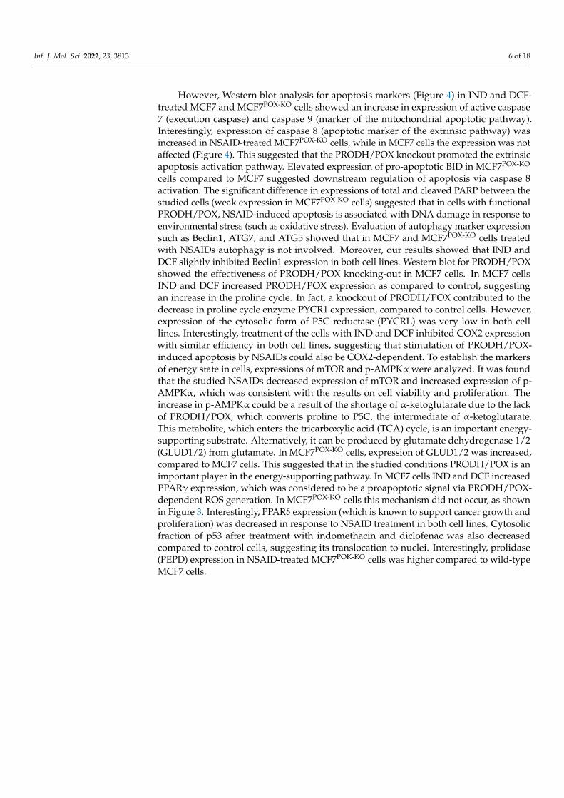
It was previously shown that IND and DCF activate PPARγ, leading to PRODH/POX-dependent ROS-mediated apoptosis [20]. Both drugs were used to validate their ability togenerate ROS. An experiment with 2′,7′-dichlorofluorescin diacetate staining confirmed thatincubation of MCF7 cells with NSAIDs strongly increased ROS generation, as visualized bythe increase in red fluorescence, compared to control (Figure 3). This effect was not shownin MCF7POX-KO cells, suggesting the critical role of PRODH/POX in NSAID-dependentROS generation.
Int. J. Mol. Sci. 2022, 23, 3813 5 of 18Int. J. Mol. Sci. 2022, 23, x FOR PEER REVIEW 5 of 18
Figure 2. Cytometric assay of apoptosis in MCF-7 and MCF7POX-KO cells. After 12 h of incubation
with 0.5 mM indomethacin and 0.375 mM diclofenac, cells were stained with fluorescent dyes using
annexin V and propidium Iodide.
It was previously shown that IND and DCF activate PPARγ, leading to
PRODH/POX-dependent ROS-mediated apoptosis [20]. Both drugs were used to validate
their ability to generate ROS. An experiment with 2′,7′-dichlorofluorescin diacetate stain-
ing confirmed that incubation of MCF7 cells with NSAIDs strongly increased ROS gener-
ation, as visualized by the increase in red fluorescence, compared to control (Figure 3).
This effect was not shown in MCF7POX-KO cells, suggesting the critical role of PRODH/POX
in NSAID-dependent ROS generation.
Figure 2. Cytometric assay of apoptosis in MCF-7 and MCF7POX-KO cells. After 12 h of incubationwith 0.5 mM indomethacin and 0.375 mM diclofenac, cells were stained with fluorescent dyes usingannexin V and propidium Iodide.
Int. J. Mol. Sci. 2022, 23, x FOR PEER REVIEW 5 of 18
Figure 2. Cytometric assay of apoptosis in MCF-7 and MCF7POX-KO cells. After 12 h of incubation
with 0.5 mM indomethacin and 0.375 mM diclofenac, cells were stained with fluorescent dyes using
annexin V and propidium Iodide.
It was previously shown that IND and DCF activate PPARγ, leading to
PRODH/POX-dependent ROS-mediated apoptosis [20]. Both drugs were used to validate
their ability to generate ROS. An experiment with 2′,7′-dichlorofluorescin diacetate stain-
ing confirmed that incubation of MCF7 cells with NSAIDs strongly increased ROS gener-
ation, as visualized by the increase in red fluorescence, compared to control (Figure 3).
This effect was not shown in MCF7POX-KO cells, suggesting the critical role of PRODH/POX
in NSAID-dependent ROS generation.
Figure 3. Fluorescence analysis of reactive oxygen species generation using 0.5 µM of 2′,7′-dichlorofluorescin diacetate staining. Analysis was performed on live cells using a survival chamberat −37 ◦C in 5% CO2. MCF7 and MCF7POX-KO cells were incubated with 0.5 mM indomethacin and0.375 mM diclofenac for 4 h. Red fluorescence intensity represents amount of generated ROS.
Int. J. Mol. Sci. 2022, 23, 3813 6 of 18
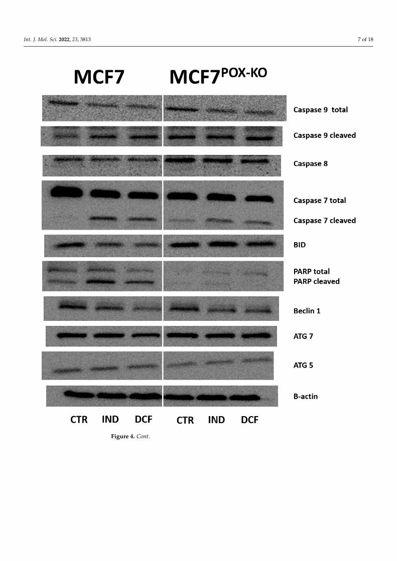
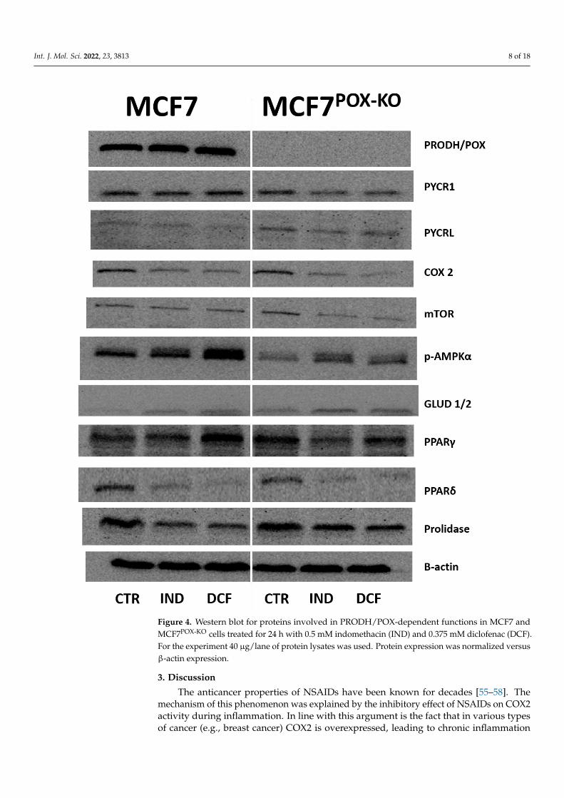
However, Western blot analysis for apoptosis markers (Figure 4) in IND and DCF-treated MCF7 and MCF7POX-KO cells showed an increase in expression of active caspase7 (execution caspase) and caspase 9 (marker of the mitochondrial apoptotic pathway).Interestingly, expression of caspase 8 (apoptotic marker of the extrinsic pathway) wasincreased in NSAID-treated MCF7POX-KO cells, while in MCF7 cells the expression was notaffected (Figure 4). This suggested that the PRODH/POX knockout promoted the extrinsicapoptosis activation pathway. Elevated expression of pro-apoptotic BID in MCF7POX-KO
cells compared to MCF7 suggested downstream regulation of apoptosis via caspase 8activation. The significant difference in expressions of total and cleaved PARP between thestudied cells (weak expression in MCF7POX-KO cells) suggested that in cells with functionalPRODH/POX, NSAID-induced apoptosis is associated with DNA damage in response toenvironmental stress (such as oxidative stress). Evaluation of autophagy marker expressionsuch as Beclin1, ATG7, and ATG5 showed that in MCF7 and MCF7POX-KO cells treatedwith NSAIDs autophagy is not involved. Moreover, our results showed that IND andDCF slightly inhibited Beclin1 expression in both cell lines. Western blot for PRODH/POXshowed the effectiveness of PRODH/POX knocking-out in MCF7 cells. In MCF7 cellsIND and DCF increased PRODH/POX expression as compared to control, suggestingan increase in the proline cycle. In fact, a knockout of PRODH/POX contributed to thedecrease in proline cycle enzyme PYCR1 expression, compared to control cells. However,expression of the cytosolic form of P5C reductase (PYCRL) was very low in both celllines. Interestingly, treatment of the cells with IND and DCF inhibited COX2 expressionwith similar efficiency in both cell lines, suggesting that stimulation of PRODH/POX-induced apoptosis by NSAIDs could also be COX2-dependent. To establish the markersof energy state in cells, expressions of mTOR and p-AMPKα were analyzed. It was foundthat the studied NSAIDs decreased expression of mTOR and increased expression of p-AMPKα, which was consistent with the results on cell viability and proliferation. Theincrease in p-AMPKα could be a result of the shortage of α-ketoglutarate due to the lackof PRODH/POX, which converts proline to P5C, the intermediate of α-ketoglutarate.This metabolite, which enters the tricarboxylic acid (TCA) cycle, is an important energy-supporting substrate. Alternatively, it can be produced by glutamate dehydrogenase 1/2(GLUD1/2) from glutamate. In MCF7POX-KO cells, expression of GLUD1/2 was increased,compared to MCF7 cells. This suggested that in the studied conditions PRODH/POX is animportant player in the energy-supporting pathway. In MCF7 cells IND and DCF increasedPPARγ expression, which was considered to be a proapoptotic signal via PRODH/POX-dependent ROS generation. In MCF7POX-KO cells this mechanism did not occur, as shownin Figure 3. Interestingly, PPARδ expression (which is known to support cancer growth andproliferation) was decreased in response to NSAID treatment in both cell lines. Cytosolicfraction of p53 after treatment with indomethacin and diclofenac was also decreasedcompared to control cells, suggesting its translocation to nuclei. Interestingly, prolidase(PEPD) expression in NSAID-treated MCF7POK-KO cells was higher compared to wild-typeMCF7 cells.
Int. J. Mol. Sci. 2022, 23, 3813 7 of 18Int. J. Mol. Sci. 2022, 23, x FOR PEER REVIEW 7 of 18
Figure 4. Cont.
Int. J. Mol. Sci. 2022, 23, 3813 8 of 18
Int. J. Mol. Sci. 2022, 23, x FOR PEER REVIEW 8 of 18
Figure 4. Western blot for proteins involved in PRODH/POX-dependent functions in MCF7 and
MCF7POX-KO cells treated for 24 h with 0.5 mM indomethacin (IND) and 0.375 mM diclofenac (DCF).
For the experiment 40 µg/lane of protein lysates was used. Protein expression was normalized ver-
sus β-actin expression.
3. Discussion
The anticancer properties of NSAIDs have been known for decades [55–58]. The
mechanism of this phenomenon was explained by the inhibitory effect of NSAIDs on
Figure 4. Western blot for proteins involved in PRODH/POX-dependent functions in MCF7 andMCF7POX-KO cells treated for 24 h with 0.5 mM indomethacin (IND) and 0.375 mM diclofenac (DCF).For the experiment 40 µg/lane of protein lysates was used. Protein expression was normalized versusβ-actin expression.
3. Discussion
The anticancer properties of NSAIDs have been known for decades [55–58]. Themechanism of this phenomenon was explained by the inhibitory effect of NSAIDs on COX2activity during inflammation. In line with this argument is the fact that in various typesof cancer (e.g., breast cancer) COX2 is overexpressed, leading to chronic inflammation
Int. J. Mol. Sci. 2022, 23, 3813 9 of 18
supporting cell proliferation, neovascularization, and cancer growth [5,59]. Interestingly, asimilar anticancer activity of NSAIDs was observed in cancer cells lacking COX2 expressionand also in models of cancer cells with knockdown expression of this enzyme [8,9,60–63].This suggested the COX2-independent anticancer mechanism of NSAIDs.
In the present study we provided evidence that NSAIDs through activation of PPARγreceptors [11] induced PRODH/POX-dependent apoptosis in MCF. This idea was basedon studies describing the pro-apoptotic effect of the activated PPARγ–PRODH/POXaxis [20,27,28,64,65].
PRODH/POX can, however, exert an opposite function. During conversion of prolineinto P5C, free electrons can support the electron transport chain producing ATP for cellsurvival [66,67]. However, in certain conditions free electrons can also be accepted directlyby oxygen, generating reactive oxygen species (ROS). In this case ROS disturb redox balancestimulating oxidative stress, known as pro-apoptotic stimuli [28,68,69]. It seems that theseopposite activities of PRODH/POX are related to the energetic state of the cell. Since cancercells have high energy requirements and because of the Warburg’s effect glucose is nota sufficient source of energy, the cells require energy from protein degradation (mostlycollagen), providing proline, among others, as a substrate for PRODH/POX resulting inATP or ROS generation [21,22,27,28].
To evaluate the role of PRODH/POX in NSAID-induced apoptosis an innovativemodel of MCF7 breast cancer cells with a knockout of PRODH/POX by CRISPR/Cas9technology was created. Unexpectedly, it was discovered that IND and DCF inducedapoptosis in both cell lines (MCF7 wild-type and MCF7POX-KO cells); however, the pro-apoptotic effect of NSAIDs was remarkably stronger in cells lacking PRODH/POX. Theeffect was accompanied by decrease in the expression of mTOR-potent cancer growthstimulator and an increase in p-AMPKα expression, a sensor of ATP level [70–72].
As a product of PRODH/POX activity, P5C can also be considered as an energysensor since it can be spontaneously converted into glutamate (reversible reaction) andthen to α-ketoglutarate (α-KG) by glutamate dehydrogenase (GLUD 1/2). Therefore,proline conversion into P5C can support the TCA cycle by production of its importantsubstrate, α-KG, supporting energy production. P5C can also be produced from ornithine,a metabolite of the urea cycle [73–75]). We found that in MCF7POX-KO cells, expressionof GLUD1/2 was elevated compared to MCF7 cells. Interestingly, the studied NSAIDsstimulated the enzyme expression, suggesting that in the absence of PRODH/POX, P5Cis generated from the glutamate or urea cycle, supporting TCA cycle function for energyproduction [27]. However, in MCF7 cells proline cycling demonstrated by increasedexpression of PRODH/POX in response to NSAIDs→ PPARγ activation was accompaniedby increased mitochondrial P5C reductase, PYCR1. In MCF7POK-KO cells (in which P5Cproduction was limited owing to PRODH/POX absence) PYCR1 expression was decreasedby NSAID treatment, suggesting that it was the mechanism for limited production of pro-energetic precursors such as P5C. Simultaneously, the cancer cell growth and expression ofPPARδ (energy-producing promotor) [76–84] was strongly inhibited by NSAIDs, enhancingthe pro-apoptotic and anti-proliferative effects, particularly in MCF7POX-KO cells.
Cytometric analysis showed that the population of MCF7POX-KO cells contained about12% dead cells, in comparison to about 1% dead cells in wild-type MCF7 cells, cultured inthe same conditions. This showed that a lack of PRODH/POX significantly decreases cellviability. Moreover, incubation of the cells with IND and DCF resulted in a further increasein number of early apoptotic and dead cells in MCF7POX-KO cells, compared to wild-typeMCF7. These results were also supported by a decrease in DNA biosynthesis in NSAID-treated MCF7POX-KO cells. In MCF7 cells the process was less pronounced. Similarly,collagen biosynthesis was inhibited in NSAID-treated cells as was found earlier [44,85–87].However, in the case of MCF7POX-KO cells, collagen biosynthesis was strongly inhibitedin response to NSAID treatment. In PRODH/POX-dependent apoptosis in MCF7 cells,this activity of NSAIDs is desired because inhibition of collagen biosynthesis (the mainprocess of proline utilization) makes proline available for PRODH/POX-dependent ROS
Int. J. Mol. Sci. 2022, 23, 3813 10 of 18
generation and apoptosis. In MCF7POX-KO cells this NSAIDs activity is also beneficialbecause proline carrying reducing potential is harmful for the cells [27,66,74,75,88,89].Interestingly, in MCF7POK-KO cells, the increase in intracellular proline was not only due tolack of the proline-degrading enzyme (PRODH/POX) but also because of the increase in theexpression of prolidase (the proline-supporting enzyme that cleaves dipeptides containingproline). The functional significance of NSAIDs inducing the above processes was foundin expression of the molecular markers of apoptosis induced in both cell lines. MCF7cells treated with NSAIDs showed increased expression of cleaved caspase 9, which is amarker of mitochondrial apoptosis. It was accompanied by an increase in the expressionof cleaved PARP, considered to be a marker of DNA damage [90,91]. DNA damage canbe induced by different stress factors such as oxidative stress, which was observed inresponse to NSAID treatment. This was proved by fluorescence analysis of generatedROS using 2′,7′-dichlorofluorescin diacetate. These results suggested that in MCF7 cellswith functional PRODH/POX, NSAIDs induced apoptosis by activating the mitochondrial(caspase 9-related) pathway in response to ROS generation as a result of PPARγ-dependentstimulation of PRODH/POX activity. In MCF7POX-KO cells, the mechanism of NSAID-induced apoptosis was different. In these cells oxidative stress was not induced (ROS werenot detected). This was also supported by a decrease in the expression of PPARγ, suggestingthat in MCF7POX-KO cells the studied NSAIDs did not induce DNA damage. Furthermore,in contrast to MCF7, in MCF7POX-KO cells caspase 8 expression was significantly increased,suggesting that the extrinsic pathway of apoptosis was induced in these cells. In fact, inMCF7POX-KO cells treated with IND or DCF expression of BID was more pronounced thanin MCF-7 cells treated with these drugs. An increase in expression of BID can activatemitochondrial caspase 9 [92,93]. On the other hand, the studied drugs, particularly DCF,contributed to a decrease in the expression of BID in both cell lines. This effect wasclearly visible in MCF-7 cells, suggesting that activation of caspase 9 in these cells could beBID-independent.
Analysis of autophagy markers (Beclin 1, Atg5, and Atg7) showed that this type ofcellular death was not activated in NSAID-treated cells in both cell lines. The mechanismof NSAID-induced apoptosis in MCF7POX-KO cells can be explained by the interplay ofthe TCA cycle, urea cycle, and proline cycle. Removing PRODH/POX from MCF7 cellsinfluenced the concentration of P5C, the intermediate in the interconversion of proline,glutamine, α-KG, and ornithine, affecting the interplay between the metabolites of thosecycles and the energy metabolism in the cells [27,94]. PRODH/POX knockout contributedto an increase in intracellular proline because of the inhibition of its degradation and theincrease in prolidase expression. Moreover, in MCF7POX-KO cells treated with NSAIDscollagen biosynthesis was strongly inhibited, contributing to a further increase in the con-centration of intracellular proline (bearing reducing potential) affecting the redox balancein the cells. These processes are potentially involved in the mechanism of PRODH/POX-independent apoptosis in breast cancer cells. The graphical visualization of the mechanismof NSAID-induced apoptosis in MCF7POX-KO cells is presented as Figure 5. Whether it is auniversal mechanism in cancer remains to be established. However, based on our previousstudies, the role of PRODH/POX in the apoptosis/survival of cancer cells is not only azero–one system but rather depends on the metabolic context of the specific cell type. Inour recent paper [95] we provided evidence that stimulation of PRODH/POX expressionby metformin induced apoptosis in both WT and PRODH/POX knockout MCF-7 cells, butonly when cultured in the absence of glutamine, while the presence of glutamine facilitateda pro-survival phenotype of the cells. Metabolomic analysis suggested that glycolysisis tightly linked to glutamine and proline metabolism in these cells, creating metabolicconditions for energy production and proline availability for PRODH/POX-dependentfunctions. Metformin treatment of both cell lines (WT and PRODH/POX knockout MCF-7cells) cultured in glutamine-free medium contributed to glucose starvation, facilitating apro-apoptotic phenotype of these cells as detected by the increase in the expression of activecaspase 7 and PARP. Caspase 7 is known as an executioner protein of apoptosis activated
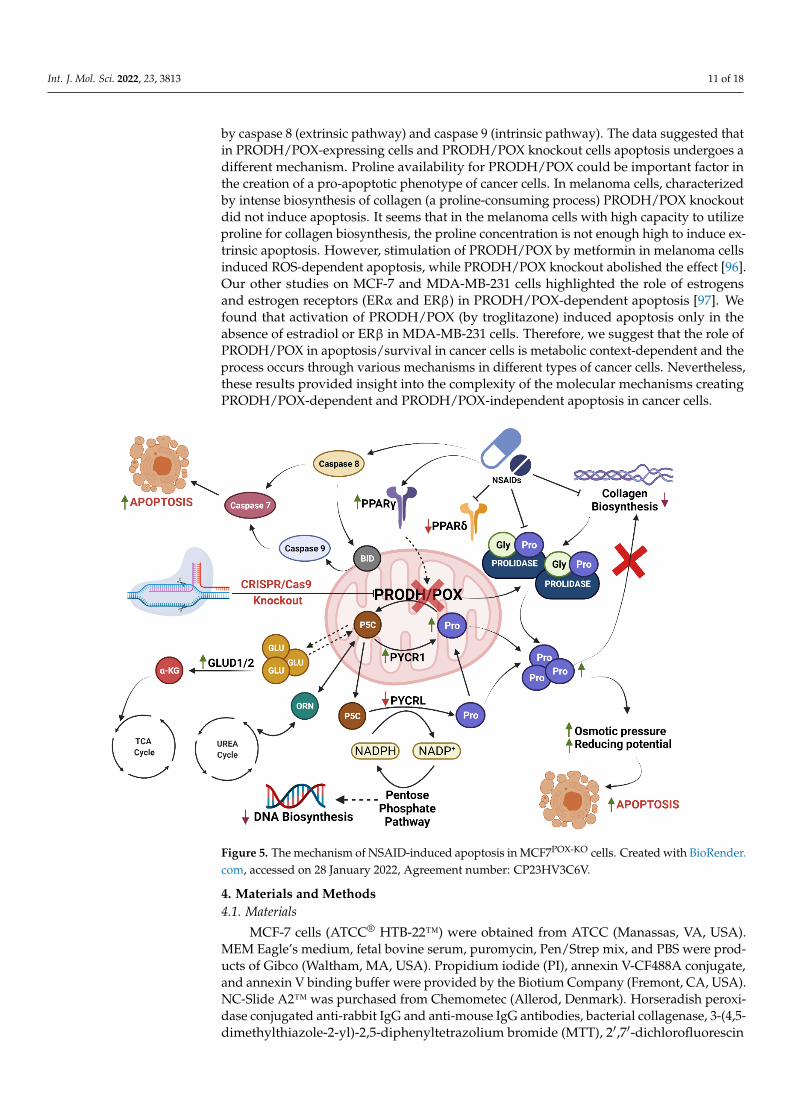
Int. J. Mol. Sci. 2022, 23, 3813 11 of 18
by caspase 8 (extrinsic pathway) and caspase 9 (intrinsic pathway). The data suggested thatin PRODH/POX-expressing cells and PRODH/POX knockout cells apoptosis undergoes adifferent mechanism. Proline availability for PRODH/POX could be important factor inthe creation of a pro-apoptotic phenotype of cancer cells. In melanoma cells, characterizedby intense biosynthesis of collagen (a proline-consuming process) PRODH/POX knockoutdid not induce apoptosis. It seems that in the melanoma cells with high capacity to utilizeproline for collagen biosynthesis, the proline concentration is not enough high to induce ex-trinsic apoptosis. However, stimulation of PRODH/POX by metformin in melanoma cellsinduced ROS-dependent apoptosis, while PRODH/POX knockout abolished the effect [96].Our other studies on MCF-7 and MDA-MB-231 cells highlighted the role of estrogensand estrogen receptors (ERα and ERβ) in PRODH/POX-dependent apoptosis [97]. Wefound that activation of PRODH/POX (by troglitazone) induced apoptosis only in theabsence of estradiol or ERβ in MDA-MB-231 cells. Therefore, we suggest that the role ofPRODH/POX in apoptosis/survival in cancer cells is metabolic context-dependent and theprocess occurs through various mechanisms in different types of cancer cells. Nevertheless,these results provided insight into the complexity of the molecular mechanisms creatingPRODH/POX-dependent and PRODH/POX-independent apoptosis in cancer cells.
Int. J. Mol. Sci. 2022, 23, x FOR PEER REVIEW 11 of 18
knockout MCF-7 cells) cultured in glutamine-free medium contributed to glucose starva-
tion, facilitating a pro-apoptotic phenotype of these cells as detected by the increase in the
expression of active caspase 7 and PARP. Caspase 7 is known as an executioner protein of
apoptosis activated by caspase 8 (extrinsic pathway) and caspase 9 (intrinsic pathway).
The data suggested that in PRODH/POX-expressing cells and PRODH/POX knockout
cells apoptosis undergoes a different mechanism. Proline availability for PRODH/POX
could be important factor in the creation of a pro-apoptotic phenotype of cancer cells. In
melanoma cells, characterized by intense biosynthesis of collagen (a proline-consuming
process) PRODH/POX knockout did not induce apoptosis. It seems that in the melanoma
cells with high capacity to utilize proline for collagen biosynthesis, the proline concentra-
tion is not enough high to induce extrinsic apoptosis. However, stimulation of
PRODH/POX by metformin in melanoma cells induced ROS-dependent apoptosis, while
PRODH/POX knockout abolished the effect [96]. Our other studies on MCF-7 and MDA-
MB-231 cells highlighted the role of estrogens and estrogen receptors (ERα and ERβ) in
PRODH/POX-dependent apoptosis [97]. We found that activation of PRODH/POX (by
troglitazone) induced apoptosis only in the absence of estradiol or ERβ in MDA-MB-231
cells. Therefore, we suggest that the role of PRODH/POX in apoptosis/survival in cancer
cells is metabolic context-dependent and the process occurs through various mechanisms
in different types of cancer cells. Nevertheless, these results provided insight into the com-
plexity of the molecular mechanisms creating PRODH/POX-dependent and
PRODH/POX-independent apoptosis in cancer cells.
Figure 5. The mechanism of NSAID-induced apoptosis in MCF7POX-KO cells. Created with BioRen-
der.com, accessed on 28 January 2022, Agreement number: CP23HV3C6V.
4. Materials and Methods
4.1. Materials
MCF-7 cells (ATCC® HTB-22™) were obtained from ATCC (Manassas, VA, USA).
MEM Eagle’s medium, fetal bovine serum, puromycin, Pen/Strep mix, and PBS were
Figure 5. The mechanism of NSAID-induced apoptosis in MCF7POX-KO cells. Created with BioRender.com, accessed on 28 January 2022, Agreement number: CP23HV3C6V.
4. Materials and Methods4.1. Materials
MCF-7 cells (ATCC® HTB-22™) were obtained from ATCC (Manassas, VA, USA).MEM Eagle’s medium, fetal bovine serum, puromycin, Pen/Strep mix, and PBS were prod-ucts of Gibco (Waltham, MA, USA). Propidium iodide (PI), annexin V-CF488A conjugate,and annexin V binding buffer were provided by the Biotium Company (Fremont, CA, USA).NC-Slide A2™ was purchased from Chemometec (Allerod, Denmark). Horseradish peroxi-dase conjugated anti-rabbit IgG and anti-mouse IgG antibodies, bacterial collagenase, 3-(4,5-dimethylthiazole-2-yl)-2,5-diphenyltetrazolium bromide (MTT), 2′,7′-dichlorofluorescin
Int. J. Mol. Sci. 2022, 23, 3813 12 of 18
diacetate (DCFDA), indomethacin, and diclofenac were purchased from Sigma Aldrich(Saint Louis, MO, USA). Primary antibodies against COX2, p-AMPKα, mTor, caspase 8,caspase 9 total and cleaved, caspase 7 total and cleaved, BID, PARP, Beclin 1, AGT5, ATG 7,and B-actin were products of Cell Signaling Technology (Danvers, MA, USA). Primary anti-bodies for PRODH/POX, PPARγ, GLUD 1/2, and prolidase were products of Santa CruzBiotechnology (Dallas, TX, USA). PYCR1, PYCRL, and PPARδ primary antibodies were ob-tained from Abnova (Taipei, Taiwan). Hoechst 33342 was obtained from Becton Dickinson(Franklin Lakes, NJ, USA). CRISPR All-In-One Non-Viral Vector and lipofectamine wereproducts of Applied Biological Materials Inc. (Richmond, BC, Canada).
4.2. Methods4.2.1. Cell Culture
MCF7 (ATCC® HTB22™) breast cancer cells were maintained in MEM Eagle’s medium,supplemented with 10% FBS, 50 U/mL of penicillin, and 50 µg/mL of streptomycin, andincubated at 37 ◦C in 5% CO2. The cells were grown on 100-mm dishes in 10 mL of completemedium. The cell culture medium was changed 2–3 times per week. For the experimentswith the investigated drugs, we used MEM Eagle’s medium without FBS and Pen/Strep.
4.2.2. Knockout of PRODH/POX in MCF7 Cells
A clone of the MCF7 cells with a knocked-out gene of PRODH/POX was preparedusing Custom CRISPR All-In-One Non-Viral Vector, provided by Applied Biological Ma-terials Inc. (Richmond, BC, Canada). A vector containing sgRNA sequence targetingPRODH/POX and puromycin resistance coding gene was introduced to the MCF7 cells bylipofectamine. As a selection factor puromycin in a concentration of 1.5 µg/µL was used.Antibiotic selection was performed for 10 days until about 98–100% of control cells died.After the selection period a single cell suspension was prepared, and cells were cultured ona 96-well plate to obtain single cell colonies. Efficiency of the PRODH/POX knockout wasperformed using the Western blot method and clone with a 100% knockout result was usedfor further experiments (named the MCF7POX-KO cell line).
4.2.3. Cell Viability Test MTT
To evaluate the cytotoxicity of the selected drugs on the MCF7 and MCF7POX-KO
cells, methyl thiazolyl tetrazolium (MTT) salt was used, as described in the Carmichaelmethod [98]. This method is based on the conversion of yellow tetrazolium bromide MTTsolution to the purple formazan derivatives in the live cells, and this is due to the activityof the mitochondrial dehydrogenases. For this assay, the cells were cultured in 12-wellplates, at a density of 0.1 × 10⁶ cells/well. When the cells reached about 70% confluency,the culture media were removed. The wells were washed with PBS, and the fresh MEMEagle’s media containing the studied drugs dissolved in DMSO (dimethyl sulfoxide) wereadded into the wells (the concentration of DMSO in the sample did not exceed 0.1%). Thesamples were prepared in triplicate. The cells were incubated with the compound for 24 h.The investigated drugs were in the following concentrations: 0.500 mM of indomethacin;0.375 mM of diclofenac. After incubation, the cell culture media containing the studieddrugs were removed, and the plates were washed twice with prewarmed PBS. Furthermore,the cells were incubated at 37 ◦C for 1 h, with MTT dissolved in PBS (0.5 mg/mL) in avolume of 1.0 mL per well. When the incubation step ended, the MTT was removed andthe formazan derivatives were dissolved in the DMSO (1.0 mL per well). To quantify theformed formazan, the absorbance at 570 nm was measured using a spectrophotometer.The viability of the cells treated with the studied drugs was calculated as a percent of thecontrol value.
4.2.4. DNA Biosynthesis
To study the effect of indomethacin and diclofenac on the MCF7 and MCF7POX-KO
cell DNA biosynthesis, an assay of the radioactive [3H]-Thymidine incorporation into

Int. J. Mol. Sci. 2022, 23, 3813 13 of 18
the DNA was performed. The cells were cultured on 12-well plates, as described above.When they reached about 70% confluency, they were treated with the studied drugs inthe described concentrations, and 1.0 µCi of [3H]-Thymidine was added to the MEMEagle’s medium. The samples were prepared in triplicate. The cells were incubated for24 h at 37 ◦C in 5% CO2. The radioactivity of the samples was measured using the Tri-Carb 2810 TR Scintillation Analyzer (PerkinElmer, Waltham, MA, USA). To calculate theDNA biosynthesis, the DPMI values of the treated cells were compared to those of thecontrol samples.
4.2.5. Collagen Biosynthesis
The collagen biosynthesis was evaluated by measuring the incorporation of the ra-dioactive 5-[3H]-Proline into the collagen. The MCF7 and MCF7POX-KO cells were culturedin 12-well plates. When the cells were about 70% confluent, the culture medium wasremoved, and the cells were treated for 24 h with the studied drugs and 5-[3H]-Proline(5 µCi/mL). Digesting proteins, with purified Clostridium histolyticum collagenase, wereused to determine the incorporation of the tracer into the collagen, as described by Pe-terkofsky et al. [99]. The radioactivity of the samples was measured by the Tri-Carb 2810TR Scintillation Analyzer (PerkinElmer, Waltham, MA, USA). To calculate the collagenbiosynthesis, the DPMI values of the treated cells were compared to those of the controls.
4.2.6. SDS-PAGE and Western Blotting
For the analysis of the protein expression by Western blotting, the cells were cultured in100 mm plates, at about 2.0× 10⁶ cells, and when they reached about 70–80% confluency, theculture medium was removed, and the cells were treated with the studied drugs (dissolvedin culture medium). After 24 h of incubation, the culture media were removed, and thecells were harvested using a cell lysis buffer supplemented with a protease/phosphataseinhibitor cocktail. The protein concentrations of the samples were determined by the Lowrymethod [100]. Then, the proteins were separated using the SDS-PAGE method describedby Laemmli [101]. After this step, the gels were washed in cold Towbin buffer (25 mM Tris,192 mM glycine, 20% (v/v) methanol, 0.025–0.1% SDS, pH 8.3). The proteins in the gels weretransferred onto the 0.2-µm nitrocellulose membranes by Trans-Blot (BioRad, Hercules, CA,USA). The transfer conditions were 200 mA, 3 h in freshly prepared Towbin buffer, and thetemperature was maintained around 4–8 ◦C. The blocking of the membranes was performedby 5% NFDM for 1 h at RT. When the blocking was complete, the membranes were washedthree times with 20 mL of TBS-T (20 mM Tris, 150 mM NaCl, and 0.1% Tween® 20). Afterthe washing step, the membranes were incubated with primary antibodies overnight at4 ◦C. The concentration of the primary antibodies was 1:1000. Furthermore, the membraneswere washed three times with 20 mL of TBS-T, and a secondary antibody conjugated withHRP solutions (1:3000) in 5% NFDM was used for 1 h at RT. Then the membranes werewashed 3 times with 20 mL of TBS-T and visualized.
4.2.7. ROS Formation
The cells were cultured on black wells in a 96-well plate, at 0.01 × 10⁴ cells/well.When the cells reached 70–80% confluency, the culture media were removed, the plate waswashed with PBS, and 100 µL of medium containing the studied drug was added into thewell. After 4 h of incubation, 0.5 µM of 2′,7′-dichlorofluorescin diacetate was added to thewells and incubated for 15 min at 37 ◦C in 5% CO2. After incubation, the culture mediawith DCFDA were removed and the cells were washed twice with prewarmed PBS. Thenthe wells were loaded with 100 µL of PBS. The cells were visualized with the BD Pathway855 Bioimaging system in an environmental control chamber (37 ◦C in 5% CO2), and ex λ
488 nm, and em λ 521 nm. In its basic state, DCFDA is a nonfluorescent compound, andwhen oxidized by ROS to DCF it becomes highly fluorescent.
Int. J. Mol. Sci. 2022, 23, 3813 14 of 18
4.2.8. Cytometric Assay for Apoptosis
The cells were cultured in 6-well plates at an initial density of 0.3 × 10⁶ cells/well.When the cells reached about 70–80% confluency, the culture media were removed. Thewells were washed with PBS, and fresh MEM Eagle’s media containing the studied drugswere added into the wells. After 24 h of incubation, the cells (including floating cells)were collected by trypsinization to 1.5 mL Eppendorf tubes. The cells were centrifuged at800× g for 5 min. The supernatant was removed, and the cell pellet was resuspended in100 µL annexin V binding buffer, and then 2 µL annexin V-CF488A conjugate was added.In the next step 2 µL Hoechst 33342 (final concentration: 10 µg/mL) was added and thesuspension was gently mixed by pipetting. After that, incubation of the cells at 37 ◦C for15 min using a heating block was performed. Next, the stained cells were centrifuged at400× g for 5 min. at room temperature, and the supernatant was removed. As the laststep, the cell pellet was resuspended in 100 µL annexin V binding buffer supplementedwith 10 µg/mL PI and then immediately analyzed in the NC-Slide A2. Cells with lowfluorescence intensities of PI (PI negative) and low fluorescence intensities of annexin Vrepresented living cells with high viability. The cells with high fluorescence intensities ofPI (PI positive) and low fluorescence intensities of annexin V represented early apoptoticcells. The cells with high fluorescence intensities of PI (PI positive) and high fluorescenceintensities of annexin V represented late apoptotic/dead cells.
4.2.9. LC-MS-Based Quantitative Analysis
LC-MS analysis of proline was performed using a 1260 Infinity II high performanceliquid chromatograph coupled to a 6530 Q-TOF mass spectrometer equipped with a dualelectrospray ionization source (Agilent Technologies, Santa Clara, CA, USA) [102]. Chro-matographic separation was conducted on a Luna HILIC column (100 × 2.0 mm, 3 µmparticle size, Phenomenex, Torrance, CA, USA) maintained at 30 ◦C. The injection volumewas 5 µL and the flow rate was 0.6 mL/min. The gradient elution of solvent A (10 mMammonium formate in water with 0.1% formic acid) and solvent B (acetonitrile) was pro-grammed as follows: 0–2 min with 90% B, 2–7 min with 90 to 30% B, 7–7.5 min with30% B, 7.5–8 min with 30 to 90% B, 8–14 min with 90% B. Data were collected in positiveionization mode. The following source parameters were applied: drying gas temperature,325 ◦C; drying gas flow, 12 L/min; nebulizer pressure, 45 psig; fragmentor voltage, 140 V;capillary voltage, 3000 V. The mass spectrometer operated in scan mode with a mass rangeof 50–1000 m/z. L-proline-d3 (Sigma Aldrich, St. Louis, MO, USA) was used as an internalstandard. Methanol extracts of cells (containing L-proline-d3) were centrifuged (14,000× g;4 ◦C; 10 min) and the supernatant was subjected to LC-MS analysis. The samples werecollected in three biological repeats, injected in duplicates, and randomized before analysis.Total protein concentration was used for normalization.
4.2.10. Statistical Analysis
In order to analyze the distribution of the data in individual groups, the Shapiro–Wilktest was performed. Within the normally distributed groups, the differences betweenthe individual groups were analyzed using a one-way ANOVA, with multiple post hoccomparisons using the Bonferroni correction. In the groups where no normal distributionswere shown, the nonparametric Mann–Whitney test was used. The data are presented asmeans ± standard error of measurement (SEM).
5. Conclusions
In this report we found that NSAIDs (IND and DCF) as PPARγ agonists activatePRODH/POX-induced ROS-dependent intrinsic apoptosis in breast cancer MCF-7 cells.However, in MCF7POX-KO cells, NSAIDs also induced apoptosis by the ROS-independentextrinsic pathway. These results provide evidence that the studied NSAIDs induce apopto-sis in both PRODH/POX-expressing and PRODH/POX knockout cells and underline theimportance of PRODH/POX as a target for NSAID-induced apoptosis in breast cancer cells.
Int. J. Mol. Sci. 2022, 23, 3813 15 of 18
Author Contributions: The authors made substantial contributions to the manuscript as follows:conception and design of the study, A.K. and A.S.; performance of experiments and tests, A.K.;knockout of PRODH/POX in MCF7 cells, A.K.; determination of amino acids concentration, K.B.;analysis and interpretation of data, A.K., M.C., J.P., K.B. and A.S.; drafting of the article A.K., J.P.and A.S.; final approval of the version to be submitted, A.S. All authors have read and agreed to thepublished version of the manuscript.
Funding: This research was supported by the National Science Centre of Poland, grant number:2017/27/N/NZ7/02370.
Institutional Review Board Statement: Not applicable.
Informed Consent Statement: Not applicable.
Data Availability Statement: This study did not report any supporting data.
Conflicts of Interest: The authors declare no conflict of interest.
References1. Chan, A.T.; Giovannucci, E.L.; Meyerhardt, J.A.; Schernhammer, E.S.; Curhan, G.C.; Fuchs, C.S. Long-term use of aspirin and
nonsteroidal anti-inflammatory drugs and risk of colorectal cancer. JAMA 2005, 294, 914–923. [CrossRef] [PubMed]2. Shebl, F.M.; Hsing, A.W.; Park, Y.; Hollenbeck, A.R.; Chu, L.W.; Meyer, T.E.; Koshiol, J. Non-steroidal anti-inflammatory drugs
use is associated with reduced risk of inflammation-associated cancers: NIH-AARP study. PLoS ONE 2014, 9, e114633. [CrossRef]3. Rothwell, P.M.; Wilson, M.; Elwin, C.-E.; Norrving, B.; Algra, A.; Warlow, C.P.; Meade, T.W. Long-term effect of aspirin on
colorectal cancer incidence and mortality: 20-year follow-up of five randomised trials. Lancet 2010, 376, 1741–1750. [CrossRef]4. Knox, S.S. From ‘omics’ to complex disease: A systems biology approach to gene-environment interactions in cancer. Cancer Cell
Int. 2010, 10, 11. [CrossRef]5. Kim, H.S.; Moon, H.-G.; Hancha, W.; Yom, C.K.; Kim, W.H.; Kim, J.H.; Noh, D.-Y. COX2 overexpression is a prognostic marker for
Stage III breast cancer. Breast Cancer Res. Treat. 2012, 132, 51–59. [CrossRef] [PubMed]6. Menter, G.D. Prostaglandins in Cancer Cell Adhesion, Migration, and Invasion. Int. J. Cell Biol. 2012, 2012, 723419. [CrossRef]7. Eibl, G.; Bruemmer, D.; Okada, Y.; Duffy, J.P.; Law, E.R.; Reber, A.H.; Hines, O.J. PGE(2) is generated by specific COX-2 activity
and increases VEGF production in COX-2-expressing human pancreatic cancer cells. Biochem. Biophys. Res. Commun. 2003, 306,887–897. [CrossRef]
8. Matos, P.; Jordan, P. Beyond COX-inhibition: ‘side-effects’ of ibuprofen on neoplastic development and progression. Curr. Pharm.Des. 2015, 21, 2978–2982. [CrossRef] [PubMed]
9. Patel, M.I.; Subbaramaiah, K.; Du, B.; Chang, M.; Yang, P.; Newman, R.A.; Cordon-Cardo, C.; Thaler, H.T.; Dannenberg, A.J.Celecoxib inhibits prostate cancer growth: Evidence of a cyclooxygenase-2-independent mechanism. Clin. Cancer Res. 2005, 11,1999–2007. [CrossRef] [PubMed]
10. Rigas, B.; Shiff, S.J. Is inhibition of cyclooxygenase required for the chemopreventive effect of NSAIDs in colon cancer? A modelreconciling the current contradiction. Med. Hypotheses 2000, 54, 210–215. [CrossRef]
11. Puhl, A.C.; Milton, F.A.; Cvoro, A.; Sieglaff, D.H.; Campos, J.L.D.O.; Bernardes, A.; Filgueira, C.; Lindemann, J.A.L.; Deng, T.;Neves, F.A.R.; et al. Mechanisms of peroxisome proliferator activated receptor γ regulation by non-steroidal anti-inflammatorydrugs. Nucl. Recept. Signal. 2015, 13, e004. [CrossRef] [PubMed]
12. Varga, T.; Czimmerer, Z.; Nagy, L. PPARs are a unique set of fatty acid regulated transcription factors controlling both lipidmetabolism and inflammation. Biochim. Biophys. Acta. 2011, 1812, 1007–1022. [CrossRef] [PubMed]
13. Okazaki, S.; Shioi, R.; Noguchi-Yachide, T.; Ishikawa, M.; Makishima, M.; Hashimoto, Y.; Yamaguchi, T. Structure-activityrelationship studies of non-carboxylic acid peroxisome proliferator-activated receptor α/δ (PPARα/δ) dual agonists. Bioorg. Med.Chem. 2016, 24, 5455–5461. [CrossRef] [PubMed]
14. Peters, J.M.; Shah, Y.M.; Gonzalez, F.J. The role of peroxisome proliferator-activated receptors in carcinogenesis and chemopreven-tion. Nat. Rev. Cancer 2012, 12, 181–195. [CrossRef] [PubMed]
15. Heudobler, D.; Rechenmacher, M.; Lüke, F.; Vogelhuber, M.; Pukrop, T.; Herr, W.; Ghibelli, L.; Gerner, C.; Reichle, A. PeroxisomeProliferator-Activated Receptors (PPAR)γ Agonists as Master Modulators of Tumor Tissue. Int. J. Mol. Sci. 2018, 19, 3540.[CrossRef] [PubMed]
16. Yousefi, B.; Samadi, N.; Baradaran, B.; Shafiei-Irannejad, V.; Zarghami, N. Peroxisome Proliferator-Activated Receptor Ligandsand Their Role in Chronic Myeloid Leukemia: Therapeutic Strategies. Chem. Biol. Drug Des. 2016, 88, 17–25. [CrossRef] [PubMed]
17. Kitamura, S.; Miyazaki, Y.; Shinomura, Y.; Kondo, S.; Kanayama, S.; Matsuzawa, Y. Peroxisome proliferator-activated receptorgamma induces growth arrest and differentiation markers of human colon cancer cells. Jpn. J. Cancer Res. 1999, 90, 75–80.[CrossRef]
18. Nazim, U.M.; Moon, J.H.; Lee, Y.J.; Seol, J.W.; Park, S.Y. PPARγ activation by troglitazone enhances human lung cancer cells toTRAIL-induced apoptosis via autophagy flux. Oncotarget 2017, 8, 26819–26831. [CrossRef] [PubMed]
Int. J. Mol. Sci. 2022, 23, 3813 16 of 18
19. Hirase, N.; Yanase, T.; Mu, Y.-M.; Muta, K.; Umemura, T.; Takayanagi, R.; Nawata, H. Thiazolidinedione Induces Apoptosis andMonocytic Differentiation in the Promyelocytic Leukemia Cell Line HL60. Oncology 1999, 57 (Suppl. S2), 17–26. [CrossRef]
20. Pandhare, J.; Cooper, S.K.; Phang, J.M. Proline oxidase, a proapoptotic gene, is induced by troglitazone: Evidence for bothperoxisome proliferator-activated receptor gamma-dependent and -independent mechanisms. J. Biol. Chem. 2006, 281, 2044–2052.[CrossRef] [PubMed]
21. Liu, W.; Phang, J.M. Proline dehydrogenase (oxidase), a mitochondrial tumor suppressor, and autophagy under the hypoxiamicroenvironment. Autophagy 2012, 8, 1407–1409. [CrossRef] [PubMed]
22. Phang, J.M.; Liu, W.; Hancock, C.; Christian, K.J. The proline regulatory axis and cancer. Front. Oncol. 2012, 2, 60. [CrossRef][PubMed]
23. Phang, J.M.; Pandhare, J.; Liu, Y. The metabolism of proline as microenvironmental stress substrate. J. Nutr. 2008, 138, 2008S–2015S.[CrossRef]
24. Donald, S.P.; Sun, X.Y.; Hu, C.A.; Yu, J.; Mei, J.M.; Valle, D.; Phang, J.M. Proline oxidase, encoded by p53-induced gene-6, catalyzesthe generation of proline-dependent reactive oxygen species. Cancer Res. 2001, 61, 1810–1815.
25. Hu, C.-A.A.; Donald, S.P.; Yu, J.; Lin, W.-W.; Liu, Z.; Steel, G.; Obie, C.; Valle, D.; Phang, J.M. Overexpression of proline oxidaseinduces proline-dependent and mitochondria-mediated apoptosis. Mol. Cell. Biochem. 2006, 295, 85–92. [CrossRef] [PubMed]
26. Tanner, J.J.; Fendt, S.M.; Becker, D.F. The Proline Cycle as a Potential Cancer Therapy Target. Biochemistry 2018, 57, 3433–3444.[CrossRef] [PubMed]
27. Phang, J.M. Proline Metabolism in Cell Regulation and Cancer Biology: Recent Advances and Hypotheses. Antioxid. Redox Signal.2019, 30, 635–649. [CrossRef] [PubMed]
28. Liu, Y.; Borchert, G.L.; Donald, S.P.; Surazynski, A.; Hu, C.-A.; Weydert, C.J.; Oberley, L.W.; Phang, J.M. MnSOD inhibits prolineoxidase-induced apoptosis in colorectal cancer cells. Carcinogenesis 2005, 26, 1335–1342. [CrossRef] [PubMed]
29. Dannenberg, A.J.; Lippman, S.M.; Mann, J.R.; Subbaramaiah, K.; DuBois, R.N. Cyclooxygenase-2 and epidermal growth factorreceptor: Pharmacologic targets for chemoprevention. J. Clin. Oncol. 2005, 23, 254–266. [CrossRef]
30. Liu, Y.; Borchert, G.L.; Surazynski, A.; Phang, J.M. Proline oxidase, a p53-induced gene, targets COX-2/PGE2 signaling to induceapoptosis and inhibit tumor growth in colorectal cancers. Oncogene 2008, 27, 6729–6737. [CrossRef]
31. Polyak, K.; Xia, Y.; Zweier, J.L.; Kinzler, K.W.; Vogelstein, B. A model for p53-induced apoptosis. Nature 1997, 389, 300–305.[CrossRef] [PubMed]
32. Maxwell, S.A.; Kochevar, G.J. Identification of a p53-response element in the promoter of the proline oxidase gene. Biochem.Biophys. Res. Commun. 2008, 369, 308–313. [CrossRef] [PubMed]
33. Myara, I.; Myara, A.; Mangeot, M.; Fabre, M.; Charpentier, C.; Lemonnier, A. Plasma prolidase activity: A possible index ofcollagen catabolism in chronic liver disease. Clin. Chem. 1984, 30, 211–215. [CrossRef] [PubMed]
34. Myara, I.; Charpentier, C.; Lemonnier, A. Prolidase and prolidase deficiency. Life Sci. 1984, 34, 1985–1998. [CrossRef]35. Mock, W.L.; Green, P.C.; Boyer, K.D. Specificity and pH dependence for acylproline cleavage by prolidase. J. Biol. Chem. 1990, 265,
19600–19605. [CrossRef]36. Adibi, S.A.; Mercer, D.W. Protein digestion in human intestine as reflected in luminal, mucosal, and plasma amino acid
concentrations after meals. J. Clin. Investig. 1973, 52, 1586–1594. [CrossRef]37. Jackson, S.H.; Dennis, A.W.; Greenberg, M. Iminodipeptiduria: A genetic defect in recycling collagen; a method for determining
prolidase in erythrocytes. Can. Med. Assoc. J. 1975, 113, 62–63.38. Miltyk, W.; Karna, E.; Pałka, J. Inhibition of prolidase activity by non-steroid antiinflammatory drugs in cultured human skin
fibroblasts. Pol. J. Pharmacol. 1996, 48, 609–613.39. Miltyk, W.; Palka, J.A. Potential role of pyrroline 5-carboxylate in regulation of collagen biosynthesis in cultured human skin
fibroblasts. Comp. Biochem. Physiol. A Mol. Integr. Physiol. 2000, 125, 265–271. [CrossRef]40. Pałka, J.A.; Miltyk, W.; Karna, E.; Wołczynski, S. Modulation of prolidase activity during in vitro aging of human skin fibroblasts
the role of extracellular matrix collagen. Tokai J. Exp. Clin. Med. 1996, 21, 207–213.41. Karna, E.; Miltyk, W.; Pałka, J.A.; Jarzabek, K.; Wołczynski, S. Hyaluronic acid counteracts interleukin-1-induced inhibition of
collagen biosynthesis in cultured human chondrocytes. Pharmacol. Res. 2006, 54, 275–281. [CrossRef]42. Palka, J.A.; Phang, J.M. Prolidase activity in fibroblasts is regulated by interaction of extracellular matrix with cell surface integrin
receptors. J. Cell. Biochem. 1997, 67, 166–175. [CrossRef]43. Galicka, A.; Wolczyñski, S.; Anchim, T.; Surazyñski, A.; Lesniewicz, R.; Palka, J. Defects of type I procollagen metabolism
correlated with decrease of prolidase activity in a case of lethal osteogenesis imperfecta. Eur. J. Biochem. 2001, 268, 2172–2178.[CrossRef] [PubMed]
44. Cechowska-Pasko, M.; Pałka, J.; Wojtukiewicz, M.Z. Enhanced prolidase activity and decreased collagen content in breast cancertissue. Int. J. Exp. Pathol. 2006, 87, 289–296. [CrossRef]
45. Karna, E.; Surazynski, A.; Orłowski, K.; Łaszkiewicz, J.; Puchalski, Z.; Nawrat, P.; Pałka, J. Serum and tissue level of insulin-likegrowth factor-I (IGF-I) and IGF-I binding proteins as an index of pancreatitis and pancreatic cancer. Int. J. Exp. Pathol. 2003, 83,239–246. [CrossRef]
46. Karna, E.; Surazynski, A.; Palka, J. Collagen metabolism disturbances are accompanied by an increase in prolidase activity inlung carcinoma planoepitheliale. Int. J. Exp. Pathol. 2000, 81, 341–347. [CrossRef]
47. Priest, R.E.; Davies, L.M. Cellular proliferation and synthesis of collagen. Lab. Investig. 1969, 21, 138–142. [PubMed]
Int. J. Mol. Sci. 2022, 23, 3813 17 of 18
48. Davis, J.M.; Cowie, H.A. The relationship between fibrosis and cancer in experimental animals exposed to asbestos and otherfibers. Environ. Health Perspect. 1990, 88, 305–309. [CrossRef] [PubMed]
49. Eddy, A.A.; Giachelli, C.M. Renal expression of genes that promote interstitial inflammation and fibrosis in rats with protein-overload proteinuria. Kidney Int. 1995, 47, 1546–1557. [CrossRef] [PubMed]
50. Liu, W.; Le, A.; Hancock, C.; Lane, A.N.; Dang, C.V.; Fan, T.W.-M.; Phang, J.M. Reprogramming of proline and glutaminemetabolism contributes to the proliferative and metabolic responses regulated by oncogenic transcription factor c-MYC. Proc.Natl. Acad. Sci. USA 2012, 109, 8983–8988. [CrossRef] [PubMed]
51. Song, G.; Jia, M.; Chen, K.; Kong, X.; Khattak, B.; Xie, C.; Li, A.; Mao, L. CRISPR/Cas9: A powerful tool for crop genome editing.Crop J. 2016, 4, 75–82. [CrossRef]
52. Liu, B.; Saber, A.; Haisma, H.J. CRISPR/Cas9: A powerful tool for identification of new targets for cancer treatment. Drug Discov.Today 2019, 24, 955–970. [CrossRef]
53. Ratner, H.K.; Sampson, T.R.; Weiss, D.S. Overview of CRISPR-Cas9 Biology; Cold Spring Harbor Laboratory Protocols; Cold SpringHarbor Laboratory Press: Cold Spring Harbor, NY, USA, 2016.
54. Wang, H.; La Russa, M.; Qi, L.S. CRISPR/Cas9 in Genome Editing and Beyond. Annu. Rev. Biochem. 2016, 85, 227–264. [CrossRef]55. Zappavigna, S.; Cossu, A.M.; Grimaldi, A.; Bocchetti, M.; Ferraro, G.A.; Nicoletti, G.F.; Filosa, R.; Caraglia, M. Anti-Inflammatory
Drugs as Anticancer Agents. Int. J. Mol. Sci. 2020, 21, 2605. [CrossRef]56. Harris, R.E.; Chlebowski, R.T.; Jackson, R.D.; Frid, D.J.; Ascenseo, J.L.; Anderson, G.; Loar, A.; Rodabough, R.J.; White, E.;
McTiernan, A. Breast cancer and nonsteroidal anti-inflammatory drugs: Prospective results from the Women’s Health Initiative.Cancer Res. 2003, 63, 6096–6101.
57. Harris, R.E.; Beebe-Donk, J.; Schuller, H.M. Chemoprevention of lung cancer by non-steroidal anti-inflammatory drugs amongcigarette smokers. Oncol. Rep. 2002, 9, 693–695. [CrossRef]
58. Hanif, R.; Pittas, A.; Feng, Y.; Koutsos, M.I.; Qiao, L.; Staiano-Coico, L.; Shiff, S.I.; Rigas, B. Effects of nonsteroidal anti-inflammatory drugs on proliferation and on induction of apoptosis in colon cancer cells by a prostaglandin-independent pathway.Biochem. Pharmacol. 1996, 52, 237–245. [CrossRef]
59. Harris, R.E. Cyclooxygenase-2 (cox-2) and the inflammogenesis of cancer. Subcell. Biochem. 2007, 42, 93–126.60. Grösch, S.; Tegeder, I.; Niederberger, E.; Bräutigam, L.; Geisslinger, G. COX-2 independent induction of cell cycle arrest and
apoptosis in colon cancer cells by the selective COX-2 inhibitor celecoxib. FASEB J. 2001, 15, 2742–2744. [CrossRef]61. Gurpinar, E.; Grizzle, W.E.; Piazza, G.A. COX-Independent Mechanisms of Cancer Chemoprevention by Anti-Inflammatory
Drugs. Front. Oncol. 2013, 3, 181. [CrossRef]62. Zhang, X.; Morham, S.G.; Langenbach, R.; Young, D.A. Malignant transformation and antineoplastic actions of nonsteroidal
antiinflammatory drugs (NSAIDs) on cyclooxygenase-null embryo fibroblasts. J. Exp. Med. 1999, 190, 451–459. [CrossRef][PubMed]
63. Gurpinar, E.; Grizzle, W.E.; Piazza, G.A. NSAIDs inhibit tumorigenesis, but how? Clin. Cancer Res. 2014, 20, 1104–1113. [CrossRef][PubMed]
64. Liu, Y.; Borchert, G.L.; Donald, S.P.; Diwan, B.A.; Anver, M.; Phang, J.M. Proline oxidase functions as a mitochondrial tumorsuppressor in human cancers. Cancer Res. 2009, 69, 6414–6422. [CrossRef] [PubMed]
65. Oscilowska, I.; Huynh, T.Y.L.; Baszanowska, W.; Prokop, I.; Surazynski, A.; Galli, M.; Zabielski, P.; Palka, J. Proline oxidasesilencing inhibits p53-dependent apoptosis in MCF-7 breast cancer cells. Amino Acids 2021, 53, 1943–1956. [CrossRef]
66. Phang, J.M.; Donald, S.P.; Pandhare, J.; Liu, Y. The metabolism of proline, a stress substrate, modulates carcinogenic pathways.Amino Acids 2008, 35, 681–690. [CrossRef] [PubMed]
67. Kazberuk, A.; Zareba, I.; Palka, J.; Surazynski, A. A novel plausible mechanism of NSAIDs-induced apoptosis in cancer cells: Theimplication of proline oxidase and peroxisome proliferator-activated receptor. Pharmacol. Rep. 2020, 72, 1152–1160. [CrossRef]
68. Ozben, T. Oxidative stress and apoptosis: Impact on cancer therapy. J. Pharm. Sci. 2007, 96, 2181–2196. [CrossRef]69. Liu, Y.; Borchert, G.L.; Surazynski, A.; Hu, C.A.; Phang, J.M. Proline oxidase activates both intrinsic and extrinsic pathways for
apoptosis: The role of ROS/superoxides, NFAT and MEK/ERK signaling. Oncogene 2006, 25, 5640–5647. [CrossRef]70. Gwinn, D.M.; Shackelford, D.B.; Egan, D.F.; Mihaylova, M.M.; Mery, A.; Vasquez, D.S.; Turk, B.E.; Shaw, R.J. AMPK Phosphoryla-
tion of Raptor Mediates a Metabolic Checkpoint. Mol. Cell 2008, 30, 214–226. [CrossRef]71. Han, D.; Li, S.J.; Zhu, Y.T.; Liu, L.; Li, M.X. LKB1/AMPK/mTOR signaling pathway in non-small-cell lung cancer. Asian Pac. J.
Cancer Prev. 2013, 14, 4033–4039. [CrossRef]72. Reiling, J.H.; Sabatini, D.M. Stress and mTORture signaling. Oncogene 2006, 25, 6373–6383. [CrossRef]73. Summitt, C.B.; Johnson, L.C.; Jönsson, T.J.; Parsonage, D.; Holmes, R.P.; Lowther, W.T. Proline dehydrogenase 2 (PRODH2) is a
hydroxyproline dehydrogenase (HYPDH) and molecular target for treating primary hyperoxaluria. Biochem. J. 2015, 466, 273–281.[CrossRef] [PubMed]
74. Phang, J.M.; Liu, W. Proline metabolism and cancer. Front. Biosci. 2012, 17, 1835–1845. [CrossRef]75. Cappelletti, P.; Tallarita, E.; Rabattoni, V.; Campomenosi, P.; Sacchi, S.; Pollegioni, L. Proline oxidase controls proline, glutamate,
and glutamine cellular concentrations in a U87 glioblastoma cell line. PLoS ONE. 2018, 13, e0196283. [CrossRef] [PubMed]76. Viswakarma, N.; Jia, Y.; Bai, L.; Vluggens, A.; Borensztajn, J.; Xu, J.; Reddy, J.K. Coactivators in PPAR-Regulated Gene Expression.
PPAR Res. 2010, 2010, 250126. [CrossRef] [PubMed]
Int. J. Mol. Sci. 2022, 23, 3813 18 of 18
77. Schoonjans, K.; Staels, B.; Auwerx, J. The peroxisome proliferator activated receptors (PPARS) and their effects on lipid metabolismand adipocyte differentiation. Biochim. Biophys. Acta 1996, 1302, 93–109. [CrossRef]
78. Blitek, A.; Szymanska, M. Peroxisome proliferator-activated receptor (PPAR) isoforms are differentially expressed in peri-implantation porcine conceptuses. Theriogenology 2017, 101, 53–61. [CrossRef] [PubMed]
79. Zhao, S.; Kanno, Y.; Li, W.; Wakatabi, H.; Sasaki, T.; Koike, K.; Nemoto, K.; Li, H. Picrasidine N Is a Subtype-Selective PPARβ/δAgonist. J. Nat. Prod. 2016, 79, 879–885. [CrossRef]
80. Wang, X.; Wang, G.; Shi, Y.; Sun, L.; Gorczynski, R.; Li, Y.-J.; Xu, Z.; Spaner, E.D. PPAR-delta promotes survival of breast cancercells in harsh metabolic conditions. Oncogenesis 2016, 5, e232. [CrossRef] [PubMed]
81. Li, Y.-J.; Sun, L.; Shi, Y.; Wang, G.; Wang, X.; Dunn, E.S.; Iorio, C.; Screaton, A.R.; Spaner, E.D. PPAR-delta promotes survival ofchronic lymphocytic leukemia cells in energetically unfavorable conditions. Leukemia 2017, 31, 1905–1914. [CrossRef]
82. Müller, R. PPARβ/δ in human cancer. Biochimie 2017, 136, 90–99. [CrossRef] [PubMed]83. Kim, M.-J.; Choi, Y.-K.; Park, S.Y.; Jang, S.Y.; Lee, J.Y.; Ham, H.J.; Kim, B.-G.; Jeon, H.-J.; Kim, J.-H.; Kim, J.-G.; et al. PPARδ
Reprograms Glutamine Metabolism in Sorafenib-Resistant HCC. Mol. Cancer Res. 2017, 15, 1230–1242. [CrossRef]84. Liu, Y.; Colby, J.K.; Zuo, X.; Jaoude, J.; Wei, D.; Shureiqi, I. The Role of PPAR-δ in Metabolism, Inflammation, and Cancer: Many
Characters of a Critical Transcription Factor. Int. J. Mol. Sci. 2018, 19, 3339. [CrossRef] [PubMed]85. Karna, E.; Szoka, L.; Huynh, T.Y.L.; Palka, J.A. Proline-dependent regulation of collagen metabolism. Experientia 2019, 77,
1911–1918. [CrossRef] [PubMed]86. Surazynski, A.; Miltyk, W.; Palka, J.; Phang, J.M. Prolidase-dependent regulation of collagen biosynthesis. Amino Acids 2008, 35,
731–738. [CrossRef] [PubMed]87. Surazynski, A.; Pałka, J.; Wołczynski, S. Acetylsalicylic acid-dependent inhibition of collagen biosynthesis and beta1-integrin
signaling in cultured fibroblasts. Med. Sci. Monit. 2004, 10, BR175–BR179. [PubMed]88. Pandhare, J.; Donald, S.P.; Cooper, S.K.; Phang, J.M. Regulation and function of proline oxidase under nutrient stress. J. Cell.
Biochem. 2009, 107, 759–768. [CrossRef] [PubMed]89. Zareba, I.; Surazynski, A.; Chrusciel, M.; Miltyk, W.; Doroszko, M.; Rahman, N.; Palka, J. Functional Consequences of Intracellular
Proline Levels Manipulation Affecting PRODH/POX–Dependent Pro-Apoptotic Pathways in a Novel in Vitro Cell Culture Model.Cell. Physiol. Biochem. 2017, 43, 670–684. [CrossRef]
90. Abeti, R.; Duchen, M.R. Activation of PARP by oxidative stress induced by β-amyloid: Implications for Alzheimer’s disease.Neurochem. Res. 2012, 37, 2589–2596. [CrossRef]
91. Catalgol, B.; Wendt, B.; Grimm, S.; Breusing, N.; Ozer, N.K.; Grune, T. Chromatin repair after oxidative stress: Role of PARP-mediated proteasome activation. Free Radic. Biol. Med. 2010, 48, 673–680. [CrossRef]
92. D’Arcy, M.S. Cell death: A review of the major forms of apoptosis, necrosis and autophagy. Cell Biol. Int. 2019, 43, 582–592.[CrossRef] [PubMed]
93. Goldar, S.; Khaniani, M.S.; Derakhshan, S.M.; Baradaran, B. Molecular mechanisms of apoptosis and roles in cancer developmentand treatment. Asian Pac. J. Cancer Prev. 2015, 16, 2129–2144. [CrossRef] [PubMed]
94. Chalecka, M.; Kazberuk, A.; Palka, J.; Surazynski, A. P5C as an Interface of Proline Interconvertible Amino Acids and Its Role inRegulation of Cell Survival and Apoptosis. Int. J. Mol. Sci. 2021, 22, 11763. [CrossRef]
95. Huynh, T.Y.L.; Oscilowska, I.; Sáiz, J.; Nizioł, M.; Baszanowska, W.; Barbas, C.; Palka, J. Metformin Treatment or PRODH/POX-Knock out Similarly Induces Apoptosis by Reprograming of Amino Acid Metabolism, TCA, Urea Cycle and Pentose PhosphatePathway in MCF-7 Breast Cancer Cells. Biomolecules 2021, 11, 1888. [CrossRef] [PubMed]
96. Oscilowska, I.; Rolkowski, K.; Baszanowska, W.; Huynh, T.Y.L.; Lewoniewska, S.; Nizioł, M.; Sawicka, M.; Bielawska, K.; Szoka, P.;Miltyk, W.; et al. Proline Dehydrogenase/Proline Oxidase (PRODH/POX) Is Involved in the Mechanism of Metformin-InducedApoptosis in C32 Melanoma Cell Line. Int. J. Mol. Sci. 2022, 23, 2354. [CrossRef] [PubMed]
97. Lewoniewska, S.; Oscilowska, I.; Huynh, T.Y.L.; Prokop, I.; Baszanowska, W.; Bielawska, K.; Palka, J. Troglitazone-InducedPRODH/POX-Dependent Apoptosis Occurs in the Absence of Estradiol or ERβ in ER-Negative Breast Cancer Cells. J. Clin. Med.2021, 10, 4641. [CrossRef] [PubMed]
98. Carmichael, J.; DeGraff, W.G.; Gazdar, A.F.; Minna, J.D.; Mitchell, J.B. Evaluation of a tetrazolium-based semiautomatedcolorimetric assay: Assessment of chemosensitivity testing. Cancer Res. 1987, 47, 936–942. [PubMed]
99. Peterkofsky, B.; Chojkier, M.; Bateman, J. Determination of collagen synthesis in tissue and cell culture systems. In Immunochemistryof the Extracellular Matrix; CRC Press: Boca Raton, FL, USA, 2017; pp. 19–47.
100. Lowry, O.H.; Rosebrough, N.J.; Farr, A.L.; Randall, R.J. Protein measurement with the Folin phenol reagent. J. Biol. Chem. 1951,193, 265–275. [CrossRef]
101. Laemmli, U.K. Cleavage of structural proteins during the assembly of the head of bacteriophage T4. Nature 1970, 227, 680–685.[CrossRef]
102. Klupczynska, A.; Misiura, M.; Miltyk, W.; Oscilowska, I.; Palka, J.; Kokot, Z.J.; Matysiak, J. Development of an LC-MS TargetedMetabolomics Methodology to Study Proline Metabolism in Mammalian Cell Cultures. Molecules 2020, 25, 4639. [CrossRef]