Embed Size (px)
Citation preview

Seediscussions,stats,andauthorprofilesforthispublicationat:https://www.researchgate.net/publication/7758811
Cell-Penetratingcis-γ-Amino-l-Proline-DerivedPeptides
ArticleinJournaloftheAmericanChemicalSociety·August2005
DOI:10.1021/ja051648k·Source:PubMed
CITATIONS
53
READS
85
5authors,including:
Someoftheauthorsofthispublicationarealsoworkingontheserelatedprojects:
PeptidesynthesisViewproject
Molecularmodeling-mechanisticstudiesViewproject
JosepFarrera-Sinfreu
BCNPeptidesS.A.
17PUBLICATIONS265CITATIONS
SEEPROFILE
ErnestGiralt
UniversityofBarcelona
542PUBLICATIONS11,066CITATIONS
SEEPROFILE
SusanaCastel
UniversityofBarcelona
27PUBLICATIONS1,182CITATIONS
SEEPROFILE
MiriamRoyo
UniversityofBarcelona
155PUBLICATIONS2,198CITATIONS
SEEPROFILE
AllcontentfollowingthispagewasuploadedbyJosepFarrera-Sinfreuon30November2016.
Theuserhasrequestedenhancementofthedownloadedfile.Allin-textreferencesunderlinedinbluearelinkedtopublicationsonResearchGate,lettingyouaccessandreadthemimmediately.
Cell-Penetrating cis -γ-Amino- L-Proline-Derived Peptides
Josep Farrera-Sinfreu,†,‡ Ernest Giralt,†,| Susanna Castel,§
Fernando Albericio,*,†,| and Miriam Royo*,‡
Contribution from the Barcelona Biomedical Research Institute, Combinatorial Chemistry Unit,and Confocal Microscopy and Cellular Micromanipulation Facility, Barcelona Science Park,
UniVersity of Barcelona, Josep Samitier 1, 08028-Barcelona, Spain, and Department of OrganicChemistry, UniVersity of Barcelona, Martı´ i Franques 1, 08028-Barcelona, Spain
Received March 15, 2005; E-mail: [email protected]; [email protected]
Abstract: The synthesis of cis-γ-amino-L-proline oligomers functionalized at the proline R-amine with severalgroups that mimic the side chains of natural amino acids, including alanine, leucine, and phenylalanine, isherein described. These γ-peptides enter into different cell lines (COS-1 and HeLa) via an endocyticmechanism. The ability of these compounds to be taken up into cells was studied at 37 °C and 4 °C byplate fluorimetry, flow cytometry, and confocal microscopy. In addition to their capacity for cellular uptake,these unnatural short length oligomers offer advantages over the well-known penetrating TAT peptide,such as being less toxic than TAT and protease resistance.
Introduction
In the past few years several peptides capable of crossingthe cell membrane, namely cell-penetrating peptides (CPPs),1
have been described in the literature.2 This capacity suggeststheir potential application as new agents for cellular deliveryof biomolecules. CPPs offer several advantages over otherknown cellular delivery systems,3,4 including low toxicity, highefficiency toward different cell lines, and even inherent thera-peutic potential. Peptides and proteins are nevertheless limitedby low protease resistance and, sometimes, low membranepermeability.5,6 Hence, compounds with greater proteolyticresistance, such as loligomers,7,8 as well as biomolecularmimetics, such asâ-peptides9-12 (nonnatural peptides, formed
by â-amino acids, that can adopt discrete and predictable well-defined secondary structures), have been evaluated as drugdelivery agents.
Peptides capable of translocating the cell membrane can beclassified into two groups: (i) cationic peptides with at leastsix charged amino acids (Lys or Arg) such as HIV-1, TATpeptide,13 penetratin,14 and chimeric transportan,15 and (ii)hydrophobic peptides, such as those based on the H-region ofsignal-sequence proteins.16 Bactericidal peptides are unusual inthat they are both charged and contain hydrophobic regions intheir primary or secondary structure.17-21 As these features areimplicated in both membrane permeabilization and pore-forming, functions which lead to microbicidal mechanisms, the
† Barcelona Biomedical Research Institute.‡ Combinatorial Chemistry Unit.§ Confocal Microscopy and Cellular Micromanipulation Facility.| Department of Organic Chemistry.
(1) Abbreviations: Amp,cis-4-amino-L-proline or (2S,4S)-4-amino-pyrrolidine-2-carboxylic acid; Boc,tert-butoxycarbonyl; (2S,4S)-Boc-Amp(Fmoc)-OH,(2S,4S)-Fmoc-4-amino-1-Boc-pyrrolidine-2-carboxylic acid; CF, 5(6)-car-boxyfluorescein; CPPs, cell-penetrating peptides; DCM, dichloromethane;DIEA, N,N-diisopropylethylamine; DIC,N,N′-diisopropylcarbodiimide;DMF, N,N-dimethylformamide; Et3N, triethylamine; Fmoc, 9-fluorenyl-methoxycarbonyl; HF, fluorhidric acid; HIV, human immunodeficiencyvirus; HOAc, acetic acid; HOBt, 1-hydroxy-1,2,3-benzotriazole;MALDI-TOF, matrix-assisted laser desorption ionization, time-of-flight;MBHA, p-methylbenzhydrylamine resin; MeCN, acetonitrile; RP-HPLC,reversed-phase high performance liquid chromatography; TBME,tert-butylmethyl ether; TFA, trifluoroacetic acid; TR-DX, Texas Red-Dextran.
(2) (a) Lindgren, M.; Ha¨llbrink, M.; Prochiantz, A.; Langel, UÄ . TiPS 2000,21, 99-103. (b) Lundberg, P., Langel, U¨ . J. Mol. Recognit.2003, 16, 227-233.
(3) (a) Chakrabarti, R.; Wylie, D. E.; Schuster S. M.J. Biol. Chem.1989,264, 15494-15500. (b) Leamon, C. P.; Low, P. S.Proc. Natl. Acad. Sci.U.S.A.1991, 88, 5572-5576. (c) Davidson, B. L.; Breakefield, X. O.Nat.ReV. Neurosci.2004, 4, 353-364.
(4) Cationic lipids have been also described to work quite well as deliverysystems: (a) Connor, J.; Huang, L.J. Cell. Biol. 1985, 101, 582-589.(b) Foldvari, M.; Mezei, C.; Mezei, M.J. Pharm. Sci.1991, 80, 1020-1028. (c) Simberg, D.; Weisman, Sarah; Talmon, Y.; Barenholz, Y.Crit.ReV. Ther. Drug Carrier Syst.2004, 21, 257-317. (d) Zhang, S.; Xu, Y.;Wang, B.; Qiao, W.; Liu, D.; Li, Z.J. Controlled Release2004, 100, 165-180.
(5) (a) Scheld, W. M.ReV. Infect. Dis. Suppl.1989, 7, 1669-1690. (b) Egleton,R. D.; Davis, T. P.Peptides1997, 18, 1431-1439. (c) Waterbeemd, H.Van de; Camenisch, G.; Folkers, G.; Chretien, J. R.; Raevsky, O. A.J. Drug Targeting1998, 6, 151-165. (d) Schwarze, S. R.; Ho, A.; Vocero-Akbani, A.; Dowdy, S. F.Science1999, 285, 1569-1572.
(6) Trehin, R.; Merkle, H. P.Eur. J. Pharm. Biopharm.2004, 58, 209-223.(7) Singh, D.; Kiarash, R.; Kawamura, K.; LaCasse, E. C.; Garie´py, J.
Biochemistry1998, 37, 5798-5809.(8) Brokx, R. D.; Bisland, S. K.; Garie´py, J.J. Controlled Release2003, 78,
115-123.(9) Umezawa, N.; Gelman, M. A.; Haigis, M. C.; Raines, R. T.; Gellman, S.
H. J. Am. Chem. Soc.2002, 124 (3), 368-369.(10) Rueping, M.; Mahajan, Y.; Sauer, M.; Seebach, D.ChemBioChem2002,
3, 257-259.(11) Garcia-Echeverria, C.; Ruetz, S.Bioorg. Med. Chem. Lett.2003, 13 (2),
247-251.(12) Potocky, T. B.; Menon, A. K.; Gellman, S. H.J. Biol. Chem.2003, 278,
50188-50194.(13) Schwarze, P. M.; Dowdy, S. F.Trends Pharmacol. Sci.2000, 21, 45-48.(14) Derossi, D.; Chassaing, G.; Prochiantz, A.Trends Cell Biol.1998, 8, 84-
87.(15) Pooga, M.; Hallbrink, M.; Zorko, M.; Langel, U.FASEB J.2000, 12, 67-
77.(16) Hawiger, J.Curr. Opin. Immunol.1997, 9, 189-194.(17) Boman, H. G.Annu. ReV. Immunol.1995, 13, 61-92.(18) Gallo, R. L.; Huttner, K. M.J. InVest. Dermatol.1998, 111, 739-743.(19) Bulet, P.; Hetru, C.; Dimarcq, J. L.; Hoffmann, D.DeV. Comput. Immunol.
1999, 23, 329-344.(20) Shai, Y.Biochim. Biophys. Acta (Biomembranes)1999, 1462, 55-70.(21) Takeshima, K.; Chikushi, A.; Lee, K.-K.; Yonehara, S.; Matsuzaky, K.
J. Biol. Chem.2003, 278, 1310-1315.
Published on Web 06/09/2005
10.1021/ja051648k CCC: $30.25 © 2005 American Chemical Society J. AM. CHEM. SOC. 2005 , 127, 9459-9468 9 9459
use of bactericidal peptides as delivery agents would appear tobe limited. However, selective modulation of the bactericidalactivity of these compounds has yielded new carriers.22
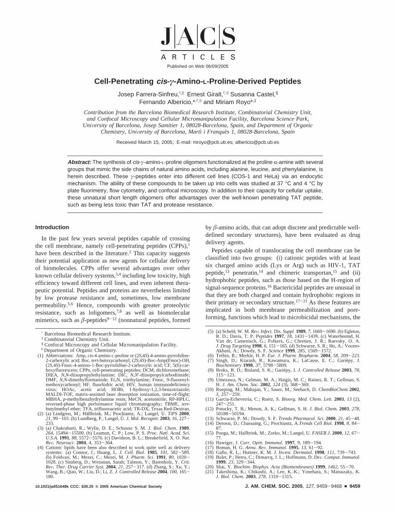
It has been reported that proline-rich peptides22-24 and prolinedendrimers25 can be internalized by eukariotic cells. The mostimportant advantage of proline-rich peptides in biologicalsystems is their solubility in water. In this context, our grouphas described a nonnatural proline-derivedγ-peptide, fromcis-γ-amino-L-proline, with the ability to adopt secondary structurein solution.26 Three differentγ-peptide families were synthe-sized, NR-acyl-γ-peptides (polyamides on the side chains),NR-alkyl-γ-peptides (polyamines on the side chains), andNR-guanidilated-γ-peptides. In the present work, some of thesepeptide structures were selected to study their cell uptakeproperties (see Figure 1).
The amphipathic character of a compound determines itspotential for internalization24,27and, in the case of the peptidesat hand, varied with side chain structure. The modulation ofthis side chain structure offers a wide range of combinationsthat open the possibility of obtaining compounds with differenthydrophobic/hydrophilic character and, consequently, withdifferent properties as carriers. Hence acyl-peptides are morehydrophobic while other families are more hydrophilic oramphipathic. Additionally, the stability imparted by theγ-pep-tide skeleton of these compounds circumvents problems associ-ated with protease liability, a major limitation of CPPs.28 Cellularuptake studies for a new family ofγ-peptides, utilizing platefluorimetry and flow cytometry quantification techniques,confocal microscopy to determine subcellular localization, andthe MTT assay to establish cytotoxicity are discussed herein.
Results and Discussion
Peptide Synthesis.Variousγ-peptides based oncis-γ-amino-L-proline (see Figure 1) were synthesized to assay their cellularuptake properties. These oligomers have a common backboneand distinct side chains introduced with different linkage typesthrough theR-amino group of the proline monomer. Based onthe linkage type, three different peptide families were obtainedand evaluated:NR-acyl-γ-peptides,NR-alkyl-γ-peptides, andNR-guanidilated-γ-peptides. The same Fmoc/Boc combinedsolid-phase strategy described in a previous work was used forthe peptide synthesis.26 Use of these two orthogonal protectinggroups on solid phase for both amino functionalities of thecis-γ-amino-L-proline leads to a flexible strategy enabling thesynthesis of a large number of diversely modifiedγ-peptides.
Theγ-peptide backbone was prepared from protected aminoacid (2S,4S)-Fmoc-4-amino-1-Boc-pyrrolidine-2-carboxylic acid[(2S,4S)-Boc-Amp(Fmoc)-OH] using DIC and HOBt as cou-pling agents. ForNR-acyl-γ-peptides, the side chains were
introduced using the corresponding carboxylic acid and the samecoupling reagents described above.NR-Alkyl- γ-peptides wereobtained via reductive amination using the correspondingaldehyde in 1% HOAc in DMF for 30 min followed by additionof NaBH3CN dissolved in MeOH. Substitution of DMF withMeOH as described previously yielded cleaner crude oligomersin higher yield, as determined by HPLC (70% for peptide3,66% for peptide4, and 67% for peptide4a). For peptide5, theNR-guanidylated-γ-peptide, the guanidinium group was intro-duced usingN,N′-di-Boc-N”-trifluoromethanesulfonyl guan-idine29 in the presence of Et3N in DCM for 4 days. All reactionsof the proline secondary amine were monitored by the chloraniltest.
Taking into account that peptide4 was not totally soluble inphysiological media at high concentrations, peptide4a, contain-ing two consecutive short polyethylenglycol chains (8-amino-3,6- dioxaoctanoic acid, Adoa) at itsC-terminal, was synthesized
(22) Sadler, K.; Eom, K. D.; Yang, J.-L.; Dimitrova, Y.; Tam, J. P.Biochemistry2002, 41, 14150-14157.
(23) Ferna´ndez-Carneado, J.; Kogan, M. J.; Castel, S.; Pujals, S.; Giralt, E.Angew. Chem., Int. Ed.2004, 43, 1811-1814.
(24) Ferna´ndez-Carneado, J.; Kogan, M. J.; Pujals, S.; Giralt, E.Biopolymers2004, 76, 196-203.
(25) Crespo, L.; Sanclimens, G.; Montaner, B.; Pe´rez-Tomas, R.; Royo, M.;Pons, M.; Albericio, F.; Giralt, E.J. Am. Chem. Soc.2002, 124, 8876-8883.
(26) Farrera-Sinfreu, J.; Zaccaro, L.; Vidal, D.; Salvatella, X.; Giralt, E.; Pons,M.; Albericio, F.; Royo, M.J. Am. Chem. Soc.2004, 126, 6048-6057.
(27) Deshayes, S.; Ple´nat, T.; Aldrian-Herrada, G.; Divita, G.; LeGrimellec, C.;Heitz, F.Biochemistry2004, 43, 7698-7706.
(28) (a) Elmquist, A.; Langel, U.Biol. Chem.2003, 384, 387-393. (b) Lindgren,M. E.; Hallbrink, M. M.; Elmquist, A.; Langel, U.Biochem. J.2004, 377,69-76.
(29) Feichtinger, K.; Zapf, C.; Sings, H. L.; Goodman, M.J. Org. Chem.1998,63, 3804-3805.
Figure 1. γ-Aminoproline monomer basedγ-peptides labeled with5(6)-carboxyfluorescein.
A R T I C L E S Farrera-Sinfreu et al.
9460 J. AM. CHEM. SOC. 9 VOL. 127, NO. 26, 2005
as a more soluble analogue.30 Peptide4a was obtained bycoupling two molecules of Fmoc-8-amine-3,6-dioxaoctanoicacid to MBHA resin using DIC/HOBt as coupling reagents,followed by the synthesis described above for peptide4.
At the end of the synthesis, after Fmoc removal, thefluorescent label 5(6)-carboxyfluorescein (CF) was introducedonto theN-terminalγ-amino group using DIC/HOBt as couplingreagents, followed by piperidine washes just before cleavageof the peptide from the resin. These washes were required inorder to remove overincorporated carboxyfluorescein.31 Whenthese washings were not carried out, compounds containing twoand three extra units of carboxyfluorescein were observed inthe crude product.
Peptides were ultimately cleaved from the resin by acidolytictreatment with anhydrous HF. The purity of the CF-γ-peptidecrudes, as determined by HPLC, ranged from 65% to 90%.Compounds were purified to more than 95% homogeneity bypreparative reverse-phase HPLC and characterized by electro-spray and/or MALDI-TOF mass spectrometry.
Enzymatic Stability. Enzymatic stability was studied forpeptide4a in trypsin and human serum and checked by HPLC,which showed total stability of the peptide, even during longincubation times (see Supporting Information). This resultrepresents a major advantage of peptide4a over CPPs, whichare labile to both trypsin and human serum and hence greatlylimited for physiological applications. This precludes the useof trypsin for the removal of cell-surface-bound peptides beforecytometry analysis (see below).
Cellular Uptake of γ-Peptides. The general idea thathydrophilic macromolecules penetrated cells by a classicalendocytotic mechanism is extended.32,33 These molecules aresegregated into different endosomal compartments by the cellfor their recycling or destruction, a fact that can hinder liberationof cargo in the cytoplasm.34 In contrast, initial reports suggestedthat CPPs penetrate cells by an energy-independent route, andstudies on the cellular entry of pAntp, Tat-derived peptides andVP22 reported that a nonclassical receptor, transporter, orendocytosis-mediated mechanism seemed to be involved.35-38
These findings would suggest that the CPPs translocate mem-branes via an energy-independent mechanism. Nevertheless,recent reevaluations of the cellular translocation of some CPPsand cargo-conjugate CPPs suggest that endocytosis is indeedthe primary internalization mechanism.39,40 Thus, the exactnature of the mechanism of CPP across the cellular membraneremains unknown, as controversies exist among different studies
published in the literature.12,41,42Despite some common featuresof CPPs, particularly their cationic nature, their structuraldiversity has fuelled the idea that the mechanisms that allowthem to cross the membrane are not the same for CPPs ofdifferent types.43
A preliminary evaluation of the cellular uptake of the differentγ-peptides was carried out in COS-1 and HeLa cells using platefluorimetry. Cells grown on 96-well plates were incubated witha range of 0.01 to 25µM of CF-peptide at 37°C or 4°C. After2 h of incubation, CF-peptide containing medium was discardedand fluorescence was measured as described in the methodssection. As shown in Figure 2A and B, peptide4awas the mostefficient at crossing the cell membranes of COS-1 and HeLacells at 37°C. The fluorescence levels of cells incubated withpeptide4a were significant in the case of HeLa cells and werehigher for COS-1 cells. In addition, peptides1, 2, 3, and5 werealso taken up by both cell types, but the cells incubated withthese CF-peptides flouresced 6 to 10 times less than thoseincubated with peptide4a. Similar results were obtained whenthe uptake experiments were performed for 8 h (see SupportingInformation). To determine if the uptake mechanism of thesepeptides was energy-dependent, the same experiments werecarried out at 4°C (Figure 2C). Under these conditions, onlypeptide4a and, to a lesser degree, peptide5 were taken up ata significant rate by HeLa cells. This result seemed to indicatethat peptides4a and 5 entered into the cells via an energy-independent mechanism, although the possibility of the peptidebinding to the plastic surface or to the cell membrane was nottotally excluded. Evidence for these phenomena was observedin control wells (wells devoid of cells) and has been describedin other studies.44 Similar results to those obtained with peptide4a were previously obtained with peptide4 (see SupportingInformation). Having completed the aforementioned experi-ments, flow cytometry was then used to differentiate peptideuptake from cell surface and/or plastic surface binding.
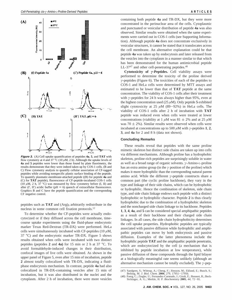
To get a more precise view of the behavior of theγ-peptidederivatives, new cell uptake experiments in COS-1 cells usingflow cytometry were performed. In our opinion flow cytometryprovides more reliable quantitative data than plate fluorimetry.The main source of imprecision in plate fluorimetry whenmeasuring the cell uptake of highly hydrophobic compoundssuch as peptide4a is an artifactual increase in the apparentuptake caused by nonspecific binding of the ligand to the plasticsurface of the plate.44 Although the preliminary flow cytometryexperiments were carried out using the entire set ofγ-peptides,focus was ultimately shifted on the two most promisingcompounds,4a and5, employing the well-known cell-penetrat-ing peptide TAT 49-57 (CF-RKKRRQRRR-NH2) as a positivecontrol. Flow cytometry analysis can quantify cellular associa-tion of CF-tagged peptides excluding any unspecific bindingof the peptides to the plastic surface of the plate. Although theuptake levels of peptides4a and5 were lower than those foundusing plate fluorimetry, suggesting that they had been partiallybound to the plate plastic, Figure 3A illustrates that thecompounds were indeed internalized by COS-1 cells. Peptide4a was taken up very efficiently by COS-1 cells and even
(30) While PEG increased peptide solubility notably, the peptide precipitates atconcentrations higher than 500µM.
(31) Fischer, R.; Mader, O.; Jung, G.; Brock, R.Bioconjugate Chem.2003, 14,653-660.
(32) Mhukerjee, S.; Ghosh, R. N.; Maxfield, F. R.Phys. ReV. 1997, 77, 759-803.
(33) Fischer, R.; Ko¨hler, K.; Fotin-Mleczek, M.; Brock, R.J. Biol. Chem.2004,279, 12625-12635.
(34) Maiolo, J. R., III.; Ottinger, E. A.; Ferrer, M.J. Am. Chem. Soc.2004,126, 15376-15377.
(35) Derossi, D.; Joliot, A. H.; Chassaing, G.; Prochiantz, A.J. Biol. Chem.1994, 269, 10444-10450.
(36) Vives, E.; Brodin, P.; Lebleu, B.J. Biol. Chem. 1997, 272, 16010-16017.(37) Derossi, D.; Calvet, S.; Trembleau, A.; Brunissen, A.; Chassaing, G.;
Prochiantz, A.J. Biol. Chem. 1996, 271, 18188-18193.(38) Elliott, G.; O’Hare, P.Cell 1997, 88, 223-233.(39) Drin, G.; Cottin, S.; Blanc, E.; Rees, A. R.; Temsamani, J.J. Biol. Chem.
2003, 278, 31192-31201.(40) Richard, J. P.; Melikov, K.; Vives, E.; Ramos, C.; Verbeure, B.; Gait, M.
J.; Chernomordik, L. V.; Lebleu, B.J. Biol. Chem.2003, 278, 585-590.
(41) Thoren, E. G.; Persson, D.; Isakson, P.; Gokso¨r, M.; Onfelt, A.; Norden,B. Biochem. Biophys. Res. Commun.2003, 307, 100-107.
(42) Vives, E.J. Mol. Recognit.2003, 16, 265-271.(43) Zorko, M.; Langel, U¨ . AdV. Drug DeliVery ReV. 2005, 57, 529-545.(44) Chico, D. E.; Given, R. L.; Miller, B. T.Peptides2003, 24, 3-9.
Cell-Penetrating cis-γ-Amino- L-Proline-Derived Peptides A R T I C L E S
J. AM. CHEM. SOC. 9 VOL. 127, NO. 26, 2005 9461
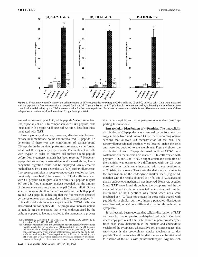
seemed to be taken up at 4°C, while peptide5 was internalizedless, especially at 4°C. In comparison withTAT peptide, cellsincubated with peptide4a flouresced 3.5 times less than thoseincubated withTAT .
Flow cytometry does not, however, discriminate betweenextracellular membrane-bound and internalized CF-peptide. Todetermine if there was any contribution of surface-boundCF-peptides in the peptide uptake measurements, we performedadditional flow cytometry experiments. The treatment of cellswith trypsin in order to remove cell-surface-bound peptidebefore flow cytometry analysis has been reported.40 However,γ-peptides are not trypsin-sensitive as discussed above; henceenzymatic digestion could not be employed. An alternativemethod based on the pH-dependence of 5(6)-carboxyfluoresceinfluorescence emission in receptor-endocytosis studies has beenpreviously described.45 As shown for COS-1 cells incubatedwith CF-peptide4a (Figure 3B) or withTAT peptide (Figure3C) for 2 h, flow cytometry analysis revealed that the amountof fluorescence was very similar at pH 7.4 and pH 6. Only asmall decrease of the fluorescence was observed in both peptide4a andTAT peptide, indicating that the fluorescence detectedby the cytometer was mainly due to internalized peptides.46
A cell uptake time-course experiment in COS-1 cells wasalso carried out for peptide4a. The progressive increase uptakeof peptide4a demonstrated that it was endocytocized by thecells, as opposed to having attached to the membrane, a process
that occurs rapidly and is temperature-independent (see Sup-porting Information).
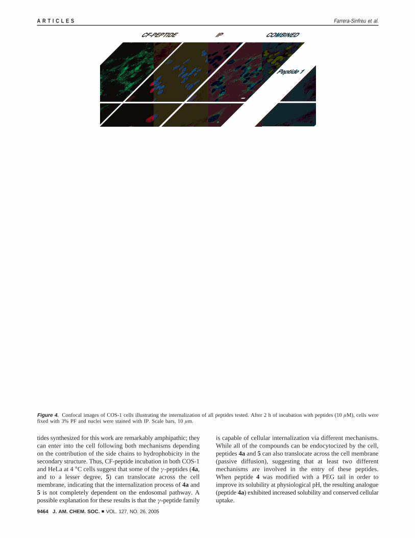
Intracellular Distribution of γ-Peptides.The intracellulardistribution of CF-peptides was examined by confocal micros-copy in both fixed and unfixed COS-1 cells recording opticalsections that allowed 3D reconstruction of the cell. Thecarboxyflouresceinated peptides were located inside the cellsand were not attached to the membrane. Figure 4 shows thedistribution of each CF-peptide tested in fixed COS-1 cellscostained with the nucleic acid marker PI. In cells treated withpeptides1, 2, and3 at 37°C, a slight vesicular distribution ofthe peptides was observed. No differences with the CF wereobserved when cells were incubated with these peptides at4 °C (data not shown). This vesicular distribution, similar tothe localization of the endocytotic marker used (Figure 5),together with the results obtained at 37°C and 4°C, suggestedthat an endocytotic mechanism was involved. However, peptides5 and TAT were found throughout the cytoplasm and in thenuclei of the cells with no punctuated pattern observed. Similardistribution of both peptides was found when cells wereincubated at 4°C (data not shown). In those cells treated withpeptide4a, a similar but more intense punctated distributionwas observed, as well as a diffuse distribution throughout thecytoplasm.
It has recently been reported that cellular distribution ofTATcan vary for live or paraformaldehyde-fixed cells.12 Confocalmicroscopy pictures ofTAT internalized in paraformaldehyde-fixed cells show distribution in the nucleus and endocytoticvesicles of the cytoplasm, whereas live-cell pictures suggest thatendocytosis is the predominant uptake mechanism of thispeptide. The difference in cellular distribution can be attributedto fixation of the cells with paraformaldehyde. Arginine-rich
(45) Chambers, J. D.; Simon, S. I.; Berger, E. M.; Sklar, L. A.; Arfors, K. E.J. Leukoc. Biol.1993, 53, 462-469.
(46) A minor contribution from any unquenched residual carboxyflouresceinatedpeptide attached to the membrane at pH 6 could still exist (at pH 6 aroundthe 80% of the carboxyfluorescein fluorescence is quenched), and as aconsequence, only a small amount of fluorescence could be due to the cell-surface-bound peptides. These experiments could not be carried out at apH lower than 5-5.5 (where carboxyfluorescein is completely quenched)because of the rapid cell death observed under our experimental conditions.
Figure 2. Fluorimetry quantification of the cellular uptake of different peptides tested (A) in COS-1 cells and (B and C) in HeLa cells. Cells were incubatedwith the peptide at a final concentration of 10µM for 2 h at 37°C (A and B) and at 4°C (C). Results were normalized by subtracting the autofluorescencecontrol value and dividing by the CF-fluorescence value for the same experiment. Error bars represent standard deviation (SD) from the mean value of threeindependent experiments of each condition.*, significantp < 0.05.
A R T I C L E S Farrera-Sinfreu et al.
9462 J. AM. CHEM. SOC. 9 VOL. 127, NO. 26, 2005
peptides such asTAT and (Arg)9 arbitrarily redistribute in thenucleus in some common cell fixation protocols.12
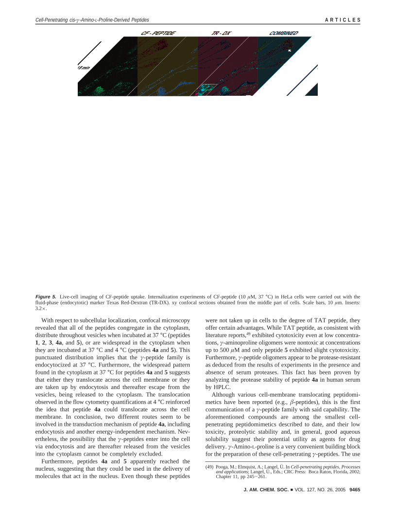
To determine whether the CF-peptides were actually endo-cytocized or if they diffused across the cell membrane, time-course uptake experiments using the fluid-phase endocytoticmarker Texas Red-Dextran (TR-DX) were performed. HeLacells were simultaneously incubated with CF-peptides (10µM,37 °C) and the endocytotic marker TR-DX. Figure 5 showsresults obtained when cells were incubated with two distinctpeptides (peptides2 and 4a) for 15 min or 2 h at 37°C. Toavoid formaldehyde-induced changes in their distribution,confocal images of live cells were obtained. As shown in theupper panel of Figure 5, even after 15 min of incubation, peptide2 almost totally colocalized with TR-DX, indicating a fluid-phase endocytosis mechanism. In contrast, peptide4a had alsocolocalized in TR-DX-containing vesicles after 15 min ofincubation, but it was also distributed in the nuclei and thecytoplasm. After 2 h of incubation, there were more vesicles
containing both peptide4a and TR-DX, but they were moreconcentrated in the perinuclear area of the cells. Cytoplasmicand punctuated or vesicular distribution of peptide4a was alsoobserved. Similar results were obtained when the same experi-ments were carried out in COS-1 cells (see Supporting Informa-tion). Although peptide4a does not concentrate exclusively invesicular structures, it cannot be stated that it translocates acrossthe cell membrane. An alternative explanation could be thatpeptide4awas taken up by endocytosis and later released fromthe vesicles into the cytoplasm in a manner similar to that whichhas been demonstrated for the human antimicrobial peptideLL-3747 and other cell-penetrating peptides.48
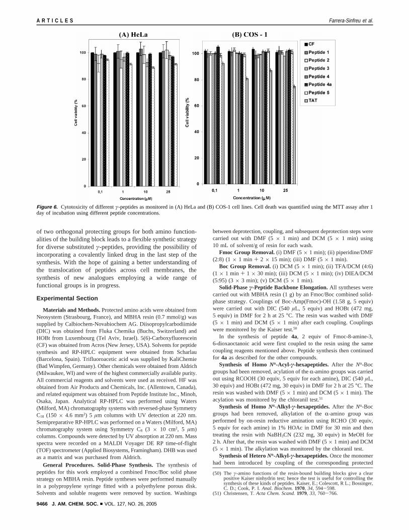
Cytotoxicity of γ-Peptides. Cell viability assays wereperformed to determine the toxicity of the proline derivedγ-peptides (Figure 6). The toxicities of each of the peptides toCOS-1 and HeLa cells were determined by MTT assays andestimated to be lower than that ofTAT peptide at the sameconcentration. The viability of COS-1 cells after their treatmentwith γ-peptides for 24 h was always higher than 95%, even atthe highest concentration used (25µM). Only peptide5 exhibitedslight cytotoxicity at 25µM (89-92%) in HeLa cells. Theviability of COS-1 cells after 2 h of incubation with TATpeptide was reduced even when cells were treated at lowerconcentrations (viability at 1µM was 81( 2% and at 25µMwas 70( 2%). Similar results were observed when cells wereincubated at concentrations up to 500µM with γ-peptides1, 2,3, and4a for 2 and 8 h (data not shown).
Concluding Remarks
These results reveal that peptides with the same prolinemimetic skeleton but distinct side chains are taken up into cellsvia different mechanisms. Although proline has a hydrophobicskeleton, proline-rich peptides are surprisingly soluble in wateras well as a broad range of organic solvents.γ-Amino-L-prolinehas an extra amino group (in theγ position of the proline) whichmakes it more hydrophilic than the corresponding natural parentamino acid. While the differentγ-peptide constructs share acommon part (the cyclic proline backbone), they vary in thetype and linkage of their side chains, which can be hydrophobicor hydrophilic. Hence the combination of skeleton, side chaintype, and side chain linkage endows each peptide with a distincthydrophobic or hydrophilic character. Peptide2 is thus clearlyhydrophobic due to the combination of a hydrophobic skeletonand the noncharged side chain linkage to its backbone. Peptides1, 3, 4, 4a, and5 can be considered special amphipathic peptidesas a result of their backbone and their charged side chainlinkages. In all cases, the side chain hydrophobicity determinesthe cell uptake properties. Hydrophobic peptides are typicallyassociated with passive diffusion while hydrophilic and amphi-pathic peptides can move by both endocytosis and passivediffusion. Examples of the latter phenomena include thehydrophilic peptideTAT and the amphipathic peptide penetratin,which are endocytocized by the cell (a mechanism that isinhibited by peptide incubation at low temperature), whilepassive diffusion of these compounds through the lipid bilayerat a biologically meaningful rate seems unlikely (although analternative mechanism cannot be totally excluded). Theγ-pep-
(47) Sandgren, S.; Wittrup, A.; Cheng, F.; Jo¨nsson, M.; Eklund, E.; Busch, S.;Belting, M. J. Biol. Chem.2004, 279, 17951-17956.
(48) Foerg, C.; Ziegler, U.; Fernandez-Carneado, J.; Giralt, E.; Rennert, R.; Beck-Sickinger, A. G.; Merkle, H. P.Biochemistry2005, 44, 72-81.
Figure 3. (A) Cell uptake quantification of peptides4a, 5, andTAT withflow cytometry at 4 and 37°C (10µM, 2 h). Although the uptake levels of4a and 5 peptides were lower than those found by plate fluorimetry, theresults demonstrate that they were indeed taken up by COS-1 cells. (B andC) Flow cytometry analysis to quantify cellular association of CF-taggedpeptides while avoiding nonspecific plastic surface binding of the peptide.To quantify plasmatic-membrane-attached peptide ((B) for peptide4a and(C) for TAT peptide), fluorescence of CF-peptide-incubated COS-1 cells(10 µM, 2 h, 37 °C) was measured by flow cytometry before (I, II) andafter (I′, II ′) acidic buffer (pH) 6) quench of extracellular fluorescence.Graphics B and C have the peptide quantification and the correspondingCF negative control.
Cell-Penetrating cis-γ-Amino-L-Proline-Derived Peptides A R T I C L E S
J. AM. CHEM. SOC. 9 VOL. 127, NO. 26, 2005 9463
tides synthesized for this work are remarkably amphipathic; theycan enter into the cell following both mechanisms dependingon the contribution of the side chains to hydrophobicity in thesecondary structure. Thus, CF-peptide incubation in both COS-1and HeLa at 4°C cells suggest that some of theγ-peptides (4a,and to a lesser degree,5) can translocate across the cellmembrane, indicating that the internalization process of4a and5 is not completely dependent on the endosomal pathway. Apossible explanation for these results is that theγ-peptide family
is capable of cellular internalization via different mechanisms.While all of the compounds can be endocytocized by the cell,peptides4aand5 can also translocate across the cell membrane(passive diffusion), suggesting that at least two differentmechanisms are involved in the entry of these peptides.When peptide4 was modified with a PEG tail in order toimprove its solubility at physiological pH, the resulting analogue(peptide4a) exhibited increased solubility and conserved cellularuptake.
Figure 4. Confocal images of COS-1 cells illustrating the internalization of all peptides tested. After 2 h of incubation with peptides (10µM), cells werefixed with 3% PF and nuclei were stained with IP. Scale bars, 10µm.
A R T I C L E S Farrera-Sinfreu et al.
9464 J. AM. CHEM. SOC. 9 VOL. 127, NO. 26, 2005
With respect to subcellular localization, confocal microscopyrevealed that all of the peptides congregate in the cytoplasm,distribute throughout vesicles when incubated at 37°C (peptides1, 2, 3, 4a, and5), or are widespread in the cytoplasm whenthey are incubated at 37°C and 4°C (peptides4a and5). Thispunctuated distribution implies that theγ-peptide family isendocytocized at 37°C. Furthermore, the widespread patternfound in the cytoplasm at 37°C for peptides4a and5 suggeststhat either they translocate across the cell membrane or theyare taken up by endocytosis and thereafter escape from thevesicles, being released to the cytoplasm. The translocationobserved in the flow cytometry quantifications at 4°C reinforcedthe idea that peptide4a could translocate across the cellmembrane. In conclusion, two different routes seem to beinvolved in the transduction mechanism of peptide4a, includingendocytosis and another energy-independent mechanism. Nev-ertheless, the possibility that theγ-peptides enter into the cellvia endocytosis and are thereafter released from the vesiclesinto the cytoplasm cannot be completely excluded.
Furthermore, peptides4a and 5 apparently reached thenucleus, suggesting that they could be used in the delivery ofmolecules that act in the nucleus. Even though these peptides
were not taken up in cells to the degree of TAT peptide, theyoffer certain advantages. While TAT peptide, as consistent withliterature reports,49 exhibited cytotoxicity even at low concentra-tions,γ-aminoproline oligomers were nontoxic at concentrationsup to 500µM and only peptide5 exhibited slight cytotoxicity.Furthermore,γ-peptide oligomers appear to be protease-resistantas deduced from the results of experiments in the presence andabsence of serum proteases. This fact has been proven byanalyzing the protease stability of peptide4a in human serumby HPLC.
Although various cell-membrane translocating peptidomi-metics have been reported (e.g.,â-peptides), this is the firstcommunication of aγ-peptide family with said capability. Theaforementioned compounds are among the smallest cell-penetrating peptidomimetics described to date, and their lowtoxicity, proteolytic stability and, in general, good aqueoussolubility suggest their potential utility as agents for drugdelivery.γ-Amino-L-proline is a very convenient building blockfor the preparation of these cell-penetratingγ-peptides. The use
(49) Pooga, M.; Elmquist, A.; Langel, U¨ . In Cell-penetrating peptides, Processesand applications; Langel, U., Eds.; CRC Press: Boca Raton, Florida, 2002;Chapter 11, pp 245-261.
Figure 5. Live-cell imaging of CF-peptide uptake. Internalization experiments of CF-peptide (10µM, 37 °C) in HeLa cells were carried out with thefluid-phase (endocytotic) marker Texas Red-Dextran (TR-DX). xy confocal sections obtained from the middle part of cells. Scale bars, 10µm. Inserts:3.2×.
Cell-Penetrating cis-γ-Amino-L-Proline-Derived Peptides A R T I C L E S
J. AM. CHEM. SOC. 9 VOL. 127, NO. 26, 2005 9465
of two orthogonal protecting groups for both amino function-alities of the building block leads to a flexible synthetic strategyfor diverse substitutedγ-peptides, providing the possibility ofincorporating a covalently linked drug in the last step of thesynthesis. With the hope of gaining a better understanding ofthe translocation of peptides across cell membranes, thesynthesis of new analogues employing a wide range offunctional groups is in progress.
Experimental Section
Materials and Methods.Protected amino acids were obtained fromNeosystem (Strasbourg, France), and MBHA resin (0.7 mmol/g) wassupplied by Calbiochem-Novabiochem AG. Diisopropylcarbodiimide(DIC) was obtained from Fluka Chemika (Buchs, Switzerland) andHOBt from Luxembourg (Tel Aviv, Israel). 5(6)-Carboxyfluorescein(CF) was obtained from Acros (New Jersey, USA). Solvents for peptidesynthesis and RP-HPLC equipment were obtained from Scharlau(Barcelona, Spain). Trifluoroacetic acid was supplied by KaliChemie(Bad Wimpfen, Germany). Other chemicals were obtained from Aldrich(Milwaukee, WI) and were of the highest commercially available purity.All commercial reagents and solvents were used as received. HF wasobtained from Air Products and Chemicals, Inc. (Allentown, Canada),and related equipment was obtained from Peptide Institute Inc., Minoh,Osaka, Japan. Analytical RP-HPLC was performed using Waters(Milford, MA) chromatography systems with reversed-phase SymmetryC18 (150 × 4.6 mm2) 5 µm columns with UV detection at 220 nm.Semipreparative RP-HPLC was performed on a Waters (Milford, MA)chromatography system using Symmetry C8 (3 × 10 cm2, 5 µm)columns. Compounds were detected by UV absorption at 220 nm. Massspectra were recorded on a MALDI Voyager DE RP time-of-flight(TOF) spectrometer (Applied Biosystems, Framingham). DHB was usedas a matrix and was purchased from Aldrich.
General Procedures. Solid-Phase Synthesis.The synthesis ofpeptides for this work employed a combined Fmoc/Boc solid phasestrategy on MBHA resin. Peptide syntheses were performed manuallyin a polypropylene syringe fitted with a polyethylene porous disk.Solvents and soluble reagents were removed by suction. Washings
between deprotection, coupling, and subsequent deprotection steps werecarried out with DMF (5× 1 min) and DCM (5× 1 min) using10 mL of solvent/g of resin for each wash.
Fmoc Group Removal.(i) DMF (5 × 1 min); (ii) piperidine/DMF(2:8) (1 × 1 min + 2 × 15 min); (iii) DMF (5 × 1 min).
Boc Group Removal.(i) DCM (5 × 1 min); (ii) TFA/DCM (4:6)(1 × 1 min + 1 × 30 min); (iii) DCM (5 × 1 min); (iv) DIEA/DCM(5:95) (3× 3 min); (v) DCM (5 × 1 min).
Solid-Phaseγ-Peptide Backbone Elongation.All syntheses werecarried out with MBHA resin (1 g) by an Fmoc/Boc combined solid-phase strategy. Couplings of Boc-Amp(Fmoc)-OH (1.58 g, 5 equiv)were carried out with DIC (540µL, 5 equiv) and HOBt (472 mg,5 equiv) in DMF for 2 h at 25°C. The resin was washed with DMF(5 × 1 min) and DCM (5× 1 min) after each coupling. Couplingswere monitored by the Kaiser test.50
In the synthesis of peptide4a, 2 equiv of Fmoc-8-amine-3,6-dioxaoctanoic acid were first coupled to the resin using the samecoupling reagents mentioned above. Peptide synthesis then continuedfor 4a as described for the other compounds.
Synthesis of HomoNr-Acyl-γ-hexapeptides.After the NR-Bocgroups had been removed, acylation of theR-amino groups was carriedout using RCOOH (30 equiv, 5 equiv for each amine), DIC (540µL,30 equiv) and HOBt (472 mg, 30 equiv) in DMF for 2 h at 25°C. Theresin was washed with DMF (5× 1 min) and DCM (5× 1 min). Theacylation was monitored by the chloranil test.51
Synthesis of HomoNr-Alkyl- γ-hexapeptides.After the NR-Bocgroups had been removed, alkylation of theR-amino group wasperformed by on-resin reductive amination using RCHO (30 equiv,5 equiv for each amine) in 1% HOAc in DMF for 30 min and thentreating the resin with NaBH3CN (232 mg, 30 equiv) in MeOH for2 h. After that, the resin was washed with DMF (5× 1 min) and DCM(5 × 1 min). The alkylation was monitored by the chloranil test.
Synthesis of HeteroNr-Alkyl- γ-hexapeptides.Once the monomerhad been introduced by coupling of the corresponding protected
(50) The γ-amino functions of the resin-bound building blocks give a clearpositive Kaiser ninhydrin test; hence the test is useful for controlling thesynthesis of these kinds of peptides. Kaiser, E.; Colescott, R L.; Bossinger,C. D.; Cook, P. I. Anal. Biochem.1970, 34, 594-598.
(51) Christensen, T.Acta Chem. Scand.1979, 33, 760-766.
Figure 6. Cytotoxicity of differentγ-peptides as monitored in (A) HeLa and (B) COS-1 cell lines. Cell death was quantified using the MTT assay after 1day of incubation using different peptide concentrations.
A R T I C L E S Farrera-Sinfreu et al.
9466 J. AM. CHEM. SOC. 9 VOL. 127, NO. 26, 2005
monomer, theNR-Boc protecting group was removed and the alkylationwas performed as above. After removal of theNγ-Fmoc protectinggroup, the reactive sequence was repeated.
Synthesis ofNr-Guanidyl-γ-hexapeptide.After theNR-Boc groupshad been removed, peptide5 was guanidilated usingN,N′-di-Boc-N”-trifluoromethanesulfonyl guanidine52 (5 equiv) and Et3N (5 equiv) inDCM for 4 days at room temperature. After the reaction, the resin waswashed with DCM (5× 1 min). The guanidilation was monitored bythe chloranil test.
5(6)-CarboxyfluoresceinNγ-Terminal Labeling. CF was coupledto theNγ-terminal group using DIC/HOBt (5 equiv/5 equiv) for 2 h.To avoid over incorporation of CF, two 30-min treatments with 20%piperidine-DMF were carried out before the cleavage of the peptidefrom the resin.31
Acidolytic Cleavage with HF. The peptide resin was washed withMeOH (3× 1 min), dried, and treated with HF in the presence of 10%anisole for 1 h at 0°C. Peptides were precipitated with cold anhydrousMTBE, dissolved in HOAc, and then lyophilized.
CF-(γAmp)6-NH2 (1): The crude peptide was purified by preparativeHPLC using a linear gradient (from 0 to 20% of MeCN in 30 min) ofMeCN (containing 1% of TFA) and H2O (containing 1% of TFA). Thepurity of each fraction was verified by analytical HPLC and MALDI-TOF and showed that the peptides were 99% pure. MS calcd forC51H61N13O12 [M + H]+: 1048.11. MALDI-TOF found: 1048.38 [M+ H]+, 1070.45 [M+ Na]+, and 1086.37 [M+ K] +.
CF-[γAmp(Nr-Ac)]6-NH2 (2): The crude peptide was purified bypreparative HPLC using a linear gradient (from 20 to 80% of MeCNin 30 min) of MeCN (containing 1% of TFA) and H2O (containing1% of TFA). The purity of each fraction was verified by analyticalHPLC and MALDI-TOF and showed that the peptides were 96% pure.MS calcd for C63H73N13O18 [M + H]+: 1300.33. MALDI-TOF found:1301.22 [M+ H]+, 1322.23 [M+ Na]+, and 1338.21 [M+ K] +.
CF-[γAmp(Nr-Me)]6-NH2 (3): The crude peptide was purified bypreparative HPLC using a linear gradient (from 0 to 20% of MeCN in30 min) of MeCN (containing 1% of TFA) and H2O (containing 1%of TFA). The purity of each fraction was verified by analytical HPLCand MALDI-TOF and showed that the peptides were 95% pure. MScalcd for C57H73N12O12 [M + H]+: 1132.26. MALDI-TOF found:1131.60 [M+ H]+ and 1153.63 [M+ Na]+.
CF-{γAmp[Nr-CH2CH2CH(CH 3)2]-γAmp(Nr-CH2CH2Ph)}3-NH2 (4): The crude peptide was purified by preparative HPLC usinga linear gradient (from 0 to 50% of MeCN in 30 min) of MeCN(containing 1% of TFA) and H2O (containing 1% of TFA). The purityof each fraction was verified by analytical HPLC and MALDI-TOFand showed that the peptides were 99% pure. MS calcd for C90H115N13O12
[M + H]+: 1570.95. MALDI-TOF found: 1570.46 [M+ H]+ and1592.91 [M+ Na]+.
CF-{γAmp[Nr-CH2CH2CH(CH3)2]-γAmp(Nr-CH2CH2Ph)}3-PEG-NH2 (4a): The crude peptide was purified by preparative HPLC usinga linear gradient (from 0 to 40% of MeCN in 30 min) of MeCN(containing 1% of TFA) and H2O (containing 1% of TFA). The purityof each fraction was verified by analytical HPLC and MALDI-TOFand showed that the peptides were 99% pure. MS calcd forC112H137N15O18 [M + H]+: 1861.26. MALDI-TOF found: 1863.18[M + H]+ and 1885.15 [M+ Na]+.
CF-[γAmp(Nr-C(NH)(NH2))]6-NH2 (5): The crude peptide waspurified by preparative HPLC using a linear gradient (from 0 to 20%of MeCN in 30 min) of MeCN (containing 1% of TFA) and H2O(containing 1% of TFA). The purity of each fraction was verified byanalytical HPLC and MALDI-TOF and showed that the peptideswere 99% pure. MS calcd for C57H73N25O12 [M + H]+: 1300.35.MALDI-TOF found: 1302.01 [M+ H]+ and 1324.36 [M+ Na]+.
Cell Culture and Peptide Treatments. HeLa and COS-1 cellswere maintained in DMEM (1000 mg/mL glucose for HeLa and4500 mg/mL for COS-1) culture medium (Biological Industries)containing 10% fetal calf serum (FCS), 2 mM glutamine, 50 u/mLpenicillin, and 0.05 g/mL streptomycin. For all experiments, exponen-tially growing cells were detached from the culture flasks using atrypsin-0.25% EDTA solution, and the cell suspension was seeded ata concentration of 21.4× 10³ cells/cm2 onto plastic dishes, glasscoverslips, 8-well Lab-Teck chambered coverglass or 96-well plates(Nalge Nunc International, Naprville, USA), depending on the experi-ment. Experiments were carried out 24 h later, at a confluence level ofapproximately 60%-70%. The carboxyfluoresceinated compounds weredissolved in PBS and sterilized with 0.22µm filters (Millex-GV, PVDF,Durapore, Millipore). The peptides and 5(6)-carboxyfluorescein stocksolutions were diluted with the cell culture medium. Nonadherent cellswere washed away and attached cells were incubated with the peptidesin DMEM at 37 °C in CO2 atmosphere or in 25 mM Hepes BufferedDMEM at 4° C.
Enzymatic Stability. Enzymatic degradation using trypsin wascarried out by incubation at 37°C of peptide4a with the enzyme in100 mM Tris-HCl at pH 8. The trypsin-peptide ratio was 1:100, using3.43 µL of a solution of trypsin from bovine pancreas E.C. 3.4.21.4(Roche, Basel) in glycerol-H2O (1:1) (6.25 mg/mL). Aliquots (50µL)were periodically taken at 2 h to 120 h, 150µL of 1 N HCl were added,and the resulting solution cooled with ice. Degradation was monitoredby HPLC.
Enzymatic degradation using human serum (Aldrich, Milwaukee)was carried out by incubation at 37°C of peptide4a with the serum(diluted 9:1 in HBSS buffer). The ratio peptide-serum was 9, andpeptide was used at a final concentration of 125µM (added to theserum dissolved). Aliquots (50µL) were periodically taken at 2 h to120 h, poured into 200µL of MeOH to precipitate the proteins, andcooled on ice. After 30 min, the sample was centrifuged and thesupernatant was analyzed by HPLC.
Uptake Measurements by Plate Fluorimetry and Flow Cyto-metry. COS-1 and HeLa cells were seeded onto 96-microwell platesat a concentration of 21.4× 10³ cells/cm2 for 24 h. The culture mediumwas discarded and replaced by new medium containing differentCF-peptides concentrations (0.01µM, 0.05µM, 0.1 µM, 1 µM, 5 µM,10 µM, and 25µM), using TAT peptide as a positive control and CFas a negative control. Cells were then incubated for 2 or 8 h at 4°Cand 37°C. After incubation, cells were washed three times with PBScontaining 1.1 mM CaCl2 and 1.3 mM MgCl2, then lysed by adding200µL of lysis buffer (0.1% Triton X-100 in 50 mM Tris, pH 8.5) toeach well. Internalized peptides were quantified by measuring thefluorescence intensity of the supernatant using a FL600 microplatefluorescence reader (Bio-Tek). Fluorescence was measured atλexcitation
) 485/20 nm andλemission) 530/25 nm. Triplicates of each measurementwere performed, and the fluorescence emitted from the blanks wassubtracted.
To analyze the internalization of CF-peptides by flow cytometry,COS-1 and HeLa cells were seeded onto 35-mm plates at a concentra-tion of 21.4× 10³ cells/cm2. After 24 h, cells were incubated withCF-peptides as previously described for fluorimetry assays. Aftervarious incubation times, cells were washed 3 times with PBS, detachedwith tripsine-EDTA 0.25%, centrifuged at 800× g, and washed again.Finally, they were resuspended in PBS containing 0.1 mM of propidiumiodide (PI). To remove fluorescence of CF or CF-peptides bound tothe plasma membrane, the pH of the PBS/PI solution was brought downto 6 by the addition of 1 N HCl just before measuring fluorescence. AtpH ) 6, extracellular fluorescence of CF is quenched without alteringcell mechanisms.45
Fluorescence analysis was performed with an Epics XL flowcytometer (Coulter). Cells stained with PI were excluded from furtheranalysis. Triplicates of each sample were performed for each condition,and results from independent experiments were normalized by subtrac-
(52) Feichtinger, K.; Zapf, C.; Sings, H. L.; Goodman, M.J. Org. Chem.1998,63, 3804-3805.
Cell-Penetrating cis-γ-Amino-L-Proline-Derived Peptides A R T I C L E S
J. AM. CHEM. SOC. 9 VOL. 127, NO. 26, 2005 9467
tion of the autofluorescence control value from each value and dividingby the fluorescence value obtained from the CF control under the sameexperimental conditions
MTT Cytotoxicity Assay.53 The viability of COS-1 and HeLa cellsin the presence of the peptides was tested using the 3-(4,5-dimethyl-thiazol-2-yl)-2,5-diphenyltetrazolium bromide (MTT) assay. To avoidsaturation in cell growth after 24 h of peptide incubation, 7× 10³ cells/well were seeded on a 96-well plate (Nange Nunc) for each assay.After 24 h, the culture medium was discarded and replaced by a newmedium containing different CF-peptide concentrations. Cells wereincubated for 2 h, 8 h, and 24 h at 37°C under 5% CO2 atmosphere,and MTT (0.5 mg/mL) was added 2 h before the end of incubation.After 2 h of incubation with MTT, the medium was discarded byaspiration and 2-propanol was added to dissolve formazan, a dark bluecolored crystal observed in the wells. Absorbance was measured at570 nm in a spectrophotometric Elx800 Universal microplate reader(Bio-Tek), 30 min after the addition of 2-propanol. Cell viability isexpressed as a percent ratio of cells treated with peptide to untreatedcells, which were used as a control.
Confocal Laser Scanning Microscopy.COS-1 and HeLa cells wereseeded onto glass coverslips at 21.4× 10³ cells/cm2 and, after 24 h,were incubated with CF-peptides as described above. For endocytosisexperiments, Texas Red-Dextran (TR-DX, 3 mg/mL, MW) 10.000,Molecular Probes) was incubated together with CF-peptide. After
CF-peptide incubation, cells were washed 3 times with PBS and fixedin 3% para-formaldehyde-2% sucrose in 0.1 M phosphate buffer (PB)for 15 min, washed 3 times in PBS, and mounted in Mowiol with 2.5%DABCO. PI (1 µg/mL) staining was performed at room temperaturefor 15 min in the presence of RNAsa in PBS (1 mg/mL). As a fixationcontrol, similar experiments were performed in cells plated onto glassbottom Lab-Tek chambers for live-cell imaging. After 2 h of incubation,cells were washed for 3 times with PBS containing 1.1 mM CaCl2,1.3 mM MgCl2, and 25 mM Hepes. Images were subsequently acquiredwithin the next 30 min.
Confocal laser scanning microscopy was performed with an OlympusFluoview 500 confocal microscope using a 60×/1.4 NA plan-apochro-matic objective. The carboxyfluorescein fluorescence was excited withthe 488-nm line of an argon laser, and its emission was detected in arange of 515-530 nm. The microscope settings were maintainedidentical for each peptide and dose. PI and TR-DX were excited at543 nm and detected with a 560-nm long pass filter. To avoid crosstalk,the two-fluorescence scanning was performed in a sequential mode.
Acknowledgment. The work was partially supported by fundsfrom CICYT (BQU2002-02047 and BQU2003-00089), theGeneralitat de Catalunya (Grup Consolidat and Centre deReferencia en Biotecnologia), and the Barcelona Science Park.
Supporting Information Available: HPLC chromatogramsand supplemental confocal microscopy images. This materialis available free of charge via the Internet at http://pubs.acd.org
JA051648K
(53) (a) MTT-cell proliferation assay, Cell Biology: A Laboratory Handbook,2nd ed.; Academic Press, 1998; Vol. 1. (b) Mechanism of cellular3-(4,5-dimethylthiazol-2-yl)-2,5-diphenyltetrazolium bromide (MTT) reduc-tion: Liu, Y.; Peterson, D. A.; Kimura, H.; Schubert, D.J. Neurochem.1997, 69, 581-593.
A R T I C L E S Farrera-Sinfreu et al.
9468 J. AM. CHEM. SOC. 9 VOL. 127, NO. 26, 2005