Embed Size (px)
Citation preview

Adaptation of an L-Proline Adenylation Domain to Use 4-Propyl-L-Proline in the Evolution of LincosamideBiosynthesisStanislav Kadlčík1, Tomáš Kučera1, Dominika Chalupská1¤a, Radek Gažák1, Markéta Koběrská1, DanaUlanová1¤b, Jan Kopecký1¤c, Eva Kutejová1,2, Lucie Najmanová1, Jiří Janata1*
1 Institute of Microbiology, Academy of Sciences of the Czech Republic, Prague, Czech Republic, 2 Department of Biochemistry and Structural Biology, Instituteof Molecular Biology, Slovac Academy of Sciences, Bratislava, Slovakia
Abstract
Clinically used lincosamide antibiotic lincomycin incorporates in its structure 4-propyl-L-proline (PPL), an unusualamino acid, while celesticetin, a less efficient related compound, makes use of proteinogenic L-proline. Biochemicalcharacterization, as well as phylogenetic analysis and homology modelling combined with the molecular dynamicssimulation were employed for complex comparative analysis of the orthologous protein pair LmbC and CcbC from thebiosynthesis of lincomycin and celesticetin, respectively. The analysis proved the compared proteins to be the stand-alone adenylation domains strictly preferring their own natural substrate, PPL or L-proline. The LmbC substratebinding pocket is adapted to accomodate a rare PPL precursor. When compared with L-proline specific ones, severallarge amino acid residues were replaced by smaller ones opening a channel which allowed the alkyl side chain ofPPL to be accommodated. One of the most important differences, that of the residue corresponding to V306 in CcbCchanging to G308 in LmbC, was investigated in vitro and in silico. Moreover, the substrate binding pocketrearrangement also allowed LmbC to effectively adenylate 4-butyl-L-proline and 4-pentyl-L-proline, substrates witheven longer alkyl side chains, producing more potent lincosamides. A shift of LmbC substrate specificity appears tobe an integral part of biosynthetic pathway adaptation to the PPL acquisition. A set of genes presumably coding forthe PPL biosynthesis is present in the lincomycin - but not in the celesticetin cluster; their homologs are found inbiosynthetic clusters of some pyrrolobenzodiazepines (PBD) and hormaomycin. Whereas in the PBD andhormaomycin pathways the arising precursors are condensed to another amino acid moiety, the LmbC protein is thefirst functionally proved part of a unique condensation enzyme connecting PPL to the specialized amino sugarbuilding unit.
Citation: Kadlčík S, Kučera T, Chalupská D, Gažák R, Koběrská M, et al. (2013) Adaptation of an L-Proline Adenylation Domain to Use 4-Propyl-L-Prolinein the Evolution of Lincosamide Biosynthesis. PLoS ONE 8(12): e84902. doi:10.1371/journal.pone.0084902
Editor: Valerie de Crécy-Lagard, University of Florida, United States of America
Received August 30, 2013; Accepted November 27, 2013; Published December 27, 2013
Copyright: © 2013 Kadlčík et al. This is an open-access article distributed under the terms of the Creative Commons Attribution License, which permitsunrestricted use, distribution, and reproduction in any medium, provided the original author and source are credited.
Funding: This work was supported by the Ministry of Education, Youth and Sports of the Czech Republic projects CZ.1.07/2.3.00/20.0055 and CZ.1.07/2.3.00/30.0003 and BIOCEV - Biotechnology and Biomedicine Centre of the Academy of Sciences and Charles University No. CZ.1.05/1.1.00/02.0109, from the European Regional Development Fund in the Czech Republic. The funders had no role in study design, data collection andanalysis, decision to publish, or preparation of the manuscript.
Competing interests: The authors have declared that no competing interests exist.
* E-mail: [email protected]
¤a Current address: Institute of Organic Chemistry and Biochemistry, Academy of Sciences of the Czech Republic, Prague , Czech Republic¤b Current address: Oceanography Section, Science Research Center, Kochi University, Kochi, Japan¤c Current address: Crop Research Institute, Prague, Czech Republic
Introduction
Lincomycin and celesticetin are the only naturalrepresentatives of the lincosamide antibiotic family (Figure 1A–C). Lincosamides are composed of an amino sugar unit linkedto an amino acid via an amide bond. While the biosynthesis ofthe amino sugar units of lincomycin and celesticetin is quitesimilar, the biosynthetic origin of the amino acid unitsprofoundly differs. N-methyl-L-proline, the amino acid unit of
celesticetin, appears to be directly derived from proteinogenicL-proline. The N-methyl-4-propyl-L-proline of lincomycin A(lincomycin, unless otherwise specified), on the other hand,arises from the unusual amino acid (2S,4R)-4-propyl-L-proline(PPL), which is synthetized from L-tyrosine via the oxidativering opening of L-3,4-dihydroxyphenylalanine (also called L-DOPA) [1-3]. Similar biosynthetic pathways for converting L-tyrosine to rare branched L-proline derivatives with two carbon(2C) or three carbon (3C) side chains are also involved in the
PLOS ONE | www.plosone.org 1 December 2013 | Volume 8 | Issue 12 | e84902

biosynthesis of several antitumor pyrrolobenzodiazepines(PBDs; Figure 1D–F) and the Streptomyces griseoflavushormone hormaomycin (Figure 1G), compounds which arestructurally and functionally dissimilar to lincomycin.Specifically, six homologs of the lincomycin biosynthetic genes,which presumably encode the proteins responsible for PPLbiosynthesis, have been identified in the anthramycin [4] andsibiromycin [5] biosynthetic clusters, but are absent in therelated celesticetin biosynthetic cluster. Five of these genes arealso present in the biosynthetic cluster of another PBD,tomaymycin [6]. The missing gene in the tomaymycin clusterappears to code for a methyltransferase, and, indeed,tomaymycin’s L-proline derivative has a 2C side chain insteadof a 3C one. In lincomycin biosynthesis, an analogous L-prolinederivative with a 2C side chain, (2S,4R)-4-ethyl-L-proline(EPL), is incorporated into a less efficient side product 4´-depropyl-4´-L-ethyllincomycin [7] (lincomycin B; Figure 1B)when the corresponding methylation step in the PPLbiosynthetic pathway is omitted. Five homologous genes havealso recently been identified in the hormaomycin biosyntheticgene cluster [8]. The presence of shared homologous genes inthe biosynthetic clusters of structurally unrelated compounds isan evidence that this set of genes has been spread among theproducing strains by horizontal gene transfer (HGT).
The intriguing aspects of the molecular evolution of thelincomycin biosynthetic pathway include not only acceptation ofthe PPL biosynthetic genes by HGT itself, but also aconsequential adaptation step by which N-demethyllincosamide synthetase (NDLS), the enzyme whichjoins the amino acid and amino sugar units, switched fromusing L-proline to using PPL. A previous study of thelincomycin biosynthetic pathway [9] suggested that NDLS is amultimeric complex, though the individual components werenot identified. An obligatory part of NDLS should be anadenylation domain (A-domain) activating the carboxylfunctional group of the recognized amino acid. Indeed, ananalysis of the lincomycin gene cluster sequence [10,11](GenBank accession no. EU124663) revealed that lmbC geneproduct shows a sequence homology to A-domains. Anorthologous gene called ccbC was detected also in the recentlysequenced celesticetin biosynthetic cluster from S. caelestisATCC 15084 (GenBank accession no. GQ844764.1). LmbCand CcbC proteins, therefore, seem to recognize and activatethe appropriate amino acid (PPL or L-proline precursor,respectively) for the condensation reaction. These twoorthologous proteins probably operate as a part of a largerNDLS heteroprotein complex and determine its overallsubstrate specificity. Although previous attempt to prove thePPL-activating function of the LmbC failed [10], based onconsiderably increasing number of sequenced andbiochemically characterized A-domains, amino acid precursoractivation function of LmbC and CcbC can be now firmlyassumed.
Several L-proline specific A-domains have beenbiochemically characterized [12-15], but no A-domainrecognizing L-proline derivatives has so far been characterizedin terms of kinetic parameters estimation. The objectives of thepresent work were to demonstrate that LmbC and CcbC
function as amino acid activating subunits of the appropriateNDLS, a key enzyme of lincosamide biosynthesis, and, also tocompare their substrate specificities and kinetic parameters toelucidate utilization of different amino acid precursors byNDLSs in lincomycin and celesticetin biosyntheses. We alsoinvestigated structural aspects of the substrate specificity ofthese A-domains by using homology models to examinedifferences in their substrate binding pocket architecture. Theresults presented here contribute to our understanding of basicprinciples involved in the molecular evolution of secondarymetabolism.
Results and Discussion
Protein sequence analysis of CcbC and LmbCProteins CcbC (505 amino acid residues; GenBank
accession no. GQ912700) and LmbC (509 residues; GenBankaccession no. ABX00600.1) share 55.7% sequence identityand contain all 10 core motifs generally conserved in A-domains [16]. A-domains normally form parts of large, multi-domain nonribosomal peptide synthetases (NRPS), butoccasionally, individual, stand-alone A-domains areencountered. BLAST search revealed that the closest relativesof CcbC and LmbC are the L-proline specific stand-alone A-domains found in several pyrrole biosynthetic pathways,including coumermycin A1, clorobiocin and prodigiosin [17-19].
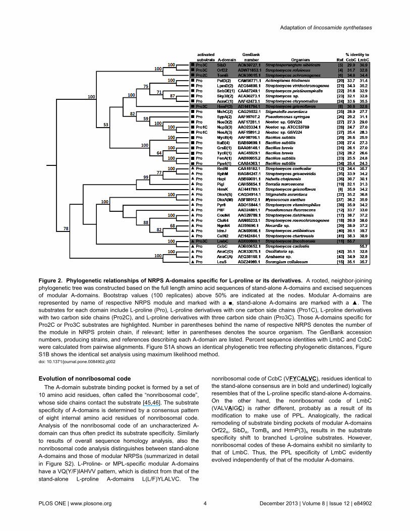
A phylogenetic analysis was carried out on a set ofsequences of A-domains specific for L-proline or L-prolinederivatives. The sequences of all available stand-alone A-domains were used along with a number of representative A-domains with proved or predictable function from multi-domainNRPSs (A-domains which are not stand-alone will hereafter becalled “modular” A-domains because they are part of multi-domain NRPS units called modules). Pairwise alignments ofeach sequence with LmbC and CcbC were carried out todetermine the levels of sequence identity. Additionally, aneighbor-joining and a maximum likelihood methods were usedto construct phylogenetic trees from a multiple sequencealignment of the entire set. Results including referencesdescribing each A-domain [4-6,8,11,12,15,17-43] aresummarized in Figure 2 and Figure S1.
The phylogenetic analysis clearly separated the stand-alone(marked ▲ in Figure 2) and modular A-domains (marked ■ inFigure 2) into two separate clades. Substrate specificityseemed to be a subordinate criterion. Modular A-domainsactivating L-proline derivatives are split into several separatebranches within the clade of L-proline specific modular A-domains. NosA(3)A and NcpB(3)A, which adenylate the Pro1Cderivative methyl-L-proline (MPL) [27,28,44] evolved from acommon ancestor independently of the A-domains activatingPro2C (TomBA from biosynthesis of tomaymycin) and Pro3Cderivatives (HrmP(3)A, SibDA and Orf22A; from biosynthesis ofhormaomycin, sibiromycin and anthramycin). The HrmP(3)A
belongs, moreover, to a separate branch than the three PBDsbiosynthesis A-domains (TomBA, SibDA and Orf22A).
On the other hand, LmbC, which recognizes a substratenearly identical to that of SibDA, clusters within the clade ofstand-alone A-domains, which otherwise adenylate only L-
Adaptation of lincosamide synthetases
PLOS ONE | www.plosone.org 2 December 2013 | Volume 8 | Issue 12 | e84902

proline. Its closest relative by a large margin is CcbC, withmore than 55% sequence identity. None of the other membersof this clade has more than 40% sequence identity with eitherLmbC or CcbC. It seems likely, therefore, that these twoproteins arose from a common ancestor. This is supported bythe fact that the average sequence identities of CcbC andLmbC with other members of this clade are quite similar(35.6% and 35.1%, respectively). CcbC’s slightly highersimilarity reflects the fact that its substrate binding pocketmatches that of the other L-proline adenylating A-domains,while that of LmbC has been altered to recognize PPL (see
below). Since A-domains which act on branched L-prolinederivatives are much less common than those which act on L-proline itself, and since no stand-alone A-domain had beenpreviously identified which acts on these derivatives, it seemslogical to assume that the ancestral specificity of both proteinswas for L-proline (as in CcbC) and that activation of PPL(LmbC) has developed more recently. Nevertheless we canonly speculate, if the ancestor was strictly L-proline specific orexhibited somewhat relaxed substrate specificity.
Figure 1. Structures of lincosamides (A–C) and other natural compounds containing branched L-proline precursors (D–G). (A) Lincomycin A, (B) Lincomycin B, (C) Celesticetin, (D) Anthramycin, (E) Sibiromycin, (F) Tomaymycin, (G) Hormaomycin.Fragments derived from L-proline are highlighted.doi: 10.1371/journal.pone.0084902.g001
Adaptation of lincosamide synthetases
PLOS ONE | www.plosone.org 3 December 2013 | Volume 8 | Issue 12 | e84902
Evolution of nonribosomal codeThe A-domain substrate binding pocket is formed by a set of
10 amino acid residues, often called the “nonribosomal code”,whose side chains contact the substrate [45,46]. The substratespecificity of A-domains is determined by a consensus patternof eight internal amino acid residues of nonribosomal code.Analysis of the nonribosomal code of an uncharacterized A-domain can thus often predict its substrate specificity. Similarlyto results of overall sequence homology analysis, also thenonribosomal code analysis distinguishes between stand-aloneA-domains and those of modular NRPSs (summarized in detailin Figure S2). L-Proline- or MPL-specific modular A-domainshave a VQ(Y/F)IAHVV pattern, which is distinct from that of thestand-alone L-proline A-domains L(L/F)YLALVC. The
nonribosomal code of CcbC (VFYCALVC), residues identical tothe stand-alone consensus are in bold and underlined) logicallyresembles that of the L-proline specific stand-alone A-domains.On the other hand, the nonribosomal code of LmbC(VALVAIGC) is rather different, probably as a result of itsmodification to make use of PPL. Analogically, the radicalremodeling of substrate binding pockets of modular A-domainsOrf22A, SibDA, TomBA and HrmP(3)A results in the substratespecificity shift to branched L-proline substrates. However,nonribosomal codes of these A-domains exhibit no similarity tothat of LmbC. Thus, the PPL specificity of LmbC evidentlyevolved independently of that of the modular A-domains.
Figure 2. Phylogenetic relationships of NRPS A-domains specific for L-proline or its derivatives. A rooted, neighbor-joiningphylogenetic tree was constructed based on the full length amino acid sequences of stand-alone A-domains and excised sequencesof modular A-domains. Bootstrap values (100 replicates) above 50% are indicated at the nodes. Modular A-domains arerepresented by name of respective NRPS module and marked with a ■, stand-alone A-domains are marked with a ▲. Thesubstrates for each domain include L-proline (Pro), L-proline derivatives with one carbon side chains (Pro1C), L-proline derivativeswith two carbon side chains (Pro2C), and L-proline derivatives with three carbon side chain (Pro3C). Those A-domains specific forPro2C or Pro3C substrates are highlighted. Number in parentheses behind the name of respective NRPS denotes the number ofthe module in NRPS protein chain, if relevant; letter in parentheses denotes the source organism. The GenBank accessionnumbers, producing strains, and references describing each A-domain are listed. Percent sequence identities with LmbC and CcbCwere calculated from pairwise alignments. Figure S1A shows an identical phylogenetic tree reflecting phylogenetic distances, FigureS1B shows the identical set analysis using maximum likelihood method.doi: 10.1371/journal.pone.0084902.g002
Adaptation of lincosamide synthetases
PLOS ONE | www.plosone.org 4 December 2013 | Volume 8 | Issue 12 | e84902

Biochemical characterization of CcbC and LmbCExcept for partially characterized HrmP(3)A, substrate
specificities of all other modular A-domains activating branchedL-proline precursors were predicted only based on theirsequences and on the formulas of their respective products.The absence of experimentally characterized branched prolinederivative specific modular A-domains, together with the lack oftheir close relatives which specifically adenylate L-proline,hamper the substrate specificity evolution analysis. On theother hand CcbC and LmbC form an orthologous pair of closelyrelated A-domains with different substrate specificities, makingthem an ideal system to study the types of changes whichoccur during the evolution of substrate specificity.
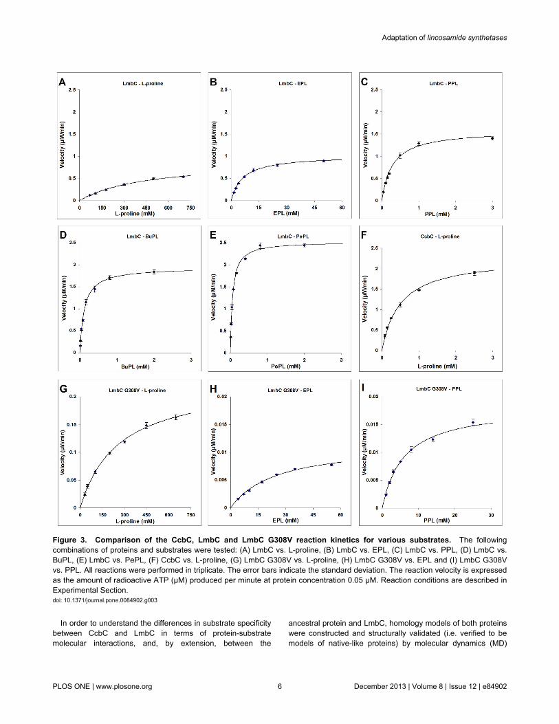
Soluble, recombinant CcbC carrying an N-terminal His6 tagand LmbC carrying a C-terminal His8 tag were produced inEscherichia coli. Unless otherwise stated, CcbC and LmbC inthe following text concerning biochemical experiments, defineHis-tagged forms of proteins mentioned above. These proteinswere stable at –20°C for a few weeks. They were purified in aone-step procedure by nickel affinity chromatography to nearhomogeneity (Figure S3). Typical yields were 9 mg of pureCcbC per 100 mL of cell culture and 3 mg of LmbC per 100 mLof cell culture. The activities of CcbC and LmbC weredetermined using an ATP-[32P]PPi exchange assay, whichmeasures the transfer of radioactivity from 32P-labeled PPi toATP. The reactions required 4 mM MgCl2 and 1 mM ATP. ATPconcentrations higher than 1.5 mM rapidly inhibited thereaction. The optimal pH for both proteins was estimated to be8.7. The kinetic parameters of CcbC and LmbC were assayedwith L-proline and its branched derivatives differing in thelength of the side chain. The results are shown in Figure 3A–Fand summarized in Table 1 and Table S1). 4-Hydroxy-L-proline, L-alanine, L-valine and L-tyrosine were also tested.
CcbC activates its natural substrate L-proline with kineticparameters (Km 0.5 ± 0.03 mM; kcat 45 ± 0.9 min–1; Figure 3F)similar to those of other L-proline specific stand-alone A-domains (examples given below) and exhibits a strict substratespecificity, having no activity in the presence of PPL or (2S,4R)-4-ethyl-L-proline (EPL); it will still act on 4-hydroxy-L-proline, but with a Km value 23 times higher than for L-proline,though its kcat value remains comparable. CcbC thus cannotutilize either of the branched L-proline precursors from thelincomycin biosynthetic pathway. No activity could be detectedeven in the presence of 50 mM EPL or PPL and at 100 timeshigher concentration of CcbC than was used in the L-prolineassay.
LmbC possesses a broader substrate specificity than CcbC;its kinetic parameters for its natural substrate, PPL (Km 0.29 ±0.03 mM; kcat 34.8 ± 1 min–1; Figure 3C), are comparable tothose of CcbC with L-proline and are within the range of valuespreviously published for the L-proline specific stand-alone A-domains CloN4 (Km 0.53 mM; kcat 13.1 min–1), CouN4 (Km 1.16mM; kcat 2.5 min–1), RedM (Km 1.54 mM; kcat 170.9 min–1), PltF(Km 0.51 mM; kcat 332.6 min–1), AnaC (Km 0.97 mM; kcat 68.5min–1) and Leu5 (Km 0.017 mM; kcat 174 min–1) [12-15]. Theaffinity of LmbC is substantially lower for EPL, an alternativenatural LmbC substrate, precursor of lincomycin B (Figure 3B).
Thus, the substrate which yields a biologically more efficientproduct is preferentially activated.
LmbC is able to activate both L-proline (Figure 3A; Table 1)and 4-hydroxy-L-proline, but its affinities for these substratesare ~103 times lower than for PPL. Proteinogenic L-proline isthus effectively excluded from incorporation into lincomycin.Similarly, both CcbC and LmbC do not act efficiently on other,inappropriate proteinogenic amino acids. For example, bothproteins activate L-alanine and L-valine, but with ~103 timeslower efficiency than their natural substrates. Neither seems toact at all on L-tyrosine, the precursor of PPL.
On the other hand, LmbC is also able to activate not only itsnatural substrates, but also synthetic L-proline derivatives withlonger alkyl side-chains, including (2S,4R)-4-butyl-L-proline(BuPL; Figure 3D) and (2S,4R)-4-pentyl-L-proline (PePL;Figure 3E). The kinetic parameters for both these substratesare even better than for PPL. The evolutionary adaptationtherefore produced an enzyme with relaxed substratespecificity. This is a frequent phenomenon among secondarymetabolism enzymes. The shift from the strict L-prolinesubstrate specificity to the relaxed one for more hydrophobicbranched L-proline derivatives of LmbC is also consistent withprevious reports that those A-domains which activatehydrophobic amino acids are generally less selective thanthose which activate polar amino acids [47,48].
The relaxed substrate specificity of LmbC, and thus of N-demethyllincomycin synthetase, along with the relaxedsubstrate specificity of the N-methyltransferase LmbJ whichcatalyzes the following and final step of lincomycin biosynthesis[49] have important practical consequences. Lincomycinderivatives with extended side-chains on the proline buildingunit exhibit higher antibacterial and antiplasmodial activities[50]. We have previously made use of the relaxed substratespecificities of LmbC and LmbJ to produce 4′-butyl-4′-depropyllincomycin and 4′-depropyl-4′-pentyllincomycinmutasynthetically in a Streptomyces lincolnensis ATCC 25466mutant strain blocked in PPL production and fed by eitherBuPL or PePL, as appropriate [51]. Taken together, theseresults suggest that if the CcbC and LmbC ancestor wasspecific for L-proline, as argued above, then the differencesbetween the substrate binding pockets of CcbC and LmbCshould reflect the changes which needed to take place in orderto transform the ancestral protein into something able to act onPPL, a new and structurally modified substrate. In parallel,these changes must also have acted to reduce the affinity ofLmbC for its original L-proline substrate which is generallyavailable in the cell pool of proteinogenic amino acids.Logically, radical remodeling of A-domain substrate bindingpocket should have been a crucial prerequisite for the NDLSsubstrate specificity adaptation. It should be noted, that the kcat
of A-domains in the adenylation reaction may be different fromthose of the same substrate in the condensation reaction [52].In the situation when the other NDLS subunits are notidentified, the ATP-[32P]PPi exchange assay characterizing theA-domain remains the most frequent method to use. Thisstandard method was previously used for the characterizationof several stand-alone L-proline specific A-domains [12-15].
Adaptation of lincosamide synthetases
PLOS ONE | www.plosone.org 5 December 2013 | Volume 8 | Issue 12 | e84902
In order to understand the differences in substrate specificitybetween CcbC and LmbC in terms of protein-substratemolecular interactions, and, by extension, between the
ancestral protein and LmbC, homology models of both proteinswere constructed and structurally validated (i.e. verified to bemodels of native-like proteins) by molecular dynamics (MD)
Figure 3. Comparison of the CcbC, LmbC and LmbC G308V reaction kinetics for various substrates. The followingcombinations of proteins and substrates were tested: (A) LmbC vs. L-proline, (B) LmbC vs. EPL, (C) LmbC vs. PPL, (D) LmbC vs.BuPL, (E) LmbC vs. PePL, (F) CcbC vs. L-proline, (G) LmbC G308V vs. L-proline, (H) LmbC G308V vs. EPL and (I) LmbC G308Vvs. PPL. All reactions were performed in triplicate. The error bars indicate the standard deviation. The reaction velocity is expressedas the amount of radioactive ATP (μM) produced per minute at protein concentration 0.05 μM. Reaction conditions are described inExperimental Section.doi: 10.1371/journal.pone.0084902.g003
Adaptation of lincosamide synthetases
PLOS ONE | www.plosone.org 6 December 2013 | Volume 8 | Issue 12 | e84902

simulation. Moreover, an LmbC with point mutation G308V incritical position of substrate binding pocket was designed andtested both in silico and in vitro.
Homology model construction and structureverification by MD simulation
LmbC and CcbC homology models were constructed basedon the structure of the phenylalanine specific A-domain of GrsA(called PheA, PDB ID 1AMU) which has bound AMP and Phe.A model of the LmbC G308V point mutant was generated by insilico mutation of G308 in the LmbC model. The L-proline (inCcbC) and PPL (in LmbC and LmbC G308V) substrates werepositioned by superimposing the models on the PheA structureand refined according to the location of the amino andcarboxylate groups of the bound L-phenylalanine in the PheAstructure. CcbC and LmbC have quite low homology to PheA(26.4% and 24.9%, respectively). In these situations, there isalways a danger that the resulting homology model may violatethe ordinary parameters of real proteins. In order to produce areliable model, 20-ns-long MD simulations were employed torelax any and all possible strains that may have arisen frommodel building. The relaxed models of LmbC and CcbC fromframe 805 corresponding to time 8.05 ns are presented inFigure 4A and B. Time-based and residue-based root-mean-square deviation (RMSD) analyses confirmed the generaloverall stability of the LmbC and LmbC G308V models duringthe whole simulation period (for details see Analysis S1).However, during the second half of the production phase, theRMSD values of the CcbC model increased and fluctuated,indicating that this model is of only limited validity. A residue-based RMSD analysis of this model at the beginning and endof the MD simulation confirmed several flexible regions.Fortunately, none of the ten amino acid residues forming the
Table 1. The activity of CcbC, LmbC and LmbC G308V forvariable substrates.
Protein Substrate[a] Km (mM) kcat (min–1) kcat/Km (mM–1min–
1)CcbC L-proline 0.5 ± 0.03 45 ± 0.9 91.7CcbC EPL ND ND NDCcbC PPL ND ND NDLmbC L-proline 470 ± 60 20 ± 1 0.043LmbC EPL 6.4 ± 0.3 22.1 ± 0.3 3.46
LmbC PPL 0.29 ± 0.03 34.8 ± 1 121LmbC BuPL 0.118 ± 0.008 42 ± 0.8 359LmbC PePL 0.0596 ± 0.003 55.2 ± 0.8 925.7LmbC G308V L-proline 260 ± 20 5 ± 0.2 0.019LmbC G308V EPL 25 ± 2 0.26 ± 0.01 0.01LmbC G308V PPL 6 ± 0.7 0.41 ± 0.02 0.068
[a] EPL - (2S,4R)-4-ethyl-L-proline; PPL - (2S,4R)-4-propyl-L-proline; BuPL - (2S,4R)-4-butyl-L-proline and PePL - (2S,4R)-4-pentyl-L-proline.Reaction conditions are described in Experimental Section. Rows showing resultsfor LmbC and CcbC with their natural substrates are highlighted. ND – tested, notdetectable. The error values indicate the standard error.doi: 10.1371/journal.pone.0084902.t001
CcbC substrate binding pocket belong to any of these regions,indicating that the CcbC substrate binding pocket remained acompact and stable structure during the whole simulationperiod. Additionally a longer 100-ns-long MD simulation wasperformed to confirm the stability of the LmbC and CcbCmodels. Time-based RMSD analysis of both models showedthe convergence and thus the stability of the system (notshown).
Evolution of the CcbC and LmbC substrate bindingpocket architecture and function
Time-based RMSD analyses of all substrate C atoms duringthe 20-ns-long MD simulations were performed to evaluate thesubstrate’s conformation and its interactions with the CcbC andLmbC binding pockets (Figure S4G). For LmbC (Figure S4D–Fand blue line in S4G), the PPL substrate remained in a stableposition and in the correct orientation in the substrate bindingpocket during the simulation period. Its RMSD fluctuated onlyslightly around a mean value of ~1.5 Å (±0.5 Å). Although L-proline also remained in contact with the substrate bindingpocket in the CcbC model (Figure S4A–C and green line inS4G), its mean RMSD reached ~5 Å during the second part ofthe production phase and exhibited substantial fluctuations (±1Å). Thus, instead of being strongly fixed, L-proline shifts androtates in the CcbC binding pocket. This is shown more clearlyin Figure 4C-D, where the L-proline rotates in the substratebinding pocket during a 0.17 ns period in the middle of theproduction phase (frames 788 and 805, between 7.88 and 8.05ns). This is likely a further indication of the limited validity of theCcbC model. Moreover during a longer 100-ns-long MDsimulation the L-proline substrate was released from the CcbCsubstrate binding pocket after ~40 ns, whereas PPL remains insubstrate binding pocket of LmbC for the whole productionphase (not shown).
Two amino acid residues of all A-domain substrate bindingpockets are highly conserved, a L-lysine which interacts withthe carboxylate group, and an L-aspartate which interacts withthe α-amino group of the substrate amino acid (colored gray inFigures 4, 5 and S4) [45,46]. All these weak interactions wereproved during MD simulations of LmbC and CcbC models withthe only exception of α-amino group of L-proline and D201 ofCcbC reflecting the above mentioned rotation of the substrate.
The remaining eight amino acids determine the substratespecificity of the A-domain (colored variously in Figures 4, 5and S4). As Figure 4 shows, the substrate binding pocket ofCcbC clearly has a smaller, tighter cavity. In addition to theinvariant residues D201 and K490, the L-proline substrate is indirect contact only with three amino acids: V202, A274 andV306. The remaining five residues of the nonribosomal codeare sterically screened by these three (and likely serve tomaintain the overall shape of the binding pocket). Similarlyformed binding pockets, in which only a few residues of thenonribosomal code directly participate in substrate binding,have previously been described for other A-domains [53-55].The CcbC homology model clearly accounts for the inability ofthe protein to accept and activate L-proline derivatives with anyside chain in position 4 (Figure 4B–D): The binding site is bothtoo small and the wrong shape to accommodate the side chain.
Adaptation of lincosamide synthetases
PLOS ONE | www.plosone.org 7 December 2013 | Volume 8 | Issue 12 | e84902

Figure 4. Homology models of the CcbC and LmbC binding pocket with the substrate. The models of LmbC (A) and CcbC(B) at frame 805 (time 8.05 ns) of a 20-ns-long, non-restrained MD simulation. Pictures C and D at frame 788 (7.88 ns) and frame805 (time 8.05 ns) represent another perspective of the CcbC homology model. The letters of the nonribosomal code at upper edgeare colored to correspond to the individual amino acid residues of the structures.doi: 10.1371/journal.pone.0084902.g004
Adaptation of lincosamide synthetases
PLOS ONE | www.plosone.org 8 December 2013 | Volume 8 | Issue 12 | e84902

The nonribosomal code of the LmbC substrate bindingpocket differs from that of CcbC in five of the eight residues
determining the substrate specificity. These differences resulttogether in a formation of the channel accommodating the alkyl
Figure 5. Homology models of the LmbC and LmbC G308V amino acid binding pocket and an RMSD analysis of thesemodels during MD simulations. Structures of the substrate binding pockets from LmbC (A–C) and LmbC G308V (D–F) homologymodels with bound PPL during the course of a 20-ns-long, non-restrained MD simulation are shown at 0 ns (left column), 8.05 ns(middle column), and 19.09 ns (right column). The nonribosomal code of each model is displayed at left. The individual letters of thecode are colored to correspond to those of the individual amino acids in the structures. A time-based RMSD analysis of thesubstrate during a 20-ns-long, non-restrained MD simulation of LmbC (blue line) and LmbC G308V (red line). The RMSD wascalculated over all substrate C atoms. The positions of the frames 0, 805 and 1909 (corresponding to the time 0 ns, 8.05 ns, and19.09 ns) are marked with vertical lines.doi: 10.1371/journal.pone.0084902.g005
Adaptation of lincosamide synthetases
PLOS ONE | www.plosone.org 9 December 2013 | Volume 8 | Issue 12 | e84902

side chain of PPL. Interestingly, only one of the three residueswhich directly contact the substrate in the CcbC binding pocketis altered in LmbC: V306 in CcbC has become G308 in LmbC(red in Figures 4B–D). The change from L-valine to glycineseems to be critical; the larger L-valine should block theaccessibility of the channel for the PPL alkyl side chain. Themodel shows that the other four differences also contribute tothe formation of a channel of a proper size, shape andhydrophobicity (Figure 4A). A207 in LmbC in contrast to F205in CcbC makes the channel more spacious (orange residue inFigure 4A), whereas more hydrophobic L246 and V274 (LmbC)in contrast to Y244 and C272 (CcbC) correspond better to theaccommodation of the hydrophobic alkyl side chain of PPL.The MD simulation indicates that the PPL substrate isconsiderably better anchored in the LmbC binding pocket thanL-proline in the CcbC pocket (Figure S4G, blue vs. green line),most likely due to an increased number of contacts betweenthe substrate and the enzyme. This feature of the modelagrees with the observed kinetic parameters of CcbC andLmbC. The substrate affinity, measured by Km, increases withthe length of the substrate’s alkyl side chain: The Km for L-proline bound to CcbC is 0.5 ± 0.03 mM, 0.29 ± 0.03 mM forPPL bound to LmbC, 0.118 ± 0.008 mM for BuPL bound toLmbC, and 0.0596 ± 0.003 mM for PePL bound to LmbC.
In silico and biochemical analysis of mutant LmbCG308V
Homology models of LmbC/CcbC indicated the importanceof the residues G308/V306. Two homology models of mutantLmbC G308V and CcbC V306G were constructed. From theCcbC V306G model it appears that the glycine residue itself inthis position does not ensure formation of a channel of theappropriate size and shape to accommodate the alkyl sidechain of PPL. It corresponds with the fact that the nonribosomalcodes of all known A-domains recognizing the 2C and 3Cbranched L-proline derivatives differ from the L-prolineconsensus in 3-6 amino acid residues. On the other hand theLmbC G308V model suggests that L-valine in this position canefficiently block the channel. The glycine residue at the testedposition thus seems to be necessary, but not sufficient toharness branched L-proline derivatives. In order to test theoutputs of the homology models, i.e. the existence of thechannel accommodating the alkyl side chain of PPL in LmbC,the above mentioned LmbC G308V mutant was tested both insilico and biochemically.
The time-based RMSD analysis of all PPL C atoms during a20-ns-long MD simulation was carried out to evaluate thesubstrate interactions with the substrate binding pocket ofLmbC G308V. The divergent character of the time-basedRMSD plot of PPL bound to LmbC G308V (Figure 5D–F andred line in 5G) does not reflect a poor homology model usedhere, but rather indicates a real incompatibility of the enzyme-substrate pair. The RMSD increases in three steps during theMD simulation: A mean value of ~3 Å for 0–4 ns, ~4 Å for 4–14ns and ~6 Å for 14–20 ns. The movement of the substrate outof the binding pocket is clearly seen in Figure 5D–F. At thebeginning of the simulation, the PPL substrate was buriedinside the substrate binding pocket, as in the wild type LmbC.
After the substrate moved out of the binding pocket aconformational change occurred, which made the pocketinaccessible, similar to the situation in CcbC. As a result, thechannel for the PPL alkyl side chain disappeared. Thissimulation suggested that much worse kinetic parameters canbe expected for the reactions of LmbC G308V with substratescontaining an alkyl side-chain compared to the wild-type LmbC.
To test these predictions experimentally, the LmbC G308Vmutant form was constructed by site-directed mutagenesis, andoverproduced, purified and assayed under the same conditionsas LmbC. The kinetic parameters for the reaction of this mutantprotein with PPL, EPL and L-proline are shown in Figure 3G–Iand summarized in Table 1. The LmbC G308V mutantexhibited a ~20× higher Km and a ~100× lower kcat, resulting ina ~2x10³-fold lower catalytic efficiency for PPL compared to thewild-type LmbC. Similarly, the kinetic parameters for EPL alsoworsened dramatically. On the other hand, the kcat/Km ratio forL-proline is almost unaffected by this mutation. Although theaffinity (Km) to the L-proline substrate may be slightly better inthe mutant form (260 ± 20 vs. 470 ± 60 mM), the catalyticefficiency declined reciprocally (0.019 vs. 00.043 mM-1min–1). Itshould be noted that the measured parameters probably reflecta combination of two independent factors. Namely, the abovementioned selective response and, to a minor extent, anonselective worsening of the overall catalytic efficiency of themutant protein. This is a common consequence of artificialchangens of natural proteins. In summary, this single G308Vpoint mutation abolishes LmbC’s natural preference for PPL,making thus PPL not much better than L-proline as thesubstrate. The results of these biochemical assays fullyconfirmed those predicted by the simulation.
Among the modular A-domains activating branched L-prolinederivatives, only HrmP(3)A from hormaomycin biosynthesisseems to have followed a similar mechanism. Compared to theconsensus pattern of the L-proline specific modular A-domains,the code of HrmP(3)A has three substitutions, all of them forsmaller residues rather than larger ones. Also, similar to LmbC,HrmP(3)A has a glycine in the position corresponding to theresidue 308 instead of a consensual L-valine. Presumably, thissubstitution plays the same role as in LmbC, namely tofacilitate the access of a substrate alkenyl side chain inside thebinding pocket. Analogously to the evolution of LmbC, also theother two substitutions in the HrmP(3)A substrate bindingpocket, V to A and I to S, could co-operate in the formation of achannel accommodating the alkenyl side-chain ofhormaomycin precursor. Recently [56], HrmP(3)A wasbiochemically proved to adenylate its putative natural substrate(2S,4R)-4-(Z)-propenyl-L-proline. Kinetic parameters have notbeen estimated but the protein highly preferred the branchedderivative over L-proline and other tested amino acids.
Conclusion
General aspects of the evolution of PPL biosynthesisand incorporation
The term “specialized metabolism” is currently often usedinstead of “secondary metabolism” [57,58] in order toemphasize the essence: more active derivatives can arise from
Adaptation of lincosamide synthetases
PLOS ONE | www.plosone.org 10 December 2013 | Volume 8 | Issue 12 | e84902

unique, i.e. specialized, biosynthetic pathways and, evenbetter, from their combinations. The biosynthesis of complexnatural compounds is encoded by biosynthetic gene clusterswhich contain subclusters, groups of genes coding forindividual specialized building units of the final product. TheHGT and a fusion of subclusters to produce new or morecomplex gene clusters is generally known as a commonmechanism in the evolution of biosynthesis of new secondarymetabolites [59]. The most puzzling seems to be the evolutionof genes coding for the enzymes linking the structural blockstogether. Such condensing enzymes are necessary for thefunctioning of a new fusion cluster, but were not required forthe ancestral gene clusters. Thus, their evolutionary origin isunclear [59,60]. Clearly, the genesis and evolution of thecondensation enzymes, particularly their substrate specificityadaptation to newly emerging intermediates, seem to be a keyelement for understanding how new biosynthetic clusters forsecondary metabolites arise.
The 2C and 3C branched L-proline derivatives are highlyspecialized building blocks integrated as precursors exclusivelyin several PBDs, lincomycin and hormaomycin. Logically, acoupled HGT of genes coding for both the biosynthetic andintegration steps appears to be the simplest evolutionarymechanism, probably involved in the evolution of PBDcompounds exhibiting high structural variability of incorporatedbuilding blocks but sharing an identical overall core structure[61]. A HGT of a whole biosynthetic cluster including genescoding for the integrating NRPSs followed by point mutations ofmodular A-domains was the most probable mechanism ofstructural diversification of PBD compounds.
The NDLS, catalyzing a condensation of building units in thebiosynthesis of lincosamides, functionally differs from typicalmodular NRPSs operating in the biosynthesis of PBDs andhormaomycin: NDLS attaches the activated amino acid to theamino sugar, but not to another amino acid, unlike the“authentic” NRPS. This is probably the most interesting aspectof evolution of lincomycin biosynthesis. The ancestral NDLSrepresents a typical example of a specialized condensingenzyme realizing a connection of two types of building units:one specialized metabolite, an amino sugar, and one primarymetabolite, proteinogenic L-proline. In the lincomycinbiosynthesis, moreover, the NDLS A-domain LmbC wasadapted from using L-proline to a new unusual specializedmetabolite, PPL, giving rise to unique connecting functionality.This new condensing activity is distinct from those found inboth ancestral clusters. From the point of view of the PPLdonor biosynthetic cluster, the NDLS attaches the precursor toa novel type of building unit (amino sugar instead of an originalamino acid). From the point of view of the acceptor biosyntheticcluster, the adaptation led to the biosynthesis of a morecomplex compound combining two specialized building units.
In the final step of lincomycin biosynthesis the amino acidmoiety of the NDL is N-methylated by LmbJ. A wide variety ofmodifications was described in PBDs, however the N-methylation of L-proline derived building unit is lincosamidespecific and arose from the ancestral biosynthetic cluster. TheN-methylation step was preserved also in the newly evolved
lincomycin biosynthetic cluster due to the relaxed substratespecificity of the N-methyltransferase enzyme [49].
Experimental Section
Construction of LmbC and CcbC expression vectorsThe lmbC gene was PCR amplified from the chromosomal
DNA of the lincomycin producing type strain Streptomyceslincolnensis ATCC 25466. The ccbC gene was PCR amplifiedfrom a SuperCos cosmid vector I (Stratagene) carrying afragment of the celesticetin gene cluster from the celesticetinproducing type strain Streptomyces caelestis ATCC 15084;GenBank GQ844764.1. The following primers were used forlmbC: lmbCf 5’-CGAATTCCATATGTCGTCCTCCGTTCGA-3’and lmbCr 5’-CCGCTCGAGCTCCCCGCGTGTGACGA-3’ (theNdeI and XhoI restriction sites are underlined). For ccbC, thefollowing primers were used: CCF 5’-CCGGAATTCCATATGAATACCTCCACTGTCCG-3’ andCCRN 5’-AACCCAAGCTTACAGCGTGACGTACCG-3’ (NdeIand HindIII restriction sites are underlined). The lmbC genewas inserted into pET42b (Novagen) via the NdeI and XhoIrestriction sites. The resulting plasmid plmbC1 was used toproduce a C-terminally His8-tagged LmbC. The ccbC gene wasinserted into pET28b (Novagen) via the NdeI and HindIIIrestriction sites. The resulting plasmid pccbC was used for theproduction of an N-terminally His6-tagged CcbC. The openreading frames of both genes were confirmed by sequencing.
Recombinant LmbC with an N-terminal His-tag was alsoproduced, but co-expression with GroES and GroELchaperonins was required to produce soluble protein. Duringthe isolation step, it was not possible to separate LmbC fromGroEL. Both the N- and C-terminally His-tagged proteinsexhibited the same activities when assayed; consequently, C-terminally His-tagged LmbC was used in the present study.
Site-directed mutagenesis of lmbC in plmbC1The lmbC gene was excised via the NdeI and XhoI restriction
sites from plmbC1 and inserted into a pJAKO [62] cloningvector, derived from pBluescript II KS+ (Stratagene) using thesame restriction sites. The resulting plmbC2 plasmid was usedfor in vitro site-directed mutagenesis using the QuickChangeSite-Directed Mutagenesis Kit (Stratagene) and mutagenicprimers G308Vf 5’-CAACATCTACGGTCCGACCGAGACCAACGTCTGTACGTACG-3’ andG308Vr 5’-CGTACGTACAGACGTTGGTCTCGGTCGGACCGTAGATGTTG-3’. The point mutation G923T, which codes for an L-valineresidue rather than an glycine at position 308 in LmbC, is inbold in the forward primer. A silent C906T mutation, introducinga TCCGAC MmeI restriction site to verify the mutation, isitalicized and underlined. The sequence of the reverse primerwas the reverse complement of the forward primer. Theresulting plmbC3 plasmid was digested with NdeI and BsiWIand the mutated segment of the lmbC gene (934 bp) wasinserted into the vector plmbC1 instead of the non-mutatedsegment using the same restriction sites to produce plmbC4.The sequence of this plasmid was confirmed by sequencing
Adaptation of lincosamide synthetases
PLOS ONE | www.plosone.org 11 December 2013 | Volume 8 | Issue 12 | e84902

and used in the production of a C-terminally His-tagged LmbCG308V mutant.
Expression and purification of LmbC, LmbC G308V andCcbC
The His-tagged LmbC, LmbC G308V and CcbC wereproduced in E. coli BL21(DE3) (Novagen), transformed byplmbC1, plmbC4 or pccbC, as appropriate. LB medium (0.1 L)with kanamycin (30 mgL–1) was inoculated and grown at 37°C.At OD600 = 0.7, the culture was cooled down to 17°C and theoverexpression was induced by 0.4 mM isopropyl-β-D-thiogalactopyranoside. Cells were grown for an additional 20hours at 17°C, harvested by centrifugation and stored frozen at–20°C.
Proteins were purified from crude cell extracts, which wereprepared by ultrasonic homogenization in TS-8 buffer (20 mMTris-HCl, 100 mM NaCl, pH 8.0), using HiTrap™ Chelating HPColumns (GE Healthcare). The His-tagged proteins wereeluted by TS-8 buffer with 250 mM imidazole. Pooled fractionscontaining the purified proteins were dialyzed overnight against50 mM Tris-HCl (pH 8.7) and stored at –20°C. Proteinconcentration was measured by the Bradford protein assay kit(Bio-Rad) with bovine serum albumin as a standard.
Enzyme activity assayEnzyme activity was assayed by amino-acid-dependent
exchange of radioactivity from [32P]-labeled PPi into ATP.[32P]Tetrasodium pyrophosphate was purchased fromPerkinElmer. The ATP-[32P]PPi reaction mixtures contained100 mM Tris-HCl (pH 8.7), 2 mM MgCl2, 1 mM DTT, 1 mMATP, 1 mM [32P]PPi (400000 CPM) and various concentrationsof amino acid substrates; the total volume was 100 μL.Reactions were started by the addition of freshly thawedenzyme in final concentrations of 0.05 μM for LmbC and CcbCor 1 μM for LmbC G308V. After 30 min incubation at 28°C, thereactions were quenched by the addition of 0.5 mL ofquenching mixture containing 1.6% (w/v) activated charcoal,4.5% (w/v) tetrasodium pyrophosphate and 3.5% perchloricacid. The quenched mixture was vortexed and pelleted bycentrifugation. The charcoal-containing pellet was washedtwice with 0.5 mL of the quenching mixture without charcoal,resuspended in 0.5 mL of water, and submitted for liquidscintillation counting using Beckman LS 6500 liquid scintillationcounter. The linearity of the reaction velocity in the 30 minutetesting range was confirmed. For each enzyme/substratecombination the reactions mixtures identical to that for thehighest used substrate concentration were prepared. Thereactions were carried out for 0, 5, 10, 20, 25 and 30 minutes.The resulting plot of product formation vs. time showed straightline. The kinetic parameters were determined by non-linearleast squares fitting.
Preparation of (2S,4R)-4-alkyl-L-prolinesSolvents and reagents were purchased from Sigma-Aldrich.
NMR spectra were recorded on a Bruker AVANCE III 400 MHzNMR (400.13 MHz for 1H and 100.62 MHz for 13C, BrukerBioSpin GmbH, Rheinstetten, Germany) in CDCl3 or DMSO-d6
at 30°C. (2S,4R)-4-Butyl-L-proline and (2S,4R)-4-pentyl-L-
proline were prepared according to a previously describedprocedure [51]. (2S,4R)-4-Propyl-L-proline was prepared byalkylation of protected L-pyroglutamic acidusing allyl bromide,followed by a two-step reduction of the resulting amide to yieldprotected 4-allyl-L-proline. Protected 4-allyl-L-proline was thenhydrogenated using a standard H2-Pd/C procedure, affording,after subsequent deprotection steps, the final product (2S,4R)-4-propyl-L-proline. The preparation of (2S,4R)-4-ethyl-L-proline was based on the controlled condensation of aprotected derivative of L-pyroglutamic acid with acetaldehyde.The resulting aldol was dehydrated using MsCl/Et3N, affording4-ethylidene-pyroglutamate. After hydrogenation (Pd/C), thisgave cis-3,5-disubstituted 2-pyrrolidone. Inversion ofconfiguration at pyroglutamate C-4 led to trans-3,5-disubstituted 2-pyrrolidone, which, after subsequent reductionof amide to amine and deprotection, led to the final product(2S,4R)-4-ethyl-L-proline. For details of the syntheticprocedures and analysis of the products, see Supportinginformation.
Phylogenetic analysisThe closest homologs of LmbC and CcbC were found using
a blastp search at the NCBI web site (http://blast.ncbi.nlm.nih.gov/Blast.cgi). The amino acid sequences ofthe A-domains were retrieved from GenBank. Sequenceidentities of these proteins to LmbC and CcbC were calculatedin Geneious 5.5.6 [63] based on pairwise alignments generatedusing MAFFT software version 7.037b at the CBRC web site(http://mafft.cbrc.jp/alignment/server) [64]. For thesealignments, the full length sequences of the stand-alone A-domains (exception: the last 64 amino acids of RphM wereremoved) and those of just the relevant A-domains extractedfrom the sequences of modular NRPSs to match the length ofLmbC and CcbC were used.
To construct the phylogenetic trees, a multiple sequencealignment was generated using MAFFT and a neighbor-joiningand maximum likelihood phylogenetic trees were constructedusing MEGA5 version 5.2 [65]. The sequence of acetyl-CoAsynthetase was used as the outgroup; bootstrapping wasperformed with 100 replications.
Homology model constructionThe structure of the L-phenylalanine specific A-domain of
GrsA (also called PheA, PDB ID 1AMU) was used as atemplate for the construction of both LmbC and CcbChomology models. To build the models, the sequences ofLmbC and CcbC were aligned to 1AMU using the ClustalXversion 2.0.10 [66]. Model structures were produced using theSWISS-MODEL server (http://swissmodel.expasy.org) inalignment mode [67]. The modeling software did notincorporate the two C-terminal residues of both proteins intothe final models. Model of LmbC G308V was generated by insilico mutation in model of LmbC. The positions of AMP, Mg2+,and the amino acid substrates in these models weredetermined by superimposing the models on the PheAtemplate in PyMOL [68] and adjusting the positions of PPL andL-proline based on the positions of the α-amino andcarboxylate groups of the L-phenylalanine bound to PheA.
Adaptation of lincosamide synthetases
PLOS ONE | www.plosone.org 12 December 2013 | Volume 8 | Issue 12 | e84902

Molecular dynamics simulationsAll molecular dynamics (MD) simulations were carried out
using the AMBER suite [69] with the parm99SB force field [70].The following simulation protocol was used: First, theprotonation states of all L-histidine residues were set to createan optimal H-bond network. Next, all remaining hydrogenatoms were added using the Leap program from the AMBERpackage. Then the structures were charge-neutralized byadding an appropriate number of Na+ ions. To prevent rotationof the entire molecule, the center of mass and the orientation ofthe protein were fixed. All systems were inserted into arectangular water box filled by TIP3P water molecules; thelayer of the water molecules was 9 Å thick. Each system wasthen minimized in the following way: The protein was frozenwhile the solvent molecules and counter ions were allowed tomove during a 1000-step minimization process, followed by a10-ps-long MD run under NpT conditions (i.e. p = 1 atm, T =298.15 K). The side chains were then relaxed by severalsequential minimizations with decreasing force constantsapplied to the backbone atoms. After relaxation, the systemwas heated to 50 K for 20 ps and then up to 298.15 K for 90ps. The particle-mesh Ewald method for treating electrostaticinteractions was used. For the production phase, allsimulations were run under periodic boundary conditions in theNpT ensemble at 298.15 K and at a constant pressure of 1 atmusing a 2-fs time integration step. The SHAKE algorithm with atolerance of 10–5 Å was applied to fix all bonds containinghydrogen atoms. A 9.0 Å cutoff was used to treat non-bondinginteractions. Coordinates were stored every 10 ps (i.e. 100frames correspond to 1 ns of simulation). The total duration ofeach production phase, along with the total numbers residues,atoms, counter ions and water molecules in each of thesystems studied, are shown in Table 2.
RMSD analysis of MD simulationsTime-based and residue-based RMSDs were used to
monitor trajectory stability and conformational changes. Fortime-based analysis, RMSDs between the starting and currentstructures were calculated in 0.1 ns intervals during the wholeproduction phase of the MD simulation. The RMSDs of theprotein models were calculated using only the backbone Cαatoms while substrate RMSDs (for PPL and L-proline) werecalculated using all substrate C atoms. For residue-based
Table 2. Production phases parameters used in MDsimulations.
Protein No. of residues Ligand Na+ No. of H2Omolecules No. of atoms
LmbC 507 PPL 6 18240 62238LmbCG308V
507 PPL 6 17156 58995
CcbC 503 L-proline 14 18746 63739
Total duration of all MD simulations was 20 ns; 1 molecule of AMP and 1 Mg2+ ionwere present in all models.doi: 10.1371/journal.pone.0084902.t002
analysis, RMSDs were calculated for every residue betweenthe starting and final structures at the end of the MD simulation.The MDTRA software package was used for all RMSDcalculations [71]. All protein structure figures were generatedby PyMOL [68].
Supporting Information
Analysis S1. Verification of homology model structure byMD simulation. (DOCX)
Figure S1. Phylogenetic trees of A-domains specific for L-proline or its derivatives. Rooted, neighbor-joining (A) andmaximum likelihood (B) phylogenetic trees were constructedbased on the full length amino acid sequences of stand-aloneA-domains and the excised sequences of modular A-domains.Bootstrap values (100 replicates) above 50 % are indicated atthe nodes. The names of A-domains are identical to those inFigure 2. The horizontal bar indicates the number of amino acidsubstitutions per site.(TIF)
Figure S2. Comparison of the nonribosomal codes of A-domains activating L-proline and L-proline derivatives. Thehighly conserved D and K residues at the boundaries ofnonribosomal codes are omitted. The same set of A-domains isshown as in Figure 2. Amino acids are numbered at the topaccording to the A-domain of GrsA (PheA) (first row), CcbC(second row), and LmbC (third row). Substrates areabbreviated as in Figure 2. Residues of stand-alone A-domainsin accordance with consensus of L-proline specific stand-aloneA-domains are in blue. Residues of modular A-domains inaccordance with consensus of L-proline specific modular A-domains are in red. *Number in parentheses behind the nameof respective NRPS denotes the number of the module inNRPS protein chain, if relevant; letter in parentheses denotesthe source organism.(TIF)
Figure S3. SDS PAGE analysis of purified CcbC, LmbCand LmbC G308V proteins. 12% (w/v) gel. Arrow shows theposition of purified proteins. Lanes: S - PageRulerTM prestainedprotein ladder; 1 - His-tagged CcbC; 2 - His-tagged LmbC; 3 -His-tagged LmbC G308V.(TIF)
Figure S4. Homology models of the CcbC and LmbCamino acid binding pocket and an RMSD analysis of thesemodels during MD simulations. Structures of the substratebinding pockets from CcbC (A–C) and LmbC (D–F) homologymodels with bound substrates during the course of a 20-ns-long, non-restrained MD simulation are shown at 0 ns (leftcolumn), 8.05 ns (middle column), and 19.09 ns (right column).The nonribosomal code of each model is displayed at left. Theindividual letters of the code are colored to correspond to thoseof the individual amino acids in the structures. L-prolinesubstrate was used in the CcbC structures while PPL was used
Adaptation of lincosamide synthetases
PLOS ONE | www.plosone.org 13 December 2013 | Volume 8 | Issue 12 | e84902

for the LmbC models. (G) A time-based RMSD analysis of thesubstrates during a 20-ns-long, non-restrained MD simulationof CcbC with L-proline (green line) and LmbC with PPL (blueline). The RMSD was calculated over all substrate C atoms.The positions of the frames 0, 788, 805 and 1909(corresponding to the time 0 ns, 7.88 ns, 8.05 ns, and 19.09ns) are marked with vertical lines.(TIF)
Table S1. Overview of all tested combinations (A-domainvs. substrate) by biochemical assay. (PDF)
Protocol S1. Preparation of (2S,4R)-4-alkyl-L-prolines.(PDF)
Acknowledgements
We thank Dr. Jacob Bauer for critical reading of themanuscript, helpful discussion and language correction and Dr.Michal Otyepka for advice on molecular modeling.
Author Contributions
Conceived and designed the experiments: SK TK JK JJ.Performed the experiments: SK TK DC RG DU. Analyzed thedata: SK TK DC LN JJ. Contributed reagents/materials/analysistools: RG MK. Wrote the manuscript: SK TK RG LN JJ EK.
References
1. Brahme NM, Gonzalez JE, Rolls JP, Hessler EJ, Mizsak S et al. (1984)Biosynthesis of the lincomycins. 1. Studies using stable isotopes on thebiosynthesis of the propyl-L-hygric and ethyl-L-hygric acid moieties oflincomycin-A and lincomycin-B. J Am Chem. Soc 106: 7873-7878.
2. Neusser D, Schmidt H, Spizèk J, Novotnà J, Peschke U et al. (1998)The genes lmbB1 and lmbB2 of Streptomyces lincolnensis encodeenzymes involved in the conversion of L-tyrosine to propylprolineduring the biosynthesis of the antibiotic lincomycin A. Arch Microbiol169: 322-332. doi:10.1007/s002030050578. PubMed: 9531633.
3. Novotná J, Honzátko A, Bednář P, Kopecký J, Janata J et al. (2004)L-3,4-Dihydroxyphenyl alanine-extradiol cleavage is followed byintramolecular cyclization in lincomycin biosynthesis. Eur J Biochem271: 3678-3683. doi:10.1111/j.1432-1033.2004.04308.x. PubMed:15355345.
4. Hu Y, Phelan V, Ntai I, Farnet CM, Zazopoulos E et al. (2007)Benzodiazepine biosynthesis in Streptomyces refuineus. Chem Biol 14:691-701. doi:10.1016/j.chembiol.2007.05.009. PubMed: 17584616.
5. Li W, Khullar A, Chou S, Sacramo A, Gerratana B (2009) Biosynthesisof sibiromycin, a potent antitumor antibiotic. Appl Environ Microbiol 75:2869-2878. doi:10.1128/AEM.02326-08. PubMed: 19270142.
6. Li W, Chou SC, Khullar A, Gerratana B (2009) Cloning andcharacterization of the biosynthetic gene cluster for tomaymycin, anSJG-136 monomeric analog. Appl Environ Microbiol 75: 2958-2963.doi:10.1128/AEM.02325-08. PubMed: 19270147.
7. Argoudelis AD, Fox JA, Eble TE (1965) U-21,669 - A new lincomycin-related antibiotic. Biochemistry 4: 698-703. doi:10.1021/bi00880a014.PubMed: 14323574.
8. Höfer I, Crüsemann M, Radzom M, Geers B, Flachshaar D et al. (2011)Insights into the biosynthesis of hormaomycin, an exceptionallycomplex bacterial signaling metabolite. Chem. Biol 18: 381-391.
9. Chung ST, Manis JJ, McWethy SJ, Patt TE, Witz DF, et al. (1997)Fermentation, biosynthesis, and molecular genetics of lincomycin. In:WR Strohl. Biotechnology of Antibiotics. New York: Drugs and thepharmaceutical sciences. pp. 165–186
10. Peschke U, Schmidt H, Zhang HZ, Piepersberg W (1995) Molecularcharacterization of the lincomycin-production gene-cluster ofStreptomyces lincolnensis-78-11. Mol Microbiol 16: 1137-1156. doi:10.1111/j.1365-2958.1995.tb02338.x. PubMed: 8577249.
11. Koběrská M, Kopecký J, Olšovská J, Jelínková M, Ulanova D et al.(2008) Sequence analysis and heterologous expression of thelincomycin biosynthetic cluster of the type strain Streptomyceslincolnensis ATCC 25466. Folia Microbiol 53: 395-401. doi:10.1007/s12223-008-0060-8.
12. Thomas MG, Burkart MD, Walsh CT (2002) Conversion of L-proline topyrrolyl-2-carboxyl-S-PCP during undecylprodigiosin and pyoluteorinbiosynthesis. Chem. Biol 9: 171-184.
13. Garneau S, Dorrestein PC, Kelleher NL, Walsh CT (2005)Characterization of the formation of the pyrrole moiety duringclorobiocin and coumermycin A1 biosynthesis. Biochemistry 44:2770-2780. doi:10.1021/bi0476329. PubMed: 15723521.
14. Méjean A, Mann S, Vassiliadis G, Lombard B, Loew D et al. (2010) Invitro reconstitution of the first steps of anatoxin-a biosynthesis inOscillatoria PCC 6506: From free L-proline to acyl carrier protein bounddehydroproline. Biochemistry 49: 103-113. doi:10.1021/bi9018785.PubMed: 19954230.
15. Kopp M, Irschik H, Gemperlein K, Buntin K, Meiser P et al. (2011)Insights into the complex biosynthesis of the leupyrrins in Sorangiumcellulosum So ce690. Mol Biosyst 7: 1549-1563. doi:10.1039/c0mb00240b. PubMed: 21365089.
16. Marahiel MA, Stachelhaus T, Mootz HD (1997) Modular peptidesynthetases involved in nonribosomal peptide synthesis. Chem Rev 97:2651-2673. doi:10.1021/cr960029e. PubMed: 11851476.
17. Wang ZX, Li SM, Heide L (2000) Identification of the coumermycin A1biosynthetic gene cluster of Streptomyces rishiriensis DSM 40489.Antimicrob Agents Chemother 44: 3040-3048. doi:10.1128/AAC.44.11.3040-3048.2000. PubMed: 11036020.
18. Pojer F, Li SM, Heide L (2002) Molecular cloning and sequenceanalysis of the clorobiocin biosynthetic gene cluster: new insights intothe biosynthesis of aminocoumarin antibiotics. Microbiology 148:3901-3911. PubMed: 12480894.
19. Harris AKP, Williamson NR, Slater H, Cox A, Abbasi S et al. (2004) TheSerratia gene cluster encoding biosynthesis of the red antibiotic,prodigiosin, shows species-and strain-dependent genome contextvariation. Microbiology-Sgm 150: 3547-3560. doi:10.1099/mic.0.27222-0.
20. Müller C, Nolden S, Gebhardt P, Heinzelmann E, Lange C et al. (2007)Sequencing and analysis of the biosynthetic gene cluster of thelipopeptide antibiotic friulimicin in Actinoplanes friuliensis. AntimicrobAgents Chemother 51: 1028-1037. doi:10.1128/AAC.00942-06.PubMed: 17220414.
21. Wang Y, Chen Y, Shen QR, Yin XH (2011) Molecular cloning andidentification of the laspartomycin biosynthetic gene cluster fromStreptomyces viridochromogenes. Gene 483: 11-21. doi:10.1016/j.gene.2011.05.005. PubMed: 21640802.
22. de Crécy-Lagard V, Blanc V, Gil P, Naudin L, Lorenzon S et al. (1997)Pristinamycin I biosynthesis in Streptomyces pristinaespiralis:Molecular characterization of the first two structural peptide synthetasegenes. J Bacteriol 179: 705-713. PubMed: 9006024.
23. Pohle S, Appelt C, Roux M, Fiedler H-P, Süssmuth RD (2011)Biosynthetic gene cluster of the non-ribosomally synthesizedcyclodepsipeptide skyllamycin: Deciphering unprecedented ways ofunusual hydroxylation reactions. J Am Chem Soc 133: 6194-6205. doi:10.1021/ja108971p. PubMed: 21456593.
24. Pfennig F, Schauwecker F, Keller U (1999) Molecular characterizationof the genes of actinomycin synthetase I and of a 4-methyl-3-hydroxyanthranilic acid carrier protein involved in the assembly of theacylpeptide chain of actinomycin in Streptomyces. J Biol Chem 274:12508-12516. doi:10.1074/jbc.274.18.12508. PubMed: 10212227.
25. Wenzel SC, Kunze B, Höfle G, Silakowski B, Scharfe M et al. (2005)Structure and biosynthesis of myxochromides S1-3 in Stigmatellaaurantiaca: Evidence for an iterative bacterial type I polyketidesynthase and for module skipping in nonribosomal peptidebiosynthesis. Chembiochem 6: 375-385. doi:10.1002/cbic.200400282.PubMed: 15651040.
26. Scholz-Schroeder BK, Soule JD, Gross DC (2003) The sypA, sypB andsypC synthetase genes encode twenty-two modules involved in thenonribosomal peptide synthesis of syringopeptin by Pseudomonassyringae pv. syringae B301D. Mol Plant-Microbe Interact 16: 271-280.doi:10.1094/MPMI.2003.16.4.271. PubMed: 12744455.
Adaptation of lincosamide synthetases
PLOS ONE | www.plosone.org 14 December 2013 | Volume 8 | Issue 12 | e84902

27. Hoffmann D, Hevel JM, Moore RE, Moore BS (2003) Sequenceanalysis and biochemical characterization of the nostopeptolide Abiosynthetic gene cluster from Nostoc sp GSV224. Gene 311: 171-180.doi:10.1016/S0378-1119(03)00587-0. PubMed: 12853152.
28. Luesch H, Hoffmann D, Hevel JM, Becker JE, Golakoti T et al. (2003)Biosynthesis of 4-methylproline in cyanobacteria: Cloning of nosE andnosF genes and biochemical characterization of the encodeddehydrogenase and reductase activities. J Org Chem 68: 83-91. doi:10.1021/jo026479q. PubMed: 12515465.
29. Duitman EH, Hamoen LW, Rembold M, Venema G, Seitz H et al.(1999) The mycosubtilin synthetase of Bacillus subtilis ATCC6633: Amultifunctional hybrid between a peptide synthetase, an aminotransferase, and a fatty acid synthase. Proc Natl Acad Sci U S A 96:13294-13299. doi:10.1073/pnas.96.23.13294. PubMed: 10557314.
30. Tsuge K, Akiyama T, Shoda M (2001) Cloning, sequencing, andcharacterization of the iturin A operon. J Bacteriol 183: 6265-6273. doi:10.1128/JB.183.21.6265-6273.2001. PubMed: 11591669.
31. Saito F, Hori K, Kanda M, Kurotsu T, Saito Y (1994) Entire nucleotide-sequence for bacillus brevis nagano GRS2 gene encoding gramicidin-Ssynthetase-2 - a multifunctional peptide synthetase. J Biochem 116:357-367. PubMed: 7822255.
32. Mootz HD, Marahiel MA (1997) The tyrocidine biosynthesis operon ofBacillus brevis: Complete nucleotide sequence and biochemicalcharacterization of functional internal adenylation domains. J Bacteriol179: 6843-6850. PubMed: 9352938.
33. Samel SA, Wagner B, Marahiel MA, Essen LO (2006) The thioesterasedomain of the fengycin biosynthesis cluster: A structural base for themacrocyclization of a non-ribosomal lipopeptide. J Mol Biol 359:876-889. doi:10.1016/j.jmb.2006.03.062. PubMed: 16697411.
34. Tsuge K, Ano T, Hirai M, Nakamura Y, Shoda M (1999) The genesdegQ, pps, and lpa-8 (sfp) are responsible for conversion of Bacillussubtilis 168 to plipastatin production. Antimicrob Agents Chemother 43:2183-2192. PubMed: 10471562.
35. Kawasaki T, Sakurai F, Nagatsuka SY, Hayakawa Y (2009) Prodigiosinbiosynthesis gene cluster in the roseophilin producer Streptomycesgriseoviridis. J Antibiot (Tokyo) 62: 271-276. doi:10.1038/ja.2009.27.PubMed: 19329986.
36. Kim D, Park YK, Lee JS, Kim JF, Jeong H et al. (2006) Analysis of aprodigiosin biosynthetic gene cluster from the marine bacteriumHahella chejuensis KCTC 2396. J Microbiol Biotechnol 16: 1912-1918.
37. Meiser P, Weissman KJ, Bode HB, Krug D, Dickschat JS et al. (2008)DKxanthene biosynthesis - Understanding the basis for diversity-oriented secondary metabolism. Chem Biol 15: 771-781. doi:10.1016/j.chembiol.2008.06.005. PubMed: 18721748.
38. Zhang XJ, Parry RJ (2007) Cloning and characterization of thepyrrolomycin biosynthetic gene clusters from Actinosporangiumvitaminophilum ATCC 31673 and Streptomyces sp strain UC 11065.Antimicrob Agents Chemother 51: 946-957. doi:10.1128/AAC.01214-06. PubMed: 17158935.
39. Maharjan S, Aryal N, Bhattarai S, Koju D, Lamichhane J et al. (2012)Biosynthesis of the nargenicin A(1) pyrrole moiety from Nocardia spCS682. Appl Microbiol Biotechnol 93: 687-696. doi:10.1007/s00253-011-3567-x. PubMed: 21927992.
40. Li CX, Roege KE, Kelly WL (2009) Analysis of the IndanomycinBiosynthetic Gene Cluster from Streptomyces antibioticus NRRL 8167.Chembiochem 10: 1064-1072. doi:10.1002/cbic.200800822. PubMed:19301315.
41. Wu QL, Liang JD, Lin SJ, Zhou XF, Bai LQ et al. (2011)Characterization of the biosynthesis gene cluster for the pyrrolepolyether antibiotic calcimycin (A23187) in Streptomyces chartreusisNRRL 3882. Antimicrob Agents Chemother 55: 974-982. doi:10.1128/AAC.01130-10. PubMed: 21173184.
42. Méjean A, Mann S, Maldiney T, Vassiliadis G, Lequin O et al. (2009)Evidence that biosynthesis of the neurotoxic alkaloids anatoxin-a andhomoanatoxin-a in the cyanobacterium Oscillatoria PCC 6506 occurson a modular polyketide synthase initiated by L-proline. J Am ChemSoc 131: 7512-7513. doi:10.1021/ja9024353. PubMed: 19489636.
43. Rantala-Ylinen A, Känä S, Wang H, Rouhiainen L, Wahlsten M et al.(2011) Anatoxin-a synthetase gene cluster of the cyanobacteriumAnabaena sp strain 37 and molecular methods to detect potentialproducers. Appl Environ Microbiol 77: 7271-7278. doi:10.1128/AEM.06022-11. PubMed: 21873484.
44. Becker JE, Moore RE, Moore BS (2004) Cloning, sequencing, andbiochemical characterization of the nostocyclopeptide biosyntheticgene cluster: molecular basis for imine macrocyclization. Gene 325:35-42. doi:10.1016/j.gene.2003.09.034. PubMed: 14697508.
45. Conti E, Stachelhaus T, Marahiel MA, Brick P (1997) Structural basisfor the activation of phenylalanine in the non-ribosomal biosynthesis of
gramicidin S. EMBO J 16: 4174-4183. doi:10.1093/emboj/16.14.4174.PubMed: 9250661.
46. Stachelhaus T, Mootz HD, Marahiel MA (1999) The specificity-conferring code of adenylation domains in nonribosomal peptidesynthetases. Chem Biol 6: 493-505. doi:10.1016/S1074-5521(99)80082-9. PubMed: 10421756.
47. Challis GL, Ravel J, Townsend CA (2000) Predictive, structure-basedmodel of amino acid recognition by nonribosomal peptide synthetaseadenylation domains. Chem Biol 7: 211-224. doi:10.1016/S1074-5521(00)00091-0. PubMed: 10712928.
48. Kurmayer R, Christiansen G, Gumpenberger M, Fastner J (2005)Genetic identification of microcystin ecotypes in toxic cyanobacteria ofthe genus Planktothrix. Microbiology 151: 1525-1533. doi:10.1099/mic.0.27779-0. PubMed: 15870462.
49. Najmanová L, Kutejová E, Kadlec J, Polan M, Olšovská J et al. (2013)Characterization of N-demethyl lincosamide methyltransferases LmbJand CcbJ ChemBioChem In press. doi:10.1002/cbic.201300389.
50. Magerlein BJ (1971) Modification of lincomycin. In: D Perlman.Structure-activity relationships among the semisynthetic antibiotics.New York: Academic Press. pp. 600-651.
51. Ulanova D, Novotná J, Smutná Y, Kameník Z, Gažák R et al. (2010)Mutasynthesis of lincomycin derivatives with activity against drug-resistant staphylococci. Antimicrob Agents Chemother 54: 927-930.doi:10.1128/AAC.00918-09. PubMed: 19917754.
52. Shaw-Reid CA, Kelleher NL, Losey HC, Gehring AM, Berg C et al.(1999) Assembly line enzymology by multimodular nonribosomalpeptide synthetases: the thioesterase domain of E. coli EntF catalyzesboth elongation and cyclolactonization. Chem Biol 6: 385-400. doi:10.1016/S1074-5521(99)80050-7. PubMed: 10375542.
53. Lautru S, Challis GL (2004) Substrate recognition by nonribosomalpeptide synthetase multi-enzymes. Microbiology 150: 1629-1636. doi:10.1099/mic.0.26837-0. PubMed: 15184549.
54. Bushley KE, Ripoll DR, Turgeon BG (2008) Module evolution andsubstrate specificity of fungal nonribosomal peptide synthetasesinvolved in siderophore biosynthesis. BMC Evol Biol 8: 328-. PubMed:19055762.
55. Yonus H, Neumann P, Zimmermann S, May JJ, Marahiel MA et al.(2008) Crystal structure of DltA. Implications for the reactionmechanism of non-ribosomal peptide synthetase adenylation domains.J Biol Chem 283: 32484-32491. doi:10.1074/jbc.M800557200.PubMed: 18784082.
56. Crüsemann M, Kohlhaas C, Piel J (2013) Evolution-guided engineeringof nonribosomal peptide synthetase adenylation domains. Chem Sci 4:1041-1045. doi:10.1039/c2sc21722h.
57. Milo R, Last RL (2012) Achieving diversity in the face of constraints:Lessons from metabolism. Science 336: 1663-1667. doi:10.1126/science.1217665. PubMed: 22745419.
58. Liscombe DK, Louie GV, Noel JP (2012) Architectures, mechanismsand molecular evolution of natural product methyltransferases. NatProd Rep 29: 1238-1250. doi:10.1039/c2np20029e. PubMed:22850796.
59. Fischbach MA, Walsh CT, Clardy J (2008) The evolution of genecollectives: How natural selection drives chemical innovation. Proc NatlAcad Sci U S A 105: 4601-4608. doi:10.1073/pnas.0709132105.PubMed: 18216259.
60. O'Brien PJ, Herschlag D (1999) Catalytic promiscuity and the evolutionof new enzymatic activities. Chem Biol 6: R91-R105. doi:10.1016/S1074-5521(99)80021-0. PubMed: 10099128.
61. Gerratana B (2012) Biosynthesis, synthesis, and biological activities ofpyrrolobenzodiazepines. Med Res Rev 32: 254-293. doi:10.1002/med.20212. PubMed: 20544978.
62. Kyselková M, Janata J, Ságová-Marečková M, Kopecký J (2010)Subunit-subunit interactions are weakened in mutant forms ofacetohydroxy acid synthase insensitive to valine inhibition. ArchMicrobiol 192: 195-200. doi:10.1007/s00203-010-0545-0. PubMed:20107768.
63. Geneious version 5.5.6 created by Biomatters. Available from http://www.geneious.com/.
64. Katoh K, Standley DM (2013) MAFFT multiple sequence alignmentsoftware version 7: Improvements in performance and usability. MolBiol Evol 30: 772-780. doi:10.1093/molbev/mst010. PubMed:23329690.
65. Tamura K, Peterson D, Peterson N, Stecher G, Nei M et al. (2011)MEGA5: Molecular evolutionary genetics analysis using maximumlikelihood, evolutionary distance, and maximum parsimony methods.Mol Biol Evol 28: 2731-2739. doi:10.1093/molbev/msr121. PubMed:21546353.
Adaptation of lincosamide synthetases
PLOS ONE | www.plosone.org 15 December 2013 | Volume 8 | Issue 12 | e84902

66. Larkin MA, Blackshields G, Brown NP, Chenna R, McGettigan PA et al.(2007) Clustal W and clustal X version 2.0. Bioinformatics 23:2947-2948. doi:10.1093/bioinformatics/btm404. PubMed: 17846036.
67. Schwede T, Kopp J, Guex N, Peitsch MC (2003) SWISS-MODEL: Anautomated protein homology-modeling server. Nucleic Acids Res 31:3381-3385. doi:10.1093/nar/gkg520. PubMed: 12824332.
68. Schrödinger LLC (2010) The PyMOL molecular graphics system,version 1.3.
69. Case DA, Cheatham TE, Darden T, Gohlke H, Luo R et al. (2005) TheAmber biomolecular simulation programs. J Comput Chem 26:1668-1688. doi:10.1002/jcc.20290. PubMed: 16200636.
70. Hornak V, Abel R, Okur A, Strockbine B, Roitberg A et al. (2006)Comparison of multiple amber force fields and development ofimproved protein backbone parameters. Proteins 65: 712-725. doi:10.1002/prot.21123. PubMed: 16981200.
71. Popov AV, Vorobjev YN, Zharkov DO (2013) MDTRA: A moleculardynamics trajectory analyzer with a graphical user interface. J ComputChem 34: 319-325. doi:10.1002/jcc.23135. PubMed: 23047307.
Adaptation of lincosamide synthetases
PLOS ONE | www.plosone.org 16 December 2013 | Volume 8 | Issue 12 | e84902