Embed Size (px)
Citation preview
BioMed CentralVirology Journal
ss
Open AcceResearchDown-regulation of cell surface CXCR4 by HIV-1Bongkun Choi*1,4, Paul J Gatti1,5, Cesar D Fermin2, Sandor Vigh3, Allyson M Haislip1 and Robert F Garry1Address: 1Department of Microbiology and Immunology, Tulane University Health Sciences Center, New Orleans, LA 70112, USA, 2College of Veterinary Medicine, Nursing & Allied Health (CVMNAH), Tuskegee University, Tuskegee, AL 36088, USA, 3Department of Structural & Cellular Biology, Tulane University Health Sciences Center, New Orleans, LA 70112, USA, 4Departments of Environmental Medicine, Pathology, and Medicine, New York University School of Medicine, Tuxedo, NY 10987, USA and 5Biocompare, Inc., 395 Oyster Point Blvd, South San Francisco, CA 94080, USA
Email: Bongkun Choi* - [email protected]; Paul J Gatti - [email protected]; Cesar D Fermin - [email protected]; Sandor Vigh - [email protected]; Allyson M Haislip - [email protected]; Robert F Garry - [email protected]
* Corresponding author
AbstractBackground: CXC chemokine receptor 4 (CXCR4), a member of the G-protein-coupledchemokine receptor family, can serve as a co-receptor along with CD4 for entry into the cell of T-cell tropic X4 human immunodeficiency virus type 1 (HIV-1) strains. Productive infection of T-lymphoblastoid cells by X4 HIV-1 markedly reduces cell-surface expression of CD4, but whetheror not the co-receptor CXCR4 is down-regulated has not been conclusively determined.
Results: Infection of human T-lymphoblastoid cell line RH9 with HIV-1 resulted in down-regulation of cell surface CXCR4 expression. Down-regulation of surface CXCR4 correlatedtemporally with the increase in HIV-1 protein expression. CXCR4 was concentrated in intracellularcompartments in H9 cells after HIV-1 infection. Immunofluorescence microscopy studies showedthat CXCR4 and HIV-1 glycoproteins were co-localized in HIV infected cells. Inducible expressionof HIV-1 envelope glycoproteins also resulted in down-regulation of CXCR4 from the cell surface.
Conclusion: These results indicated that cell surface CXCR4 was reduced in HIV-1 infected cells,whereas expression of another membrane antigen, CD3, was unaffected. CXCR4 down-regulationmay be due to intracellular sequestering of HIV glycoprotein/CXCR4 complexes.
BackgroundChemokine receptors are seven-transmembrane G-pro-tein-coupled receptors that upon ligand binding transmitsignals, such as calcium flux, resulting in chemotacticresponses [1-3]. Chemokine receptors are divided intofour families that reflect differential binding of the CXC,CC, CX3C and XC subfamilies of chemokines [4]. Severalmembers of the chemokine receptor family function ascoreceptors with the primary receptor CD4 to allow entryof various strains of human immunodeficiency virus type
1 (HIV-1) into the cells [5-8]. T-cell-tropic X4 HIV-1 useCD4 and chemokine receptor CXCR4 for entry into targetcells, whereas macrophage-tropic R5 HIV-1 use CD4 andchemokine receptor CCR5. Dual-tropic strains can useeither CCR5 and CXCR4 as co-receptors. In addition,CCR3, CCR2, CXCR6 (Bonzo/STLR6) among other chem-okine receptors can function as coreceptors and supportinfection by a more restricted subset of macrophage-tropicor dual-tropic HIV strains [9,10,5,11,12].
Published: 11 January 2008
Virology Journal 2008, 5:6 doi:10.1186/1743-422X-5-6
Received: 21 December 2007Accepted: 11 January 2008
This article is available from: http://www.virologyj.com/content/5/1/6
© 2008 Choi et al; licensee BioMed Central Ltd. This is an Open Access article distributed under the terms of the Creative Commons Attribution License (http://creativecommons.org/licenses/by/2.0), which permits unrestricted use, distribution, and reproduction in any medium, provided the original work is properly cited.
Page 1 of 10(page number not for citation purposes)
Virology Journal 2008, 5:6 http://www.virologyj.com/content/5/1/6
CXCL12 (stromal derived factor 1 α/β, SDF-1α/β) is thenatural ligand for CXCR4, whereas CC chemokines, CCL3(macrophage inflammatory factor 1α, MIP-1α/chemok-ine LD78α), CCL3-L1 (LD78β), CCL4 (MIP-1β), andCCL5 (RANTES), are ligands for CCR5 [13-16]. CXCL12,CCL3, CCL4 and CCL5 as well as other natural and syn-thetic chemokine receptor ligands are able to inhibit cellfusion and infection by various strains of HIV-1, depend-ent or independent of co-receptor usage [17-21]. Thesefindings have encouraged the development of antiHIVtherapeutics targeting chemokine receptors [22-25].
Productive infection of CD4+ cells with HIV-1 markedlyreduces cell-surface expression of CD4, which follows aclassic mechanism for retroviral interference [26,27].Down-regulation of CD4 by HIV-1 has been attributed tothe formation of intracellular complexes consisting ofHIV-1 envelope glycoproteins and CD4 receptors [28],although other mechanisms may also be involved in a celltype dependent manner [29,30]. Chemokine receptors,including CCR5 and CXCR4, can be down-regulated afterbinding of their respective chemokine ligands by a mech-anism involving endocytosis of the complex [31-33]. Theenvelope glycoproteins of HIV-1 competitively antago-nize signaling by coreceptors CXCR4 and CCR5 [34,35].Exogenously added recombinant soluble HIV-1 surfaceglycoprotein (SU, gp120) can be coprecipitated from thecell surface into a complex with CD4 and CXCR4, thatmay lead to the formation of a trimolecular complexbetween HIV SU, CD4 and CXCR4 [36,37]. However,prior studies have suggested that although CCR5 corecep-tors are down-modulated during infection by R5 HIV-1,CXCR4 co-receptor is not down-regulated after productiveX4 HIV-1 infection [38]. CXCR4 was shown to be selec-tively down-regulated from the cell surface by HIV-2/vcpin the context of CD4-independent infection [39] or fromcells infected with CD4-independent HIV-1 isolate thatenters directly via CXCR4 [40]. Furthermore, exogenousexpression of the HIV-1 Nef protein reduced cell surfacelevels of CCR5 or CXCR4 [41,42]. Here, we examinewhether or not productive infection by HIV-1 alters thecell surface expression of CXCR4. Our results indicate thatCXCR4 is down-regulated from the surface of CD4+ T-lymphoblastoid cells infected by HIV-1 and that HIV-1Env and CXCR4 are colocalized in infected cells.
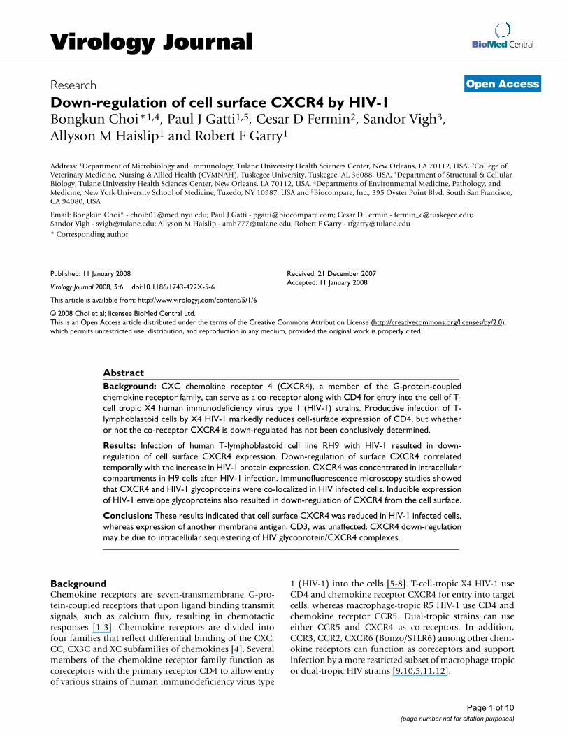
ResultsHIV-1 infection down-regulates surface expression of CXCR4 in RH9 cellsTo determine whether HIV infection alters cell surfaceCXCR4 levels, RH9 T-lymphoblastoid cells were infectedwith HIV-1LA1 at a MOI of 4 or mock-infected. At 1, 4 and7 days post infection (PI), the level of cell surface CXCR4on RH9 cells and HIV-1-infected RH9 cells were deter-mined by flow cytometric analysis using CXCR4 mono-
clonal antibody (MAb) 12G5 [39]. Relative binding of12G5 monoclonal antibody was significantly reducedcompared to uninfected cells at 4, and 7 days postinfec-tion, respectively (Fig. 1A). As a control, we also deter-mined the effect of HIV infection on CD3 in RH9 cells. H9cells infected with HIV maintained surface CD3 expres-sion at a similar level to that of uninfected H9 cells (Fig.1B). To determine the relationship between the expres-sion of surface CXCR4 and HIV-1 protein expression, HIV-1 production by infected cells was quantified by a antigen-capture enzyme-linked immunosorbant assay (Ag-captureELISA; Abbott Laboratories) and the number of HIV-1antigen expressing cells were measured by indirectimmunofluorescence microscopy. The decline in CXCR4expression was accompanied by a rapid increase in HIV-1protein expression in infected RH9 cells.
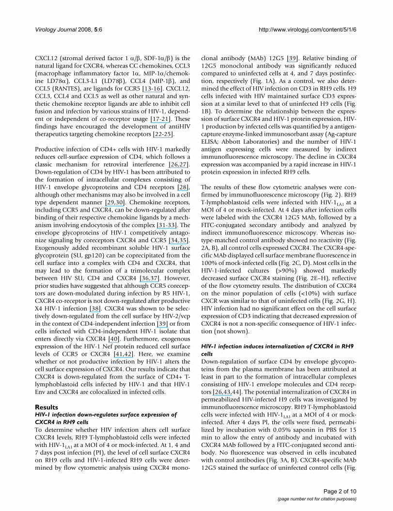
The results of these flow cytometric analyses were con-firmed by immunofluorescence microscopy (Fig. 2). RH9T-lymphoblastoid cells were infected with HIV-1LA1 at aMOI of 4 or mock-infected. At 4 days after infection cellswere labeled with the CXCR4 12G5 MAb, followed by aFITC-conjugated secondary antibody and analyzed byindirect immunofluorescence microscopy. Whereas iso-type-matched control antibody showed no reactivity (Fig.2A, B), all control cells expressed CXCR4. The CXCR4-spe-cific MAb displayed cell surface membrane fluorescence in100% of mock-infected cells (Fig. 2C, D). Most cells in theHIV-1-infected cultures (>90%) showed markedlydecreased surface CXCR4 staining (Fig. 2E–H), reflectiveof the flow cytometry results. The distribution of CXCR4on the minor population of cells (<10%) with surfaceCXCR was similar to that of uninfected cells (Fig. 2G, H).HIV infection had no significant effect on the cell surfaceexpression of CD3 indicating that decreased expression ofCXCR4 is not a non-specific consequence of HIV-1 infec-tion (not shown).
HIV-1 infection induces internalization of CXCR4 in RH9 cellsDown-regulation of surface CD4 by envelope glycopro-teins from the plasma membrane has been attributed atleast in part to the formation of intracellular complexesconsisting of HIV-1 envelope molecules and CD4 recep-tors [26,43,44]. The potential internalization of CXCR4 inpermeabilized HIV-infected H9 cells was investigated byimmunofluorescence microscopy. RH9 T-lymphoblastoidcells were infected with HIV-1LA1 at a MOI of 4 or mock-infected. After 4 days PI, the cells were fixed, permeabi-lized by incubation with 0.05% saponin in PBS for 15min to allow the entry of antibody and incubated withCXCR4 MAb followed by a FITC-conjugated second anti-body. No fluorescence was observed in cells incubatedwith control antibodies (Fig. 3A, B). CXCR4-specific MAb12G5 stained the surface of uninfected control cells (Fig.
Page 2 of 10(page number not for citation purposes)
Virology Journal 2008, 5:6 http://www.virologyj.com/content/5/1/6
3C, D). A weak additional intracellular signal observed insome control cells may be attributed to newly synthesizedCXCR4 molecules in intracellular compartments of secre-tory pathways. In cultures productively infected with HIV-1, intracellular CXCR4 staining was markedly increased inapproximately 50% of the cells, with a redistribution ofthe staining that is consistent with the intracellular accu-mulation of the receptor (Fig. 3E–H).
HIV-1 SU and CXCR4 are colocalized in HIV-1 productively-infected RH9 cellsExogenously added HIV SU or SU expressed from recom-binant vectors can form a complex with CD4 and chem-okine receptor [36,37]. Double labeling was used todetermine if an analogous complex of CXCR4 and HIV-1glycoprotein can be detected in HIV-1 productivelyinfected cells. RH9 T-lymphoblastoid cells were infectedwith HIV-1LA1 at a MOI of 4 or mock-infected. After 4 daysPI, the cells were fixed, permeabilized with saponin andincubated with 12G5 CXCR4 MAb followed by a FITC-conjugated second antibody. For staining of HIV-1 glyco-proteins, cells were incubated with rhodamine-conju-gated antibodies to the HIV-1 proteins and double-fluorescence analysis was performed. A phase contrastmicrograph of a multinucleated HIV-1 infected cell is
shown in Figure 4A. Figure 4C and Figure 4D representstaining for anti-HIV-1 proteins (red) and anti-CXCR4(green) MAb, respectively. Superpositions of the two colorchannels appear in yellow representing the degree of colo-calization of CXCR4 and HIV-1 proteins (Fig. 4B). Similarresults were observed in nonsyncytial cells expressingHIV-1 proteins. These results suggest that HIV-1 SU andCXCR4 are colocalized in HIV-1 productively-infectedRH9 cells.
Inducible expression of HIV-1 Env down-regulates cell surface CXCR4 expressionHIV-1 Env have been suggested to play a role in down-reg-ulation of surface CD4 molecules from the plasma mem-brane [28,45,46]. The effect of inducible expression of theHIV-1 envelope protein (strain HXB2) on CXCR4 expres-sion was analyzed in CD4+ Jurkat lymphocytes with awell-characterized tetracycline inducible expression sys-tem [47,48]. Env expression was monitored by syncitialformation and immunofluorecence staining for Env pro-teins. In the presence of tetracycline, no fluorescence wasobserved in Jurkat cells, indicating that Env expressionwas repressed. When Jurkat cells were cultured in theabsence of tetracycline to induce Env expression, >95% ofcells stained positive for HIV-1 Env. In the presence of tet-
Flow cytometry analysis demonstrating reduced CXCR4 expression in HIV-1 infected RH9 cellsFigure 1Flow cytometry analysis demonstrating reduced CXCR4 expression in HIV-1 infected RH9 cells. Panel A: RH9 T-lymphoblast-oid cells infected with HIV-1LA1. On days 1, 4, and 7 postinfection cells were fixed with 4% paraformaldehyde, stained with mouse MAb 12G5 anti-CXCR4 (10 μg/ml) or isotype-matched control antibody followed by fluorescein isothiocyanate (FITC)-conjugated goat anti-mouse immunoglobulin G, and analyzed by flow cytometry. Median fluorescence intensity was calculated as an indicator of the level of cell surface CXCR4 expression. Data are presented as single-color histograms with FITC fluores-cence (CD3 expression) along the horizontal axis and relative cell number along the vertical axis. RH9 cells (control cells), heavy solid line: H9 cells infected with HIV, dotted line; H9 with an isotype-matched control antibody, thin solid line. Panel B: Analysis of surface CD3 expression in HIV-1 and mock infected RH9 cells by FACS analyzed on day 7 post-infection.
Page 3 of 10(page number not for citation purposes)
Virology Journal 2008, 5:6 http://www.virologyj.com/content/5/1/6
Page 4 of 10(page number not for citation purposes)
Immunofluorescence microscopy demonstrating reduced cell surface expression of CXCR4 in HIV-1 infected RH9 cellsFigure 2Immunofluorescence microscopy demonstrating reduced cell surface expression of CXCR4 in HIV-1 infected RH9 cells. Panel A: Immunofluorescence staining control with isotype-matched monoclonal antibody. Panel C: CXCR4 immunofluorescence staining of H9 cells. Panels E and G: CXCR4 immunofluorescence staining of H9 cells acutely infected by HIV-1. Panels B, D, F and H show phase contrast images of the same fields of cells shown in left panels. The fluorescent syncytial cell in panel G is representative of a minor population of cells in the infected culture (<10%) with a CXCR4 surface distribution similar to unin-fected cells.
Virology Journal 2008, 5:6 http://www.virologyj.com/content/5/1/6
Page 5 of 10(page number not for citation purposes)
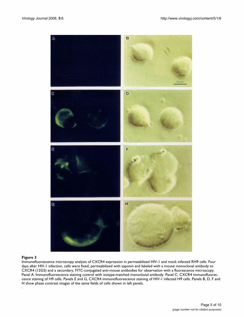
Immunofluorescence microscopy analysis of CXCR4 expression in permeabilized HIV-1 and mock infected RH9 cellsFigure 3Immunofluorescence microscopy analysis of CXCR4 expression in permeabilized HIV-1 and mock infected RH9 cells. Four days after HIV-1 infection, cells were fixed, permeabilized with saponin and labeled with a mouse monoclonal antibody to CXCR4 (12G5) and a secondary, FITC-conjugated anti-mouse antibodies for observation with a fluorescence microscopy. Panel A: Immunofluorescence staining control with isotype-matched monoclonal antibody. Panel C: CXCR4 immunofluores-cence staining of H9 cells. Panels E and G, CXCR4 immunofluorescence staining of HIV-1 infected H9 cells. Panels B, D, F and H show phase contrast images of the same fields of cells shown in left panels.
Virology Journal 2008, 5:6 http://www.virologyj.com/content/5/1/6
racycline, i.e, no Env expression, cells expressed a similaramount of CXCR4 as Jurkat cells without the Env expres-sion plasmid (Fig. 5A–D). In contrast, a decrease in thelevel of CXCR4 expression was seen in >95% of Jurkatcells expressing Env proteins (Fig 5E–G), indicating thatEnv expression leads to down-regulation of cell surfaceCXCR4 expression. There was a strong correlationbetween a lack of Env expression and expression ofCXCR4 in cells of the induced cultures. The distribution ofCXCR4 on the minor population of induced Jurkat cells(<5%) with surface CXCR4 was similar to that of unin-duced cells (Fig. 2G, H).
DiscussionCellular receptors for viruses are often down-regulatedfrom the plasma membrane following productive infec-tion, making infected cells refractory to superinfection byother viruses that use the same receptor for entry [49-51,27,52]. The decrease in surface expression may becaused in part by the formation of a complex between theviral receptor binding protein and cellular receptors in
intracellular compartments. Both HIV-1 and simianimmunodeficiency virus down-regulate cell surfaceexpression of CD4, their primary receptor [26,53]. Severalmechanisms have been proposed to account for thedown-regulation of CD4 following primate lentivirusinfection [26,28,54,55]. Internalization of CD4 can occurupon binding of HIV-1 envelope glycoproteins [45,46].Down-regulation of CD4 may also be mediated by theHIV-1 Nef and Vpu accessory proteins [55]. Nef isexpressed early and Vpu late preventing CD4 expressionthroughout the HIV-1 replication cycle. Nef links CD4 tocomponents of clathrin-dependent trafficking pathwaysresulting in internalization and delivery of CD4 to lyso-somes for degradation [56-59]. Vpu links CD4 to a ubiq-uitin ligase thereby facilitating degradation of CD4 in theendoplasmic reticulum [60].
Here we demonstrate that during productive acute cyto-pathic infection of CD4+ T-lymphoblastoid cells by HIV-1 there is an extensive down-regulation of cell surfaceCXCR4 expression, which correlated with the increase inHIV-1 protein expression. CXCR4 appears to be concen-trated in intracellular compartments in H9 cells after HIV-1 infection. Colocalization of both CXCR4 and HIV-1glycoproteins was detected in HIV-1 infected cells.Epitope masking is unlikely to be responsible for the lossof CXCR4 surface staining since intracellular complexeswere readily detected. Down-regulation of the CXCR4coreceptor during productive infection by CD4-depend-ent X4 HIV-1 strains was not observed in a previous studyby Chenine and coworkers [38]. In contrast to results withthe X4 HIV-1 strains they tested, Chenine and coworkersobserved a complete loss of CCR5 staining on the surfaceof cells chronically infected with R5 viruses [38]. Further-more, it has been shown that CXCR4 is down-regulated byHIV-2 isolates that use CXCR4 as their primary receptor[39]. CXCR4 is also down-regulated in cells infected withCD4-independent X4 HIV-1 isolate m7NDK [40]. How-ever, another CD4-independent HIV-1 isolate, HIV-1/IIIBx, failed to down-regulate CXCR4 on chronicallyinfected cells [61].
There are several plausible explanations for the differencesin the results we obtained in the current study with thoseobtained previously by Chenine et al.[38]. As with the twoCD4-independent HIV-1 isolates tested that differ inCXCR4 down-regulation [40,61], it is possible that Env ofthe two X4 strains of HIV-1 we used (LA1, HXB2) differ intheir ability to down-modulate CXCR4 from the Env ofthe X4 viruses (HX10, MN) used by Chenine and cowork-ers. HIV-1 strain LA1 grows to high titers and the Tet-Offsystem in Jurkat cells produces significant amounts ofHXB2 Env. LA1 is highly cytopathic and significant CPE isobserved in the inducible HXB2 Env expression system[48]. In contrast, "little syncytium formation and cell
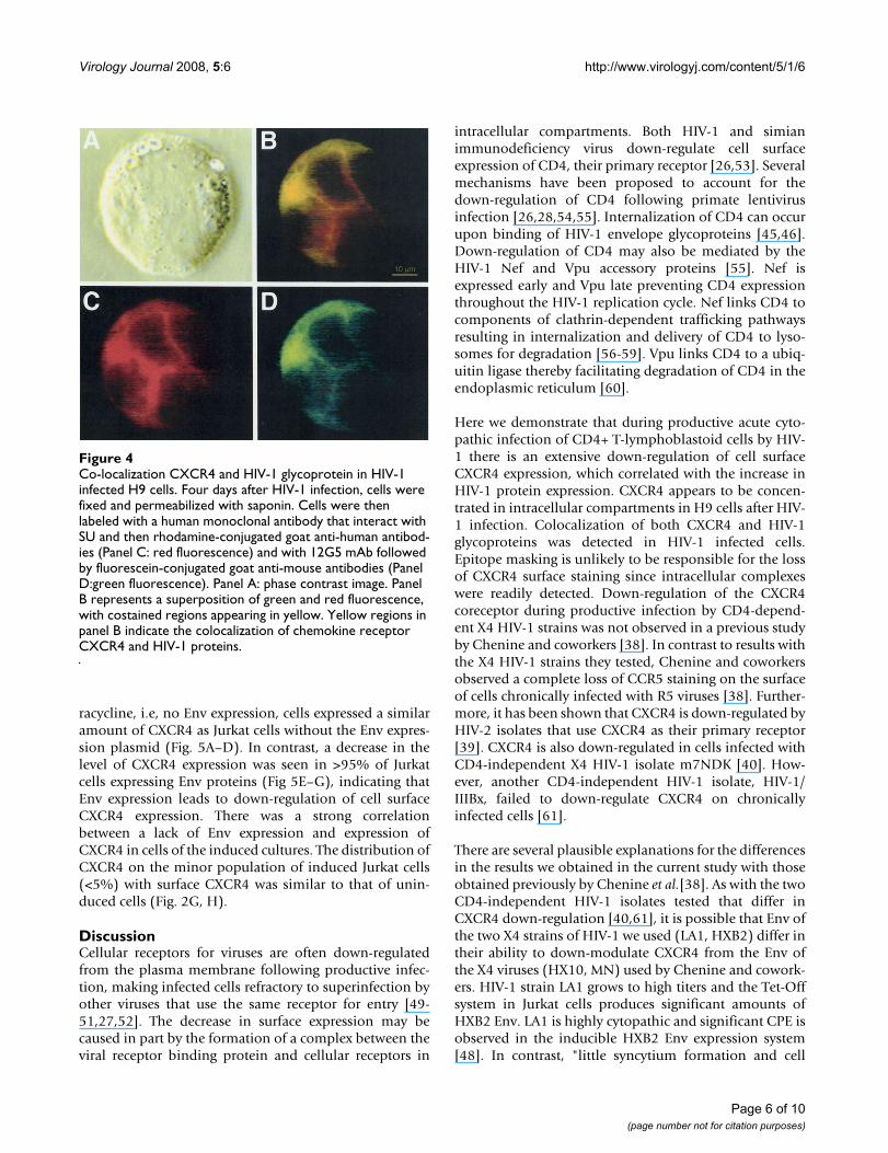
Co-localization CXCR4 and HIV-1 glycoprotein in HIV-1 infected H9 cellsFigure 4Co-localization CXCR4 and HIV-1 glycoprotein in HIV-1 infected H9 cells. Four days after HIV-1 infection, cells were fixed and permeabilized with saponin. Cells were then labeled with a human monoclonal antibody that interact with SU and then rhodamine-conjugated goat anti-human antibod-ies (Panel C: red fluorescence) and with 12G5 mAb followed by fluorescein-conjugated goat anti-mouse antibodies (Panel D:green fluorescence). Panel A: phase contrast image. Panel B represents a superposition of green and red fluorescence, with costained regions appearing in yellow. Yellow regions in panel B indicate the colocalization of chemokine receptor CXCR4 and HIV-1 proteins.
Page 6 of 10(page number not for citation purposes)
Virology Journal 2008, 5:6 http://www.virologyj.com/content/5/1/6
Page 7 of 10(page number not for citation purposes)
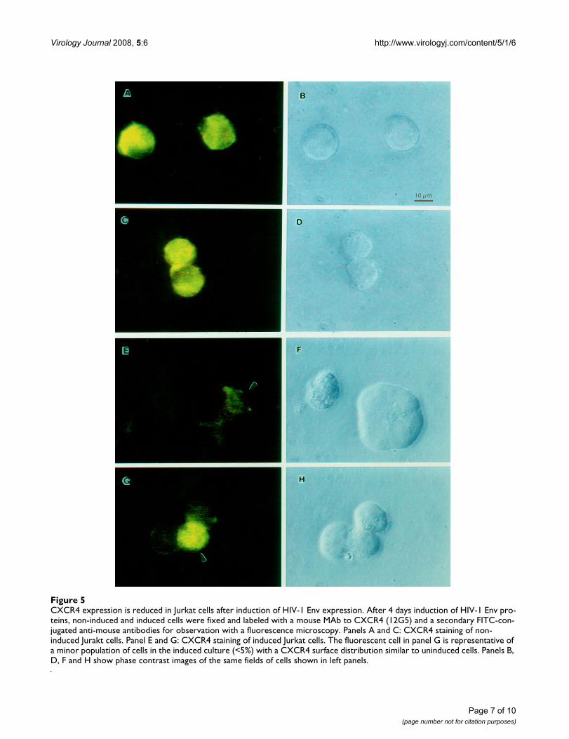
CXCR4 expression is reduced in Jurkat cells after induction of HIV-1 Env expressionFigure 5CXCR4 expression is reduced in Jurkat cells after induction of HIV-1 Env expression. After 4 days induction of HIV-1 Env pro-teins, non-induced and induced cells were fixed and labeled with a mouse MAb to CXCR4 (12G5) and a secondary FITC-con-jugated anti-mouse antibodies for observation with a fluorescence microscopy. Panels A and C: CXCR4 staining of non-induced Jurakt cells. Panel E and G: CXCR4 staining of induced Jurkat cells. The fluorescent cell in panel G is representative of a minor population of cells in the induced culture (<5%) with a CXCR4 surface distribution similar to uninduced cells. Panels B, D, F and H show phase contrast images of the same fields of cells shown in left panels.
Virology Journal 2008, 5:6 http://www.virologyj.com/content/5/1/6
death" was observed in the X4 HIV-1 infected culturesused by Chenine and coworkers [38]. The CD4 independ-ent HIV-2 strain that down-regulates CXCR4 used byEndres et al. (1996) was also highly cytopathic. However,it is unlikely that cytopathic effects are responsible for thedecrease in surface CXCR4 by simply selecting for cells inthe culture with a low level of CXCR4. CXCR4 is uni-formly present on the cells in the RH9 and Jurkat cultures.It is possible that other strains of HIV-1, which grow tolower titers than LA1 or produce less HIV-1 Env than theHXB2 inducible expression system, may have a smallerimpact on cell surface CXCR4 for stochastic reasons. TheEnv of the strains used here may also have a higher affin-ities for CXCR4 than certain other X4 viruses, allowingdirect CXCR4-Env complexing intracellularly. It is alsopossible that differences in the ability to down-regulateCXCR4 are cell specific. However, we used two differentcell lines, RH9 and Jurkat, in the current studies andobserved HIV-1 induced CXCR4 down-regulation in both.We also observed a partial down-regulation of CXCR4 inprimary human peripheral blood mononuclear cells afterinfection of HIV-1 (not shown).
Alteration in CXCR4 expression after infection by HIV-1could result from sequestration of CXCR4 intracellularlyor from the direct effects of other HIV-1 proteins on thesynthesis of CXCR4 or its transport to the cell surface. Sev-eral studies have shown that HIV-1 SU can displace chem-okines from their receptors [34,35]. Interactions betweenSU, CD4, and CXCR4 have also been well established[62,36]. Previous studies demonstrated that treatmentwith the HIV-1 SU increased colocalization of CD4 withCXCR4 and cocapping of the gp120-CD4-CXCR4 com-plexes resulted in the cointernalization of a proportion ofthe gp120-CXCR4 complexes into intracellular vesicles[37]. We did observe down-regulation of surface CXCR4in an inducible system for Env (and Rev) in which acces-sory proteins Nef and Vpu are not expressed. However,given other studies suggesting that Nef and Vpu may beable to down-regulate CXCR4 independently of Env, therole these proteins should be considered in future work.HHV-6 and HHV-7 induce down-regulation of CXCR4[63]. These viruses do not use CXCR4 for cell entry, andinduce a markedly decreased level of CXCR4 gene tran-scription without any significant alteration of the post-transcriptional stability of CXCR4 mRNA. Reduced levelsof CXCR4 mRNA transcripts were observed in cellsinfected with CD4-independent HIV-1 isolate [26]. Fur-thermore, the modulation of CCR5 expression by the R5viruses is at the level of transcription [38]. Further experi-ments will be needed to determine the mechanisms ofdown-modulation of surface CXCR4 by HIV-1.
ConclusionThe amount of surface CXCR4 was greatly reduced in T-lymphoblastoid cells infected with HIV-1 strain LA1, butexpression of another membrane antigen, CD3, was unaf-fected. CXCR4 was concentrated in intracellular compart-ments in RH9 cells after HIV-1 infection.Immunofluorescence microscopy studies showed thatCXCR4 and HIV-1 glycoproteins were co-localized in HIV-1 infected cells. Inducible expression of HIV-1 envelopeglycoproteins also resulted in down-regulation of CXCR4from the cell surface. CXCR4 down-regulation may be duein part to intracellular sequestering of HIV glycoprotein/CXCR4 complexes.
MethodsCells and virusCells of the RH9 subclone of the CD4+ human T-lym-phoblastoid cell line RH9 were the kind gift of Dr. SuraiyaRasheed (University of Southern California), and weremaintained in RPMI 1640 supplemented with 10% fetalbovine serum (GIBCO, Long Island, NY), penicillin (100U/ml) and streptomycin (100 μg/ml). Joseph Sodroski(Harvard University) kindly provided the Env-inducibleJurkat cell line [48].
Flow cytometry and immunofluorescence microscopyRH9 T-lymphoblastoid cells were infected with HIV-1LA1at a MOI of 4 or mock-infected. At various times after theaddition of virus, cells were fixed in 4% paraformalde-hyde for 15 min at room temperature, washed and stainedwith the mouse MAb 12G5 (10 μg/ml) against humanCXCR4 followed by fluorescein isothiocyanate (FITC)-conjugated goat anti-mouse immunoglobulin G (Sigma).In some experiments cells were permeabilized by incuba-tion with 0.05% saponin in PBS for 15 min prior to addi-tion of antibody. CXCR4 monoclonal antibody 12G5derived by Dr. James Hoxie [39] was obtained through theAIDS Research and Reference Reagent Program, Divisionof AIDS, NIAID, NIH. Mouse isotype-matched antibodies(Sigma) were used as a negative control for the gating ofthose cells staining negative for a cell surface marker. Flowcytometry was performed on a Coulter EPICS fluores-cence-activated flow cytometer (Coulter Electronics,Hialeah, Fla.). For immunofluorescence microscopy cellswere analyzed with a Nikon microscope equipped for epi-fluorescence. Fluorescent images were acquired with anOlympus microscope, a 100 W UV source, appropriateexciter and blocking filters, captured with a CCD, andprocessed with Adobe PhotoShop.
Competing interestsThe author(s) declare that they have no competing inter-ests.
Page 8 of 10(page number not for citation purposes)
Virology Journal 2008, 5:6 http://www.virologyj.com/content/5/1/6
Authors' contributionsBC performed all experiments with substantial help fromPJG and AH. RFG, SV and CDF provided guidance, exper-tise, equipment, and funding for these experiments. Allauthors have read and approved this manuscript.
AcknowledgementsThis research was supported by Public Health Service grants AI054238, AI054626 and AI068230 from the National Institute of Allergy and Infec-tious Diseases. We thank Drs. Rasheed, Sodroski and Hoxie for making materials available.
References1. Murphy PM: Viral exploitation and subversion of the immune
system through chemokine mimicry. Nat Immunol 2001,2(2):116-122.
2. Baggiolini M, Dewald B, Moser B: Human chemokines: an update.Annu Rev Immunol 1997, 15:675-705.
3. Allen SJ, Crown SE, Handel TM: Chemokine: receptor structure,interactions, and antagonism. Annu Rev Immunol 2007,25:787-820.
4. Bacon K, Baggiolini M, Broxmeyer H, Horuk R, Lindley I, MantovaniA, Maysushima K, Murphy P, Nomiyama H, Oppenheim J, Rot A,Schall T, Tsang M, Thorpe R, Van Damme J, Wadhwa M, Yoshie O,Zlotnik A, Zoon K: Chemokine/chemokine receptor nomen-clature. J Interferon Cytokine Res 2002, 22(10):1067-1068.
5. Feng Y, Broder CC, Kennedy PE, Berger EA: HIV-1 entry cofactor:functional cDNA cloning of a seven-transmembrane, G pro-tein-coupled receptor [see comments]. Science 1996,272(5263):872-877.
6. Moore JP: Coreceptors: implications for HIV pathogenesisand therapy. Science 1997, 276(5309):51-52.
7. Berger EA, Murphy PM, Farber JM: Chemokine receptors as HIV-1 coreceptors: roles in viral entry, tropism, and disease. AnnuRev Immunol 1999, 17:657-700.
8. Bjorndal A, Deng H, Jansson M, Fiore JR, Colognesi C, Karlsson A,Albert J, Scarlatti G, Littman DR, Fenyo EM: Coreceptor usage ofprimary human immunodeficiency virus type 1 isolates var-ies according to biological phenotype. J Virol 1997,71(10):7478-7487.
9. Dragic T, Litwin V, Allaway GP, Martin SR, Huang Y, Nagashima KA,Cayanan C, Maddon PJ, Koup RA, Moore JP, Paxton WA: HIV-1entry into CD4+ cells is mediated by the chemokine recep-tor CC- CKR-5 [see comments]. Nature 1996,381(6584):667-673.
10. Alkhatib G, Combadiere C, Broder CC, Feng Y, Kennedy PE, MurphyPM, Berger EA: CC CKR5: a RANTES, MIP-1alpha, MIP-1betareceptor as a fusion cofactor for macrophage-tropic HIV-1.Science 1996, 272(5270):1955-1958.
11. He J, Chen Y, Farzan M, Choe H, Ohagen A, Gartner S, Busciglio J,Yang X, Hofmann W, Newman W, Mackay CR, Sodroski J, GabuzdaD: CCR3 and CCR5 are co-receptors for HIV-1 infection ofmicroglia. Nature 1997, 385(6617):645-649.
12. Choe H, Farzan M, Sun Y, Sullivan N, Rollins B, Ponath PD, Wu L,Mackay CR, LaRosa G, Newman W, Gerard N, Gerard C, Sodroski J:The beta-chemokine receptors CCR3 and CCR5 facilitateinfection by primary HIV-1 isolates. Cell 1996,85(7):1135-1148.
13. Bleul CC, Farzan M, Choe H, Parolin C, Clark-Lewis I, Sodroski J,Springer TA: The lymphocyte chemoattractant SDF-1 is a lig-and for LESTR/fusin and blocks HIV-1 entry. Nature 1996,382(6594):829-833.
14. Combadiere C, Ahuja SK, Tiffany HL, Murphy PM: Cloning andfunctional expression of CC CKR5, a human monocyte CCchemokine receptor selective for MIP-1(alpha), MIP-1(beta),and RANTES. J Leukoc Biol 1996, 60(1):147-152.
15. Samson M, Labbe O, Mollereau C, Vassart G, Parmentier M: Molec-ular cloning and functional expression of a new human CC-chemokine receptor gene. Biochemistry 1996, 35(11):3362-3367.
16. Blanpain C, Migeotte I, Lee B, Vakili J, Doranz BJ, Govaerts C, VassartG, Doms RW, Parmentier M: CCR5 binds multiple CC-chemok-
ines: MCP-3 acts as a natural antagonist. Blood 1999,94(6):1899-1905.
17. Jansson M, Popovic M, Karlsson A, Cocchi F, Rossi P, Albert J, WigzellH: Sensitivity to inhibition by beta-chemokines correlateswith biological phenotypes of primary HIV-1 isolates. ProcNatl Acad Sci U S A 1996, 93(26):15382-15387.
18. Oravecz T, Pall M, Norcross MA: Beta-chemokine inhibition ofmonocytotropic HIV-1 infection. Interference with a post-binding fusion step. J Immunol 1996, 157(4):1329-1332.
19. Capobianchi MR, Abbate I, Antonelli G, Turriziani O, Dolei A, Dian-zani F: Inhibition of HIV type 1 BaL replication by MIP-1alpha,MIP-1beta, and RANTES in macrophages. AIDS Res Hum Ret-roviruses 1998, 14(3):233-240.
20. Stantchev TS, Broder CC: Consistent and significant inhibitionof human immunodeficiency virus type 1 envelope-mediatedmembrane fusion by beta-chemokines (RANTES) in primaryhuman macrophages. J Infect Dis 2000, 182(1):68-78.
21. Pugach P, Marozsan AJ, Ketas TJ, Landes EL, Moore JP, Kuhmann SE:HIV-1 clones resistant to a small molecule CCR5 inhibitoruse the inhibitor-bound form of CCR5 for entry. Virology 2007,361(1):212-228.
22. Simmons G, Clapham PR, Picard L, Offord RE, Rosenkilde MM,Schwartz TW, Buser R, Wells TNC, Proudfoot AE: Potent inhibi-tion of HIV-1 infectivity in macrophages and lymphocytes bya novel CCR5 antagonist. Science 1997, 276(5310):276-279.
23. Clapham PR, Reeves JD, Simmons G, Dejucq N, Hibbitts S, McKnightA: HIV coreceptors, cell tropism and inhibition by chemok-ine receptor ligands. Mol Membr Biol 1999, 16(1):49-55.
24. Simmons G, Reeves JD, Hibbitts S, Stine JT, Gray PW, Proudfoot AE,Clapham PR: Co-receptor use by HIV and inhibition of HIVinfection by chemokine receptor ligands. Immunol Rev 2000,177:112-126.
25. Trkola A, Ketas TJ, Nagashima KA, Zhao L, Cilliers T, Morris L,Moore JP, Maddon PJ, Olson WC: Potent, broad-spectrum inhi-bition of human immunodeficiency virus type 1 by the CCR5monoclonal antibody PRO 140. J Virol 2001, 75(2):579-588.
26. Hoxie JA, Alpers JD, Rackowski JL, Huebner K, Haggarty BS, Cedar-baum AJ, Reed JC: Alterations in T4 (CD4) protein and mRNAsynthesis in cells infected with HIV. Science 1986,234(4780):1123-1127.
27. Potash MJ, Volsky DJ: Viral interference in HIV-1 infected cells.Rev Med Virol 1998, 8(4):203-211.
28. Crise B, Buonocore L, Rose JK: CD4 is retained in the endoplas-mic reticulum by the human immunodeficiency virus type 1glycoprotein precursor. J Virol 1990, 64(11):5585-5593.
29. Hoxie JA, Rackowski JL, Haggarty BS, Gaulton GN: T4 endocytosisand phosphorylation induced by phorbol esters but not bymitogen or HIV infection. J Immunol 1988, 140(3):786-795.
30. Geleziunas R, Bour S, Wainberg MA: HIV-1 associated down-modulation of CD4 gene expression is differentiallyrestricted in lymphocytic and monocytic cell lines. J LeukocBiol 1994, 55(5):589-595.
31. Amara A, Gall SL, Schwartz O, Salamero J, Montes M, Loetscher P,Baggiolini M, Virelizier JL, Arenzana-Seisdedos F: HIV coreceptordownregulation as antiviral principle: SDF-1alpha- depend-ent internalization of the chemokine receptor CXCR4 con-tributes to inhibition of HIV replication. J Exp Med 1997,186(1):139-146.
32. Aramori I, Ferguson SS, Bieniasz PD, Zhang J, Cullen B, Cullen MG:Molecular mechanism of desensitization of the chemokinereceptor CCR-5: receptor signaling and internalization aredissociable from its role as an HIV-1 co-receptor. Embo J 1997,16(15):4606-4616.
33. Brandt SM, Mariani R, Holland AU, Hope TJ, Landau NR: Associa-tion of chemokine-mediated block to HIV entry with core-ceptor internalization. J Biol Chem 2002, 277(19):17291-17299.
34. Madani N, Kozak SL, Kavanaugh MP, Kabat D: gp120 envelopeglycoproteins of human immunodeficiency viruses competi-tively antagonize signaling by coreceptors CXCR4 andCCR5. Proc Natl Acad Sci U S A 1998, 95(14):8005-8010.
35. Wang JM, Ueda H, Howard OM, Grimm MC, Chertov O, Gong X,Gong W, Resau JH, Broder CC, Evans G, Arthur LO, Ruscetti FW,Oppenheim JJ: HIV-1 envelope gp120 inhibits the monocyteresponse to chemokines through CD4 signal-dependentchemokine receptor down-regulation. J Immunol 1998,161(8):4309-4317.
Page 9 of 10(page number not for citation purposes)
Virology Journal 2008, 5:6 http://www.virologyj.com/content/5/1/6
Publish with BioMed Central and every scientist can read your work free of charge
"BioMed Central will be the most significant development for disseminating the results of biomedical research in our lifetime."
Sir Paul Nurse, Cancer Research UK
Your research papers will be:
available free of charge to the entire biomedical community
peer reviewed and published immediately upon acceptance
cited in PubMed and archived on PubMed Central
yours — you keep the copyright
Submit your manuscript here:http://www.biomedcentral.com/info/publishing_adv.asp
BioMedcentral
36. Lapham CK, Ouyang J, Chandrasekhar B, Nguyen NY, Dimitrov DS,Golding H: Evidence for cell-surface association between fusinand the CD4-gp120 complex in human cell lines [see com-ments]. Science 1996, 274(5287):602-605.
37. Ugolini S, Moulard M, Mondor I, Barois N, Demandolx D, Hoxie J,Brelot A, Alizon M, Davoust J, Sattentau QJ: HIV-1 gp120 inducesan association between CD4 and the chemokine receptorCXCR4. J Immunol 1997, 159(6):3000-3008.
38. Chenine AL, Sattentau Q, Moulard M: Selective HIV-1-induceddownmodulation of CD4 and coreceptors. Arch Virol 2000,145(3):455-471.
39. Endres MJ, Clapham PR, Marsh M, Ahuja M, Turner JD, McKnight A,Thomas JF, Stoebenau-Haggarty B, Choe S, Vance PJ, Wells TN,Power CA, Sutterwala SS, Doms RW, Landau NR, Hoxie JA: CD4-independent infection by HIV-2 is mediated by fusin/CXCR4.Cell 1996, 87(4):745-756.
40. Valente ST, Chanel C, Dumonceaux J, Olivier R, Marullo S, Briand P,Hazan U: CXCR4 is down-regulated in cells infected with theCD4-independent X4 human immunodeficiency virus type 1isolate m7NDK. J Virol 2001, 75(1):439-447.
41. Michel N, Allespach I, Venzke S, Fackler OT, Keppler OT: The Nefprotein of human immunodeficiency virus establishes super-infection immunity by a dual strategy to downregulate cell-surface CCR5 and CD4. Curr Biol 2005, 15(8):714-723.
42. Venzke S, Michel N, Allespach I, Fackler OT, Keppler OT: Expres-sion of Nef downregulates CXCR4, the major coreceptor ofhuman immunodeficiency virus, from the surfaces of targetcells and thereby enhances resistance to superinfection. JVirol 2006, 80(22):11141-11152.
43. Cefai D, Ferrer M, Serpente N, Idziorek T, Dautry-Varsat A, DebreP, Bismuth G: Internalization of HIV glycoprotein gp120 isassociated with down-modulation of membrane CD4 andp56lck together with impairment of T cell activation. J Immu-nol 1992, 149(1):285-294.
44. Bour S, Boulerice F, Wainberg MA: Inhibition of gp160 and CD4maturation in U937 cells after both defective and productiveinfections by human immunodeficiency virus type 1. J Virol1991, 65(12):6387-6396.
45. Fujita K, Omura S, Silver J: Rapid degradation of CD4 in cellsexpressing human immunodeficiency virus type 1 Env andVpu is blocked by proteasome inhibitors. J Gen Virol 1997, 78 (Pt 3):619-625.
46. Su SB, Ueda H, Howard OM, Grimm MC, Gong W, Ruscetti FW,Oppenheim JJ, Wang JM: Inhibition of the expression and func-tion of chemokine receptors on human CD4+ leukocytes byHIV-1 envelope protein gp120. Chem Immunol 1999, 72:141-160.
47. Gossen M, Bujard H: Studying gene function in eukaryotes byconditional gene inactivation. Annu Rev Genet 2002, 36:153-173.
48. Cao J, Park IW, Cooper A, Sodroski J: Molecular determinants ofacute single-cell lysis by human immunodeficiency virus type1. J Virol 1996, 70(3):1340-1354.
49. Vogt PK, Ishizaki R: Patterns of viral interference in the avianleukosis and sarcoma complex. Virology 1966, 30(3):368-374.
50. Temin HM: Mechanisms of cell killing/cytopathic effects bynonhuman retroviruses. Rev Infect Dis 1988, 10(2):399-405.
51. Weller SK, Joy AE, Temin HM: Correlation between cell killingand massive second-round superinfection by members ofsome subgroups of avian leukosis virus. J Virol 1980,33(1):494-506.
52. Nethe M, Berkhout B, van der Kuyl AC: Retroviral superinfectionresistance. Retrovirology 2005, 2:52.
53. Salmon P, Olivier R, Riviere Y, Brisson E, Gluckman JC, Kieny MP,Montagnier L, Klatzmann D: Loss of CD4 membrane expressionand CD4 mRNA during acute human immunodeficiencyvirus replication. J Exp Med 1988, 168(6):1953-1969.
54. Crise B, Rose JK: Human immunodeficiency virus type 1 glyc-oprotein precursor retains a CD4-p56lck complex in theendoplasmic reticulum. J Virol 1992, 66(4):2296-2301.
55. Lindwasser OW, Chaudhuri R, Bonifacino JS: Mechanisms of CD4downregulation by the Nef and Vpu proteins of primateimmunodeficiency viruses. Curr Mol Med 2007, 7(2):171-184.
56. Gama Sosa MA, DeGasperi R, Kim YS, Fazely F, Sharma P, RuprechtRM: Serine phosphorylation-independent downregulation ofcell-surface CD4 by nef. AIDS Res Hum Retroviruses 1991,7(11):859-860.
57. Kim YH, Chang SH, Kwon JH, Rhee SS: HIV-1 Nef plays an essen-tial role in two independent processes in CD4 down-regula-tion: dissociation of the CD4-p56(lck) complex and targetingof CD4 to lysosomes. Virology 1999, 257(1):208-219.
58. Stoddart CA, Geleziunas R, Ferrell S, Linquist-Stepps V, Moreno ME,Bare C, Xu W, Yonemoto W, Bresnahan PA, McCune JM, GreeneWC: Human immunodeficiency virus type 1 Nef-mediateddownregulation of CD4 correlates with Nef enhancement ofviral pathogenesis. J Virol 2003, 77(3):2124-2133.
59. Chaudhuri R, Lindwasser OW, Smith WJ, Hurley JH, Bonifacino JS:Downregulation of CD4 by human immunodeficiency virustype 1 Nef is dependent on clathrin and involves direct inter-action of Nef with the AP2 clathrin adaptor. J Virol 2007,81(8):3877-3890.
60. Willey RL, Maldarelli F, Martin MA, Strebel K: Human immunode-ficiency virus type 1 Vpu protein induces rapid degradationof CD4. J Virol 1992, 66(12):7193-7200.
61. Hoxie JA, LaBranche CC, Endres MJ, Turner JD, Berson JF, DomsRW, Matthews TJ: CD4-independent utilization of the CXCR4chemokine receptor by HIV-1 and HIV-2. J Reprod Immunol1998, 41(1-2):197-211.
62. Trkola A, Dragic T, Arthos J, Binley JM, Olson WC, Allaway GP,Cheng-Mayer C, Robinson J, Maddon PJ, Moore JP: CD4-depend-ent, antibody-sensitive interactions between HIV-1 and itsco- receptor CCR-5 [see comments]. Nature 1996,384(6605):184-187.
63. Yasukawa M, Hasegawa A, Sakai I, Ohminami H, Arai J, Kaneko S,Yakushijin Y, Maeyama K, Nakashima H, Arakaki R, Fujita S: Down-regulation of CXCR4 by human herpesvirus 6 (HHV-6) andHHV-7. J Immunol 1999, 162(9):5417-5422.
Page 10 of 10(page number not for citation purposes)


