Embed Size (px)
Citation preview

Int.J.Curr.Microbiol.App.Sci (2019) 8(7): 1201-1211
1201
Original Research Article https://doi.org/10.20546/ijcmas.2019.807.143
Study of Microbiological Profile and Antibiotic Susceptibility of Blood
Stream Infections in Tertiary Care Hospital
K. Vidyasagar* and D. Venkatesha
Department of Microbiology, Adichunchanagiri Institute of Medical Sciences, BG Nagara,
Nagamangala (Taluk), Mandya (Dist)-PIN: 571448, Karnataka, India
*Corresponding author
A B S T R A C T
Introduction
Blood stream infections (BSIs) can lead to
life-threatening sepsis and are globally
associated with high morbidity and
mortality.[1]
Blood stream infections (BSI) are
defined by the presence and active
multiplication of microorganisms in the blood
stream.[2]
BSI by the place of acquisition is
categorized either community associated or
hospital associated. Blood stream infections
occur when bacteria enter the blood stream
from either a primary focus of infection in an
organ (UTI, Pneumonia, meningitis...), a
wound or via an indwelling or implanted
device.[3]
Health care associated (HCA) BSIs
can occur as complications following medical
and surgical procedures or the insertion of an
intravascular or indwelling device.[4]
Blood
stream infections (BSIs) have serious
International Journal of Current Microbiology and Applied Sciences ISSN: 2319-7706 Volume 8 Number 07 (2019) Journal homepage: http://www.ijcmas.com
Blood stream infections (BSIs) can lead to life-threatening sepsis and are globally
associated with high morbidity and mortality. Early diagnosis plays a crucial role in
managing BSI. Objective is to identify the pathogens causing blood stream infections
and to know their antibiotic sensitivity pattern. This was a retrospective study of 1
year duration. A total of 1332 blood samples from clinically diagnosed cases of blood
stream infections received in the microbiology laboratory were included in the study.
Blood samples were processed and isolates were identified by standard biochemical
tests and antibiotic susceptibility testing was done by Kirby Bauer disc diffusion
methods as per CLSI guidelines. Out of 1332 blood samples received, 204 (15.3%)
samples showed growth and 1128 (84.68%) samples showed no growth, with total
percentage of culture positivity being 15.3%. Among 204 positive cultures, 202 (99%)
showed bacterial growth and 2 (0.98%) were Candida spp. Bacteremia due to Gram-
positive pathogens was more common compared to Gram-negative pathogens. The
present study provides information about pathogens responsible for blood stream
infections and their antibiotic susceptibility. Antibiotic susceptibility pattern of
isolates provides useful guidelines to clinicians in initiating empiric therapy and help
in management of blood stream infections.
K e y w o r d s
Blood stream
infections (BSI),
Microbiological
profile, Antimicrobial susceptibility
Accepted:
10 June 2019
Available Online:
10 July 2019
Article Info

Int.J.Curr.Microbiol.App.Sci (2019) 8(7): 1201-1211
1202
consequences such as shock, disseminated
intravascular coagulation, multiple organ
failure, and even death. Early diagnosis plays
a crucial role in managing BSI, and hence,
prompt detection of such infections is a
critical function of clinical microbiology
laboratories. [5]
BSI can be caused by both Gram-positive and
Gram-negative microorganisms as well as
fungi. Common Gram negative bacteria are
Escherichia coli, Klebsiella spp, Enterobacter
spp, Proteus spp, Salmonella typhi,
Pseudomonas aeruginosa, Acinetobacter spp,
Haemophilus influenza, Brucella spp and
Neisseria meningitidis. Common Gram
positive bacteria are Staphylococcus aureus,
Coagulase negative Staphylococci (CONS),
Enterococci and alpha haemolytic (viridans)
streptococci. [6]
Blood culture is the gold standard for the
detection of blood stream infection.[7]
One of
the main complication in the treatment of BSI
is the increasing resistance of bacteria to
antibiotics.
Emerging drug resistance among blood stream
pathogens limit therapeutic options and
complicate patient’s management. [8]
Today
the only way to reduce mortality due to blood
stream infection is early diagnosis and
appropriate antimicrobial therapy at the
earliest.
The aim of the present study to identify the
pathogens causing blood stream infection and
to know their antibiotic sensitivity pattern,
thus providing useful guidance to clinicians to
antibiotic therapy.
Materials and Methods
This was a retrospective study conducted for a
period of one year in a tertiary care hospital. A
total of 1332 blood samples from clinically
diagnosed cases of blood stream infections
received in the microbiology laboratory were
included in the study.
Blood samples were collected from clinically
suspected bacteremia cases before the
administration of antibiotics under aseptic
precautions and inoculated into brain heart
infusion broth. A volume of 5–10 ml from
adults and 2–3 ml from pediatric patients were
obtained for culture. The culture bottles were
incubated at 37°C aerobically for 18-24 hours
and periodic subcultures were done onto
MacConkey agar and blood agar on day 2, day
4 and finally on day 7 and samples were
reported as no growth after 7 days of aerobic
incubation. Bacterial growth on the
subcultures was identified by colony
morphology, Gram staining, and standard
biochemical tests. [6]
Antibiotic susceptibility
testing was done by Kirby Bauer disc
diffusion methods as per CLSI guidelines.
Cefoxitin disc diffusion method used to
identify Methicillin resistant Staphylococcus
aureus (MRSA) and Methicillin resistant
Coagulase negative Staphylococci
(MRCONS) among Staphylococcus aureus
and Coagulase negative Staphylococci
respectively.[9]
Extended spectrum beta-
lactamases (ESBL) in Gram-negative bacilli
were studied by phenotypic method with
ceftazidime (30µg) and ceftazidime +
clavulanic acid (30µg+10µg) as per CLSI
guidelines.[9]
MDR (Multi drug resistant) was
defined as non-susceptibility to at least one
agent in three or more antimicrobial
categories. [9]
Results and Discussion
In the present study, 1332 blood samples were
received and processed for aerobic culture.
Out of 1332 blood samples, 204 (15.3%)
samples showed growth, 1128(84.68%)
samples showed no growth and with total
percentage of culture positivity being 15.3%.

Int.J.Curr.Microbiol.App.Sci (2019) 8(7): 1201-1211
1203
Out of 1332 blood samples received, 793
(59.53%) were from male and 539(40.46%)
were female, maximum from the age group of
<1year (37.31%), followed by 1-10 year
(19.29%), 21-30years (7.73%) and 31-40
years (7.28%) respectively (Figure 1).
The ward wise distribution of samples
includes 1254(94.19%) received from IPD,
72(5.40%) from ICU and 6(0.45%) from
OPD.
Among 204 positive cultures, 202(99%)
showed bacterial growth and 2(0.98%) were
Candida spp. Out of 202 (99%) bacterial
growth, Gram positive organisms were
128(62.74%) and Gram negative organisms
were 74(36.27%) respectively (Table 1).
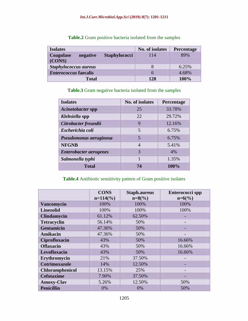
Among Gram positive isolates (128), the most
predominant isolate was Coagulase negative
Staphylococci (CONS) 114(89%) followed by
Staphylococcus aureus 8(6.25%) and
Enterococcus faecalis 6(4.68%) (Table 2).
Methicillin resistance Coagulase negative
Staphylococci (MRCONS) was found in
82.45% of total CONS isolates and Methicillin
resistance Staphylococcus aureus (MRSA) in
50% of total Staphylococcus aureus isolates.
Among Gram negative isolates (74), the
predominant isolate was Acinetobacter spp
25(33.78%) followed by Klebsiella spp
22(29.72%), Citrobacter freundii 9 (12.16%),
Escherichia coli 5 (6.75%), Pseudomonas
aeruginosa 5(6.25%), NFGNB 4 (5.4%),
Enterobacter aerogenes 3(4%) and
Salmonella typhi 1 (1.35%) (Table 3).
Antibiotic sensitivity pattern of Gram positive
and Gram negative organisms was studied.
CONS was 100% sensitive to Vancomycin
and Linezolid, followed by Clindamycin
(61.12%), Tetracycline (56.14%), Amikacin
and Gentamicin (47.76% each).
Staphylococcus aureus was 100% sensitive to
Vancomycin and Linezolid, followed by
Clindamycin (62.5%) and Ciprofloxacin,
Oflaxacin, Levofloxacin, Amikacin,
Gentamicin and Tetracycline (50% each)
respectively. Enterococcus spp showed 100%
sensitive to Vancomycin and Linezolid,
followed by 50% Amoxy-Clav and Penicillin
(Table 4).
Out of total 114 CONS isolates, 94 were
MRCONS and among 8 Staphylococcus
aureus isolates, 4 were MRSA and the
incidence of MRCONS and MRSA being
82.45% and 50% respectively.
The most effective antibiotic against Gram
positive organisms were Vancomycin and
Linezolid (100% each) followed by
Clindamycin (60.12%) and Tetracycline
(56.14%).
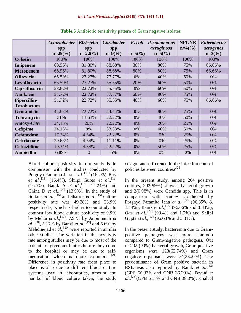
The most effective antibiotic against
Acinetobacter spp was Colistin (100%),
followed by Imipenem and Meropenem
(68.96% each), Oflaxacin and Levofloxacin
(65.50% each), Ciprofloxacin (58.62%),
Amikacin and Pipercillin-Tazobactam
(51.72% each). Klebsiella spp showed 100%
sensitive to Colistin, followed by 81.8% to
Imipenem and Meropenem. Citrobacter spp
was 100% sensitive to Colistin, followed by
88.88% to Imipenem and Meropenem, 77.77%
to Amikacin and Oflaxacin, 55.55% to
Pipercillin-Tazobactam, Ciprofloxacin and
Levofloxacin. Escherichia coli showed 100%
sensitive to Colistin, followed by Imipenem
and Meropenem (80% each), Amikacin (60%)
(Table 5).
MDR was found in 60.80% of Gram negative
isolates and ESBL were 20.27%. MDR was
found high among Enterobacteriaceae.
Carbapenem resistance was seen more among
Nonfermenters (31%) as compared to
Enterobacteriaceae (20.2%)

Int.J.Curr.Microbiol.App.Sci (2019) 8(7): 1201-1211
1204
The most effective antibiotic against Gram
negative organisms were Colistin followed by
Imipenem, Meropenem, Amikacin and
Oflaxacin.
In the present study, majority of the isolates
showed high resistance to commonly used
antibiotics belongs to Penicillins (Ampicillin
&Amoxy-Clav), Cephalosporins (Cefotaxime,
Ceftriaxone and Ceftazidime),
Fluoroquinolones (Ciprofloxacin,Ofloxacin
and Levofloxacin) and Aminoglycosides
(Gentamicin and Amikacin).
Blood stream infections constitute one of the
most serious conditions and associated with
high morbidity and mortality as a result,
timely detection, identification and
antimicrobial susceptibility testing of blood
stream pathogens are important.
The gold standard for diagnosis of BSIs is
blood culture. [7]
The present study gives
information about pathogens causing blood
stream infections. It also provides information
about antibiotic sensitivity pattern that plays
an important role in management of
septicaemia cases.
In this study, total 1332 blood samples were
received, out of which 204 (15.3%) samples
showed growth and 1128(84.68%) showed no
growth, with culture positivity being 15.3%.
Fig.1 Age distribution of patients
0
50
100
150
200
250
300
350
400
450
500
<1 Yr 1-10 yrs 11-20yrs
21-30yrs
31-40yrs
41-50yrs
51-60yrs
61-70yrs
71-80yrs
>80 yrs
497 (37.31%)
257 (19.29%)
89 (6.68%)
103 (7.73%)
97 (7.28%)86 (6.45%)
78 (5.85%)80 (6%)
34 (2.55%)11 (0.82%)
Table.1 Pathogens isolated from samples
Isolates No. of isolates Percentage
Gram positive bacteria 128 62.74%
Gram negative bacteria 74 36.27%
Candida spp 2 0.98%
Total 204 100%
Int.J.Curr.Microbiol.App.Sci (2019) 8(7): 1201-1211
1205
Table.2 Gram positive bacteria isolated from the samples
Isolates No. of isolates Percentage
Coagulase negative Staphylococci
(CONS)
114 89%
Staphylococcus aureus 8 6.25%
Enterococcus faecalis 6 4.68%
Total 128 100%
Table.3 Gram negative bacteria isolated from the samples
Isolates No. of isolates Percentage
Acinetobacter spp 25 33.78%
Klebsiella spp 22 29.72%
Citrobacter freundii 9 12.16%
Escherichia coli 5 6.75%
Pseudomonas aeruginosa 5 6.75%
NFGNB 4 5.41%
Enterobacter aerogenes 3 4%
Salmonella typhi 1 1.35%
Total 74 100%
Table.4 Antibiotic sensitivity pattern of Gram positive isolates
CONS
n=114(%)
Staph.aureus
n=8(%)
Enterococci spp
n=6(%)
Vancomycin 100% 100% 100%
Linezolid 100% 100% 100%
Clindamycin 61.12% 62.50% -
Tetracyclin 56.14% 50% -
Gentamicin 47.36% 50% -
Amikacin 47.36% 50% -
Ciprofloxacin 43% 50% 16.66%
Oflaxacin 43% 50% 16.66%
Levofloxacin 43% 50% 16.66%
Erythromycin 21% 37.50% -
Cotrimoxazole 14% 12.50% -
Chloramphenicol 13.15% 25% -
Cefotaxime 7.90% 37.50% -
Amoxy-Clav 5.26% 12.50% 50%
Penicillin 0% 0% 50%
Int.J.Curr.Microbiol.App.Sci (2019) 8(7): 1201-1211
1206
Table.5 Antibiotic sensitivity pattern of Gram negative isolates
Acinetobacter
spp
n=25(%)
Klebsiella
spp
n=22(%)
Citrobacter
spp
n=9(%)
E. coli
n=5(%)
Pseudomonas
aeruginosa
n=5(%)
NFGNB
n=4(%)
Enterobacter
aerogenes
n=3(%)
Colistin 100% 100% 100% 100% 100% 100% 100%
Imipenem 68.96% 81.80% 88.68% 80% 80% 75% 66.66%
Meropenem 68.96% 81.80% 88.68% 80% 80% 75% 66.66%
Ofloxacin 65.50% 27.27% 77.77% 0% 40% 50% 0%
Levofloxacin 65.50% 27.27% 55.55% 20% 60% 50% 0%
Ciprofloxacin 58.62% 22.72% 55.55% 0% 60% 50% 0%
Amikacin 51.72% 22.72% 77.77% 60% 80% 75% 0%
Pipercillin-
Tazobactam
51.72% 22.72% 55.55% 40% 60% 75% 66.66%
Gentamicin 44.82% 22.72% 44.44% 40% 80% 75% 0%
Tobramycin 31% 13.63% 22.22% 0% 40% 50% 0%
Amoxy-Clav 24.13% 20% 22.22% 0% 20% 25% 0%
Cefipime 24.13% 9% 33.33% 0% 40% 50% 0%
Cefotaxime 17.24% 4.54% 22.22% 0% 0% 25% 0%
Ceftriaxone 20.68% 4.54% 11.11% 0% 0% 25% 0%
Ceftazidime 10.34% 4.54% 22.22% 0% 50% 25% 0%
Ampicillin 6.89% 0 5% 0% 0% 0% 0%
Blood culture positivity in our study is in
comparison with the studies conducted by
Pragnya Paramita Jena et al.,[10]
(16.2%), Roy
et al.,[11]
(16.4%), Shilpi Gupta et al.,[12]
(16.5%), Banik A et al.,[13]
(14.24%)
and
China D et al.,[14]
(13.9%). In the study of
Sultana et al., [15]
and Sharma et al.,[16]
culture
positivity rate was 49.28% and 33.9%
respectively, which is higher to our study. In
contrast low blood culture positivity of 9.9%
by Mehta et al.,[17]
, 7.9 % by Anbumanni et
al.,[18]
, 5.17% by Barati et al.,[19]
and 5.6% by
Mehdinejad et al.,[20]
were reported in similar
other studies. The variation in the positivity
rate among studies may be due to most of the
patient are given antibiotics before they come
to the hospital or may be due to self-
medication which is more common. [21]
Difference in positivity rate from place to
place is also due to different blood culture
systems used in laboratories, amount and
number of blood culture taken, the study
design, and difference in the infection control
policies between countries.[21]
In the present study, among 204 positive
cultures, 202(99%) showed bacterial growth
and 2(0.98%) were Candida spp. This is in
comparison with studies conducted by
Pragnya Paramita Jena et al.,[10]
(96.85% &
3.14%), Banik et al.,[13]
(96.66% and 3.33%),
Qazi et al.,[22]
(98.4% and 1.5%) and Shilpi
Gupta et al.,[12]
(96.68% and 3.31%).
In the present study, bacteremia due to Gram-
positive pathogens was more common
compared to Gram-negative pathogens. Out
of 202 (99%) bacterial growth, Gram positive
organisms were 128(62.74%) and Gram
negative organisms were 74(36.27%). The
predominance of Gram positive bacteria in
BSIs was also reported by Banik et al.,[13]
(GPB 60.37% and GNB 36.29%), Pavani et
al.,[23]
(GPB 61.7% and GNB 38.3%), Khaleel
Int.J.Curr.Microbiol.App.Sci (2019) 8(7): 1201-1211
1207
et al.,[24]
(GPB59.85% and GNB40.15 %),
Gohel et al.,[25]
(GPB 58.3% and GNB
40.2%), Bhavna Bhadauria et al.,[26]
(GPB
57.28% and GNB 42.74%), Ashima Katayi et
al.,[27]
(GPB 57.14% and GNB 42.85%),
Dagnew et al.,[28]
(GPB69% and GNB31%)
and Wasihun et al.,[29]
(GPB 72.2% and GNB
27.8%) respectively.
In the present study, among Gram positive
isolates (128), the most predominant isolate
was CONS 114(89%) followed by
Staphylococcus aureus 8(6.25%) and
Enterococcus spp 6(4.68%). This finding is in
accordance with studies conducted by
Karlowsky et al.,[30]
(CONS 42%, Staph.
aureus 16.2% and Enterococcus spp 8.3%),
Ashima Katayi et al.,[27]
(CONS 55.5%,
Staph.aureus 34% and Enterococcus spp
10.4%), Alam et al.,[31]
(CONS 63.5%, Staph.
aureus 23.1% and Enterococcus spp 5.8%),
Nazir et al.,[32]
(CONS 67.9%, Staph. aureus
24.5%& Enterococcus spp 7.5%), Pragnya
Paramita Jena et al.,[10]
(CONS 40.5%,
Staph.aureus 7.87% and Enterococcus spp
3.1%)
where CONS reported as the most
common isolate causing BSIs.
CONS were mainly recognized as mere
contaminants till 1970’s; however, several
studies have now reported an increasing
incidence of infection by this group of
bacteria. [32]
Over the past two decades,
CONS, the usual skin commensals are
increasingly being considered blood stream
pathogens in select settings. Coagulase
negative Staphylococcus is the third most
common cause of BSI and the most common
cause of nosocomial BSI. [33]
Incidence of
nosocomial bacteremia due to CONS is
increasing due to frequent use of vascular
access devices. Improper methods of blood
collection and the presence of long standing
intravascular catheters are recognized as
possible modes of spread of BSI by CONS.
[13] Some authors have demonstrated that
coagulase-negative Staphylococcus adheres to
the catheter surface, and produces slime,
which are risk factors for BSI. [34]
According
to Souvenir et al., clinical significance of
CONS was defined as at least two blood
cultures positive for CONS within 5 days or
one positive blood culture plus clinical
evidence of infection, which includes
abnormal leukocyte count and temperature or
blood pressure. [35]
In this study, among Gram negative isolates
(74), Acinetobacter spp was the most
predominant organism isolated (39.18%),
followed by Klebsiella spp (29.72%),
Citrobacter spp (12.16%), E.coli (6.75%),
Pseudomonas aeruginosa (6.25%),
Enterobacter spp (4%) and S.typhi (1.35%).
These findings are consistent with other
studies conducted by Pragnya Paramita Jena
et al.,[10]
, Banik et al.,[13]
, Ashima Katayi et
al.,[27]
and Nazir et al.,[32]
where Acinetobacter
spp and Klebsiella spp have been found to be
predominant isolates among Gram negative
organisms.
The reason for high rate of isolation of
Acinetobacter spp among Gram-negative
bacteria may be because of acquisition of
infection during hospital stay, as it is one of
the commonest pathogen seen in nosocomial
infections. Also, their ubiquitous nature in the
hospital environment and inadequate infection
control practice has continuously raised the
incidence of Acinetobacter infections over the
past two decades. [36]
Apart from Gram positive and Gram negative
organisms, Candida albicans were isolated in
two positive blood cultures (0.98%). Similar
observation was made by Qazi et al.,[22]
.
The results of antibiotic sensitivity of Gram-
positive bacteria showed CONS,
Staphylococcus aureus and Enterococcus spp
were 100% sensitive to Vancomycin and
Int.J.Curr.Microbiol.App.Sci (2019) 8(7): 1201-1211
1208
Linezolid followed by Clindamycin and
Tetracycline and were least sensitive to
Penicillins, Cephalosporins and
Fluoroquinolones and this finding similar to
other studies. The incidence of MRCONS was
82.45% and MRSA was 50%. Methicillin
resistance rate was higher in CONS as
compared with Staphylococcus aureus, which
is similar to study by Mathur et al.,[37]
and Mir
et al.,[38]
.
These organisms are notorious since they do
not respond to the broad class of beta lactam
antibiotics and acquire resistance to newer
antibiotics quite rapidly. This effectively
complicates the management of such BSIs. [10]
In the present, the most effective antibiotic
against Gram positive organisms were
Vancomycin and Linezolid followed by
Clindamycin and Tetracycline.
Among Gram negative bacteria,
Acinetobacter spp, Klebsiella spp,
Citrobacter spp, E. coli and NFGNB showed
100% sensitive to Colistin followed by
Imipenem and Meropenem (80% each),
Oflaxacin and Levofloxacin (60% each) and
Amikacin (55%) and least sensitive to
Ampicillin, Amoxicillin+clavulanic acid
combination and Cephalosporins and this is
similar to other studies.
In our study MDR was found in 60.80% of
Gram negative isolates and is in comparison
with study conducted by Shilpi Gupta et
al.,[12]
and Nazir et al.,[32]
and ESBL were
20.27% which is similar to study conducted
by Anathan et al.,[39]
(25.4%).
MDR was
found high among Enterobacteriaceae.
Carbapenem resistance was seen more among
Nonfermenters (31%) as compared to
Enterobacteriaceae (20.2%) and this may be
due to inappropriate empirical use of
Carbapenem as the first line treatment.
The greatest threat with MDR and
Carbapenem resistant Gram negative bacteria
is that the infections are usually untreatable
due to the limited options of the antibiotics
available, resulting into increased mortality.
Worldwide, their incidence is rising with
variations due to regional and geographical
differences as stated by Jadhav et al.,[40]
. With
the shortage of newer drugs availability and
increasing resistance, use of limited option
drugs such as colistin by clinicians could soon
lead to the condition of so called pan drug
resistance.[12]
In the present study, the most
effective antibiotic against Gram negative
organisms were Colistin followed by
Imipenem, Meropenem, Amikacin and
Oflaxacin.
The information of predominant organisms
and their sensitivity among sepsis patients is
essential for making the right choice of
antibiotics in the management of sepsis.
Hence, blood cultures must be obtained from
all suspected cases of bacteraemia or sepsis
before prescribing antibiotics. The main
factors causing the increase in antimicrobial
resistant bacteria are poor infection control
practices and inappropriate use of antibiotics.
Strict infection control measures along with
antibiotic policy for judicious antibiotic
therapy should be implemented in the
hospitals as control measures against blood
stream infections and to check the emergence
of resistance. [41]
Blood stream infections are an important
nosocomial infection responsible for
morbidity and mortality in the patients. The
present study provides information on the
spectrum of pathogens causing blood stream
infections and their antimicrobial
susceptibility profile, helping the clinicians in
early diagnosis and guiding in the
management of blood stream infections. The
study identified both Gram-positive and
Gram-negative bacteria to be responsible for
blood stream infections and most of them
were found to be MDR. Inappropriate
antibiotic use and poor infection control

Int.J.Curr.Microbiol.App.Sci (2019) 8(7): 1201-1211
1209
practices contributes to the emergence of
antimicrobial resistance in bacteria. The key
to control of antibiotic resistant pathogens is
to strictly adhere to infection control practices
and mandates antibiotic policy for rational use
of antibiotics. Also, Routine surveillance of
antimicrobial resistance in frequently
encountered bacterial pathogens will be useful
for deciding on empirical treatment strategies
and also devising an effective antimicrobial
stewardship program in hospitals.
References
1. Meremkwer MM, Nwachukwu CE,
Asuquo AE, Okebe J, Utsalo SJ.
Bacterial isolates from blood cultures of
children with suspected septicaemia in
Calabar, Nigeria. BMC Infect
Dis. 2005; 5:110–5.
2. Claudio Viscoli Virulence. 2016 Apr;
7(3): 248–251.
3. Healthcare Related Infection
Surveillance and Prevention (CHRISP)
Signal Infection Surveillance Manual,
Section 3 Blood Stream Infection
Signal. Jun 2013.
4. CDC/NHSN Surveillance HAI Criteria.
Jun 2013.
5. Seifert H, Wisplinghoff H. Bloodstream
infection and endocarditis. In: Borriello
SP, Murray PR, Funke G, editors.
Topley and Wilson’s Microbiology and
Microbial Infections, Bacteriology. 10th
ed., Vol. 1. Ch. 4.1. London: Hodder
Arnold ASM Press; 2005. p. 1181‑ 235.
6. Basic laboratory procedures in clinical
bacteriology. 2nd
edition. World Health
Organization, Geneva. 2004; pp. 20-21.
7. Wadud ABMA. Bacteriological profiles
of blood culture isolates by
BacT/ALERT 3D automated system.
Journal of Shaheed Suhrawardy
Medical College. 2009; 1(2): 213-219.
8. French GL. Clinical impact and
relevance of antibiotic resistance. Adv
Drug Deliv Rev. 2005; 57(10): 1514-27.
9. Clinical and Laboratory Standards
Institute. Performance Standards for
Antimicrobial Susceptibility Testing:
Twenty‑ Fourth Informational
Supplement. CLSI Document
M100‑ S27. Wayne, PA: Clinical and
Laboratory Standards Institute; 2017.
10. Pragnya Paramita Jena, Renu Gur,
Shalini Dewan Duggal.Microbiological
Profile and Antibiogram of Blood
Stream Isolates at a Referral Hospital in
North Delhi: A One Year Study.
International Journal of Biomedical
Research 2015; 6(10): 819-824.
11. Roy I, Jain A, Kumar M, Agarwal SK.
Bacteriology of neonatal septicaemia in
a tertiary care hospital of Northern
India. Indian J Med Microbiol 2002; 20:
156-159.
12. Shilpi Gupta, Bineeta Kashyap.
Bacteriological profile and antibiogram
of blood culture isolates from a tertiary
care hospital of North India. Trop J Med
Res 2016; 19: 94-9.
13. Banik A, Bhat SH, Kumar A, Palit A,
Snehaa K. Blood stream infections and
trends of antimicrobial sensitivity
patterns at Port Blair. J Lab Physicians
2018; 10: 332-7.
14. Chinna D et al., Bacterial profile and
antimicrobial susceptibility pattern of
blood isolates from a tertiary care
hospital in North India. IJPRBS 2013;
2(2): 24-35.
15. Sultana Q, Ansari H, Ansari WMA.
Bacteriological profile and
antimicrobial susceptibility patterns of
organisms responsible for blood stream
infections. Indian J Microbiol Res 2016;
3(2): 1137.
16. Sharma M, Goel N, Chaudhary U,
Aggarwal R, Arora DR. Bacteraemia in

Int.J.Curr.Microbiol.App.Sci (2019) 8(7): 1201-1211
1210
children. Indian J Pediatr 2002; 69:
1029: 32.
17. Mehta M. et al., antimicrobial
susceptibility pattern of blood isolates
from a teaching hospital in north India.
Jap J Infect Dis 2005; 58: 174-76.
18. Anbumani N. et al., Distribution and
Antimicrobial Susceptibility of Bacteria
Isolated from Blood Cultures of
Hospitalized Patients in a Tertiary Care
Hospital. Indian journal of practicing
doctor 2008; 5(2): 75-79.
19. Barati M. et al., Bacterial profile and
antimicrobial susceptibility of blood
culture isolates; Iran J. Med Sci 2009;
4(2): 87-95.
20. Mehdinejad M. et al., Study of
prevalence and antimicrobial
susceptibility pattern of bacteria isolated
from blood cultures. J. Biologic Sci
2009; 9(3): 249-53.
21. Lee A, Mirrett S, Reller LB, Weinstein
MP. Detection of blood stream
infections in adults: How many blood
cultures are needed? J ClinMicrobiol
2007; 45: 3546‑ 8.
22. Dr M.S. Qazi, Dr Bhawana Bajare, Dr
Neha Baid. Study of microbial profile
and antibiogram of blood stream
infections in adults with septicemia.
Indian journal of research 2017; 6 (12):
414-416.
23. Pavani, G., et al., Bacterial pathogens
responsible for blood stream infection
and pattern of drug resistance in a
tertiary care hospital of Lahore.
Biomedica 2009; 25 (3): 142-149.
24. Khaleel M.E. et al., Study of Microbial
Isolates from Blood at a University
Teaching Hospital Annals 2010; 16(3):
25-29.
25. Gohel, K. et al., Bacteriological Profile
and Drug Resistance Patterns of Blood
Culture Isolates in a Tertiary Care
Nephrourology Teaching Institute.
BioMed Research International 2014: 1-
5.
26. Bhadauria B, Farooq U, Singh S, Dayal
N, Mashkoor S, Sridhar D.
Bacteriological Profile and Antibiogram
of Gram Negative Bacteria Isolated
from Blood Culture. Int Arch BioMed
Clin Res 2017; 3(2): 91-95.
27. Ashima Katayi, Deepinder Singh,
Madhu Sharma, Uma Chaudhary.
Bacteriological profile and antibiogram
of blood culture isolates from intensive
care units in a teaching tertiary care
hospital. Journal of Health Sciences and
Reasearch 2018; 9(1): 6-10.
28. Dagnew M, Yismaw G, Gizachew M,
Gadisa A, Abebe T, Tadesse T, Alemu
A. Bacterial profile and antimicrobial
susceptibility pattern in septicemia
suspected patients attending Gonder
University Hospital, Nortwest
Ethiopia.BMC Res Notes 2013; 6: 283.
29. Wasihun AG, Wlekidan LN,
Geremariam SA, Dejene TA,
Welderufael AL, Haile TD,
Muthupandian S. Bacteriological profile
and antimicrobial susceptibility pattern
of blood culture isolates among febrile
patients in Mekelle Hospital, Northern
Ethiopia. Springer plus 2015; 4: 314.
30. Karlowsky JA, Jones ME, Draghi DC,
Thornsberry C, Sahm DF, Volturo GA,
et al., Prevalence and antimicrobial
susceptibilities of bacteria isolated from
blood cultures of hospitalized patients
in the United States in 2002. Ann Clin
Microbiol Antimicrob 2004; 3: 7.
31. Alam MS, Pilai PK, Kapur P, Pillai
KK.Resistant pattern of bacteria
isolated from blood stream infections at
a university hospital in Delhi. J Pharm
Bioallied Sci 2011; 3(4): 525-530.
32. Asifa Nazir, Ifshana Sana, Bushra
Yousuf Peerzada, Tabindah Farooq.
Study of prevalence and antimicrobial
susceptibility pattern of blood culture
Int.J.Curr.Microbiol.App.Sci (2019) 8(7): 1201-1211
1211
isolates from a tertiary care hospital of
North India. Int J Res Med Sci 2018;
6(12): 4046-4052.
33. Pfaller MA, Jones RN, Doern GV,
Sader HS, Kugler KC. Survey of blood
stream infections attributable to gram-
positive cocci: frequency of occurrence
and antimicrobial susceptibility of
isolates collected in 1997 in the United
States, Canada and Latin America from
the SENTRY Antimicrobial
Surveillance Program, SENTRY
Participants Group. Diagn Microbiol
Infect Dis 1999; 33: 283-297.
34. Raad, I., et al., “Serious complications
of vascular catheter-related
Staphylococcus aureus bacteremia in
cancer patients.” European Journal of
Clinical Microbiology and Infectious
Diseases Vol 1992; 11(8): 675-682.
35. Souvenir D, Donald E, Anderson J,
Palpant S, Mroch H, Askin S, Anderson
J, Claridge J, Eiland J, Malone C,
Garrison MW, Watson P, Campbell
DM. Blood Cultures Positive for
Coagulase-Negative Staphylococci:
Antisepsis, Pseudobacteremia, and
Therapy of Patients. J Clin Microbiol
1998; 36(7): 1923-1926.
36. Prashanth K, Badrinath S. Nosocomial
infections due to Acinetobacter species:
Clinical findings, risk and prognostic
factors. Indian J Med Microbiol 2006;
24: 39-44.
37. Mathur P, Varghese P, Tak V, Gunjiyal
J, Lalwani S, Kumar S, Misra MC.
Epidemiology of blood stream
infections at a level-1 trauma care
center of India. J Lab Physicians 2014;
6(1): 22-27.
38. Mir AB, Srikant. Prevalence and
antimicrobial susceptibility of
methicillin resistant Staphylococcus
aureus and coagulase negative
Staphylococci in a tertiary care hospital.
Asian J Pharm Clin Res 2013; 6(3):
231-134.
39. Ananthan S, Subha A. Cefoxitin
resistance mediated by loss pf a porin in
clinical strains of Klebsiella
pneumoniae and E.coli. Indian J Med
Microbiol 2005; 23(1): 20-23
40. Jadhav S, Gandham N, Paul R, Misra
RN, Ujagare MT, Angadi K.
Bacteriological profile of septicaemia
and antimicrobial susceptibility of
isolates from tertiary care hospital in
India. Res J Pharm Biol Chem Sci 2012;
3: 1100‑ 8.
41. Vijay Prakash Singha, Abhishek Mehta.
Bacteriological profile of blood stream
infections at a Rural tertiary care
teaching hospital of Western Uttar
Pradesh. Indian Journal of Basic and
Applied Medical Research 2017; 6(3):
393-401.
How to cite this article:
Vidyasagar, K. and Venkatesha, D. 2019. Study of Microbiological Profile and Antibiotic
Susceptibility of Blood Stream Infections in Tertiary Care Hospital.
Int.J.Curr.Microbiol.App.Sci. 8(07): 1201-1211. doi: https://doi.org/10.20546/ijcmas.2019.807.143