Embed Size (px)
Citation preview
Citation Dec M Stepien-Pysniak
D Szczepaniak K Turchi B
Urban-Chmiel R Virulence Profiles
and Antibiotic Susceptibility of
Escherichia coli Strains from Pet
Reptiles Pathogens 2022 11 127
httpsdoiorg103390pathogens
11020127
Academic Editors Gabriela Jorge
Da Silva and Sara Domingues
Received 10 November 2021
Accepted 19 January 2022
Published 21 January 2022
Publisherrsquos Note MDPI stays neutral
with regard to jurisdictional claims in
published maps and institutional affil-
iations
Copyright copy 2022 by the authors
Licensee MDPI Basel Switzerland
This article is an open access article
distributed under the terms and
conditions of the Creative Commons
Attribution (CC BY) license (https
creativecommonsorglicensesby
40)
pathogens
Article
Virulence Profiles and Antibiotic Susceptibility of Escherichiacoli Strains from Pet ReptilesMarta Dec 1 Dagmara Stepien-Pysniak 1 Klaudiusz Szczepaniak 2 Barbara Turchi 3 andRenata Urban-Chmiel 1
1 Department of Veterinary Prevention and Avian Diseases Faculty of Veterinary Medicine University of LifeSciences in Lublin Akademicka 12 20-033 Lublin Poland dagmarastepienuplublinpl (DS-P)renataurbanuplublinpl (RU-C)
2 Department of Parasitology and Fish Diseases Faculty of Veterinary Medicine University of Life Sciences inLublin Akademicka 12 20-033 Lublin Poland klaudiuszszczepaniakuplublinpl
3 Department of Veterinary Science University of Pisa Viale delle Piagge 2 56124 Pisa Italybarbaraturchiunipiit
Correspondence martadecuplublinpl Tel +48-81-445-60-36
Abstract Exotic reptiles are increasingly being bred as pets in many countries around the worldincluding Poland However the close contact between reptiles and their owners provides favourableconditions for the transmission of zoonotic pathogens In this work we examined E coli isolates from67 captive reptiles regarding their virulence antibiotic susceptibility phylogenetic affiliation andgenetic diversity The incidence of E coli was highest in snakes (516 16 isolates31 samples) andslightly lower in turtles (444 818) and lizards (444 818) Genes encoding virulence factorswere confirmed in 50 of isolates and the most common were the traT (375 n = 12) fyuA (2187n = 7) and irp-2 (1562 n = 5) The majority (7187 n = 23) of E coli isolates were susceptible to allof the antimicrobial substances used in the study Streptomycin resistance (2187 n = 7) was themost frequent while resistance to other antimicrobial substances was sporadic One strain (312)was classified as multidrug-resistant The presence of resistance genes (aadA tetA tetB tetM andblaTEM) was confirmed in 125 (n = 4) of the isolates The majority (656 n = 21) of E coli isolatesrepresented the B1 phylogenetic group (GTG)5-PCR fingerprinting showed considerable geneticvariation in the pool of tested isolates The frequency of E coli in reptiles is much lower than inmammals or birds Due to the presence of virulence genes characteristic of both intestinal pathogenicE coli (IPEC) and extraintestinal pathogenic E coli (ExPEC) reptilian strains of E coli have pathogenicpotential and therefore people in contact with these animals should follow good hygiene practices
Keywords reptiles E coli virulence genes antibiotic susceptibility phylogenetic group rep-PCR
1 Introduction
In the last few years exotic reptiles have risen in popularity as pets with a populationof over 9 million in European households The estimated number of captive reptiles inPoland is 215000 [1] (pp 44 50) The corn snake ball python steppe tortoise Greektortoise and lizards such as the bearded agama chameleon and geckos are very popular [2]Pet reptiles are sometimes kept in terrariums but often they are free to move about inhomes and treated as companion animals In addition reptile exhibitions organized inpublic spaces provide opportunities not only to observe these animals but also to touchor hold them However close contact between reptiles and humans poses a public healthrisk as these animals may harbour and excrete potentially pathogenic microorganismsReptiles are well recognized as asymptomatic carriers of Salmonella including serotypeswhich can cause infections in humans known as reptile-associated salmonellosis (RAS)At the same time data on the characteristics of reptilian E coli strains (RepEC) whichsimilarly to Salmonella belong to the Enterobacteriaceae family are very limited [34]
Pathogens 2022 11 127 httpsdoiorg103390pathogens11020127 httpswwwmdpicomjournalpathogens
Pathogens 2022 11 127 2 of 12
Within the E coli species apart from commensals commonly colonizing the intestinesof mammals and birds there are also intestinal pathogenic E coli (IPEC) and extraintestinalpathogenic E coli (ExPEC) strains The formers are diarrhoeagenic pathogens and thelatter colonize other parts of the hostrsquos body but can also exist as commensals Each ofthese two groups contains a number of pathotypes that differ in their range of virulencefactors and the type of disease they cause However E coli has high genome plasticity andhybrid strains carrying a combination of both IPEC and ExPEC virulence-associated genesare known as well [5] ExpPEC strains ie avian pathogenic E coli (APEC) uropathogenicE coli (UPEC) and neonatal meningitis E coli (NMEC) are responsible for many diseasesof humans and animals such as urinary tract infections (UTIs) pneumonia meningitisdiverse intra-abdominal infections soft tissue infections osteomyelitis and sepsis [6] Dueto the high genetic similarity of virulent ExpPEC strains from animals and humans it hasbeen suggested that livestock or pets may be a reservoir for the transmission of E coliinfections [78] Among IPECs the enterohaemorrhagic E coli (EHEC) strains that produceShiga toxin are considered the most dangerous for humans The symptoms of EHECinfection are bloody diarrhoea and severe colitis and some patients develop haemolyticuraemic syndrome (HUS) which can be fatal [9] The main reservoirs of Shiga-toxin-producing strains of E coli are ruminants (cattle sheep and goats) but these strains havealso been detected in reptiles Moreover lizards can be carriers of other IPECs as well ieenteroaggregative E coli (EAEC) enterotoxigenic E coli (ETEC) and enteropathogenic Ecoli (EPEC) [910]
Bearing in mind the above as well as the growing interest in reptile breeding andthe lack of information on the occurrence and characteristics of RepEC in this study weaimed to determine the prevalence of E coli strains in captive reptiles and their virulenceand antibiotic sensitivity profiles as well as their phylogenetic affiliation and geneticvariation Monitoring the presence of microorganisms that are dangerous to humanssuch as pathogenic and drug-resistant E coli and identifying reservoirs of such strains isimportant for assessing the risk associated with their spread and possible infection
2 Results21 Identification of E coli
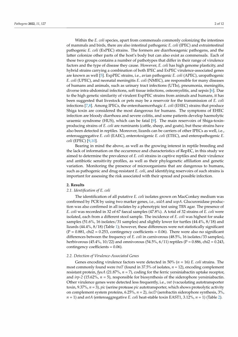
The identification of all putative E coli isolates grown on MacConkey medium wasconfirmed by PCR by using two marker genes ie uidA and uspA Glucuronidase produc-tion was also confirmed in all isolates by a phenotypic test using TBX agar The presence ofE coli was recorded in 32 of 67 faecal samples (478) A total of 32 strains of E coli wereisolated each from a different stool sample The incidence of E coli was highest for snakesamples (516 16 isolates31 samples) and slightly lower for turtles (444 818) andlizards (444 818) (Table 1) however these differences were not statistically significant(P = 0881 chi2 = 0253 contingency coefficients = 006) There were also no significantdifferences between the frequency of E coli in carnivorous (485 16 isolates33 samples)herbivorous (454 1022) and omnivorous (545 611) reptiles (P = 0886 chi2 = 0243contingency coefficients = 006)
22 Detection of Virulence-Associated Genes
Genes encoding virulence factors were detected in 50 (n = 16) E coli strains Themost commonly found were traT (found in 375 of isolates n = 12) encoding complementresistant protein fyuA (2187 n = 7) coding for the ferric yersiniabactin uptake receptorand irp-2 (1562 n = 5) responsible for biosynthesis of the siderophore yersiniabactinOther virulence genes were detected less frequently ie vat (vacuolating autotransportertoxin 937 n = 3) pic (serine protease pic autotransporter which shows proteolytic activityon complement system proteins 625 n = 2) iucD (aerobactin siderophore synthesis 3n = 1) and astA (enteroaggregative E coli heat-stable toxin EAST1 312 n = 1) (Table 2)
Pathogens 2022 11 127 3 of 12
Table 1 Frequency of isolation of Escherichia coli from captive reptiles in Poland
Group ofReptiles
Number ofSamples
Species (Number ofSamples) Diet Group
Number ofE coli
Isolates ()
Snakes 31
Pantheropsis guttatus (14)Python regius (3)Boa constrictor (4)
Lampropeltis triangulum (4)Morelia pilota (1)
Orthrophis teaniurus (5)
CarnivoreCarnivoreCarnivoreCarnivoreCarnivoreCarnivore
7 (50)3 (100)3 (75)2 (50)
1 (100)0
Lizards 18
Pogona vitticeps (6)Iguana iguana (4)
Eublepharis macularius (3)Furcifer pardali (5)
OmnivoreHerbivoreCarnivoreOmnivore
5 (83)2 (50)
01 (20)
Turtles 18Testudo horsfieldii (8)Testudo hermanni (9)
Chelonoidis carbonaria (1)
HerbivoreHerbivoreHerbivore
5 (625)2 (22)
1 (100)
Total 67 32 (478)
Table 2 Phenotypic and genotypic drug resistance profiles virulence profiles and phylogeneticgroups of E coli strains isolated from reptiles
Isolate Species PhylogeneticGroup
Antibiotic PhenotypePattern (Including
Resistant andIntermediate Strains) a
ResistanceGenes Virulence Genes
6 Corn snake (Pantheropsis guttatus) B1 AMP-AK-S-CIP - traT
13a Ball python (Python regius) B1 AMP - irp-2 iucD traTfyuA
15a Ball python (Python regius) A - - -16a Corn snake (Pantheropsis guttatus) A AMP-AMC-AK-S - pic astA26 Boa constrictor snake (Boa constrictor) B1 S - -
30a Corn snake (Pantheropsis guttatus) B2 S - irp-2 vat traT fyuA31a Corn snake (Pantheropsis guttatus) B1 S aadA traT32a Milk snake (Lampropeltis triangulum) B2 S - irp-2 vat traT fyuA34 Ball python (Python regius) B1 S - traT
35a Boa constrictor snake (Boa constrictor) D S - vat36a Boa constrictor snake (Boa constrictor) A - - traT37a Corn snake (Pantheropsis guttatus) B1 CN-AK-S-T-W b - -39 Corn snake (Pantheropsis guttatus) B1 - -41 Milk snake (Lampropeltis triangulum) B2 S - pic irp-2 traT fyuA47 Corn snake (Pantheropsis guttatus) B1 S - -48 Carpet python (Morelia spilota) B1 S - traT fyuA
54a Panther chameleon (Furcifer pardalis) B1 S - -46 Central bearded dragon (Pogona vitticeps) A S-T tetA -55 Central bearded dragon (Pogona vitticeps) D CN-AK-S-T-CIP tetB tetM irp-2 fyuA61 Central bearded dragon (Pogona vitticeps) B1 AMP-S blaTEM traT62 Central bearded dragon (Pogona vitticeps) B1 S - -63 Central bearded dragon (Pogona vitticeps) B1 S - -44 Green iguana (Iguana iguana) B1 S - traT65 Green iguana (Iguana iguana) B1 AMP-S - -
17a Steppe tortoise (Testudo horsfieldii) B1 - - traT18a Steppe tortoise (Testudo horsfieldii) A S - fyuA19a Steppe tortoise (Testudo horsfieldii) A - - -25a Red-footed tortoise (Chelonoidis carbonaria) B1 AK-S - -67 Greek tortoise (Testudo hermanni) B1 S - -69 Steppe tortoise (Testudo horsfieldii) B1 S - -70 Steppe tortoise (Testudo horsfieldii) B1 S - -71 Greek tortoise (Testudo hermanni) B1 S - -
a bold symbols indicate resistance non-bold symbols indicate intermediate susceptibility AMPmdashampicillinAMCmdashamoxicillinclavulanic acid AKmdashamikacin CIPmdashciprofloxacin CNmdashgentamicin Smdashstreptomycin Tmdashtetracycline Wmdashtrimethoprim b MDR strain
Pathogens 2022 11 127 4 of 12
In seven isolates the coexistence of between two and four virulence genes wasrecorded and interestingly as many as six of these strains were obtained from snakes(Table 2) Moreover 1 strain (16a from a corn snake) with a pic-astA profile can be classifiedas IPEC 1 strain (41 from milk snake) with a pic-irp-2-traT-fyuA profile is a hybrid strain(the pic gene is characteristic of IPEC and irp-2 traT and fyuA for ExPEC) while the re-maining strains contain virulence genes characteristic of ExPEC No E coli strain containedany of the remaining virulence genes characteristic of either IPEC (stx1 stx2 hlyA eaeA saaescV ent bfpB elt estIa estIb invE ipaH and aggR) or ExPEC (pap-C ompT cvacvi iss iutAand kpsII)
23 Antibiotic Susceptibility Testing
Most of the E coli isolates (7187 2332) were found to be susceptible to all of thetested antimicrobial agents Among the remaining isolates the phenotype of resistance tostreptomycin (2187 732) was the most frequent while resistance to other substancesie gentamicin (625 232) amikacin (625 232) tetracycline (625 232) ampi-cillin (312 132) amoxicillinclavulanic acid (312 132) and trimethoprim (312132) was observed much less frequently A total of 3 strains (937) showed resistance tomore than 1 drug but only 1 strain (3125) E coli 37a from corn snake was classified asmultidrug-resistant (MDR defined as resistance to at least 3 or more antibiotics belonging todifferent antimicrobial categories) It is also interesting that intermediate streptomycin sus-ceptibility was recorded for the majority of the strains tested (5937 1932) The 2 strainsthat showed resistance to AMC or AMP and 4 strains with intermediate susceptibility toAMP were susceptible to cephalosporins (2G 3G and 4G) and carbapenems (Table 2)
24 Detection of Resistance Genes
The occurrence of the resistance genes was recorded for only 4 (125) E coli strains More-over 1 strain (312) was detected with the aadA gene encoding streptomycinspectinomycinadenylyltransferase 2 strains (625) were confirmed with the tetA tetB and tetM genes con-ferring resistance to tetracyclines and 1 isolate (312) carried the TEM-type beta-lactamase(blaTEM) For each of these strains a correlation was found between the phenotype and thepresence of resistance genes (Table 2)
25 Determination of E coli Phylogenetic Groups
Phylogenetic groups of E coli were determined based on the electrophoretic profilesof multiplex PCR amplicons (yja TspE4C2 chuA svg and uidA) (Figure 1) Over 65 of theisolates (656 n = 21) belonged to group B1 187 (n = 6) were assigned to group A 94(n = 3) to B2 and 62 (n = 2) to group D None of the isolates belonged to group B21 (noisolate contained the svg gene) (Figure 1) It is worth noting that all the strains representinggroup B2 were collected from snakes (Table 2)
Pathogens 2022 11 x FOR PEER REVIEW 5 of 13
25 Determination of E coli Phylogenetic Groups Phylogenetic groups of E coli were determined based on the electrophoretic profiles
of multiplex PCR amplicons (yja TspE4C2 chuA svg and uidA) (Figure 1) Over 65 of the isolates (656 n = 21) belonged to group B1 187 (n = 6) were assigned to group A 94 (n = 3) to B2 and 62 (n = 2) to group D None of the isolates belonged to group B21 (no isolate contained the svg gene) (Figure 1) It is worth noting that all the strains repre-senting group B2 were collected from snakes (Table 2)
Figure 1 Multiplex PCR patterns for E coli phylogenetic groups
26 Rep-PCR Fingerprinting DNA amplification with the (GTG)5 primer revealed significant genotypic diversity
among the E coli isolates The electrophoretic profiles were reproducible in the 2 separate PCR reactions and the fingerprinting generated distinct amplification bands ranging in size from 350 bp to 2700 bp The profiles obtained contained between 2 and 12 PCR prod-ucts and allowed the 32 strains to be grouped into 27 rep-types (Figure 2) This indicates considerable genetic diversity of the E coli isolates with a maximum percentage of dis-similarity of 41 There was no correlation between the electrophoretic profile and the phylogenetic group or the host species although several strains representing the same phylogenetic group had identical fingerprints (Figure 3)
Figure 2 Agarose gel patterns of products amplified by (GTG)5-PCR
Figure 1 Multiplex PCR patterns for E coli phylogenetic groups
Pathogens 2022 11 127 5 of 12
26 Rep-PCR Fingerprinting
DNA amplification with the (GTG)5 primer revealed significant genotypic diversityamong the E coli isolates The electrophoretic profiles were reproducible in the 2 separatePCR reactions and the fingerprinting generated distinct amplification bands ranging in sizefrom 350 bp to 2700 bp The profiles obtained contained between 2 and 12 PCR products andallowed the 32 strains to be grouped into 27 rep-types (Figure 2) This indicates considerablegenetic diversity of the E coli isolates with a maximum percentage of dissimilarity of 41There was no correlation between the electrophoretic profile and the phylogenetic group orthe host species although several strains representing the same phylogenetic group hadidentical fingerprints (Figure 3)
Pathogens 2022 11 x FOR PEER REVIEW 5 of 13
25 Determination of E coli Phylogenetic Groups Phylogenetic groups of E coli were determined based on the electrophoretic profiles
of multiplex PCR amplicons (yja TspE4C2 chuA svg and uidA) (Figure 1) Over 65 of the isolates (656 n = 21) belonged to group B1 187 (n = 6) were assigned to group A 94 (n = 3) to B2 and 62 (n = 2) to group D None of the isolates belonged to group B21 (no isolate contained the svg gene) (Figure 1) It is worth noting that all the strains repre-senting group B2 were collected from snakes (Table 2)
Figure 1 Multiplex PCR patterns for E coli phylogenetic groups
26 Rep-PCR Fingerprinting DNA amplification with the (GTG)5 primer revealed significant genotypic diversity
among the E coli isolates The electrophoretic profiles were reproducible in the 2 separate PCR reactions and the fingerprinting generated distinct amplification bands ranging in size from 350 bp to 2700 bp The profiles obtained contained between 2 and 12 PCR prod-ucts and allowed the 32 strains to be grouped into 27 rep-types (Figure 2) This indicates considerable genetic diversity of the E coli isolates with a maximum percentage of dis-similarity of 41 There was no correlation between the electrophoretic profile and the phylogenetic group or the host species although several strains representing the same phylogenetic group had identical fingerprints (Figure 3)
Figure 2 Agarose gel patterns of products amplified by (GTG)5-PCR Figure 2 Agarose gel patterns of products amplified by (GTG)5-PCR
Pathogens 2022 11 x FOR PEER REVIEW 6 of 13
0 5 10 15 20 25 30 35 40 45 50
percent disagreement
65-green iguana-B162-bearded dragon-B1
48-carpet python-B147-corn snake-B136a-boa snake-A
70-steppe tortoise-B135a-boa snake-D
25a-red-footed tortoise-B119a-steppe tortoise-A18a-steppe tortoise-A
17a-steppe tortoise-B146-bearded dragon-A
44-green iguana-B134-ball python-B116a-corn snake-A
55-bearded dragon-D15a-ball python-A
13a-ball python-B137a-corn snake-B131a-corn snake-B1
69-steppe tortoise-B141-milk snake-B2
32a-milk snake-B230a-corn snake-B2
54a-panther chameleon-B139-corn snake-B1
61-bearded dragon-B171-Greek tortoise-B167-Greek tortoise-B1
26-boa snake-B16-corn snake-B1
Figure 3 Dendrogram generated by UPGMA clustering from (GTG)5-PCR fingerprinting of E coli strains
3 Discussion The scientific literature abounds with information on the drug susceptibility and vir-
ulence of E coli isolated from humans mammals and birds but reports on the occurrence and characteristics of E coli in reptiles are scarce The prevalence of the E coli strains re-ported in these studies (478) is in line with that reported by Gopee et al [11] (52) for reptiles from the Emperor Valley Zoo Trinidad Spain The authors recorded the highest incidence of E coli in Crocodylidae (833) lower incidences in turtles (6808) and snakes (375ndash511 depending on the family) and the lowest for lizards (333) In con-trast with our observations a higher frequency of E coli was recorded in herbivorous rep-tiles (667) than in carnivorous reptiles (464)
A significantly higher frequency of E coli in captive and free-living reptiles was rec-orded by Ramos et al [3] in Brazil (68) and as in our study they reported a higher inci-dence of E coli in snakes (844) than in lizards (466) and turtles (40ndash50) Similar re-sults were obtained by Sylvester et al [12] and Bautista-Trujillo et al [10] who showed the presence of E coli in approximately 40 of green iguanas Several other authors have also described the presence of E coli in reptiles however they did not take into account the frequency of this bacterium in individual groups of reptiles Książczyk et al [4] iso-lated a total of 35 E coli strains from 103 stool samples from reptiles at the zoo in Wrocław Poland These data indicate 34 prevalence assuming each strain was isolated from a different sample Much higher prevalence of E coli in reptiles is suggested by the findings of Unger et al [13] who isolated a total of 142 E coli isolates from 150 faecal samples derived from gt 60 exotic reptile species crossing the veterinary border at Frankfurt airport
Warm-blooded animals (mammals and birds) are already known to be reservoirs of both IPEC and ExPEC E coli strains [14ndash16] but knowledge of the occurrence of patho-genic E coli in cold-blooded animals is scant In terms of the presence of virulence genes
Figure 3 Dendrogram generated by UPGMA clustering from (GTG)5-PCR fingerprinting of E coli strains
Pathogens 2022 11 127 6 of 12
3 Discussion
The scientific literature abounds with information on the drug susceptibility andvirulence of E coli isolated from humans mammals and birds but reports on the occurrenceand characteristics of E coli in reptiles are scarce The prevalence of the E coli strainsreported in these studies (478) is in line with that reported by Gopee et al [11] (52) forreptiles from the Emperor Valley Zoo Trinidad Spain The authors recorded the highestincidence of E coli in Crocodylidae (833) lower incidences in turtles (6808) and snakes(375ndash511 depending on the family) and the lowest for lizards (333) In contrast withour observations a higher frequency of E coli was recorded in herbivorous reptiles (667)than in carnivorous reptiles (464)
A significantly higher frequency of E coli in captive and free-living reptiles wasrecorded by Ramos et al [3] in Brazil (68) and as in our study they reported a higherincidence of E coli in snakes (844) than in lizards (466) and turtles (40ndash50) Similarresults were obtained by Sylvester et al [12] and Bautista-Trujillo et al [10] who showedthe presence of E coli in approximately 40 of green iguanas Several other authors havealso described the presence of E coli in reptiles however they did not take into account thefrequency of this bacterium in individual groups of reptiles Ksiazczyk et al [4] isolated atotal of 35 E coli strains from 103 stool samples from reptiles at the zoo in Wrocław PolandThese data indicate 34 prevalence assuming each strain was isolated from a differentsample Much higher prevalence of E coli in reptiles is suggested by the findings of Ungeret al [13] who isolated a total of 142 E coli isolates from 150 faecal samples derived fromgt60 exotic reptile species crossing the veterinary border at Frankfurt airport
Warm-blooded animals (mammals and birds) are already known to be reservoirs ofboth IPEC and ExPEC E coli strains [14ndash16] but knowledge of the occurrence of pathogenicE coli in cold-blooded animals is scant In terms of the presence of virulence genes ourresearch is largely in line with the findings of Ramos et al [3] who detected only 2 ofthe many virulence genes characteristic for ETEC EHEC and NTEC ie the cnf1 geneencoding cytotoxic necrotizing factor 1 (found in 92 of RepEC isolates) and the astA geneencoding EAST1 toxin (26) The frequency described in this study (312) of the astAgene coding for heat-stable enterotoxin EAST1 characteristic of EAEC is also consistentwith results reported by Ksiazczyk et al [4] In the case of virulence genes characteristic forIPEC it should be emphasized that our research is the first report on the presence of the picgene in RepEC characteristic for the EHEC pathotype Completely different results werepresented by Bautista-Trujillo et al [10] who classified 62 of 100 E coli isolates from captivegreen iguanas in Mexico as diarrhoeagenic E coli (DEC) strains Among them STEC strains(403) carrying the stx1 (387) or stx2 (16) gene were the most prevalent pathotypefollowed by EAEC (274 aap-positive strains) ETEC (274 each lt- or st-positive) andEPEC (49 bfpA- or eaeA-positive) The frequency of tratT (375 coding for complementresistance protein) and fyuA (2187 coding for the ferric yersiniabactin uptake receptor)genes reported in these studies was about twice as high as in the case of RepEC strainsisolated in Poland (traT-167 fyuA-83) by another research team [4] The fyuA geneis common in both UPEC and APEC strains while the traT gene is characteristic of theUPEC pathotype [4] The occasional presence of the irp-2 gene in RepEC (83) was alsodemonstrated by Ksiazczyk et al [4] However in contrast to our results they did notdetect the iucD or vat gene in any strain and in several strains they confirmed the presenceof other virulence genes characteristic of ExPEC ie papC iss and tsh
The results of this study on drug resistance in E coli most closely match the findingsof Sylvester et al [12] who showed that among E coli strains from wild and pet greeniguanas (from Grenada) resistance to streptomycin (12) and amoxicillin-clavulanic acid(12) was the most common and only 7 of strains were multi-drug resistant A muchhigher prevalence of resistant E coli strains was recorded in reptiles housed at zoo inTrinidad [11] and resistance to ampicillin (667) tetracycline (576) and gentamicin(121) was the most frequent Interestingly the prevalence of streptomycin resistance(364) demonstrated by these studies was somewhat similar to our data (219) An
Pathogens 2022 11 127 7 of 12
agreement was also noted in the lack of resistance to fluoroquinolones A high percentageof resistant E coli strains was also demonstrated by Bautista-Trujillo et al [10] who classifiedas many as 823 of DEC isolates (from captive green iguanas in Mexico) as MDR andmost frequently noted resistance to amikacin (~40) and ampicillin (~30)
The higher incidence of drug-resistant strains in captive reptiles from Trinidad andMexico may be due to different veterinary practices and the different prevalence of drug-resistant E coli in the reptile diet and environment Details on the type and frequency ofantibiotic use in reptiles in these countries are not known but Mexico was among the topten countries in the world with the highest use of antibiotics in veterinary medicine in2017 [16]
The higher frequency of resistance to streptomycin (~22) compared to resistanceto other antimicrobial substances (0ndash625) reported in these studies may be due to thewidespread use of this antibiotic in veterinary medicine as well as in horticulture as aplant protection product The resulting streptomycin-resistant strains may spread in theenvironment and eventually be acquired by the reptiles via their feed (plants rodentsmeat) Moreover the results obtained in this study and some literature data suggestthat the reptilian Enterobacteriaceae may have a reduced susceptibility to streptomycinStreptomycin resistance was the most frequently reported antibiotic resistance phenotypein reptile-derived Salmonella strains in Poland (25) [17] and Indonesia (75) [18] SimilarlyBertolini et al [19] showed a high percentage (79) of Salmonella strains intermediatelysusceptible to streptomycin in reptiles
According to our best knowledge this work is the first full report of the occurrence ofresistance genes in E coli from reptiles The aadA (aminoglycoside (3rdquo) (9) adenylyltrans-ferase) tetA (tetracycline efflux protein TetA) tetB (tetracycline efflux protein TetB) andblaTEM genes detected in several RepEC strains are commonly found in drug-resistantEnterobacteriaceae isolated from humans and farm animals [2021] The tetM gene codingfor tetracycline ribosomal protection protein has thus far been found sporadically in E colimainly in pig and chicken strains [21] Its presence confirmed in this work in one RepECisolate may be the result of genetic transfer from Enterococcus or Lactobacillaceae commoncarriers of tetM [2223] The TEM family of β-lactamases covers a large group of enzymesand determining which TEM lactamase has been detected in these studies requires moredetailed sequencing-based analyzes In E coli strains the most common is the prototypenarrow-spectrum blaTEM-1 lactamase from which the broad-spectrum lactamases (ESBL)have evolved by mutation [2425] The blaTEM-1 gene was also found in two strains of Ecoli from reptiles transported across the veterinary border in Frankfurt where it coexistedwith many other resistance genes [13]
Phylogenetic group B1 which dominated (656) among the RepEC isolates is char-acteristic of non-pathogenic E coli strains while groups A and B2 recorded in this studyfor a total of 9 isolates is characteristic of ExPEC ie UPEC and APEC [426] It is there-fore worth noting that 3 of 4 RepEC strains containing 4 virulence genes simultaneouslybelonged to the B2 phylogenetic group The results presented in this study are consistentwith the reports of Gordon and Cowling [27] who assigned nearly 70 of E coli isolatesfrom snakes and lizards to phylogenetic group B1 while strains of groups A B2 and Daccounted for 151 61 and 91 respectively A higher percentage (25) of strainsrepresenting the B2 phylogenetic group was recorded in turtles [28] A higher frequency ofstrains of the B1 group in RepEC was reported by Ramos et al [3] (884) and Ksiazczyket al [4] (958)
4 Materials and Methods41 Collection of Faecal Samples
The research material consisted of faecal samples collected between 2017 and 2020from 67 reptiles ie snakes (n = 31) lizards (n = 18) and turtles (n = 18) living in captivityor as pets The animals did not show any symptoms of disease They were obtained fromprivate owners (n = 53) and from pet shops (n = 14) in the Lubelskie Province Poland
Pathogens 2022 11 127 8 of 12
42 E coli Isolation
Stool specimens collected with a swab were suspended in peptone water and incubatedfor approximately 18 hours inoculated onto MacConkey agar (Oxoid Ltd AltrinchamUK) and incubated at 37 C for 24 h under aerobic conditions Single pink colonies wereinoculated on TSB (trypticase soy broth) (Oxoid Ltd Altrincham UK) and pure culturessupplemented with 20 glycerol were stored at minus80 C for further analysis To determinethe ability of the strains to produce glucuronidase they were additionally plated on TXBagar and incubated ~24 h at 44 C
43 Identification of E coli
Presumptive E coli isolates were identified based on detection of the uidA glucuronidasegene and the E coli-specific flanking region of the uspA gene (coding for universal stressprotein) PCR was performed using DreamTaq polymerase (Thermo Fisher Scientific VilniusLithuania) and the following two pairs of primers uidAF-TATGGAATTTCGCCGATTTT anduidAR-TGTTTGCCTCCCTGCTGCGG (uidA amplicon size 166 bp) and uspAF-CCGATACGCTGCCAATCAGT and uspAR-ACGCAGACCGTAGGCCAGAT (uspA amlicon 884 bp) [25]The PCR reaction were performed using Dream Taq polymerase and the following thermal-cycling program initial denaturation at 94 C for 5 min 30 cycles of 94 C for 45 s 57 Cfor 45 s and 72 C for 1 min final extension step at 72 C for 8 min PCR products (8 microL~600 ng) were separated by electrophoresis (100 V 1 h) on 18 agarose gels and visualizedby SimplySafe (Eurx Gdansk PL) staining PCR product sizes were determined by com-parison with a Nova 100 bp DNA ladder (Novazym Poznan Poland) using Image Lab 61Software (Bio-Rad Hercules CA USA)
44 DNA Extraction
Whole-genome DNA was extracted from all of the tested E coli isolates (n = 32) usingthe Gene MATRIX Bacterial amp Yeast Genomic DNA Purification Kit (Eurx Gdansk Popland)following the manufacturerrsquos instructions and stored at minus20 C DNA concentration wasdetermined by measuring the absorbance of 2microL of the sample at 260280 nm using theNanoDrop Lite spectrophotometer (Thermo Fisher Scientific Waltham MA USA) and thequality of DNA was analyzed by agarose (15 wv) gel electrophoresis No signs of DNAdegradation (smearing) were observed in any of the samples and its concentration was~20 ngmicroL
45 Detection of Virulence Genes
Uniplex or multiplex PCR (Table 3) using gene-specific primers (Table S1) wasused to detect the presence of 29 genes associated with virulence in both intestinal andextraintestinal pathogenic E coli strains Two E coli strains from wild mammals previouslyconfirmed to have some virulence genes [16] ie astA-stx1-stx2-hlyA (E coli 22a) and astA-escV-eaeA (E coli 19) were used as positive controls All PCR reactions were performedin an Eppendorf Mastercycler using Dream Taq polymerase (Thermo Scientific) PCRproducts (8 microL ~600 ng) were separated by electrophoresis (100 V 1 h) on 18 agarose gelsand visualized by SimplySafe (Eurx Gdansk Poland) staining PCR product sizes weredetermined by comparison with a Nova 100 bp DNA ladder (Novazym Poznan Poland)using Image Lab 61 Software (Bio-Rad Herkules CA USA)
Pathogens 2022 11 127 9 of 12
Table 3 PCR schemes used to detect virulence genes in E coli
Detected Genes AnnealingTemperature (C) Reference
Multiplex I stx1 stx2 hylA eaeAsaa
65 (10 cycles) then62 (20 cycles) [14]
Multiplex IIecsV ent bfpB invEastA aggR pic ipaH
elt estIa estIb62 [14]
Multiplex III ompT iutA 63 [14]
Multiplex IV tsh pap-C iss irp-2 57
[29]Multiplex V iucD vat 54
Uniplex I cvacvi 58
Uniplex II cnf 55 [30]
Uniplex III kpsII 56
[4]Uniplex IV traT 59
Uniplex V fyuA 59
46 Antimicrobial Susceptibility Testing
Antibiotic susceptibility testing was performed by the agar disk diffusion methodaccording to Clinical and Laboratory Standards Institute (CLSI) recommendations [31]Isolates were revitalized on tryptone soy agar (TSA) plates (Oxoid Altrincham UK) Bacte-rial suspensions with a turbidity equivalent to McFarland Standard 05 were swabbed onMuumleller Hinton agar plates (Oxoid Altrincham UK) with a sterile cotton swab Antibioticdisks (Oxoid Altrincham UK) containing ampicillin (AMP 10 microg) amoxicillinclavulanicacid (AMC 2010 microg) cefotaxime (CTX 30 microg) chloramphenicol (C 30 microg) tetracycline(TE 30 microg) trimethoprim (W 5 microg) ciprofloxacin (CIP 5 microg) streptomycin (S 10 microg)gentamicin (CN 10 microg) amikacin (AK 30 microg) and nitrofurantoin (F 100 microg) were placedon the plates Strains that showed resistance or intermediate susceptibility to AMP or AMCwere additionally tested against cefoxitin (FOX 30 microg) cefepime (FEP 30 microg) ceftazidime(CAZ 30 microg) ceftazidimeclavulanic acid (CZC 3010 microg) imipenem (IMP 10 microg) andmeropenem (MEM 10 microg) Inhibition zone diameters including the diameter of the 6-mmdisks were measured after incubation at 35 C for 20 h Isolates were classified as resistantI intermediate (I) and susceptible (S) according to breakpoints provided by CLSI [31]only in the case of nitrofurantoin was the interpretation of the results based on EUCASTguidelines [32] E coli ATCC 25922 reference strain was used as a quality control
47 Detection of Resistance Genes
All of hte E coli isolates that showed phenotypic resistance or intermediate suscep-tibility to specific antimicrobial agents were tested for the presence of relevant resistancegenes using a uniplex or multiplex PCR assay Specifically genes conferring resistanceto beta-lactams (blaTEM blaOXY blaCTX and blaSHV) tetracyclines (tetA tetB tetC tetKtetL tetM and tetO) aminoglycosides (aadA strAstrB aac(3)-II aac(3)-IV aphA1 and aphA2)trimethoprim and sulfonamides (sul1 sul2 sul3 dfrA1 dfrA5 and drfA7-A17) were detectedusing the primers and annealing temperatures shown in Table S2 Analysis of the PCRproducts was performed as above
48 Determination of E coli Phylogenetic Groups
To determine the phylogenetic groups of the E coli isolates five sets of primers for thefive sets of primers for the genes yja TspE4C2 chuA svg and uidA (Table S3) were used in amultiplex PCR as previously described [33] PCR products were separated by electrophore-sis in 25 (wtvol) high resolution agarose (Blirt Gdansk Poland) The amplicons sizes
Pathogens 2022 11 127 10 of 12
were determined by comparison with a Nova 100 bp DNA ladder (Novazym PoznanPoland) using Image Lab 61 Software (Bio-Rad Herkules CA USA) The phylogeneticgroups were determined based on the PCR gel pattern [33]
49 Rep-PCR Fingerprinting
The genetic diversity of E coli isolates was assessed by repetitive extragenic palin-dromic (REP)-PCR using the (GTG)5 primer -5prime-GTGGTGGTGGTGGTG-3prime [34] The PCRreactions were carried out in 18 microL of a reaction mixture containing 75 microL of 2x PCR MixPlus (AampA Biotechnology Gdynia Poland) 075 microL of (GTG)5 primer 075 microL (~20 ngmicroL)of DNA template and 9 microL of H2O Amplification was performed in the following se-quence initial denaturation 5 min 95 C 40 cycles at 95 C for 45 s 42 C for 35 s and72 C for 2 min and final extension at 72 C for 8 min PCR products were separated byelectrophoresis in 15 agarose The gels were run at 80 V in 1X TBE buffer stained withSimplySafe (Eurx Gdansk Poland) and visualized under an UV source using the GelDocGo imaging system (BioRad Herkules CA USA) Profiles were analysed with Image LabSoftware (BioRad Herkules CA USA) by comparison with the Nova 100 bp DNA ladder(Novazym Poznan Poland)
The presence of a given band was coded as 1 and the absence of a given band wascoded as 0 in a data matrix (Excel Microsoft Office) and the unweighted pair group methodwith arithmetic averages (UPGMA) was used to generate a dendrogram of dissimilarity inStatistica software (Ver 131 Tulsa OK USA)
410 Statistical Analysis
Pearsonrsquos chi-squared test was used to determine the relationship between diet (car-nivores omnivores herbivores) and the frequency of E coli as well as between the typeof reptile (snakes lizards turtles) and the frequency of E coli isolation The level ofsignificance was set as p lt 005 The statistical analysis was performed using MicrosoftExcel 2019
5 Conclusions
The results presented in this study contribute to knowledge of the occurrence andphenotypic and genotypic features of RepEC strains Although the incidence of drugresistance in E coli strains from reptiles in Poland is low the possibility of transmissionof resistance genes to other members of Enterobacteriaceae including Salmonella whichcommonly inhabits reptile intestines should be taken into account In addition RepECstrains contain virulence genes characteristic of ExPEC and less often of IPEC IPEC strainsare diarrhoeagenic pathogens while ExPEC strains may exist as commensals but underfavourable conditions can cause infections Due to the pathogenic potential of RepECstrains they may pose a health risk to humans and reptile keepers should use goodhygiene practices when handling these animals
Supplementary Materials The following are available online at httpswwwmdpicomarticle103390pathogens11020127s1 Table S1 Sequences of primers used to detect virulence genesamplicon sizes and references [142930] Table S2 Sequences of primers used to detect resistancegenes [142335ndash37] Table S3 Primer sequences used to determine phylogenetic groups in E colistrains [35]
Author Contributions Conceptualization MD methodology MD and BT validation DS-Pformal analysis MD investigation MD DS-P resources MD and KS data curation MDwritingmdashoriginal draft preparation MD writingmdashreview and editing RU-C and BT visualiza-tion MD funding acquisition RU-C and KS All authors have read and agreed to the publishedversion of the manuscript
Funding This research received no external funding
Institutional Review Board Statement The research was carried out in accordance with the prin-ciples of bioethics in force in Poland (Act of 15 January 2015 on the protection of animals used for
Pathogens 2022 11 127 11 of 12
scientific or educational purposes Journal of Laws 2015 item 266) as well as in the EU (Directive201063EU of the European Parliament and of the Council of 22 September 2010 on the protectionof animals used for scientific purposes) according to which collecting stool samples for scientificresearch is not a procedure on animals and does not require the approval of the Local Ethical Commit-tee Pet and pet store owners have agreed to collect reptile feces when cleaning terrariums withoutinteracting with animal welfare
Informed Consent Statement Not applicable
Data Availability Statement All the data presented in the study are included in the article Furtherenquires can be directed to the corresponding author
Acknowledgments Not applicable
Conflicts of Interest The authors declare no conflict of interest
References1 FEDIAF (The European Pet Food Industry) FEDIAF Annual Report 2021 p 44 50 Available online httpswwwfediaforg
annual-reporthtml (accessed on 20 January 2022)2 Valdez JW Using Google Trends to Determine Current Past and Future Trends in the Reptile Pet Trade Animals 2021 11 676
[CrossRef]3 Ramos CP Santana JA Morcatti Coura F Xavier RGC Leal CAG Oliveira Junior CA Heinemann MB Lage AP
Lobato FCF Silva ROS Identification and Characterization of Escherichia coli Salmonella Spp Clostridium perfringens andC difficile Isolates from Reptiles in Brazil Biomed Res Int 2019 2019 9530732 [CrossRef]
4 Ksiazczyk M Dudek B Kuczkowski M OrsquoHara R Korzekwa K Wzorek A Korzeniowska-Kowal A Upton M JunkaA Wieliczko A et al The Phylogenetic Structure of Reptile Avian and Uropathogenic Escherichia coli with Particular Referenceto Extraintestinal Pathotypes Int J Mol Sci 2021 22 1192 [CrossRef]
5 Lindstedt BA Finton MD Porcellato D Brandal LT High frequency of hybrid Escherichia coli strains with combinedIntestinal Pathogenic Escherichia coli (IPEC) and Extraintestinal Pathogenic Escherichia coli (ExPEC) virulence factors isolatedfrom human faecal samples BMC Infect Dis 2018 18 544 [CrossRef]
6 Wasinski B Extra-intestinal pathogenic Escherichia colimdashThreat connected with food-borne infections Ann Agric Environ Med2019 26 532ndash537 [CrossRef]
7 Garciacutea A Fox JG A One Health Perspective for Defining and Deciphering Escherichia coli Pathogenic Potential in MultipleHosts Comp Med 2021 71 3ndash45 [CrossRef]
8 Rodriguez-Siek KE Giddings CW Doetkott C Johnson TJ Fakhr MK Nolan LK Comparison of Escherichia coli isolatesimplicated in human urinary tract infection and avian colibacillosis Microbiology 2005 151 2097ndash2110 [CrossRef] [PubMed]
9 Gomes TA Elias WP Scaletsky IC Guth BE Rodrigues JF Piazza RM Ferreira LC Martinez MB DiarrheagenicEscherichia coli Braz J Microbiol 2016 47 3ndash30 [CrossRef] [PubMed]
10 Bautista-Trujillo GU Gutieacuterrez-Miceli FA Mandujano-Garciacutea L Oliva-Llaven MA Ibarra-Martiacutenez C Mendoza-NazarP Ruiz-Sesma B Tejeda-Cruz C Peacuterez-Vaacutezquez LC Peacuterez-Batrez JE et al Captive Green Iguana Carries DiarrheagenicEscherichia coli Pathotypes Front Vet Sci 2020 7 99 [CrossRef]
11 Gopee NV Adesiyun AA Caesar K A longitudinal study of Escherichia coli strains isolated from captive mammals birdsand reptiles in Trinidad J Zoo Wildl Med Off Publ Am Assoc Zoo Vet 2000 31 353ndash360 [CrossRef]
12 Sylvester WRB Amadi V Hegamin-Younger C Pinckey R Macpherson CNL Mckibben JS Bruhl-Day R John-SylvesterKD Hariharan H Occurrence of Antibiotic Resistant Escherichia coli in Green Iguanas (Iguana Iguana) in Grenada West IndiesInt J Vet Med Res Rep 2014 2014 260412 [CrossRef]
13 Unger F Eisenberg T Prenger-Berninghoff E Leidner U Ludwig ML Rothe M Semmler T Ewers C Imported reptiles asa risk factor for the global distribution of Escherichia coli harbouring the colistin resistance gene mcr-1 Int J Antimicrob Agents2017 49 122ndash123 [CrossRef] [PubMed]
14 Nowaczek A Dec M Stepien-Pysniak D Urban-Chmiel R Marek A Roacutezanski P Antibiotic Resistance and VirulenceProfiles of Escherichia coli Strains Isolated from Wild Birds in Poland Pathogens 2021 10 1059 [CrossRef] [PubMed]
15 Turchi B Dec M Bertelloni F Winiarczyk S Gnat S Bresciani F Viviani F Cerri D Fratini F Antibiotic Susceptibilityand Virulence Factors in Escherichia coli from Sympatric Wildlife of the Apuan Alps Regional Park (Tuscany Italy) Microb DrugResist 2019 25 772ndash780 [CrossRef] [PubMed]
16 Tiseo K Huber L Gilbert M Robinson TP Van Boeckel TP Global Trends in Antimicrobial Use in Food Animals from 2017to 2030 Antibiotics 2020 9 918 [CrossRef]
17 Zajac M Skarzynska M Lalak A Kwit R Smiałowska-Weglinska A Pasim P Szulowski K Wasyl D Salmonella inCaptive Reptiles and Their Environment-Can We Tame the Dragon Microorganisms 2021 9 1012 [CrossRef]
18 Arnafia W Ningrum SG Adji RS Lukman DW Pasaribu FH Wibawan IWT Isolation of Salmonella from reptiles in petshop and its susceptibility to antibiotics in Indonesia Hum Vet Med 2016 8 177ndash181
Pathogens 2022 11 127 12 of 12
19 Bertelloni F Chemaly M Cerri D Gall FL Ebani VV Salmonella infection in healthy pet reptiles Bacteriological isolationand study of some pathogenic characters Acta Microbiol Immunol Hung 2016 63 203ndash216 [CrossRef]
20 Mazurek J Pusz P Bok E Stosik M Baldy-Chudzik K The phenotypic and genotypic characteristics of antibiotic resistancein Escherichia coli populations isolated from farm animals with different exposure to antimicrobial agents Pol J Microbiol 201362 173ndash179 [CrossRef]
21 Jurado-Rabadaacuten S de la Fuente R Ruiz-Santa-Quiteria JA Orden JA de Vries LE Agersoslash Y Detection and linkage tomobile genetic elements of tetracycline resistance gene tet(M) in Escherichia coli isolates from pigs BMC Vet Res 2014 10 155[CrossRef] [PubMed]
22 Dec M Stepien-Pysniak D Gnat S Fratini F Urban-Chmiel R Cerri D Winiarczyk S Turchi B Antibiotic Susceptibilityand Virulence Genes in Enterococcus Isolates from Wild Mammals Living in Tuscany Italy Microb Drug Resist 2020 26 505ndash519[CrossRef]
23 Dec M Nowaczek A Stepien-Pysniak D Wawrzykowski J Urban-Chmiel R Identification and antibiotic susceptibility oflactobacilli isolated from turkeys BMC Microbiol 2018 18 168 [CrossRef] [PubMed]
24 Ur Rahman S Ali T Ali I Khan NA Han B Gao J The Growing Genetic and Functional Diversity of Extended SpectrumBeta-Lactamases Biomed Res Int 2018 2018 9519718 [CrossRef] [PubMed]
25 Paterson DL Bonomo RA Extended-spectrum beta-lactamases A clinical update Clin Microbiol Rev 2005 18 657ndash686[CrossRef]
26 Xu X Sun Q Zhao L Virulence Factors and Antibiotic Resistance of Avian Pathogenic Escherichia Coli in Eastern China J VetRes 2019 63 317ndash320 [CrossRef]
27 Gordon DM Cowling A The distribution and genetic structure of Escherichia coli in Australian vertebrates Host andgeographic effects Microbiology 2003 149 3575ndash3586 [CrossRef]
28 Godambe LP Bandekar J Shashidhar R Species specific PCR based detection of Escherichia coli from Indian foods 3 Biotech2017 7 130 [CrossRef] [PubMed]
29 Ewers C Janssen T Kiessling S Philipp HC Wieler LH Rapid detection of virulence-associated genes in avian pathogenicEscherichia coli by multiplex polymerase chain reaction Avian Dis 2005 49 269ndash273 [CrossRef]
30 Toacuteth I Heacuterault F Beutin L Oswald E Production of cytolethal distending toxins by pathogenic Escherichia coli strainsisolated from human and animal sources Establishment of the existence of a new cdt variant (Type IV) J Clin Microbiol 2003 414285ndash4291 [CrossRef]
31 CLSI Performance Standards for Antimicrobial Susceptibility Testing 28th ed CLSI Supplement M100 Clinical and LaboratoryStandards Institute Wayne PA USA 2018
32 EUCAST The European Committee on Antimicrobial Susceptibility Testing Breakpoint Tables for Interpretation of MICs andZone Diameters Version 110 2021 Available online httpwwweucastorg (accessed on 10 December 2021)
33 Feng Y Mannion A Madden CM Swennes AG Townes C Byrd C Marini RP Fox JG Cytotoxic Escherichia coli strainsencoding colibactin and cytotoxic necrotizing factor (CNF) colonize laboratory macaques Gut Pathog 2017 9 71 [CrossRef]
34 Cruz-Soto AS Toro-Castillo V Munguiacutea-Magdaleno CO Torres-Flores JE Flores-Pantoja LE Loeza-Lara PD Jimeacutenez-Mejiacutea R Genetic relationships biofilm formation motility and virulence of Escherichia coli isolated from bovine mastitis RevMex Cienc Pecu 2020 11 167ndash182 [CrossRef]
35 Fang H Ataker F Hedin G Dornbusch K Molecular epidemiology of extended-spectrum beta-lactamases among Escherichiacoli isolates collected in a Swedish hospital and its associated health care facilities from 2001 to 2006 J Clin Microbiol 2008 46707ndash712 [CrossRef] [PubMed]
36 Kozak GK Boerlin P Janecko N Reid-Smith RJ Jardine C Antimicrobial resistance in Escherichia coli isolates from swineand wild small mammals in the proximity of swine farms and in natural environments in Ontario Canada Appl EnvironMicrobiol 2009 75 559ndash566 [CrossRef] [PubMed]
37 Grape M Motakefi A Pavuluri S Kahlmeter G Standard and real-time multiplex PCR methods for detection of trimethoprimresistance dfr genes in large collections of bacteria Clin Microbiol Infect 2007 13 1112ndash1118 [CrossRef]
Pathogens 2022 11 127 2 of 12
Within the E coli species apart from commensals commonly colonizing the intestinesof mammals and birds there are also intestinal pathogenic E coli (IPEC) and extraintestinalpathogenic E coli (ExPEC) strains The formers are diarrhoeagenic pathogens and thelatter colonize other parts of the hostrsquos body but can also exist as commensals Each ofthese two groups contains a number of pathotypes that differ in their range of virulencefactors and the type of disease they cause However E coli has high genome plasticity andhybrid strains carrying a combination of both IPEC and ExPEC virulence-associated genesare known as well [5] ExpPEC strains ie avian pathogenic E coli (APEC) uropathogenicE coli (UPEC) and neonatal meningitis E coli (NMEC) are responsible for many diseasesof humans and animals such as urinary tract infections (UTIs) pneumonia meningitisdiverse intra-abdominal infections soft tissue infections osteomyelitis and sepsis [6] Dueto the high genetic similarity of virulent ExpPEC strains from animals and humans it hasbeen suggested that livestock or pets may be a reservoir for the transmission of E coliinfections [78] Among IPECs the enterohaemorrhagic E coli (EHEC) strains that produceShiga toxin are considered the most dangerous for humans The symptoms of EHECinfection are bloody diarrhoea and severe colitis and some patients develop haemolyticuraemic syndrome (HUS) which can be fatal [9] The main reservoirs of Shiga-toxin-producing strains of E coli are ruminants (cattle sheep and goats) but these strains havealso been detected in reptiles Moreover lizards can be carriers of other IPECs as well ieenteroaggregative E coli (EAEC) enterotoxigenic E coli (ETEC) and enteropathogenic Ecoli (EPEC) [910]
Bearing in mind the above as well as the growing interest in reptile breeding andthe lack of information on the occurrence and characteristics of RepEC in this study weaimed to determine the prevalence of E coli strains in captive reptiles and their virulenceand antibiotic sensitivity profiles as well as their phylogenetic affiliation and geneticvariation Monitoring the presence of microorganisms that are dangerous to humanssuch as pathogenic and drug-resistant E coli and identifying reservoirs of such strains isimportant for assessing the risk associated with their spread and possible infection
2 Results21 Identification of E coli
The identification of all putative E coli isolates grown on MacConkey medium wasconfirmed by PCR by using two marker genes ie uidA and uspA Glucuronidase produc-tion was also confirmed in all isolates by a phenotypic test using TBX agar The presence ofE coli was recorded in 32 of 67 faecal samples (478) A total of 32 strains of E coli wereisolated each from a different stool sample The incidence of E coli was highest for snakesamples (516 16 isolates31 samples) and slightly lower for turtles (444 818) andlizards (444 818) (Table 1) however these differences were not statistically significant(P = 0881 chi2 = 0253 contingency coefficients = 006) There were also no significantdifferences between the frequency of E coli in carnivorous (485 16 isolates33 samples)herbivorous (454 1022) and omnivorous (545 611) reptiles (P = 0886 chi2 = 0243contingency coefficients = 006)
22 Detection of Virulence-Associated Genes
Genes encoding virulence factors were detected in 50 (n = 16) E coli strains Themost commonly found were traT (found in 375 of isolates n = 12) encoding complementresistant protein fyuA (2187 n = 7) coding for the ferric yersiniabactin uptake receptorand irp-2 (1562 n = 5) responsible for biosynthesis of the siderophore yersiniabactinOther virulence genes were detected less frequently ie vat (vacuolating autotransportertoxin 937 n = 3) pic (serine protease pic autotransporter which shows proteolytic activityon complement system proteins 625 n = 2) iucD (aerobactin siderophore synthesis 3n = 1) and astA (enteroaggregative E coli heat-stable toxin EAST1 312 n = 1) (Table 2)
Pathogens 2022 11 127 3 of 12
Table 1 Frequency of isolation of Escherichia coli from captive reptiles in Poland
Group ofReptiles
Number ofSamples
Species (Number ofSamples) Diet Group
Number ofE coli
Isolates ()
Snakes 31
Pantheropsis guttatus (14)Python regius (3)Boa constrictor (4)
Lampropeltis triangulum (4)Morelia pilota (1)
Orthrophis teaniurus (5)
CarnivoreCarnivoreCarnivoreCarnivoreCarnivoreCarnivore
7 (50)3 (100)3 (75)2 (50)
1 (100)0
Lizards 18
Pogona vitticeps (6)Iguana iguana (4)
Eublepharis macularius (3)Furcifer pardali (5)
OmnivoreHerbivoreCarnivoreOmnivore
5 (83)2 (50)
01 (20)
Turtles 18Testudo horsfieldii (8)Testudo hermanni (9)
Chelonoidis carbonaria (1)
HerbivoreHerbivoreHerbivore
5 (625)2 (22)
1 (100)
Total 67 32 (478)
Table 2 Phenotypic and genotypic drug resistance profiles virulence profiles and phylogeneticgroups of E coli strains isolated from reptiles
Isolate Species PhylogeneticGroup
Antibiotic PhenotypePattern (Including
Resistant andIntermediate Strains) a
ResistanceGenes Virulence Genes
6 Corn snake (Pantheropsis guttatus) B1 AMP-AK-S-CIP - traT
13a Ball python (Python regius) B1 AMP - irp-2 iucD traTfyuA
15a Ball python (Python regius) A - - -16a Corn snake (Pantheropsis guttatus) A AMP-AMC-AK-S - pic astA26 Boa constrictor snake (Boa constrictor) B1 S - -
30a Corn snake (Pantheropsis guttatus) B2 S - irp-2 vat traT fyuA31a Corn snake (Pantheropsis guttatus) B1 S aadA traT32a Milk snake (Lampropeltis triangulum) B2 S - irp-2 vat traT fyuA34 Ball python (Python regius) B1 S - traT
35a Boa constrictor snake (Boa constrictor) D S - vat36a Boa constrictor snake (Boa constrictor) A - - traT37a Corn snake (Pantheropsis guttatus) B1 CN-AK-S-T-W b - -39 Corn snake (Pantheropsis guttatus) B1 - -41 Milk snake (Lampropeltis triangulum) B2 S - pic irp-2 traT fyuA47 Corn snake (Pantheropsis guttatus) B1 S - -48 Carpet python (Morelia spilota) B1 S - traT fyuA
54a Panther chameleon (Furcifer pardalis) B1 S - -46 Central bearded dragon (Pogona vitticeps) A S-T tetA -55 Central bearded dragon (Pogona vitticeps) D CN-AK-S-T-CIP tetB tetM irp-2 fyuA61 Central bearded dragon (Pogona vitticeps) B1 AMP-S blaTEM traT62 Central bearded dragon (Pogona vitticeps) B1 S - -63 Central bearded dragon (Pogona vitticeps) B1 S - -44 Green iguana (Iguana iguana) B1 S - traT65 Green iguana (Iguana iguana) B1 AMP-S - -
17a Steppe tortoise (Testudo horsfieldii) B1 - - traT18a Steppe tortoise (Testudo horsfieldii) A S - fyuA19a Steppe tortoise (Testudo horsfieldii) A - - -25a Red-footed tortoise (Chelonoidis carbonaria) B1 AK-S - -67 Greek tortoise (Testudo hermanni) B1 S - -69 Steppe tortoise (Testudo horsfieldii) B1 S - -70 Steppe tortoise (Testudo horsfieldii) B1 S - -71 Greek tortoise (Testudo hermanni) B1 S - -
a bold symbols indicate resistance non-bold symbols indicate intermediate susceptibility AMPmdashampicillinAMCmdashamoxicillinclavulanic acid AKmdashamikacin CIPmdashciprofloxacin CNmdashgentamicin Smdashstreptomycin Tmdashtetracycline Wmdashtrimethoprim b MDR strain
Pathogens 2022 11 127 4 of 12
In seven isolates the coexistence of between two and four virulence genes wasrecorded and interestingly as many as six of these strains were obtained from snakes(Table 2) Moreover 1 strain (16a from a corn snake) with a pic-astA profile can be classifiedas IPEC 1 strain (41 from milk snake) with a pic-irp-2-traT-fyuA profile is a hybrid strain(the pic gene is characteristic of IPEC and irp-2 traT and fyuA for ExPEC) while the re-maining strains contain virulence genes characteristic of ExPEC No E coli strain containedany of the remaining virulence genes characteristic of either IPEC (stx1 stx2 hlyA eaeA saaescV ent bfpB elt estIa estIb invE ipaH and aggR) or ExPEC (pap-C ompT cvacvi iss iutAand kpsII)
23 Antibiotic Susceptibility Testing
Most of the E coli isolates (7187 2332) were found to be susceptible to all of thetested antimicrobial agents Among the remaining isolates the phenotype of resistance tostreptomycin (2187 732) was the most frequent while resistance to other substancesie gentamicin (625 232) amikacin (625 232) tetracycline (625 232) ampi-cillin (312 132) amoxicillinclavulanic acid (312 132) and trimethoprim (312132) was observed much less frequently A total of 3 strains (937) showed resistance tomore than 1 drug but only 1 strain (3125) E coli 37a from corn snake was classified asmultidrug-resistant (MDR defined as resistance to at least 3 or more antibiotics belonging todifferent antimicrobial categories) It is also interesting that intermediate streptomycin sus-ceptibility was recorded for the majority of the strains tested (5937 1932) The 2 strainsthat showed resistance to AMC or AMP and 4 strains with intermediate susceptibility toAMP were susceptible to cephalosporins (2G 3G and 4G) and carbapenems (Table 2)
24 Detection of Resistance Genes
The occurrence of the resistance genes was recorded for only 4 (125) E coli strains More-over 1 strain (312) was detected with the aadA gene encoding streptomycinspectinomycinadenylyltransferase 2 strains (625) were confirmed with the tetA tetB and tetM genes con-ferring resistance to tetracyclines and 1 isolate (312) carried the TEM-type beta-lactamase(blaTEM) For each of these strains a correlation was found between the phenotype and thepresence of resistance genes (Table 2)
25 Determination of E coli Phylogenetic Groups
Phylogenetic groups of E coli were determined based on the electrophoretic profilesof multiplex PCR amplicons (yja TspE4C2 chuA svg and uidA) (Figure 1) Over 65 of theisolates (656 n = 21) belonged to group B1 187 (n = 6) were assigned to group A 94(n = 3) to B2 and 62 (n = 2) to group D None of the isolates belonged to group B21 (noisolate contained the svg gene) (Figure 1) It is worth noting that all the strains representinggroup B2 were collected from snakes (Table 2)
Pathogens 2022 11 x FOR PEER REVIEW 5 of 13
25 Determination of E coli Phylogenetic Groups Phylogenetic groups of E coli were determined based on the electrophoretic profiles
of multiplex PCR amplicons (yja TspE4C2 chuA svg and uidA) (Figure 1) Over 65 of the isolates (656 n = 21) belonged to group B1 187 (n = 6) were assigned to group A 94 (n = 3) to B2 and 62 (n = 2) to group D None of the isolates belonged to group B21 (no isolate contained the svg gene) (Figure 1) It is worth noting that all the strains repre-senting group B2 were collected from snakes (Table 2)
Figure 1 Multiplex PCR patterns for E coli phylogenetic groups
26 Rep-PCR Fingerprinting DNA amplification with the (GTG)5 primer revealed significant genotypic diversity
among the E coli isolates The electrophoretic profiles were reproducible in the 2 separate PCR reactions and the fingerprinting generated distinct amplification bands ranging in size from 350 bp to 2700 bp The profiles obtained contained between 2 and 12 PCR prod-ucts and allowed the 32 strains to be grouped into 27 rep-types (Figure 2) This indicates considerable genetic diversity of the E coli isolates with a maximum percentage of dis-similarity of 41 There was no correlation between the electrophoretic profile and the phylogenetic group or the host species although several strains representing the same phylogenetic group had identical fingerprints (Figure 3)
Figure 2 Agarose gel patterns of products amplified by (GTG)5-PCR
Figure 1 Multiplex PCR patterns for E coli phylogenetic groups
Pathogens 2022 11 127 5 of 12
26 Rep-PCR Fingerprinting
DNA amplification with the (GTG)5 primer revealed significant genotypic diversityamong the E coli isolates The electrophoretic profiles were reproducible in the 2 separatePCR reactions and the fingerprinting generated distinct amplification bands ranging in sizefrom 350 bp to 2700 bp The profiles obtained contained between 2 and 12 PCR products andallowed the 32 strains to be grouped into 27 rep-types (Figure 2) This indicates considerablegenetic diversity of the E coli isolates with a maximum percentage of dissimilarity of 41There was no correlation between the electrophoretic profile and the phylogenetic group orthe host species although several strains representing the same phylogenetic group hadidentical fingerprints (Figure 3)
Pathogens 2022 11 x FOR PEER REVIEW 5 of 13
25 Determination of E coli Phylogenetic Groups Phylogenetic groups of E coli were determined based on the electrophoretic profiles
of multiplex PCR amplicons (yja TspE4C2 chuA svg and uidA) (Figure 1) Over 65 of the isolates (656 n = 21) belonged to group B1 187 (n = 6) were assigned to group A 94 (n = 3) to B2 and 62 (n = 2) to group D None of the isolates belonged to group B21 (no isolate contained the svg gene) (Figure 1) It is worth noting that all the strains repre-senting group B2 were collected from snakes (Table 2)
Figure 1 Multiplex PCR patterns for E coli phylogenetic groups
26 Rep-PCR Fingerprinting DNA amplification with the (GTG)5 primer revealed significant genotypic diversity
among the E coli isolates The electrophoretic profiles were reproducible in the 2 separate PCR reactions and the fingerprinting generated distinct amplification bands ranging in size from 350 bp to 2700 bp The profiles obtained contained between 2 and 12 PCR prod-ucts and allowed the 32 strains to be grouped into 27 rep-types (Figure 2) This indicates considerable genetic diversity of the E coli isolates with a maximum percentage of dis-similarity of 41 There was no correlation between the electrophoretic profile and the phylogenetic group or the host species although several strains representing the same phylogenetic group had identical fingerprints (Figure 3)
Figure 2 Agarose gel patterns of products amplified by (GTG)5-PCR Figure 2 Agarose gel patterns of products amplified by (GTG)5-PCR
Pathogens 2022 11 x FOR PEER REVIEW 6 of 13
0 5 10 15 20 25 30 35 40 45 50
percent disagreement
65-green iguana-B162-bearded dragon-B1
48-carpet python-B147-corn snake-B136a-boa snake-A
70-steppe tortoise-B135a-boa snake-D
25a-red-footed tortoise-B119a-steppe tortoise-A18a-steppe tortoise-A
17a-steppe tortoise-B146-bearded dragon-A
44-green iguana-B134-ball python-B116a-corn snake-A
55-bearded dragon-D15a-ball python-A
13a-ball python-B137a-corn snake-B131a-corn snake-B1
69-steppe tortoise-B141-milk snake-B2
32a-milk snake-B230a-corn snake-B2
54a-panther chameleon-B139-corn snake-B1
61-bearded dragon-B171-Greek tortoise-B167-Greek tortoise-B1
26-boa snake-B16-corn snake-B1
Figure 3 Dendrogram generated by UPGMA clustering from (GTG)5-PCR fingerprinting of E coli strains
3 Discussion The scientific literature abounds with information on the drug susceptibility and vir-
ulence of E coli isolated from humans mammals and birds but reports on the occurrence and characteristics of E coli in reptiles are scarce The prevalence of the E coli strains re-ported in these studies (478) is in line with that reported by Gopee et al [11] (52) for reptiles from the Emperor Valley Zoo Trinidad Spain The authors recorded the highest incidence of E coli in Crocodylidae (833) lower incidences in turtles (6808) and snakes (375ndash511 depending on the family) and the lowest for lizards (333) In con-trast with our observations a higher frequency of E coli was recorded in herbivorous rep-tiles (667) than in carnivorous reptiles (464)
A significantly higher frequency of E coli in captive and free-living reptiles was rec-orded by Ramos et al [3] in Brazil (68) and as in our study they reported a higher inci-dence of E coli in snakes (844) than in lizards (466) and turtles (40ndash50) Similar re-sults were obtained by Sylvester et al [12] and Bautista-Trujillo et al [10] who showed the presence of E coli in approximately 40 of green iguanas Several other authors have also described the presence of E coli in reptiles however they did not take into account the frequency of this bacterium in individual groups of reptiles Książczyk et al [4] iso-lated a total of 35 E coli strains from 103 stool samples from reptiles at the zoo in Wrocław Poland These data indicate 34 prevalence assuming each strain was isolated from a different sample Much higher prevalence of E coli in reptiles is suggested by the findings of Unger et al [13] who isolated a total of 142 E coli isolates from 150 faecal samples derived from gt 60 exotic reptile species crossing the veterinary border at Frankfurt airport
Warm-blooded animals (mammals and birds) are already known to be reservoirs of both IPEC and ExPEC E coli strains [14ndash16] but knowledge of the occurrence of patho-genic E coli in cold-blooded animals is scant In terms of the presence of virulence genes
Figure 3 Dendrogram generated by UPGMA clustering from (GTG)5-PCR fingerprinting of E coli strains
Pathogens 2022 11 127 6 of 12
3 Discussion
The scientific literature abounds with information on the drug susceptibility andvirulence of E coli isolated from humans mammals and birds but reports on the occurrenceand characteristics of E coli in reptiles are scarce The prevalence of the E coli strainsreported in these studies (478) is in line with that reported by Gopee et al [11] (52) forreptiles from the Emperor Valley Zoo Trinidad Spain The authors recorded the highestincidence of E coli in Crocodylidae (833) lower incidences in turtles (6808) and snakes(375ndash511 depending on the family) and the lowest for lizards (333) In contrast withour observations a higher frequency of E coli was recorded in herbivorous reptiles (667)than in carnivorous reptiles (464)
A significantly higher frequency of E coli in captive and free-living reptiles wasrecorded by Ramos et al [3] in Brazil (68) and as in our study they reported a higherincidence of E coli in snakes (844) than in lizards (466) and turtles (40ndash50) Similarresults were obtained by Sylvester et al [12] and Bautista-Trujillo et al [10] who showedthe presence of E coli in approximately 40 of green iguanas Several other authors havealso described the presence of E coli in reptiles however they did not take into account thefrequency of this bacterium in individual groups of reptiles Ksiazczyk et al [4] isolated atotal of 35 E coli strains from 103 stool samples from reptiles at the zoo in Wrocław PolandThese data indicate 34 prevalence assuming each strain was isolated from a differentsample Much higher prevalence of E coli in reptiles is suggested by the findings of Ungeret al [13] who isolated a total of 142 E coli isolates from 150 faecal samples derived fromgt60 exotic reptile species crossing the veterinary border at Frankfurt airport
Warm-blooded animals (mammals and birds) are already known to be reservoirs ofboth IPEC and ExPEC E coli strains [14ndash16] but knowledge of the occurrence of pathogenicE coli in cold-blooded animals is scant In terms of the presence of virulence genes ourresearch is largely in line with the findings of Ramos et al [3] who detected only 2 ofthe many virulence genes characteristic for ETEC EHEC and NTEC ie the cnf1 geneencoding cytotoxic necrotizing factor 1 (found in 92 of RepEC isolates) and the astA geneencoding EAST1 toxin (26) The frequency described in this study (312) of the astAgene coding for heat-stable enterotoxin EAST1 characteristic of EAEC is also consistentwith results reported by Ksiazczyk et al [4] In the case of virulence genes characteristic forIPEC it should be emphasized that our research is the first report on the presence of the picgene in RepEC characteristic for the EHEC pathotype Completely different results werepresented by Bautista-Trujillo et al [10] who classified 62 of 100 E coli isolates from captivegreen iguanas in Mexico as diarrhoeagenic E coli (DEC) strains Among them STEC strains(403) carrying the stx1 (387) or stx2 (16) gene were the most prevalent pathotypefollowed by EAEC (274 aap-positive strains) ETEC (274 each lt- or st-positive) andEPEC (49 bfpA- or eaeA-positive) The frequency of tratT (375 coding for complementresistance protein) and fyuA (2187 coding for the ferric yersiniabactin uptake receptor)genes reported in these studies was about twice as high as in the case of RepEC strainsisolated in Poland (traT-167 fyuA-83) by another research team [4] The fyuA geneis common in both UPEC and APEC strains while the traT gene is characteristic of theUPEC pathotype [4] The occasional presence of the irp-2 gene in RepEC (83) was alsodemonstrated by Ksiazczyk et al [4] However in contrast to our results they did notdetect the iucD or vat gene in any strain and in several strains they confirmed the presenceof other virulence genes characteristic of ExPEC ie papC iss and tsh
The results of this study on drug resistance in E coli most closely match the findingsof Sylvester et al [12] who showed that among E coli strains from wild and pet greeniguanas (from Grenada) resistance to streptomycin (12) and amoxicillin-clavulanic acid(12) was the most common and only 7 of strains were multi-drug resistant A muchhigher prevalence of resistant E coli strains was recorded in reptiles housed at zoo inTrinidad [11] and resistance to ampicillin (667) tetracycline (576) and gentamicin(121) was the most frequent Interestingly the prevalence of streptomycin resistance(364) demonstrated by these studies was somewhat similar to our data (219) An
Pathogens 2022 11 127 7 of 12
agreement was also noted in the lack of resistance to fluoroquinolones A high percentageof resistant E coli strains was also demonstrated by Bautista-Trujillo et al [10] who classifiedas many as 823 of DEC isolates (from captive green iguanas in Mexico) as MDR andmost frequently noted resistance to amikacin (~40) and ampicillin (~30)
The higher incidence of drug-resistant strains in captive reptiles from Trinidad andMexico may be due to different veterinary practices and the different prevalence of drug-resistant E coli in the reptile diet and environment Details on the type and frequency ofantibiotic use in reptiles in these countries are not known but Mexico was among the topten countries in the world with the highest use of antibiotics in veterinary medicine in2017 [16]
The higher frequency of resistance to streptomycin (~22) compared to resistanceto other antimicrobial substances (0ndash625) reported in these studies may be due to thewidespread use of this antibiotic in veterinary medicine as well as in horticulture as aplant protection product The resulting streptomycin-resistant strains may spread in theenvironment and eventually be acquired by the reptiles via their feed (plants rodentsmeat) Moreover the results obtained in this study and some literature data suggestthat the reptilian Enterobacteriaceae may have a reduced susceptibility to streptomycinStreptomycin resistance was the most frequently reported antibiotic resistance phenotypein reptile-derived Salmonella strains in Poland (25) [17] and Indonesia (75) [18] SimilarlyBertolini et al [19] showed a high percentage (79) of Salmonella strains intermediatelysusceptible to streptomycin in reptiles
According to our best knowledge this work is the first full report of the occurrence ofresistance genes in E coli from reptiles The aadA (aminoglycoside (3rdquo) (9) adenylyltrans-ferase) tetA (tetracycline efflux protein TetA) tetB (tetracycline efflux protein TetB) andblaTEM genes detected in several RepEC strains are commonly found in drug-resistantEnterobacteriaceae isolated from humans and farm animals [2021] The tetM gene codingfor tetracycline ribosomal protection protein has thus far been found sporadically in E colimainly in pig and chicken strains [21] Its presence confirmed in this work in one RepECisolate may be the result of genetic transfer from Enterococcus or Lactobacillaceae commoncarriers of tetM [2223] The TEM family of β-lactamases covers a large group of enzymesand determining which TEM lactamase has been detected in these studies requires moredetailed sequencing-based analyzes In E coli strains the most common is the prototypenarrow-spectrum blaTEM-1 lactamase from which the broad-spectrum lactamases (ESBL)have evolved by mutation [2425] The blaTEM-1 gene was also found in two strains of Ecoli from reptiles transported across the veterinary border in Frankfurt where it coexistedwith many other resistance genes [13]
Phylogenetic group B1 which dominated (656) among the RepEC isolates is char-acteristic of non-pathogenic E coli strains while groups A and B2 recorded in this studyfor a total of 9 isolates is characteristic of ExPEC ie UPEC and APEC [426] It is there-fore worth noting that 3 of 4 RepEC strains containing 4 virulence genes simultaneouslybelonged to the B2 phylogenetic group The results presented in this study are consistentwith the reports of Gordon and Cowling [27] who assigned nearly 70 of E coli isolatesfrom snakes and lizards to phylogenetic group B1 while strains of groups A B2 and Daccounted for 151 61 and 91 respectively A higher percentage (25) of strainsrepresenting the B2 phylogenetic group was recorded in turtles [28] A higher frequency ofstrains of the B1 group in RepEC was reported by Ramos et al [3] (884) and Ksiazczyket al [4] (958)
4 Materials and Methods41 Collection of Faecal Samples
The research material consisted of faecal samples collected between 2017 and 2020from 67 reptiles ie snakes (n = 31) lizards (n = 18) and turtles (n = 18) living in captivityor as pets The animals did not show any symptoms of disease They were obtained fromprivate owners (n = 53) and from pet shops (n = 14) in the Lubelskie Province Poland
Pathogens 2022 11 127 8 of 12
42 E coli Isolation
Stool specimens collected with a swab were suspended in peptone water and incubatedfor approximately 18 hours inoculated onto MacConkey agar (Oxoid Ltd AltrinchamUK) and incubated at 37 C for 24 h under aerobic conditions Single pink colonies wereinoculated on TSB (trypticase soy broth) (Oxoid Ltd Altrincham UK) and pure culturessupplemented with 20 glycerol were stored at minus80 C for further analysis To determinethe ability of the strains to produce glucuronidase they were additionally plated on TXBagar and incubated ~24 h at 44 C
43 Identification of E coli
Presumptive E coli isolates were identified based on detection of the uidA glucuronidasegene and the E coli-specific flanking region of the uspA gene (coding for universal stressprotein) PCR was performed using DreamTaq polymerase (Thermo Fisher Scientific VilniusLithuania) and the following two pairs of primers uidAF-TATGGAATTTCGCCGATTTT anduidAR-TGTTTGCCTCCCTGCTGCGG (uidA amplicon size 166 bp) and uspAF-CCGATACGCTGCCAATCAGT and uspAR-ACGCAGACCGTAGGCCAGAT (uspA amlicon 884 bp) [25]The PCR reaction were performed using Dream Taq polymerase and the following thermal-cycling program initial denaturation at 94 C for 5 min 30 cycles of 94 C for 45 s 57 Cfor 45 s and 72 C for 1 min final extension step at 72 C for 8 min PCR products (8 microL~600 ng) were separated by electrophoresis (100 V 1 h) on 18 agarose gels and visualizedby SimplySafe (Eurx Gdansk PL) staining PCR product sizes were determined by com-parison with a Nova 100 bp DNA ladder (Novazym Poznan Poland) using Image Lab 61Software (Bio-Rad Hercules CA USA)
44 DNA Extraction
Whole-genome DNA was extracted from all of the tested E coli isolates (n = 32) usingthe Gene MATRIX Bacterial amp Yeast Genomic DNA Purification Kit (Eurx Gdansk Popland)following the manufacturerrsquos instructions and stored at minus20 C DNA concentration wasdetermined by measuring the absorbance of 2microL of the sample at 260280 nm using theNanoDrop Lite spectrophotometer (Thermo Fisher Scientific Waltham MA USA) and thequality of DNA was analyzed by agarose (15 wv) gel electrophoresis No signs of DNAdegradation (smearing) were observed in any of the samples and its concentration was~20 ngmicroL
45 Detection of Virulence Genes
Uniplex or multiplex PCR (Table 3) using gene-specific primers (Table S1) wasused to detect the presence of 29 genes associated with virulence in both intestinal andextraintestinal pathogenic E coli strains Two E coli strains from wild mammals previouslyconfirmed to have some virulence genes [16] ie astA-stx1-stx2-hlyA (E coli 22a) and astA-escV-eaeA (E coli 19) were used as positive controls All PCR reactions were performedin an Eppendorf Mastercycler using Dream Taq polymerase (Thermo Scientific) PCRproducts (8 microL ~600 ng) were separated by electrophoresis (100 V 1 h) on 18 agarose gelsand visualized by SimplySafe (Eurx Gdansk Poland) staining PCR product sizes weredetermined by comparison with a Nova 100 bp DNA ladder (Novazym Poznan Poland)using Image Lab 61 Software (Bio-Rad Herkules CA USA)
Pathogens 2022 11 127 9 of 12
Table 3 PCR schemes used to detect virulence genes in E coli
Detected Genes AnnealingTemperature (C) Reference
Multiplex I stx1 stx2 hylA eaeAsaa
65 (10 cycles) then62 (20 cycles) [14]
Multiplex IIecsV ent bfpB invEastA aggR pic ipaH
elt estIa estIb62 [14]
Multiplex III ompT iutA 63 [14]
Multiplex IV tsh pap-C iss irp-2 57
[29]Multiplex V iucD vat 54
Uniplex I cvacvi 58
Uniplex II cnf 55 [30]
Uniplex III kpsII 56
[4]Uniplex IV traT 59
Uniplex V fyuA 59
46 Antimicrobial Susceptibility Testing
Antibiotic susceptibility testing was performed by the agar disk diffusion methodaccording to Clinical and Laboratory Standards Institute (CLSI) recommendations [31]Isolates were revitalized on tryptone soy agar (TSA) plates (Oxoid Altrincham UK) Bacte-rial suspensions with a turbidity equivalent to McFarland Standard 05 were swabbed onMuumleller Hinton agar plates (Oxoid Altrincham UK) with a sterile cotton swab Antibioticdisks (Oxoid Altrincham UK) containing ampicillin (AMP 10 microg) amoxicillinclavulanicacid (AMC 2010 microg) cefotaxime (CTX 30 microg) chloramphenicol (C 30 microg) tetracycline(TE 30 microg) trimethoprim (W 5 microg) ciprofloxacin (CIP 5 microg) streptomycin (S 10 microg)gentamicin (CN 10 microg) amikacin (AK 30 microg) and nitrofurantoin (F 100 microg) were placedon the plates Strains that showed resistance or intermediate susceptibility to AMP or AMCwere additionally tested against cefoxitin (FOX 30 microg) cefepime (FEP 30 microg) ceftazidime(CAZ 30 microg) ceftazidimeclavulanic acid (CZC 3010 microg) imipenem (IMP 10 microg) andmeropenem (MEM 10 microg) Inhibition zone diameters including the diameter of the 6-mmdisks were measured after incubation at 35 C for 20 h Isolates were classified as resistantI intermediate (I) and susceptible (S) according to breakpoints provided by CLSI [31]only in the case of nitrofurantoin was the interpretation of the results based on EUCASTguidelines [32] E coli ATCC 25922 reference strain was used as a quality control
47 Detection of Resistance Genes
All of hte E coli isolates that showed phenotypic resistance or intermediate suscep-tibility to specific antimicrobial agents were tested for the presence of relevant resistancegenes using a uniplex or multiplex PCR assay Specifically genes conferring resistanceto beta-lactams (blaTEM blaOXY blaCTX and blaSHV) tetracyclines (tetA tetB tetC tetKtetL tetM and tetO) aminoglycosides (aadA strAstrB aac(3)-II aac(3)-IV aphA1 and aphA2)trimethoprim and sulfonamides (sul1 sul2 sul3 dfrA1 dfrA5 and drfA7-A17) were detectedusing the primers and annealing temperatures shown in Table S2 Analysis of the PCRproducts was performed as above
48 Determination of E coli Phylogenetic Groups
To determine the phylogenetic groups of the E coli isolates five sets of primers for thefive sets of primers for the genes yja TspE4C2 chuA svg and uidA (Table S3) were used in amultiplex PCR as previously described [33] PCR products were separated by electrophore-sis in 25 (wtvol) high resolution agarose (Blirt Gdansk Poland) The amplicons sizes
Pathogens 2022 11 127 10 of 12
were determined by comparison with a Nova 100 bp DNA ladder (Novazym PoznanPoland) using Image Lab 61 Software (Bio-Rad Herkules CA USA) The phylogeneticgroups were determined based on the PCR gel pattern [33]
49 Rep-PCR Fingerprinting
The genetic diversity of E coli isolates was assessed by repetitive extragenic palin-dromic (REP)-PCR using the (GTG)5 primer -5prime-GTGGTGGTGGTGGTG-3prime [34] The PCRreactions were carried out in 18 microL of a reaction mixture containing 75 microL of 2x PCR MixPlus (AampA Biotechnology Gdynia Poland) 075 microL of (GTG)5 primer 075 microL (~20 ngmicroL)of DNA template and 9 microL of H2O Amplification was performed in the following se-quence initial denaturation 5 min 95 C 40 cycles at 95 C for 45 s 42 C for 35 s and72 C for 2 min and final extension at 72 C for 8 min PCR products were separated byelectrophoresis in 15 agarose The gels were run at 80 V in 1X TBE buffer stained withSimplySafe (Eurx Gdansk Poland) and visualized under an UV source using the GelDocGo imaging system (BioRad Herkules CA USA) Profiles were analysed with Image LabSoftware (BioRad Herkules CA USA) by comparison with the Nova 100 bp DNA ladder(Novazym Poznan Poland)
The presence of a given band was coded as 1 and the absence of a given band wascoded as 0 in a data matrix (Excel Microsoft Office) and the unweighted pair group methodwith arithmetic averages (UPGMA) was used to generate a dendrogram of dissimilarity inStatistica software (Ver 131 Tulsa OK USA)
410 Statistical Analysis
Pearsonrsquos chi-squared test was used to determine the relationship between diet (car-nivores omnivores herbivores) and the frequency of E coli as well as between the typeof reptile (snakes lizards turtles) and the frequency of E coli isolation The level ofsignificance was set as p lt 005 The statistical analysis was performed using MicrosoftExcel 2019
5 Conclusions
The results presented in this study contribute to knowledge of the occurrence andphenotypic and genotypic features of RepEC strains Although the incidence of drugresistance in E coli strains from reptiles in Poland is low the possibility of transmissionof resistance genes to other members of Enterobacteriaceae including Salmonella whichcommonly inhabits reptile intestines should be taken into account In addition RepECstrains contain virulence genes characteristic of ExPEC and less often of IPEC IPEC strainsare diarrhoeagenic pathogens while ExPEC strains may exist as commensals but underfavourable conditions can cause infections Due to the pathogenic potential of RepECstrains they may pose a health risk to humans and reptile keepers should use goodhygiene practices when handling these animals
Supplementary Materials The following are available online at httpswwwmdpicomarticle103390pathogens11020127s1 Table S1 Sequences of primers used to detect virulence genesamplicon sizes and references [142930] Table S2 Sequences of primers used to detect resistancegenes [142335ndash37] Table S3 Primer sequences used to determine phylogenetic groups in E colistrains [35]
Author Contributions Conceptualization MD methodology MD and BT validation DS-Pformal analysis MD investigation MD DS-P resources MD and KS data curation MDwritingmdashoriginal draft preparation MD writingmdashreview and editing RU-C and BT visualiza-tion MD funding acquisition RU-C and KS All authors have read and agreed to the publishedversion of the manuscript
Funding This research received no external funding
Institutional Review Board Statement The research was carried out in accordance with the prin-ciples of bioethics in force in Poland (Act of 15 January 2015 on the protection of animals used for
Pathogens 2022 11 127 11 of 12
scientific or educational purposes Journal of Laws 2015 item 266) as well as in the EU (Directive201063EU of the European Parliament and of the Council of 22 September 2010 on the protectionof animals used for scientific purposes) according to which collecting stool samples for scientificresearch is not a procedure on animals and does not require the approval of the Local Ethical Commit-tee Pet and pet store owners have agreed to collect reptile feces when cleaning terrariums withoutinteracting with animal welfare
Informed Consent Statement Not applicable
Data Availability Statement All the data presented in the study are included in the article Furtherenquires can be directed to the corresponding author
Acknowledgments Not applicable
Conflicts of Interest The authors declare no conflict of interest
References1 FEDIAF (The European Pet Food Industry) FEDIAF Annual Report 2021 p 44 50 Available online httpswwwfediaforg
annual-reporthtml (accessed on 20 January 2022)2 Valdez JW Using Google Trends to Determine Current Past and Future Trends in the Reptile Pet Trade Animals 2021 11 676
[CrossRef]3 Ramos CP Santana JA Morcatti Coura F Xavier RGC Leal CAG Oliveira Junior CA Heinemann MB Lage AP
Lobato FCF Silva ROS Identification and Characterization of Escherichia coli Salmonella Spp Clostridium perfringens andC difficile Isolates from Reptiles in Brazil Biomed Res Int 2019 2019 9530732 [CrossRef]
4 Ksiazczyk M Dudek B Kuczkowski M OrsquoHara R Korzekwa K Wzorek A Korzeniowska-Kowal A Upton M JunkaA Wieliczko A et al The Phylogenetic Structure of Reptile Avian and Uropathogenic Escherichia coli with Particular Referenceto Extraintestinal Pathotypes Int J Mol Sci 2021 22 1192 [CrossRef]
5 Lindstedt BA Finton MD Porcellato D Brandal LT High frequency of hybrid Escherichia coli strains with combinedIntestinal Pathogenic Escherichia coli (IPEC) and Extraintestinal Pathogenic Escherichia coli (ExPEC) virulence factors isolatedfrom human faecal samples BMC Infect Dis 2018 18 544 [CrossRef]
6 Wasinski B Extra-intestinal pathogenic Escherichia colimdashThreat connected with food-borne infections Ann Agric Environ Med2019 26 532ndash537 [CrossRef]
7 Garciacutea A Fox JG A One Health Perspective for Defining and Deciphering Escherichia coli Pathogenic Potential in MultipleHosts Comp Med 2021 71 3ndash45 [CrossRef]
8 Rodriguez-Siek KE Giddings CW Doetkott C Johnson TJ Fakhr MK Nolan LK Comparison of Escherichia coli isolatesimplicated in human urinary tract infection and avian colibacillosis Microbiology 2005 151 2097ndash2110 [CrossRef] [PubMed]
9 Gomes TA Elias WP Scaletsky IC Guth BE Rodrigues JF Piazza RM Ferreira LC Martinez MB DiarrheagenicEscherichia coli Braz J Microbiol 2016 47 3ndash30 [CrossRef] [PubMed]
10 Bautista-Trujillo GU Gutieacuterrez-Miceli FA Mandujano-Garciacutea L Oliva-Llaven MA Ibarra-Martiacutenez C Mendoza-NazarP Ruiz-Sesma B Tejeda-Cruz C Peacuterez-Vaacutezquez LC Peacuterez-Batrez JE et al Captive Green Iguana Carries DiarrheagenicEscherichia coli Pathotypes Front Vet Sci 2020 7 99 [CrossRef]
11 Gopee NV Adesiyun AA Caesar K A longitudinal study of Escherichia coli strains isolated from captive mammals birdsand reptiles in Trinidad J Zoo Wildl Med Off Publ Am Assoc Zoo Vet 2000 31 353ndash360 [CrossRef]
12 Sylvester WRB Amadi V Hegamin-Younger C Pinckey R Macpherson CNL Mckibben JS Bruhl-Day R John-SylvesterKD Hariharan H Occurrence of Antibiotic Resistant Escherichia coli in Green Iguanas (Iguana Iguana) in Grenada West IndiesInt J Vet Med Res Rep 2014 2014 260412 [CrossRef]
13 Unger F Eisenberg T Prenger-Berninghoff E Leidner U Ludwig ML Rothe M Semmler T Ewers C Imported reptiles asa risk factor for the global distribution of Escherichia coli harbouring the colistin resistance gene mcr-1 Int J Antimicrob Agents2017 49 122ndash123 [CrossRef] [PubMed]
14 Nowaczek A Dec M Stepien-Pysniak D Urban-Chmiel R Marek A Roacutezanski P Antibiotic Resistance and VirulenceProfiles of Escherichia coli Strains Isolated from Wild Birds in Poland Pathogens 2021 10 1059 [CrossRef] [PubMed]
15 Turchi B Dec M Bertelloni F Winiarczyk S Gnat S Bresciani F Viviani F Cerri D Fratini F Antibiotic Susceptibilityand Virulence Factors in Escherichia coli from Sympatric Wildlife of the Apuan Alps Regional Park (Tuscany Italy) Microb DrugResist 2019 25 772ndash780 [CrossRef] [PubMed]
16 Tiseo K Huber L Gilbert M Robinson TP Van Boeckel TP Global Trends in Antimicrobial Use in Food Animals from 2017to 2030 Antibiotics 2020 9 918 [CrossRef]
17 Zajac M Skarzynska M Lalak A Kwit R Smiałowska-Weglinska A Pasim P Szulowski K Wasyl D Salmonella inCaptive Reptiles and Their Environment-Can We Tame the Dragon Microorganisms 2021 9 1012 [CrossRef]
18 Arnafia W Ningrum SG Adji RS Lukman DW Pasaribu FH Wibawan IWT Isolation of Salmonella from reptiles in petshop and its susceptibility to antibiotics in Indonesia Hum Vet Med 2016 8 177ndash181
Pathogens 2022 11 127 12 of 12
19 Bertelloni F Chemaly M Cerri D Gall FL Ebani VV Salmonella infection in healthy pet reptiles Bacteriological isolationand study of some pathogenic characters Acta Microbiol Immunol Hung 2016 63 203ndash216 [CrossRef]
20 Mazurek J Pusz P Bok E Stosik M Baldy-Chudzik K The phenotypic and genotypic characteristics of antibiotic resistancein Escherichia coli populations isolated from farm animals with different exposure to antimicrobial agents Pol J Microbiol 201362 173ndash179 [CrossRef]
21 Jurado-Rabadaacuten S de la Fuente R Ruiz-Santa-Quiteria JA Orden JA de Vries LE Agersoslash Y Detection and linkage tomobile genetic elements of tetracycline resistance gene tet(M) in Escherichia coli isolates from pigs BMC Vet Res 2014 10 155[CrossRef] [PubMed]
22 Dec M Stepien-Pysniak D Gnat S Fratini F Urban-Chmiel R Cerri D Winiarczyk S Turchi B Antibiotic Susceptibilityand Virulence Genes in Enterococcus Isolates from Wild Mammals Living in Tuscany Italy Microb Drug Resist 2020 26 505ndash519[CrossRef]
23 Dec M Nowaczek A Stepien-Pysniak D Wawrzykowski J Urban-Chmiel R Identification and antibiotic susceptibility oflactobacilli isolated from turkeys BMC Microbiol 2018 18 168 [CrossRef] [PubMed]
24 Ur Rahman S Ali T Ali I Khan NA Han B Gao J The Growing Genetic and Functional Diversity of Extended SpectrumBeta-Lactamases Biomed Res Int 2018 2018 9519718 [CrossRef] [PubMed]
25 Paterson DL Bonomo RA Extended-spectrum beta-lactamases A clinical update Clin Microbiol Rev 2005 18 657ndash686[CrossRef]
26 Xu X Sun Q Zhao L Virulence Factors and Antibiotic Resistance of Avian Pathogenic Escherichia Coli in Eastern China J VetRes 2019 63 317ndash320 [CrossRef]
27 Gordon DM Cowling A The distribution and genetic structure of Escherichia coli in Australian vertebrates Host andgeographic effects Microbiology 2003 149 3575ndash3586 [CrossRef]
28 Godambe LP Bandekar J Shashidhar R Species specific PCR based detection of Escherichia coli from Indian foods 3 Biotech2017 7 130 [CrossRef] [PubMed]
29 Ewers C Janssen T Kiessling S Philipp HC Wieler LH Rapid detection of virulence-associated genes in avian pathogenicEscherichia coli by multiplex polymerase chain reaction Avian Dis 2005 49 269ndash273 [CrossRef]
30 Toacuteth I Heacuterault F Beutin L Oswald E Production of cytolethal distending toxins by pathogenic Escherichia coli strainsisolated from human and animal sources Establishment of the existence of a new cdt variant (Type IV) J Clin Microbiol 2003 414285ndash4291 [CrossRef]
31 CLSI Performance Standards for Antimicrobial Susceptibility Testing 28th ed CLSI Supplement M100 Clinical and LaboratoryStandards Institute Wayne PA USA 2018
32 EUCAST The European Committee on Antimicrobial Susceptibility Testing Breakpoint Tables for Interpretation of MICs andZone Diameters Version 110 2021 Available online httpwwweucastorg (accessed on 10 December 2021)
33 Feng Y Mannion A Madden CM Swennes AG Townes C Byrd C Marini RP Fox JG Cytotoxic Escherichia coli strainsencoding colibactin and cytotoxic necrotizing factor (CNF) colonize laboratory macaques Gut Pathog 2017 9 71 [CrossRef]
34 Cruz-Soto AS Toro-Castillo V Munguiacutea-Magdaleno CO Torres-Flores JE Flores-Pantoja LE Loeza-Lara PD Jimeacutenez-Mejiacutea R Genetic relationships biofilm formation motility and virulence of Escherichia coli isolated from bovine mastitis RevMex Cienc Pecu 2020 11 167ndash182 [CrossRef]
35 Fang H Ataker F Hedin G Dornbusch K Molecular epidemiology of extended-spectrum beta-lactamases among Escherichiacoli isolates collected in a Swedish hospital and its associated health care facilities from 2001 to 2006 J Clin Microbiol 2008 46707ndash712 [CrossRef] [PubMed]
36 Kozak GK Boerlin P Janecko N Reid-Smith RJ Jardine C Antimicrobial resistance in Escherichia coli isolates from swineand wild small mammals in the proximity of swine farms and in natural environments in Ontario Canada Appl EnvironMicrobiol 2009 75 559ndash566 [CrossRef] [PubMed]
37 Grape M Motakefi A Pavuluri S Kahlmeter G Standard and real-time multiplex PCR methods for detection of trimethoprimresistance dfr genes in large collections of bacteria Clin Microbiol Infect 2007 13 1112ndash1118 [CrossRef]
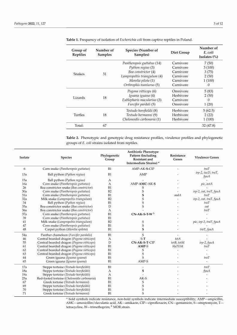
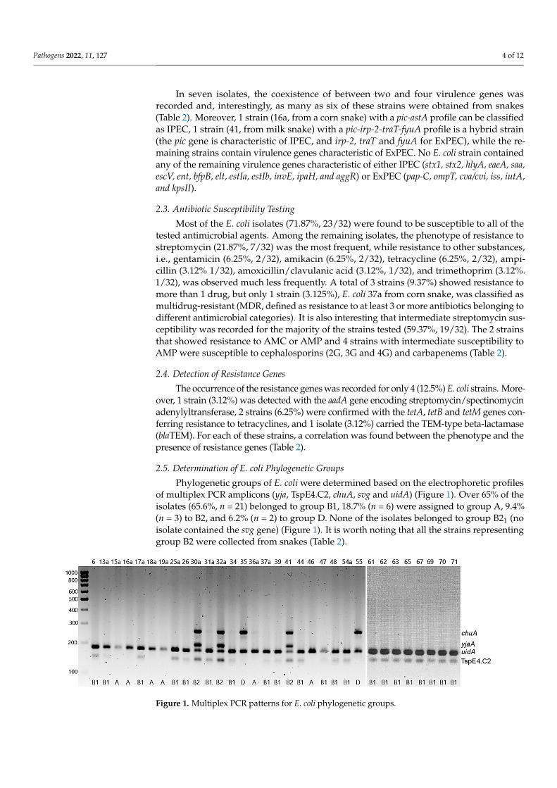
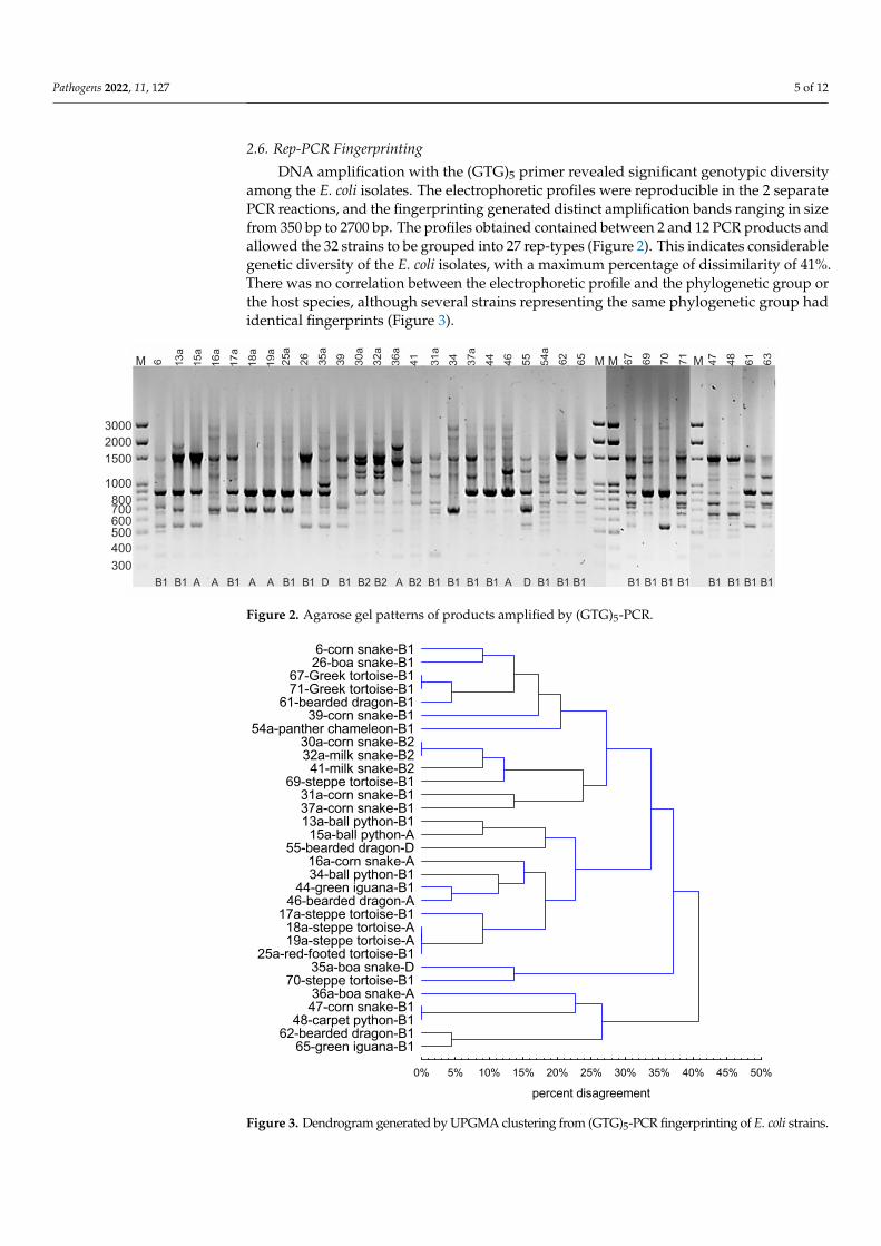
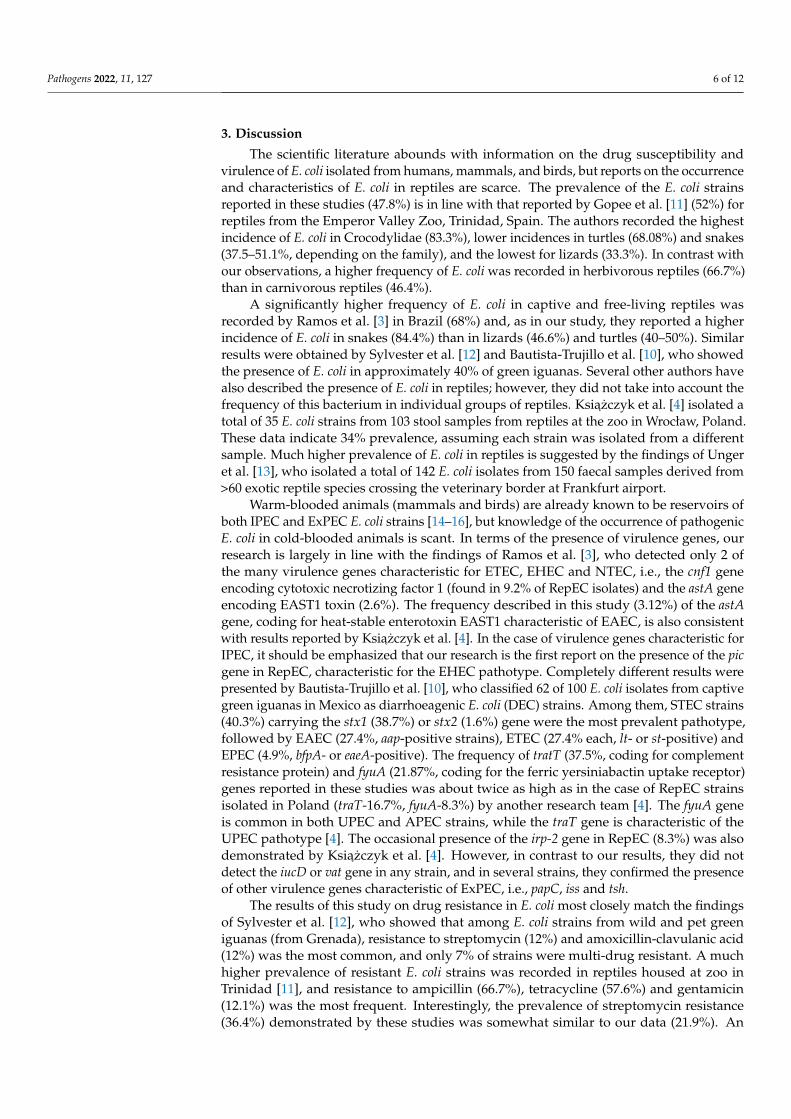
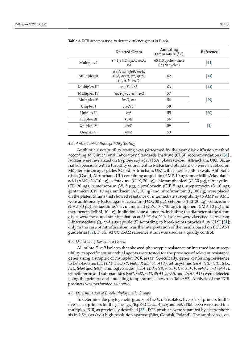
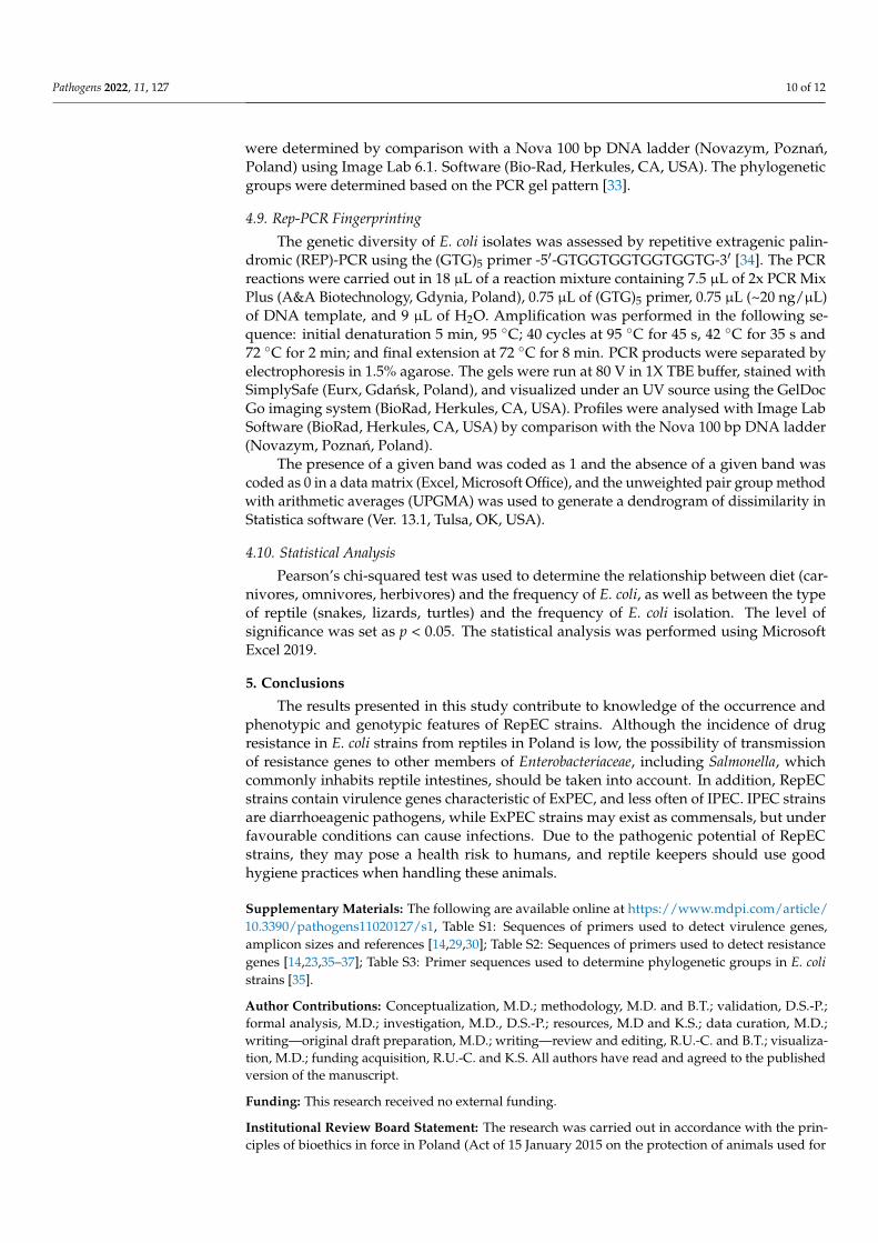
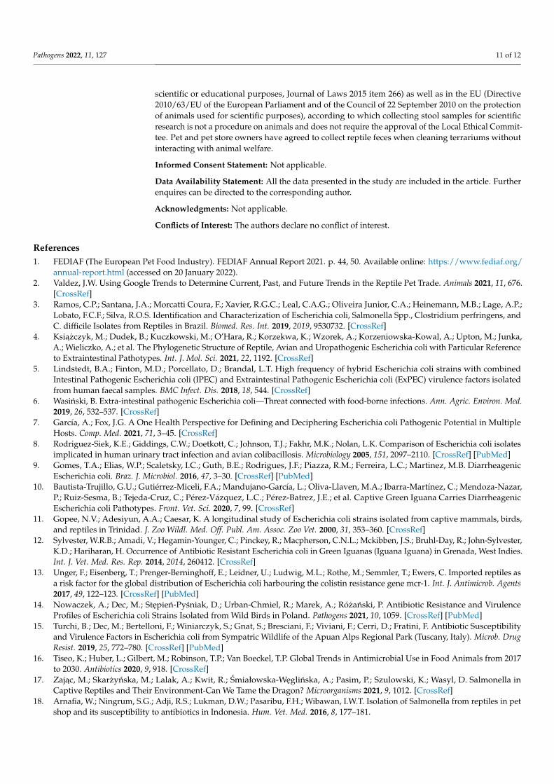


