Embed Size (px)
Citation preview
General rights Copyright and moral rights for the publications made accessible in the public portal are retained by the authors and/or other copyright owners and it is a condition of accessing publications that users recognise and abide by the legal requirements associated with these rights.
Users may download and print one copy of any publication from the public portal for the purpose of private study or research.
You may not further distribute the material or use it for any profit-making activity or commercial gain
You may freely distribute the URL identifying the publication in the public portal If you believe that this document breaches copyright please contact us providing details, and we will remove access to the work immediately and investigate your claim.
Downloaded from orbit.dtu.dk on: Jun 29, 2022
Profiling evolutionary landscapes underlying drug resistance
Hickman, Rachel
Publication date:2017
Document VersionPublisher's PDF, also known as Version of record
Link back to DTU Orbit
Citation (APA):Hickman, R. (2017). Profiling evolutionary landscapes underlying drug resistance. Novo Nordisk FoundationCenter for Biosustainability.
Profiling evolutionary landscapes underlying drug resistance.
PhD Thesis: Rachel Amanda Hickman 14th February 2017
Novo-Nordisk Foundation Center of Biosustainability
Technical University of Denmark
Profiling evolutionary landscapes underlying drug resistance
PhD thesis written by Rachel A. Hickman
Supervisor: Prof. Morten Sommer
Co-Supervisor: Prof. Søren Molin
Copyright © 2017 by Rachel A. Hickman
Novo Nordisk Foundation Center for Biosustainability
Technical University of Denmark
Kemitorvet 220, 2800 Kgs.Lyngby
Denmark
Preface
This PhD thesis was prepared at Novo Nordisk Foundation Center for Bio-sustainability at the Technical University of Denmark (DTU) in fulfillment of the requirements for the PhD degree. Research work was performed both at the Centre for Systems Microbiology (2014-2015) and at the Novo Nordisk Foundation Center for Biosustainability (2015-2017).
The work was carried out under the supervision of Professor Morten Som-mer (Supervisor) and Professor Søren Molin (co-supervisor) and funded by a PhD stipend from DTU.
14th February 2017
Rachel Amanda Hickman
Acknowledgements
There are many people I would like for making my time through my PhD enjoyable and do-able. Firstly, and most importantly, I would like to thank Morten for giving me this opportunity to this PhD and allowing me to research cool and exciting science and pushing me outside my comfort zone and Søren for his support when I was overwhelmed. I would also like to thank Christian for guiding me and supervising my as a new lost PhD student and Andreas his partner in crime that has help on many occasions, with R and the Danish abstract within this PhD thesis! I would also like to thank Lejla for providing DNA extracts that formed the pilot study for the second manuscript, Helle for her depth reading and guid-ance of this manuscript and Jakob for his tolerance and help in this project. I would also like to thank Lumeng for helping on the crazy FACs project and tips on Pseudomonas aeruginosa PCR! There are so many people from the Sommer lab group that I’m also grateful for such as Gitte for group organization and oracle of the tem system, Mari for finding obscure lab items, PhD comradery from the Sommer PhD office (Ruben, Micheal, Kira, Gonzalo and un-officially Christian N) and other members of the Sommer lab group both past and present as well as other CFB members. Finally, I’m also very grateful for the support from my family and my part-ner Erik and would like to dedicate this PhD thesis to them as without them it would be impossible. Tusind tak!
Abstract
Bacteria have existed on earth for 3.5 million years and their ability to evolve has allowed for their survival in almost all global niches. Bacteria evolve and adapt easily due to their short generation times, plastic genomes, acquisition (external) DNA and their ability to form protective bacterial communities i.e. biofilms or dormant metabolic states.
Antibiotic drugs are currently our best medicine to treat (against) bacterial pathogens due to antibiotics unique properties of being small molecules that are soluble and act systemically. These qualities allow for many modern medical procedures to occur due to antibiotics preventative/ prophylactic and therapeutic qualities.
Despite bacterial antibiotic resistance mechanisms always being present in nature, the overuse and misuse of antibiotics by humans are accelerating the rise and dissemination of bacterial antibiotic resistance. Bacterial antibiotic resistance is global threat to public health; especially because of lack of new drugs. It has been highlighted that understanding antibiotic resistance by further elucidating mechanisms of evolution, molecular mechanisms of ac-tion and reservoirs of resistance are essential Therefore, the work involved in this PhD thesis, examines the evolution of antibiotic resistance in bacterial populations. Two main studies were performed: the first to elucidate the molecular mech-anisms of collateral sensitive drug pairs and collateral resistance drug pairs in adaptation of Escherichia coli populations; and the second exploring mu-tant variant dynamics in cystic fibrosis lung, by analyzing sputum samples from chronic carriers of Pseudomonas aeruginosa undergoing antibiotic treatment. Both studies explore the trajectories of antibiotic resistance within bacterial populations: the first study by exploring antibiotic resistance loci, and the in the second by whole-gene sequencing. The desired outcome from both stud-ies is to find methods to use antibiotic therapy more rationally to treat infec-tion efficiently and effectively whilst reducing the evolution of antibiotic resistance.

Dansk resumé
Bakterier har eksisteret på jorden i 3,5 millioner år, og i den tid har deres evne til at udvikle sig tilladt deres overlevelse i stort set alle tænkelige ni-cher. Bakterier udvikles og tilpasses let på grund af deres korte generations-tider, plastiske genomer, erhvervelsen af eksternt DNA samt deres evne til at danne beskyttende bakterielle samfund dvs. biofilm.
Antibiotika er i øjeblikket vores bedste medicin til behandling af bakterielle patogener på grund af dets opløselighed systemiske virkning. Disse kvalite-ter er vitale for mange moderne medicinske procedurer på grund af antibioti-kas profylaktiske og terapeutiske egenskaber.
På trods af at antibiotika resistensmekanismer altid har været til stede i natu-ren, har samfundets overforbrug og misbrug af antibiotika fremskyndet stig-ningen og formidling af bakteriel antibiotikaresistens. Bakteriel antibiotika-resistens er en global trussel mod den offentlige sundhed; især på grund af manglen på nye lægemidler. En grundigere forståelse af antibiotikaresistens ved yderligere at belyse evolutionsmekanismer, molekylære virkningsmeka-nismer og reservoirer er afgørende. Derfor belyser dette PhD-arbejde udvik-lingen af resistens i bakterielle.
To hovedundersøgelser blev udført: den første bestod i, yderligere at belyse de molekylære mekanismer i antbiotikapar som indbyrdes forstærker eller svækker resistensevolution i Escherichia coli populationer. Det andet studie udforsker mutantvariant dynamik i CF lunge-prøver fra kronisk Pseudomo-nas aeruginosa inficerede cystisk fibrose patienter under igangværende anti-biotisk behandling på hospitalet. Begge undersøgelser udforsker udviklings-veje for antibiotikaresistens i bakterielle populationer: det første studie ved at udforske kendte antibiotikaresistensmutationer i genomet, og det andet ved sekventering af hele gener involveret i resistens. Det ønskede resultat fra begge undersøgelser er at finde metoder til at bruge antibiotika, der både behandler patogen effektivt og samtidig hindre udviklingen af antibiotikaresistens.
List of Publications
This thesis is based on the following papers, which are referred to in the text by their Roman numerals.
I. R.A. Hickman, C. Munck and M.O.A Sommer. Time-resolved tracking
of mutations reveals strong clonal interference during antimicrobial adaptive of Escherichia coli to single and drug pairs. (Currently in inter-active review to Frontiers in Microbiology)
II. R.A. Hickman, J. Frimodt-Møller, E. Rossi, H.K. Johansen, S. Mølin and M.O.A. Sommer. Direct sequencing of Pseudomonas aeruginosa from sputum of cystic fibrosis patients undergoing antimicrobial thera-py. (Manuscript in preparation).
Contents
PROFILINGEVOLUTIONARYLANDSCAPESUNDERLYINGDRUGRESISTANCE. I
PREFACE III
ACKNOWLEDGEMENTS IV
ABSTRACT V
DANSKRESUMÉ VI
ABBREVIATIONS X
OUTLINEOFPHDTHESIS 11
BACTERIALEVOLUTION 12
PROCESSESTHATALLOWBACTERIALGENOMICMODIFICATION 12EVOLUTIONARYTHEORY:NEO-LAMARCKISMVERSUSNEO-DARWINISM? 14BACTERIALPOPULATIONADAPTATIONSTONOVELENVIRONMENTALCHALLENGES 15
ANTIBIOTICDRUGS 17
DISCOVERYOFANTIBIOTICS 17ANTIBIOTICCLASSES 18ANTIBIOTICRESISTANCEANDPERSISTENCE 19IMPACTSANDDISSEMINATIONOFANTIBIOTICRESISTANCE 21
PROFILINGANDEXPLORINGDRUGTREATMENTANDRESISTANCE 22
PHENOTYPICQUANTIFICATIONOFANTIBIOTICRESISTANCE 22MOLECULARQUANTIFICATIONOFANTIBIOTICRESISTANCE 23VERIFICATIONOFMOLECULARDETECTIONOFANTIBIOTICRESISTANCE 24GENERATIONOFCLINICALANDENVIRONMENTALSAMPLECOLLECTIONS 25GENERATIONOFLABORATORYGENERATEDSAMPLECOLLECTION 25
APPLICATIONSOFPROFILINGDRUGTREATMENTANDRESISTANCELANDSCAPES 28
TRADITIONALAPPROACHESTOAPPLYINGDRUGPROFILINGFORTREATMENT,WITHLIMITEDANTIBIOTICRESISTANCEOUTCOMES 28NOVELMETHODSTOSYSTEMATICALLYREVIEWDRUGREACTIONS 29TRANSFERIN-VITRODRUGPROFILINGRESULTSINTOPOSSIBLECLINICALAPPLICATION 30
CHAPTER5.CONCLUDINGREMARKSANDFUTUREPERSPECTIVES 32
REFERENCES 34
MANUSCRIPTS 43
MANUSCRIPTI:TIME-RESOLVEDTRACKINGOFMUTATIONSREVEALSSTRONGCLONALINTERFERENCEDURINGANTIMICROBIALADAPTIVEOFESCHERICHIACOLITOSINGLEANDDRUGPAIRS. 43MANUSCRIPTII:DIRECTSEQUENCINGOFPSEUDOMONASAERUGINOSAFROMSPUTUMOFCYSTICFIBROSISPATIENTSUNDERGOINGANTIMICROBIALTHERAPY 44
79
Abbreviations
ALE Adaptive Laboratory Evolution AMK Amikacin CHL Chloramphenicol CIP Ciprofloxacin EUCAST The European committee on antimicrobial susceptibility testing HGT Horizontal gene transfer IC Inhibitory Concentration I Intermediate antimicrobial susceptible type INDELs Insertion or deletions sequences MDR Multi-drug resistance NGS Next generation sequencing ROS Reactive oxygen species R Resistant antimicrobial susceptible type SNPs Single nucleotide polymorphisms S Susceptible antimicrobial susceptible type WGS Whole genome sequencing WHO World Health Organization
11
Outline of PhD Thesis
This thesis is composed of 5 chapters: Bacterial Evolution, Antibiotic drugs, Profiling and Exploring Drug Treatment and Resistance, Applications of Profiling of Drug Treatment and Resistance, and Concluding Remarks and Future Perspectives. Each chapter composes of a short introduction, to provide the overview of the importance of the chapter, followed by sub-chapters. Within each of the sub-chapter’s ideas, theories and links to scientific literature are given and then summarized with how this applies to the to the research work done in this PhD time-period.
12
Bacterial Evolution
Within most natural environments a diverse consortia of bacteria species exist. Following the evolutionary laws of nature, bacteria have adapted to a variety of physiological demands such as pH [1], osmotic pressures [2], tem-perature [3], nutrient availability [4] and chemical perturbations like disin-fectants [5], metal ions [6] and antibiotics [7]. Bacteria have several evolu-tionary constraints e.g. asexual reproduction that limits chromosomal ge-nomic recombination and a high codon bias genome of ca. 90% [8]. Despite these constraints, bacteria can easily and rapidly evolve due to their short generation time (e.g. E. coli K-12 sub-strain MG1655 in Luria-Bertani broth at 37oC has a doubling time during steady-state of 20 minutes [9]), their large population size capability (e.g. up to 2x10⁹ CFU/mL [10]), their rate of spontaneous mutation (e.g. ~ 0.03 per genome per replication [11]), and their genetic exchange between taxa [12]). These advantageous characteristics promote both anagenesis, which allows evolution within the bacterial species population, and cladogenesis, which allows new bacterial species to develop in different biological niches [13].
Processes that allow bacterial genomic modification To accomplish these abilities, bacteria rely on mechanisms to acquire novel genetic information and manipulate their genome. This achieved by three main processes: bacterial transduction, transformation and de novo muta-tions. The first two processes occur by horizontal gene transfer (HGT), which unlike other forms of life defy species specific boundaries. Therefore, a microbial habitat can quickly possess abilities to defy the abilities of given antimicrobials, metal ions and disinfectants. An infamous example of this is the CTX-M-15 plasmid that confers resistance to antimicrobials, metal ions and biocides [14]. Furthermore, it is easily disseminated in Enterobacteri-aceae and as a consequence several clinical outbreaks have occurred [15–17]. De novo mutations, however, occur intrinsically within a cell and if remain uncorrected by cellular correction systems these mutations will be passed on to its future daughter cells. As mentioned earlier HGT resistance mechanisms defy species specific boundaries. It could therefore be easy to assume that more scientific effort should be concentrated on HGT resistance
13
mechanism rather than de-novo mutational resistance mechanisms, however both play an integral role in adaptation evolution. For some bacterial species – such as P. aeruginosa in chronically infected cystic fibrosis patients [18,19] and Mycobacterium tuberculosis where HGT [20] – evolution is generated mainly or solely by the latter mechanism. Therefore, scientific research in both types of acquired resistance are important, especially when it comes to understanding bacterial evolution in a wide range of organisms and perturbations. With the research involved in this PhD doctorate, I have looked into the types of de novo mutations that occur in the bacterial population. De novo mutations are a result of genomic instability. Genomic instability is induced by the constant threat that bacterial cells are exposed to, such as operations of their DNA replication and repair systems, mobile elements, phages, chem-ical entitles and environmental factors [21]. De novo mutations manifest themselves as point mutations, genomic rearrangements or translocations, and can be inherited to future prodigy if remained un-corrected by cellular repair systems. Point mutations, or single nucleotide changes (SNPs), can induce three types of amino acid coding changes. The effects of these chang-es are silent, when no effects are observed to coding amino acid, missense, where a different amino acid is coded for and is also referred to as a non-synonymous change, and nonsense mutations, which encodes for a stop-codon that terminates the transcription of the RNA prematurely. Genomic rearrangements are induced by insertion or deletions sequences (INDELs) or the combination thereof, amplifications, or translocations. The result of IN-DELs in the genome cause change to the encoded amino acid sequence of a transcribed mRNA strand by either the addition or subtraction of codons or by causing frame shifts to encode entirely different amino acids. The results of gene duplication are common in both bacteria and eukaryotes [22]. A possible model for gene duplication is the innovation-amplification-divergence model. In this model an ancestral gene has a weak secondary activity, and in a given favorable selection pressure a cell that duplicate this gene create a different gene from the ancestor by genetic modification [23]. From the research work performed during this PhD, my main focus was on missense and nonsense mutations SNPs, INDELs and possible gene duplica-tion events that occur during antibiotic adaptation or during antibiotic treat-ment.
14
Evolutionary theory: Neo-Lamarckism versus Neo-Darwinism? Genomic modifications which lead to bacterial phenotypes that are better suited to a given environment are essential for bacterial evolutionary devel-opment, but which evolution hypothesis that induce these effects is uncer-tain. Therefore, the theory that best answer the evolutionary development process is under scientific debate again, as the impact of understanding pro-cess that lead to genomic stability play an important role in curbing antibi-otic resistance. The two main theories are Neo-Lamarckism and Neo-Darwinism. The Neo-Lamarckism theory assumes that external environmental cues causes indi-viduals to change their phenotype in response. Individuals that are successful in changing their phenotype to adapt will pass this on to their future progeny. The Neo-Darwinism theory follows that the basic unit of evolution is a population, where genomic variation occurs by random genetic drift and selection for given phenotypes is driven by environmental factors. Evidence for both theories has been produced by the use of next generation sequencing (NGS) and other typing methods. Direct evidence for bacterial Neo-Lamarckism can be found in the CRISPR-cas system, where integration of small segments of HGT DNA into specific loci allows for host defense (Koonin and Wolf 2009). Most de-novo antibiotic resistance is nonetheless assumed to be the result of Neo-Darwinism. This is seen in bacterial popula-tions where contact with sub-lethal concentrations of antibiotics generates random mutants either by inducing direct effects (e.g. bleomycin inducing double-strand DNA breaks [24]) or in-direct effects (e.g. ampicillin down-stream production of reactive oxygen species (ROS) that incudes DNA dam-age [25]). These mutations establish themselves in the population and can be fixed or lost in the bacterial population by clonal interference. However, a quasi-Lamarckism mechanism coupled with neo-Darwinian mechanism, permits bacterial cell to adapt to various environmental perturbations by allowing a regulation of genomic instability to occur followed by natural selection [26]. The quasi-Lamarckism mechanism was first suggested by Radman, circulated privately in 1970, where she states that “‘SOS-replication’ mechanism can be induced by a variety of mutagenic treatments, which cause inhibition of ‘vegetative’ DNA replication…This replication mechanism is an inaccurate, mutation-prone process” (Embedded in [27]). Interestingly many antibiotic drugs classes used in the experimental work of this PhD thesis also induce SOS- replication response. The drug classes that induced these effects are: aminoglycosides [28], chloramphenicol [29] and fluoroquinolones [30]. Therefore, it is interesting to note that, sub-lethal exposure to antibiotics can induce DNA damage and trigger genomic insta-bility that therefore induced genetic diversity of microbial pathogens
15
(Shapiro 2015). Despite none of these quasi-larmarckian mechanisms being validated experimentally in this PhD thesis, the neo-Darwinian mutations I observe could also be a result of the antibiotics inducing genomic instability facilitated by quasi-larmarckian mechanisms. Therefore, this is something I would like to return to in the concluding remarks and future perspectives.
Bacterial population adaptations to novel environmental challenges For bacteria populations to adapt to a new environmental challenge or per-turbation, novel mutation cells must occur, reproduce in quantities as not to be lost by random genetic drift. This process is known as bacterial mutant allelic establishment, where a sub-population of this clone type develop within a population. If this mutant allelic sub-population is able to take over the population and dominate 100% of the population this is known as fixa-tion. Two variables govern a bacterial population’s ability to survive a new envi-ronmental challenge these are: the range of beneficial mutations that are able to be produced in the population and the size of the population [31]. In a small bacterial population, the range of beneficial mutants is less, therefore that population has less likelihood of surviving is also less [32]. However, if a beneficial mutation allelic sub-population does arise and become estab-lished it has an almost certain probability of fixing in the population. This is opposite in larger population due to the range of beneficial mutations being higher (e.g. clonal divergence), so likelihood of surviving is also higher. However, the beneficial mutation allelic sub-population or clone has less chance of becoming fixed in the population. This is due to clonal interfer-ence, where two novel genotypes or more compete with each other to estab-lish themselves within the population [33]. Different genotypes can occur in different genetic backgrounds within the population, therefore the large the bacterial population, the higher diversity in bacterial genetic background and novel beneficial mutational allelic types [34]. All these characteristics play an important role in evolution population mutagenic allelic dynamics, where periodic selection of various clones of mutant alleles occur. The overall re-sult of clonal interference in a large population acts as a natural selection process that selects beneficial mutations that has the largest effect at the smallest fitness cost, known as periodic selection [35]. The work I presented in this PhD thesis explores the characteristics of evolu-tion of bacterial population mutagenic allelic dynamics by time-resolved tracking known mutagenic loci in in-vitro antibiotic E. coli adaption (manu-
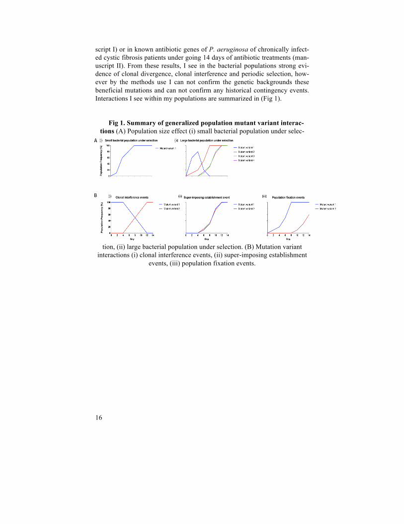
16
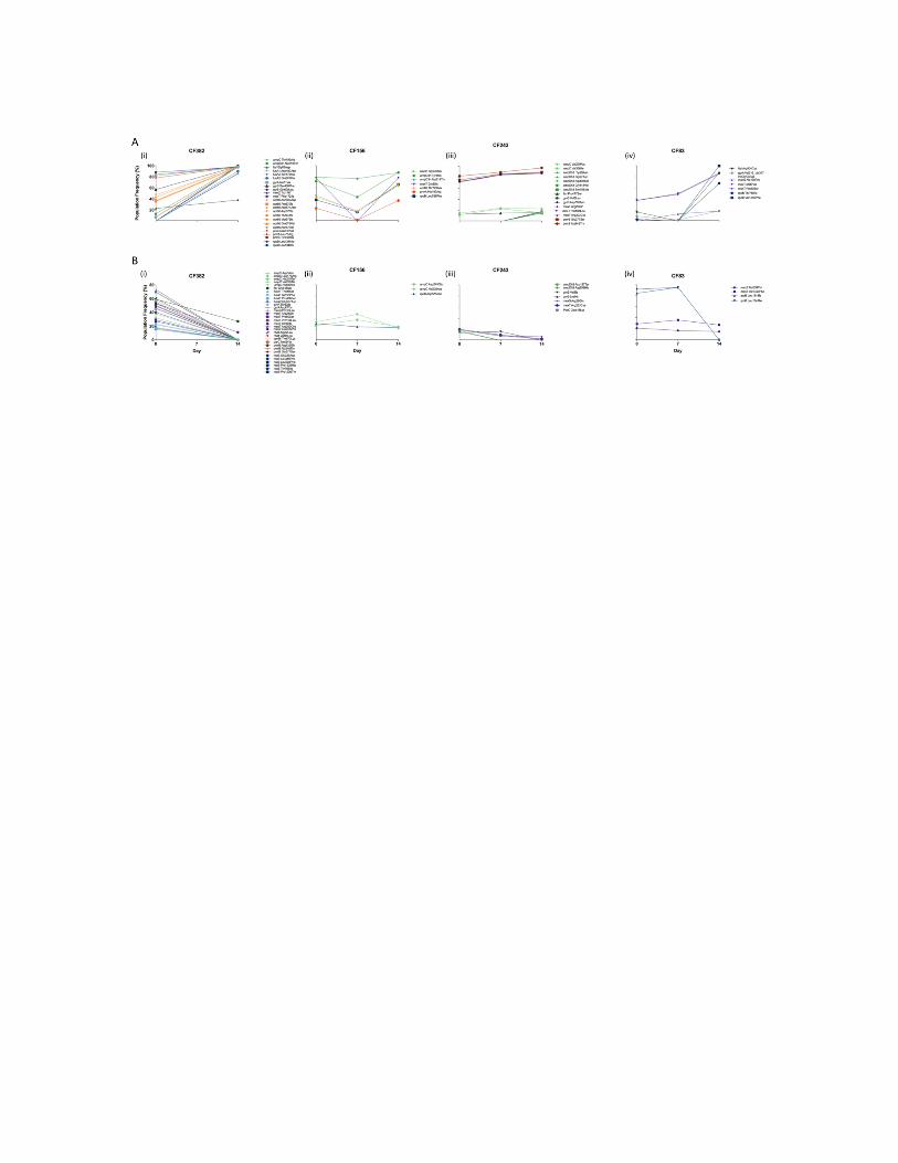
script I) or in known antibiotic genes of P. aeruginosa of chronically infect-ed cystic fibrosis patients under going 14 days of antibiotic treatments (man-uscript II). From these results, I see in the bacterial populations strong evi-dence of clonal divergence, clonal interference and periodic selection, how-ever by the methods use I can not confirm the genetic backgrounds these beneficial mutations and can not confirm any historical contingency events. Interactions I see within my populations are summarized in (Fig 1).
Fig 1. Summary of generalized population mutant variant interac-tions (A) Population size effect (i) small bacterial population under selec-
tion, (ii) large bacterial population under selection. (B) Mutation variant interactions (i) clonal interference events, (ii) super-imposing establishment
events, (iii) population fixation events.
17
Antibiotic drugs
The discovery and use of antibiotic drugs allowed the birth of modern medi-cine to occur by allowing medical procedures to be performed whilst reduc-ing the risk of pathogenic bacteria. Antibiotic drugs are currently our best medicine to treat against bacterial pathogens due to antibiotics’ unique prop-erties of being small molecules that are soluble and act systemically [36]. Bacteria have a tremendous ability to evolve and develop genotypes to over-come antibiotic drug effects, and when this occurs in invading bacterial pathogens they cause mortality and morbidity. It is essential to understand where antibiotic drugs come from, what classes of drugs we have, how bac-teria become resistant to these drugs and how we can counteract the effects of antibiotic resistance.
Discovery of antibiotics The potential of antibiotics was first demonstrated following the discovery and use of salvarsan, which was used in the first antibiotic drug trial of late stage syphilis patients and where impressive outcomes were observed [37]. Antibiotic discovery and use was further invigorated by the discovery of penicillin from a contaminated agar plate by Alexander Flemming [38]. Fol-lowing the purification steps developed by Florey and Chain, the medicinal use of penicillin came into fruition[39]. The importance of this discovery was that microorganisms themselves had the ability to produce small chemi-cal compounds that inhibited or killed other microorganisms. This led to the Golden era of antimicrobial discovery with most success being discovered by the Waksman antimicrobial discovery platform [40]. Following the boom in antibiotic discovery it was naively believed that chemistry would be able to produce novel antimicrobials. This approach generated fewer effective novel antimicrobials than expected at the time, but did generate better for-mulations, drug penetrations and drug delivery [41]. The impact of this cre-ated the antibiotic discovery deficit (Fig 2), and – coupled with the rise in antibiotic resistance – there is now a strong emphasis to discover novel anti-biotics but also to further our understanding of our current antibiotics to op-timize their use. The research involved in this PhD thesis of profiling drug resistance, in both the laboratory and from the clinical setting, aims at under-

18
standing bacterial antibiotic resistance evolution with the long-term goals of using antibiotics more effectively.
Fig 2. Time-line of antibiotic drug introduction date
Antibiotic Classes To further understand our current antibiotic drugs it is important to know their chemical structure, as drugs that have the same structural class tend exhibit similar traits of effectiveness, toxicity and allergy potential [40]. Using structural chemistry to classify antibiotic drugs allows us to construct a generalized view of which antibiotic classes disrupt particular biological processes in the bacterial cell (Fig 3). Overall antibiotics disrupt three bio-logical structures and synthesis processes essential for cell growth and maintenance. These are cell structure synthesis and maintenance, nucleic acid synthesis and protein synthesis. Further categorization can be done on the anitbiotic’s other traits, such as their bacterial effect (i.e. bactericidal or bacteriostatic), the type of pathogens they target (i.e. Gram negative or posi-tive or both), the spectrum of pathogens they target (i.e. narrow or broad-spectrum) and their pharmacological abilities. The classification and antibac-terial traits of all antimicrobials are vital to know to be able to utilize these drugs to their maximum efficacy.

19
Fig 3. Generalized view of biological processes disrupted by different an-
tibiotic classes. Top left are antibiotics that disrupt protein synthesis, top right is antibiotics that disrupt cell structure and maintenance and bottom
right antibiotics that disrupt nucleic acid synthesis.
Antibiotic Resistance and Persistence As previously mentioned bacteria have a tremendous capacity to evolve and overcome several survival limiting environmental factors. It is therefore no surprise that they also evolve to resist antibiotics. Antibiotic resistance from a bacteriologist perspective is the genetic adaptation of bacterial cells, which permits genetic altered cells to grow and divide in the presence of antibiotic concentrations that would normally kill or inhibit unaltered cells. Antibiotic resistance from a clinical perspective is related to the drug concentration tolerance and pharmacokinetics of the patient and the effects on the bacteria at this concentration [42,43].
Bacterial cells can alter their genome by acquiring plasmids (such as the CTX-M-15 plasmid disseminated in Enterobacteriaceae species [17]); by acquiring novel genomic material via viral vector (such as phages within the microbiome [44]) and by de-novo mutations (such as mutations observed in cystic fibrosis patients chronically infected with P. aeruginosa [45–47]). Despite the origins of the genomic antibiotic resistance adaptions, bacterial cells use four different types of antibiotic resistance mechanisms to evade drug effects. These mechanisms are: induction of multi-drug resistance (MDR) efflux pumps that expel the antibiotic drug from inside the cell; mod-ification of the drug-target that prevent drug-target complexes forming; membrane modification that reduce permeability of the drug in to the cell; and production of enzymes that degrade the drug (Fig 4).
20
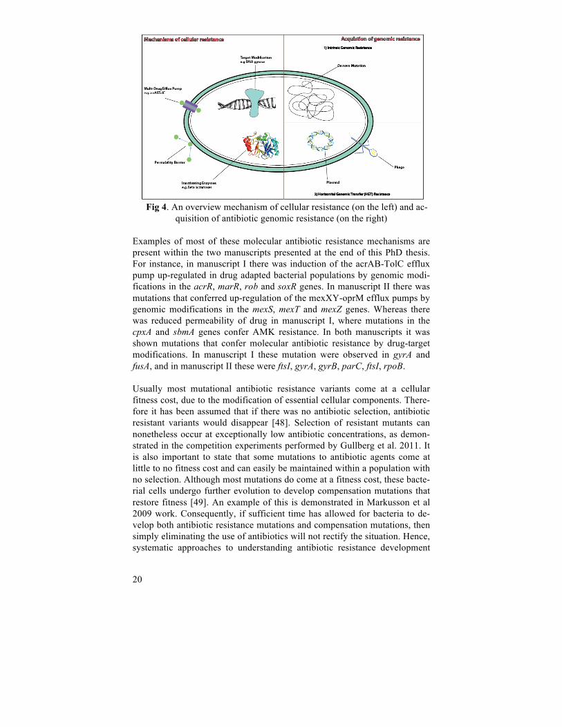
Fig 4. An overview mechanism of cellular resistance (on the left) and ac-quisition of antibiotic genomic resistance (on the right)
Examples of most of these molecular antibiotic resistance mechanisms are present within the two manuscripts presented at the end of this PhD thesis. For instance, in manuscript I there was induction of the acrAB-TolC efflux pump up-regulated in drug adapted bacterial populations by genomic modi-fications in the acrR, marR, rob and soxR genes. In manuscript II there was mutations that conferred up-regulation of the mexXY-oprM efflux pumps by genomic modifications in the mexS, mexT and mexZ genes. Whereas there was reduced permeability of drug in manuscript I, where mutations in the cpxA and sbmA genes confer AMK resistance. In both manuscripts it was shown mutations that confer molecular antibiotic resistance by drug-target modifications. In manuscript I these mutation were observed in gyrA and fusA, and in manuscript II these were ftsI, gyrA, gyrB, parC, ftsI, rpoB. Usually most mutational antibiotic resistance variants come at a cellular fitness cost, due to the modification of essential cellular components. There-fore it has been assumed that if there was no antibiotic selection, antibiotic resistant variants would disappear [48]. Selection of resistant mutants can nonetheless occur at exceptionally low antibiotic concentrations, as demon-strated in the competition experiments performed by Gullberg et al. 2011. It is also important to state that some mutations to antibiotic agents come at little to no fitness cost and can easily be maintained within a population with no selection. Although most mutations do come at a fitness cost, these bacte-rial cells undergo further evolution to develop compensation mutations that restore fitness [49]. An example of this is demonstrated in Markusson et al 2009 work. Consequently, if sufficient time has allowed for bacteria to de-velop both antibiotic resistance mutations and compensation mutations, then simply eliminating the use of antibiotics will not rectify the situation. Hence, systematic approaches to understanding antibiotic resistance development
21
and methods to exploit it in antibiotic treatment are essential and will be discussed in the chapter application of profiling drug resistance. From either a bacteriologist or clinical perspective, the best measurement of antibiotic resistance is using Minimal Inhibitory Concentration (MIC). MIC values by antimicrobial susceptible testing are required to deem if a bacterial species or sample is susceptible (S) (where treatment is likely to be success-ful), intermediate (I) (where its unknown if treatment will be successful) or resistant (R) (where treatment is unlikely to be successful to a given antibi-otic) [50]. This will be covered in more detail in the next chapter on profiling drug resistance.
Impacts and dissemination of Antibiotic Resistance The impacts of antibiotic resistance are considerable due to the fact that we are all possible stakeholders in requirement for antibacterial therapy (e.g. economically due to the cost of health care and for most medical treatment [51]). Many modern medical procedures rely on the use of antibiotics (e.g. prophylaxis and post-operative care for surgery [52], treatment for immuno-comprised individuals [53] and treatment of sexually transmitted diseases [54]). There is a clear correlation that the more antibiotics we use the more bacterial antibiotic resistance occurs due to the large selective pressure we induce [55]. It is also important to state that there has always been a natural reservoir of antibiotic resistance that has existed before antibiotic usage. For instance, putative antibiotic resistance genes have been detected in ancient DNA studies (e.g. permafrost cores [56]) and the gut microbiome of 11th century pre-Columbian Andean mummy [57]). However, our misuse and over use will act as a strong selection to further disseminate antibiotic re-sistance and cause further antibiotic resistance evolution. This is especially so if the selection pressure is strong enough, such those encountered during human or animal antibiotic treatment [58], effluent from manufacturing plants [59] or wastewater treatment plants [60]. Therefore, to prevent or limit antibiotic resistance it is important for academics, clinical and industry to work together in developing new antimicrobials, optimizing treatment strat-egies and finding ways to minimize antibiotic usage to impede future antibi-otic resistance.
22
Profiling and exploring drug treatment and resistance
Profiling drug resistance mainly relies on categorizing different isolates from different samples on their drug concentration abilities. Isolates can be col-lected from clinical samples, environmental samples and in-vitro adaptation experiments. The future of profiling drug resistance will also rely on mo-lecular techniques on bacterial populations to provide a more comprehensive profiling of drug resistance. These methods will be elaborated on in this chapter.
Phenotypic quantification of antibiotic resistance As mentioned in the previous chapter the best initial form of categorizing antibiotic resistance is by the drug’s in-vitro MIC value, which allows the strain’s antibiotic susceptible type be known. This can be measured either by broth dilution in incremental concentration steps or by epsilometer (E-test) assay. To perform these tests effectively, guidelines should be strictly ad-hered to [61]. This is to ensure the only variable of the assay is antibiotic drug concentration. Once a MIC value has been obtained following the clini-cal breakpoints on EUCAST website in accordance to the bacterial species [62], an antimicrobial susceptible type can be stated which can be S, I or R. The EUCAST clinical break-points have been determined by epidemiologi-cal studies of a large number of bacterial isolates and are regularly updated and can be viewed on method (MIC or disk diffusion), antimicrobial and species (EUCAST Antimicrobial wild type distribution of microorganism). However, these guidelines are often slightly relaxed in research facilities, where MIC is often translated into antibiotic inhibitory concentrations (IC) at different population levels such as IC90 and IC50 values [63–65]. Phenotypic quantification of two or more antibiotic drug agents can also be assessed. To evaluate this the fractional inhibitory concentration (FIC) can be measured following the checkerboard FIC methods [66], where agents are tested alone and together. From this an FIC index value can be calculated. Antibiotic combinational effects can be categorized as synergistic (more effective when used together), additive (the same effective when used to-
23
gether, equivalent to using a double dose) and antagonistic (less effective when used together) [67]. Further phenotypic evaluation of an adapting bacterial population can be performed by assessing the evolution of an antibiotic drug-pair compared to its single drug components. This is calculated by using the formula presented in Munck et al 2014 work, which relies on IC90 values of the drug-pairs and the single drug components before and after adaptation. From the calculated evolvability value, if a value ≥ 1 this implies that resistance to the drug-pair evolves to the same extent as the single drug components, whilst a evolvabil-ity value of ≤ 1 implies that resistance to the drug-pair evolves to the lesser extent than the single drug components. This calculation helps to provide information that can indicate drug cross-resistance or more importantly col-lateral sensitivity, a term that will be explained in the next chapter. For man-uscript I the cryopreserved samples were obtain from Munck et al. 2014 work on phenotypic assessment on evolved bacterial populations drug-pair and single drug, where we further evaluated the populations by using molec-ular techniques.
Molecular quantification of antibiotic resistance Molecular methods can reveal ‘omics’ changes (i.e. genomic, transcriptomic and proteomic changes) to bacterial isolates or populations depending on the procedure and method used. These methods can provide rapid and sensitive determination of antibiotic resistance from a wide range of samples. The simplest method is PCR, where DNA from an isolate or a sample can be amplified with designed primers to see whether a resistance gene is present or not. An advance of this method is quantitative PCR (qPCR), where moni-toring the amplification of the PCR product can verify the presence of given genes and their amount in the sample. The qPCR is seen as an attractive diagnostic tool as it is rapid, accurate and sensitive compared to cultivation based determination and can provide information in epidemiological route of transmission etc. PCR methods have been used to determine tetracycline resistance genes [68], methicillin resistance encoded mecA gene in Staphylo-cocci and rifampicin resistance in Mycobacterium tuberculosis [69] and plasmid mediated ampC in Gram negative species [70]. However, there are two main limitations to these PCR approaches. The first limitation being that the presence of these antibiotic resistance genes does not mean that treatment of certain antibiotics will fail, due to the expression of these genes being low; and the second limitation being that novel molecular mechanism can easily be missed [69].
24
The first problem can be overcome by using transcriptomic or proteomic methods; where with transcriptomics direct gene expression can be analyzed, and with proteomics protein levels and modification can be evaluated. Over-coming the second problem requires a more in-depth analysis, such as using sanger sequencing and whole genome sequencing (WGS) on bacterial iso-lates to capture the novel mutations. This can easily be performed but is ex-pensive. These sequencing methods are frequently used for clinical isolates, adaptation laboratory evolution studies, and environmental samples. Other approaches can be taken such as metagenomics where sequencing the DNA of the whole community can take occur. This allows for detection of the whole resistome, but is limited on the annotated antibiotic resistance genes in public gene databases [71]. Another limitation to this technique is that most metagenomic methods rely on NGS platforms that generate short se-quencing reads which can make assembly of genomes into long contigs hard with limited mapping resolution. But unlike other methods it does allow for the whole uncultured community to be sequenced. Therefore, our approach was in manuscript I to PCR amplify loci of interest in known antibiotic resistance genes and for manuscript II PCR amplify well known antibiotic resistance genes of interested and then use population se-quencing on the MiSeq platform. We were fortunate that we only wanted to know the molecular resistance changes to our organism of interest that we could easily map to a reference. This allowed for us to generate amplicons to eliminate background noise, sequencing reads that were easy to map and directly detected variant and establish their frequency in the population.
Verification of molecular detection of antibiotic resistance Unlike phenotypic assays, NGS data sets require validation of variants de-tected as these are mere observations. This can be done by two different approaches: using gene editing tools or literature mining to find validation results. In the first approach, modifications in an ancestral wild type are done by recombineering techniques such as homologous recombineering where SNPs and INDELS can be reintroduce [72], or by high-throughput SNP CRISPER-Cas9 technologies [73]. The second approach is the simpler of the two where validations have been previously been preformed, analyzed and values reported. In manuscript I selected mutations were recombineered, and relative fitness to the ancestral wild type, IC90 to a given drug and persistence at different drug concentrations was reported. For manuscript II all treatment enriched
25
non-synonymous mutations were in the literature mined, those where re-combineering and MIC difference were reported are complied in table 4.
Generation of clinical and environmental sample collections Most bacterial collections comprise of isolates from samples where microbi-ology laboratories have processed the samples. Examples of these are P. aeruginosa isolate collection from sputum samples from cystic fibrosis pa-tients [74,75] Staphylococcus aureus isolates from a teaching hospital [76]. However, especially in environmental samples, it is well known that only a small percentage of bacterial species can be isolated. Therefore many sample collections comprise of extracted DNA, which can later be analyzed by met-agenomic techniques (e.g. pyrosequencing of antibiotic resistance-contaminated river sediments [77]). In manuscript II we used culture-independent methods to analyze our P. aerugionsa populations by extracting the DNA, amplifying our genes of interest and sequencing on the Miseq NGS platform. Validation of results was done with literature mining.
Generation of laboratory generated sample collection To generate antibiotic resistance, sample collections tend to rely on adapta-tion evolution experiments to evolve isogenic naïve wild types to become drug resistant. Unlike clinical bacterial isolate, where evolutionary changes lead to antibiotic resistance, adaptation evolution experiments allow to regu-larly freeze samples to form cryopreserved stocks [78]. As the experiments are performed in controlled laboratory settings, scientific analysis of evolu-tionary phenomena are easier to analyze [79]. Therefore, in turn, replay ex-periments and tracking experiments can easily be performed.
26
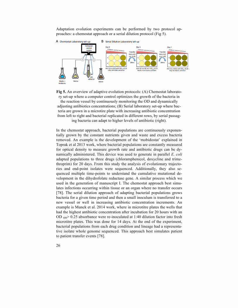
Adaptation evolution experiments can be performed by two protocol ap-proaches: a chemostat approach or a serial dilution protocol (Fig 5).
Fig 5. An overview of adaptive evolution protocols: (A) Chemostat laborato-
ry set-up where a computer control optimizes the growth of the bacteria in the reaction vessel by continuously monitoring the OD and dynamically
adjusting antibiotics concentrations; (B) Serial laboratory set-up where bac-teria are grown in a microtire plate with increasing antibiotic concentration from left to right and bacterial replicated in different rows, by serial passag-
ing bacteria can adapt to higher levels of antibiotic (right). In the chemostat approach, bacterial populations are continuously exponen-tially grown by the constant nutrients given and waste and excess bacteria removed. An example is the development of the ‘mobidostat’ explained in Toprak et al 2013 work, where bacterial populations are constantly measured for optical density to measure growth rate and antibiotic drugs can be dy-namically administered. This device was used to generate in parallel E. coli adapted populations to three drugs (chloramphenicol, doxycline and trime-throprim) for 20 days. From this study the analysis of evolutionary trajecto-ries and end-point isolates were sequenced. Additionally, they also se-quenced multiple time-points to understand the cumulative mutational de-velopment in the dihydrofolate reductase gene. A similar process which we used in the generation of manuscript I. The chemostat approach best simu-lates infections occurring within tissue or an organ where no transfer occurs [78]. The serial dilution approach of adapting bacterial populations grows bacteria for a given time period and then a small inoculum is transferred to a new vessel or well in increasing antibiotic concentration increments. An example is Munck et al. 2014 work, where in microtitre plates the wells that had the highest antibiotic concentration after incubation for 20 hours with an OD 600> 0.25 absorbance were re-inoculated at 1:40 dilution factor into fresh microtitre plates. This was done for 14 days. At the end of the experiment, bacterial populations from each drug condition and lineage had a representa-tive isolate whole genome sequenced. This approach best simulates patient to patient transfer events [78].

27
Another alternative method for systematically examining antibiotic re-sistance is to use strain collections consisting of genetic mutants. Examples are the use of the KEIO strain collection where systematically studying the intrinsic resistome of E. coli to various drugs and examining the phenotypic response yielded important results [80,81]. This has also been applied to P. aeruginosa by using transposon generated libraries, which then can be tested in a similar manner to examine the intrinsic resistome[82–84]. In relation to the research work performed, in manuscript I genomic evalua-tion of the cryopreserved population samples was generated by serial dilu-tion adaption evolution experiments. Whilst in manuscript II, if to be re-performed under strict laboratory conditions, a chemostat laboratory set up would probably be more applicable, due to the bacteria’s permeant residence in the cystic fibrosis patient lung.
28
Applications of profiling drug treatment and resistance landscapes
The implication of profiling evolutionary landscapes in regards to antibiotic resistance are vital, for the ability to use antibiotic as an essential medical resource. Clinical antibiotic resistance is unfortunately not hard for bacteria to achieve due to the micro-niches in the body e.g. in the lungs, epidermis and joints [85]. Especially in these micro-niches bacteria experience sub-inhibitory antibiotic concentrations, which impede but not prevent growth [85,86] and even allow evolution to occur to circumvent any negative ef-fects. Therefore, through this next chapter I will discuss the application of profiling drug resistance landscapes.
Traditional approaches to applying drug profiling for treatment, with limited antibiotic resistance outcomes Within microbiology it has been naively accepted that the development of bacterial antibiotic resistance can be impeded by the use of drug combina-tions, especially those that exhibit synergistic effects. The assumption was that bacteria developing resistance to one drug was more likely than devel-oping resistance to two drugs simultaneously [87]. Traditionally in the clini-cal setting most antibiotic drug combinations are chosen due to their differ-ent cellular targets [88], but despite many antibiotics attacking different cel-lular targets bacteria often evolve the same mechanisms to resistant antibiot-ics. These mechanisms are commonly known as cross-resistance mechanisms (e.g. are the multi-drug resistant efflux pumps and modifica-tions to pleiotropic genes [89]). However, there are exceptions; e.g. it has been frequently found by in-vitro assays that many classes of antibiotics have synergy with aminoglycosides [90–92]. This did have great clinical success and now with understanding the molecular mechanisms we can elu-cidate ways to utilize antibiotics more effectively. In the next sub-chapter, I will further explain the mechanisms of why aminoglycosides exhibit syner-gistic qualities with other antibiotic drug classes.

29
Novel methods to systematically review drug reactions Traditional microbiology could only assess antibiotic resistance by pheno-typic assays or by low throughput sanger sequencing to reveal genomic changes. With developments in molecular and automation technologies it is easier to study phenotypic drug responses and study cellular molecular changes. These new methods have been able to resurrect old hypothesis, such as collateral sensitivity, test them and provide important information to ascertain the application validity. Systematic studies can explore a bacterial species’ reaction to large volume of antibiotics. This can be done by investigating bacterial collections of gene knockouts, transposon mutant libraries, adaptation populations and endpoint isolates, and clinical or environmental isolate collections. Most studies have focused on WGS and reported where genomic resistance has occurred. This could provide critical intrinsic antibiotic resistance information (e.g. the dihydrofolate reductase gene in trimethoprim resistance [93]). Other studies have lead in promoting antibiotic treatment ideas and strategies, which hope-fully can be used in clinical application once certain parameters have been satisfied. Ideas promoting novel targets were generated following the development of the Keio collection of approximately 4000 single gene knockouts. This al-lowed scientists to test the Keio collection with different antibiotics. The first study by Tamae et al 2008 showed that with high-throughput screening of 7 drugs, there were genes that made a given bacterial strain hypersensi-tivity to a given drug. This was further extended in Lui et al 2010 work with 22 additional antibiotics. Both studies showed the complexity of the intrinsic resistome, and especially by targeting pleiotropic genes by co-drugs or anti-biotic adjuvants, there was renewed hope in antibiotic treatments. Transpos-on libraries in P. aeruginosa also have shown promise in exploiting intrinsic resistome weak-points that could be useful for co-drugs or antibiotic adju-vants [82–84]. This produced the revival of an old idea of collateral sensi-tivity [94]. In this work bacteria that were made resistant to a given antibi-otic agent became more sensitive to another. This phenomenon was the main focus in Imamovic and Sommer 2013 work. By adapting E. coli bacterial populations to 23 antibiotics and evaluating drug susceptibility profiles, col-lateral sensitivity and cross-resistance drug networks were established. After finding drug pairs that exhibited collateral sensitivity, a demonstration of drug cycling with gentamycin and cefuroxime was shown to be successful. Adaptation to each of the drugs was done sequentially and MICs of both drugs performed; the drug that was used for adaptation showed increased resistance whilst the other drug had a severely decreased MIC value. The use of drug networks displaying collateral sensitivity has also been explored by

30
Lazar et al. 2014, combining in-vitro adaptation of E. coli and whole genome sequencing to 12 antibiotic drugs with 5-6 replicates. It was later elucidated that aminoglycoside mutants often change key components required for en-ergy production and permeability, whereas other resistance mutants e.g. chloramphenicol mutants require a higher demand of energy, e.g. mutants that up-regulate MDR efflux pumps. Using a similar approach, in Munck et al. 2014 work the experiment was performed on 10 drug-pairs and 5 single drugs done in triplicate for 14 days, showing that the drug pairs which in-volve amikacin and another antibiotic agent such as chloramphenicol can limit the development of antibiotic resistance. To further elucidate these findings within the adaptive bacterial populations I used amplicon deep sequencing of around Munck et al 2014 end-point muta-tions. From this we discovered that the collateral sensitive drug pair AMK-CHL still developed resistance mutations in acrAB-TolC multi-drug resistant efflux transcriptional repressor genes, but did not develop mutations in the fusA and sbmA genes that is typical for AMK resistance development. These studies reported the genomic trade-offs that bacteria must make to become resistant to a given antibiotic drug, but this could leave them more vulnera-ble to another type of antibiotic drug. Therefore, using collateral sensitive drug cycling networks and collateral sensitive pairs tools could be effective to increase the longevity of antibiotic therapy until new agents are available [95].
Transfer in-vitro drug profiling results into possible clinical application To transfer these in-vitro findings from the laboratory to clinical practice, an expansion of data must occur regarding the use of different clinically rele-vant pathogens and isolates [95]. Another consideration is antibiotic’s phar-macodynamics parameters (that describe the impact of an antimicrobial agent on the target organism) and the pharmacokinetic parameters (that de-scribe the availability of an antimicrobial agent in the target organ or tissue), which will provide information regarding the plausibility of using these agents within the patients [43]. It would be of interest to look into the long term effects of using antibiotic collateral sensitive treatment as a tool for possibly treating chronic infections, such as chronic pseudomonas infections. However today little is known of the bacterial population genomic diver-gence of antibiotic resistant variants especially in individuals inflicted with chronic infections. In manuscript II we begin to look into this by studying 19 genes in cystic fibrosis patients that have chronic p. aeruginosa lung infec-tions whilst undergoing hospitalized treatment. We could see a vast array of

31
genomic mutant sub-populations present in the sputum samples. Therefore, understanding bacterial population divergence, sub-populations and the uses of different antibiotic and their deployment could help towards the treatment of bacterial pathogens.
32
Chapter 5. Concluding remarks and future perspectives
As stated through out this PhD thesis, bacteria have many advantageous traits that allow them to evolve to a vast array of environmental conditions. These traits are their: short generation time, large population capabilities and their rate of spontaneous mutations. By following adaptive evolution of bac-teria, we can observe evolution in real time [96] and freeze regular samples for later tracking genomic events as seen with in the two manuscripts of this PhD.
The bacterial evolution studies within this PhD thesis are all related to intrin-sic antibiotic resistance, within bacterial populations. Our molecular pipe-lines could detect SNP and INDEL variants but not clearly detect gene du-plication events. In manuscript I, deep amplicon sequencing of antibiotic loci were tracked over time to establish genomic events occurring in adapted collateral sensi-tive drug-pair bacterial populations, in adapted collateral resistant drug-pair bacterial populations and in adapted single drug bacterial populations. Our finding from this research indicated that adapted collateral sensitive drug-pair populations e.g. populations adapted to AMK-CHL stopped developing fusA and sbmA mutants but continue to develop mutants in other gene loci. In the adapted collateral resistance drug-pair these finding did not occur. Consequently, by using antibiotic collaterally sensitive drug pairs, evolution of antibiotic resistance can be limited but not completely inhibited it. We also observe how mutational variants appear in the population and how their dynamics follow the rules describe by others such as clonal interference, population fixation, and periodic selection occur [34,35,97,98]. Whilst in manuscript II, 19 P. aerugiosa antibiotic resistance and pathoga-daptive whole genes were sequenced and analyzed from DNA extracted from chronically infected cystic fibrosis patients undergoing hospitalized antimicrobial therapy. The results of this manuscript indicated that there was a vast array of genomic variants produced during antimicrobial treatment; most of these were synonymous mutations. Therefore, we thought there could be environmental factors such as quorum sensing mechanisms endog-
33
enous to P. aerugiosa [99] inducing genomic divergence or the use of the antibiotics themselves that could be inducing genomic divergence by mech-anisms such as SOS response[100]. We then examined the non-synonomous mutations within the population and observed effects of clonal interference that has been well documented through literature by adaptation experiments in other Gram negative species [98,101]. These events are probably im-portant for the development of antibiotic resistance within the bacterial pop-ulations to find mutations are effective against antimicrobials that compro-mise fitness the least. Most of our novel enriched variants were not described within the current scientific literature, with the exception of 11 variants, where three had follow up studies where the MIC increase reported. These studies have generated important but limited information. As men-tioned by Hancock 2014, an expansion in antibiotic resistance information is required in different bacterial species, to several antibiotics and treatments, from different bacterial population locations. From the work here I hope I can expand in the future by developing larger data-sets regarding antibiotic resistance within bacterial populations, by ana-lyzing data from both systematic in-vitro experiments and clinical samples. As well as developing sequencing pipelines to capture genomic events across the whole genome within bacterial species populations, so duplication events could also be captured. Maybe it is an ambitious statement to finish on, but by understanding factors that induce genomic variation we can hopefully find the mechanisms to im-pede it, such as the quasi-Lamarckian mechanisms that induce neo-Darwinism evolution in antibiotic resistance. Then using co-drug or antibi-otic adjuvants [81,84,102,103] that target these quasi-Lamarckian mecha-nisms, suppression of antibiotic resistance development can ensure the lon-gevity of modern medical treatments.
34
References
1. Hughes BS, Cullum AJ, Bennett AF. EVOLUTIONARY ADAPTATION TO ENVIRONMENTAL pH IN EXPERIMENTAL LINEAGES OF ESCHERICHIA COLI. Evolution (N Y). Blackwell Publishing Inc; 2007;61: 1725–1734. doi:10.1111/j.1558-5646.2007.00139.x
2. Wood JM. Bacterial responses to osmotic challenges. J Gen Physiol. The Rockefeller University Press; 2015;145: 381–8. doi:10.1085/jgp.201411296
3. Shapiro RS, Cowen LE. Uncovering cellular circuitry controlling temperature-dependent fungal morphogenesis. Virulence. 2012;3: 400–4. doi:10.4161/viru.20979
4. LaCroix RA, Sandberg TE, O’Brien EJ, Utrilla J, Ebrahim A, Guzman GI, et al. Use of adaptive laboratory evolution to discover key mutations enabling rapid growth of Escherichia coli K-12 MG1655 on glucose minimal medium. Appl Environ Microbiol. American Society for Microbiology (ASM); 2015;81: 17–30. doi:10.1128/AEM.02246-14
5. Gerba CP. Quaternary ammonium biocides: efficacy in application. Appl Environ Microbiol. American Society for Microbiology (ASM); 2015;81: 464–9. doi:10.1128/AEM.02633-14
6. Sütterlin S, Edquist P, Sandegren L, Adler M, Tängdén T, Drobni M, et al. Silver Resistance Genes Are Overrepresented among Escherichia coli Isolates with CTX-M Production. Appl Env Microbiol. 2014;80: 6863–6869. doi:10.1128/AEM.01803-14
7. Sommer MOA, Church GM, Dantas G. The human microbiome harbors a diverse reservoir of antibiotic resistance genes. http://dx.doi.org/104161/viru1412010. Taylor & Francis; 2010;
8. Mira A, Ochman H, Moran NA. Deletional bias and the evolution of bacterial genomes. Trends Genet. 2001;17: 589–596. doi:10.1016/S0168-9525(01)02447-7
9. Sezonov G, Joseleau-Petit D, D’Ari R. Escherichia coli physiology in Luria-Bertani broth. J Bacteriol. American Society for Microbiology (ASM); 2007;189: 8746–9. doi:10.1128/JB.01368-07
10. Gonidakis S, Longo VD. Assessing chronological aging in bacteria. Methods Mol Biol. NIH Public Access; 2013;965: 421–37. doi:10.1007/978-1-62703-239-1_28
11. Drake JW, Charlesworth B, Charlesworth D, Crow JF. Rates of Spontaneous Mutation. Genetics. 1998;148.
12. Jones D, Sneath PH. Genetic transfer and bacterial taxonomy.
35
Bacteriol Rev. American Society for Microbiology (ASM); 1970;34: 40–81. Available: http://www.ncbi.nlm.nih.gov/pubmed/4909647
13. Koeppel AF, Wertheim JO, Barone L, Gentile N, Krizanc D, Cohan FM. Speedy speciation in a bacterial microcosm: new species can arise as frequently as adaptations within a species. ISME J. Nature Publishing Group; 2013;7: 1080–1091. doi:10.1038/ismej.2013.3
14. Gullberg E, Albrecht LM, Karlsson C, Sandegren L, Andersson DI. Selection of a multidrug resistance plasmid by sublethal levels of antibiotics and heavy metals. MBio. American Society for Microbiology (ASM); 2014;5: e01918-14. doi:10.1128/mBio.01918-14
15. Mshana SE, Hain T, Domann E, Lyamuya EF, Chakraborty T, Imirzalioglu C. Predominance of Klebsiella pneumoniaeST14 carrying CTX-M-15 causing neonatal sepsis in Tanzania. BMC Infect Dis. BioMed Central; 2013;13: 466. doi:10.1186/1471-2334-13-466
16. Moremi N, Manda E V, Falgenhauer L, Ghosh H, Imirzalioglu C, Matee M, et al. Predominance of CTX-M-15 among ESBL Producers from Environment and Fish Gut from the Shores of Lake Victoria in Mwanza, Tanzania. Front Microbiol. Frontiers Media SA; 2016;7: 1862. doi:10.3389/fmicb.2016.01862
17. Poulou A, Voulgari E, Vrioni G, Koumaki V, Xidopoulos G, Chatzipantazi V, et al. Outbreak caused by an ertapenem-resistant, CTX-M-15-producing Klebsiella pneumoniae sequence type 101 clone carrying an OmpK36 porin variant. J Clin Microbiol. American Society for Microbiology (ASM); 2013;51: 3176–82. doi:10.1128/JCM.01244-13
18. Hancock REW, Speert DP. Antibiotic resistance in Pseudomonas aeruginosa: mechanisms and impact on treatment. Drug Resist Updat. 2000;3: 247–255. doi:10.1054/drup.2000.0152
19. Fajardo A, Martínez-Martín N, Mercadillo M, Galán JC, Ghysels B, Matthijs S, et al. The Neglected Intrinsic Resistome of Bacterial Pathogens. Falagas M, editor. PLoS One. Public Library of Science; 2008;3: e1619. doi:10.1371/journal.pone.0001619
20. Eldholm V, Balloux F. Antimicrobial Resistance in Mycobacterium tuberculosis: The Odd One Out. Trends Microbiol. 2016;24: 637–648. doi:10.1016/j.tim.2016.03.007
21. Darmon E, Leach DRF. Bacterial genome instability. Microbiol Mol Biol Rev. American Society for Microbiology; 2014;78: 1–39. doi:10.1128/MMBR.00035-13
22. Bratlie MS, Johansen J, Sherman BT, Huang DW, Lempicki RA, Drabløs F. Gene duplications in prokaryotes can be associated with environmental adaptation. BMC Genomics. BioMed Central; 2010;11: 588. doi:10.1186/1471-2164-11-588
23. Näsvall J, Sun L, Roth JR, Andersson DI. Real-Time Evolution of New Genes by Innovation, Amplification, and Divergence. Science (80- ). 2012;338.
24. Hecht SM. Bleomycin: New Perspectives on the Mechanism of
36
Action1. American Chemical Society; 1999; doi:10.1021/NP990549F 25. Dwyer DJ, Belenky PA, Yang JH, MacDonald IC, Martell JD,
Takahashi N, et al. Antibiotics induce redox-related physiological alterations as part of their lethality. Proc Natl Acad Sci U S A. National Academy of Sciences; 2014;111: E2100-9. doi:10.1073/pnas.1401876111
26. Koonin E V, Wolf YI. Is evolution Darwinian or/and Lamarckian? Biol Direct. BioMed Central; 2009;4: 42. doi:10.1186/1745-6150-4-42
27. Bridges BA. Error-prone DNA repair and translesion DNA synthesis: II: The inducible SOS hypothesis. DNA Repair (Amst). 2005;4: 725–739. doi:10.1016/j.dnarep.2004.12.009
28. Vestergaard M, Paulander W, Ingmer H. Activation of the SOS response increases the frequency of small colony variants. BMC Res Notes. BioMed Central; 2015;8: 749. doi:10.1186/s13104-015-1735-2
29. Higashitani N, Higashitani A, Horiuchi K. SOS induction in Escherichia coli by single-stranded DNA of mutant filamentous phage: monitoring by cleavage of LexA repressor. J Bacteriol. American Society for Microbiology (ASM); 1995;177: 3610–2. Available: http://www.ncbi.nlm.nih.gov/pubmed/7768876
30. Cirz RT, Chin JK, Andes DR, de Crécy-Lagard V, Craig WA, Romesberg FE. Inhibition of Mutation and Combating the Evolution of Antibiotic Resistance. Waldor M, editor. PLoS Biol. Public Library of Science; 2005;3: e176. doi:10.1371/journal.pbio.0030176
31. Fogle CA, Nagle JL, Desai MM. Clonal Interference, Multiple Mutations and Adaptation in Large Asexual Populations. Genetics. 2008;108: 2163–2173.
32. Wilke CO. The speed of adaptation in large asexual populations. Genetics. Genetics Society of America; 2004;167: 2045–53. doi:10.1534/genetics.104.027136
33. Gerrish PJ, Lenski RE. The fate of competing beneficial mutations in an asexual population. Genetica. Kluwer Academic Publishers; 1998;102/103: 127–144. doi:10.1023/A:1017067816551
34. Good BH, Rouzine IM, Balick DJ, Hallatschek O, Desai MM. Distribution of fixed beneficial mutations and the rate of adaptation in asexual populations. Proc Natl Acad Sci U S A. National Academy of Sciences; 2012;109: 4950–5. doi:10.1073/pnas.1119910109
35. de Visser JAGM, Rozen DE. Clonal interference and the periodic selection of new beneficial mutations in Escherichia coli. Genetics. Genetics Society of America; 2006;172: 2093–100. doi:10.1534/genetics.105.052373
36. Hughes D, Karlén A. Discovery and preclinical development of new antibiotics. Ups J Med Sci. Informa Healthcare; 2014;119: 162–9. doi:10.3109/03009734.2014.896437
37. Kaufmann SHE. Paul Ehrlich: founder of chemotherapy. Nat Rev Drug Discov. Nature Publishing Group; 2008;7: 373–373.
37
doi:10.1038/nrd2582 38. Ligon BL. Sir Alexander Fleming: Scottish researcher who
discovered penicillin. Semin Pediatr Infect Dis. 2004;15: 58–64. doi:10.1053/j.spid.2004.02.002
39. Lee Ligon B. Sir Howard Walter Florey—the force behind the development of penicillin. Semin Pediatr Infect Dis. 2004;15: 109–114. doi:10.1053/j.spid.2004.04.001
40. Lewis K. Platforms for antibiotic discovery. Nat Rev Drug Discov. Nature Research; 2013;12: 371–387. doi:10.1038/nrd3975
41. Brown ED, Wright GD. Antibacterial drug discovery in the resistance era. Nature. Nature Research; 2016;529: 336–343. doi:10.1038/nature17042
42. MacGowan AP. Clinical implications of antimicrobial resistance for therapy. J Antimicrob Chemother. Oxford University Press; 2008;62: ii105-ii114. doi:10.1093/jac/dkn357
43. Rybak MJ. Pharmacodynamics: Relation to antimicrobial resistance. Am J Infect Control. 2006;34: S38–S45. doi:10.1016/j.ajic.2006.05.227
44. Modi SR, Lee HH, Spina CS, Collins JJ. Antibiotic treatment expands the resistance reservoir and ecological network of the phage metagenome. Nature. Nature Research; 2013;499: 219–222. doi:10.1038/nature12212
45. Greipel L, Fischer S, Klockgether J, Dorda M, Mielke S, Wiehlmann L, et al. Molecular Epidemiology of Mutations in Antimicrobial Resistance Loci of Pseudomonas aeruginosa Isolates from Airways of Cystic Fibrosis Patients. Antimicrob Agents Chemother. American Society for Microbiology; 2016;60: 6726–6734. doi:10.1128/AAC.00724-16
46. Fischer S, Greipel L, Klockgether J, Dorda M, Wiehlmann L, Cramer N, et al. Multilocus amplicon sequencing of Pseudomonas aeruginosa cystic fibrosis airways isolates collected prior to and after early antipseudomonal chemotherapy. J Cyst Fibros. 2016; doi:10.1016/j.jcf.2016.10.013
47. Markussen T, Marvig RL, Gómez-Lozano M, Aanæs K, Burleigh AE, Høiby N, et al. Environmental heterogeneity drives within-host diversification and evolution of Pseudomonas aeruginosa. MBio. American Society for Microbiology (ASM); 2014;5: e01592-14. doi:10.1128/mBio.01592-14
48. Hall BG. Innovation: Predicting the evolution of antibiotic resistance genes. Nat Rev Microbiol. Nature Publishing Group; 2004;2: 430–435. doi:10.1038/nrmicro888
49. Andersson DI, Hughes D. Antibiotic resistance and its cost: is it possible to reverse resistance? Nat Rev Microbiol. 2010;8: 260–271. doi:10.1038/nrmicro2319
50. Leclercq R, Cantón R, Brown DFJ, Giske CG, Heisig P, MacGowan AP, et al. EUCAST expert rules in antimicrobial susceptibility testing. Clin Microbiol Infect. 2013;19: 141–160. doi:10.1111/j.1469-
38
0691.2011.03703.x 51. Cantas L, Shah SQA, Cavaco LM, Manaia CM, Walsh F, Popowska
M, et al. A brief multi-disciplinary review on antimicrobial resistance in medicine and its linkage to the global environmental microbiota. Front Microbiol. Frontiers; 2013;4: 96. doi:10.3389/fmicb.2013.00096
52. Poggio JL. Perioperative strategies to prevent surgical-site infection. Clin Colon Rectal Surg. Thieme Medical Publishers; 2013;26: 168–73. doi:10.1055/s-0033-1351133
53. Vincent J-L, Bassetti M, François B, Karam G, Chastre J, Torres A, et al. Advances in antibiotic therapy in the critically ill. Crit Care. BioMed Central; 2016;20: 133. doi:10.1186/s13054-016-1285-6
54. Shaskolskiy B, Dementieva E, Leinsoo A, Runina A, Vorobyev D, Plakhova X, et al. Drug Resistance Mechanisms in Bacteria Causing Sexually Transmitted Diseases and Associated with Vaginosis. Front Microbiol. Frontiers Media SA; 2016;7: 747. doi:10.3389/fmicb.2016.00747
55. O ’neill J. TACKLING DRUG-RESISTANT INFECTIONS GLOBALLY: FINAL REPORT AND RECOMMENDATIONS THE REVIEW ON ANTIMICROBIAL RESISTANCE. 2016;
56. D’Costa VM, King CE, Kalan L, Morar M, Sung WWL, Schwarz C, et al. Antibiotic resistance is ancient. Nature. Nature Research; 2011;477: 457–461. doi:10.1038/nature10388
57. Santiago-Rodriguez TM, Fornaciari G, Luciani S, Dowd SE, Toranzos GA, Marota I, et al. Gut Microbiome of an 11th Century A.D. Pre-Columbian Andean Mummy. Wilson BA, editor. PLoS One. Public Library of Science; 2015;10: e0138135. doi:10.1371/journal.pone.0138135
58. Wegener HC. ANTIBIOTIC RESISTANCE—LINKING HUMAN AND ANIMAL HEALTH. National Academies Press (US); 2012;
59. Marathe NP, Regina VR, Walujkar SA, Charan SS, Moore ERB, Larsson DGJ, et al. A Treatment Plant Receiving Waste Water from Multiple Bulk Drug Manufacturers Is a Reservoir for Highly Multi-Drug Resistant Integron-Bearing Bacteria. Zhou Z, editor. PLoS One. Public Library of Science; 2013;8: e77310. doi:10.1371/journal.pone.0077310
60. Li D, Yang M, Hu J, Zhang J, Liu R, Gu X, et al. Antibiotic-resistance profile in environmental bacteria isolated from penicillin production wastewater treatment plant and the receiving river. Environ Microbiol. Blackwell Publishing Ltd; 2009;11: 1506–1517. doi:10.1111/j.1462-2920.2009.01878.x
61. EUCAST: Videos from EUCAST [Internet]. [cited 13 Feb 2017]. Available: http://www.eucast.org/videos_from_eucast/
62. European Committee on Antimicrobial Susceptibility Testing Breakpoint tables for interpretation of MICs and zone diameters.
63. Imamovic L, Sommer MOA. Use of Collateral Sensitivity Networks to Design Drug Cycling Protocols That Avoid Resistance
39
Development. Sci Transl Med. 2013;5: 204ra132-204ra132. Available: http://stm.sciencemag.org/content/5/204/204ra132%5Cnhttp://stm.sciencemag.org/content/5/204/204ra132.full%5Cnhttp://stm.sciencemag.org/content/5/204/204ra132.full.pdf
64. Munck C, Gumpert HK, Wallin AI, Wang HH, Sommer MO. Prediction of resistance development against drug combinations by collateral responses to component drugs. Sci Transl Med. 2014;6: 262ra156. Available: http://www.ncbi.nlm.nih.gov/pubmed/25391482
65. Chevereau G, Dravecká M, Batur T, Guvenek A, Ayhan DH, Toprak E, et al. Quantifying the Determinants of Evolutionary Dynamics Leading to Drug Resistance. Balaban N, editor. PLOS Biol. 2015;13: e1002299. doi:10.1371/journal.pbio.1002299
66. Orhan G, Bayram A, Zer Y, Balci I. Synergy tests by E test and checkerboard methods of antimicrobial combinations against Brucella melitensis. J Clin Microbiol. American Society for Microbiology (ASM); 2005;43: 140–3. doi:10.1128/JCM.43.1.140-143.2005
67. Odds FC. Synergy, antagonism, and what the chequerboard puts between them. J Antimicrob Chemother. Oxford University Press; 2003;52: 1–1. doi:10.1093/jac/dkg301
68. Aminov RI, Chee-Sanford JC, Garrigues N, Mehboob A, Mackie RI. Detection of Tetracycline Resistance Genes by PCR Methods. Public Health Microbiology. New Jersey: Humana Press; 2004. pp. 003–014. doi:10.1385/1-59259-766-1:003
69. Fluit AC, Visser MR, Schmitz FJ. Molecular detection of antimicrobial resistance. Clin Microbiol Rev. American Society for Microbiology (ASM); 2001;14: 836–71, table of contents. doi:10.1128/CMR.14.4.836-871.2001
70. Mohd Khari FI, Karunakaran R, Rosli R, Tee Tay S. Genotypic and Phenotypic Detection of AmpC β-lactamases in Enterobacter spp. Isolated from a Teaching Hospital in Malaysia. Galdiero M, editor. PLoS One. Public Library of Science; 2016;11: e0150643. doi:10.1371/journal.pone.0150643
71. Mande SS, Mohammed MH, Ghosh TS. Classification of metagenomic sequences: methods and challenges. Brief Bioinform. Oxford University Press; 2012;13: 669–681. doi:10.1093/bib/bbs054
72. Lennen RM, Nilsson Wallin AI, Pedersen M, Bonde M, Luo H, Herrgård MJ, et al. Transient overexpression of DNA adenine methylase enables efficient and mobile genome engineering with reduced off-target effects. Nucleic Acids Res. Oxford University Press; 2016;44: e36–e36. doi:10.1093/nar/gkv1090
73. Garst AD, Bassalo MC, Pines G, Lynch SA, Halweg-Edwards AL, Liu R, et al. Genome-wide mapping of mutations at single-nucleotide resolution for protein, metabolic and genome engineering. Nat Biotechnol. 2016;35: 48–55. doi:10.1038/nbt.3718
40
74. Sommer LM, Marvig RL, Luján A, Koza A, Pressler T, Molin S, et al. Is genotyping of single isolates sufficient for population structure analysis of Pseudomonas aeruginosa in cystic fibrosis airways? BMC Genomics. BioMed Central; 2016;17: 589. doi:10.1186/s12864-016-2873-1
75. Workentine ML, Sibley CD, Glezerson B, Purighalla S, Norgaard-Gron JC, Parkins MD, et al. Phenotypic Heterogeneity of Pseudomonas aeruginosa Populations in a Cystic Fibrosis Patient. Battista JR, editor. PLoS One. Public Library of Science; 2013;8: e60225. doi:10.1371/journal.pone.0060225
76. Chen K, Huang Y, Song Q, Wu C, Chen X, Zeng L. Drug-resistance dynamics of Staphylococcus aureus between 2008 and 2014 at a tertiary teaching hospital, Jiangxi Province, China. BMC Infect Dis. BioMed Central; 2017;17: 97. doi:10.1186/s12879-016-2172-0
77. Kristiansson E, Fick J, Janzon A, Grabic R, Rutgersson C, Weijdegård B, et al. Pyrosequencing of antibiotic-contaminated river sediments reveals high levels of resistance and gene transfer elements. PLoS One. Public Library of Science; 2011;6: e17038. doi:10.1371/journal.pone.0017038
78. Jansen G, Barbosa C, Schulenburg H. Experimental evolution as an efficient tool to dissect adaptive paths to antibiotic resistance. Drug Resist Updat. 2013;16: 96–107. doi:10.1016/j.drup.2014.02.002
79. Dragosits M, Mattanovich D. Adaptive laboratory evolution -- principles and applications for biotechnology. Microb Cell Fact. BioMed Central; 2013;12: 64. doi:10.1186/1475-2859-12-64
80. Tamae C, Liu A, Kim K, Sitz D, Hong J, Becket E, et al. Determination of antibiotic hypersensitivity among 4,000 single-gene-knockout mutants of Escherichia coli. J Bacteriol. American Society for Microbiology; 2008;190: 5981–8. doi:10.1128/JB.01982-07
81. Liu A, Tran L, Becket E, Lee K, Chinn L, Park E, et al. Antibiotic Sensitivity Profiles Determined with an Escherichia coli Gene Knockout Collection: Generating an Antibiotic Bar Code. Antimicrob Agents Chemother. 2010;54: 1393–1403. doi:10.1128/AAC.00906-09
82. Gerits E, Blommaert E, Lippell A, O’Neill AJ, Weytjens B, De Maeyer D, et al. Elucidation of the Mode of Action of a New Antibacterial Compound Active against Staphylococcus aureus and Pseudomonas aeruginosa. Seleem MN, editor. PLoS One. Public Library of Science; 2016;11: e0155139. doi:10.1371/journal.pone.0155139
83. Dötsch A, Becker T, Pommerenke C, Magnowska Z, Jänsch L, Häussler S. Genomewide identification of genetic determinants of antimicrobial drug resistance in Pseudomonas aeruginosa. Antimicrob Agents Chemother. American Society for Microbiology; 2009;53: 2522–31. doi:10.1128/AAC.00035-09
84. Turner KH, Wessel AK, Palmer GC, Murray JL, Whiteley M.
41
Essential genome of Pseudomonas aeruginosa in cystic fibrosis sputum. Proc Natl Acad Sci U S A. National Academy of Sciences; 2015;112: 4110–5. doi:10.1073/pnas.1419677112
85. Girgis HS, Hottes AK, Tavazoie S. Genetic Architecture of Intrinsic Antibiotic Susceptibility. Herman C, editor. PLoS One. Public Library of Science; 2009;4: e5629. doi:10.1371/journal.pone.0005629
86. Gullberg E, Cao S, Berg OG, Ilbäck C, Sandegren L, Hughes D, et al. Selection of resistant bacteria at very low antibiotic concentrations. PLoS Pathog. Department of Medical Biochemistry and Microbiology, Uppsala University, Uppsala, Sweden.; 2011;7: e1002158. Available: http://eutils.ncbi.nlm.nih.gov/entrez/eutils/elink.fcgi?dbfrom=pubmed&id=21811410&retmode=ref&cmd=prlinks
87. Oz T, Guvenek A, Yildiz S, Karaboga E, Tamer YT, Mumcuyan N, et al. Strength of selection pressure is an important parameter contributing to the complexity of antibiotic resistance evolution. Mol Biol Evol. Oxford University Press; 2014;31: 2387–401. doi:10.1093/molbev/msu191
88. Leekha S, Terrell CL, Edson RS. General Principles of Antimicrobial Therapy. Mayo Clin Proc. 2011;86: 156–167. doi:10.4065/mcp.2010.0639
89. Pál C, Papp B, Lázár V. Collateral sensitivity of antibiotic-resistant microbes. Trends Microbiol. 2015;23: 401–407. doi:10.1016/j.tim.2015.02.009
90. Watanakunakorn C, Glotzbecker C. Synergism with Aminoglycosides of Penicillin, Ampicillin and Vancomycin Against Non-Enterococcal Group-D Streptococci and Viridans Streptococci. J Med Microbiol. Microbiology Society; 1977;10: 133–138. doi:10.1099/00222615-10-1-133
91. Clark RB, Pakiz CB, Hostetter MK. Synergistic activity of aminoglycoside-beta-lactam combinations against Pseudomonas aeruginosa with an unusual aminoglycoside antibiogram. Med Microbiol Immunol. 1990;179: 77–86. Available: http://www.ncbi.nlm.nih.gov/pubmed/2113159
92. Neu HC. Synergy and antagonism of combinations with quinolones. Eur J Clin Microbiol Infect Dis. 1991;10: 255–61. Available: http://www.ncbi.nlm.nih.gov/pubmed/1864285
93. Toprak E, Veres A, Michel J-B, Chait R, Hartl DL, Kishony R. Evolutionary paths to antibiotic resistance under dynamically sustained drug selection. Nat Genet. Nature Research; 2011;44: 101–105. doi:10.1038/ng.1034
94. Szybalski W, Bryson V. GENETIC STUDIES ON MICROBIAL CROSS RESISTANCE TO TOXIC AGENTS I. CROSS RESISTANCE OF ESCHERICHIA COLI TO FIFTEEN ANTIBIOTICS’ 2. J Bacteriol. 1952;62: 489–499.
95. Hancock REW. Collateral damage. Nat Biotechnol. Nature Research;

42
2014;32: 66–68. doi:10.1038/nbt.2779 96. Jerison ER, Desai MM. Genomic investigations of evolutionary
dynamics and epistasis in microbial evolution experiments. Curr Opin Genet Dev. 2015;35: 33–39. doi:10.1016/j.gde.2015.08.008
97. Berg OG. Periodic selection and hitchhiking in a bacterial population. J Theor Biol. Academic Press; 1995;173: 307–320. doi:10.1006/jtbi.1995.0064
98. Fogle CA, Nagle JL, Desai MM. Clonal Interference, Multiple Mutations and Adaptation in Large Asexual Populations. Genetics. 2008;180.
99. Skindersoe ME, Alhede M, Phipps R, Yang L, Jensen PO, Rasmussen TB, et al. Effects of antibiotics on quorum sensing in Pseudomonas aeruginosa. Antimicrob Agents Chemother. American Society for Microbiology (ASM); 2008;52: 3648–63. doi:10.1128/AAC.01230-07
100. Cabot G, Zamorano L, Moyà B, Juan C, Navas A, Blázquez J, et al. Evolution of Pseudomonas aeruginosa Antimicrobial Resistance and Fitness under Low and High Mutation Rates. doi:10.1128/AAC.02676-15
101. Elena SF, Lenski RE. Microbial genetics: Evolution experiments with microorganisms: the dynamics and genetic bases of adaptation. Nat Rev Genet. Nature Publishing Group; 2003;4: 457–469. doi:10.1038/nrg1088
102. Gallagher LA, Shendure J, Manoil C. Genome-scale identification of resistance functions in Pseudomonas aeruginosa using Tn-seq. MBio. American Society for Microbiology (ASM); 2011;2: e00315-10. doi:10.1128/mBio.00315-10
103. Gill EE, Franco OL, Hancock REW. Antibiotic adjuvants: diverse strategies for controlling drug-resistant pathogens. Chem Biol Drug Des. Wiley-Blackwell; 2015;85: 56–78. doi:10.1111/cbdd.12478
43
Manuscripts
Manuscript I: Time-resolved tracking of mutations reveals strong clonal interference during antimicrobial adaptive of Escherichia coli to single and drug pairs.

Time-resolved tracking of mutations reveals1
strong clonal interferenceduringEscherichia2 coliantimicrobialadaptiveevolutiontosingle3
anddrug-pairs.4
Authors:RachelAmandaHickman1,ChristianMunck1andMortenOttoAlexander5 Sommer1 6
Affiliation: 1Novo Nordisk Foundation, Center for Biosustainability, Technical7 University of Denmark, Kemitorvet, Building 220, Kngs. Lyngby, DK-28008 (Denmark) 9
Correspondingauthor:[email protected] 10
11
Abstract 12
Understandingtheevolutionaryprocessesleadingtoantibioticresistancecanhelp13
to achieve better treatment strategies. Genomic characterization of antimicrobial14
adaptationevolutionexperimentsiscommonlydonebysequencingrepresentative15
isolates fromend-point adaptedpopulations. Consequently, little is knownabout16
thedynamicsof the resistanceallelesduringadaptation.Hereweusepopulation17
sequencingtomonitorgeneticchangesinkeyadaptivelociatseveraltime-points18
duringanadaptiveevolutionexperimenttofivedifferentantibioticconditions.We19
monitorthemutationalspectruminputativeresistanceallelesinlineagesevolved20
to single antibiotics (amikacin, chloramphenicol and ciprofloxacin) as well as21
antibiotic combinations (amikacin + chloramphenicol and chloramphenicol +22
ciprofloxacin). We find that lineages evolved to antibiotic combinations exhibit23
differentdynamicresistanceadaptationprofilescomparedtosingledrugevolved24
lineages. Analysis of allele frequency dynamics indicates interactions between25
specificmutations.Wetermedtheseclonalinterferenceallelicinteractions,super-26
imposing allelic interactions and population allelic fixation events. To assess our27
observationsasub-setofmutationswereintroducedintotheancestralwildtype.28
The resulting stainswere assessedon relative fitness, IC90 fold change from the29
ancestralwildtypeandmutantallelicpersistenceabilities.30
31
Keywords:Antibiotics,allelicdynamics,populationfrequencysequencing32
33
34
35
36
37
38
39

Introduction 40
Bacteriahaveanimpressiveabilitytoadapttochangesintheirenvironment.Their41
largepopulationsizesandshortgenerationtimesallowthemtorapidlyevolveand42
adapt in response to environmental perturbations. In addition, their ability to43
horizontally acquire new genetic material enables them to quickly expand their44
functional capabilities. Consequently, bacteria have evolved to overcome stress45
fromawiderangeofxenobiotic includingdisinfectants(Gerba,2015),metal ions46
(Sütterlinetal.,2014)andantibiotics(DantasandSommer,2014;Gullbergetal.,47
2011;Piddock,2012;Sommeretal.,2010). 48
Inthecaseofmoresevereperturbationssuchashigh-levelantibioticexposure,or49
other xenobiotic perturbations the regulatory plasticity is not sufficient and the50
bacteria have to evolve new traits to survive (Lindsey et al., 2013). This either51
occurs via horizontal acquisition of new genes or throughmutations in existing52
genes. In the latter case, random single nucleotide variants (SNVs) or53
insertions/deletions(INDELs)inthegenomedrivetheevolutionofnewtraits.The54
abilityofamutatedalleletoestablishwithinapopulationdependsonthecomplex55
interplaybetweenthepositiveselectionfortheconferredbenefitofthemutation56
and negative selection against any associated fitness cost of the mutation. By57
identifyingSNVsandINDELsatthepopulationlevelatdifferenttimepointsduring58
adaptation to a physical or chemical perturbation, it is possible to uncover the59
trajectories through which a population evolves. Studies of such evolutionary60
trajectories have identified the mutational landscape that populations undergo61
when adapting to different diverse perturbations such as; glucose as a limiting62
nutrient(Barricketal.,2009;LaCroixetal.,2015)andantibioticexposure(Fenget63
al.,2016;Topraketal.,2011;Zhangetal.,2015). 64
Aninterestingphenomenonthatoftenaccompaniesthemutationalresponsestoan65
environmental perturbation is the emergence of collateral effects. This66
phenomenon describes the situation where the adaptive mutations acquired in67
responsetooneperturbationaffectthecell'stolerancetootherperturbations.Such68
collateraleffectshavebeenfoundinbacteria(ImamovicandSommer,2013;Lázár69
et al., 2013; Munck et al., 2014; Oz et al., 2014; Pál et al., 2015; Szybalski and70
Bryson,1952),viruses(Miedemaetal.,2013)andhumancell lines(Cerezoetal.,71
2015;Zhaoetal.,2016). 72
Fundamentally, collateral changes can be divided into two categories; collateral73
resistance and collateral sensitivity. Collateral resistance is the situation where74
increasedtolerancetoonexenobioticperturbationalsogives increasedtolerance75
to other xenobiotic perturbations. In contrast, collateral sensitivity describes the76
situation where increased tolerance to one perturbation is accompanied by77
increasedsensitivitytootherperturbations.Inarecentstudyweinvestigatedhow78
bacterial populations respond to a dual selection pressure relative to a single79
selectionpressure(Muncketal.,2014).Overthecourseoffourteendaysbacterial80
populationswereadaptivelyevolvedtoeithersingleantibioticsorcombinationsof81
two antibiotics. Interestingly,we found that thepresence of collateral sensitivity82
between two drugs significantly limited the populations' capacity to evolve83
resistance. In the current studywe follow theevolutionaryevents at keygenetic84
lociinbacterialpopulationsthathavebeenexposedtoincreasingconcentrationsof85
eitherasingleantibioticor combinationsof twoantibiotics(Muncketal.,2014).86
We investigated lineages evolved to amikacin [AMK], chloramphenicol [CHL],87
ciprofloxacin [CIP],amikacin+chloramphenicol [AMK-CHL]andchloramphenicol88
+ ciprofloxacin [CHL-CIP]. The AMK-CHL drug combinationwas selected since it89
wasfoundtogenerateevolutionarytensionsuppressingevolutionofantimicrobial90
resistance, whereas the CHL-CIP combination was chosen since the component91
drugs have complementary evolutionary trajectories that enhance resistance92
evolutiontowardsthecombination(Figure1A).Inthisstudyampliconpopulation93
sequencing and genome engineeringwere used to characterize the evolutionary94
dynamics in response to the individual antibiotics and antibiotic combinations95
(Figure1B).96
97
98
MaterialsandMethods 99
BacterialStrains 100
ThebacterialstrainusedinthisstudywasMG1655,anE.coliK-12MG1655wild-101
type(Blattneretal.1997)fortheadaptationevolutionexperimentandgeneration102
of sample collection. Further characterization experiments were done on103
successfullyrecombinedstrainstoreintroducegivenmutations, thestrainswere:104
E. coli K-12 MG1655:marR C328T, E. coli K-12 MG1655:rob G467A, E. coli K-12105
MG1655:marRC328T+robG467A,E.coliK-12MG1655:gyrAA260G,E.coliK-12106
MG1655:marRT251A,E. coliK-12MG1655:gyrAA260G+marRT251A,E. coliK-107
12MG1655:soxR 378_380delGCG and E. coli K-12MG1655: gyrA A260G + soxR108
378_380delGCG. 109
SamplerevivalandDNAextraction 110
SavedmicrotitreplatesfromMuncketal2014weredefrostedfor30minat4°C,to111
prevent aerosol contamination plates were centrifuged at 300g for 5 sec before112
seal removal. To revive our samples, 150μL samples were transferred to a new113
tubewith500μLLBmedia and incubatedat37°C for threehours.GenomicDNA114
was extracted by using Genomic Mini kit in accordance to the protocol (A+A115
biotechnologies). 116
ResistanceallelePrimerdesign 117
The loci selected for sequencing were chosen based on knowledge of their118
involvement in antibiotic resistance and the mutation profile in the end-point119
sequenced original evolved lineages and population level loci sequencing was120
performedonday2,4,6,8,10and10(Figure1A).Forthisstudywemonitored121
mutationsforAMK:cpxA,fusAandsbmA;forCHL:acrR,marR,robandsoxR;forCIP122
acrR,gyrAandsoxR;forAMK+CHL:acrR,cpxA,fusA,marR,robandsoxR;forCHL+123
CIP: acrR, gyrA,marR, rob, soxR.We believe these loci cover important genomic124
locationstobegintounravelimportantinteractioninintrinsicantibioticresistance.125
PrimersweredesignedwithoverhangstoallowbarcodingwithNexteraXTindices126
in accordance to Illumina 16S Metagenomic Sequencing Protocol (Illumina127
ProtocolOnline)toamplify12differentgenomicregions(SupplementalTable1)128
in knownwhere resistance causing SNPs. All primerswere tested onE. coli-K12129
wild type genomic DNA and PCR products were verified by 1% agarose gel130
electrophoresiswithGelRed(Biotium). 131
Ampliconlibrarypreparationandsequencing 132
All samples were amplified using the primers in (Supplemental table 1) and133
following thedescription in the above section for eachdrug condition.EachPCR134
reactionwasdonein0.2mLsterilePCRtubesandconsistsof10�Lphusionflash135
high-hidelityPCRmastermix(ThermoFischerScientific),0.5�Mforwardprimer,136
0.5� Mreverseprimer,1�LDNAtemplateandH20toatotalreactionvolumeof137
20�L. The PCR amplification used an initial denaturation of for 98oC 30 s, 30138
cyclesconsistingofa98oCfor10sdenaturation,65oCfor10sannealing,72oCfor139
15selongation,72oCfinalextensionfor60secthenholdat4oC.Tovalidatecorrect140
amplificationgelelectrophoresiswasperformedwith1%agarosegelandstained141
with ethidium bromide. After verification all the different loci amplicons that142
belongedtothesamedrug-condition,lineageandtime-pointwerepooledtogether.143
PCRproductswerecleanedbyAgencourtAMPureXPPCRpurificationsystemused144
according to theironlineprotocol (BeckmanCoulter2013).Thebarcodingof the145
ampliconswasdonebyasecondPCRamplificationandwasdonein0.2mLsterile146
PCR tubes and consists of 25�L Phusion Flash High-Fiedelity PCR master mix147
(ThermoFischerScientific),5� LNexteraXT index1(N7XX),5�MNexteraXT148
index 2 (S5XX) (Illumina), 5� L pooled amplicons and H20 to a total reaction149
volumeof50�L.ThePCRamplificationusedaninitialdenaturationoffor98oC30150
s,8cyclesconsistingofa98oCfor10sdenaturation,65oCfor10sannealing,72oC151
for15selongation,then72oCfinalextensionfor60secthenholdat4oC.Barcoded152
amplicons were cleaned according to Agencourt AMPure XP PCR purification153
systems (Beckman Coulter 2013). Barcoded amplicons were measured for DNA154
concentration with Qubit dsDNA HS Assay kits and measured on Qubit155
Fluorometer(ThermoFischerScientificInc2015)andaveragefragmentsizewith156
Agilent DNA 1000 kit and measured on Agilent 2100 Bioanalyzer instrument157
(Agilent Technologies 2013). Then barcoded amplicons were pooled from158
fragment size andDNAconcentrationvalues to forma sequencing librarybased.159
The sequencing library was sequenced on the MiSeq sequencing platform160
(Illumina). 161
SequenceAnalysis 162
Allrawsequencingreadfromourampliconsequencinghavebeendepositedinthe163
Sequence Read Archive (SRA) under bioproject accession PRJNA328094.164
Sequencing reads were mapped to the reference genome (GenBank accession165
NC_000913)usingCLCgenomicsworkbench (Qiagen).These fileswere exported166
andusedforcoverageanalysis.Toverifysequencingcoveragemappingreadfiles167
fromCLCgenomicworkbenchandusing thestatisticalprogramR(RCoreTeam,168
2014a).Coverageplotsweregeneratedandanyampliconsthanwerebelowourset169
thresholdof1000readcoveragewerere-doneuntilourthresholdwasachieved,to170
ensure good quality data (Supplemental Figure 1-5). To detect variants, all171
mappingswere analyzedwith the basic variant detection calling function in CLC172
genomicworkbench(Qiagen).FileswerefurtheranalyzedusingtheprogramR(R173
CoreTeam,2014b),ourdetectedvariantsarelistedinSupplementalTable2. 174
Recombineeringfordirectmutagenesis 175
For single mutant variants, the ancestral wild-type E. coli K-12 MG1655 was176
transformed with the transient mutator plasmid PMA7sacB (GenBank accession177
KT285941)(Lennen et al., 2016). For each single variant mutations two178
recombineeringcyclingroundsweredonewithE.coliK-12MG1655harboringthe179
PMA7sacB plasmid this was in accordance to theMAGE cyclingwith the single-180
stranded oligonucleotides (ss-oligo) with verification primers (Supplemental181
Table3)andplasmidcuringdescribedbyLehnenetal.2016.Directmutagenesis182
verificationwasconfirmedbySangersequencing,donewithEurofinsMix2Seqkit183
and following their instructions (Eurofins Genomics). After verification, double184
mutant variants were created by taking one single mutant variants from the185
observed double-mutant pair and transforming with the with the PMA7sacB186
plasmid, recombineeringwith theothermutantvariant ss-oligowasdone in two187
roundsaspreviouslydescribedandplasmidcuringwasalsoperformed. 188
Characterisationofrecombineeredmutationvariantstrains 189
Relativegrowthrateas indictorof fitnesswasdoneforallrecombineeredstrains190
and wild type. To measure relative growth fitness, overnight cultures in LB191
mediumwerediluted to1x106CFU/mL, thenadded to96-wellmicrotitreplate192
with amedium negative control all performedwith 8 biological replicates, with193
twotechnicalreplicatestoallowcalculationoftherelativegrowthrate.Growthof194
thesampleswasdoneat37oCwithshakingfor10sandkineticmeasurementson195
ELx808 absorbance microplate reader with Gen5 software (BioTek Instruments196
Inc).Opticaldensitymeasurementswere takenat630nm (OD630) every10min.197
ExponentialgrowthwasdefinedasOD630valuesbetween0.02and0.1.Tocalculate198
relative growthR statistical softwarewasused to find thedoubling time (Td)of199
eachof the strains and the standarddeviation (TdSD), then to calculate relative200
growth rate, the recombineered strainTdvaluewasdividedby thewild typeTd201
value.SelectiveadvantagewasmeasuredbyperformingIC90determinationassays202
to the antibiotic drug condition where the mutations were observed. IC90203
determination was performed in 96-well microtitre plates with 2-fold drug204
gradientconsistingof10drugconcentrationspreparedinLBbroth,eachwellhada205
volume100 � Landwasinoculatedwith1� Lbacterialculturetoprovidewell206
start inoculum1x10 CFU/mL, each plate had 8 biological replicates. Plateswere207
incubated for 18h at 37oC, and OD600 was read on Synergy H1 plate reader208
(BioTekInstrumentsInc). 209
Results 210
Selectionofallelesforpopulationsequencing. 211
WespecificallytargetedputativeresistanceallelesidentifiedbyMuncketal.2014212
throughwholegenomesequencingofevolved isolates.As templateweusedDNA213
isolatedfromevolvingpopulationssampledevery48hoursoftheexperiment.The214
following genomic loci (and drug conditions)were studied: cpxA, fusA and sbmA215
(AMK);marR and rob (CHL);acrR,gyrA and soxR (CIP); cpxA andmarR (AMK +216
CHL);acrR,marR,gyrAandsoxR(CHL+CIP).Thesegenesareimplicatedineither217
multi-drugresistance(collateralresistance)orspecific-drugresistance(collateral218
sensitivity). The genes implicated in multi-drug resistance include global219
regulators that induceup-regulationof acrAB-TolCmulti-drug efflux systems, i.e.220
acrR, marR, rob and soxR (Chubiz et al., 2012a). Mutations in these global221
regulators often have generalized cross resistance effects to many antibiotic222
classes.Consequently,wetermedtheseasmutationsincrossresistancegeneloci.223
Incontrast,genesimplicatedinspecific-drugresistancearegyrAmutations(Fuet224
al.,2013),thatconferCIPresistance,andcpxA(Girgisetal.,2009),fusA(Johanson225
andHughes,1994)andsbmA(Chubizetal.,2012b;Puckettetal.,2012),thatconfer226
AMKresistance.227
Mutant allele dynamics differ between single drug and drug228
combinationevolvedpopulations 229
Oursamplecollectionconsistedof90samplesfrom15bacterialpopulations.These230
weretriplicatesofadaptiveevolutionexperimentsto5drugconditions.3ofthese231
were single drugs: AMK, CHL, CIP; and 2were drug combinations: AMK-CIP and232
CHL-CIP.Thetriplicateparallel lineageswereassigned letters fromA-Candwere233
sampledon6time-points(Day:2,4,6,8,10and12)(Figure1A).Fromoursample234
collection,weextractedgenomicDNA,amplifiedlociofinterest,verifiedproducts235
by gel electrophoresis, pooled together amplicons comprising targeted alleles236
basedondrug-conditionandtime-point,barcodedtheampliconswithindicesina237
second PCR step, and pooled them together to form a sequencing library. The238
library was then sequenced using the MiSeq sequencing platform to establish239
mutationalpopulation frequencydynamics inantibioticresistanceconferring loci240
(MaterialandMethodsandFigure1B).Aminimumcoverageof1000sequencing241
reads per ampliconwas required before further data processing (Supplemental242
Figure1-5).243
Fromour resultswewanted to compare the allele frequency dynamics of single244
drug evolved populations versus combination drug evolved populations. We245
observeddifferencesinthecumulativefrequenciesofmutationsofsomegenesin246
thecombinationconditioncomparedtotheirsingledrugcomponents(Figure2A).247
There were minor differences in gene loci mutational cumulative frequencies,248
except for the collateral sensitive gene loci of AMK-CHL compared to AMK. It is249
assumed, that antibiotic combinations would suppress antibiotic resistance250
evolutioncomparedtodrugmonotherapy.Yet,wehavepreviouslyshownthatthis251
is not always the case; instead only combinations with collateral sensitivity252
interactionsseemtosuppressdrugevolution(Muncketal.,2014).WedidaHolm-253
Sidakmulti-comparisontwo-wayANOVAstatisticaltesttocomparethecumulative254
mutationalfrequenciesbetweendifferentdrugconditionsandsequencedgeneloci255
characterized in this study. We found spurious differences between single and256
combination drug conditions in their respective cumulative mutational257
frequencies. However, two gene loci showed consecutive significant differences258
over two ormore time-points. Thesewere the fusA gene and sbmA gene,where259
mutationswere significantly less frequent inAMK-CHLdrugevolvedpopulations260
compared to the AMK drug evolved populations (p-value ≤0.001, Holm-Sidak261
multi-comparison two-way ANOVA test) (Figure 2B). Thiswas also observed in262
theworkofMuncketal2014andlikelyresults fromcollateralsensitivitytoCHL263
causedbythesemutations(deCristobaletal.,2008;Macvaninetal.,2005).264

Populations exhibit complex dynamics of mutant alleles during265
antibioticresistanceevolution 266
To elucidate important mutant alleles within our populations, we selected all267
allelesthatappearedinatleastonetime-pointwithapopulationfrequency≥30%268
(Supplemental Figure 6). We observed 5 mutations that occurred in parallel269
evolution events in different adapted populations (AMK: cpxA Trp184Arg, fusA270
Pro610Gln;AMK-CHL:acrR Lys55Glu; CIP andCHL-CIP:gyrA Ser83del; CHL-CIP:271
soxRArg20Cys).Outofthesemutations,2havenotpreviouslybeenreportedinthe272
literature,namelyacrRLys55GluinAMK-CHLpopulationsandcpxATrp184Argin273
AMKpopulations.WespeculatethattheacrRLys55Glumutationconfersresistance274
totheCHLcomponentintheAMK-CHLpopulationsbyup-regulationoftheacrAB-275
TolCmulti-drugeffluxpumpwhereascpxATrp184ArgconfersAMKresistanceby276
modifyingmembranestressresponse.277
The most prevalent mutation in the evolved bacterial populations was gyrA278
Ser83del,as itwasobserved in3CHL-CIPand2CIPpopulationsatseveral time-279
points.ThegyrAgeneoftenmutatesataminoacidposition83howeverithasonly280
beenreportedonce that themutationwasdue toadeletionof thecodon(Jaktaji281
andMohiti), as seen within our data.We saw this mutation in high prevalence,282
especiallyintheCHL-CIPlineageA,whereonday12itwasobservedatafrequency283
of100%inthepopulation.However,thismutantallelewasnotdetectedinMunck284

et al 2014 work possibly due to the use of whole genome sequencing of a285
representativeisolatefromthepopulation.286
Within our data we observed three distinct accumulation patterns of different287
mutant alleles (Figure 3A). We termed these: discordant allele frequency288
dynamics,where themutantalleleshaveopposing frequency trajectories toeach289
other (Figure 3A (i-iii)); super-imposing allele frequency dynamics, where the290
mutant alleles have the same frequency trajectories (Figure 3B (i-iii)); and291
accumulating allele frequency dynamics, where one allele accumulates in the292
populationfollowingtheaccumulationofanotherallele(Figure3C(i-iii)).293
We observed discordant allele frequency dynamics in three populations: CIP294
lineageB, AMK lineageB andCHLLineage C. For CIP lineageBwewere able to295
inferdirectantagonisticinteractionsasthemutationswerewithinthedistanceofa296
singleread.Weobservedthattheindividualreadsonlyhadanaminoacidchange297
in gyrA at position Ser83del or Asp87Gly for the time-points 4 to12. These298
interference patterns could be a result of mutational incompatibility or clonal299
interference, but further experimentation is required to deduce which effect is300
responsibleforourobservations.301
Weobservedsuper-imposingallelefrequencydynamicsinthreepopulations:AMK302
LineageC,CHL-CIPlineageAandCIPlineageA.Forallthreepopulationsthetwo303
mutant alleles have similar population frequency over multiple-time points304

(Figure 3 (C) (i-iii)). Such accumulation patterns likely result from the quick305
succession of two separate mutations within a cell. Since, our population306
sequencingwas performed only every 48-hourswe have likely not been able to307
resolveoccurrenceofsuchsuccessiveevents.308
For the accumulating allele frequency dynamics we observed one allele that309
occurred at high frequency in the population followed by the accumulation of a310
subsequent allele. This was observed in three populations: AMK lineage B, CHL311
Lineage B and CIP Lineage A. These observations could be the result of two312
possible effects, firstly thehitchhiking effectwhere aneutralmutant allele is co-313
occurswithabeneficialmutantalleledueto theselectiveadvantageprovidedby314
the beneficial allele the neutral mutation is therefore irreversible (Taddei et al.,315
1997; Tenaillon et al., 1999) or secondly through positive or neutral epistasis316
(Fogleetal.,2008;Sniegowskietal.,1997).Unlikethesuper-imposingtrajectories,317
theoriginalmutationcanbereadilyidentified.318
Recombineeringpermitsvalidationofallelicinteractions319
Tofollowuponourobservationsweusedrecombineeringtoengineerstrainswith320
ourobservedmutantalleles.Thiswasdonewithasub-setofdetectedmutations321
exhibitingsuper-imposingordiscordantaccumulationpatterns.Bymeasuringthe322
relativegrowthrateandantibiotictoleranceoftheisogenicmutantscomparedto323
the ancestralwild typewewere able todeducewhy somemutation interactions324
occurred(Figure4A(i-ii)).325
For our discordant mutational allelic frequency dynamic pair observed in CHL326
lineageCpopulationweobserved that the robG467Aallelebecamedominant in327
thepopulationandmarRC328Twaslostfromthepopulationfromday6onward328
(Figure 3A (iii)). Therefore we recombineered 3 bacterial strains MG1655:rob329
G467A, MG1655: marR C328T and MG1655:marR C328T+rob and tested each330
strainforrelativegrowthrateandCHLIC90foldincreasefromwildtype(Figure331
4A(i)).FromourrecombineeringresultswededucedthattheMG1655:robG467A332
strainwasselectedduetoitsselectiveadvantagetotheantibioticperturbationin333
comparisontoMG1655:marRC328Tstrain(Figure4A(i)).Astherecombineered334
strainMG1655:robG467AhadIC90foldincreaseof6fromtheancestralwildtype335
comparedtoMG1655:marRC328Tstrainwithafoldincreaseof5.5.Accordingly,336
we hypothesize that the observed accumulation patterns in the CHL lineage C337
populationresult fromclonal interferenceandnotmutational incompatibility.To338
validatethisassumptionwerecombineeredthestrainMG1655:marRC328T+rob339
G467A,thisstrainwasviableandalsohadastrongCHLtoleranceasithadanIC90340
fold increaseof 11 compared to the ancestralwild type.However, the straindid341
have a reduced relative growth rate, in comparison to the single mutate allele342
componentsandintheancestralwildtype.343
For our super-imposing mutations we tested our observation in the CHL-CIP344
lineageApopulation,wheregyrA247_249delTCGandmarRT251Aappearedatthe345
same frequency from day 8 (Figure 3B (ii)). Therefore we recombineered 3346
bacterial strains MG1655:gyrA 247_249delTCG MG1655: marR T251A and347
MG1655:gyrA247_249delTCG+marRT251Aandtestedeachengineeredstrainfor348
relative growth rate and CHL-CIP tolerance compared to the ancestralwild type349
(Figure4A(ii)).350
WefindthatthebacterialcellspossessingbothmutantallelesgyrA247_249delTCG351
andmarR T251A have a strong selective advantage, as the IC90 of the double352
mutantwas11-foldhigherthantheancestralwildtype,comparedto4.3-foldand353
6.6-foldforthesinglemutantsgyrA247_249delTCGandmarRT251A,respectively.354
Thedoublemutantdidhaveareducedrelativegrowthrateof0.75whengrownin355
antibiotic free conditions. We believe these mutations complement each other:356
gyrA247_249delTCG confersdirect resistance againstCIP (Fuet al., 2013)while357
marR T251A induces up-regulation of the acrAB-TolC multi-drug efflux pump358
causingcollateralresistancetobothCHLandCIP(Chubizetal.,2012a).359
Our previous observations coupled with our recombineering results show that360
bacteriasamplemanymutantalleletypes.Mutantallelesthataredisadvantageous361
dissipate quickly from the bacterial population such as the three mutant allelic362
losersindiscordantmutationallelicdynamics(CIPLineageB:gyrASer83del;AMK363
LineageB:cpxATrp184Arg;CHLLineageC:marRGln110*).Whilstthosethataid364
survival persist such as the interactions demonstrated in the super-imposing or365
accumulatingmutationalallelicdynamics (AMKLineageB:cpxAAsp31Tyr+ fusA366
Pro610Gln;CHLLineageB:marRArg86Trp+robArg150His;CIPLineageA:gyrA367
Asp87Tyr+gyrAGln75Ser;CHL-CIPLineageB:gyrASer83del+marRVal84Glu).368
Thesemutantalleleswilleitherbecomepartofthepopulation’sadaptationhistory369
orwillbeeventuallylosttoanewmutantallelesuccessor.370
371
Discussion372
FrompreviousworkbyMuncketal2014weobservedmutationsthatcounteract373
the effects of antibiotics both by collateral resistance and collateral sensitive374
mechanisms. TheMunck et al 2014 study, aswell asmanyothers relies on end-375
point genome sequencing of representative isolates to investigate evolved376
populations. In this studywe show the importance of using next generation re-377
sequencingtoolstofurtherevaluateadaptationevolutioninmutatedlociovertime378
atthepopulationlevel.Usingthisapproach,wefindthatselectingdrug-pairsbased379
on collateral sensitivity can reduce or impede antibiotic resistance development.380
Also, we show that longitudinal population sequencing can identify putative381
epistatic effects (e.g. CIP Lineage B clonal interference in gyrA between mutant382
allelesSer83delandAsp87Gly).383
Usingpopulation amplicon sequencing allowedus to discover severalmutations,384
numeratetheirfrequencyinthewholepopulationsandfollowtheirfrequencyover385
theexperimentaltimecourse.However,weareunabletoestablishanysubsidiary386
antibioticmutationsoutsideourgenelociorresolveanycompensationsmutations.387
Nonethelesswebelievethesedirectapproacheswillplayacriticalrolein in-vitro388
adaptation evolution experiments and with methodology modification could389
extendtopopulationsequencingstudies in-vivo.Thiswillbe important inseveral390
casesincludingmonitoringofchronicconditionsthatrequirelong-termantibiotic391
treatment i.e. chronic urinary tract infections (Blango et al., 2014; Nolan et al.,392
2015) or cystic fibrosis patients colonized with Pseudomonas aeruginosa393
(Folkesson et al., 2012) and Staphylococcus aureus infections (Vanderhelst et al.,394
2012).395
Authorcontribution396 Author contribution for study conception and design: R.AH, C.M and M.O.A.S;397
acquisition of data: R.A.H; analysis and interpretation of data: R.A.H and C.M;398
draftingofmanuscript:R.A.H;criticalrevision:R.A.H,C.MandM.O.A.S; fundingof399
research:M.O.A.S400
401 402 403
Funding 404 Thisresearchwasfundedby;EUH2020ERC-20104-STGLimitMDR(638902)and405
theDanishCouncilforIndependentResearchSapereAudeprogrammeDFF-4004-406
00213.MOASacknowledgesadditionalfundingfromtheNovoNordiskFoundation407
andTheLundbeckFoundation.408
409
Acknowledgments 410
We would like to thank Andreas Porse for developing the R growth script,411
describing theparametersand technical adviceaswell as theBacterial Synthetic412
Group at the Novo Nordic Foundation Center for Biosustainability members for413
interesting scientific discussion to help stimulate our research and develop new414
ideas.415
416
References417
Barrick,J.E.,Yu,D.S.,Yoon,S.H.,Jeong,H.,Oh,T.K.,Schneider,D.,etal.(2009).418 Genomeevolutionandadaptationinalong-termexperimentwithEscherichia419 coli.Nature461,1243–1247.doi:10.1038/nature08480.420
Blango,M.G.,Ott,E.M.,Erman,A.,Veranic,P.,andMulvey,M.A.(2014).Forced421 resurgenceandtargetingofintracellularuropathogenicEscherichiacoli422 reservoirs.PLoSOne9,e93327.doi:10.1371/journal.pone.0093327.423
Cerezo,D.,Cánovas,M.,García-Peñarrubia,P.,andMartín-Orozco,E.(2015).424 CollateralsensitivitytocoldstressanddifferentialBCL-2familyexpressionin425 newdaunomycin-resistantlymphoblastoidcelllines.Exp.CellRes.331,11–20.426 doi:10.1016/j.yexcr.2014.11.017.427
Chubiz,L.M.,Glekas,G.D.,andRao,C.V.(2012a).TranscriptionalCrossTalkwithin428 themar-sox-robReguloninEscherichiacoliIsLimitedtotherobandmarRAB429 Operons.J.Bacteriol.194,4867–4875.doi:10.1128/JB.00680-12.430
Chubiz,L.M.,Lee,M.-C.,Delaney,N.F.,andMarx,C.J.(2012b).FREQ-Seq:arapid,431 cost-effective,sequencing-basedmethodtodetermineallelefrequencies432 directlyfrommixedpopulations.PLoSOne7,e47959.Availableat:433 http://eutils.ncbi.nlm.nih.gov/entrez/eutils/elink.fcgi?dbfrom=pubmed&id=2434
3118913&retmode=ref&cmd=prlinks.435 deCristobal,R.E.,Vincent,P.A.,andSalomon,R.A.(2008).ACombinationofsbmA436
andtolCMutationsinEscherichiacoliK-12Tn10-CarryingStrainsResultsin437 HypersusceptibilitytoTetracycline.J.Bacteriol.190,1491–1494.438 doi:10.1128/JB.01844-07.439
Dantas,G.,andSommer,M.O.A.(2014).HowtoFightBackAgainstAntibiotic440 Resistance.Am.Sci.102,42.doi:10.1511/2014.106.42.441
Feng,Y.,Jonker,M.J.,Moustakas,I.,Brul,S.,andterKuile,B.H.K.(2016).Dynamics442 ofMutationsduringDevelopmentofResistancebyPseudomonasaeruginosa443 againstFiveAntibiotics.Antimicrob.AgentsChemother.60,4229–4236.444 doi:10.1128/AAC.00434-16.445
Fogle,C.A.,Nagle,J.L.,andDesai,M.M.(2008).ClonalInterference,Multiple446 MutationsandAdaptationinLargeAsexualPopulations.Genetics108,2163–447 2173.448
Folkesson,A.,Jelsbak,L.,Yang,L.,Johansen,H.K.,Ciofu,O.,Høiby,N.,etal.(2012).449 AdaptationofPseudomonasaeruginosatothecysticfibrosisairway:an450 evolutionaryperspective.Nat.Rev.Microbiol.Publ.online13Novemb.2012;|451 doi10.1038/nrmicro290710,841.doi:10.1038/NRMICRO2907.452
Fu,Y.,Zhang,W.,Wang,H.,Zhao,S.,Chen,Y.,Meng,F.,etal.(2013).Specific453 patternsofgyrAmutationsdeterminetheresistancedifferenceto454 ciprofloxacinandlevofloxacininKlebsiellapneumoniaeandEscherichiacoli.455 BMCInfect.Dis.13,8.doi:10.1186/1471-2334-13-8.456
Gerba,C.P.(2015).Quaternaryammoniumbiocides:efficacyinapplication.Appl.457 Environ.Microbiol.81,464–9.doi:10.1128/AEM.02633-14.458
Girgis,H.S.,Hottes,A.K.,andTavazoie,S.(2009).GeneticArchitectureofIntrinsic459 AntibioticSusceptibility.PLoSOne4,e5629.460 doi:10.1371/journal.pone.0005629.461
Gullberg,E.,Cao,S.,Berg,O.G.,Ilbäck,C.,Sandegren,L.,Hughes,D.,etal.(2011).462 Selectionofresistantbacteriaatverylowantibioticconcentrations.PLoS463 Pathog.7,e1002158.Availableat:464 http://eutils.ncbi.nlm.nih.gov/entrez/eutils/elink.fcgi?dbfrom=pubmed&id=2465 1811410&retmode=ref&cmd=prlinks.466
Imamovic,L.,andSommer,M.O.A.(2013).Useofcollateralsensitivitynetworksto467 designdrugcyclingprotocolsthatavoidresistancedevelopment.Sci.Transl.468 Med.5,204ra132.Availableat:469 http://eutils.ncbi.nlm.nih.gov/entrez/eutils/elink.fcgi?dbfrom=pubmed&id=2470 4068739&retmode=ref&cmd=prlinks.471
Jaktaji,R.P.,andMohiti,E.StudyofMutationsintheDNAgyrasegyrAGeneof472 Escherichiacoli.473
Johanson,U.,andHughes,D.(1994).Fusidicacid-resistantmutantsdefinethree474 regionsinelongationfactorGofSalmonellatyphimurium.doi:10.1016/0378-475 1119(94)90604-1.476
LaCroix,R.A.,Sandberg,T.E.,O’Brien,E.J.,Utrilla,J.,Ebrahim,A.,Guzman,G.I.,et477 al.(2015).Useofadaptivelaboratoryevolutiontodiscoverkeymutations478 enablingrapidgrowthofEscherichiacoliK-12MG1655onglucoseminimal479 medium.Appl.Environ.Microbiol.81,17–30.doi:10.1128/AEM.02246-14.480
Lázár,V.,PalSingh,G.,Spohn,R.,Nagy,I.,Horváth,B.,Hrtyan,M.,etal.(2013).481 Bacterialevolutionofantibiotichypersensitivity.Mol.Syst.Biol.9,700.482 doi:10.1038/msb.2013.57.483
Lennen,R.M.,NilssonWallin,A.I.,Pedersen,M.,Bonde,M.,Luo,H.,Herrgård,M.J.,484 etal.(2016).TransientoverexpressionofDNAadeninemethylaseenables485 efficientandmobilegenomeengineeringwithreducedoff-targeteffects.486 NucleicAcidsRes.44,e36–e36.doi:10.1093/nar/gkv1090.487
Lindsey,H.A.,Gallie,J.,Taylor,S.,andKerr,B.(2013).Evolutionaryrescuefrom488 extinctioniscontingentonalowerrateofenvironmentalchange.Nature494,489 463–467.Availableat:490 http://eutils.ncbi.nlm.nih.gov/entrez/eutils/elink.fcgi?dbfrom=pubmed&id=2491 3395960&retmode=ref&cmd=prlinks.492
Macvanin,M.,Hughes,D.,Rodnina,M.V.,Savelsbergh,A.,Katunin,V.I.,493 Wintermeyer,W.,etal.(2005).Hyper-susceptibilityofafusidicacid-resistant494 mutantofSalmonellatodifferentclassesofantibiotics.FEMSMicrobiol.Lett.495 247,215–20.doi:10.1016/j.femsle.2005.05.007.496
Miedema,F.,Hazenberg,M.D.,Tesselaar,K.,vanBaarle,D.,deBoer,R.J.,and497 Borghans,J.A.M.(2013).ImmuneactivationandcollateraldamageinAIDS498 pathogenesis.Front.Immunol.4,298.doi:10.3389/fimmu.2013.00298.499
Munck,C.,Gumpert,H.K.,Wallin,A.I.,Wang,H.H.,andSommer,M.O.(2014).500 Predictionofresistancedevelopmentagainstdrugcombinationsbycollateral501 responsestocomponentdrugs.Sci.Transl.Med.6,262ra156.Availableat:502 http://www.ncbi.nlm.nih.gov/pubmed/25391482.503
Nolan,L.K.,Li,G.,andLogue,C.M.(2015).OriginandDisseminationof504 AntimicrobialResistanceamongUropathogenicEscherichiacoli.Microbiol.505 Spectr.3.doi:10.1128/microbiolspec.UTI-0007-2012.506
Oz,T.,Guvenek,A.,Yildiz,S.,Karaboga,E.,Tamer,Y.T.,Mumcuyan,N.,etal.(2014).507 Strengthofselectionpressureisanimportantparametercontributingtothe508
complexityofantibioticresistanceevolution.Mol.Biol.Evol.31,2387–401.509 doi:10.1093/molbev/msu191.510
Pál,C.,Papp,B.,andLázár,V.(2015).Collateralsensitivityofantibiotic-resistant511 microbes.TrendsMicrobiol.23,401–407.doi:10.1016/j.tim.2015.02.009.512
Piddock,L.J.V(2012).Thecrisisofnonewantibiotics--whatisthewayforward?513 Lancet.Infect.Dis.12,249–53.doi:10.1016/S1473-3099(11)70316-4.514
Puckett,S.E.,Reese,K.A.,Mitev,G.M.,Mullen,V.,Johnson,R.C.,Pomraning,K.R.,et515 al.(2012).BacterialResistancetoAntisensePeptidePhosphorodiamidate516 MorpholinoOligomers.Antimicrob.AgentsChemother.56,6147–6153.517 doi:10.1128/AAC.00850-12.518
RCoreTeam(2014a).R:ALanguageandEnvironmentforStatisticalComputing.519 Availableat:https://www.r-project.org.520
RCoreTeam(2014b).R:ALanguageandEnvironmentforStatisticalComputing.521 Availableat:https://www.r-project.org.522
Sniegowski,P.D.,Gerrish,P.J.,andLenski,R.E.(1997).Evolutionofhighmutation523 ratesinexperimentalpopulationsofE.coli.Nature387,703–705.524 doi:10.1038/42701.525
Sommer,M.O.A.,Church,G.M.,andDantas,G.(2010).Thehumanmicrobiome526 harborsadiversereservoirofantibioticresistancegenes.527 http://dx.doi.org/10.4161/viru.1.4.12010.528
Sütterlin,S.,Edquist,P.,Sandegren,L.,Adler,M.,Tängdén,T.,Drobni,M.,etal.529 (2014).SilverResistanceGenesAreOverrepresentedamongEscherichiacoli530 IsolateswithCTX-MProduction.ApplEnv.Microbiol.80,6863–6869.531 doi:10.1128/AEM.01803-14.532
Szybalski,W.,andBryson,V.(1952).GENETICSTUDIESONMICROBIALCROSS533 RESISTANCETOTOXICAGENTSI.CROSSRESISTANCEOFESCHERICHIACOLI534 TOFIFTEENANTIBIOTICS’2.J.Bacteriol.62,489–499.535
Taddei,F.,Radman,M.,Maynard-Smith,J.,Toupance,B.,Gouyon,P.H.,andGodelle,536 B.(1997).Roleofmutatorallelesinadaptiveevolution.Nature387,700–2.537 doi:10.1038/42696.538
Tenaillon,O.,Toupance,B.,LeNagard,H.,Taddei,F.,andGodelle,B.(1999).539 Mutators,populationsize,adaptivelandscapeandtheadaptationofasexual540 populationsofbacteria.Genetics152,485–93.Availableat:541 http://www.ncbi.nlm.nih.gov/pubmed/10353893[AccessedSeptember16,542 2016].543
Toprak,E.,Veres,A.,Michel,J.-B.,Chait,R.,Hartl,D.L.,andKishony,R.(2011).544
Evolutionarypathstoantibioticresistanceunderdynamicallysustaineddrug545 selection.Nat.Genet.44,101–105.doi:10.1038/ng.1034.546
Vanderhelst,E.,DeMeirleir,L.,Verbanck,S.,Piérard,D.,Vincken,W.,andMalfroot,547 A.(2012).PrevalenceandimpactonFEV1declineofchronicmethicillin-548 resistantStaphylococcusaureus(MRSA)colonizationinpatientswithCystic549 Fibrosis:Asingle-center,casecontrolstudyof165patients.J.Cyst.Fibros.11,550 2–7.doi:10.1016/j.jcf.2011.08.006.551
Zhang,G.,Wang,C.,Sui,Z.,andFeng,J.(2015).Insightsintotheevolutionary552 trajectoriesoffluoroquinoloneresistanceinStreptococcuspneumoniae.J.553 Antimicrob.Chemother.70,2499–2506.doi:10.1093/jac/dkv134.554
Zhao,B.,Sedlak,J.C.,Srinivas,R.,Creixell,P.,Pritchard,J.R.,Tidor,B.,etal.(2016).555 ExploitingTemporalCollateralSensitivityinTumorClonalEvolution.Cell165,556 234–246.doi:10.1016/j.cell.2016.01.045.557
558 FigureLegends 559 560
Figure 1. Experimental overview of investigation from sample collection561
description, adaptation laboratory evolution (ALE) overview and amplicon562
frequencywork-flow.(A)Backgroundtoourfrozensamplecondition,showing5563
drug conditions (AMK, CHL, CIP, AMK-CHL and CHL-CIP) each with 3 lineages564
(Lineages A, B, C) covering 6 time-points (Day 0, 2, 4, 6, 8,10 and 12) from the565
adaptation laboratoryexperiments.(B)Theworkflowusedtoassessdifferent loci566
from different drug conditions, lineages and time-points, where (1) DNA was567
extractedfromthedifferentsample,(2)eachsamplehasampliconsforeachlocus568
theseweregeneratedbyprimersthathadaDNAamplifyingregionandanIllumina569
NexteraXTcompatibleoverhang,(3)thesewerethenbarcodedusingindicesfrom570
theIlluminaNexteraXTlibrarypreparationkit, (4)allgeneratedampliconswere571
pooledtogetherbasedonfragmentsizeandconcentration,(5)thepooledamplicon572
library was sequenced on MiSeq Illumina sequencing platform, (6) each sample573
wassortedbybarcodeandeachgenerateda filetomaptothereferencegenome574
andcallvariantsinthebacterialpopulationandcalculatetheirfrequency. 575
576
Figure2.Comparativecumulativemutationfrequenciesofgenelociforeach577
drugconditionoverdeep-sequencingtime-points(A)Thecumulativemutation578
frequencycalculatedforeachtime-point,afterremovalofallmutationsbelow5%579
(i)fromlefttorightAMK(showingcpxA,fusAandsbmAgeneloci),CHL(showing580
acrR,marR,robandsoxRgeneloci)andAMK-CHL(showingacrR,cpxA,fusA,marR,581
rob,sbmAandsoxRgeneloci),(ii)fromlefttorightCHL(showingacrR,marR,rob582
andsoxRgeneloci),CIP(showingacrR,gyrA,rob,andsoxRgeneloci)andCHL-CIP583
(showing acrR, gyrA, marR, rob, and soxR gene loci). (B) Heat-map of showing584
difference cumulative mutational frequencies for fusA and sbmA gene loci from585
AMKandAMK-CHLdrug conditions at each time-point byHolm-Sidak’smultiple586
comparisontwo-wayANOVAbetween(Alldarkgreypointsarenotsignificantand587
allbluepointsaresignificantwithaP-valuebelow0.0001). 588
589
Figure3. Epistaticmutation interactionobservationonthewholebacterial590
population. Epistatic mutation interaction observation on the whole bacterial591
population with the three types of mutational interactions shown with three592
examplesofeach(A)Discordantmutationalallelicinteractions,wheremutational593
areasformdifferentfractionsofthepopulationeitherredfillorgreyfill,fromleft594
to right (i) CIP-Lineage B (with gyrA Asp87Gly in grey line and fill and gyrA595
Ser83del in red line), (ii)AMK-LineageB (with sbmAAla169Glu in grey line and596
cpxATrp184Arg in red line) and (iii)CHL-LineageC (with robArg156His in grey597
andmarRGln110*inredline)(B)Super-imposingmutationalallelic interactions,598
where both mutations are present at the same time therefore the mutational599
frequencyareaispurplefillasitassumedthatbothmutationsareco-existinginthe600
bacterialcells,fromlefttoright,(i)AMK-LineageC(withcpxAAla79Valingreyline601
andfusAAla608Valredline),(ii)CHL-CIPLinageA(withgyrASer83delingreyline602
andmarRVal84Glured line)and(iii)CIPLineageA (withgyrASer83del ingrey603
line and soxR Ser126_Arg127delinsSer in red line) (C) Accumulating mutational604
allelic interaction, where mutation arises the frequency area shown in grey fill,605
thenthesecondmutationarisesinthebackgroundofthefirstthereforeweassume606
that both are presentwithin the bacterial cells so purple frequency area fillwas607
used, from left to right (i)AMK–LineageB (with fusAPro610Gln ingrey lineand608
cpxAAsp31Tyr in red line), (ii)CHLLinageB (withmarR Arg86Trp ingrey line609
and rob Arg156His in red line) and (iii) CIP- LineageA (withgyrA Asp87Tyr in610
greylineandgyrAGly75Serinredline).611
612
Figure 4. Evaluation of strains based on relative growth rate and IC90 fold613
change from ancestral wild-type, where green on the plots represent ≥ 1614
relativegrowthrateandaIC90foldchangefromwildtypeabove≥�10and615
red≤0.5relativegrowthrateandaIC90foldchangefromwildtypeabove≤2.616
(A)(i)Recombineedstrainstoassessthediscordantmutationallelic interactions617
based on mutation found in CHL-Lineage C (strains labelled accordingly on the618
righthand-side)(ii)Recombineeredstrainstoaccessthesuperimposingmutation619
allelicinteractionsbasedonmutationfoundinCHL-CIPLinageA(strainslabelled620
accordinglyontherighthand-side).621
622
SupportingInformationLegends623 624
SITable1.Primersforgenerationofamplicons625
626
SITable2.Variantdetectiontable627
628
SITable3.Homologousrecombineeringoligosandverificationprimers 629
630
SI Figure 1. Coverage plots for each investigatory amplicon for AMK drug631
condition 632
633
SI Figure2. Coverage plots for each investigatory amplicon for CHL drug634
condition635
636
SI Figure3. Coverage plots for each investigatory amplicon for CIP drug637
condition 638
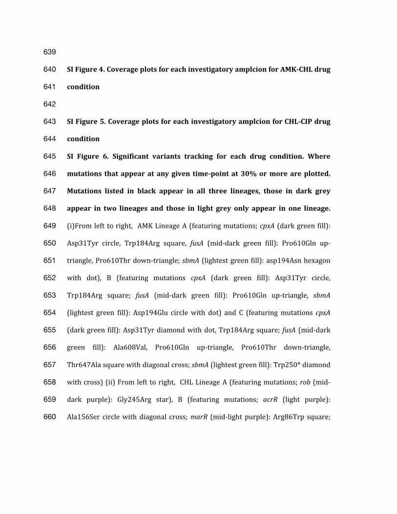
639
SIFigure4.CoverageplotsforeachinvestigatoryamplcionforAMK-CHLdrug640
condition641
642
SIFigure5.CoverageplotsforeachinvestigatoryamplcionforCHL-CIPdrug643
condition644
SI Figure 6. Significant variants tracking for each drug condition. Where645
mutations thatappearatanygiven time-pointat30%ormoreareplotted.646
Mutations listed in black appear in all three lineages, those in dark grey647
appear in two lineages and those in light grey only appear in one lineage.648
(i)Fromleft toright, AMKLineageA(featuringmutations;cpxA (darkgreenfill):649
Asp31Tyr circle, Trp184Arg square, fusA (mid-dark green fill): Pro610Gln up-650
triangle,Pro610Thrdown-triangle;sbmA(lightestgreenfill):asp194Asnhexagon651
with dot), B (featuring mutations cpxA (dark green fill): Asp31Tyr circle,652
Trp184Arg square; fusA (mid-dark green fill): Pro610Gln up-triangle, sbmA653
(lightest green fill): Asp194Glu circlewith dot) and C (featuringmutations cpxA654
(darkgreen fill):Asp31Tyrdiamondwithdot,Trp184Argsquare; fusA (mid-dark655
green fill): Ala608Val, Pro610Gln up-triangle, Pro610Thr down-triangle,656
Thr647Alasquarewithdiagonalcross;sbmA(lightestgreenfill):Trp250*diamond657
withcross)(ii)Fromlefttoright, CHLLineageA(featuringmutations;rob(mid-658
dark purple): Gly245Arg star), B (featuring mutations; acrR (light purple):659
Ala156Ser circlewithdiagonal cross;marR (mid-light purple):Arg86Trp square;660

rob (mid-dark purple): Arg156His circle) andC (featuringmutations;acrR (light661
purple): Arg209fs hexagon;marR (mid-light purple): Arg86Trp square, Gln110*662
down-triangle;rob(mid-darkpurple):Arg156Hiscircle).(iii)Fromlefttoright,CIP663
LineageA(featuringmutations;gyrA (mid-lightgreen):asp87Tyrcircle,Gly75Ser664
up-triangle,ser83delsquare;soxR(darkpurple):Ser126_Arg127delinSerdiamond665
with cross), B (featuring mutations; gyrA (mid-light green): asp87Gly diamond,666
ser83delsquare;rob (mid-darkpurple):Arg156Hishexagonwithdiagonalcross)667
and C (featuringmutations;acrR (light purple): Ser25* hexagon;gyrA (mid-light668
green): Gly81Cys circle with diagonal cross, soxR (dark purple):669
Arg127_Ser128delinsArgdown-triangle).(iv)Fromlefttoright,AMK-CHLLineage670
A (featuring mutations; acrR (light purple): Lys55Glu circle; cpxA (dark green):671
Ala187Thr square, Ile382_Thr383delinMetLeu up-triangle, Ser175Cys down-672
triangle; soxR (Dark purple): Ala18Glu hexagon, glu150fs square with diagonal673
cross),B(A(featuringmutations;acrR(lightpurple):Asp157Asnstar,Cys205Phe674
square, Lys55Glu circle and Met175Arg) and C (featuring acrR (light purple):675
Ala156Ser diamond with Ala156Ser, Arg209fs square with diagonal cross,676
Lys55Glu; cpxA (dark green): Ala187Thr and soxR (dark purple) Ala18Glu677
hexagon). (V) From left to right, CHL-CIP Lineage A (featuring mutations gyrA678
(mid-light green): Phe96fs circle, Ser83del square; marR (mid-light purple):679
Cys51Ser half-dark up-triangle, Val84Glu down triangle; soxR (dark purple):680
Arg20Cyssquare)B(featuringmutationsacrR(lightpurple):Cys205delinsCysArg681
circle,Thr5Alacirclehalfdark;gyrA(mid-lightgreen):Ser83delsquare;soxR(dark682

purple): Arg20Cys square, Ser126_Arg127delinsCys diamond) and C (featuring683
mutationsacrR (lightpurple):Cys205delinsCysArgcircle;gyrA (mid-lightgreen):684
Ser83del square; soxR (dark purple): Arg20Cys square, Ser126_Arg127delinsSer685
up-triangle).686
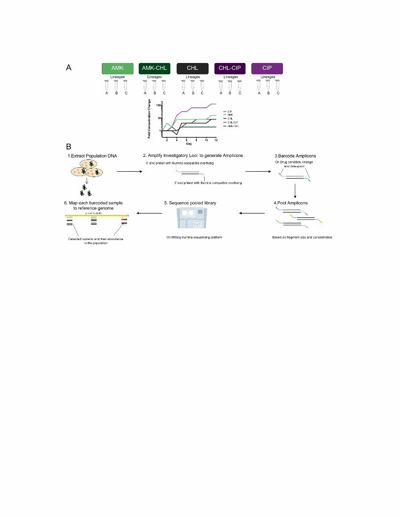
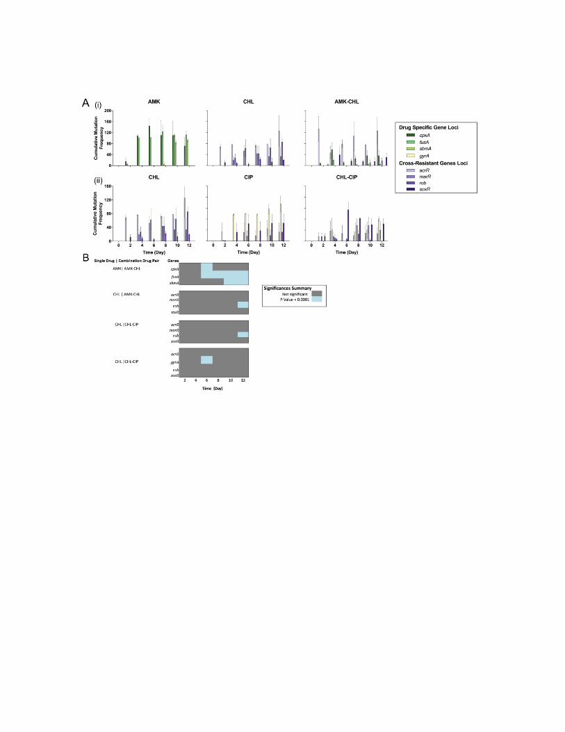
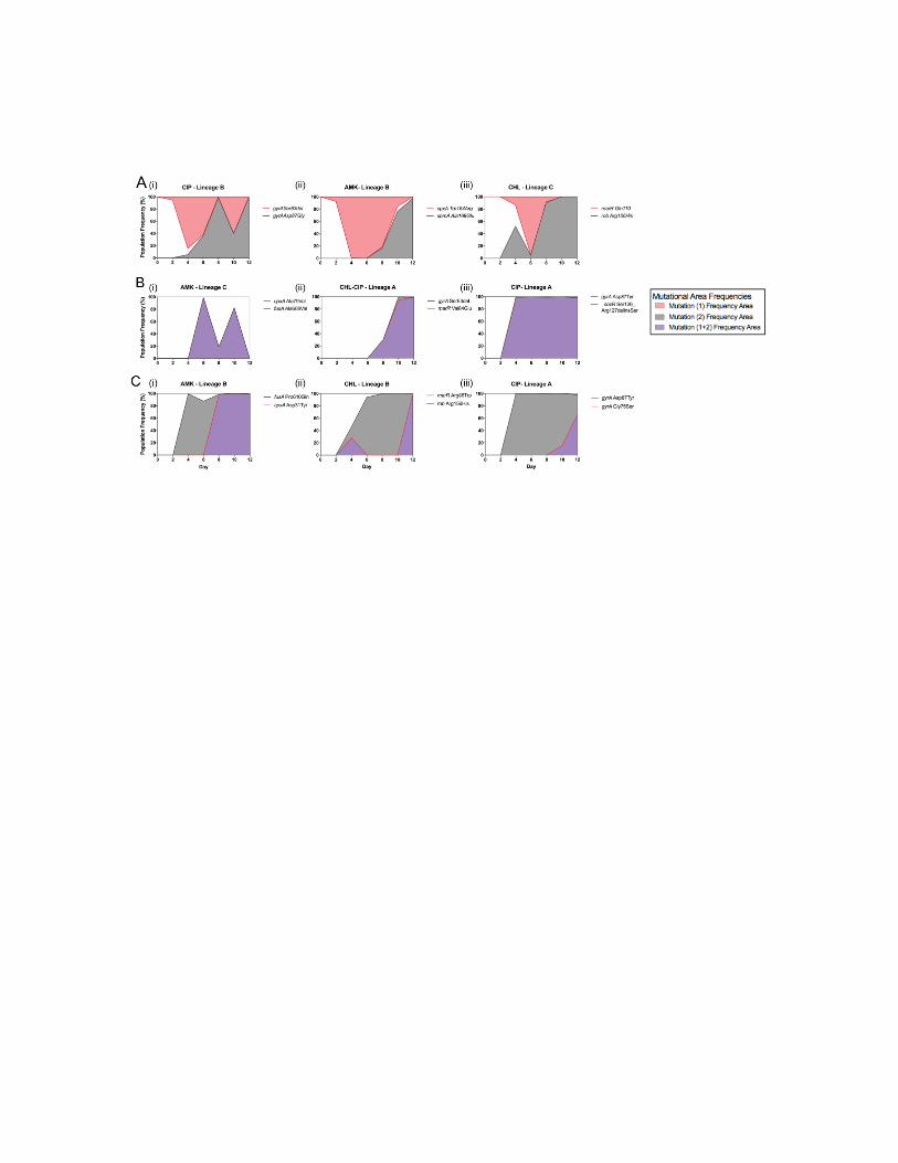

44
Manuscript II: Direct sequencing of Pseudomonas aeruginosa from sputum of cystic fibrosis patients
undergoing antimicrobial therapy
79

DirectsequencingofPseudomonasaeruginosa1
fromsputumofcysticfibrosispatients2
undergoingantimicrobialtherapy3
Authors:4
RachelA.Hickman1,JakobFrimodt-Møller1,ElioRossi2,HelleKroghJohansen2*,5 SørenMolin1*,MortenO.A.Sommer1*6
Affiliation:1NovoNordiskFoundation,CenterforBiosustainability,Technical7 UniversityofDenmark,Kemitorvet,Building220,Kngs.Lyngby,DK-28008 (Denmark)9
2Rigshospitalet,DepartmentofClinicalMicrobiology,afsnit9301,JulianeMariesVej10 22,CopenhagenØ,DK-2100(Denmark)11
Correspondingauthors*:MortenSommer:[email protected];HelleKrogh12 Johansen:[email protected];SørenMolin:[email protected]
14
Abstract15
Cystic fibrosis (CF) patients due to inherited genomic mutations suffer from16 dehydrated mucous that provide the perfect matrix to support opportunistic17 pathogens.Thisisofcriticalconcerninthelungswhereinfectionscaneasilyoccur18 and debilitate lung function. Antimicrobial therapy has been good at delaying19 chronic lung infections but despite best efforts, patients often become chronically20 infected with chronic Pseudomonas aeruginosa in their lungs. Aggressive21 antimicrobial treatment helps to lower the P. aerugionsa bacteria population to22 restorebetterlungfunction,butover-timeP.aerugionsaevolvetobecomeantibiotic23 resistantand the therapybecome lesseffectiveovertime.This studyaddressesCF24 patients thatare inflictedwithchronicPseudomonasaeruginosa lung infectionsby25 directlyanalyzingtheirbydirectlyanalyzingtheirsputumduringhospitalizationfor26 antimicrobial therapy. This is first study to directly analyze un-perturbed sputum27 samples in 19 known antibiotic resistance and patho-adaptive genes (ampC,28 ampDh3,fusA1,fusA2,gyrA,gyrB,mexR,mexS,mexT,mexZ,nfxB,opr86,phoP,PhoQ,29 pmrA,pmrBandrpoB) tounderstandabundanceof individualmutational variants30 andmutationalvariantpopulationseffects.Byusingwholegenesequencingdirectly31 on sputum samples from CF patient we yield important results regarding P.32 aeruginosa lungpopulation,whichcouldhelpoptimizingantibiotictreatmentsand33 reduce antibiotic resistance development in persistence and chronically P.34 aeruginosainfectedCFpatients.35
36
Keyword:Pseudomonasaeruginosa,cysticfibrosis,directwholegenepopulation37 sequencing,sputum,andclonalinterference38
39
Introduction 40
Cysticfibrosis(CF)isanautosomalrecessivelyinheriteddiseasethataffectsabout741
in100,000peopleintheEUcountries(Farrell,2008).Itiscausedbyamutationin42
the CFTR protein that function as a chloride channels causing viscous dehydrate43
mucous on muco-epithial linings, which is difficult to expel. Un-expelled mucus44
provides the perfect habitat for a consortium of opportunistic microorganisms45
(Tang et al., 2014) and consequently lung infections are the major cause of46
morbidity andmortality in CF patients. Therefore, it is routine for CF patients to47
have recurrent antimicrobial therapy to treat the lung infections and tomaintain48
lung function (Johansen et al., 2004). Antimicrobial therapy has help to delay the49
development of chronic infections from infancy to early adolescents ages (Döring50
andHoiby,2004;Frederiksenetal.,1997;Valeriusetal.,1991).However,CFpatient51
oftenbecomeinfectedwithubiquitousenvironmentalbacterialpathogen,especially,52
Pseudomonasaeruginosa,whichoftenbecomesestablishedasachronicinfection.53
54
Many studies focus on the adaptive process of P. aeruginosa to becoming an55
establishedchronicinfection(Marvigetal.,2015;Smithetal.,2006;Sommeretal.,56
2016) or developing resistance against antimicrobial therapy (Feng et al., 2016;57
Jochumsenetal.,2016).58
59
Herewepresentanin-vivomolecularstudy,followingfourCFpatientshospitalized60
forantimicrobial treatment.Bydirectlyextracting theDNA fromthesputum from61
three time-points at day 0, 7, and 14; we can observe the un-biased mutational62
variantsthatareoccurwithinthegenesbyusingourmolecularpipeline(Figure1).63
Our pipeline, unlike other proceeding studies analyzes the whole gene, by PCR64
amplificationupanddownstreamofourgenesofinterest.Withtheinformation,we65
can access the amount of mutated alleles occurring at each time point and state66
whattranslationaleffecttheycause.Byunderstandingthepopulationdynamicsand67
mutational alleles that are enriched during antimicrobial therapywe can acquire68
knowledge about bacterial populations that can be targeted in CF patients with69
chronicP.aeruginosa infections.FutureapplicationforCFpatients fromthisstudy70
are: deployment of antimicrobial treatment that reduced the evolution of71
antimicrobial resistance; and for CF patients that suffer from persistent P.72
aeruginosainfections,withthemotivationhowtodelaychroniccolonization.73
74
Materialandmethods75
76
Patient77
Our studyconsistedof4malesCFpatientsundergoingantimicrobial treatmentat78
theCFclinicatRigshospitalet,Copenhagen,Denmark.Theageofthepatientsranged79
from 26 to 53 years, all were diagnosed with chronic P. aeruginosa infection in80
accordancetotheCopenhagencriteria(Johansenetal.,2004).Patient’streatmentat81
the hospital is recorded in (Table 1). Sampleswere stored at -20 after collection,82
untilDNAextractionwasperformed.83
84
Microbiology85
All sputum samples were Gram-stained and microscopically evaluated and 86
cultured aerobically on selective agar media. These media include a Sabouraud 87
plate, a 7% NaCl plate, a B. cepacia plate containing colistin and gentamicin and a 88
“blue plate” (modified Conradi Drigalski's medium) selective for Gram-negative 89
rods and non-selective media including 5% Danish blood agar and chocolate agar. 90
Direct plating of sputum samples on a 14-cm blood agar plate (State Serum 91
Institute, Copenhagen, Denmark) with discs containing anti-pseudomonas 92
antibiotics including colistin, tobramycin, meropenem, ciprofloxacin, aztreonam, 93
piperacillin/tazobactam and ceftazidime. Susceptibility testing of the sputum 94
samples was done by the agar diffusion method on Danish blood agar plates 95
(Statens Serum Institut, Copenhagen, Denmark), employing Neosensitabs® 96
(Rosco, Roskilde, Denmark)(Ciofu et al., 1996). Isolated bacteria were identified 97
as described previously (Johansen et al., 2008) Before 2011, biochemical profiling of P. 98
aeruginosa was based on API 20NE (bioMérieux), and from 2011 on MALDI-TOF mass 99
spectrometry (Bruker, Germany). 100
101
102
103
DNAextractionfromsputumsamples104
All thawed sputum samples were treated with 30µL (1M) tris (2-carboxyethyl)105
phosphine,and10µLproteinaseK(20mg/mL)(ThermoFisherScientifc)and1mLof106
DNA/RNAshield(ZymoResearch)andvortexedfor30seconds.Sampleswerethen107
added to a 2mL impact resistant screw-top tubewith 300mL zirconia/glassbeads108
with a diameter 0.1mM (Carl Roth International) and vortexed on a secure and109
horizontalholderatmaximumspeedfor5mins.ThengenomicDNAwasextracted110
fromallsputumsamplesbyZR–DuetRNA/DNAmini-prepkit(ZymoResearch)and111
doneinaccordancetothekitsrecommendations. 112
113
PrimerDesign114
Thegenesselectedwerechosenbasedonknowledgeoftheir involvementde-novo115
antibiotic resistance. The following genes of the P. aeruginosa core genomewere116
selected for sequencing: ampC, ampDh3, ftsI, fusA1, fusA2, gyrA, gyrBmexS,mexR,117
mexT,mexZ,nfxB,opr86,parC,pmrA,pmrB,phoQ,phoR,rpoB.Primersweredesigned118
toamplify19genesassociatedwithantibioticsusceptibility(SupplementalFig.1).119
Allprimerswere testedwithgenomicDNA fromP.aeruginosa strains:PAO1,DK2120
and PA14 the sequences of the 19 target genes were extracted from the121
Pseudomonasdatabase(Winsoretal.,2016).PCRproductsweretestedwithPAO1122

and DK-2 DNA extracts verified by 1% agarose gel electrophoresis with GelRed123
(Biotium).124
125
Wholegenelibrarypreparationandsequencing126
All sampleswereamplifiedusing the19primerpairs in (Supplemental table 1),127
PCRs from sampleDNA extractswere done independently to each other to avoid128
cross contamination. EachPCR reactionwasdone in 0.2mL sterile PCR tubes and129
consistsof10µLPhusionHigh-FidelityPCRmastermix(ThermoFischerScientific),130
0.5µM forwardprimer, 0.5µM reverseprimer, 1µLDNA template andH20 to a131
totalreactionvolumeof20µL.ThePCRamplificationusedaninitialdenaturationof132
for 98oC 30 seconds, 30 cycles consisting of a 98oC for 10 seconds denaturation,133
60oC for10secondsannealing,72oC for3minelongation,72oC finalextension for134
180secondsthenholdat4oC.Tovalidatecorrectamplificationgelelectrophoresis135
wasperformedwitha96-well2%E-gelwithSYBRsafeDNAgelstain(Invitrogen)136
on the E-base electrophoresis device for 13 min (Invitrogen) and gel images137
analyzedontheE-editor2.02software(Invitrogen).Eachsamplewascleanedwith138
1.8x Ampure bead and done in accordance an online protocol (Beckman Coulter139
2013). The 19 PCR reactions for each sample DNA extractwere pooled together;140
eachpooledPCRproductinaccordancetoitsoriginalsample.200ngofpooledDNA141
inavolumeof130µLwasshearedinaCovarisfocused-ultrasonicatorE220,for55142
secondswithapeakincidencepowerof105W,dutyfactor5%andcyclesperburst143
200.ShearedpooledDNAsampleswerethenpreparedforsequencingbytheKapa144

HyperPlusDNAlibrarypreparationkit(KapaBiosystems)andbarcodedusingLT-145
Truseq single-index adapter kit (Illumina Inc). Barcoded librariesweremeasured146
for DNA concentration with Qubit dsDNA HS Assay kits and measured on Qubit147
Fluorometer (ThermoFischerScientific Inc2015)andaverage fragment sizewith148
Fragment analyzer (Advanced Analytical Technologies, Inc). Barcoded libraries149
werepooledfromfragmentsizeandDNAconcentrationvaluestoformasequencing150
library based. The sequencing librarywas sequenced on theMiSeq platformwith151
MiseqreagentkitV3,sequencereadlength2x300bp(IlluminaInc).152
153
SequenceAnalysis154
All raw sequencing read from the gene sequencing has been deposited in the155
SequenceReadArchive(SRA)underbioprojectaccessionnumberxxxx.Sequencing156
readsweremappedtothereferencegenome(GenBankaccessionAE004091)using157
CLC genomics workbench version 8.5.1(Qiagen). To verify sequencing coverage,158
mapping read files fromCLC genomicworkbenchdatawas analyzed in Excel and159
plots designed in Prism6 (GraphPad Software Inc). To ensure good coverage our160
thresholdforaveragecoveragewassetat100,samplesthathadlowcoveragewere161
re-read until sufficient coveragewas achieved (Supplemental Fig 1-4). Variants162
weredetectedbyusingthelowfrequencyvariantdetectioncallingfunctioninCLC163
genomicworkbenchV8.5.1(Qiagen),provideallthevariantsthathadapopulation164
frequencyof≥1%.(SupplementalTable2).165
166
Results167
The study consisted of 4 CF patients chronically with P. aerugionsa with an age168
rangefrom26to53years.Eachpatientprovidedthreesputumsamplesonday0,7169
and 14 during hospitalized antimicrobial treatment, minus one patient who was170
unabletoprovideaday7coughsample.FollowingourpipelineweextractedDNA171
from each of the samples and using our pipeline (Figure 1) the intrinsic172
antimicrobial resistance development was assessed in 19 genes that have been173
previouslyreportedtoconferresistanceagainstcertainclassesofantibiotics.These174
were:foraminoglycoside(genesofinterestwere:fusA1(Marvigetal.,2013),fusA2175
(Marvig et al., 2013), and rpoB (Qi et al., 2014)); for peptidoglycan synthesis176
inhibitingclasses(genesofinterestwere:ampC(Marvigetal.,2013),ampDh3(Lee177
et al., 2013) and ftsI (Smith et al., 2006)); for fluoroquinolone (genes of interest178
were:gyrA(Fengetal.,2016;Marvigetal.,2013),gyrB(Fengetal.,2016;Marviget179
al.,2013),parC(Fengetal.,2016)andrpoB(Pietschetal.,2017));polymixin(genes180
of interest were: opr86 (Jochumsen et al., 2016), phoP (Smith et al., 2006) phoQ181
(Gutuetal.,2013;Moskowitzetal.,2012),pmrA(Moskowitzetal.,2012)andpmrB182
(Moskowitzetal.,2012;Smithetal.,2006));andpatho-adaptivegenesthatconfer183
cross-resistance (genes of interest were: mexR (Vestergaard et al., 2016),184
mexS(Richardotetal.,2016),mexT(Smithetal.,2006),mexZ(Smithetal.,2006)and185
nfxB(Montietal.,2013)).186
187

Only two patients provide a day 0 and day 14 samples for antimicrobial188
susceptibility testing samples to the department of clinical microbiology189
department,thesewerepatientCF156andCF83(Table2).Fromtheantimicrobial190
susceptibility testing we observed phenotypic changes before and after in both191
patients. For CF156 in the non-mucoidP.aeruginosa we saw changes in: colistin,192
from susceptible to resistant; imipenem, from susceptible to intermediate; and193
meropenem,andfromintermediatetoresistant.Inthesamepatientwealsosawin194
themucoidP.aeruginosasampleaphenotypicchangeofimpenemfromday0being195
intermediate to susceptibleonday14.Whereas forpatientCF83 in themucoidP.196
aeruginosa antimicrobial susceptible typing results we saw changes in:197
ciprofloxacin, fromsusceptibletointermediate;andimipenem,fromsusceptibleto198
intermediate. The overall observation from these two patients is that the use199
antimicrobialtherapyleadstoinductionofresistancetoseveralantibiotics.Thisis200
notsurprisingasantimicrobialsarewellknowforselectingpre-existingandde-novo201
mutants(AnderssonandHughes,2014).202
203
Detection of mutational variants reveal the high abundance of204 mutageniceventsofP.aeruginosaintheCFlung205
Followingcoverageanalysis,whereallsampleshadtomeettherequirementsofall206
genes having an average coverage of 100 sequencing reads per base in the open207
readingframeofthegene,thevariantdetectionandanalysiswasperformed.From208
our results we able to plot an overview of all the variant types detected with a209
population frequency≥5% for eachpatient and time-point (Figure2). From these210
plots, we found that the maximum total individual variant detected were was in211

patient CF382 at day 0 with 514 different variants detected and the minimum212
individual variant detected in patient CF83 at day 0 with 143 different variants213
detected. Most of the detected variants were synonymous mutations, the second214
most abundantmutation typewas non- synonymous SNPs, followed by the third215
most abundant mutation type being frameshift mutations and the fourth most216
abundantmutationtypewasprematurestopmutationswherefewtonomutations217
were detected. Similar distribution of mutational variant types from amplicon218
sequencedP.aerugionsaisolatesfromtheCFlunghavebeenreportedbyGriepleet219
al.2016.220
221
Therewas also a general trend amongst three of the patients that the amount of222
individual mutation variants detected increased during the 14-day antimicrobial223
treatment. This was highest in patient CF83 where the individual mutational224
variants for day 0, 7 and 14 were: 143; 173; and 233 (Figure 2 (iv)). In patient225
CF243 the individualmutational variantsdetected forday0, 7 and14were: 253;226
204; and 305 (Figure 2 (iii)). In patient CF243 the individualmutational variants227
detectedforday0,7and14were:175;156;and201(Figure2(ii)).Anexceptionto228
thistrendwaspatientCF382,wheretheindividualmutationalvariantsdetectedfor229
day 0 and 14 were 514 and 158 (Figure 2 (i)). These results imply that when230
patientsarereceivingantimicrobial therapy theP.aeruginosabacterialpopulation231
inthelungexperiencespossiblyanexpansioningenomicdiversity.Whetherthisis232
a direct or downstream effect of antibiotics, such as the production of reactive233
oxygen species (Kohanski et al., 2007) or by antibiotics inducing the quorum234
sensingmechanismsendogenous toP.aeruginosathat canhavepleiotropiceffects235
(Skindersoeetal.,2008)needtobefurtherelucidated.236
237
Evaluation of non-synonymous mutations during antimicrobial238 treatment show mutational enrichment and clonal inference239 withinthebacterialpopulation240
We then further analyzed our data by evaluating the non-synonymousmutations,241
wewere interested to followthe trajectoriesofmutations thatwereenrichedand242
un-selected for during antimicrobial therapy (Figure 3). To achieve this we243
examined the non-synonymous mutations that were present at a least one time-244
point ≥5%. In this figure, we could see in all patients that there was a clear245
enrichmentofmutagenicallelicvariantsoverthetreatmenttimeperiod(Figure3A246
(i-iv)).Wewere interested if thesemutationshadbeenpreviouslydetectedand if247
there was any information regarding these mutations conferring antibiotic248
resistance. From the 41 non- synonymous mutations, 11 of these have been249
reported in literature, with 3 mutations all from patient CF382 with results250
confirmingthatthesemutationsconferresistance(Table3).Ourmolecularpipeline251
detected severalmutations that have not previously been reported, this could be252
due to our direct DNA extraction, amplification and sequencing, capturing all253
presentvariants.254
255
Fromtheenrichedmutationvariants,wecouldseethateachpatienthadaunique256
responseintheindividualvariantsthatwereproduced.Thiscouldbearesultofthe257
historicalcontingencyoftheP.aeruginosabacteriainthelung,theclone-typethey258
wereinfectedwithandthetreatmentused(Table1).PatientCF382isknowntobe259

infected with a hypermutator strain, and was treated with drug classes:260
aminoglycosides,cephalosporin, fluoroquinoloneandpolymixins.Therefore, itwas261
not surprisingwe sawenrichedmutations in the following genes:ampC,ampDh3,262
fusA1, fusA2, gyrA, gyrB,mexZ, opr86, phoQ,pmrA,pmrB and rpoB.For CF156, the263
patient was treated with polymixin and fluoroquinole classes of antibiotics. This264
patienthadenrichedmutationsin:ampC,ampD3h,mexT,opr86,pmrAandrpoB.The265
ampCandampD3hmutationsthatwereenrichedduringtreatmentwereananomaly266
wedidn’texpecttoobserve,astheampCandampDh3mutationsweexpecttoconfer267
beta-lactam,cephalosporinandcarbapenemresistance.Wesuspectthatthesenew268
mutants in mexT, opr86, pmrA and rpoB that appeared had ampC and ampDh3269
mutations in their genomicbackground, andwere selected as a result of genomic270
hitchhiking a common phenomenon often seen in adaption laboratory evolution271
experiments (Elena and Lenski, 2003). Patient CF243 was treatment with272
aminoglycosides,carbapenem,fluoroquinoloneandpolymixinclassesofantibiotics.273
ThehighlevelsofenrichmentconsistedinmutationsinpmrBgenethatisknownto274
confer resistance to polymixins (Moskowitz et al., 2012; Owusu-Anim and Kwon,275
2012;Schureketal.,2009)andmexT,whichisknowntoconfercross-resistanceby276
the expression of efflux pumps (Alyaseen et al., 2005). Patient CF83 was treated277
with aminoglycosides, cephloasporin, fluoroquinolones and polymixins. High278
frequencyenrichmentwasfoundinrpoB,mexT,andftsIandlow-levelenrichmentin279
mexSandgyrA.WheretherpoBmutantsconferaminoglycosideresistance(Qietal.,280
2014)andciprofloxacin(Pietschetal.,2017),ftsImutantsconferresistanceagainst281
carbapenems (Cabot et al.), and mexT mutants provide general cross-resistance282
(Alyaseenetal.,2005).283
284
Wealsoobservedstrongclonalinterferenceisexhibitedintwopatients,CF382and285
CF83 (Figure3A (i) and (iv),3B (i) and (iv)),withmarginal clonal interference in286
CF156andCF243(Figure3A(ii)and (iii),3B (i)and (iv)). Inallpatients,wesee287
thattherearesub-populationsofbacterialmutationalvariants,atraitalsoseenby288
othersregardingsub-populationsofvirulentP.aeruginosaintheCFlung(O’Brienet289
al 2017). Therefore, establishment of novelmutations seems to readily occur, but290
fixation in the population is a rare event, unlike in-vitro adaptation evolution291
experimentsinotherGram-negativespecies(WongandSeguin,2015).Thiscouldbe292
an artifact of spatial structural andheterogeneity in theCF lung (Folkesson et al.,293
2012;Markussenetal.,2014;Winstanleyetal.,2016)ortheshorttimeperiodour294
studywaspreformedin.Thus,showingthecomplexbacterialpopulationdynamics295
withinCFpatients.296
297
Discussion298
Within this study we use whole gene population sequencing method to study299
individual mutation variant events and mutagenic populations dynamics. Our300
approachwastoextracttheDNAfromthepatientsputumsamples,amplifygenesof301
interest and sequence with no P. aerugionsa isolation cultivation steps. With the302
development and growing accessibility of next generation sequencing platforms,303
studies analyzing evolution in bacterial populations are growing. In particular, P.304
aerugionsa population sequencing following cultivation from CF patient samples305
(Fischeretal.,2016;Greipeletal.,2016;O’Brienetal.,2017).306
307
InCFpatientsthatarechronicallyinfectedwithP.aeruginosa,wehaveobservedthe308
vastarrayofmutantclonesoccupyoneenvironment,aphenomenonalsodescribed309
byGreiple et al 2016. Yet by following events longitudinallywe can observe how310
these compete by clonal interference to establish the clone types that best aids311
populationsurvival.312
313
The bacterial populations we study also display similar characteristics that have314
been reported in-vitro adaptation studies with other Gram-negative bacterial315
species, such as clonal interference andmultiplemutant enrichment (Fogle et al.,316
2008;GerrishandLenski,1998;Sniegowskietal.,1997;WongandSeguin,2015).317
318
Ourresultsareimportantbutlimitedthereforeinfuturewewouldliketoexaminea319
largercohortofchronicallyinfectedpatientandforlongerperiodsoftime.Thiswill320
allow us to examine key factors such as: which antibiotics treatment cause the321
highest and lowest generationofmutational variants; andby studyingpopulation322
dynamics for longerperiodsof timecanwecaptureclonal fixationeventsand the323
implicationsofthesepopulationfixationsbyin-vitroexperimentation.324
325
Therefore, our long-term version is to optimize antimicrobial treatment in326
chronically infected CF patients with P. aeruginosa and in patients that have327
persistentcolonizationand findstrategies todelay thechronicestablishmentofP.328
aeruginosa.329
330
Acknowledgements331
TheCFpatientsandnursesattheadultCFcenteratRigshospitaletarethankedfor332 theircollaborationinthisproject.WewouldalsoliketothankAnnaKozafromCFB’s333 nextgenerationsequencinglaboratoryforhertechnicalsupportandadvice.HKJ334 wassupportedbytheNovoNordiskFoundationasaclinicalresearchstipendand 335 RigshospitaletRammebevilling2015-17andLundbeckfondenGrantR167-2013-336 15229.MOASfundingby,EUH2020ERC-20104-STGLimitMDR(638902)andthe337 DanishCouncilforIndependentResearchSapereAudeprogrammeDFF-4004-338 00213additionalfundingfromtheNovoNordiskFoundationandTheLundbeck339 Foundation 340 341
342
References343
Alyaseen,S.A.,Piper,K.E.,Rouse,M.S.,Steckelberg,J.M.,andPatel,R.(2005).344
Selectionofcross-resistancefollowingexposureofPseudomonasaeruginosa345
clinicalisolatestociprofloxacinorcefepime.Antimicrob.AgentsChemother.49,346
2543–5.doi:10.1128/AAC.49.6.2543-2545.2005.347
Andersson,D.I.,andHughes,D.(2014).Microbiologicaleffectsofsublethallevelsof348
antibiotics.Nat.Rev.Microbiol.12,465–478.doi:10.1038/nrmicro3270.349
Bezuidt,O.K.,Klockgether,J.,Elsen,S.,Attree,I.,Davenport,C.F.,andTümmler,B.350
(2013).IntraclonalgenomediversityofPseudomonasaeruginosaclonesCHA351
andTB.BMCGenomics14,416.doi:10.1186/1471-2164-14-416.352
Bruchmann,S.,Dötsch,A.,Nouri,B.,Chaberny,I.F.,andHäussler,S.(2013).353
Quantitativecontributionsoftargetalterationanddecreaseddrug354
accumulationtoPseudomonasaeruginosafluoroquinoloneresistance.355
Antimicrob.AgentsChemother.57,1361–8.doi:10.1128/AAC.01581-12.356
Cabot,G.,López-Causapé,C.,Ocampo-Sosa,A.A.,Sommer,L.M.,Domínguez,M.Á.,357
Zamorano,L.,etal.(2016).DecipheringtheResistomeoftheWidespread358
PseudomonasaeruginosaSequenceType175InternationalHigh-RiskClone359
throughWhole-GenomeSequencing.Antimicrob.AgentsChemother.60,7415–360
7423.doi:10.1128/AAC.01720-16.361
Cabot,G.,Zamorano,L.,Moyà,B.,Juan,C.,Navas,A.,Blázquez,J.,etal.Evolutionof362
PseudomonasaeruginosaAntimicrobialResistanceandFitnessunderLowand363
HighMutationRates.doi:10.1128/AAC.02676-15.364
Ciofu,O.,Jensen,T.,Pressler,T.,Johansen,H.K.,Koch,C.,andH⊘iby,N.(1996).365
MeropenemincysticfibrosispatientsinfectedwithresistantPseudomonas366
aeruginosaorBurkholderiacepaciaandwithhypersensitivitytoβ-lactam367
antibiotics.Clin.Microbiol.Infect.2,91–98.doi:10.1111/j.1469-368
0691.1996.tb00212.x.369
Döring,G.,andHoiby,N.(2004).Earlyinterventionandpreventionoflungdisease370
incysticfibrosis:aEuropeanconsensus.J.Cyst.Fibros.3,67–91.371
doi:10.1016/j.jcf.2004.03.008.372
Elena,S.F.,andLenski,R.E.(2003).Microbialgenetics:Evolutionexperimentswith373
microorganisms:thedynamicsandgeneticbasesofadaptation.Nat.Rev.Genet.374
4,457–469.doi:10.1038/nrg1088.375
Farrell,P.M.(2008).TheprevalenceofcysticfibrosisintheEuropeanUnion.376
doi:10.1016/j.jcf.2008.03.007.377
Feng,Y.,Jonker,M.J.,Moustakas,I.,Brul,S.,andterKuile,B.H.K.(2016).Dynamics378
ofMutationsduringDevelopmentofResistancebyPseudomonasaeruginosa379
againstFiveAntibiotics.Antimicrob.AgentsChemother.60,4229–4236.380
doi:10.1128/AAC.00434-16.381
Fischer,S.,Greipel,L.,Klockgether,J.,Dorda,M.,Wiehlmann,L.,Cramer,N.,etal.382
(2016).MultilocusampliconsequencingofPseudomonasaeruginosacystic383
fibrosisairwaysisolatescollectedpriortoandafterearlyantipseudomonal384
chemotherapy.J.Cyst.Fibros.doi:10.1016/j.jcf.2016.10.013.385
Fogle,C.A.,Nagle,J.L.,andDesai,M.M.(2008).ClonalInterference,Multiple386
MutationsandAdaptationinLargeAsexualPopulations.Genetics108,2163–387
2173.388
Folkesson,A.,Jelsbak,L.,Yang,L.,Johansen,H.K.,Ciofu,O.,Høiby,N.,etal.(2012).389
AdaptationofPseudomonasaeruginosatothecysticfibrosisairway:an390
evolutionaryperspective.Nat.Rev.Microbiol.Publ.online13Novemb.2012;|391
doi10.1038/nrmicro290710,841.doi:10.1038/NRMICRO2907.392
Frederiksen,B.,Koch,C.,andHøiby,N.(1997).Antibiotictreatmentofinitial393
colonizationwithPseudomonasaeruginosapostponeschronicinfectionand394
preventsdeteriorationofpulmonaryfunctionincysticfibrosis.Pediatr.395
Pulmonol.23,330–5.Availableat:396
http://www.ncbi.nlm.nih.gov/pubmed/9168506[AccessedFebruary9,2017].397
Gerrish,P.J.,andLenski,R.E.(1998).Thefateofcompetingbeneficialmutationsin398
anasexualpopulation.Genetica102/103,127–144.399
doi:10.1023/A:1017067816551.400
Greipel,L.,Fischer,S.,Klockgether,J.,Dorda,M.,Mielke,S.,Wiehlmann,L.,etal.401
(2016).MolecularEpidemiologyofMutationsinAntimicrobialResistanceLoci402
ofPseudomonasaeruginosaIsolatesfromAirwaysofCysticFibrosisPatients.403
Antimicrob.AgentsChemother.60,6726–6734.doi:10.1128/AAC.00724-16.404
Gutu,A.D.,Sgambati,N.,Strasbourger,P.,Brannon,M.K.,Jacobs,M.A.,Haugen,E.,et405
al.(2013).PolymyxinresistanceofPseudomonasaeruginosaphoQmutantsis406
dependentonadditionaltwo-componentregulatorysystems.Antimicrob.407
AgentsChemother.57,2204–15.doi:10.1128/AAC.02353-12.408
Jochumsen,N.,Marvig,R.L.,Damkiær,S.,Jensen,R.L.,Paulander,W.,Molin,S.,etal.409
(2016).TheevolutionofantimicrobialpeptideresistanceinPseudomonas410
aeruginosaisshapedbystrongepistaticinteractions.Nat.Commun.7,13002.411
doi:10.1038/ncomms13002.412
Johansen,H.K.,Moskowitz,S.M.,Ciofu,O.,Pressler,T.,andHøiby,N.(2008).Spread413
ofcolistinresistantnon-mucoidPseudomonasaeruginosaamongchronically414
infectedDanishcysticfibrosispatients.J.Cyst.Fibros.7,391–397.415
doi:10.1016/j.jcf.2008.02.003.416
Johansen,H.K.,Nørregaard,L.,Gøtzsche,P.C.,Pressler,T.,Koch,C.,andHøiby,N.417
(2004).AntibodyresponsetoPseudomonasaeruginosaincysticfibrosis418
patients:Amarkeroftherapeuticsuccess?-A30-yearCohortstudyofsurvival419
inDanishCFpatientsafteronsetofchronicP.aeruginosalunginfection.Pediatr.420
Pulmonol.37,427–432.doi:10.1002/ppul.10457.421
Kohanski,M.A.,Dwyer,D.J.,Hayete,B.,Lawrence,C.A.,andCollins,J.J.(2007).A422
CommonMechanismofCellularDeathInducedbyBactericidalAntibiotics.Cell423
130,797–810.doi:10.1016/j.cell.2007.06.049.424
Lee,M.,Artola-Recolons,C.,Carrasco-López,C.,Martínez-Caballero,S.,Hesek,D.,425
Spink,E.,etal.(2013).Cell-WallRemodelingbytheZinc-ProteaseAmpDh3426
fromPseudomonasaeruginosa.J.Am.Chem.Soc.135,12604–12607.427
doi:10.1021/ja407445x.428
Llanes,C.,Köhler,T.,Patry,I.,Dehecq,B.,vanDelden,C.,andPlésiat,P.(2011).Role429
oftheMexEF-OprNeffluxsysteminlow-levelresistanceofPseudomonas430
aeruginosatociprofloxacin.Antimicrob.AgentsChemother.55,5676–84.431
doi:10.1128/AAC.00101-11.432
Markussen,T.,Marvig,R.L.,Gómez-Lozano,M.,Aanæs,K.,Burleigh,A.E.,Høiby,N.,433
etal.(2014).Environmentalheterogeneitydriveswithin-hostdiversification434
andevolutionofPseudomonasaeruginosa.MBio5,e01592-14.435
doi:10.1128/mBio.01592-14.436
Marvig,R.L.,Dolce,D.,Sommer,L.M.,Petersen,B.,Ciofu,O.,Campana,S.,etal.437
(2015).Within-hostmicroevolutionofPseudomonasaeruginosainItalian438
cysticfibrosispatients.BMCMicrobiol.15,218.doi:10.1186/s12866-015-0563-439
9.440
Marvig,R.L.,Johansen,H.K.,Molin,S.,andJelsbak,L.(2013).Genomeanalysisofa441
transmissiblelineageofpseudomonasaeruginosarevealspathoadaptive442
mutationsanddistinctevolutionarypathsofhypermutators.PLoSGenet.9,443
e1003741.doi:10.1371/journal.pgen.1003741.444
Monti,M.R.,Morero,N.R.,Miguel,V.,andArgaraña,C.E.(2013).nfxBasaNovel445
TargetforAnalysisofMutationSpectrainPseudomonasaeruginosa.PLoSOne446
8,e66236.doi:10.1371/journal.pone.0066236.447
Moskowitz,S.M.,Brannon,M.K.,Dasgupta,N.,Pier,M.,Sgambati,N.,Miller,A.K.,et448
al.(2012).PmrBmutationspromotepolymyxinresistanceofPseudomonas449
aeruginosaisolatedfromcolistin-treatedcysticfibrosispatients.Antimicrob.450
AgentsChemother.56,1019–30.doi:10.1128/AAC.05829-11.451
O’Brien,S.,Williams,D.,Fothergill,J.L.,Paterson,S.,Winstanley,C.,andBrockhurst,452
M.A.(2017).Highvirulencesub-populationsinPseudomonasaeruginosalong-453
termcysticfibrosisairwayinfections.BMCMicrobiol.17,30.454
doi:10.1186/s12866-017-0941-6.455
Owusu-Anim,D.,andKwon,D.H.(2012).DifferentialRoleofTwo-Component456
RegulatorySystems(phoPQandpmrAB)inPolymyxinBSusceptibilityof457
Pseudomonasaeruginosa.Adv.Microbiol.2.doi:10.4236/aim.2012.21005.458
Pietsch,F.,Bergman,J.M.,Brandis,G.,Marcusson,L.L.,Zorzet,A.,Huseby,D.L.,etal.459
(2017).CiprofloxacinselectsforRNApolymerasemutationswithpleiotropic460
antibioticresistanceeffects.J.Antimicrob.Chemother.72,75–84.461
doi:10.1093/jac/dkw364.462
Qi,Q.,Preston,G.M.,andMacLean,R.C.(2014).Linkingsystem-wideimpactsof463
RNApolymerasemutationstothefitnesscostofrifampinresistancein464
Pseudomonasaeruginosa.MBio5,e01562.doi:10.1128/mBio.01562-14.465
Richardot,C.,Juarez,P.,Jeannot,K.,Patry,I.,Plésiat,P.,andLlanes,C.(2016).Amino466
AcidSubstitutionsAccountforMostMexSAlterationsinClinicalnfxCMutants467
ofPseudomonasaeruginosa.Antimicrob.AgentsChemother.60,2302–10.468
doi:10.1128/AAC.02622-15.469
Rodríguez-Martínez,J.-M.,Poirel,L.,andNordmann,P.(2009).Extended-spectrum470
cephalosporinasesinPseudomonasaeruginosa.Antimicrob.AgentsChemother.471
53,1766–71.doi:10.1128/AAC.01410-08.472
Schurek,K.N.,Sampaio,J.L.M.,Kiffer,C.R.V,Sinto,S.,Mendes,C.M.F.,andHancock,473
R.E.W.(2009).InvolvementofpmrABandphoPQinpolymyxinBadaptation474
andinducibleresistanceinnon-cysticfibrosisclinicalisolatesofPseudomonas475
aeruginosa.Antimicrob.AgentsChemother.53,4345–51.476
doi:10.1128/AAC.01267-08.477
Skindersoe,M.E.,Alhede,M.,Phipps,R.,Yang,L.,Jensen,P.O.,Rasmussen,T.B.,etal.478
(2008).EffectsofantibioticsonquorumsensinginPseudomonasaeruginosa.479
Antimicrob.AgentsChemother.52,3648–63.doi:10.1128/AAC.01230-07.480
Smith,E.E.,Buckley,D.G.,Wu,Z.,Saenphimmachak,C.,Hoffman,L.R.,D’Argenio,D.481
A.,etal.(2006).GeneticadaptationbyPseudomonasaeruginosatotheairways482
ofcysticfibrosispatients.Proc.Natl.Acad.Sci.U.S.A.103,8487–92.483
doi:10.1073/pnas.0602138103.484
Sniegowski,P.D.,Gerrish,P.J.,andLenski,R.E.(1997).Evolutionofhighmutation485
ratesinexperimentalpopulationsofE.coli.Nature387,703–705.486
doi:10.1038/42701.487
Sommer,L.M.,Marvig,R.L.,Luján,A.,Koza,A.,Pressler,T.,Molin,S.,etal.(2016).Is488
genotypingofsingleisolatessufficientforpopulationstructureanalysisof489
Pseudomonasaeruginosaincysticfibrosisairways?BMCGenomics17,589.490
doi:10.1186/s12864-016-2873-1.491
Tang,A.C.,Turvey,S.E.,Alves,M.P.,Regamey,N.,Tümmler,B.,andHartl,D.(2014).492
Currentconcepts:host–pathogeninteractionsincysticfibrosisairwaysdisease.493
Eur.Respir.Rev.23.494
Valerius,N..,Koch,C.,andHo̸iby,N.(1991).PreventionofchronicPseudomonas495
aeruginosacolonisationincysticfibrosisbyearlytreatment.Lancet338,725–496
726.doi:10.1016/0140-6736(91)91446-2.497
Vestergaard,M.,Paulander,W.,Marvig,R.L.,Clasen,J.,Jochumsen,N.,Molin,S.,etal.498

(2016).Antibioticcombinationtherapycanselectforbroad-spectrum499
multidrugresistanceinPseudomonasaeruginosa.Int.J.Antimicrob.Agents47,500
48–55.doi:10.1016/j.ijantimicag.2015.09.014.501
Winsor,G.L.,Griffiths,E.J.,Lo,R.,Dhillon,B.K.,Shay,J.A.,andBrinkman,F.S.L.502
(2016).Enhancedannotationsandfeaturesforcomparingthousandsof503
PseudomonasgenomesinthePseudomonasgenomedatabase.NucleicAcids504
Res.44,D646-53.doi:10.1093/nar/gkv1227.505
Winstanley,C.,O’Brien,S.,andBrockhurst,M.A.(2016).Pseudomonasaeruginosa506
EvolutionaryAdaptationandDiversificationinCysticFibrosisChronicLung507
Infections.TrendsMicrobiol.24,327–337.doi:10.1016/j.tim.2016.01.008.508
Wong,A.,andSeguin,K.(2015).Effectsofgenotypeonratesofsubstitutionduring509
experimentalevolution.Evolution(N.Y).69,1772–1785.510
doi:10.1111/evo.12700.511
512
FigureLegends513
Figure1.Methodologyworkflowoverview (1)Thawcollectedsputumsamples,514 (2)ExtractDNA fromeach sample (3)PCRall19genes foreachDNAextract and515 verifyPCRproducts(4)ShearDNAbysonicationforDNAfragmentsca.600bp(5)516 Prepare sequencing library by kit and barcode then pool, (6) Sequence Miseq517 Illuminaplatform.518
519
Figure2.Overviewofallvariant typesdetectedwithapopulation frequency520 ≥5%throughouttreatmentcourseforallgenescombined(i)PatientCF382,(ii)521 CF156,(iii)CF243and(iv)CF83.Varianttypescorrespondtokeyontheright.522
523
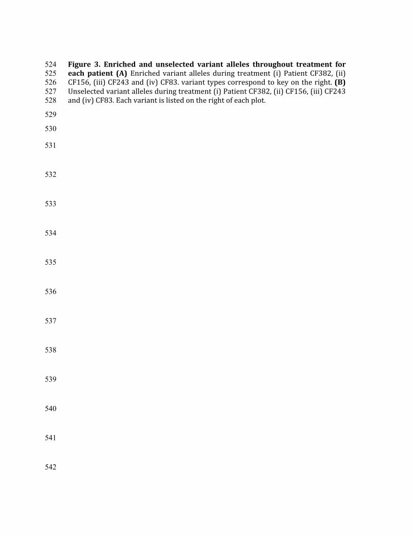
Figure 3. Enriched and unselected variant alleles throughout treatment for524 each patient (A)Enrichedvariant allelesduring treatment (i) PatientCF382, (ii)525 CF156,(iii)CF243and(iv)CF83.varianttypescorrespondtokeyontheright.(B)526 Unselectedvariantallelesduringtreatment(i)PatientCF382,(ii)CF156,(iii)CF243527 and(iv)CF83.Eachvariantislistedontherightofeachplot.528
529
530
531
532
533
534
535
536
537
538
539
540
541
542

Table1.Patienthistory543
Patient Daily treatment over hospitalization period
P. aeruginosa Clone Type
Hypermutator strain
CF382 Intravenous3gCeftazidime3gx3Intravenous500mgTobramycinx1InhalationPromixin3mioIEx1Oral750mgCiprofloxacinx2Oral250mgAzithromycinx1
DK32 Yes
CF156 Oral 750mgCiprofloxacinx2Intravenous9mioIEColistinx1
DK1 and DK2 No
CF243 Intravenous9mioIEColistinx1Intravenous2gMeropenemx2Inhalation240mgCiprofloxacinx2Inhalation300mgTobramycinx2Oral250mgAzithromycinx1
DK2 No
CF83 InhalationPromixin3mioIEx1Intravenous3gCeftazidime3gx3Intravenous500mgTobramycinx1InhalationPromixin3mioIEx1Oral750mgCiprofloxacinx2Oral250mgAzithromycinx1
Unknown No
544
545
546
547
548
549
550
551
552
553
554
555
556
557
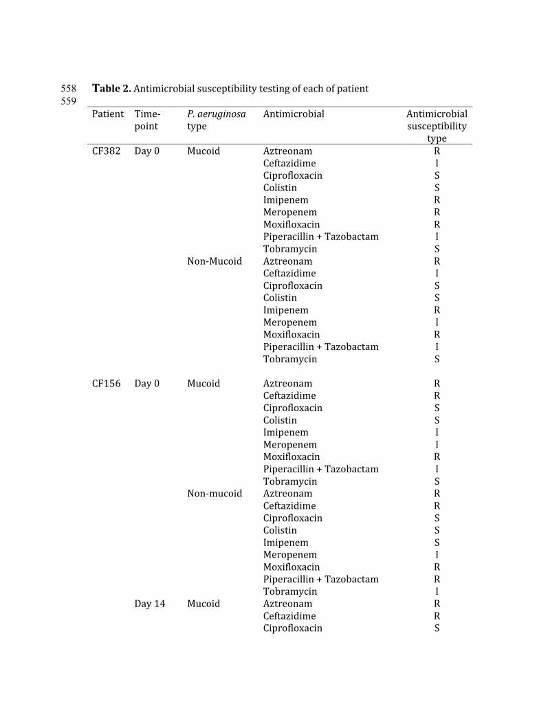
Table2.Antimicrobialsusceptibilitytestingofeachofpatient558 559 Patient Time-
pointP.aeruginosatype
Antimicrobial Antimicrobialsusceptibility
typeCF382 Day0 Mucoid
Non-Mucoid
AztreonamCeftazidimeCiprofloxacinColistinImipenemMeropenemMoxifloxacinPiperacillin+TazobactamTobramycinAztreonamCeftazidimeCiprofloxacinColistinImipenemMeropenemMoxifloxacinPiperacillin+TazobactamTobramycin
RISSRRRISRISSRIRIS
CF156 Day0Day14
MucoidNon-mucoidMucoid
AztreonamCeftazidimeCiprofloxacinColistinImipenemMeropenemMoxifloxacinPiperacillin+TazobactamTobramycinAztreonamCeftazidimeCiprofloxacinColistinImipenemMeropenemMoxifloxacinPiperacillin+TazobactamTobramycinAztreonamCeftazidimeCiprofloxacin
RRSSIIRISRRSSSIRRIRRS
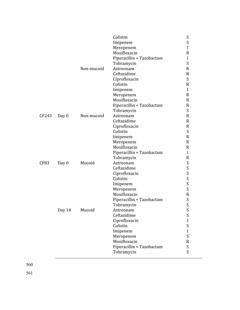
Non-mucoid
ColistinImipenemMeropenemMoxifloxacinPiperacillin+TazobactamTobramycinAztreonamCeftazidimeCiprofloxacinColistinImipenemMeropenemMoxifloxacinPiperacillin+TazobactamTobramycin
SSIRISRRSRIRRRS
CF243 Day0
Non-mucoid
AztreonamCeftazidimeCiprofloxacinColistinImipenemMeropenemMoxifloxacinPiperacillin+TazobactamTobramycin
RRRSRRRIR
CF83 Day0Day14
MucoidMucoid
AztreonamCeftazidimeCiprofloxacinColistinImipenemMeropenemMoxifloxacinPiperacillin+TazobactamTobramycinAztreonamCeftazidimeCiprofloxacinColistinImipenemMeropenemMoxifloxacinPiperacillin+TazobactamTobramycin
SSSSSSRSSSSISISRSS
560
561
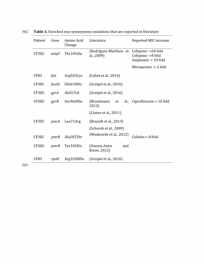
Table3.Enrichednon-synonymousmutationsthatarereportedinliterature562
Patient Gene AminoAcid
Change
Literature ReportedMICincrease
CF382 ampC Thr105Ala(Rodríguez-Martínez et
al.,2009)
Cefapime->64fold
Cefepime->8fold
Imipenem->10fold
Meropenem->2fold
CF83 ftsI Arg503Cys (Cabotetal.,2016)
CF382 fusA2 Gln610His (Greipeletal.,2016)
CF382 gyrA Ala51Val (Greipeletal.,2016)
CF382 gyrB Ser466Phe (Bruchmann et al.,
2013)
(Llanesetal.,2011)
Ciprofloxacin->10fold
CF382 pmrA Leu71Arg (Bezuidtetal.,2013)
(Schureketal.,2009)
CF382 pmrB Ala282Thr(Moskowitzetal.,2012)
Colistin->8fold
CF382 pmrB Tyr345His (Owusu-Anim and
Kwon,2012)
CF83 rpoB Arg1038His (Greipeletal.,2016)
563