Embed Size (px)
Citation preview

apv
h(vi
dev
Virology 287, 105–111 (2001)doi:10.1006/viro.2001.1023, available online at http://www.idealibrary.com on
Quantitative Analysis of Herpesvirus Sequences from Normal Tissueand Fibropapillomas of Marine Turtles with Real-Time PCR
Sandra L. Quackenbush,* Rufina N. Casey,† Rebecca J. Murcek,† Thomas A. Paul,† Thierry M. Work,‡Colin J. Limpus,§ Anny Chaves,¶ Leslie duToit,¶ Javier Vasconcelos Perez,i A. Alonso Aguirre,** Terry R. Spraker,††
Julia A. Horrocks,‡‡ Lotus A. Vermeer,§§ George H. Balazs,¶¶ and James W. Casey†,1
*Department of Molecular Biosciences, University of Kansas, Lawrence, Kansas 66045-2106; †Cornell University, Department of Microbiology andImmunology, Ithaca, New York 14853; ‡United States Geological Survey, National Wildlife Health Center, Honolulu Field Station, Honolulu, Hawaii
96850; §Queensland Parks and Wildlife Service, P.O. Box 155, Brisbane, Albert St. Q4002, Australia; ¶San Pablo Heredia, Costa Rica; iInstitutoNacional de la Pesca, Centro Mexicano de la Tortuga, Mazunte, Tonameca km 7 Carretera San Antonio-Puerto Angel Apdo., Postal 16, PuertoAngel, Oaxaca National; **Wildlife Trust, 1200 Lincoln Avenue, Suite 2, Prospect Park, Pennsylvania 19076; ††State Veterinary Diagnostic Laboratory,
Colorado State University, Fort Collins, Colorado 80523; ‡‡Department of Biological and Chemical Sciences, University of the West Indies,Cave Hill, St. Michael, Barbados; §§Barbados Sea Turtle Project, Bellairs Research Institute, St. James, Barbados; and ¶¶National
Marine Fisheries Service, Southwest Fisheries Science Center, Honolulu Laboratory, 2570 Dole St., Honolulu, Hawaii 96822
Received January 9, 2001; returned to author for revision February 7, 2001; accepted May 22, 2001; published online July 23, 2001
Quantitative real-time PCR has been used to measure fibropapilloma-associated turtle herpesvirus (FPTHV) pol DNA loadsin fibropapillomas, fibromas, and uninvolved tissues of green, loggerhead, and olive ridley turtles from Hawaii, Florida, CostaRica, Australia, Mexico, and the West Indies. The viral DNA loads from tumors obtained from terminal animals were relativelyhomogenous (range 2–20 copies/cell), whereas DNA copy numbers from biopsied tumors and skin of otherwise healthyturtles displayed a wide variation (range 0.001–170 copies/cell) and may reflect the stage of tumor development. FPTHV DNAloads in tumors were 2.5–4.5 logs higher than in uninvolved skin from the same animal regardless of geographic location,further implying a role for FPTHV in the etiology of fibropapillomatosis. Although FPTHV pol sequences amplified from tumorsare highly related to each other, single signature amino acid substitutions distinguish the Australia/Hawaii, Mexico/CostaRica, and Florida/Caribbean groups. © 2001 Academic Press
Key Words: herpesvirus; fibropapilloma; real-time PCR; marine turtles.
Fta
Hmmc(
ocraoa
INTRODUCTION
Fibropapillomatosis (FP) is a neoplastic disease ofmarine turtles characterized by the presence of epithelialfibropapillomas and internal fibromas. FP of marine tur-tles is a recently emerging disease and likely presents anew viral epizootic (Herbst, 1994; Lackovich et al., 1999;Quackenbush et al., 1998). FP has appeared worldwidewith sporadic but generally increasing frequency ingreen (Chelonia mydas), loggerhead (Caretta caretta),
nd olive ridley (Lepidochely olivacea) turtles and mayotentially pose a significant threat to the long-term sur-ival of marine turtles (Balazs and Pooley, 1991).
The etiologic agent of FP is most likely a herpesvirus;owever, environmental cofactors may be involved
Herbst and Klein, 1995; Landsberg et al., 1999). Herpes-irus sequences have been detected in fibropapillomas
n green, loggerhead, and olive ridley turtles from Hawaii,
1 To whom correspondence and reprint requests should be ad-ressed at Department of Microbiology and Immunology, C5-153 Vet-rinary Medical Center, College of Veterinary Medicine, Cornell Uni-ersity, Ithaca, NY 14853-6401. E-mail: [email protected].
g105
lorida, and Costa Rica using consensus PCR primershat recognize conserved regions of the DNA polymer-se gene of herpesviruses (Lackovich et al., 1999;
Quackenbush et al., 1998; VanDevanter et al., 1996).erpesvirus-like particles have been visualized in tu-ors by electron microscopy, and experimental trans-ission of fibropapilloma to green turtles has been suc-
essful using filtered, cell-free tumor homogenatesHerbst et al., 1995, 1996; Jacobson et al., 1991). Herpes-
virus sequences were not detected in skin samples fromturtles without FP, which suggests this virus is not acommon infectious agent of marine turtles (Lackovich etal., 1999; Quackenbush et al., 1998).
Study of fibropapilloma-associated turtle herpesvi-ruses (FPTHV) has been limited to in vivo assessment ofviral DNA, mostly by PCR, and further constrained by thefailure of all attempts at in vitro cultivation to date (Lack-
vich et al., 1999). Standard PCR demonstrates a strongorrelation between the presence of the turtle herpesvi-us DNA polymerase gene and FP, but does not provide
measure of the levels of FPTHV DNA in tumors andther infected tissues. To further implicate FPTHV as thegent of FP, we quantified viral DNA loads in tissues from
reen, olive ridley, and loggerhead turtles afflicted with0042-6822/01 $35.00Copyright © 2001 by Academic PressAll rights of reproduction in any form reserved.

a
D
qipop
vshe
CpA
pgvDow
G
L
G
O
106 QUACKENBUSH ET AL.
FP from the Pacific and Atlantic Ocean and evaluated thegenetic relatedness of the FPTHV pol gene from these
reas.
RESULTS
etection of FPTHV pol sequences in turtles fromAustralia, Barbados, and Mexico
To determine whether fibropapillomas from green andloggerhead turtles from Moreton Bay, Australia contain aherpesvirus, DNA was extracted from tumors and sub-jected to PCR using turtle herpesvirus-specific primers(GTHV1 and GTHV2). These primers were designed fromturtle herpesvirus DNA polymerase sequences previ-ously found to be associated with fibropapillomas frommarine turtles and amplify a 165-bp fragment of the DNApolymerase gene (Quackenbush et al., 1998). A total of20 tumors were assayed from 14 green, 2 loggerhead,and 4 olive ridley turtles. DNA from uninvolved skin from14 tumor-bearing turtles and skin from 31 tumor-freeanimals was also assayed (Table 1). With the exceptionof one tumor from a green turtle, herpesvirus pol se-
uences were detected in all tumors. Five of 11 histolog-cally normal skin samples from green turtles with fibro-apillomas also were found to harbor herpesvirus. Fivef 27 tumor-free Australian turtles demonstrated theresence of THV pol sequences in skin samples.
A fibropapilloma and uninvolved skin were obtainedfrom one green turtle collected in Barbados. Using theFPTHV-specific pol primers, both of these samples werefound to be positive by PCR (Table 1). Four tumors col-lected from olive ridley turtles from Mexico were alsofound to contain FPTHV pol sequences. Skin samplesfrom four olive ridley turtles without any presence oftumors were negative for FPTHV (Table 1).
These results are consistent with those from a previ-
TABLE 1
Detection of Turtle Herpesvirus Sequences in Sea Turtles
n No. positive (%)
reen turtles—AustraliaFibropapillomas 13 12 (92)Skin from tumor positive turtles 11 5 (45)Skin from tumor free turtles 14 3 (21)
oggerhead turtles—AustraliaFibropapillomas 2 2 (100)Skin from tumor positive turtles 2 0 (0)Skin from tumor free turtles 13 2 (15)
reen turtles—BarbadosFibropapillomas 1 1 (100)Skin from tumor positive turtle 1 1 (100)
live Ridley turtles—MexicoFibropapillomas 4 4 (100)Skin from tumor free turtles 4 0 (0)
ous study of green and loggerhead turtles from Hawaii
and Florida and olive ridley turtles from Costa Rica(Quackenbush et al., 1998). However, in contrast to pre-
ious studies, herpesvirus sequences were detected inkin samples from both tumor-free green and logger-ead turtles from Moreton Bay, Australia (Quackenbusht al., 1998).
loning and sequence analysis of herpesvirus DNAolymerase amplicons from tumors collected inustralia, Barbados, and Mexico
To further characterize the herpesvirus that wasresent in fibropapillomas collected from new geo-raphic locations, a 483-bp fragment of the turtle herpes-irus DNA polymerase gene was cloned and sequenced.NA isolated from tumors from green, loggerhead, andlive ridley turtles from Australia, Barbados, and Mexicoas PCR amplified with the FPTHV pol primers, GTHV2
and GTHV3. The amino acid sequence derived from eachof these fragments was aligned with the turtle herpesvi-rus sequences previously described (Fig. 1) (Quacken-bush et al., 1998). The Australian loggerhead sequence(LTHV-Aust) is identical to that of the Hawaiian greenturtle sequence (GTHV-Ha). Two nucleotide changes re-sulting in one amino acid substitution [leucine (L) toserine (S)] are present in the Australian green turtlesequence (GTHV-Aust) compared to the GTHV-Ha se-quence. The Barbados green turtle sequence (GTHV-Bar)is more closely related to the Florida green (GTHV-Fl)and loggerhead sequences (LTHV-Fl). The Barbados se-quence differs from the Florida sequences by three nu-cleotide changes, which result in two amino acid substi-tutions. The olive ridley herpesvirus sequence from tur-tles collected in Mexico (ORTHV-Mex) differs from theolive ridley sequence (ORTHV-CR) previously identified inturtles from Costa Rica by three nucleotide changes,which result in one amino acid substitution [histidine (H)to asparagine (N)]. The H-to-N amino acid substitution isalso found in the GTHV-Bar sequence.
FPTHV DNA levels in tumors and tissues of turtleswith fibropapillomatosis
Standard PCR analysis does not provide a quantitativemeasure of the levels of FPTHV DNA present in tumorsand infected tissues. To accurately assess the herpes-virus DNA load in different tissues, real-time quantitativePCR was employed. The number of copies of FPTHVDNA pol gene present in tumors and other tissues fromturtles sampled from Hawaii and Florida are displayed inFig. 2. All assays contained 100 ng DNA, which corre-sponds to approximately 20,000 cells, assuming a valueof 6 pg of DNA per cell. As a reference point, a copynumber of 2 3 104 FPTHV genomes would be equivalentto one copy of FPTHV per tumor cell under these assay
conditions; however, no assumption can be made re-
0
Fs
itfDtAiFR
107HERPESVIRUS OF MARINE TURTLES WITH REAL-TIME PCR
garding the distribution of FPTHV genomes within thetumor. FPTHV pol DNA loads in fibropapillomas werefound to range from 3.3 3 103 to 4.9 3 105 copies per 100ng DNA. Two Hawaiian turtles (12377 and 12368) hadinternal fibromas in the heart, lung, and kidney in whichthe viral load ranged from 9.4 3 103 to 1.3 3 105 copiesof FPTHV. Thirty-eight histologically normal samples fromvarious tissues of 10 turtles were also assayed for thepresence of FPTHV (Fig. 2). Sixteen of the 38 sampleswere positive for FPTHV; however, at significantly lowerlevels (mean 5 12.2 6 8.267 copies per 100 ng DNA, P ,
.006) than those found in tumors (mean 5 8.85 3 104 68.0692 3 104 copies per 100 ng DNA).
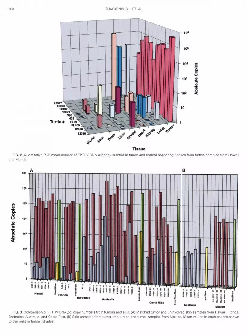
PTHV DNA levels in tumors and uninvolved skinamples from distinct geographical locations
To further implicate FPTHV as the etiologic agent of FPn marine turtles, real-time PCR was used to determinehe level of FPTHV DNA in tumors and uninvolved skinrom the same animal. Real-time PCR was performed on
NA extracted from uninvolved skin and tumors from 30urtles with FP collected in Hawaii, Florida, Barbados,ustralia, and Costa Rica. The absolute numbers of cop-
es of FPTHV pol are presented in Fig. 3A. The range ofPTHV pol in uninvolved skin from the Australia/Costaica/Barbados tumor turtles was 2.8 to 1.9 3 104 copies.
A paired t test was performed on these data and it wasfound that there is significantly (P , 0.007) less FPTHVpol in the skin samples than that found in the tumors.Importantly, 15 of the 21 skin samples contained lessthan 100 copies of FPTHV pol DNA, six of which did notcontain any FPTHV viral DNA. Skin samples from threeloggerhead and two green tumor-free turtles were also
FIG. 1. Amino acid sequence alignment of FPTHV pol genes. Aminoregions amplified from fibropapillomas from green turtles (GTHV), loggeAustralia (Aust), Barbados (Bar), Costa Rica (CR), and Mexico (Mex).
quantitated (Fig. 3B). The mean value for skin was 2.5 3
102. The tumors assayed from the four olive ridley turtlesfrom Mexico contained between 1.1 3 104 and 2.6 3 104
copies (mean 1.85 3 104) of FPTHV pol DNA (Fig. 3B).Matched skin samples from these animals were notavailable for analysis. However, FPTHV was not detectedin skin from four Mexican olive ridley turtles without FP(Table 1).
DISCUSSION
FP occurs in turtles throughout the world; however,only animals in Hawaii, Florida, and Costa Rica havepreviously been positively identified as harboring FPTHV(Quackenbush et al., 1998). We now extend these data toinclude turtles from Australia, Barbados, and PacificMexico. Ninety-five percent of tumors from these animalswere positive for FPTHV DNA pol sequences. In addition,FPTHV was also found in five of 25 skin samples thatwere analyzed from healthy foraging and nesting turtlesfrom Australia. Since FPTHV’s primary target appears tobe skin, as with most alphaherpesviruses, the presenceof FPTHV DNA in skin from tumor-free animals likelyrepresents early infection. Thus detection of FPTHV byPCR in tumor-free animals may be indicative of thoseanimals that will eventually develop FP. Corroborativedata, such as serological testing, is essential to validatethis assumption.
Comparison of the new herpesvirus sequences fromAustralia, Barbados, and Pacific Mexico with those pre-viously identified confirmed the high degree of related-ness among all these viruses and suggests that virusesisolated from animals from broader geographic regionsare still closely related. FPTHV sequences amplified fromtumors from Hawaii and Australia are very similar to
equences were derived from the nucleotide sequences of FPTHV polurtles (LTHV), and olive ridley turtles (ORTHV) from Hawaii (Ha), FL (Fl),
acid srhead t
each other, differing only by one amino acid substitution.
108 QUACKENBUSH ET AL.
FIG. 2. Quantitative PCR measurement of FPTHV DNA pol copy number in tumor and normal appearing tissues from turtles sampled from Hawaiiand Florida.
FIG. 3. Comparison of FPTHV DNA pol copy numbers from tumors and skin. (A) Matched tumor and uninvolved skin samples from Hawaii, Florida,Barbados, Australia, and Costa Rica. (B) Skin samples from tumor-free turtles and tumor samples from Mexico. Mean values in each set are shown
to the right in lighter shades.
ctFtf
audpstacEhm
109HERPESVIRUS OF MARINE TURTLES WITH REAL-TIME PCR
The olive ridley sequences from Mexico (Pacific) andCosta Rica (Pacific) also differ by only one amino acidsubstitution. Although GTHV-Bar contains unique aminoacid substitutions, the sequences isolated from turtles inthe Caribbean (Florida and Barbados) are highly related.The significance of these unique changes in the highlyconserved DNA polymerase gene with regard to definedturtle populations may be substantiated by comparisonsof sequences of less conserved herpesviral genes andinclusion of additional tumor samples.
A mean of 15.21 copies per cell (range 5 0.001–170)of FPTHV DNA pol sequences were detected in tumors
ollected from turtles from six different locationshroughout the world. This value would suggest thatPTHV plays a dominant role in maintenance of the
umor state. As stated earlier, the single copy numberor FPTHV under these PCR conditions is 2 3 104
copies, the threshold single copy value if it is assumedthat FPTHV is distributed equally among all cells in thetumor. The viral DNA load in individual tumors sam-pled from Hawaii and Florida were relatively homog-enous, ranging from 1.7 to 25 copies of FPTHV pertumor cell, a value in agreement with previous esti-mates by Southern blot analysis (Quackenbush et al.,1998) and within the threshold value. In contrast, real-time PCR revealed that the copy numbers of viral DNAin tumors from Costa Rica and Australia varied by asmuch as 5 logs. Corresponding skin samples from thesame turtles of these distinct geographical regionsalso showed the same trend in copy number variationcompared to the Hawaii, Florida, and Barbados sam-ples. Seven of 18 tumor samples were below thesingle copy threshold value. The hypothesis thatFPTHV exerts a dominant role in tumor maintenance ischallenged by these data, especially considering tu-mors where only 10 to a few hundred copies arepresent (Fig. 3A). This copy number variation mayreflect the stage of tumor development. The Hawaii,Florida, and Barbados samples were obtained frommoribund turtles while the Australia, Costa Rica, andMexican samples were obtained from otherwisehealthy freshly dead, turtles. Spontaneous regressionof fibropapillomas has been documented photograph-ically in some field studies and may also result in viralcopy number variation (Balazs et al., 2000; Bennett et
l., 2000). Further, the cell(s) targeted by FPTHV arenknown and the viral load may be different in theistinct cellular layers that comprise the tumor. Fibro-apillomas consist of papillary epidermal hyperplasiaupported on broad fibrovascular stromal stalks and
he ratio of epidermal to dermal proliferation variesmong lesions (Harshbarger, 1991; Herbst, 1994; Ja-obson et al., 1989; Lucke’, 1938; Norton et al., 1990).osinophilic intranuclear inclusions associated witherpesvirus particles have been observed in epider-
al cells from some tumors by histopathology (Herbstet al., 1999; Jacobson et al., 1991). FPTHV expressionand loads could also change during tumor progres-sion. In situ hybridization analysis will be of value inaddressing these questions by examining the distribu-tion of viral DNA in tumors.
FPTHV sequences are also present in some normaltissues (in addition to skin) of animals with tumors, how-ever, at significantly lower levels (mean 5 0.0012 60.0017 copies per cell, range 5 0–0.002) than thosefound in tumors (Fig. 2). Some tissues in which FPTHVhas been detected, heart, kidney, and lung, are associ-ated with tumor development, suggesting that the lowlevel of viral DNA detected may be indicative of earlyinfection or likely tumor metastasis since FPTHV has notbeen found in blood.
To ascertain the requirement for FPTHV in tumor de-velopment, real-time PCR performed on samples from 38animals determined that FPTHV DNA loads in tumorswere 2.5–4.5 logs higher than in uninvolved skin from thesame animal regardless of geographic location. Thequantitative PCR data described here further substanti-ates FPTHV as the causative agent of fibropapillomato-sis. Molecular cloning and DNA sequence analysis ofthis virus will allow for the generation of diagnosticreagents to monitor infected animals as well as aid inmanagement decisions necessary to maintain survival ofthese endangered species.
MATERIALS AND METHODS
PCR amplification, cloning, and sequencing
Biopsies of tumors and nontumored skin were col-lected from live-captured immature and adult green andloggerhead turtles from Moreton Bay, Australia and adultfemale olive ridley turtles nesting in Oaxaca, Mexico(Pacific coast). Additionally, tumored tissues (skin andinternal tumors) were obtained from freshly dead greenturtles from Hawaii, Florida, and Barclays Park, Barbadosthat were stranded with severe fibropapillomatosis. DNAwas isolated from tissues as previously described(Quackenbush et al., 1998). One microgram of DNA in atotal volume of 100 ml was subjected to PCR amplifica-tion with turtle-specific herpesvirus primers: GTHV1(59TGTCTGGAGGTGGCGGCCACG39), GTHV2 (59GA-CACGCAGGCCAAAAAGCGA39), or GTHV3 (59AGCAT-CATCCAGGCCCACAA39). The PCR mixture consisted of20 mM Tris–HCl (pH 8.3), 2 mM MgCl2, 50 mM KCl, 2.5%DMSO, 200 mM of each dNTP, 10 pmol of each primer,and 2.5 U of Taq DNA polymerase (Life Technologies,Gaithersburg, MD). All samples were denatured at 94°Cfor 5 min and then amplified for 30 s at 94°C, 30 s at62°C, and 30 s at 72°C for 35 cycles, followed by 10 minat 72°C for one cycle. Fifteen microliters of each PCRamplification reaction was separated on a 2% agarosegel. PCR products were gel purified with a Qiaex II gel
extraction kit (Qiagen Inc., Chatsworth, CA) and cloned
l
C
110 QUACKENBUSH ET AL.
into pCR2.1 TOPO vector (Invitrogen Corp., Carlsbad,CA). Automated sequencing was done with an ABI 373Aautomated sequencer (Applied Biosystems, Inc., FosterCity, CA) at the Biotechnology Resource Center at Cor-nell University.
Real-time quantitative PCR
An ABI 7700 PRISM quantitative PCR instrument wasused to process and quantify levels of THV DNA. Theprimers and probe used to quantitate FPTHV were se-lected from the GTHV-Ha DNA polymerase gene. Theprimers used were turtle 59-pol (59ACTGGCTGGCACT-CAGGAAA39) and turtle 39-pol (59CAGCTGCTGCTTGTC-CAAAA39), which generates a product of 86 bp. A fluoro-genic probe, turtle pol-probe, which contained a FAM(6-carboxy-fluorescein) reporter molecule attached to the59 end and a TAMRA (6-carboxy-tetramethyl-rhodamine)quencher linked at the 39 end (59-[6FAM]-CGATGAAAAC-CGCACCGAGCGA-[TAMRA]-39) was synthesized by PEBiosystems. PCR amplification was performed in a 50 mlreaction volume containing dNTPs at a concentration of200 mM for dATP, dCTP, dGTP, and 400 mM for dUTP, 5pmol of each primer, 20 pmol of probe, 0.5 U of AmpEr-ase (PE Biosystems) uracil N-glycosylase (UNG), 0.25 mlof AmpliTaq Gold (PE Biosystems), and 5 ml of 103TaqMan buffer. All assays contained 100 ng of genomicDNA. PCR mixtures were subjected to 2 min at 50°C toactivate UNG (to degrade possible contaminating ampli-cons) and 10 min at 95°C to activate the Taq polymerasefollowed by 40 cycles of 15 s at 95°C and 1 min at 62°Cwith an ABI 7700 PRISM sequence detector (PE Biosys-tems).
For each reaction, the amount of fluorescence wasmeasured as a function of the quantity of a reporter dye
FIG. 4. Standard curve for real-time PCR. Serial log dilutions of pGTT values, which correspond to the PCR cycle number in which the va
plasmid DNA.
(FAM) that was released during amplification due to the
59 to 39 exonuclease activity of Taq polymerase. Serialog dilutions of the GTHV pol plasmid (Quackenbush et
al., 1998) were subjected to real-time PCR to establishstandard curves. The threshold cycle (CT) value for eachsample was determined as the number of the cycle atwhich the measured fluorescence first exceeded thethreshold limit (10 times the standard deviation of thebaseline). The CT values were plotted against the corre-sponding input DNA copy number and standard curveswere fitted by linear regression with correlation coeffi-cients of .0.95 (Fig. 4). The CT values obtained fromtissue DNA were used to calculate the viral genomecopy number for each experimental sequence amplified.The limit of sensitivity of the real-time PCR was esti-mated as the highest plasmid dilution that yielded com-parable CT values in replicate samples.
Statistics
Student’s t test and a 95% confidence interval basedon a t distribution were used for all statistic analyses. AP value less than 0.05 was considered significant.
Nucleotide sequence accession numbers
The sequences described in this paper have been de-posited with the GenBank database and assigned the fol-lowing Accession Nos.: AF299107, AF299108, AF299109,and AF299110.
ACKNOWLEDGMENTS
We express our gratitude to Dr. Allan Eaglesham for critical readingof this manuscript. T.A.P. and R.J.M. were supported by a training Grant
lasmid ranging from 1 to 107 copies per reaction were amplified. Thebove the threshold limit, are plotted against the copy number of input
HVpol plue is a
5T32CA09682 from the National Institutes of Health.

B
B
H
H
H
H
H
H
J
J
L
L
L
N
Q
V
111HERPESVIRUS OF MARINE TURTLES WITH REAL-TIME PCR
REFERENCES
Balazs, G. H., Murakawa, S. K. K., Ellis, D. M., and Aguirre, A. A. (2000).Manifestation of fibropapillomatosis and rates of growth of greenturtles in Kaneohe Bay, Hawaii. Proceedings of the Eighteenth An-nual Symposium on Sea Turtle Biology and Conservation, pp. 112–113. U.S. Dept. of Commerce, NOAA Tech Memo NMFS-SEFSC.
alazs, G. H., and Pooley, S. (1991). “Research plan for marine turtlefibropapilloma” (G. H. Balazs, and S. Pooley, Eds.), U.S. Dept. ofCommerce, NOAA Tech Memo NMFS-SWFSC-156.
ennett, P. U., Keuper-Bennett, U., and Balazs, G. H. (2000). Photo-graphic evidence for the regression of fibropapilloma afflicting greenturtles at Honokowai, Maui in the Hawaiian Islands. Proceedings ofthe Nineteenth Annual Symposium on Sea Turtle Biology and Con-servation. U.S. Dept. of Commerce, NOAA Tech Memo NMFS-SEFSC, 443, 37–39.
arshbarger, J. C. (1991). Sea turtle fibropapilloma cases in the registryof tumors in lower animals. Research plan for marine turtle fibropap-illoma (G. H. Balazs, and S. G. Pooley, Eds.), U.S. Dept. of Commerce,NOAA Tech Memo NMFS-SWFSC-156.
erbst, L. H. (1994). Fibropapillomatosis of marine turtles. Annu. Rev.Fish Dis. 4, 389–425.
erbst, L. H., Jacobson, E. R., Klein, P. A., Balazs, G. H., Moretti, R.,Brown, T., and Sundberg, J. P. (1999). Comparative pathology andpathogenesis of spontaneous and experimentally induced fibropap-illomas of green turtles (Chelonia mydas). Vet. Pathol. 36(6), 551–564.
erbst, L. H., Jacobson, E. R., Moretti, R., Brown, T., Sundberg, J. P., andKlein, P. A. (1995). Experimental transmission of green turtle fibro-papillomatosis using cell-free tumor extracts. Dis. Aquat. Org. 22,1–12.
erbst, L. H., and Klein, P. A. (1995). Green turtle fibropapillomatosis:Challenges to assessing the role of environmental cofactors. Envi-ron. Health Perspect. 103(Suppl. 4), 27–30.
erbst, L. H., Moretti, R., Brown, T., and Klein, P. A. (1996). Sensitivity of
the transmissible green turtle fibropapillomatosis agent to chloro-form and ultracentrifugation conditions. Dis. Aquat. Org. 25, 225–228.
acobson, E. R., Buergelt, C., Williams, B., and Harris, R. K. (1991).Herpesvirus in cutaneous fibropapillomas of the green turtle, Che-lonia mydas. Dis. Aquat. Org. 12, 1–6.
acobson, E. R., Mansell, J. L., Sundberg, J. P., Hajjar, L., Reichmann,M. E., Ehrhart, L. M., Walsh, M., and Murru, F. (1989). Cutaneousfibropapillomas of green turtles (Chelonia mydas). J. Comp. Pathol.101, 39–52.
ackovich, J. K., Brown, D. R., Homer, B. L., Garber, R. L., Mader, D. R.,Moretti, R. H., Patterson, A. D., Herbst, L. H., Oros, J., Jacobson, E. R.,Curry, S. S., and Klein, P. A. (1999). Association of herpesvirus withfibropapillomatosis of the green turtle Chelonia mydas and the log-gerhead turtle Caretta caretta in Florida. Dis. Aquat. Org. 37(2),89–97.
andsberg, J. H., Balazs, G. H., Steidinger, K. A., Baden, D. G., Work,T. W., and Russell, D. J. (1999). The potential role of natural tumorpromoters in marine turtle fibropapillomatosis. J. Aquat. Anim. Health11, 199–210.
ucke’, B. (1938). Studies on tumors in cold-blooded vertebrates. Annu.Rep. Tortugas Lab. Carnegie Institute, Washington, DC 1937–38,92–94.
orton, T. M., Jacobson, E. R., and Sundberg, J. P. (1990). Cutaneousfibropapillomas and renal myxofibroma in a green turtle, Cheloniamydas. J. Wildl. Dis. 26, 265–270.
uackenbush, S. L., Work, T. M., Balazs, G. H., Casey, R. N., Rovnak, J.,Chaves, A., duToit, L., Baines, J. D., Parrish, C. R., Bowser, P. R., andCasey, J. W. (1998). Three closely related herpesviruses are associ-ated with fibropapillomatosis in marine turtles. Virology 246(2), 392–399.
anDevanter, D. R., Warrener, P., Bennett, L., Schultz, E. R., Coulter, S.,Garber, R. L., and Rose, T. M. (1996). Detection and analysis ofdiverse herpesviral species by consensus primer PCR. J. Clin. Mi-crobiol. 34(7), 1666–1671.