Embed Size (px)
Citation preview

1
Lectures 6 & 7
Colonic Tumors & Polyp
{ حياته طوال.. تجرع ذل الجهل ساعة لم يذق مر التعلم ومن }
Red: Important. Grey: Extra Notes Doctors Notes will be in text boxes

2
Objectives:
A. Know the classification of intestinal tumors (small intestine and colon)
B. Know the definition of a polyp.
C. Compare adenomatous polyps and hyperplastic polyps with respect to pathology (gross
and microscopic features).
D. Know the three subtypes of adenomatous polyps, eg, tubular adenoma, villous adenoma,
tubulovillous adenoma.
E. Describe the adenomatous polyp-cancer sequence and the features associated with risk of
malignancy, eg, polyp size, histologic architecture, and severity of epithelial dysplasia.
F. Describe the classification of the hereditary syndromes involving the GI tract and the
syndromes associated with an increased risk of cancer (Peutz-Jeghers syndrome, familial
adenomatous polyposis, and hereditary nonpolyposis colorectal carcinoma)
G. Know common types of intestinal polyps
A. Non-neoplastic polyps no dysplasia
4 common types (hyperplastic, hamartomatous, inflammatory, lymphoid)
B. Neoplastic polyps there is dysplasia
3 types (tubular, tubulovilous, villous)
Colon cancer:
A. Describe the epidemiology of colon cancer.
B. Compare the pathology (gross and microscopic features) and clinical features of right-sided
colonic adenocarcinoma and left-sided colorectal adenocarcinoma.
C. Describe the relationship between prognosis and the various stages of cancer of the colon
and rectum as noted in the TNM (tumor-nodes-metastasis) classification and staging
system.
D. Describe the relationship between carcinoembryonic antigen (CEA) and recurrence
following resection of the primary tumor.
E. Mention the significant of carcinoid tumor and its features
H. Know the clinical presentation of left and right sided colon cancer, and the environmental
factors that increase its risk
C. Left colon: frank bleeding, obstruction
D. Right colon: iron deficiency anaemia
E. Tumor markers: CEA
I. Understand the pathogenesis of colon cancer
F. Adenoma to Carcinoma Pathway
G. Two genetic pathways APC/B-catenin and DNA mismatch repair genes
H. Familial Polyposis Syndrome
J. Describe the Pathological features of colon cancer
I. Adenocarcinoma most common: carcinoid tumor {neurosecretory granules}
J. 70% are in the rectum and/or sigmoid
K. Duke classification is used for staging
References: Lecture slides & Robbins

3
Tumors of the small and large intestines:
o Polyps o Carcinoma o Carcinoid tumor o Lymphoma
Sigmoid Colon: Most common site of GI polyps, diverticula and cancer
Introduction:
Polyps are most common in the colon
but may occur in the esophagus,
stomach, or small intestine. It has two
types:
A. Sessile: without stalks, which is
proliferation of cells adjacent to
the polyp and the effects of
traction on the luminal
protrusion.
B. Pedunculated: may combine to
create a stalk.
- The majority of small and large intestine tumors appears as a polyp. - Polyp: is protrusion or growth above the surface, which could be benign or malignant. - If the polyp is big you can’t discriminate from the morphology whether it’s benign or malignant unless you examine it under the microscope, but if it’s small like 2 mm it is benign.
The opposite of polyp is the diverticula which is out pouches in the wall of the colon, common in people who have chronic constipation, due to increased pressure on the wall and weakness of the muscle, but it’s still lined by mucosa, & could lead to inflammation and sometimes to perforation, and it could be
giant
Sessile polyp usually has a wide base with finger like projections above the surface, appears velvety under the microscope. Pedunculated polyp: like mushroom.

4
Polyps:
In general, intestinal polyps can be classified as neoplastic or non- neoplastic.
▪ Non-neoplastic polyps (90%)
Hyperplastic polyps
Hamartomatous polyps (Juvenile & Peutz-Jeghers polyps)
Inflammatory polyps
Lymphoid polyps
▪ Neoplastic polyps (10%)
Adenoma: The most common neoplastic polyp is the adenoma
Hyperplastic Polyps:
▪ Asymptomatic.
▪ > 50% are located in the rectosigmoid.
▪ Sawtooth surface.
▪ Star shaped crypts.
▪ Composed of well-formed glands and crypts lined by differentiated goblet or
absorptive cells.
Hamartomatous polyps:
Hamartoma is a tumour composed of tissue elements normally found at that site
of the organ, but which are growing in a disorganized mass. not a malignant
tumor.
Hamartomatous
Polyps
Juvenile Polyps Retention Polyp
Peutz-Jeghers Polyps
The most common type that Dr. Maha saw in the clinic is the neoplastic one because they do not take biopsy from hyperplastic polyps.
Hyperplastic polyp: no stalk, only increases the number of cells (goblet cells and enterocytes) in the lining epithelium (because of more mitosis and less apoptosis) without any premalignant changes (no dysplasia). - The elongation of the crypts with the folds of cells gives the sawtooth appearance. - It could be 2 or three but not polyposis (polyposis: more than a hundred polyps).
Hamartoma: normal tissue in disorganized manner, such as pulmonary hamartoma (usually it is
congenital anomaly)
In addition to the dilated glands there is smooth muscles in the lamina propria which start to branch. - That’s why it’s called hamartoma.
5
Inflammatory Polyps:
▪ Longstanding IBD, especially in chronic ulcerative colitis.
▪ Represent an exuberant reparative response to longstanding mucosal injury
called pseudopolyps.
Lymphoid polyps:
It’s a normal polyp
Juvenile Polyps (retention polyp) Peutz-Jeghers syndrome
▪ Developmental malformations
affecting the glands and lamina
propria.
▪ Commonly occur in children
under 5 years old in the rectum.
▪ In adult called retention polyp.
▪ No malignant potential.
▪ Rare, autosomal dominant
▪ Hamartomatous polyps accompanied by
mucosal and cutaneous pigmentation around
the lips, oral mucosa, face and genitalia,
present with red blood in stool.
▪ On gross evaluation, Polyps tend to be large
and pedunculated. with a lobulated contour
▪ Increased risk of developing carcinoma of the
pancreas, breast, lung, ovary and uterus. - Could cause abdominal pain and rectal bleeding. - Could lead to polyposis in the colon or stomach. -Juvenile POLYPOSIS could lead to malignancies, notice not juvenile POLYPs! - Cronkhite-Canada syndrome: it is related to auto immune disorders not mutations, the patients have high risk of rheumatoid arthritis and other autoimmune disorders.
Juvenile Polyps (retention polyp): Pedunculated polyp, the stroma is edematous with cystic dilatation.
Under the microscope you will see a dilated gland containing mucus, and surrounded by stroma, this stroma has inflammatory
cells & the surface is slightly ulcerated
Smooth eroded surface with numerous mucus retention
cysts, typical of sporadic juvenile polyps.
Multiple ulcers, in between these ulcers the mucosa is inflamed, edematous and protrudes so it appears like a polyp, so it’s the normal tissue and surrounded by ulcers (pseudopolyps)
Stimulation and activation of
Peyer’s patch, lead to enlargement of it.
Patient requires CT screening every year Important ↓
Mainly it’s dilated glands + smooth muscles in the lamina propria, blood in stool, pigmentation in oral mucosa + gentile skin + face and lips. Large pedunculated polyps
سرطان خطر ففيه كبيرة انها بما
it’s the normal tissue between two ulcers, it gets inflamed and projects like polyp so it’s called pseudopolyps, there’s a risk of cancer.
6
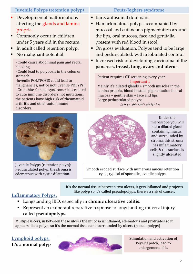
Adenomatous Polyp (adenoma): (Compare it with hyperplastic)
▪ Occur mainly in large bowel.
▪ Sporadic and familial.
▪ Vary from small pedunculated to large sessile.
▪ Epithelium proliferation and dysplasia.
Divided into:
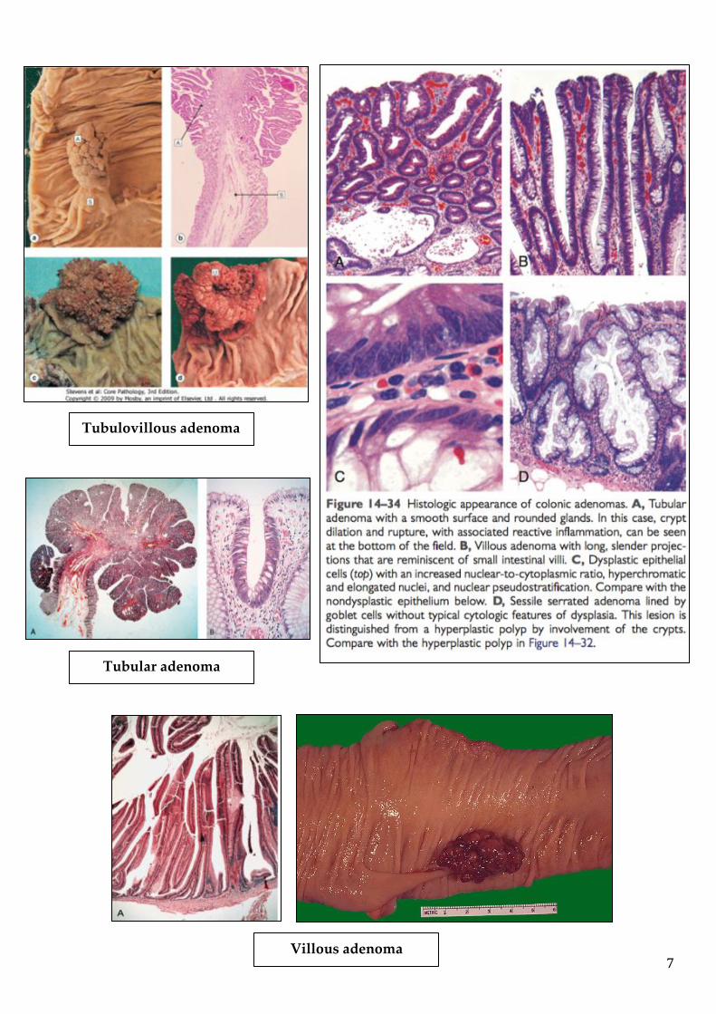
1. Tubular adenoma: less than 25% villous architecture
▪ Represents 75% of all neoplastic polyps.
▪ 75 % occur in the distal colon and rectum
▪ Sigmoid colon most common site.
2. Villous adenoma: villous architecture over 50%
▪ The least common, largest and most ominous of epithelial polyps (most likely
to undergo malignant transformation).
▪ Age: 60 to 65 years, 75% located in rectosigmoid area.
▪ Present with rectal bleeding or anemia, large ones may secrete copious
amounts of mucoid material rich in protein and potassium.
▪ Large tumors can produce hypoalbuminemia and hypokalemia.
3. Tubulovillous adenoma: villous architecture between 25 and 50%.
▪ 20%–30% of polyps
▪ Intermediate in size, degree of dysplasia and malignant potential between
tubular and villous adenomas.
Villous: (highest possibility of developing carcinoma). Tubular: (lowest possibility of developing carcinoma)
- Sporadic (1 or 2 polyps especially in old age) - Familial (> 500 polyps)
Tubular adenoma has a smooth surface unlike the villous adenoma which has finger like projections. In adenoma, you will see dysplasia (nuclear enlargement, hyperchromatism, pleomorphism and abnormal mitosis)
Benign tumor, it’s epithelium proliferation + Dysplasia (so
important!!!)
Thin core + dysplasia, finger like projection without stalk, least common but largest سرطان لنا تسبب وممكن وحدة أخطر فهي وحده اكبر انها بماو .
we will see: Diarrhea, hypoalbuminemia and hypokalemia
7
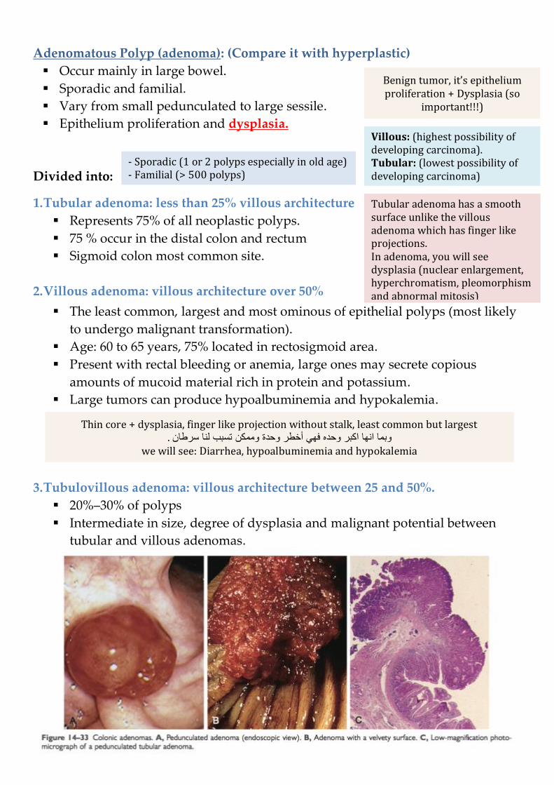
Tubulovillous adenoma
Tubular adenoma
Villous adenoma

8
Relationship of Neoplastic Polyps to Carcinoma: Very important (Memorize the
genes) Adenoma to carcinoma sequence is documented by several genetic
alterations.
Polyp removal leads to CRC prevention; Polyp is surrogate marker.
The probability of carcinoma occurring in a neoplastic polyp is related to:
o The size of the polyp. Size is the most important characteristic that correlates
with risk of malignancy.
o The relative proportion of its villous features.
o The presence of significant cytologic atypia (dysplasia) in the neoplastic cells.
o Multiple polyps.
o Accumulation of genetic mutation.
Familial Polyposis Syndrome:
Patients have genetic tendencies to develop neoplastic polyps.
Familial polyposis coli (FPC):
A. Genetic defect of Adenomatous polyposis coli (APC).
B. APC gene located on the long arm of chromosome 5 (5q21).
C. APC gene is a tumor suppressor gene.
D. Innumerable neoplastic polyps in the colon (Need ≥
100 for diagnosis, 500 to 2500).
E. Polyps are also found elsewhere in alimentary tract
F. The risk of colorectal cancer is 100% by midlife.
9
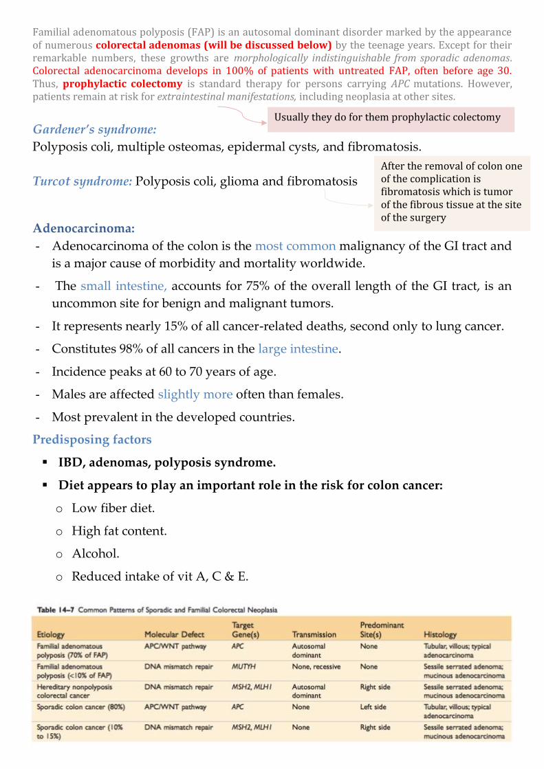
Familial adenomatous polyposis (FAP) is an autosomal dominant disorder marked by the appearance of numerous colorectal adenomas (will be discussed below) by the teenage years. Except for their remarkable numbers, these growths are morphologically indistinguishable from sporadic adenomas. Colorectal adenocarcinoma develops in 100% of patients with untreated FAP, often before age 30. Thus, prophylactic colectomy is standard therapy for persons carrying APC mutations. However, patients remain at risk for extraintestinal manifestations, including neoplasia at other sites.
Gardener’s syndrome:
Polyposis coli, multiple osteomas, epidermal cysts, and fibromatosis.
Turcot syndrome: Polyposis coli, glioma and fibromatosis
Adenocarcinoma:
- Adenocarcinoma of the colon is the most common malignancy of the GI tract and
is a major cause of morbidity and mortality worldwide.
- The small intestine, accounts for 75% of the overall length of the GI tract, is an
uncommon site for benign and malignant tumors.
- It represents nearly 15% of all cancer-related deaths, second only to lung cancer.
- Constitutes 98% of all cancers in the large intestine.
- Incidence peaks at 60 to 70 years of age.
- Males are affected slightly more often than females.
- Most prevalent in the developed countries.
Predisposing factors
▪ IBD, adenomas, polyposis syndrome.
▪ Diet appears to play an important role in the risk for colon cancer:
o Low fiber diet.
o High fat content.
o Alcohol.
o Reduced intake of vit A, C & E.
Usually they do for them prophylactic colectomy
After the removal of colon one of the complication is fibromatosis which is tumor of the fibrous tissue at the site of the surgery
10
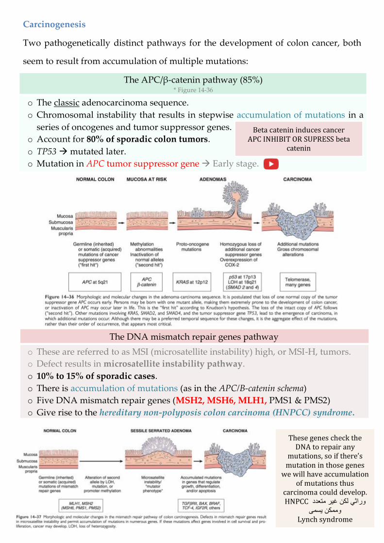
Carcinogenesis
Two pathogenetically distinct pathways for the development of colon cancer, both
seem to result from accumulation of multiple mutations:
The APC/β-catenin pathway (85%) * Figure 14-36
o The classic adenocarcinoma sequence.
o Chromosomal instability that results in stepwise accumulation of mutations in a
series of oncogenes and tumor suppressor genes.
o Account for 80% of sporadic colon tumors.
o TP53 mutated later.
o Mutation in APC tumor suppressor gene Early stage.
The DNA mismatch repair genes pathway
o These are referred to as MSI (microsatellite instability) high, or MSI-H, tumors.
o Defect results in microsatellite instability pathway.
o 10% to 15% of sporadic cases.
o There is accumulation of mutations (as in the APC/B-catenin schema)
o Five DNA mismatch repair genes (MSH2, MSH6, MLH1, PMS1 & PMS2)
o Give rise to the hereditary non-polyposis colon carcinoma (HNPCC) syndrome.
Beta catenin induces cancer APC INHIBIT OR SUPRESS beta
catenin
These genes check the DNA to repair any
mutations, so if there’s mutation in those genes
we will have accumulation of mutations thus
carcinoma could develop. HNPCC متعدد غير لكن وراثي
يسمى وممكن Lynch syndrome
11
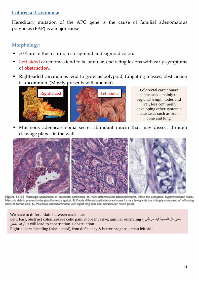
Colorectal Carcinoma:
Hereditary mutation of the APC gene is the cause of familial adenomatous
polyposis (FAP) is a major cause.
Morphology:
▪ 70% are in the rectum, rectosigmoid and sigmoid colon.
▪ Left-sided carcinomas tend to be annular, encircling lesions with early symptoms
of obstruction.
▪ Right-sided carcinomas tend to grow as polypoid, fungating masses, obstruction
is uncommon. (Mostly presents with anemia).
▪ Mucinous adenocarcinoma secret abundant mucin that may dissect through
cleavage planes in the wall.
Right-sided Left-sided Colorectal carcinomas metastasize mainly to
regional lymph nodes and liver, less commonly
developing other systemic metastases such as brain,
bone and lung.
We have to differentiate between each side: Left: Fast, obstruct colon, severe colic pain, more invasive, annular encircling ( سرطان فيه المحيط كل يعني
أخطر وهذا ) it will lead to constriction + obstruction Right: ulcers, bleeding (black stool), iron deficiency & better prognosis than left side

12
Signs and symptoms:
▪ If located closer to the anus: change in bowel habit, feeling of incomplete
defecation, PR bleeding.
▪ A tumor that is large enough to fill the entire lumen of the bowel may cause
bowel obstruction.
▪ Right-sided lesions are more likely to bleed while left-sided tumors are usually
detected later and could present with bowel obstruction.
▪ Serum levels of carcinoembryonic antigen (CEA) are related to tumor size and
extent of spread. They are helpful in monitoring for recurrence of tumor after
resection.
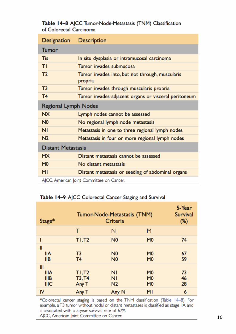
TNM Staging of Colon Cancers is used for staging:
Duke classification is used for staging.
Tumor markers:
A tumor marker is a substance found in the blood, urine or body tissues that can be
elevated in cancer, among other tissue types.
▪ Carcinoembryonic antigen (CEA) & Carbohydrate antigen (CA19-9) are
Useful to assess disease recurrence (late stage)
▪ Elevated in:
CEA Some non-neoplastic conditions like ulcerative colitis pancreatitis,
cirrhosis COPD, Crohn's disease as well as in smokers.
CA19-9
Colon cancer, pancreatic cancer, esophageal cancer and
hepatocellular carcinoma. Apart from cancer, pancreatitis,
cirrhosis.
▪ Tissue inhibitor of metalloproteinases 1 (TIMP1) = Early as well as late stage
disease.

13
Malignant Small Intestinal Neoplasms:
In descending order of frequency:
o Carcinoid.
o Adenocarcinomas.
o Lymphomas.
o Leiomyosarcomas.
Carcinoid Tumors:
These tumors were called “carcinoid” because they are slower growing than
carcinomas.
▪ Neoplasms arising from endocrine cells found along the length of GIT mucosa.
▪ The peak incidence: sixth decade, but they may appear at any age.
▪ They compose less than 2% of colorectal malignancies.
▪ Almost half of small intestinal malignant tumors:
o 60 to 80% appendix and terminal ileum.
▪ 10 to 20% rectum.
Ultrastructural features: neurosecretory electron dense bodies in the cytoplasm
Clinical features:
❖ Asymptomatic.
❖ May cause obstruction, intussusception or bleeding.
❖ May elaborate hormones: Zollinger-Ellison (Gastrin secretion), Cushing’s
carcinoid or other syndromes.
Local tumor of endocrine cells that secrete serotonin (vasoactive amine), it’s more malignant in ileum, benign in rectum. Carcinoid tumor is asymptomatic, why? Because secreted serotonin goes to the liver through the portal vein and gets detoxified, it does not enter the systemic circulation thus it’s asymptomatic. Morphology: Low grade tumor, cells have the same size (coin cells) & neurosecretory vesicles contain serotonin
Cushing’s secretes: ACTH like hormone increases the cortisone

14
Carcinoid syndrome:
❖ 1% of carcinoid tumor & in 20% of those of widespread metastasis.
❖ Paroxymal flushing, episodes of asthma-like wheezing, right-sided heart
failure, attacks of watery diarrhea, abdominal pain.
❖ The principal chemical mediator is serotonin.
❖ The syndrome is classically associated with ileal carcinoids with hepatic
metastases.
Serotonin and Diarrhea:
Patients with carcinoid syndrome often suffer from diarrhea, which has both a
secretory and a motor component. The secretory component of carcinoid diarrhea is
attributable to excessive serotonergic stimulation of submucosal secretomotor
neurons; the motor component includes faster small bowel and colon transit and an
exaggerated tonic response of the colon to ingestion of a meal.
Lymphoma: the doctors said that it’s not important
❖ Most often low-grade lymphomas arising in mucosal-associated lymphoid
tissue (MALT) lymphoma or high-grade non-Hodgkin's lymphomas of B cell
type.
❖ May occur in any part of the intestine; particularly the stomach
❖ The ileocecal region is a favored site for Burkitt's lymphoma.
Malignant glands of an adenocarcinoma of the
colon infiltrating the muscularis propria.
What is the mode of spread of this cancer?
Colonic carcinomas spread by local extension to
adjacent structures. The favored sites of metastases
are regional lymph nodes, liver, lungs, and bones.
Syndrome means that the tumor metastasized to liver which means that serotonin will get out to the circulation through hepatic vein > thus symptoms appear.
Symptoms:
Flushing, abdominal pain, asthma like, watery diarrhea (because serotonin will increase the motility of the intestine.)
15
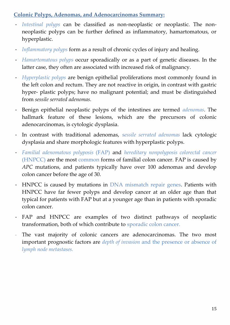
Colonic Polyps, Adenomas, and Adenocarcinomas Summary:
- Intestinal polyps can be classified as non-neoplastic or neoplastic. The non-
neoplastic polyps can be further defined as inflammatory, hamartomatous, or
hyperplastic.
- Inflammatory polyps form as a result of chronic cycles of injury and healing.
- Hamartomatous polyps occur sporadically or as a part of genetic diseases. In the
latter case, they often are associated with increased risk of malignancy.
- Hyperplastic polyps are benign epithelial proliferations most commonly found in
the left colon and rectum. They are not reactive in origin, in contrast with gastric
hyper- plastic polyps; have no malignant potential; and must be distinguished
from sessile serrated adenomas.
- Benign epithelial neoplastic polyps of the intestines are termed adenomas. The
hallmark feature of these lesions, which are the precursors of colonic
adenocarcinomas, is cytologic dysplasia.
- In contrast with traditional adenomas, sessile serrated adenomas lack cytologic
dysplasia and share morphologic features with hyperplastic polyps.
- Familial adenomatous polyposis (FAP) and hereditary nonpolyposis colorectal cancer
(HNPCC) are the most common forms of familial colon cancer. FAP is caused by
APC mutations, and patients typically have over 100 adenomas and develop
colon cancer before the age of 30.
- HNPCC is caused by mutations in DNA mismatch repair genes. Patients with
HNPCC have far fewer polyps and develop cancer at an older age than that
typical for patients with FAP but at a younger age than in patients with sporadic
colon cancer.
- FAP and HNPCC are examples of two distinct pathways of neoplastic
transformation, both of which contribute to sporadic colon cancer.
- The vast majority of colonic cancers are adenocarcinomas. The two most
important prognostic factors are depth of invasion and the presence or absence of
lymph node metastases.
16
17
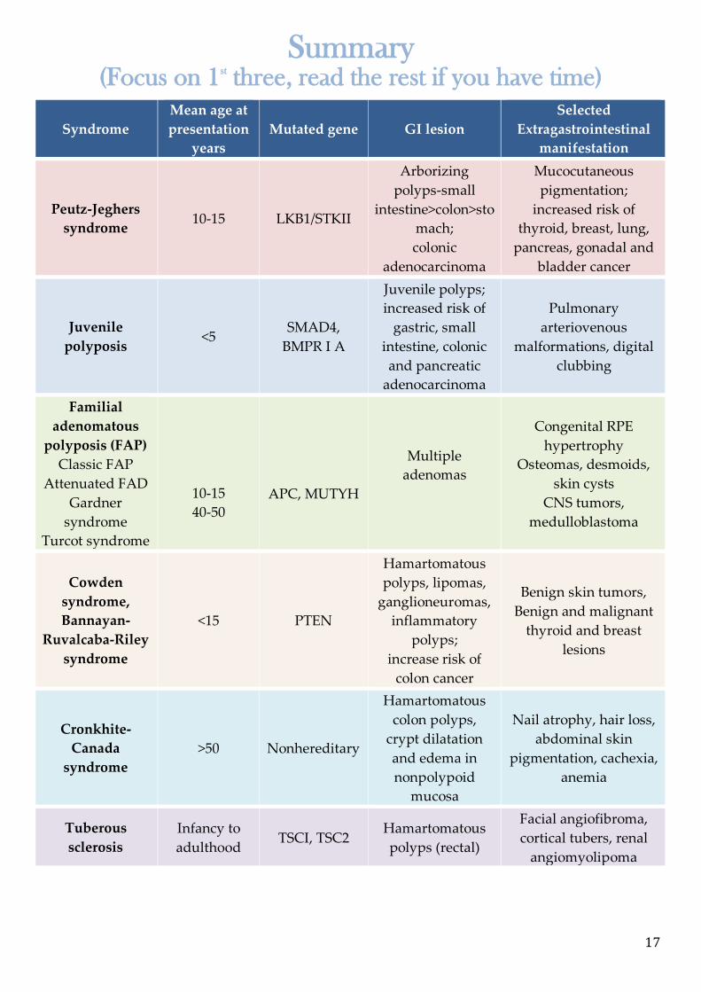
Summary (Focus on 1st three, read the rest if you have time)
Syndrome
Mean age at
presentation
years
Mutated gene GI lesion
Selected
Extragastrointestinal
manifestation
Peutz-Jeghers
syndrome 10-15 LKB1/STKII
Arborizing
polyps-small
intestine>colon>sto
mach;
colonic
adenocarcinoma
Mucocutaneous
pigmentation;
increased risk of
thyroid, breast, lung,
pancreas, gonadal and
bladder cancer
Juvenile
polyposis <5
SMAD4,
BMPR I A
Juvenile polyps;
increased risk of
gastric, small
intestine, colonic
and pancreatic
adenocarcinoma
Pulmonary
arteriovenous
malformations, digital
clubbing
Familial
adenomatous
polyposis (FAP)
Classic FAP
Attenuated FAD
Gardner
syndrome
Turcot syndrome
10-15
40-50
APC, MUTYH
Multiple
adenomas
Congenital RPE
hypertrophy
Osteomas, desmoids,
skin cysts
CNS tumors,
medulloblastoma
Cowden
syndrome,
Bannayan-
Ruvalcaba-Riley
syndrome
<15 PTEN
Hamartomatous
polyps, lipomas,
ganglioneuromas,
inflammatory
polyps;
increase risk of
colon cancer
Benign skin tumors,
Benign and malignant
thyroid and breast
lesions
Cronkhite-
Canada
syndrome
>50 Nonhereditary
Hamartomatous
colon polyps,
crypt dilatation
and edema in
nonpolypoid
mucosa
Nail atrophy, hair loss,
abdominal skin
pigmentation, cachexia,
anemia
Tuberous
sclerosis
Infancy to
adulthood TSCI, TSC2
Hamartomatous
polyps (rectal)
Facial angiofibroma,
cortical tubers, renal
angiomyolipoma
18
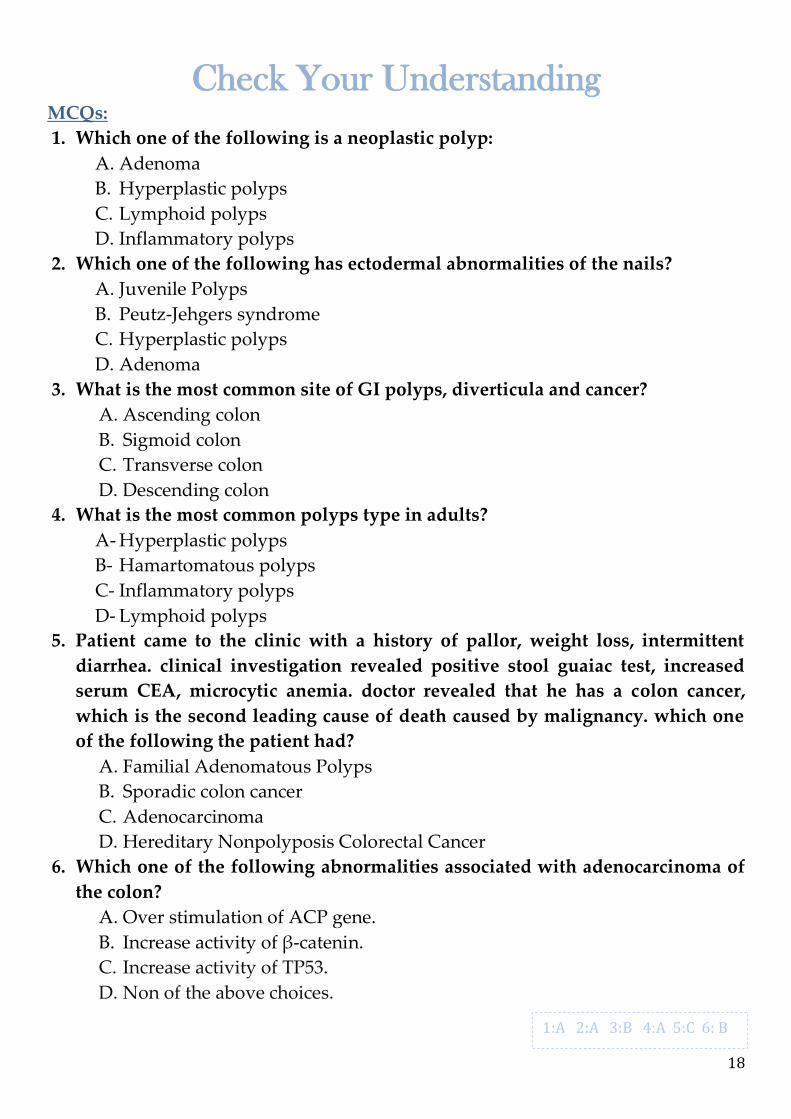
Check Your Understanding MCQs:
1. Which one of the following is a neoplastic polyp:
A. Adenoma
B. Hyperplastic polyps
C. Lymphoid polyps
D. Inflammatory polyps
2. Which one of the following has ectodermal abnormalities of the nails?
A. Juvenile Polyps
B. Peutz-Jehgers syndrome
C. Hyperplastic polyps
D. Adenoma
3. What is the most common site of GI polyps, diverticula and cancer?
A. Ascending colon
B. Sigmoid colon
C. Transverse colon
D. Descending colon
4. What is the most common polyps type in adults?
A- Hyperplastic polyps
B- Hamartomatous polyps
C- Inflammatory polyps
D- Lymphoid polyps
5. Patient came to the clinic with a history of pallor, weight loss, intermittent
diarrhea. clinical investigation revealed positive stool guaiac test, increased
serum CEA, microcytic anemia. doctor revealed that he has a colon cancer,
which is the second leading cause of death caused by malignancy. which one
of the following the patient had?
A. Familial Adenomatous Polyps
B. Sporadic colon cancer
C. Adenocarcinoma
D. Hereditary Nonpolyposis Colorectal Cancer
6. Which one of the following abnormalities associated with adenocarcinoma of
the colon?
A. Over stimulation of ACP gene.
B. Increase activity of β-catenin.
C. Increase activity of TP53.
D. Non of the above choices.
1:A 2:A 3:B 4:A 5:C 6: B
19
7. Which one of the following mechanisms is associated with morphological
identifiable changes in adenocarcinoma of the colon?
A. APC mutation\ β-catenin mechanism.
B. The microsatellite instability pathway.
8. An early symptom of left-sided carcinoma (Colorectal carcinoma)?
A. Fibrosis
B. Obstruction
C. Bleeding
D. Stricture
9. Right-sided carcinoma (Colorectal carcinoma) associated with?
A. Sickle cell anemia
B. Hypochromic microcytic anemia
C. Iron deficiency anemia
D. Bleeding
10. Survival is decreased in Colorectal carcinoma in case of?
A. lymph node metastases
B. lung metastases
C. mucosal gland metastases
D. brain metastases
11. Most common site of metastatic lesions?
A. Heart
B. spleen
C. Pancreas
D. liver
12. Under the microscope we found a polyp which is 0.4 cm in diameter, the type
of it is:
A- Hyperplasic polyp.
B- Adenomatous polyp.
C- The diameter cannot guide us in this case.
13. The hall mark of adenomatous polyps:
1- Epithelial dysplasia.
2- mature goblet and absorptive cells
3- nuclear hyperchromasia
14. Which one of these polyps will result because the epithelium fails to mature
as cells migrate out of the crypt?
A- Hyperplasic polyp.
B- Adenomatous polyp.
C- Both.
7: A 8:B 9:C 10:A 11:D 12:C 13:A 14:B
Q12: The explanation:
Hyperplastic polyps are less than 0.5 cm
adenomatous polyps range from 0.3 – 10 cm
20
Contact us: [email protected]
Team Members:
نوف التويجري فهد العبداللطيف
فاطمة الدين
تون الصالحف كوثر الموسى لميس آل تميم
الصغير هلولو
مريم سعيدان
منيرة العيوني
عقيلمي ال
نورة الخراز
نورة الطويل
نوف الرشيد
نوف العبدالكريم
أثير النشوان الجوهرة المزروع
إلهام الزهراني بدور جليدان
خولة العماري دانيا الهنداوي
دانة عمله ديما الفارس
رزان السبتي
رغد المنصور
سارة القحطاني
شما السهيلي
محمد الدغيثر
معاذ باعشن
عبدالناصر الوابل
عبدالرحمن الزامل
محمد الزاحم
الزيدان عبدالعزيز
عبدهللا الفريح
ماجد العسبلي
عبدهللا العليوي
عبدالرحمن الناصر
محمد الفضل
قال صلى هللا عليه وسلم: }من سلك طريقا يلتمس فيه علما سهل هللا له به
طريقا إلى الجنة{
دعواتنا لكم بالتوفيق