Embed Size (px)
Citation preview
Complimentary Contributor Copy

Complimentary Contributor Copy
SURGERY - PROCEDURES, COMPLICATIONS, AND RESULTS
ANASTOMOSES
TYPES, TECHNIQUES/PROCEDURES,
CLINICAL OUTCOMES
AND COMPLICATIONS
No part of this digital document may be reproduced, stored in a retrieval system or transmitted in any form orby any means. The publisher has taken reasonable care in the preparation of this digital document, but makes noexpressed or implied warranty of any kind and assumes no responsibility for any errors or omissions. Noliability is assumed for incidental or consequential damages in connection with or arising out of informationcontained herein. This digital document is sold with the clear understanding that the publisher is not engaged inrendering legal, medical or any other professional services.
Complimentary Contributor Copy

SURGERY - PROCEDURES,
COMPLICATIONS, AND RESULTS
Additional books in this series can be found on Nova’s website
under the Series tab.
Additional e-books in this series can be found on Nova’s website
under the e-book tab.
Complimentary Contributor Copy

SURGERY - PROCEDURES, COMPLICATIONS, AND RESULTS
ANASTOMOSES
TYPES, TECHNIQUES/PROCEDURES,
CLINICAL OUTCOMES AND
COMPLICATIONS
FRANCES C. KING
AND
MCKINLEY A. MALLOY
EDITORS
New York
Complimentary Contributor Copy

Copyright © 2013 by Nova Science Publishers, Inc.
All rights reserved. No part of this book may be reproduced, stored in a retrieval system or
transmitted in any form or by any means: electronic, electrostatic, magnetic, tape,
mechanical photocopying, recording or otherwise without the written permission of the
Publisher.
For permission to use material from this book please contact us:
Telephone 631-231-7269; Fax 631-231-8175
Web Site: http://www.novapublishers.com
NOTICE TO THE READER
The Publisher has taken reasonable care in the preparation of this book, but makes no
expressed or implied warranty of any kind and assumes no responsibility for any errors or
omissions. No liability is assumed for incidental or consequential damages in connection
with or arising out of information contained in this book. The Publisher shall not be liable
for any special, consequential, or exemplary damages resulting, in whole or in part, from
the readers’ use of, or reliance upon, this material. Any parts of this book based on
government reports are so indicated and copyright is claimed for those parts to the extent
applicable to compilations of such works.
Independent verification should be sought for any data, advice or recommendations
contained in this book. In addition, no responsibility is assumed by the publisher for any
injury and/or damage to persons or property arising from any methods, products,
instructions, ideas or otherwise contained in this publication.
This publication is designed to provide accurate and authoritative information with regard
to the subject matter covered herein. It is sold with the clear understanding that the
Publisher is not engaged in rendering legal or any other professional services. If legal or any
other expert assistance is required, the services of a competent person should be sought.
FROM A DECLARATION OF PARTICIPANTS JOINTLY ADOPTED BY A
COMMITTEE OF THE AMERICAN BAR ASSOCIATION AND A COMMITTEE OF
PUBLISHERS.
Additional color graphics may be available in the e-book version of this book.
Library of Congress Cataloging-in-Publication Data
Library of Congress Control Number: 2013935913
Published by Nova Science Publishers, Inc. † New York
)ISBN: 978-1-62618-658-3 (eBook)
Complimentary Contributor Copy

Contents
Preface vii
Chapter I Bilioenteric Anastomoses 1
Miguel Ángel Mercado
and Julio Alfaro Varela
Chapter II Intestinal Anastomosis Education and Training 47
José C. Manuel-Palazuelos, Federico Castillo,
Carlos Gavilanes, Manuel Gómez-Fleitas
and Juan C. Rodríguez-Sanjuán
Chapter III Invaginating Colonic Anastomosis 77
Aly Saber
Chapter IV Expandable Devices for Easier, Quicker and More
Efficient Aortic-Prosthesis Anastomosis 103
Stefano Nazari
Chapter V Bowel Anastomosis: Types, Techniques/Procedures,
Clinical Outcomes and Complications 139
Jair Santos-Torres, Jaime Ruiz-Tovar,
Antonio Arroyo and Rafael Calpena
Index 161
Complimentary Contributor Copy

Complimentary Contributor Copy
Preface
In this book, the authors present current research in the study of the types,
techniques/procedures, clinical outcomes and complications of surgical
anastomoses. Topics discussed include bilioenteric anastomoses; intestinal
anastomosis education and training; invaginating colonic anastomosis;
expandable devices for easier, quicker and more efficient aortic-prosthesis
anastomosis; and potential postoperative complications associated with bowel
anastomosis.
Chapter I – Bilioenteric anastomoses (BEA) are a frequent surgical
procedure performed under different scenarios. The main objective of BEA is
to allow adequate bile outflow into the gastrointestinal tract and prevent future
biliary strictures that will compromise adequate bile drainage, leading to
repeated episodes of cholangitis, secondary biliary cirrhosis and death.
Indications for this procedure include benign diseases (strictures of the biliary
tract, iatrogenic bile duct injuries, choledochal cysts) and malignant diseases
(pancreatic cancer and distal cholangiocarcinoma). Since Winiwaters’ first
description of a bilioenteric reconstruction more than 100 years ago, major
technical advances have occurred through time. Bilioenteric anastomoses can
be classified as extra-hepatic and intra-hepatic, depending of the anatomical
area of the biliary tract used. Extra-hepatic anastomoses include
choledochoduodenostomy, choledochojejunostomy, and hepatojejunostomy.
Intra-hepatic anastomoses include hepaticojejunostomy (Hepp-Couinaud) and
peripheral cholangiojejunostomy (Longmire-Sanford). The Hepp-Couinaud
technique is the most frequently performed anastomosis. This approach
exposes the extra-hepatic course of the left hepatic duct and allows a wide
anastomosis to a Roux-en-Y jejunal loop. The use of trans-anastomotic stents
is no longer necessary and has limited indications. Complications of BEA
Complimentary Contributor Copy

Frances C. King and McKinley A. Malloy viii
include cholangitis, biliary fistula, intra-abdominal abscesses and strictures.
Long-term follow-up report excellent results in 90% of the patients. Sixty-five
percent of recurrent strictures develop during the first 2 years and 80% during
the first 5- years after surgery. In this chapter the authors present the most
frequently BEA techniques performed, their complications and outcome.
Chapter II – The aim of a gastro-intestinal suture is to provide a hermetic
closure in the intestine or in an anastomosis.
To achieve a successful suture, a proper technique is essential, with strict
adherence to surgical principles, such as suture tension, border vascularization
and intestinal diameter. Also the patient biological condition has to be
considered. All of these factors influence suture healing but the experience and
skill of the surgeon are probably most important in the final outcome of the
anastomosis.
Until recently the learning of any surgical procedure was based on direct
operation on the patient, with initial supervision by an experienced surgeon.
This has several drawbacks such as risks for the patient, long learning curves
and an increase in operating room costs because of greater operation times.
Laparoscopic procedures need even longer and more complex training periods
due to the lack of tactile sensation and two-dimension view. The problem is
even greater in the case of residents who are less experienced in surgery in
general. To speed up learning and avoid direct training on patients, training
laboratories have been designed, where physical and virtual reality simulators
can be used.
The usefulness of training using simulation in basic surgical techniques
has been shown in improving general surgical skills and performance of
intestinal anastomosis with synthetic materials. Training in intestinal
anastomosis using dead animal viscera is very similar to the clinical setting
and has advantages over other options such as live animals, simulators or
corpses. The training on live animals reproduces real clinical settings although
it has drawbacks such as high costs or the sacrifice of the animal. Cadaver
surgery is also very similar to real clinical settings but is hardly available.
Virtual reality is very different from real clinical settings and evidence for
validation of most designed devices is lacking. These models could be used as
the first contact with laparoscopic training, according to the conclusions of the
systematic reviews published to date. However, the advantage in resident
training with some laparoscopic experience has not been shown. On the other
hand these systems are expensive, although less so than direct training on
patients.
Complimentary Contributor Copy

Preface ix
As a result, the aim of this work is to expose which is the best training
method in every anastomosis type and the influence that the different
simulators or training with animals have on learning surgical skills.
Chapter III – The earliest reports of surgical suture date back to 3000 BC
in ancient Egypt, and the oldest known suture is in a mummy from 1100 BC.
The ancient Egyptians and Babylonians and the later Greeks and Romans used
the intestines of herbivorous animals for much the same purposes. Detailed
records of sigmoid volvulus were found in the Egyptian Papyrus Ebers and in
ancient Greek and Roman writings. The ancient Egyptian Ebers papyrus
describes the natural history of sigmoid volvulus as either reducing
spontaneously, or the sigmoid colon being ‘rotted’. Written in 500 BCE, the
detailed description of a wound suture and the suture materials used in it is by
the Indian sage and physician Sushruta.
Early in the first century AD, Celsus recorded attempts to suture the
intestine but and Abulkasem in 87 AD, recommended using the jaws of large
ants to unite intestinal wounds and referred to catgut made from the intestines
of sheep as suture material. Other ancient surgical methods involved the use of
a few large-diameter sutures; use of bone, trachea, or wood stents; or attempt
to invaginate the cut ends of intestine. The oldest reported intestinal suturing
technique is the Glover’s suture that was a simple continuous stitch in which
the ends, instead of being tied, were left long and pulled externally through the
abdominal wound.
Chapter IV – Open thoracic aorta prosthetic substitution still carries
significant mortality and serious complications risk, in particular to CNS. Risk
is mostly correlated to the length of clamping/circulatory arrest time, i.e.
essentially to the time required for vascular anastomosis construction.
We developed devices for easier, quicker and more efficient aortic-
prosthesis anastomosis based on a new working principle: i.e. compression of
vascular stump between inner (nitinol wireframe) and outer structures
(external ligature or nitinol wireframe) instead of sewing with full-thickness
perforation of the vessel wall.
The device consists of loops of nitinol wires, wrapped within a Dacron
fabric and connected to a prosthesis end (Type I and III). The nitinol wire
loops can be expanded and tightened by activating a removable guide in such a
way that device varies its diameter, while maintaining a regular cylindrical
shape. This allows the easy and quick insertion of the retracted device into the
vascular stump and then its expansion to perfectly fit with the vessel diameter.
Haemostasis and permanent device fixation are provided by external
ligature/suture.
Complimentary Contributor Copy

Frances C. King and McKinley A. Malloy x
Three main models (Type I, II and III) applying the same working
mechanism, but with different configurations, allow to fit with all aorta
segments as well as special conditions of use.
Device type I, previously connected with a tube graft end, is used for the
first anastomosis, either proximal or distal; device type II is then used for the
second anastomosis after having tailored the graft tube at its appropriate
length.
Device type III is ideally used for anastomosis in dissection cases,
allowing in particular to include even the concavity of arch. Single graft layer
type I devices for small diameters (6-14 mm) can be used for supraortic trunks.
The regularly expandable configuration of the ring device allows to solve
all the insertion, positioning and stability problems of the 70ies intraluminal
prosthesis. That makes performing anastomosis a very simple task, which can
be carried out in seconds vs the 10-15 min per anastomosis at best required
with hand suture.
The aortic wall being not perforated by the suture, the coupling is
immediately blood-thigh (“air-tight” in fact!) and then independent by the
integrity of the physiological coagulation mechanisms.
In summary favorable effects on complications rate, particularly in aortic
arch substitution, related to circulatory arrest, hypothermia and CNS perfusion
and dissection layers reconstructions can be expected due to:
1. dramatic reduction of the time required for completing aortic
prosthetic anastomosis because of a) great simplification of
anastomosis technique, which is performed at once with b) double
strip graft vascular stump buttressing and c)"air-tight" sealing
dissection layers re-approximation
2. easy and quick supraortic trunks anastomosis previously prepared on
the main tube graft.
Anastomosis immediate blood-tightness not dependent on coagulation
integrity may predictably decrease intra- and postoperative blood losses. Use
of these devices may also enhance mininvasive access in prosthetic open
substitution of any aortic segments.
Chapter V – Despite development of improved surgical techniques,
advances in perioperative and critical care and introduction of broad-spectrum
antibiotics, colorectal surgery continues to present with as a great challenge.
Postoperative complications are common, occurring in 18-57% of patients
after elective surgery and in 39.3-72% after emergency one. Potential
Complimentary Contributor Copy

Preface xi
postoperative complications associated with the colorrectal surgery are
complications related to anastomosis.
There is nothing that provokes greater anxiety and consternation to the
gastrointestinal surgeon than the prospect of a leak from a colonic or colorectal
anastomosis. The consequences to the patient from such a complication can be
significant and not infrequently life-threatening. A surgeon can only control
some of the many variables in anastomotic construction. The fundamental
principles of preservation of an adequate blood supply, total absence of tension
on the suture line and healthy bowel for both the proximal and distal ends
without thickening or inflammation have remained constant. The necessity of
bowel preparation is now a topic of considerable debate, defending many
surgeons not to be performed. The technical requirements include the creation
of an airtight suture line, in some circumstances protected by a proximal
diverting procedure, and/or omental wrap. Whether the anastomosis is hand-
sewn in one or two layers, performed with interrupted or running suture
technique, or constructed with a stapling device has no impact on leak rates.
Factors often beyond the surgeon’s control are immutable comorbidities and
the patient’s body habitus.
A safe anastomosis should include: not leak, cause no persistent bleeding,
cause no stricture of the lumen and create no risk for internal hernia. An ideal
anastomosis must be also easy to construct, consistently reproducible, and easy
to teach.
The aim of paper is to review types, techniques, procedures, clinical
outcomes and complications of colorectal anastomoses, including mechanical
and hand-sewn sutures, of the colonic and colorectal anastomoses. The authors
expect to help surgeons and surgical fellows to learn about this topic with
particular attention to the risk factors and procedure-related complications.
Complimentary Contributor Copy

Complimentary Contributor Copy

In: Anastomoses ISBN: 978-1-62618-657-6
Editors: F. King, McKineley A. Malloy © 2013 Nova Science Publishers, Inc.
Chapter I
Bilioenteric Anastomoses
Miguel Ángel Mercado*
and Julio Alfaro Varela†
Department of Surgery, Instituto Nacional de Ciencias Médicas y
Nutrición “Salvador Zubirán”,
México City, México
Abstract
Bilioenteric anastomoses (BEA) are a frequent surgical procedure
performed under different scenarios. The main objective of BEA is to
allow adequate bile outflow into the gastrointestinal tract and prevent
future biliary strictures that will compromise adequate bile drainage,
leading to repeated episodes of cholangitis, secondary biliary cirrhosis
and death. Indications for this procedure include benign diseases
(strictures of the biliary tract, iatrogenic bile duct injuries, choledochal
cysts) and malignant diseases (pancreatic cancer and distal
cholangiocarcinoma). Since Winiwaters’ first description of a bilioenteric
reconstruction more than 100 years ago, major technical advances have
* Corresponding author: Professor and Chairman Department of Surgical Division, Chief of
Hepatobiliary and Pancreatic Surgery, Department of Surgery, Instituto Nacional de
Ciencias Médicas y Nutrición “Salvador Zubirán”, Vasco de Quiroga 15, Colonia Sección
XVI, Delegación Tlalpan, CP 14000, México City, México. Tel: +52 (55) 5487 0900,
email: [email protected]. † Fellow of Hepatobiliary and Pancreatic Surgery, Instituto Nacional de Ciencias Médicas y
Nutrición “Salvador Zubirán,” email: [email protected].
Complimentary Contributor Copy

Miguel Ángel Mercado and Julio Alfaro Varela 2
occurred through time. Bilioenteric anastomoses can be classified as
extra-hepatic and intra-hepatic, depending of the anatomical area of the
biliary tract used. Extra-hepatic anastomoses include choledo-
choduodenostomy, choledochojejunostomy, and hepatojejunostomy.
Intra-hepatic anastomoses include hepaticojejunostomy (Hepp-Couinaud)
and peripheral cholangiojejunostomy (Longmire-Sanford). The Hepp-
Couinaud technique is the most frequently performed anastomosis. This
approach exposes the extra-hepatic course of the left hepatic duct and
allows a wide anastomosis to a Roux-en-Y jejunal loop. The use of trans-
anastomotic stents is no longer necessary and has limited indications.
Complications of BEA include cholangitis, biliary fistula, intra-
abdominal abscesses and strictures. Long-term follow-up report excellent
results in 90% of the patients. Sixty-five percent of recurrent strictures
develop during the first 2 years and 80% during the first 5- years after
surgery. In this chapter we present the most frequently BEA techniques
performed, their complications and outcome.
Introduction
Historical Background
Hepatobiliary diseases have been described in ancient manuscripts from
civilizations dating centuries ago (Egypt, Greece, and Mesopotamia). [1] As
knowledge of anatomy and physiology improved, breakthroughs occurred
through time in the field of surgical treatment of hepatobiliary diseases. One of
the first surgical interventions reported was surgical removal of gallstones by
Fabricus in 1618. Jean-Louis Petit is considered as the founder of gall bladder
surgery, suggesting the creation of biliary fistula in 1733. [1] Simms
performed the first elective surgery for jaundice in 1878 (Cholecystostomy).
Langenbuch iconic first cholecystectomy in 1882, opened a new era in
hepatobiliary surgery. [2, 3] History of bilioenteric anastomoses (BEA) began
with Winiwater (cholecystoenterostomy) in 1881, [4] Mayo
(choledochoduodenostomy) in 1905, [4] followed by Monprofit’s BEA
(hepaticojejunostomy) using a Roux-en-Y intestinal loop. [4] Bilioenteric
anastomoses continued to evolve with different modifications, as the mucosal
graft technique described by Rodney-Smith and Hepp-Couinaud´s approach to
intra-hepatic biliary tract. [4] Different techniques began to be used for
challenging bilioenteric anastomoses, some of these techniques were described
by Longmire-Sanford (partial liver resection) and Blumgart (hilar
dissection for intra-hepatic anastomoses). [5] Gupta et al. [6] has even
Complimentary Contributor Copy

Bilioenteric Anastomoses 3
published the use of the appendix for hepaticoporto-appendico-jejunostomy
for biliary atresia. [6] In 1986, Mouret performed the first laparoscopic
cholecystectomy [7] and since then, laparoscopic approach has become more
important in surgery every day and is now being applied in BEA. These
innovations couldn´t have been possible without the contribution of great
anatomists and courageous surgeons that have played a crucial role through
history of medicine and especially in the surgical field. New developments in
tissue engineer will modify in the near future reconstructions of the biliary
tract. [8, 9] Along with advancements in surgery, auxiliary methods have
contributed to improve results. Interventional radiology, endoscopic treatment
and modern imaging techniques have also allowed marked improvements in
BEA.
Indications of Bilioenteric Anastomoses
A wide range of diseases that affects the biliary tract may cause biliary
obstruction, stressing the need of surgical procedures (bilioenteric
anastomoses) to adequately drain bile from the liver. Bilioenteric anastomoses
are necessary to relieve bile outflow obstruction secondary to benign or
malignant disease of the biliary tree, as well as secondary to surgical
procedures in which the biliary tract has been surgically removed and needs to
be reconstructed. The main objective of these derivations is to drain bile from
the liver into the gastrointestinal tract, therefore, preventing short and long
term complications as repeated episodes of cholangitis, secondary biliary
cirrhosis, portal hypertension and eventually death.
The main indication for a bilioenteric derivation is to relieve obstructive
jaundice, whether secondary to a benign or malignant disease or as part of
reconstruction due to surgical resection of the biliary tract. Table 1 shows the
most frequent etiologies of obstructive jaundice.
Not all causes of obstructive jaundice require a BEA; many problems can
be actually treated by endoscopic or interventional radiology techniques. The
type of anastomosis depends on the etiology of the obstruction or the type of
surgical resection/reconstruction of the biliary tract and will be discuss with
further detail in the chapter.
Complimentary Contributor Copy

Miguel Ángel Mercado and Julio Alfaro Varela 4
Table 1. Etiology of Obstructive Jaundice
Benign Etiology
Choledocholithiasis
Mirizzi’s Syndrome
Primary Sclerosing Cholangitis
Biliary Tuberculosis
Parasites (Ascaris lumbricoides, Liver flukes)
Pancreatitis
Biliary Strictures (Trauma, Iatrogenic, Radiation)
Congenital Strictures (Atresia, Congenital Cysts)
Malignant Etiology
Cholangiocarcinoma (Klatskins’ Tumor, Distal cholangiocarcinoma)
Pancreatic Cancer
Ampullary Carcinomas
Gallbladder Cancer
Secondary Adenopathies in the Porta Hepatis
Types of Anastomoses
Different types of BEA have been described along history. Many are
considered obsolete and have only historical value. We classify BEA
according to the anatomical location of the biliary tract that will be used and
the gastrointestinal viscera that is going to be anastomosed to the biliary tract.
Bilioenteric Anastomoses can be classified into two types [10]:
1) Extrahepatic Anastomoses
Choledochoduodenostomy, Choledochojejunostomy
Cholecystojejunostomy, Cholescystoduodenostomy
Hepatojejunostomy
2) Intrahepatic Anastomoses
Central Cholangiojejunostomy
a) Hepaticojejunosotomy (Hepp-Couinaud)
b) Rodney-Smith (mucosal graft)
c) Abdo-Machado
Complimentary Contributor Copy

Bilioenteric Anastomoses 5
Peripheral Cholangiojejunostomy
a) Longmire-Sanford
b) Dogliotti
c) Soupault-Couinaud
Extrahepatic Anastomoses
Cholecystojejunostomy, Cholecystoduodenostomy and
Chole-cystogastrostomy The gallbladder can be anastomosed to different parts of the
gastrointestinal tract, and that will determine the type of anastomosis. These
procedures include anastomosis to the jejunum, stomach and duodenum. They
have the advantage of being a technically easy procedure.
Cholecystojejunostomy for palliation of jaundice in advanced pancreatic and
periampullary cancer is safe and well established. [11] Anastomoses to the
gallbladder should be avoided as far as possible, and it should only be reserved
for palliation with proven distal carcinoma and if the patient is expected to live
a few months. [12] The only contraindications are the involvement of the
cystic duct by tumor or a low cystic duct insertion. [13] The junction of the
cystic duct and the common hepatic duct has to be at least 1 cm away from the
malignant obstruction. The disadvantage is that the gallbladder usually
becomes infected and has a major risk of perforation and cholangitis. [12] The
type of anastomoses does not appear to have much effect on the complication
rate. [12] Roux-en-Y cholecystojejunostomy seems to improve the
complication rates, but it has the disadvantage of being more ulcerogenic.[12]
Hepatojejunostomy is preferred over these anastomoses, the more distant this
surgical bypass is from the gallbladder duct, the less likely it is to be involved
early in the progression of the disease. [14-18]
Laparoscopic approach is an option for this type of procedures.
Laparoscopic cholecystojejunostomy is the most often performed palliative
procedure being much easier than laparoscopic hepatojejunostomy, but has the
disadvantage of the same negative results as the open technique. [18-19]
Choledochoduodenostomy Anastomoses between the common bile duct and the duodenum may be
performed end-to-end or side-to-side, being the former more frequently done.
They are indicated in the treatment of multiple calculi of the common bile
duct, retained stones, distal common bile duct strictures, ampullary stenosis,
Complimentary Contributor Copy

Miguel Ángel Mercado and Julio Alfaro Varela 6
benign ampullary tumors, [20] dilated common bile duct (>20mm), failure of
endoscopic retrograde cholangiopancreatography (ERCP) or not availability of
ERCP.[21-25] It has the advantage that guarantees physiological bile flow into
the duodenum and endoscopic anastomoses control. The main disadvantage of
the procedure is ascending cholangitis and “sump syndrome”. [23, 26-28]
Many authors now advocate laparoscopic choledochoduodenostomy as a
safe and effective procedure. [29-31] Laparoscopic BEA have to be performed
in highly specialized centers under trained personnel.
Endoscopic
ultrasonography-guided choledochoduodenostomy can be safely performed
and is now being described by some groups. [32] Nevertheless, some authors
consider Choledochoduodenostomy as an obsolete procedure.
[33-35]
Choledochoduodenostomy is contraindicated in common bile duct <15mm,
perivaterian diverticulum and sclerosing cholangitis. [36]
Choledochojejunostomy Choledochojejunostomy is the anastomoses between the common bile
duct and a Roux-en-Y jejunal loop. The main indications are benign; mainly
iatrogenic, biliary strictures and malignant obstruction of the biliary tract
caused by pancreatic or duct wall tumors. [37] A defunctionalized intestinal
loop of the proximal jejunum is used to construct a Roux-en-Y anastomosis to
the biliary tract, preventing in this manner the reflux of food debris.
Laparoscopic approach has also been proposed for this type of anastomoses.
[38] The anastomoses can be performed side-to-side or end-to-side. Roux-en-
Y anastomoses was established to reduce the “Sump syndrome”; nevertheless,
it doesn´t completely eliminates the risk of postoperative cholangitis [39-41]
and is associated with other serious complications unique to this procedure,
such as jejunal loop herniation, intussusception, variceal jejunal loop
hemorrhage [42, 43] and Roux-en-Y limb-associated motility abnormality that
can also lead to enterobiliary reflux. [44] Contraindications for this procedure
are patients with a short life expectancy (< 6 months) and with very poor
functional status. [45] Patients not suited for this BEA should undergo less
invasive palliative procedures, including percutaneous biliary drainage or
endoscopic stenting.
Hepatojejunostomy It consists of anastomoses between the common hepatic duct and a Roux-
en-Y jejunal loop. It can be performed end-to-side or side-to-side. The main
indications are benign strictures, generally iatrogenic bile duct injuries, [46]
biliary fibrosis secondary to chronic pancreatitis and previous bilioenteric
Complimentary Contributor Copy

Bilioenteric Anastomoses 7
procedures that suffer stenosis. Among malignant indications are
cholangiocarcinoma and gallbladder carcinoma that infiltrates the common
bile duct, and is considered as a final step for palliative treatment. [47] End-to-
side anastomoses are preferred over side-to-side, because the latter cannot
eliminate the risk of tumor ingrowth and obliteration of the anastomoses. [37]
Laparoscopic approach has also been proven to be a safe procedure. [48] The
main contraindication for this technique is a short life expectancy (< 6
months).
Intrahepatic Anastomoses
Hepaticojejunostomy (Hepp-Couinaud) This technique was first described in 1956 by Hepp and Couinaud as an
approach to the extrahepatic course of the left hepatic bile duct. [49] This
technique is also known as segment 3 hepatojejunostomy or B3
cholangiojejunostomy. [50] The most common indication are benign strictures,
especially iatrogenic bile duct injuries. It is particularly useful in high
strictures (benign or malignant) just below the confluence of the left and right
hepatic duct. [51] Hepp-Couinaud approach in bile duct injury is the most
frequent type of reconstruction technique performed. It has the advantage of
allowing anastomoses over adequate tissue to ensure a high quality
anastomoses. Contraindications for this procedure are the presence of an
atrophic left lobe, a percentage of hepatic parenchyma to be drained less than
30% or less than two segments and presence of portal hypertension. [52-53] It
is a BEA that provides the best outcome and follow-up results.
Cholangiojejunostomy (Longmire-Sanford) It was described in 1949 by Longmire and Sanford as anastomoses to bile
duct of segment II/III. This technique requires partial resection of segment III
to expose dilated intrahepatic ducts and perform anastomoses to a Roux-en-Y
jejunal loop. [55] The main indication for this procedure is proximal malignant
obstruction of the biliary tract as a palliative treatment. The main
complications of this technique are bleeding and dysfunction of the
anastomoses. Less invasive maneuvers are preferred, such as percutaneous
trans-hepatic biliary drainage. This type of procedure is now rarely performed
due to better palliative procedures with less morbidity.
Complimentary Contributor Copy

Miguel Ángel Mercado and Julio Alfaro Varela 8
Techniques
The main objective of all BEA techniques is to accomplish adequate
biliary outflow into the gastrointestinal tract. Several principles must be
achieved to obtain a high quality anastomoses: [56]
Tension free anastomoses
Well vascularized
Widely patent anastomoses
Mucosa-to-Mucosa
Anastomoses that drain all parts of the liver
In the present chapter we will discuss the most frequently performed
bilioenteric anastomoses; the technical aspects of Roux-en-Y anastomosis will
not be mentioned with detail.
Type of Suture
The types of sutures utilized are important for adequate functioning of the
anastomoses. The ideal suture is hydrolysable monofilament absorbable 4-0 or
5-0. Multiple knots are avoided and should be placed outside the lumen to
prevent sludge formation and bile stasis. Silk and Catgut are not recommended
due to intense inflammatory response generated. Generally, anastomoses are
monolayer with interrupted stitches, but a continuous suture can be used if the
anastomosis is wide or according to the surgeon’s preference.
Recommended sutures:
Polyglicolic acid
Polydioxanone
Polypropylene monofilament
Polyglecaprone 25
Complimentary Contributor Copy

Bilioenteric Anastomoses 9
Cholecystojejunostomy (Open Technique)
Incisions Different types of incisions may be used to explore the abdomen. A right
subcostal incision with or without a vertical extension is an excellent option
(Hockey stick). A bilateral subcostal incision (Chevron) can also be used or a
midline incision. (Figure 1).
Figure 1. Types of incisions.
Surgical Aspects Once the abdominal cavity has been reached, the surgeon must be sure
that the cystic duct insertion is at least 1 cm away from the tumor and that the
cystic duct is patent. The Gallbladder is left in situ and the body is used for the
anastomosis. A simple jejunal loop that reaches the sub-hepatic space in an
easy and tension free manner is used, or if the surgeon’s preference is a Roux-
en-Y jejunal limb is used and passed in a retrocolic or antecolic manner. The
jejunum is approximated to the gallbladder using a posterior row of interrupted
absorbable sutures (3-0). The body of the gallbladder is opened and the
incision is prolonged as much as possible (at least 2-3 cm).
The anti-mesenteric border of the jejunum is opened in a parallel manner,
but shorter. Full thickness of the gallbladder and jejunal wall is included in the
anastomoses, using continuous absorbable sutures (3-0 or 4-0). The
Complimentary Contributor Copy

Miguel Ángel Mercado and Julio Alfaro Varela 10
anastomoses begin in the middle (posterior wall) and runs to both corners;
once the corner is reached, the anterior wall anastomoses is continued using
Connell stitches. The anterior serosal layer is not necessary to approximate. A
drain may be left in place. (Figure 2).
Figure 2. Anastomoses between the Gallbladder and a jejunal loop.
Figure 3. Laparoscopic port placement.
Complimentary Contributor Copy

Bilioenteric Anastomoses 11
Cholecystojejunostomy (Laparoscopic)
Port Placement The patient is placed in a supine position and the surgeon is placed in the
right side or standing between the legs of the patient. Four ports are necessary.
The first port is placed periumbilically (10-12 mm), a second (5 mm)
subxiphoid trocar is placed, a third port (10-12 mm) placed in the right
subcostal area in the nipple line and the fourth port (5 mm) is placed in the left
upper quadrant. (Figure 3).
Surgical Aspects A 30 degree-angle scope is recommended. Once the ports are placed, a
general inspection of the abdominal cavity is performed and the organs to
anastomose are evaluated. The jejunal loop to be used is verified that it reaches
tension free and easily the sub-hepatic space. A cholecystostomy and
jejunostomy is performed using cautery or ultrasonic scalpel. Two stay sutures
can be placed to aid during the anastomoses. Side-to-side anastomosis is
performed using an endoscopic stapler (45 mm length and 2.5 mm thickness).
(Figure 4) The common enterotomy can be closed with interrupted absorbable
suture (3-0) or using a second cartridge of stapler. A close drain is placed.
Figure 4. Laparoscopic cholecystojejunostomy using and endoscopic stapler.
Complimentary Contributor Copy

Miguel Ángel Mercado and Julio Alfaro Varela 12
Choledochoduodenostomy (Side-to-Side)
Incisions A right subcostal incision with or without a vertical extension is an
excellent option (Hockey stick). A bilateral subcostal incision (Chevron) can
also be used or a midline incision. (Figure 5).
Figure 5. Types of incisions.
Surgical Aspects The first step is to mobilize the colon inferiorly to allow adequate
exposure of the gallbladder, duodenum and hepatoduodenal ligament. (Figure
6) If the patient has previous surgeries, careful dissection and adhesiolysis has
to be performed. A Kocher maneuver is performed to allow adequate
mobilization of the duodenum to the site of the planned anastomosis. Close
attention must be paid to a tension free mobilization of the duodenum, being a
critical step to achieve a tension free anastomosis. (Figure 7) Cholecystectomy
is then performed in the classical manner. The hepatoduodenal ligament is
incised and a dilated common bile duct is exposed. Stay sutures (3-0) are
placed laterally to help with exposition and traction. Extensive dissection of
the bile duct has to be avoided. A choledochotomy is performed of at least 2.5
cm, close to the proximal superior border of the duodenum. A perpendicular
longitudinal duodenotomy is performed that has to be smaller than the
Complimentary Contributor Copy

Bilioenteric Anastomoses 13
choledochotomy. (Figure 8) The posterior wall anastomoses are performed
using interrupted absorbable sutures (3-0 or 4-0) (Figure 9). The first suture is
placed in the 6 o´clock position and more sutures are placed to the 3 o´clock
and 9 o´clock position. The sutures must include all the wall of the common
bile duct and duodenum. The sutures are tied and the stay sutures are released.
All sutures are cut but the ones placed in the 3 o´clock and 9 o´clock position.
The anterior wall anastomosis is performed with interrupted absorbable
sutures (3-0 or 4-0) making sure the knot is left outside. Too many sutures are
not recommended. A close drain is placed.
Figure 6. Adequate exposition of the gallbladder, duodenum and hepatoduodenal
ligament is achieved after inferior mobilization of the colon.
Figure 7. A generous Kocher maneuver is performed to allow tension-free
anastomoses.
Complimentary Contributor Copy

Miguel Ángel Mercado and Julio Alfaro Varela 14
Figure 8. The common bile duct is exposed and stay sutures are placed to help with
traction, a choledochotomy of at least 2.5 cm is performed close to the superior border
of the duodenum that will be anastomosed and the enterotomy is performed
perpendicular to the choledochotomy (solid line).
Figure 9. (a) Posterior wall anastomoses using interrupted sutures, stay sutures are
placed to help with traction. (b) Final appearance of the Choledochoduodenostomy.
Complimentary Contributor Copy

Bilioenteric Anastomoses 15
Choledochojejunostomy (Side-to-side)
Incisions A right subcostal incision with or without a vertical extension is an
excellent option (Hockey stick). A bilateral subcostal incision (Chevron) can
also be used or a midline incision. Chevron incision provides the best
exposure. (Figure 10).
Figure 10. Types of incisions.
Surgical Aspects The colon must be mobilized inferiorly to allow adequate exposure of the
hepatoduodenal ligament. In redo operations adhesiolysis must be carefully
done. Cholecystectomy is performed in the traditional manner. Once the cystic
duct has been ligated it can be used as a reference landmark to be followed to
its junction with the common bile duct; the hepatic artery can also be used as a
reference landmark. A fine-needle syringe can be of aid in the identification of
the biliary tract or intraoperative ultrasound can also be used. As the
hepatoduodenal ligament has been opened and the common bile duct identify,
care must be taken to avoid excessive dissection of the biliary tract to avoid
injury to axial vascular structures. Sutures (3-0 or 4-0) that serve as traction
are placed laterally and above the area of stricture. A Roux-en-Y jejunal limb
is prepared for the anastomosis. The common bile duct is opened (at least 2
Complimentary Contributor Copy

Miguel Ángel Mercado and Julio Alfaro Varela 16
cm) and is cleared of stones and sludge using randall forceps and biliary
fogartys. A smaller enterotomy is performed in the antimesenteric border of
the jejunal limb at 5 cm from the close end using cautery or ultrasonic scalpel.
The anastomosis is started with sutures on both corners, and interrupted
absorbable sutures (4-0) are placed with 3mm distance and include all the wall
of the common bile duct and jejunum. (Figure 11) The anterior wall
anastomosis is performed in the same manner. A close drain is placed. (Figure
12).
Figure 11. Choledochojejunostomy with a Roux-en-Y jejunal loop.
Figure 12. Final appearance of a choledochojejunostomy with Roux-en-Y retrocolic
jejunal limb.
Complimentary Contributor Copy

Bilioenteric Anastomoses 17
Hepatojejunostomy (End-to-Side)
Incisions A right subcostal incision with or without a vertical extension is an
excellent option (Hockey stick). A bilateral subcostal incision (Chevron) can
also be used or a midline incision. Chevron incision provides the best exposure
(Figure 13).
Figure 13. Types of incisions.
Surgical Aspects Once the abdominal cavity has been reached, adequate mobilization of
structures is of great importance. The colon is mobilized inferiorly and the
hepatoduodenal ligament is adequately exposed. The common hepatic duct is
searched by opening the heptoduodenal ligament. A fine needle-syringe may
be used to find the bile duct. Once the bile duct has been exposed, stay sutures
(3-0) that aid in traction are placed laterally above the area of stricture. The
common bile duct is ligated below the area of stricture and transected between
the ligatures. After adequately exposing the common hepatic duct, a Roux-en-
Y jejunal loop is position in a retrocolic manner into the subhepatic space
(Figure 14a). Excessive dissection has to be avoided of the proximal stump.
The proximal biliary tract is cleaned from stones and sludge with saline
irrigation and the use of randall forceps or biliary fogartys. An enterotomy is
performed in the antimesenteric border of the jejunal limb at 5 cm from the
Complimentary Contributor Copy

Miguel Ángel Mercado and Julio Alfaro Varela 18
close end. A suture can be placed in the anterior wall of the hepatic duct to be
used as traction. It is important to create mucosa-to-mucosa anastomoses. The
posterior wall anastomoses is performed from left to right using interrupted
absorbable sutures (4-0 or 5-0) that are placed first in the posterior wall of the
common hepatic duct in an outside/inside manner and into the jejunal limb
Inside/outside. It has to be noticed that these are tied until the last suture is
placed; keeping each suture in an orderly fashion to avoid crossing of the
sutures. The knots are left outside the lumen. After the posterior wall
anastomoses is performed, the anterior wall anastomoses is performed with
interrupted absorbable sutures (4-0 or 5-0). A close drain is placed (Figure
14b).
Figure 14. (a) The common hepatic duct is exposed and the Roux-en-Y jejunal limb is
placed in the sub-hepatic space. (b) Final appearance of the hepatojejunostomy.
Hepaticojejunostomy (Hepp-Couinaud)
Incisions A right subcostal incision with or without a vertical extension is an
excellent option (Hockey stick). A bilateral subcostal incision (Chevron) can
also be used or a midline incision. Chevron incision provides the best
exposure. The authors preferred approach is a right subcostal incision. (Figure
15)
a b
Complimentary Contributor Copy
Bilioenteric Anastomoses 19
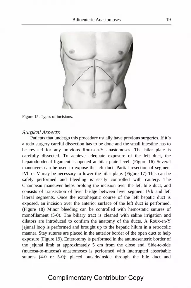
Figure 15. Types of incisions.
Surgical Aspects Patients that undergo this procedure usually have previous surgeries. If it’s
a redo surgery careful dissection has to be done and the small intestine has to
be revised for any previous Roux-en-Y anastomoses. The hilar plate is
carefully dissected. To achieve adequate exposure of the left duct, the
hepatoduodenal ligament is opened at hilar plate level. (Figure 16) Several
maneuvers can be used to expose the left duct. Partial resection of segment
IVb or V may be necessary to lower the hilar plate. (Figure 17) This can be
safely performed and bleeding is easily controlled with cautery. The
Champeau maneuver helps prolong the incision over the left bile duct, and
consists of transection of liver bridge between liver segment IVb and left
lateral segments. Once the extrahepatic course of the left hepatic duct is
exposed, an incision over the anterior surface of the left duct is performed.
(Figure 18) Minor bleeding can be controlled with hemostatic sutures of
monofilament (5-0). The biliary tract is cleaned with saline irrigation and
dilators are introduced to confirm the anatomy of the ducts. A Roux-en-Y
jejunal loop is performed and brought up to the hepatic hilum in a retrocolic
manner. Stay sutures are placed in the anterior border of the open duct to help
exposure (Figure 19). Enterotomy is performed in the antimesenteric border of
the jejunal limb at approximately 5 cm from the close end. Side-to-side
(mucosa-to-mucosa) anastomoses is performed with interrupted absorbable
sutures (4-0 or 5-0); placed outside/inside through the bile duct and
Complimentary Contributor Copy

Miguel Ángel Mercado and Julio Alfaro Varela 20
inside/outside in the jejunum. (Figure 20) The sutures are placed from left to
right and are not tied until the last suture of the posterior wall has been placed.
After the posterior wall anastomoses is finished, the anterior wall is performed
in the same manner with interrupted absorbable sutures (4-0 or 5-0). The
jejunal limb is fixed to the liver capsule and a close drain is placed. (Figure
21).
Figure 16. The hilar plate is dissected to expose the biliary tract confluence.
Figure 17. Partial resection of segment IV-V is performed to lower the hilar plate and
expose the extrahepatic course of the left hepatic duct. The incision is done parallel to
the hilar plate, between the gallbladder fossa and the round ligament. The dotted line is
the limit between segment IV and V.
Complimentary Contributor Copy

Bilioenteric Anastomoses 21
Figure 18. Once the hilar plate has been lowered and the biliary tract confluence
exposed, an incision over the anterior surface of the left hepatic duct is performed.
Complimentary Contributor Copy

Miguel Ángel Mercado and Julio Alfaro Varela 22
Figure 19. After the extrahepatic course of the left duct is opened, stay sutures are
placed to help exposition of the biliary tract lumen, and dilators are introduce to
identify the anatomy.
Complimentary Contributor Copy

Bilioenteric Anastomoses 23
Figure 20. Absorbable hydrolysable monofilament sutures (5-0) are placed on both
corners, outside/inside in the biliary tract and inside/outside in the jejunal loop.
Figure 21. Final view of the Hepp-Couinaud anastomosis with intestinal loop fixed to
the liver capsule.
Complimentary Contributor Copy

Miguel Ángel Mercado and Julio Alfaro Varela 24
To Stent Or Not to Stent a Bilioenteric Anastomoses?
Controversy exists with regards the use of stents or not in bilioenteric
anastomoses. The author doesn´t uses stents in a routinely manner.
Benefits of stents:
Preventing dehiscence by control of ductal and jejunal limb pressure.
Prevents stenosis.
Allows manipulation and/or dilation of the anastomoses in the
postoperative period.
Allows radiological control of the anastomoses.
Disadvantages of stents:
No data supporting its routine use.
Duration of stent placement is random and depends on the surgeon´s
experience.
Use of stents is cause of complications.
They cause inflammatory reaction and cause bile stasis.
When bilioenteric anastomoses are performed electively, without
cholangitis or infection, with a normal biliary tract (not dilated); stents are not
necessary. Mercado et al. [57] reported more postoperative complications in
patients with stents, including neo-formation of bile stones and complex
fistulas (arterio-biliary and bilio-pleural fistulas). There is no benefit
demonstrated with the use of biliary acids to prevent stent occlusion. [58]
The author recommends the use of stent in the following situations:
1) Thin bile ducts with diameter less than 4 mm.
2) Inflammation of the anastomosed ducts.
Complications
Bilioenteric anastomoses represent major surgical procedures and
postoperative complications are divided into early and late complications.
Many patients are acutely or chronically ill, making them more susceptible to
suffer postoperative complications. Complications after BEA may require
Complimentary Contributor Copy

Bilioenteric Anastomoses 25
reoperation and result in long-term morbidity. [59] Early postoperative
morbidity rate for BEA is 20-30% and mortality rate 0-2%. [60-63]
The most frequent early postoperative complication following BEA is
wound infection (8-12%). [62, 63] Other early complications are cholangitis
(5.7%), biloma/ intra-abdominal abscess (3.4%), biliary fistula, biliary-enteric
anastomosis dehiscence, peritonitis and death (0-2%). [62, 64, 65] There are
several factors associated to complications, such as patient’s age, co-morbid
conditions, and type of anastomosis performed, influencing outcome of BEA.
[59] Other factors that should be considered as risk factors for complications
are bile duct injuries, specifically when associated to vasculobiliary injuries.
Some postoperative complications have worst outcome and can potentially
lead to reoperation, prolong hospital stay and death. Bile leakage is one of the
major postoperative complications because is an important cause of morbidity
and extended hospital stays. [66] Bile leakage secondary to BEA is presents in
4.6% of the cases. [62] The placement of drains helps detect this complication
in the early postoperative period, allowing different treatments to prevent
major complications.
Bile leakage is defined as [67]:
a) Bile discharge from an abdominal wound and/or drain, with a total
bilirubin level of >5mg/mL or three times the serum level.
b) Intra-abdominal collections of bile confirmed by percutaneous
aspiration.
c) Cholangiographic evidence of dye leaking from the opacified bile
ducts.
Bile leaks can also be classified as minor and major. Minor bile leaks are
small, manifested as self-limited drainage of bile-containing fluid via an
external drain or as small postoperative sub-hepatic collections which resolve
spontaneously. Major bile leaks include biliary fistulas, bilomas, bile ascites or
bile peritonitis. [68] Bile drainage of more than 100 cc per day over a period of
2 weeks is unlikely to close spontaneously without manifesting long-term
complications. [68]
Many patients don´t develop a clear fistula, but show a biloma; which is a
confined collection of bile, usually in juxtaposition to the source of a bile leak.
[68] Imaging techniques as abdominal ultrasound and Computer Tomography
(CT) will show a large low density collection, its location and morphology.
[69] Bilomas can be treated by interventional radiology by percutaneous
drainage. Imaging is usually not indicated unless there is suspicion of early or
Complimentary Contributor Copy

Miguel Ángel Mercado and Julio Alfaro Varela 26
late complications developing, and the most important evaluation tool is daily
clinical assessment.
Late complications of BEA are strictures of the anastomoses. Several
imaging techniques are available to confirm patency or stricture of the
anastomosis. Radiological evaluation of the anastomosis should be performed
whenever strictures are suspected. Magnetic Resonance Cholangio-
pancreatography (MRCP) has proved to be a reliable noninvasive technique to
visualize the biliary anastomosis. [70] The disadvantage of MRCP is that it is
not easily available in many hospitals.
Katz et al. [71] described that sensitivity and specificity of MRCP was of
94.4% and 88.9%, respectively, with positive and negative predictive values of
94.4% and 89.9%, respectively. Beltrán et al. [72] had a sensitivity of 93%,
and specificity of 97.6%, with a global diagnostic accuracy of 95.6%. MRCP
is an excellent non-invasive method to evaluate BEA.
Once strictures are confirmed, Endoscopic Retrograde Cholangio-
pancreatography (ERCP) or Percutaneous Transhepatic Cholangiography is
used as a therapeutic instrument. Cantwell et al. [73] analyzed the
effectiveness of percutaneous balloon dilation (PBBD) (Figure 22) as
treatment of benign postoperative biliary strictures and the probability of a
patient not having clinically significant restenosis at 2.5 and 3 years after
primary PBBD was 0.66 and 0.56, respectively. Previous studies demonstrated
that 38%–67% of patients did not have clinically significant stenosis. [74-76].
Figure 22. (a) Stenosis of the BEA is evident through a percutaneous trans-hepatic
cholangiography. (b) Balloon dilation is being performed by interventional radiology.
(c) Control cholangiography demonstrates the patency of the BEA after balloon
dilation.
Complimentary Contributor Copy

Bilioenteric Anastomoses 27
Cholecystojejunostomy
Many long-term complications will not be observed in different BEA,
because they are performed in patients with malignant disease. Oishi et al. [77]
reported a morbidity of 32% and late complications in 15% of the cases,
including recurrent biliary stones with obstruction and anastomotic stricture;
however, despite the morbidity, 85% of patients had a successful and durable
biliary decompression during an 8 year follow-up. Cholecystojejunostomy is
not the ideal BEA for benign disease, and it should be limited to malignant
obstruction. Thomson et al. [78] described a series of patients in which all
developed cholangitis and concluded that cholelithiasis and cholangitis are
inevitable when this BEA is used for benign obstruction
Rare complications can occur and have been described as intussusception
(cause of recurrent obstructive jaundice), right upper quadrant pain,
cholangitis, or GI bleeding in patient with this BEA. [79] Bleeding varicose
have been reported as late complications of palliative biliary surgery for
chronic pancreatitis. [80, 81] Salam et al. [82] reported varices through the
cholecystojejunostomy in a patient with concomitant obstruction of the
common bile duct and the portal vein.
Cholecystojejunostomy is not recommended for definitive treatment of
benign disease. When a benign biliary obstruction is suspected, other BEA has
to be considered, due to the survival of these patients and the risk of
cholangitis and malignancy. [77, 78, 83] Gallbladder carcinoma is a possible
late complication of cholecystojejunostomy and should be remembered when
dealing with patients that had this BEA. [83] Cholecystojejunostomy has a risk
of malignancy and is another important reason why this procedure is
abandoned in benign obstruction.
Choledochoduodenostomy (CDD)
Choledochoduodenostomy (CDD) is one of the BEA that has been more
thoroughly described. Riedel was the first to describe them more than a
century ago (1892). CDD is a controversial procedure; with a lot of
complications described in the literature. Ascending cholangitis, sump
syndrome and alkaline reflux gastritis are some of the documented
complications. Although it has good long-term results in some studies, it is not
the best procedure for lower common bile duct obstruction. [84] The most
Complimentary Contributor Copy

Miguel Ángel Mercado and Julio Alfaro Varela 28
common complications are intra-abdominal abscess (26%), wound infection
(20%), and biliary leakage (13%). [44]
CDD has a morbidity of 23% and a mortality of 3%. [86] CDD risk for
cholangitis is between 0% to 12%, usually associated with stricture of the
anastomosis and remnant or recurrent stones. [86] The width of CDD
anastomoses has been of debate for some time, considered by some authors to
range between 2-2.5 cm. Stricture of the anastomoses and subsequent
development of cholangitis are the most frequently described long-term
complications, and to avoid it several authors recommend a BEA width of at
least 2. 0 cm. Several authors recommend a stoma size greater than 2.5 cm to
prevent cholangitis. [87]
Sphincter of Oddi regulates the flow of bile and pancreatic juice into the
duodenum preventing reflux of duodenal content back into the biliary tract,
function that is lost with CDD. CDD is associated to pneumobilia,
regurgitation of food debris and duodenal content into the biliary tract. [88]
Although CDD are one of the most physiological BEA allowing bile outflow
into the duodenum, most of its complications are associated to the loss of
sphincter of Oddi function.
Side-to-side CDD has great risk of developing “sump syndrome.” Sump
syndrome is defined as the accumulation of biliary and duodenal contents in a
poorly drained distal stump of the biliary tree. [89] Sump syndrome has been
reported in 0 to 9.6% of the cases. [23, 90, 91] The presence of symptoms
following food accumulation within the bile duct is what characterizes the
syndrome. [92] Complications of sump syndrome are cholangitis, pancreatitis,
hepatic abscesses, and secondary biliary cirrhosis. [92] Sump syndrome has a
low incidence and appears to be related to BEA stricture and not a true sump
syndrome. [89] Limiting the definition of sump syndrome to cholangitis and
hepatic abscess may underreport true incidence of this syndrome. [44] (Figure
23).
Treatment of sump syndrome begins with endoscopic sphincterotomy in
order to decompress the common bile duct, with good results. [93-96]
Sometimes endoscopic treatment fails due to previous heavy or multiple
stones, large food debris accumulated through the stoma that cannot be cleared
with endoscopy. [92] Caroli-Bosc et al. [93] described their experience with
patients with sump syndrome managed endoscopically and found out that 60%
had food debris and 33% biliary calculi. Surgical treatment includes Roux-en-
Y hepaticojejunostomy, with resection of the distal portion of the CBD. [92]
Complimentary Contributor Copy

Bilioenteric Anastomoses 29
Figure 23. CT scan with multiple pericholangitic abscesses and mild dilation of
intrahepatic bile ducts, suggestive of BEA stricture and ascending cholangitis.
The incidence of duodenogastric reflux (DGR) after CDD, assessed by
endoscopy and histology, ranges from 11.5% to 70%, even though dyspeptic
symptoms attributable to DGR have been reported in only 15% of patients.
The symptoms related to DGR depend not only the presence but also in the
severity of reflux; the presence of an intact pylorus in these patients could
prevent the severity of DGR. [93] Lujan-Mompean et al. evaluated DGR in
patients with cholecystectomy alone and in patients with cholecystectomy plus
CDD. This last group had higher reflux rates than patients who underwent
simple cholecystectomy, attributing it to unregulated bile flow to the
duodenum following bypass of the sphincter of Oddi and altered motility of
the pyloroduodenum due to surgical manipulation of the duodenum. [94] Most
of the patients with CDD are asymptomatic and the degree of DGR does not
necessarily produce symptoms in all patients. [93]
CDD have been associated to malignant. Reflux of food debris and
substances into the biliary tract cause changes in the biliary epithelium that
may lead to malignant transformation. [64] Eleftheriadis et al. [98] evaluated
duct mucosa in patients who had a CDD performed and found that the mucosa
showed hyperplasia, metaplastic goblet cells, and pyloric like gland formation;
changes that are also found in patients with hepatolithiasis and congenital
choledochal cyst, which have been considered as premalignant disease. [99,
100] Tocchi et al. [63] described an incidence of cholangiocarcinoma of 5.5%
which was higher in patients with CDD, compared with hepaticojejunostomy
Complimentary Contributor Copy

Miguel Ángel Mercado and Julio Alfaro Varela 30
or transduodenal sphincteroplasty (7.6% vs. 1.9% vs. 4.8%, respectively).
Patients that underwent this type of BEA are recommended to have close
follow-up in the long-term, especially if they had several episodes of
cholangitis.
Choledochojejunostomy (CJ)
Most surgeons prefer a Roux-en-Y CJ in the management of benign
biliary disease. [85] Postoperative morbidity of CJ is of 20% to 33%, with a
mortality of 0% to 2%. [74, 100, 101] CJ are more complicated and takes
longer; requiring circumferential dissection of the common bile duct and a
Roux-en-Y anastomoses. [44] Hepaticojejunostomy is preferred over this
BEA.
Hepaticojejunostomy
Hepaticojejunostomy has a morbidity rate of 19-49% and a mortality of 0-
6% for benign disease [59, 100, 102-103] The most common complications
include wound infection, cholangitis, bile leak, hemorrhage, pancreatitis,
delayed gastric emptying, cardiopulmonary complications, systemic sepsis,
renal failure, abscess formation, fistula formation, and stenosis. Factors
associated to BEA complications are low serum albumin levels and worse
American Association of Anesthesiologists (ASA) physical status. [59]
Excellent results are expected in 90% of the cases when they are performed
under expert hands.
Stricture following HJ occurs in 5% to 17%. [100, 102-104] Factors
associated to stricture formation are vasculobiliary injuries, multiple repair
attempts, biloma, external or internal biliary fistula, anastomosis in non-dilated
duct, injury at or above the level of the biliary bifurcation, preoperative and
postoperative percutaneous biliary drainage, and patient comorbidities. [74,
102] BEA strictures can be treated with endoscopic dilation, percutaneous
dilation or redo surgery. Kucukay et al. [105] described the efficacy of
percutaneous biliary balloon dilation (PBBD) of benign HJ strictures, and
found a morbidity of 5.6%. Recurrent biliary stricture presents in two thirds of
patients within 2–3 years after reconstruction, 80% of patients within 5 years,
and 90% of patients within 7 years. [60, 106-107] Biliary leakage has been
reported in 2.3% of the cases. [108] Initial management of biliary leakage may
Complimentary Contributor Copy

Bilioenteric Anastomoses 31
be with endoscopic treatment, interventional radiology and surgery in more
complex cases. (Figure 24).
Figure 24. Image (a) corresponds to a percutaneous cholangiography through a
previously placed external drainage catheter in which a biliary leakage (arrow) from
the BEA was evident. Image (b) shows a percutaneous transhepatic internal-external
catheter placed through the BEA to control the biliary fistula.
Outcome and Follow-Up
Time Interval and Duration of Follow-up
Time interval for follow-up after a bilioenteric anastomoses is of 3 months
during the first year, every 6 months during the second year and yearly
evaluation is recommended after the third year. The total minimum time
interval for follow-up is of 5 years; when most bilioenteric anastomoses tend
to dysfunction. Some author recommend follow-up for 2 to 5 years and other
groups up to 10 to 20 years. [54, 60, 64, 109-110] It is important to remember
that most patients with malignancy are not expected to reach this minimum
time interval.
Complimentary Contributor Copy

Miguel Ángel Mercado and Julio Alfaro Varela 32
Laboratory Tests and Imaging
Several biochemical parameters and imaging techniques are used during
follow-up to evaluate anastomoses patency:
Liver function tests: Liver function test elevation after bilioenteric
anastomoses is no infrequent. AST/ALT are highly sensible
biomarkers of liver damage. Bilirrubin level is closely related to
biliary obstruction. Alkaline phosphatase (AP) increase before there is
clinical evidence of jaundice. In most cases AP levels return to normal
value after bilioenteric anastomoses. AP persists high but with normal
bilirubin levels when there is partial obstruction of a liver segment.
[111] When total bilirubin and direct bilirubin levels rise, complete
obstruction has to be ruled out. Many patients that undergo
bilioenteric anastomoses never return to normal levels of AP and
don’t represent higher risk of cholangitis in the long-term.
Liver Ultrasound (US): It is an imagine technique easily available.
The disadvantage of this method is its limitation to evaluate liver
hilum due to abundant interposition of structures and gas. It is useful
to detect early complications as fluid collections. During follow-up it
may evidence dilated intrahepatic bile ducts as a sign of BEA
dysfunction.
Computer Tomography (CT): It is useful in the diagnosis of early and
late complications during the postoperative period and during follow-
up.
Magnetic Cholangioresonance: Is an excellent non-invasive method to
evaluate patency of the anastomoses and is the first option for
evaluating BEA. It has the disadvantage of not allowing any
therapeutic action in the presence of obstruction. (Figure 25).
Percutaneous cholangiography: It is used when cholangioresonance is
not available. It has the advantage of allowing direct manipulation of
the BEA if it is required with balloon dilation or drainage catheter
need to be placed to control biliary leakage or progressive dilation of
the BEA. (Figure 26)
Complimentary Contributor Copy

Bilioenteric Anastomoses 33
Figure 25. Image (a) corresponds to a T2 coronal magnetic cholangioresonance with a
patent BEA. Image (b) corresponds to a 3D cholangioresonance volumetric
reconstruction of a patent BEA with adequate biliary outflow into the jejunal limb in
which the biliary tract has normal diameter.
Figure 26. Percutaneous trans-hepatic cholangiography in which a stenotic BEA and
dilation of the intrahepatic biliary tract is evident.
Imaging studies are not routinely indicated unless suspicion of early or
late complications exists or stricture of the anastomoses needs to be ruled out
during follow-up. Imaging studies are determined according to biochemical
and clinical symptoms. Biochemical parameters are useful and are evaluated in
every check-up.
Complimentary Contributor Copy

Miguel Ángel Mercado and Julio Alfaro Varela 34
Classifications
There are several classifications used to evaluate outcome and patency of
bilioenteric anastomoses. These classifications include laboratory findings and
clinical parameters. The most frequently used classifications are the
Terblanche [110] scale (Table 2) and Mc Donald [65] classification. (Table 3).
Table 2. Terblanche Classification
Grade I Excellent Results No biliary symptoms with normal liver
function test.
Grade II Good Results Transitory symptoms, currently no
symptoms and normal liver function test.
Grade III Fair Results Clearly related symptoms requiring
medical therapy and/or deteriorating
liver function tests.
Grade IV Poor Results Recurrent stricture requiring correction
or related death.
Table 3. McDonald Classification
Grade A No clinical symptoms from the biliary tract, normal
laboratory liver function
tests.
Grade B No clinical signs, laboratory liver function tests slightly
elevated liver or periodical episodes of pain or fever.
Grade C Pain, cholangitis with the presence of fever, jaundice and
abnormal laboratory tests.
Grade D Condition requiring surgical or endoscopic correction.
Patients with Grade A and B are considered as having an excellent
outcome. These patients are amenable to follow-up twice a year to determine
their clinical symptoms and laboratory findings. [65] In the case patients
evolve to Grade C, the presence of cholangitis must be assessed, patency of
the anastomosis and liver parenchyma status through US or
cholangioresonance. The need for hospital stay and endoscopic, radiological or
surgical treatment has to be evaluated. Grade C and D are considered as poor
outcome; therefor, closer follow-up is recommended in this group. Follow-up
is mandatory in all patients that undergo bilioenteric anastomoses because
Complimentary Contributor Copy

Bilioenteric Anastomoses 35
60% of therapeutic failure occurred in the first 3 years and 80% during the first
5 years. [106, 107]
Three scenarios may occur in patients with Grade III or Grade C:
1) Recurrent episodes of cholangitis, abnormal liver function tests,
normal liver parenchyma and a patent anastomosis. These cases are
treated with antibiotics and ursodesoxycolic acid.
2) Episodes of cholangitis, abnormal liver function tests, especially AP,
dilated intrahepatic bile ducts and stricture of the anastomosis.
Treatment of choice is ERCP and/or interventional radiology and as
last resource redo surgery.
3) Recurrent episodes of cholangitis, pericholangitic abscesses and
stricture of the anastomosis. Treatment may begin with antibiotics,
ERCP and/or interventional radiology, redo surgery with possible
hepatectomy if less invasive strategies fail.
Good long-term results can be achieved in 70-90% of the cases. [112] The
John Hopkins group reported a series of 142 patients with a success rate of
90.8% at 5 year follow-up [64] Other authors report successful results between
80-90% of the cases at 5 year follow-up. [113] The authors experience with
355 patients reported success in 94% of the cases (most of them secondary to
iatrogenic bile duct injury). An important factor associated to successful
results is the experience of the surgeon and patients that require complex
biliary surgery and reconstruction should be referred to a specialized center.
Two-third of strictures will appear during the first 2 years and 90% during
the first 5 years. [106] The authors recommendation for treatment of recurrent
strictures is to begin with less invasive measures to dilate the anastomoses.
When strictures occur, endoscocopic or transhepatic balloon dilation with stent
placement is effective. Costamagna et al. [114] proposed progressive
endoscopic dilation of the stenosis with placement of multiple stents. Patients
that will benefit with endoscopic treatment are the ones diagnosed soon after
surgery and have better outcome than those who develop strictures after
surgery. [115] Strictures in the proximal segments of the biliary tract are more
difficult to treat and require surgical treatment. [115] Davids et al. [116]
performed a comparative study between surgery and endoscopic treatment,
reporting similar long-term success with recurrence in 17% of patients.
Percutaneos treatment require multiple sessions of balloon dilation and long-
term placement of stents, [117] with morbidity associated to bleeding and bile
leak up to 40%. Twenty percent of patients may eventually require redo
Complimentary Contributor Copy

Miguel Ángel Mercado and Julio Alfaro Varela 36
surgery. [118]Pitt et al. [74] compared surgical repair (choledochojejunostomy
vs. Hepaticojejunostomy) with percutaneous balloon dilation, reporting
patency rates of 88% and 55% at 5 years for the surgical reconstruction group
and percutaneous treatment.
Quality of Life
World Health Organization defines health as “a state of complete physical,
mental, and social well-being, and not merely the absence of disease.”[119]
Scarce information is available in the literature about Quality Of Life (QOL)
after BEA. A general idea exists that patients that have a BEA will have a
worst QOL; but no data is available and most information is related to surgical
outcome after biliary tract reconstruction secondary to iatrogenic bile duct
injury. The John Hopkins group presented a study assessing the QOL between
patients who underwent surgical reconstruction of bile duct injuries and
laparoscopic cholecystectomy. Their study evaluated 3 domains (Physical,
psychological and social) and demonstrated lower scores in the psychological
domain in patients with bile duct reconstruction (p<0.05). [120] Boema et al.
[121] study had reduced QOL scores in the physical and mental domains;
independently from the type of treatment (surgery vs. endoscopic) and the
severity of the injury. De Reuver et al. [122] demonstrated a worse QOL in
patients involved in litigations.
References
[1] Beal JM. Historical perspective of gallstone disease. Surg Gynecol
Obstet 1984; 158: 181-189
[2] Hardy KJ. Carl Langenbuch and the Lazarus Hospital: events and
circumstances surrounding the first cholecystectomy. Aust N Z J Surg
1993; 63: 56-64.
[3] van Gulik TM. Langenbuch's cholecystectomy, once a remarkably
controversial operation. Neth J Surg. 1986; 38: 138-141.
[4] Braasch JW. Historical perspectives of biliary tract injuries. Surg Clin
North Am. 1994; 74: 731-740.
[5] Blumgart LH. Hilar and intrahepatic biliary enteric anastomosis. Surg
Clin North Am. 1994; 74: 845-863
Complimentary Contributor Copy

Bilioenteric Anastomoses 37
[6] Gupta DK, Rohatgi M. Use of appendix in biliary atresia. Indian J
Pediatr. 1989 Jul-Aug; 56(4):479-82.
[7] Mouret P. From the first laparoscopic cholecystectomy to the frontiers of
laparoscopic surgery: the prospective futures. Dig Surg 1991; 8: 124.
[8] Barralet JE, Wallace LL, Strain AJ. Tissue engineering of human biliary
epithelial cells on polyglycolic acid/polycaprolactone scaffolds
maintains long-term phenotypic stability. Tissue Eng. 2003
Oct;9(5):1037-45
[9] Miyazawa M, Torii T, Toshimitsu Y, Okada K, Koyama I, Ikada Y. A
tissue-engineered artificial bile duct grown to resemble the native bile
duct. Am J Transplant. 2005 Jun;5(6):1541-7
[10] Ferraina P, Merello Laurdies J. Anastomosis biliodigestivas. Cirugía
digestiva 2009; (IV); 461, pág. 1-14
[11] Buckwalter JA, Lawton RL, Tidrick RT. Bypass operation for neoplasic
biliary tract obstruction. Am J Surg 1965: 109-100-6.
[12] Hess W, Berei G. Textbook of Bilio-Pancreatic Diseases, pathology,
diagnosis, medical treatment, surgical technique and tactics.1st Edition.
(Vol III) PICCIN 1997, Padua, Italy.
[13] Khan T, Lwin M, Ulah S. Bilio-enteric anastomoses: Results in benign
and malignant conditions. Singapore Med J 1993; Vol 34: 545-550.
[14] Sarfeh IJ, Rypins EB, Jakowatz JG et al. A prospective, randomized
clinical investigation of cholecystoenterostomy and
choledochoenterostomy. Am J Surg 1988; 155:411
[15] Watanapa P, Williamson RC. Surgical palliation for pancreatic cancer:
developments during the past two decades. Br J Surg 1992; 79:8
[16] Urbach DR, Bell CM, Swanstrom LL et al. Cohort study of surgical
bypass to the gallbladder or bile duct for the palliation of jaundice due to
pancreatic cancer. Ann Surg 2003; 237:86.
[17] House MG, Choti MA. Palliative therapy for pancreatic/biliary cancer.
Surg Clin North Am 2005; 85:359.
[18] Siquini W. Surgical Treatment of Pancreatic Diseases. Springer –
Verlag, Italy 2009. Pg. 456.
[19] Ravindra SD, Siriwardena AK. Laparoscopic biliary bypass and current
management algorithms for palliation of malignant obstructive jaundice.
Ann Surg Oncol 2004 11:815
[20] Degenshein GA. Choledochoduodenostomy: an 18 year study of 175
consecutive cases. Surgery. 1974; 76:319-324.
[21] Capper WM. External choledochoduodenostomy. An evaluation of 125
cases. Br J Surg. 1961; 49:292-300.
Complimentary Contributor Copy

Miguel Ángel Mercado and Julio Alfaro Varela 38
[22] Wright NL. Evaluation of the results of choledochoduodenostomy. Br J
Surg. 1968; 55:33-36.
[23] Madden JL, Chun JY, Kandalaft S, Parekh M. Choledochoduo-
denostomy: an unjustly maligned surgical procedure? Am J Surg 1970;
119:45-54.
[24] Johnson AG, Rains AJ. Prevention and treatment of recurrent bile duct
stones by choledochoduodenostomy. World J Surg. 1978; 2:487-496.
[25] Lygidakis NJ. Choledochoduodenostomy in calculous biliary tract
disease. Br J Surg. 1981; 68:762-765.
[26] Baker A.R et al. Sump Syndrome following choledochoduodenostomy
and its Endoscopic treatment. Br J Surg. 1985, 72: 433-435
[27] Jones SA. et al. Sphincteroplasty in the treatment of biliary tract disease.
Surg. Clin. N. Am. 1983, 53: 1123-37.
[28] Baker AR, Neoptolemos JP, Leese T, Fossard DP. Choledochoduo-
denostomy, transduodenal sphincteroplasty and sphincterotomy for
calculi of the common bile duct. Surg Gynecol Obstet. 1987; 164:245-
251.
[29] Tang CN, Siu WT, Ha JP, Li MK. Laparoscopic choledochoduo-
denostomy: an effective drainage procedure for recurrent pyogenic
cholangitis. Surg Endosc. 2003; 17:1590-1594.
[30] Jeyapalan M. Almeida JA, Michaelson RL, Franklin ME Jr.
Laparoscopic choledochoduodenostomy: Review of a 4 year experience
with an uncommon problem. Surg Laparosc Endosc Percutan Tech
2002;12:148-53.
[31] Chander J, Mangla V, Vindal A, Lal P, Ramteke VK. Laparoscopic
choledochoduodenostomy for biliary stone disease: a single-center 10-
year experience. J Laparoendosc Adv Surg Tech A. 2012 Jan-Feb;
22(1):81-4.
[32] Artifon EL, Aparicio D, Paione JB, Lo SK, Bordini A, Rabello C, Otoch
JP, Gupta K. Biliary drainage in patients with unresectable, malignant
obstruction where ERCP fails: endoscopic ultrasonography-guided
choledochoduodenostomy versus percutaneous drainage. J Clin
Gastroenterol. 2012 Oct; 46(9):768-74.
[33] de Almeida AC, dos Santos NM, Aldeia FJ. Choledochoduodenostomy
in the management of common duct stones or associated pathology—an
obsolete method? HPB Surg. 1996; 10:27–33.
[34] Miros M, Kerlin P, Strong R, et al. Post-choledochoenterostomy “sump
syndrome”. Aust N Z J Surg. 1990;60:109–112
Complimentary Contributor Copy

Bilioenteric Anastomoses 39
[35] Venerito M, Fry LC, Rickes S, et al. Cholangitis as a late complication
of choledochoduodenostomy: the sump syndrome. Endoscopy.
2009;41(suppl 2):E142–E143
[36] Ajaz A Malik, Shiraz A Rather, Shams UL Bari and Khursheed Alam
Wani. Long-term results of choledochoduodenostomy in benign biliary
obstruction. World J Gastrointest Surg. 2012 February 27; 4(2): 36-40.
[37] Fisher J. Mastery of Surgery. 6th Edition. Philadelphia; USA: Lippincott
Williams and Wilkins; 2012.
[38] Tang CN, Siu WT, Ha JP, Tai CK, Tsui KK, Li MK. Laparoscopic
biliary bypass—a single centre experience. Hepatogastroenterology
2007;54:503–507.
[39] Cuschieri A, Adamson GD: Multimedia article. Laparoscopic
transection choledochoduodenostomy. Surg Endosc 2005, 19(5):728.
[40] Marangoni G, Ali A, Faraj W, Heaton N, Rela M: Clinical features and
treatment of sump syndrome following hepatico-
jejunostomy. Hepatobiliary, 10(3):261–264.
[41] Morrissey PE, Burns GA, Cohn SM. Sump syndrome complicating
Roux-en-Y hepaticojejunostomy: case report and review of the
literature. Surgery 1996, 119(4):403–405.
[42] Suo T, Song LJ, Tong SX: Gallstone in jejunal limb with jejunocolonic
fistula 10 years after Roux-en-Y choledochojejunostomy. World J of
Radiology, 3(1):38–40.
[43] Chida T, Kageyama F, Yamada M, Yoshii S, Honjo Y, Takai T, Iwaoka
Y, Terai T, Kagami T, Yamazaki S et al. A case of hemorrhage from
varices of an interposed jejunum after choledochojejunostomy treated
successfully by endoscopic injection using alpha-cyanoacrylate
monomer. Nippon Shokakibyo Gakkai Zasshi 1661, 107(10):1661–1668.
[44] Leppard WM, Shary TM, Adams DB and Morgan K. Choledochoduo-
denostomy: Is It Really So Bad? J Gastrointest Surg 2011; 15 (5) 754-7
[45] Clavien PA. Atlas of upper gastrointestinal and hepato-pancreato-
biliary surgery. New York: Springer; 2007.
[46] Fletcher DR, Hobbs MST, Tan P, et al: Complications of
cholecystectomy: Risks of the laparoscopic approach and protective
effects of operative cholangiography. A population-based study. Ann
Surg 229:449-457, 1999
[47] Sarmiento J. Hepaticojejunostomy: Indications and Surgical Technique.
Operative Techniques in General Surgery, Vol 2, No 4 (December),
2000: pp 295-303
Complimentary Contributor Copy

Miguel Ángel Mercado and Julio Alfaro Varela 40
[48] Diao M, Li L, Cheng W. Laparoscopic versus Open Roux-en-
Y hepatojejunostomy for children with choledochal cysts: intermediate-
term follow-up results. Surg Endosc. 2011 May; 25(5):1567-73.
[49] Hepp J, Couinaud C. L´abord et l´utilisation du canal hepatique dans le
reparations de la voie biliare principale. Presse Med 1956; 64: 947
[50] Guglielmi A, Ruzzenente A, Iacono C. Surgical Treatment of Hilar and
Intrahepatic Cholangiocarcinoma. Springer-Verlag Italia 2008.
[51] Myburgh JA. The Hepp-Couinaud Approach to Strictures of the Bile
Ducts I. Injuries, Choledochal Cysts,and Pancreatitis. Annals of Surgery
1993; Vol. 218, No. 5, 615-620.
[52] Singhal D, van Gulik TM, Gouma DJ. Palliative management of hilar
cholangiocarcinoma. Surg Oncol 2005; 14(2):59–74
[53] Connors S, Wigmore SJ, Madhavan KK et al. Surgical palliation for
unresectable hilar cholangiocarcinoma. HPB Surg 2005; 7:273–277.
[54] Tochi A, Costa G, Lepre L, et al. The long-term outcome of
hepaticojejunostomy in the Treatment of bening bile duct strictures. Ann
Surg 1996; 224(2): 162-167.
[55] Traverso LW. The Longmire I, II, and III operations. Am J Surg. 2003
May, Volume 185, Issue 5
[56] Winslow E, Fialkowski E, Linehan D, Hawkins W, Picus D, Strasberg S.
“Sideways”: Results of Repair of Biliary Injuries Using a Policy of Side-
To-Side Hepatico-Jejunostomy. Ann Surg, Vol 249 (3): 2009; 426-434.
[57] Mercado MA, Chan C, Orozco H, Cano-Gutiérrez G, Chaparro JM,
Galindo E, Vilatobá M, Samaniego-Arvizu G. To stent or not to stent
bilioenteric anastomosis after iatrogenic injury: a dilemma not
answered? Arch Surg 2002; 137: 60-63
[58] De Ledinghen VPerson BLegoux JL et al. Prevention of biliary stent
occlusion by ursodeoxycholic acid plus norfloxacin: a multicenter
randomized trial. Dig Dis Sci.2000;45145- 150
[59] Zafar S, Rizwan M, Raza R, N Khan M, Kasi M. Early complications
after biliary enteric anastomosis for benign diseases: A retrospective
analysis. BMC Surgery 2011, 11:19.
[60] Hall JG, Pappas TN. Current management of biliary strictures. J
Gastrointest Surg 2004; 8: 1098-1110.
[61] Ahrendt SA, Pitt HA. Surgical therapy of iatrogenic lesions of biliary
tract. World J Surg 2001; 25: 1360-1365.
[62] Sicklick JK, Camp MS, Lillemoe KD, Melton GB, Yeo CJ, Campbell
KA, Talamini MA, Pitt HA, Coleman J, Sauter PA, Cameron JL.
Surgical management of bile duct injuries sustained during laparoscopic
Complimentary Contributor Copy

Bilioenteric Anastomoses 41
cholecystectomy: perioperative results in 200 patients. Ann Surg 2005;
241: 786-792; discussion 793-795.
[63] Tocchi A, Mazzoni G, Liotta G, Lepre L, Cassini D, Miccini M. Late
development of bile duct cancer in patients who had biliary-enteric study
of more than 1,000 patients. Ann Surg 2001, 234(2):210-214.
[64] Lillemoe KD, Melton GB, Cameron JL, Pitt HA, Campbell KA,
Talamini MA, Sauter PA, Coleman J, Yeo CJ. Postoperative bile duct
strictures: management and outcome in the 1990s. Ann Surg 2000; 232:
430-441.
[65] McDonald ML, Farnell MB, Nagorney DM, Ilstrup DM, Kutch JM.
Benign biliary strictures: repair and outcome with a contemporary
approach. Surgery 1995; 118: 582-590; discussion 590-591.
[66] Shimizu T, Yoshida H, Mamada Y, Taniai N, Matsumoto S, Mizuguchi
Y, Yokomuro S, Arima Y, Akimaru K, Tajiri T. Postoperative bile
leakage managed successfully by intrahepatic biliary ablation with
ethanol. World J Gastroenterol 2006 June 7; 12(21): 3450-3452.
[67] Kapoor S, Nundy S. Bile Duct Leaks from the Intrahepatic Biliary Tree:
A Review of Its Etiology, Incidence, and Management. HPB Surgery
2012, Article ID 752932
[68] Mongestern L, Berci G, Pasternak E. Bile leakage after biliary tract
surgery. A laparoscopic perspective. Surg Endosc 1993; 7: 432-438.
[69] Kapoor V, Baron R, Peterson M. Bile Leaks After Surgery. AJR 2004;
182:451–458.
[70] Pecchi A, De Santis M, Gibertini, Tarantino G, Gerunda G, Torricelli P,
Di Benedetto F. Role of Magnetic Resonance Imaging in the Detection
of Anastomotic Biliary Strictures After Liver Transplantation.
Transplantation Proceedings 2011; 43, 1132–1135.
[71] Katz LH, Benjaminov O, Belinki A, Geler A, Braun M, Knizhnik M,
Aizner S, Shaharabani E, Sulkes J, Shabtai E, Pappo O, Atar E, Tur-
Kaspa R, Mor E, Ben-Ari Z. Magnetic resonance
cholangiopancreatography for the accurate diagnosis of biliary
complications after liver transplantation: comparison with endoscopic
retrograde cholangiography and percutaneous transhepatic
cholangiography – long-term follow-up. Clin Transplant 2010: 24:
E163–E169.
[72] Beltrán MM, Marugán RB, Oton E, Blesa C, Nun˜o J. Accuracy of
magnetic resonance cholangiography in the evaluation of late biliary
complications after orthotopic liver transplantation. Transplant Proc
2005: 7: 3924.
Complimentary Contributor Copy

Miguel Ángel Mercado and Julio Alfaro Varela 42
[73] Cantwell C, Pena C, Gervais D, Hahn P, Dawson S, Mueller P. Thirty
Years’ Experience with Balloon Dilation of Benign Postoperative
Biliary Strictures: Long-term Outcomes. Radiology 2008 (3): 249.
[74] Pitt HA, Kaufman SL, Coleman J, White RI, Cameron JL. Benign
postoperative biliary strictures: operate or dilate? Ann Surg 1989;
210:417–425.
[75] Williams HJ, Bender CE, May GR. Benign postoperative biliary
strictures: dilation with fluoroscopic guidance. Radiology 1987; 163:
629–634.
[76] Mueller PR, vanSonnenberg E, Ferrucci JT, et al. Biliary stricture
dilation: multicenter review of clinical management in 73 patients.
Radiology 1986; 160:17–22.
[77] Oishi AJ, Sarr MG, Nagorney DM, Traynor MD. Long-Term Outcome
of Cholecystoenterostomy as a Definitive Biliary Drainage Procedure for
Benign Disease. World J. Surg 1995; 19: 616-620, 1995.
[78] Thomson SR. Sequelae of cholecystojejunostomy for benign
intrapancreatic biliary strictures. S Afr Surg 1998;36(1):26–29.
[79] Porter DH, Kim D, Austin RM. Cholecystojejunostomy Intussusception.
Gastrointest Radiol 13:227-229 (1988).
[80] Carpenter S, Brown KA. Chronic complications after
cholecystojejunostomy. Am J Gastroenterol 1994; 89: 2073-2075.
[81] Miller JT Jr, De Odorico I, Marx MV. Cholecystojejunostomy varices
demonstrated by enteroclysis. Abdom Imaging 1997;22: 474-476.
[82] Salam AA, Goldman M, Smith D, et al. Gastric, intestinal, and
gallbladder varices: hemodynamic and therapeutic considerations. South
Med J 1979; 72:402–408.
[83] Pilgrim CHC, Satgunaseelan L, Ward SM, Evans PM. Gallbladder
Carcinoma as a Long-Term Complication of Cholecystojejunostomy. J
Gastrointest Surg (2009) 13:2330–2332.
[84] Malik AA, Rather S, Bari S, Wani KA. Long-term results of
choledochoduodenostomy in benign biliary obstruction. World J
Gastrointest Surg 2012 February 27; 4(2): 36-40.
[85] de Aretxabala X, Bahamondes JC. Choledochoduodenostomy for
common bile duct stones. World J Surg. 1998; 22:1171.
[86] Panis Y, Fagniez PL, Brisset D, Lacaine F, Levard H, Hay JM. Long
term results of choledochoduodenostomy versus choledochojejunostomy
for choledocholithiasis. The French Association for Surgical Research.
Surg Gynecol Obstet. 1993 Jul; 177(1):33-7.
Complimentary Contributor Copy

Bilioenteric Anastomoses 43
[87] Gupta BS. Choledochoduodenostomy: a study of 28 consecutive cases.
Kathmandu University Medical Journal (2003) Vol. 2, No. 3, Issue7,
193 – 197.
[88] LING Xiao-feng, XU Zhi, WANG Li-xin, HOU Chun-sheng, XIU Dian-
rong, ZHANG Tong-lin and ZHOU Xiao-si. Long-term outcomes of
choledochoduodenostomy for hepatolithiasis. Chinese Medical Journal
2010; 123(2):137-141.
[89] Khajanchee YS, Cassera MA, Hammill CW, Swanström L, Hansen P.
Outcomes Following Laparoscopic Choledochoduodenostomy in the
Management of Benign Biliary Obstruction. J Gastrointest Surg (2012)
16:801–805.
[90] De Almeida A, Cruz A, Aldeia F. Side to side choledochodenostomy in
the management of cholelithiasis and associated disease: facts and
fiction. American Journal of Surgery 1984; 147: 253–259.
[91] Baker A, Neoptolemos J, Leese T et al. Long term follow-up of patients
with side to side choledochoduodenostomy and transduodenal
sphincteroplasty. Annals of the Royal College of Surgeons of England.
1987; 68: 253–257.
[92] Qadan M, Clarke S, Morrow S, Triadafilopoulos G, Visser B. Sump
Syndrome as a Complication of Choledochoduodenostomy. Dig Dis Sci
(2012) 57:2011–2015
[93] Caroli-Bosc FX, Demarquay JF, Peten EP, et al. Endoscopic
management of sump syndrome after choledochoduodenostomy:
retrospective analysis of 30 cases. Gastrointest Endosc. 2000; 51:180.
[94] Siegel JH. Duodenoscopic sphincterotomy in the treatment of the ‘‘sump
syndrome’’. Dig Dis Sic. 1981; 26:922.
[95] Hallstone A, Triadafilopoulos G. Spontaneous sump syndrome:
successful treatment by duodenoscopic sphincterotomy. Am J
Gastroenterol. 1990; 85:1518.
[96] Marbet UA, Stalder GA, Faust H, Harder F, Gyr K. Endoscopic
sphincterotomy and surgical approaches in the treatment of the ‘sump
syndrome’. Gut. 1987; 28:142.
[97] Eleftheriadis E, Tzioufa V, Kotzampassi K, Aletras H. Common bile
duct mucosa in choledochoduodenostomy patients— histological and
histochemical study. HPB Surg. 1988; 1:15–20.
[98] Nakamura Y, Ohta G, Nagakawa T, Matsubara F. Pathological findings
of hepatolithiasis. Jpn J Gastroenterol. 1981; 78:874–882.
Complimentary Contributor Copy

Miguel Ángel Mercado and Julio Alfaro Varela 44
[99] Komi N, Tamura T, Tsuge S, et al. Relation of patient age to
premalignant alterations in choledochal cyst epithelium. J Pediatr Surg.
1986; 21:430–433.
[100] Rothlin MA, Lopfe M and Largiader F. Long-Term Results of
Hepaticojejunostomy for Benign Lesions of the Bile Ducts. The
American Journal of Surgery. 1998; 1: 22–25.
[101] Ross CB, H’Doubler WZ, Sharp KW et al. Recent experience with
benign biliary strictures. American Surgeon. 1989; 1: 64–70.
[102] Schmidt SC, Langrehr JM, Hintze RE, Neuhaus P. Long-term results
and risk factors influencing outcome of major bile duct injuries
following cholecystectomy. Br J Surg 2005; 92:76–82.
[103] Mercado MÁ, Franssen B. Transition from a low: to a high-volume
centre for bile duct repair: changes in technique and improved outcome.
HPB 2011; 13:767–773.
[104] House M, Cameron J, Schulick R. Incidence and outcome of biliary
strictures after pancreaticoduodenectomy. Annals of Surgery 2006 vol.
243, no. 5, pp. 571–576.
[105] Kucukay F, Okten R, Yurdakul M, Ozdemir E, Erat S, Parlak E,
Disibeyaz S, Ozer I, Bostanci EB, Olcer T, Tola M. Long-term Results
of Percutaneous Biliary Balloon Dilation Treatment for Benign
Hepaticojejunostomy Strictures: Are Repeated Balloon Dilations
Necessary? J Vasc Interv Radiol 2012; 23:1347–1355.
[106] Pitt HA, Miyamoto T, Parapatis SK, Tompkins RK, Longmire WP Jr.
Factors influencing outcome in patients with postoperative biliary
strictures. Am J Surg 1982; 144:14–21.
[107] Pellegrini CA, Thomas MJ, Way LW. Recurrent biliary stricture.
Patterns of recurrence and outcome of surgical therapy. Am J Surg 1984;
147:175–180.
[108] de Castro S, Kuhlmann K, Busch O, van Delden O, Lameris J, van Gulik
T, Obertop H, Gouma D. Incidence and management of biliary leakage
after hepaticojejunostomy. J Gastrointest Surg 2005;9:1163–1173.
[109] Lillemoe KD, Martin SA, Cameron JL, Yeo CJ, Talamini MA, Kaushal
S, Coleman J, Venbrux AC, Savader SJ, Osterman FA, Pitt HA. Major
bile duct injuries during laparoscopic cholecystectomy. Follow-up after
combined surgical and radiologic management. Ann Surg 1997; 225:
459-468; discussion 468-471.
[110] Terblanche J, Worthley CS, Spence RA, Krige JE. High or low
hepaticojejunostomy for bile duct strictures? Surgery 1990; 108: 828-
834.
Complimentary Contributor Copy

Bilioenteric Anastomoses 45
[111] Rege RV. Adverese effects of biliary obstruction: Implications for
treatment of patients with obstructive jaundice. AJR 1995; 164: 287-93.
[112] Schmidt SC, Langrehr JM, Hintze RE, Neuhaus P. Longterm results and
risk factors influencing outcome of major bile duct injuries following
cholecystectomy. Br J Surg 2005; 92:76-82.
[113] Bauer TW, Morris FB, Lowenstein A, et al. The consequences of major
bile duct injury during laparoscopic cholecystectomy. J Gastrointest
Surg 1998; 2: 61-66.
[114] Costamagna G, Pandolfi M, Mutignani M, Spada C, Perri V. Long-term
results of endoscopic management of postoperative bile duct strictures
with increasing numbers of stents. Gastrointest Endosc. 2001;54:162–
168.
[115] Jacques J. G. H. M. Bergman, Lotje Burgemeister, Marco J. Bruno, Erik
A. J. Rauws, Dirk J. Gouma, Guido N. J. Tytga, Kees Huibregtse. Long-
term follow-up after biliary stent placement for postoperative bile duct
stenosis. Gastrointestinal endoscopy 2001; (54): 154-161.
[116] Davids PH, Tanka AK, Rauws EA, van Gulik TM, van Leeuwen DJ, de
Wit LT, et al. Benign biliary strictures. Surgery or endoscopy? Ann Surg
1993; 217:237-43.
[117] Jan YY, Chen MF, Hung CF. Balloon dilatation of intrahepatic duct and
biliary-enteric anastomosis strictures. Long term results. Int Surg
1994;79:103-5.
[118] Vos PM, van Beek EJ, Smits NJ, Rauws EA, Gouma DJ,Reeders JW.
Percutaneous balloon dilatation for benign hepaticojejunostomy
strictures. Abdom Imaging 2000; 25 :134-8.
[119] Preamble to the Constitution of the World Health Organization as
adopted by the International Health Conference, New York, 19–22 June,
1946; signed on 22 July 1946 by the representatives of 61 States
(Official Records of the World Health Organization, no. 2, p. 100) and
entered into force on 7 April 1948.
[120] Melton GB, Lillemoe KD, Cameron JL, Sauter PA, Coleman J, Yeo CJ.
Major bile duct injuries associated with laparoscopic cholecystectomy:
Effect of surgical repair on quality of life. Annals of surgery 2002;
235:888-895.
[121] Boerma D, Rauws EAJ, Keulemans YLA, et al. Impaired quality of life
5 years after bile duct injury during the laparoscopic cholecystectomy: A
prospective analysis. Ann Surg 2001; 234:750–757.
Complimentary Contributor Copy

Miguel Ángel Mercado and Julio Alfaro Varela 46
[122] deReuver P, Sprangers M, Rauws E, et al. Impact of bile duct injury
after laparoscopic cholecystectomy on quality of life: a longitudinal
study after multidisciplinary treatment. Endoscopy 2008;40(8):637–43.
Complimentary Contributor Copy
In: Anastomoses ISBN: 978-1-62618-657-6
Editors: F. King, McKineley A. Malloy © 2013 Nova Science Publishers, Inc.
Chapter II
Intestinal Anastomosis
Education and Training
José C. Manuel-Palazuelos, Federico Castillo,
Carlos Gavilanes, Manuel Gómez-Fleitas
and Juan C. Rodríguez-Sanjuán Department of General Surgery, University Hospital Marqués de
Valdecilla, University of Cantabria, Santander, Spain
Abstract
The aim of a gastro-intestinal suture is to provide a hermetic closure
in the intestine or in an anastomosis.
To achieve a successful suture, a proper technique is essential, with
strict adherence to surgical principles, such as suture tension, border
vascularization and intestinal diameter. Also the patient biological
condition has to be considered. All of these factors influence suture
healing but the experience and skill of the surgeon are probably most
important in the final outcome of the anastomosis.
Until recently the learning of any surgical procedure was based on
direct operation on the patient, with initial supervision by an experienced
surgeon. This has several drawbacks such as risks for the patient, long
learning curves and an increase in operating room costs because of
greater operation times. Laparoscopic procedures need even longer and
more complex training periods due to the lack of tactile sensation and
two-dimension view. The problem is even greater in the case of residents
Complimentary Contributor Copy

J. C. Manuel-Palazuelos, F. Castillo, C. Gavilanes et al. 48
who are less experienced in surgery in general. To speed up learning and
avoid direct training on patients, training laboratories have been designed,
where physical and virtual reality simulators can be used.
The usefulness of training using simulation in basic surgical
techniques has been shown in improving general surgical skills and
performance of intestinal anastomosis with synthetic materials. Training
in intestinal anastomosis using dead animal viscera is very similar to the
clinical setting and has advantages over other options such as live
animals, simulators or corpses. The training on live animals reproduces
real clinical settings although it has drawbacks such as high costs or the
sacrifice of the animal. Cadaver surgery is also very similar to real
clinical settings but is hardly available. Virtual reality is very different
from real clinical settings and evidence for validation of most designed
devices is lacking. These models could be used as the first contact with
laparoscopic training, according to the conclusions of the systematic
reviews published to date. However, the advantage in resident training
with some laparoscopic experience has not been shown. On the other
hand these systems are expensive, although less so than direct training on
patients.
As a result, the aim of this work is to expose which is the best
training method in every anastomosis type and the influence that the
different simulators or training with animals have on learning surgical
skills.
Introduction
Postgraduate training in surgery lasts currently 5 or more years after
graduation. Such teaching occurs in hospital based residency programs [1].
The concept of surgical residency in a hospital is attributed to William S.
Halsted which was one of his first achievements at The Johns Hopkins
Hospital. He designed a training program for surgeons based primarily on the
German, Austrian, and Swiss models. The Germanic system of training for
young surgeons consisted of assistants spending many years in a university
surgical clinic who eventually achieved the position of first assistant to the
professor. Halsted introduced a system in which the medical school graduates
entered into a university sponsored, hospital-based surgical training program
that, over a several-year period of increasing responsibility, slowly led to the
training of young surgeons who were well versed in anatomy, pathology,
bacteriology, and physiology.
The training program culminated in a final period of near-total
independence and autonomous activity. This eventually made the surgical
Complimentary Contributor Copy

Intestinal Anastomosis Education and Training 49
residency training programs as we know them today [2, 3]. Surgical training
has undergone dramatic changes in recent years. Knowledge development and
new technologies have led to the gradual increase in the surgical therapeutic
arsenal, so that the general surgeon extensively trained, able to treat a wide
variety of processes in multiple organ systems, has become a rarity. Over the
past 30 years, general surgery has given way to several subspecialties [1].
For many procedures, the observed associations between hospital volume
and operative mortality are largely mediated by surgeon volume. Patients can
often substantially improve their chances of survival, even in high-volume
hospitals, by selecting surgeons who perform the operations frequently. The
key mechanism could simply be “practical” clinical judgment and technical
skill that are achieved only by surgeons who perform a specific procedure with
sufficient frequency [4].
Currently, many graduate residents in general surgery choose to receive
additional training in a subspecialty, before starting independent exercise.
Many residents, particularly in university hospital and reference system
programs, contact almost exclusively with subspecialists during their
residence. Since more interventions are performed by young specialists after
residency training and fellowships, the training achieved during the residence
becomes increasingly less complete. Consequently, many residents believe
that training in general surgery in the current format does not prepare them
properly for practice, and therefore they feel compelled to find additional
training. There is now a generalized concept among members of the
profession, that graduates in residency programs are not as capable as those of
a generation ago [1].
Until recently, learning surgical procedure was based on the operation
realization, initially monitored, on the patients themselves. This implies a long
learning curve with increased morbidity, and possibly mortality, and worse
long-term outcomes [5]. Today the residents are much more supervised and
have less exposure to the operating room environment. Also, current trainees
have less independent surgical experience because of pressures for greater
operating room efficiencies and the shorter workweek for residents due to the
introduction of European directives on working time seeking the conciliation
of work and family life. All of these have led to a reassessment of training
methods [6, 7, 8].
Much of the problem of inexperience could be solved by simulating
clinical scenarios. Residents could learn technical skills and gain experience in
decision-making models, in safe simulated environments but the clinical
simulation tools today remain relatively expensive and under development.
Complimentary Contributor Copy

J. C. Manuel-Palazuelos, F. Castillo, C. Gavilanes et al. 50
Nevertheless, simulation promises to transform completely surgical education
in the years ahead [1].
The ability to acquire surgical skills requires consistent practice, and
evidence suggests that many of these technical skills can be learned out of the
operating theater [9, 10, 11, 12]. Surgical simulation offers the opportunity for
trainees to practice their surgical skills prior to entering the operating room,
allowing detailed feedback and objective assessment of their performance.
This enables better patient safety and standards of care. Surgical simulators
can be divided into organic or inorganic types. Organic simulators, consisting
of live animal and fresh human cadaver models, are considered to be of high-
fidelity. Inorganic types comprise virtual reality simulators and synthetic
bench models. Current evidence suggests that skills acquired through training
with simulators positively transfers to the clinical setting and improves
operative outcome. The major challenge for the future revolves around
understanding the value of this new technology and developing an educational
curriculum that can incorporate surgical simulators [13].
While there may be compelling reasons to reduce reliance on patients,
cadavers, and animals for surgical training, none of the methods of simulated
training (including computer simulation) has yet been shown to be better than
other forms of surgical training. In addition, little is known about the real costs
(including adverse outcomes in patients) of either simulated or standard
surgical training [14].
Manually and Stapled Bowel Anastomosis
Within the past 200 years, gastrointestinal anastomosis has been
transformed from a dangerous venture into a safe and routinely performed
procedure. Among these advances was the transition to scientifically-based
medicine, chiefly the knowledge of the importance of serosa apposition
introduced by Lembert [15]. The French physician and surgeon Antoine
Thomas Alfred Étienne Lembert, born April 19, 1802 in Nancy and died in
Paris of a stomach cancer at the age of 49 in 1851 is worth mentioning [16]. In
1826 Lembert developed a suture technique using interrupted sutures that
passed through the entire bowel wall except for the mucous membrane [17].
Later in 1887, Halsted described that the submucosa provides most of the
tensile strength to the gastro-intestinal tract. The bulk of collagen is contained
within this layer, along with blood vessels, lymphatics, and nerve fibres. For
this reason he altered Lembert's suture technique, passing the needle through
Complimentary Contributor Copy

Intestinal Anastomosis Education and Training 51
the submucosa but not into the bowel lumen [18]. Other suture patterns carry
no less important eponyms such as Cushing’s suture: A type of variation on
continuous mattress sutures. The suture starts with Lembert’s technique and,
after knotting, the bites are taken parallel to wound edges alternately on either
side to the end. The suture runs parallel to the wound edges through the tissue
and the exposed part of the suture runs perpendicular to the wound edges. The
submucosa is engaged but not the mucosa. Connell’s suture is similar to
Cushing’s except that complete penetration into the bowel lumen is performed.
An additional modern advance in bowel anastomosis has been the advent
of staplers, although the Murphy Button device described in 1892 was the first
popular stapling prototype [19, 20]. Surgical stapling devices were first
introduced by Hültl in 1908, although they did not gain popularity then or for
some time afterwards because the early instruments were cumbersome and
unreliable. Further progress was not remarkable until the early 1960’s when
the Institute for Experimental Apparatus and Instruments in Moscow
developed a group of instruments capable of performing gastrointestinal tract
anastomosis [21].
Anastomotic Healing
The process of intestinal anastomotic healing is similar to wound healing
elsewhere in the body and can be divided into (A) acute inflammatory - lag -
phase, (B) proliferative phase, and (C) remodelling or maturation phase.
Collagen is the single most important molecule for determining intestinal wall
strength, which makes its metabolism of particular interest for understanding
anastomotic healing. A critical stage in collagen formation is the
hydroxylation of proline during the maturation phase to hydroxyproline, which
gives the molecule its structural strength. The bursting pressure of an
anastomosis is often used to gauge the strength of the healing process. This
pressure has been found to increase rapidly in the early postoperative period,
reaching 60% of the strength of the surrounding bowel within three to four
days and 100% in one week [22, 23]. We cannot forget the serosa on the bowel
wall which also influences the ease with which both ends of the intestine may
be joined. This effect is emphasized by the increased technical difficulty of
joining extraperitoneal bowel ends, for example the thoracic esophagus and
the rectum.
Complimentary Contributor Copy

J. C. Manuel-Palazuelos, F. Castillo, C. Gavilanes et al. 52
Factors Associated with Anastomotic Failure
Complications associated with anastomotic breakdown increase morbidity
and mortality. Reported failure rates range from 1 to 24%, depending on what
type of anastomosis was performed and whether the operation was scheduled
or emergent [24]. Signs and symptoms suggesting postoperative anastomotic
leak appear usually between days four and seven, and include abdominal pain
or peritonitis, often leading to a systemic inflammatory response syndrome
(SIRS) and sepsis.
Factors contributing to anastomotic failure may be local or systemic.
Among the systemic factors are malnutrition, vitamin deficiencies, anemia,
diabetes mellitus, uremia, sepsis, previous irradiation or chemotherapy, steroid
use, certain disorders such as Crohn’s disease - which is associated with poor
anastomotic healing and increased anastomotic leak rates [25, 26], smoking
and heavy alcohol consumption [27], or hypothermia [28].
Among the local factors, blood flow is a critical factor in tissue healing.
The increased vascularity of the bowel wall is the reason why gastric and
small bowel anastomoses heal more rapidly than anastomoses involving the
esophagus or the large bowel. In preparing the bowel ends for anastomosis, it
is imperative that the mesentery be handled carefully and to preserve
vascularization. Prevention of tension at the anastomosis is also critical.
Inverting the cut bowel edges in colorectal surgery is also important [29].
Bowel preparation may not be essential and it may actually be harmful. A
Finnish randomized prospective study published in 2000 first suggested that
patients with bowel preparation had no influence on leak, infection, or
restoration of bowel function rates [30]. Subsequent studies have showed that
rates of both anastomotic leakage and wound infection were actually
significantly higher in patients receiving bowel preparation compared to those
who did not [31]. This may be related to the change in native intestinal flora
after bowel preparation.
In practice the choice of anastomosis may be influenced by the diameter
of the bowel ends, edema, accessibility and site of anastomosis, contamination,
available time and equipment and underlying pathology. Anastomoses can be
described as follows: sutured: (1) interrupted or continuous; (2) one or two-
layer; (3) end-to-end or side-to-side (or any combination); (4) various suture
materials; (5) extramucosal or full thickness sutures; and (6) size of and
spacing between each suture; and stapled: (1) side-to-side or end-to-end (or
any combination); (2) staple lines oversewn, buried or not; and (3) various
stapling devices [32].
Complimentary Contributor Copy

Intestinal Anastomosis Education and Training 53
What does the evidence say on the topic? Prospective, randomized trials
have not demonstrated any differences between stapled and hand-sewn
anastomosis in terms of leakage rates, length of hospital stay, or overall
morbidity [33].
In 2007 the Cochrane Collaboration published a meta-analysis of
randomized controlled trials regarding ileocolic anastomoses. A stapled side-
to-side anastomosis is recommended following a right hemicolectomy,
particularly if this operation is performed for a colonic adenocarcinoma [34].
Most evidence currently favors a stapled side-to-side ileocolic anastomosis in
Crohn’s disease [35] or suggests that suturing and stapling are equivalents
[36]. No evidence favors sutured end-to-end anastomosis. In contrast, level 1a
evidence has shown that sutured or endoluminal circular stapled techniques are
equally suitable for colorectal anastomoses [37].
In trauma patients, level 3 currently remains our best available evidence,
and suggests that stapled small bowel anastomoses may be best avoided in
trauma [38, 39]. The question of stapled colonic anastomosis remains
uncertain.
Training According to Surgical Approach
Open Approach
Intestinal anastomosis is one of the most important and frequent
procedures in clinical practice of a surgeon. As a result, a proper performance
is needed and the best way to achieve this is adequate training before
undertaking anastomoses in the patient. Despite the recent technological
advances and the development of laparoscopic and robotic-assisted surgery,
training in open surgery must not be underestimated since it is the base to start
any type of surgical training as well as the last resort in case of trouble when
operating on laparoscopically.
Bench training is advisable before starting clinical practice. Learning
curves are shortened since the techniques can be repeated many times, which
could take months or years or be virtually impossible in the clinical ground.
The abilities acquired by means of simulation can be transferred to the
operating room. As a result, simulation-based training is the ideal tool to
increase patient safety and to correct the lack of clinical experience as well as
coordination failures of the multidisciplinary team [6]. Any type of surgical
training should start by the open approach. In the case of intestinal
Complimentary Contributor Copy

J. C. Manuel-Palazuelos, F. Castillo, C. Gavilanes et al. 54
anastomosis, it is necessary to know the basic techniques and maneuvers, such
as the different types of stitches, knots and anastomoses. Then, attention
should be paid to the handling of the abdominal wall. Finally, specific training
on anastomosis can be started to avoid the complications seen in clinical
practice, many as a result of technical errors [40]. Virtually every anastomosis
type can be, currently, susceptible of training. The most used models are the
pig intestine ex-vivo and the pig or sheep in-vivo. The ex-vivo model allows
practicing mainly entero-enteric, gastro-jejunal and ileo-colic anastomosis.
This model provides better control of the setting for the initial anastomosis
training since it allows placing the intestinal segments as needed. Its main
inconvenience is tissue conservation damage, which occasionally makes wall
layer differentiation more difficult. For this reason, a good model for
beginners is the gastro-jejunal anastomosis since the gastric wall is wider.
In-vivo models allow more types of anastomoses and even more complex,
such as esophago-gastric, low and ultralow colo-rectal and colo-anal
anastomosis, in a biological and anatomic setting similar to a real operation.
In this phase of the training, attention must be paid to the teaching of
manual anastomosis, more technically demanding and dependant on the
human factor. As a result, more training is needed.
The teaching procedure we do begins with the watching of videos with the
technique to learn and a description by an expert of the essential steps and
technical details. Then, the trainee begins making side-to-side anastomoses in
different viscuses in an ex-vivo model.
For the training of side-to-side anastomoses, the two intestines are placed
in parallel and joined by two stitches at both ends. Then, a seromuscular suture
(either with running or interrupted stitches) is carried out in the posterior
surface of the future anastomosis. The intestines are opened and another suture
is started in the middle, including the entire wall of both bowels. The suture
progresses to one corner. At this point a Connell’s stitch is done: when the
needle is inside the intestinal lumen, it is passed outside into the same intestine
side; then, in the other bowel the needle is passed out-to-in and then in-to-out
(Figure 1). This Connell’s stitch must be repeated in the other intestine by new
movements out-to-in and then in-to-out to allow the needle to be well placed
to begin the suture on the anterior surface of the anastomosis (Figure 2). The
distance between the needle passes must be short to prevent the initial stitch
remaining hidden. With a second thread, another suture is also started in the
middle of the posterior surface, going to the opposite side. At the corner,
Connell’s stitch is also done, and upon anastomosis completion, both threads
are tied in the middle of the anterior anastomosis surface.
Complimentary Contributor Copy

Intestinal Anastomosis Education and Training 55
Figure 1.
Figure 2.
In this phase, training with staplers can also be done in esophago-gastric,
low colo-rectal and esophago-jejunal anastomoses, which are frequently used
in clinical practice. They are especially difficult anastomoses, with a high risk
of failure for anatomic reasons. As a result it is important to train on them to
minimize technical errors when they have to be done in the real patient.
Laparoscopic Approach
This approach produces important perception changes in the surgeon.
Vision is in two dimensions and the touch sensation is lacking so the
consistency transmitted from the tissues is much decreased. Specific training
must therefore be done in laparoscopic techniques. The essentials of the open
techniques are the same, although adapted to the laparoscopic instruments and
some movement limitations. Some threads have “memory” and are more
difficult to manage, especially in small spaces. The needle is also hard to
Complimentary Contributor Copy

J. C. Manuel-Palazuelos, F. Castillo, C. Gavilanes et al. 56
manage, first due to the difficulty of properly placing it in the tip of the needle-
holder and second, because of precision loss due to holder length. This could
be a cause of lesions in the abdominal structures. Today some three-dimension
equipment is available which facilitates vision, increases depth perception and,
therefore, surgical precision, although specific training is also needed.
Another problem found in laparoscopy is fatigue due to the demanding
positions the surgeon has to adopt. Ergonomics is, therefore, another
suggestion for improvement, especially in anastomosis training, since they are
demanding techniques with high concentration necessity which can be
disturbed because of bad positions or use of poor ergonomic instruments. As a
result, not only does the technique itself need to be trained in but also the way
to do it to avoid surgeon lesions and increase efficacy.
There are some specific laparoscopic instruments to make manual
anastomosis, such as the Endo StitchTM
device (Covidien Surgical) and dentate
threads, which help suture performance.
However, due to greater complexity of manual techniques, with
intracorporeal knots and ergonomic problems, stapled anastomoses - less
influenced by the surgeon - are preferred in many laparoscopic procedures.
The available laparoscopic staplers allow performing any type of intestinal
anastomosis.
For an adequate training in laparoscopy, successive phases with increasing
complexity are needed, since the learning curve is longer than in open surgery.
The training should begin in the endotrainer and, then, should continue in in-
vivo models. Some difficult anastomoses, such as the low-rectal type, ought to
be complemented with training on cadavers, the most realistic model [41].
Robotic-Assisted Techniques
Robotic-assisted surgery shares most features with laparoscopic surgery
with the main advantage of higher precision. This is due to more intuitive
movements of the instruments, movement scale, tremor suppression, increased
ambidexterity, three-dimension vision and, most importantly, the ability to
imitate human wrist movements with the possibility of 360º rotation. This
greatly facilitates surgical work, increasing skills with better ergonomics and,
therefore, making manual anastomoses easier in cases where conventional
laparoscopy would be very limited. The main disadvantages of robotic-assisted
surgery are high costs, big size of the equipment and limited surgical range
[42]. Also, the robot does not currently have adaptors for staplers, so they have
Complimentary Contributor Copy

Intestinal Anastomosis Education and Training 57
to be introduced by the assistant surgeon, who occasionally has trouble doing
so due to the trocar placement and even to the robot arms.
Intestinal Anastomoses: Need for Laboratory Training
In the mid XIX century Halsted and other surgeons thought that the
learning method consisted of watching, doing and teaching. An expert
surgeon, as Halsted was, taught colleagues or fellows surgical techniques in
the operating-room - even those he designed himself - for intestinal
anastomosis, such as the U-shaped stitch. Other surgeons like Lembert,
Cushing or Connell follow the same principles and also designed intestinal
suture techniques, with the aim of performing hermetic anastomosis, with little
tissue damage and therefore, low stenosis and leak rates.
In those years there was already important controversy concerning which
technique was best: one or two layers, interrupted or running suture. Halsted
highlighted the advantages of one layer for gastro-intestinal sutures, since he
thought that the two layers technique was harmful because it was considered
more traumatic, slower, more expensive and predisposing to stenosis.
Nowadays, evidence concerning the superiority of one layer running
suture is lacking [18]. The higher speed during performance makes this the
most accepted technique although surgical societies do not opt for any
particular suture type.
By the end of the XX century, the anastomosis techniques had become
standardized. New materials arrived (Polyglactin, Polyglactin 910,
polydioxanona), which allowed easy handling and reabsorbing several months
later, thus favoring the development of running suture [43, 44, 45]. Also, at the
end of the XX century manufacturers developed a variety of devices –staplers-
which made it possible to perform automatic anastomosis. Clinical studies
have shown lower leak rates, especially when dealing with high risk
anastomoses such as those located in the esophagus or rectum [34]. As a
result, staplers became extensively used in most hospitals, also under the
influence of manufacturers.
Progress on surgery and the birth and development of laparoscopy have
meant that surgical techniques such as intestinal anastomoses, apparently
consolidated, need revision as well as teaching methods, considering the
different conditions of the laparoscopic view. This is even more important due
Complimentary Contributor Copy

J. C. Manuel-Palazuelos, F. Castillo, C. Gavilanes et al. 58
to the time restriction on residents in operating-rooms. As a result, training
laboratories become decisive in training in surgical techniques.
In the beginning of laparoscopy, the surgeon had to adapt to the new
conditions but using manual techniques, leading to a combination of
anastomoses performed through laparoscopic and open approaches. This is the
case of colectomy or gastrectomy where the anastomoses, either stapled or
hand-performed, were extracorporeal, although the increasing demand of less
invasive surgery favors intracorporeal anastomosis. These are more technically
demanding and, therefore, there is a need for more complex training [46].
On the other hand residents nowadays spend less time in operating-rooms
than before, have more complex techniques to learn (due to laparoscopic
surgery), have to respond to society’s demands for safer procedures and to
better cost management. As a result, the need for new approaches in surgical
teaching has become apparent.
Also, training medical students in basic surgical maneuvers such as
intestinal anastomosis could allow identification of those with special skills to
be surgeons, as in the case of aviation [6].
Although the clinical impression, before the laparoscopic era, was that
extramucosal one-layer running suture is as safe as interrupted suture in two
layers, the need of simpler laparoscopic techniques has generalized the use of
the former type and standardized learning [46, 47]. The difficulty associated
with these laparoscopic anastomoses favored stapler use, with the consequent
increase in financial costs, with no improvement in safety by comparison with
a manual anastomosis performed by a skilled surgeon [1]. Nevertheless, other
studies performed on ileo-colic anastomosis suggest higher leak rates in
manual than in stapled anastomosis [34].
The appearance of assisted robotic surgery has increased the need for
simplification of technical maneuvers, favoring the “manual” –through robotic
arms- anastomosis, since the stapling devices are not incorporated into the
robotic mechanisms.
Training Models
The first question that arises is: Which is the best model for teaching a
basic technical skill such as intestinal anastomosis? Currently there are several
options available: live animals, human bodies, inanimate models and virtual
reality simulators. Although live animals and human bodies provide a more
real anatomy and better tissue resistance, they are expensive. Technology
Complimentary Contributor Copy

Intestinal Anastomosis Education and Training 59
improvement in virtual simulators has made them more attractive but they are
also expensive and have limited synaptic feedback. Inanimate models are
currently the best choice for anastomosis teaching. They are cheap and can be
placed in any type of hospital or faculty, thus allowing easy access to staff
surgeons, residents and medical students [5]. As a result they have become the
central method for training in these skills.
Artificial Inanimate Models
These types of models have proven to be a good initial step in the learning
of anastomosis technique, both open and laparoscopic, although their realism
is poor.
Foam sponges: These were used with tubular shapes as the initial model
both for manual and stapled intestinal anastomoses. They were cheap but with
the disadvantages of lack of realism and are a difficult model to learn a proper
technique [49].
Polyurethane foam: Later there appeared the polyurethane foams DASIE
(Dog Abdominal Surrogate for Instructional Exercises) (DASIE International,
Elora, Ontario, Canada), a model designed for developing some psychomotor
skills needed for surgery. This model is used as an alternative to live animals
to teach the sterile technique, instrument handling, suture procedures and
abdominal surgery in general. It has an internal tube –the “intestine”- to
practice different gastrointestinal sutures (anastomoses, ostomies). Its
acceptance was assessed by means of a questionnaire answered by the staff of
the Faculty where it was used. The results showed that DASIE worked well
and was well accepted by trainees. It is considered an effective preparative
method before practicing on live animals [50]. Other materials (collagen,
polyester, expanded polytetrafluoroethylene (ePTFE): These materials, used
for vascular prostheses, have also been applied as simulators, by using both
ends as intestines which allow performing anastomoses of any type: end-to-
end, end-to-side, side-to-side with the suture type designed by Lembert [51].
Flexible plastic materials: The original products consisted of a single
layer flexible tubular structure. They let anastomosis be performed but this
could not be “extramucosal”. The trainee perception was that of stiffness
higher than that of a human intestine. Later, these materials gained flexibility
and manufacturers achieved a structure with two layers, very similar to the
human intestine, and this allows performing a simulated extramucosal suture.
The materials have also become more elastic and, therefore, more similar to
Complimentary Contributor Copy
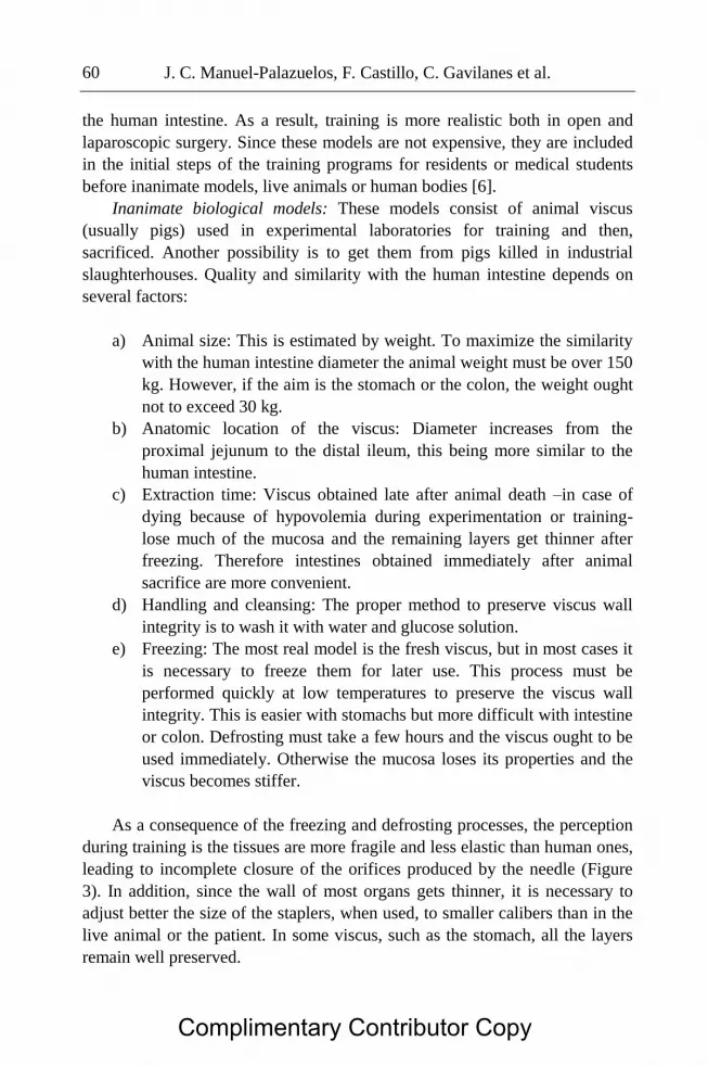
J. C. Manuel-Palazuelos, F. Castillo, C. Gavilanes et al. 60
the human intestine. As a result, training is more realistic both in open and
laparoscopic surgery. Since these models are not expensive, they are included
in the initial steps of the training programs for residents or medical students
before inanimate models, live animals or human bodies [6].
Inanimate biological models: These models consist of animal viscus
(usually pigs) used in experimental laboratories for training and then,
sacrificed. Another possibility is to get them from pigs killed in industrial
slaughterhouses. Quality and similarity with the human intestine depends on
several factors:
a) Animal size: This is estimated by weight. To maximize the similarity
with the human intestine diameter the animal weight must be over 150
kg. However, if the aim is the stomach or the colon, the weight ought
not to exceed 30 kg.
b) Anatomic location of the viscus: Diameter increases from the
proximal jejunum to the distal ileum, this being more similar to the
human intestine.
c) Extraction time: Viscus obtained late after animal death –in case of
dying because of hypovolemia during experimentation or training-
lose much of the mucosa and the remaining layers get thinner after
freezing. Therefore intestines obtained immediately after animal
sacrifice are more convenient.
d) Handling and cleansing: The proper method to preserve viscus wall
integrity is to wash it with water and glucose solution.
e) Freezing: The most real model is the fresh viscus, but in most cases it
is necessary to freeze them for later use. This process must be
performed quickly at low temperatures to preserve the viscus wall
integrity. This is easier with stomachs but more difficult with intestine
or colon. Defrosting must take a few hours and the viscus ought to be
used immediately. Otherwise the mucosa loses its properties and the
viscus becomes stiffer.
As a consequence of the freezing and defrosting processes, the perception
during training is the tissues are more fragile and less elastic than human ones,
leading to incomplete closure of the orifices produced by the needle (Figure
3). In addition, since the wall of most organs gets thinner, it is necessary to
adjust better the size of the staplers, when used, to smaller calibers than in the
live animal or the patient. In some viscus, such as the stomach, all the layers
remain well preserved.
Complimentary Contributor Copy

Intestinal Anastomosis Education and Training 61
Figure 3.
Figure 4. Pig viscus "ex vivo".
f) Anastomosis type: Small intestine is appropriate for manual or stapled
side-to-side intestinal anastomosis, either open or laparoscopic, as
well as for gastro-enteric anastomosis (Figure 4). However, it is less
adequate for end-to-end anastomosis due to higher fragility and
smaller size. The same situation occurs with the colon when
performing a stapled end-to-end anastomosis [5, 52, 53].
Biological Models These types increase the model complexity. They incorporate homeostatic
phenomena characteristic of the biological elements. Their effects can be
difficult to predict and control. Therefore, variability is a feature of biological
Complimentary Contributor Copy

J. C. Manuel-Palazuelos, F. Castillo, C. Gavilanes et al. 62
models. As a result, experimental design is essential when dealing with these
models.
The use of biological models, either animal or cadaveric, is associated
with some ethical, moral and legal implications that it is necessary to bear in
mind before planning training with these materials.
Animal Models Live animal models are associated with the highest fidelity level by
comparison with other models for training intestinal anastomosis performance.
This is due to the possibility of operating on live viscus, with blood perfusion,
not available in other models. Nevertheless, they cannot precisely represent
human anatomy. Even the most similar and used, the porcine model, is flawed
by important anatomic differences.
Live animal laboratories are expensive due to the need of anesthetic
equipment, experienced personnel, and stabulation facilities. Also, several
ethical considerations must have to be borne in mind when using live animals.
Pigs, rabbits and dogs have only one stomach and the small intestine is
similar to the human one so they are useful for intestinal anastomosis training,
either manual or stapled and irrespective of the approach, open or
laparoscopic. For colo-rectal or ileo-colic anastomosis, pigs and dogs have
more anatomical similarities to humans [54, 55], although ethical objections
arise when dealing with dog use because of their affective proximity. The
embryological development of the rabbit and the fact that it is a false ruminant
make its large intestine wall stiffer and, therefore, a worse model. All those
animals allow highly realistic training models. In addition it is possible to
match their different sizes with those of newborns – rabbits -, children – small
pigs – or adults - big pigs [56].
Since cadavers are seldom available and they are expensive, it has been
shown that training on live animals is the best step before starting surgical
practice on humans. As a result, the FLS (Fundamentals of Laparoscopic
Surgery, a program offered by the American College of Surgeons and the
SAGES-Society of American Gastrointestinal and Endoscopic Surgeons)
includes training on pigs for residents and, for more advanced trainees,
includes also practicing on cadavers [57].
Human Cadavers The best option is to get fresh cadavers directly from pathology wards.
This is the most realistic model but their continual availability to perform
successive training sessions is limited [58]. An alternative is the fixation and
Complimentary Contributor Copy

Intestinal Anastomosis Education and Training 63
preservation of cadavers by means of Walther Thiel’s method [59] which
allows body conservation in natural colors and texture. This is based on body
immersion in a specific solution for a limited time. This method allows
keeping the body out of the liquid and storing it in a closed container. If the
body becomes too desiccated, it can be reintroduced in the solution to keep the
tissues wet. This process leads to more convenient body handling and use.
Also, since this process avoids formaldehyde use, there is no emission of
irritant or damaging gases. The final result is a non-irritant and almost odorless
body with high articular mobility, which retains tissue elasticity in a similar
way to fresh bodies and, therefore, very adequate for training on invasive
procedures.
A recent paper, which reviewed different studies concerning surgical
training on human cadavers, suggested that technical skills acquired in low
fidelity models (non-live models) could be as good as training on high fidelity
models (cadavers), since the authors found that the learning process appears
more important that the model itself [58]. This is especially true for procedures
such as suture training which can be practiced on simulation models. These
models, however, are probably not so advantageous when anatomical details
and tissue fidelity play a more important role. This is the case of procedures
such as viscus resection followed by stapled anastomosis (anterior low rectal
resection) [60]. Another possible inconvenience to practice intestinal
anastomosis is the higher stiffness of the intestinal wall and, as a result, more
probabilities of tissue tear.
Nevertheless, the relationship between skills learned on cadavers and
improvement in surgical results when operating directly on patients after
objective assessment is not proven.
Anastakis describes cadaver models as the “gold standard for technical
training”. This can express a generalized opinion suggesting that it has to be
the best model since it reproduces live human anatomy well. Cadaver models
can be useful to allow trainees practicing procedures, with the possibility of
making mistakes in a safe setting, before performing operations on patients
[58].
Training Simulators
Physical simulators: These consist of containers to lodge ex-vivo viscus to
perform anastomosis laparoscopically. They allow the placing of trocars to
introduce the laparoscopic optical system and the instruments to perform
Complimentary Contributor Copy

J. C. Manuel-Palazuelos, F. Castillo, C. Gavilanes et al. 64
different procedures. Physical simulators are very useful to do a wide variety
of surgical procedures, without the need of using live animals (Figure 5). Also,
they have been proven to be a good previous step before training on live
animals, cadavers or patients [46].
Virtual simulators: Since the organism is very complex, it is necessary to
simulate only the tissue biomechanical features, avoiding physiological
processes such as bleeding. Even so, realistic simulation of the biomechanical
behavior of human tissues is very difficult. The difficulty arises first from the
scanty knowledge of the parameters which control tissue deformation. There is
currently very little information concerning live tissue biomechanical features.
This lack of information is due to technical troubles and ethical objections to
measure these parameters on live tissues. As a result, the few available data
come from dead pigs and human viscus. Since these viscuses do not have
blood perfusion, the fast coagulation process inside and temperature difference
significantly alter elasticity by comparison with live tissues. Therefore, these
measures are hardly reliable.
Of note, the first virtual simulator used in anastomosis training was that of
Boston Dynamics for vascular anastomosis, designed by Marc Raibert [61].
Simulator technology has developed fast since then. Concerning intestinal
anastomosis simulators, however, only two current modules of Simbionix
allow performing stapled anastomosis, for gastric by-pass [62] and another in
the module of colo-rectal anastomosis. Hand suture is not possible in these
simulators, which is a considerable limitation in training.
Figure 5. Physical simulator.
Complimentary Contributor Copy

Intestinal Anastomosis Education and Training 65
Training Models as a Mean to Assess Ability
Several scientific societies such as the American College of Surgeons
(ACS), the Society of American Gastrointestinal and Endoscopic Surgeons
(SAGES) with their training program FLS, the Royal College of Physicians
and Surgeons of Canada, the Royal College of Surgeons of England and the
Latin-American Federation of Surgery (FELAC) advise gastrointestinal
anastomosis performance, both open and laparoscopic, for the resident
curriculum to achieve basic technical ability in digestive surgery [63, 64, 65,
66, 67]. Moreover, the higher technical demands of laparoscopic surgery have
forced many staff surgeons to train on anastomosis.
Several questions arise to know whether the above described models can
fully develop surgical abilities.
a) Which training model could best help to achieve ability?
It has been shown that artificial models are a useful first step to adapt to
the suture materials and staplers. Lauscher showed that intestinal anastomosis
practicing in endotrainer with either a plastic digestive tube or animal viscus
provides similar ability levels [68].
Virtual simulators cannot currently provide proper development of these
techniques. Lewis suggests that the gastric by-pass module of the Simbionix
virtual simulator only allows practicing the first steps of gastro-enteric stapled
anastomosis [61].
Several authors have shown that inanimate biological models allow
achieving technical ability and they state that this skill ought to be a previous
step to direct training on live animals or human cadavers. Also, because of
their low cost and high realism, the inanimate biological models ought to be
used to decrease animal sacrifice. They allow the surgical techniques to be
repeated many times, considering that the estimated training procedures
necessary for proficiency are, at least, 8 anastomoses for open surgery, 4 in
case of stapler use and 40 for manual laparoscopic anastomosis [5, 6, 46, 52].
Other authors describe, for the first steps of residents, ability achievement in
terms of quality after doing 7 manual anastomoses, although the time spent is
longer than the ideal [65].
In our opinion, the live biological models (experimental animals) and
human body model allow achieving surgical abilities before the trainees
Complimentary Contributor Copy
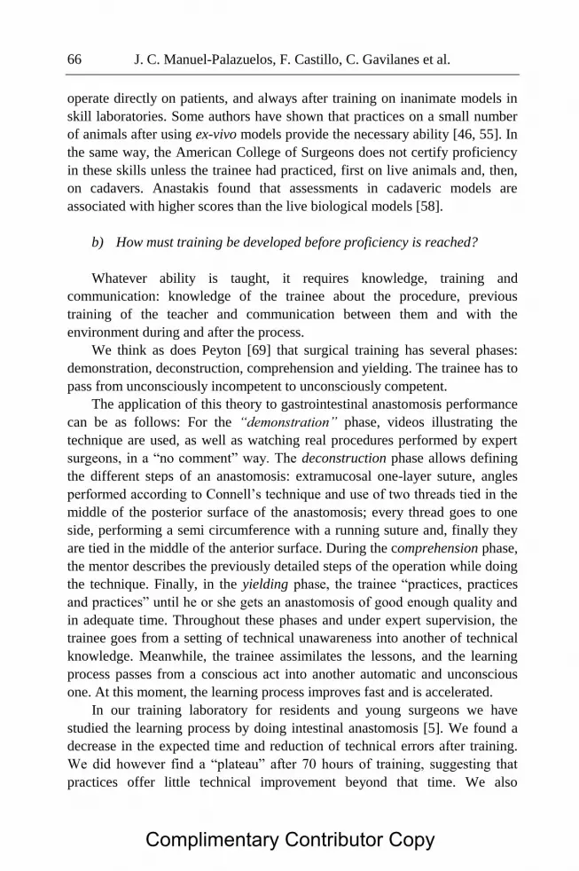
J. C. Manuel-Palazuelos, F. Castillo, C. Gavilanes et al. 66
operate directly on patients, and always after training on inanimate models in
skill laboratories. Some authors have shown that practices on a small number
of animals after using ex-vivo models provide the necessary ability [46, 55]. In
the same way, the American College of Surgeons does not certify proficiency
in these skills unless the trainee had practiced, first on live animals and, then,
on cadavers. Anastakis found that assessments in cadaveric models are
associated with higher scores than the live biological models [58].
b) How must training be developed before proficiency is reached?
Whatever ability is taught, it requires knowledge, training and
communication: knowledge of the trainee about the procedure, previous
training of the teacher and communication between them and with the
environment during and after the process.
We think as does Peyton [69] that surgical training has several phases:
demonstration, deconstruction, comprehension and yielding. The trainee has to
pass from unconsciously incompetent to unconsciously competent.
The application of this theory to gastrointestinal anastomosis performance
can be as follows: For the “demonstration” phase, videos illustrating the
technique are used, as well as watching real procedures performed by expert
surgeons, in a “no comment” way. The deconstruction phase allows defining
the different steps of an anastomosis: extramucosal one-layer suture, angles
performed according to Connell’s technique and use of two threads tied in the
middle of the posterior surface of the anastomosis; every thread goes to one
side, performing a semi circumference with a running suture and, finally they
are tied in the middle of the anterior surface. During the comprehension phase,
the mentor describes the previously detailed steps of the operation while doing
the technique. Finally, in the yielding phase, the trainee “practices, practices
and practices” until he or she gets an anastomosis of good enough quality and
in adequate time. Throughout these phases and under expert supervision, the
trainee goes from a setting of technical unawareness into another of technical
knowledge. Meanwhile, the trainee assimilates the lessons, and the learning
process passes from a conscious act into another automatic and unconscious
one. At this moment, the learning process improves fast and is accelerated.
In our training laboratory for residents and young surgeons we have
studied the learning process by doing intestinal anastomosis [5]. We found a
decrease in the expected time and reduction of technical errors after training.
We did however find a “plateau” after 70 hours of training, suggesting that
practices offer little technical improvement beyond that time. We also
Complimentary Contributor Copy

Intestinal Anastomosis Education and Training 67
observed (data not published) that the “automation” phase in case of manual
laparoscopic anastomoses done in endotrainer is reached as of 40 procedures,
although enough quality extramucosal suture and low leak rate had been
achieved with 20. This means that the trainee has reached the conscious
competent phase. Others have established as 50 manual anastomoses assisted
by the Da Vinci robot the number necessary to equal in time and quality the
anastomoses performed through an open approach [70, 71].
Since women have higher hand skill and men show better spatial
coordination, possible learning differences have been proposed. However,
studies have shown no difference in the final learning [6].
c) How can ability learning be assessed?
The ideal assessing system ought to be easy to use and to interpret, be
accurate, reliable and acceptable. Objective systems of assessment by experts
are the most reliable and useful, although only a few have the aforementioned
features. Assessments based on video watching by experts need long time
consumption and, therefore, are expensive and difficult to perform routinely.
Currently, assessment of technical skills depends on mentor qualification
to a large degree.
Ability assessment based on objective models has several advantages.
First, the abilities can be standardized in programs and, therefore, to evaluate
specific skills. Second, objective assessment is possible but the scores could
not correlate well with evaluation in the operating room. Actually, higher
scores could induce the belief that the trainee is able to start practice on the
patient before real proficiency has been reached.
To perform this, the Objective Structured Assessment of Technical Skills
(OSATS) has been developed as a reliable method to assess abilities. This
examination includes several items such as tissue handling, time to
completion, knowledge and use of instruments, assistant managing, planning
and development of operation and specific knowledge of the trained technique.
A rating scale is built scoring each item from 0 to 5, according to a
proper/improper performance criterion. Reznick [72] designed a type of
OSATS examination to evaluate intestinal anastomoses in live animals.
The same authors as well as others [73, 74], use task check-lists to
evaluate intestinal anastomosis addressing suture and knot quality as well as
care of tissue handling. In our own experience, we assess anastomosis size,
time to completion, suture quality (extramucosal stitches, eversion edge) and
absence of leaks, as shown by means of hydrostatic tests [5]. This evaluation
Complimentary Contributor Copy

J. C. Manuel-Palazuelos, F. Castillo, C. Gavilanes et al. 68
can be done by watching the videos recorded during training or by direct
observation by the mentor.
Figure 6. Correlation between time to anastomosis completion and hours of training.
The former allows evaluation by several experts and in different moments.
The latter obliges the presence of evaluators in the training room and is usually
limited to one or two people. The more structured and precise the check-lists
are and the better experience is described, the higher the evaluation quality is.
Quality is even higher when mentors have been properly trained in this
methodology. The original OSATS were developed to be used as direct
watching and then they were modified by authors such as Grantcharov to
evaluate recorded procedures, where he found the rating scales were reliable
enough to assess these tasks [75].
We did, in our training laboratory, direct structured watching by only one
trained evaluator. After more than 5,000 anastomoses in a physical simulator,
we found this assessing method is reliable to evaluate technical progression of
surgical residents [5]. We think entero-enteric and gastro-enteric anastomoses
0 50 100 150
0
50
100
150
200
Digestive anastomoses on endotrainer
Training hours
Time(mn)
gastro-jejunal both jejuno-jejunal
Complimentary Contributor Copy

Intestinal Anastomosis Education and Training 69
are convenient techniques for laparoscopic training for several reasons. First,
the time to completion can be measured and its decreasing can be correlated
with time spent on training (Figure 6). Second, error rate decreases with
training. However, learning does not improve over 70 hours’ training, since
beyond this period the time to completion and error rate decreasing is very
low.
Assessment of Technical Skill Transfer from Simulation Models to the Patient
in the Operating Room
Out of the reliable investigational setting, skill assessment in the operating
room can be difficult. This is due to the possible variations in the operation
designated to serve as evaluation, to the different academic standards and to
the variation among the surgeons and residents in the way they perform the
procedures. All of these lead to a lack of standardization and, therefore, to
difficult evaluation.
Several questions arise before evaluating the transfer to the operating
room. The first is whether endotrainer-based training is equivalent to the
animal model.
We think as does Hamad [46] the surgical technique can be fully learned
on a laparoscopic endotrainer, with repetition at least 40 times in a very
standardized way. From our experience (data not published), a standardized
technique such as the side-to-side gastro-enteric or entero-enteric anastomosis
allows progression to the animal model when performed at least 40 times, with
no improvement after further training.
Progression to live animal models demands adaptation to a less stable
surgical field, more contaminated by fluids (blood, intestinal content),
although the adaptation is usually fast. Hamad found in laparoscopic surgery a
slight time increase in the performance of the different type of intestinal
anastomoses (about 13 minutes), although without leaks or stenosis [46].
Others, such as Gonzalo Soto, found a mean time of 50 minutes with a failure
rate of 3%, with no stenosis, after performance of 37 intestinal anastomoses
[54].
When a bench model with open surgery instruments is used, the number
of anastomoses and time required to equal expert results are lower, estimated
as eight [6]. Staplers can help in the first steps of anastomoses.
Complimentary Contributor Copy

J. C. Manuel-Palazuelos, F. Castillo, C. Gavilanes et al. 70
In summary, we think the anastomoses ought to be trained in a
standardized way, beginning on a bench or endotrainer model, and later pass to
a live animal model. This skill can be achieved after doing the technique
laparoscopically on both models about 50 times, and about 15 with an open
approach. However, the transfer to patient is not yet established.
There are experiences with virtual simulators for training on laparoscopic
cholecystectomy and laparoscopic gastric by-pass. Improvement in previous
training to performing them directly on patients has been observed but, for the
moment, virtual simulators for intestinal anastomosis exist only in the training
field [61, 74].
Another question is whether the scoring systems (check-lists, OSATS) are
able to discriminate, both in inanimate models and live models, the proficiency
level achieved and whether this is enough to be transferred to the patient.
Since there are no validated check-lists or OSATS for anastomoses, score
systems of other procedures can be used, measuring time to completion and
anastomosis quality. Most authors also include a pressure test to evaluate
possible leaks.
Unfortunately, more factors influence anastomosis performance on
patients, such as vascularization, end tension or patient-related factors
(diabetes, corticosteroid intake). As a result, not only the technical skill, but
also surgical clinical experience are determinant in succeeding when dealing
with a patient.
References
[1] Bell RH Jr, Banker MB, Rhodes RS, Biester TW, Lewis FR. Graduate
medical education in surgery in the United States. Surg. Clin. North Am.,
2007 Aug; 87(4): 811-23.
[2] Halsted WS. The training of the surgeon. Bull. Johns. Hop. Hosp., 1904;
15:267-75.
[3] Cameron JL. William Stewart Halsted. Our surgical heritage. Ann. Surg.,
1997 May; 225(5): 445-58.
[4] Birkmeyer JD, Stukel TA, Siewers AE, Goodney PP, Wennberg DE,
Lucas FL. Surgeon volume and operative mortality in the United States.
N. Eng. J. Med., 2003 Nov 27; 349 (22): 2117-27.
[5] Rodríguez-Sanjuán JC, Manuel-Palazuelos C, Fernández-Díez MJ,
Gutiérrez JM, Alonso-Martín J, Redondo C, Herrera LA, Gómez-Fleitas
M. Assessment of resident training in laparoscopic surgery based on a
Complimentary Contributor Copy

Intestinal Anastomosis Education and Training 71
digestive system anastomosis model in the laboratory. [Article in
Spanish] Cir. Esp., 2010 Jan; 87(1): 20-5.
[6] Masud D, Undre S, Darzi A. Using manual dexterity to predict the
quality of the final product in the small bowel anastomosis after a period
of training. Am. J. Surg., 2012 Jun; 203(6): 776-81.
[7] Bann S, Darzi A. Selection of individuals for training in surgery. Br. J.
Surg., 2005; 190: 98 –102.
[8] Debas HT, Bass BL, Brennan MF, Flynn TC, Folse JR, Freischlag JA, et
al. American Surgical Association Blue Ribbon Committee Report on
Surgical Education: 2004. Ann. Surg., 2005 Jan; 241(1):1-8.
[9] Scott DJ, Bergen PC, Laycock R, Tesfay ST, Valentine RJ, Euhus DM,
et al. Laparoscopic training on bench models: better and more cost
effective than operating room experience? J. Am. Coll. Surg., 2000;191:
272-83.
[10] Seymour NE, Gallagher AG, Roman SA, O’Brien MK, Bansal VK,
Andersen DK, et al. Virtual reality training improves operating room
performance: results of a randomized, double-blinded study. Ann. Surg.,
2002; 236:458-63.
[11] Fried GM, Feldman LS, Vassiliou MC, Fraser SA, Stanbridge D,
Ghitulescu G, et al. Proving the value of simulation in laparoscopic
surgery. Ann. Surg., 2004; 240:518-25.
[12] Sroka G, Feldman LS, Vassiliou MC, Kaneva PA, Fayez R, Fried GM.
Fundamentals of laparoscopic surgery simulator training to proficiency
improves laparoscopic performance in the operating room---a
randomized controlled trial. Am. J. Surg., 2010; 199:115-20.
[13] Tan SS, Sarker SK. Simulation in surgery: a review. Scott. Med. J., 2011
May;56(2):104-9.
[14] Sutherland LM, Middleton PF, Anthony A, Hamdorf J, Cregan P, Scott
D, Maddern GJ. Surgical simulation: a systematic review. Ann. Surg.,
2006 Mar; 243 (3): 291-300.
[15] Chen C. The art of bowel anastomosis. Scand. J. Surg.,
2012;101(4):238-40.
[16] Bernard CL, Huette C. Procédé de Lembert. In Précis iconographique de
médecine opératoire et d’anatomie chirurgicale. Paris: Méquignon-
Marvis, 1855, p. 257, pl. 59 p. 260-261 (495 p.).
[17] Lembert A: Memoire sur l’enterorraphie avec description d’un precede
nouveau pour pratiquer cette operation chirurgicale. Rep. Gen. D’Anat.
Physiol. Pathol. Clin. Chir., 1826;2:100–107.
Complimentary Contributor Copy

J. C. Manuel-Palazuelos, F. Castillo, C. Gavilanes et al. 72
[18] Halsted WS: Circular structure of the intestine: an experimental study.
Am. J. Med. Sci., 1887; 94:436–461.
[19] Murphy JB. Cholecysto-intestinal, gastrointestinal, enterointestinal
anastomosis and approximation without sutures. Med. Rec., 1892;
42:665-676.
[20] Forde KA, McLarty AJ, Tsai J, Ghalili K, Delany HM. Murphy’s button
revisited: Clinical experience with the biofragmentable anastomotic ring.
Ann. Surg., 1993; 217:78–81.
[21] Ravitch MM, Brown IW, Daviglus GF: Experimental and clinical use of
the Soviet Bronchus Stapling Instruments. Surgery, 1959; 46:1-97.
[22] Hesp FL, Hendriks T, Lubbers EJ, deBoer HH. Wound healing in the
intestinal wall. A comparison between experimental ileal and colonic
anastomoses. Dis. Colon. Rectum., 1984 Feb; 27(2): 99-104.
[23] Wise L, McAlister W, Stein T, Schuck P. Studies on the healing of
anastomoses of small and large intestines. Surg. Gynecol. Obstet., 1975
Aug;141(2):190-194.
[24] Matthiessen P, Hallböök O, Rutegård J, Simert G, Sjödahl R.
Defunctioning stoma reduces symptomatic anastomotic leakage after
low anterior resection of the rectum for cancer: a randomized
multicentre trial. Ann. Surg., 2007 Aug;246(2):207-214.
[25] Schrock TR, Deveney CW, Dunphy JE. Factors contributing to leakage
of colonic anastomoses. Ann. Surg., 1973; 177(5):513-518.
[26] Paterson-Brown S, Beck DE, Whitlow CB. A Companion to Specialist
Surgical Practice: Core Topics in General and Emergency Surgery. 4th
ed. Philadelphia: Elsevier. Saunders, 2009: 167-192.
[27] Sørensen LT, Jørgensen T, Kirkeby LT, Skovdal J, Vennits B, Wille-
Jørgensen P. Smoking and alcohol abuse are major risk factors for
anastomotic leakage in colorectal surgery. Br. J. Surg., 1999; 86(7):927-
931.
[28] Witte MB, Barbul A: repair of full-thickness bowel injury. Crit. Care.
Med., 2003;31:S 538–546.
[29] Goligher J, McAdam W. A controlled trial of inverting versus everting
intestinal suture in clinical large bowel surgery. Br. J. Surg., 1970;
57:817.
[30] Miettinen RP, Laitinen ST, Mäkelä JT, Pääkkönen ME. Bowel
preparation with oral polyethylene glycol electrolyte solution vs. no
preparation in elective open colorectal surgery: prospective, randomized
study. Dis. Colon. Rectum., 2000; 43 (5); 669-675.
Complimentary Contributor Copy

Intestinal Anastomosis Education and Training 73
[31] Bucher P, Gervaz P, Morel P: Should preoperative mechanical bowel
preparation be abandoned? Ann. Surg., 2007; 245(4): 662.
[32] Goulder F. Bowel anastomoses: The theory, the practice and the
evidence baseWorld J. Gastrointest. Surg., 2012 September 27; 4(9):
208-213.
[33] Docherty JG, McGregor JR, Akyol AM, Murray GD, Galloway DJ.
Comparison of manually constructed and stapled anastomoses in
colorectal surgery. West of Scotland and Highland Anastomosis Study
Group. Ann. Surg., 1995 Feb; 221(2): 176-184.
[34] Choy PY, Bissett IP, Docherty JG, Parry BR, Merrie AE. Stapled versus
handsewn methods for ileocolic anastomoses. Cochrane Database Syst.
Rev., 2007: CD004320.
[35] Simillis C, Purkayastha S, Yamamoto T, Strong SA, Darzi AW, Tekkis
PP. A meta-analysis comparing conventional end-to-end anastomosis vs.
other anastomotic configurations after resection in Crohn's disease. Dis.
Colon. Rectum., 2007 Oct; 50(10): 1674-1687.
[36] McLeod RS, Wolff BG, Ross S, Parkes R, McKenzie M. Recurrence of
Crohn’s disease after ileocolic resection is not affected by anastomotic
type: results of a multicenter, randomized, controlled trial. Dis. Colon.
Rectum., 2009; 52: 919-927.
[37] Taflampas P, Christodoulakis M, Tsiftsis DD. Anastomotic leakage after
low anterior resection for rectal cancer: facts, obscurity, and fiction.
Surg. Today, 2009; 39(3):183-188.
[38] Brundage SI, Jurkovich GJ, Grossman DC, Tong WC, Mack CD, Maier
RV. Stapled versus sutured gastrointestinal anastomoses in the trauma
patient. J. Trauma., 1999; 47: 500-507; discussion 500-507.
[39] Brundage SI, Jurkovich GJ, Hoyt DB, Patel NY, Ross SE, Marburger R,
Stoner M, Ivatury RR, Ku J, Rutherford EJ, Maier RV. Stapled versus
sutured gastrointestinal anastomoses in the trauma patient: a multicenter
trial. J. Trauma, 2001; 51: 1054-1061.
[40] Jyoti Shah, Yaron Munz, Joanne Manson, Krishna Moorthy, Ara Darzi.
Objective Assessment of Small Bowel Anastomosis Skill in Trainee
General Surgeons and Urologists. World J. Surg., (2006) 30: 248–251.
[41] Jörn Gröne, Jörg-Peter Ritz, Andrea Stroux, Kai S. Lehmann, Johannes
C. Lauscher. Measurable learning effects after a 1-week skills course in
digestive surgery. Int. J. Colorectal. Dis., (2010) 25:1133–1139.
[42] Slawomir J. Marecik, Vivek Chaudhry, Azam Jan, Russell K. Pearl,
John J. Park, Leela M. Prasad. A comparison of robotic, laparoscopic,
Complimentary Contributor Copy

J. C. Manuel-Palazuelos, F. Castillo, C. Gavilanes et al. 74
and hand-sewn intestinal sutured anastomoses performed by residents.
The American Journal of Surgery, 193 (2007) 349–355.
[43] Goligher JC, Lee PW, Simpkins KC, Lintott DJ. A controlled
comparison one- and two-layer techniques of suture for high and low
colorectal anastomoses. Br. J. Surg., 1977; 64: 609-614.
[44] Maurya SD, Gupta HC, Tewari A, Khan SS, Sharma BD. Double layer
versus single layer intestinal anastomosis: a clinical trial. Int. Surg.,
1984; 69:339-340.
[45] Shikata S, Yamagishi H, Taji Y, Shimada T, Noguchi Y. Single- versus
two- layer intestinal anastomosis: a meta-analysis of randomized
controlled trials. BMC Surg., 2006; 6:2.
[46] Hamad MA, Mentges B, Buess G. Laparoscopic sutured anastomosis of
the bowel; Technique and learning curve. Surg. Endosc., 2003; 17:
1840-1844.
[47] Max E, Sweeney WB, Bailey HR, Oommen SC, Butts DR, Smith KW,
Zamora LF, Skakun GB. Results of 1000 single-layer continuous
polypropylene intestinal anastomoses. Am. J. Surg., 1991; 162:461– 467.
[48] Mohr Z, Willis S. Intestinal anastomoses and techniques in the lower
gastrointestinal tract. Chirurg, 2011;82:34-40.
[49] Ramos-Salgado F, Quintero-Becerra J, Hernández-Toriz N. Modelo para
entrenamiento de cirugía laparoscópica urológica. Rev. Mex. Urol.,
2010; 70:31-35.
[50] Holmberg D.L, Cockshutt JR, Basher A. Use of a dog abdominal
surrogate for teaching surgery. J. Vet. Med. Educ., 1993; 20:107-111.
[51] Molina Martínez JL, Silveira Prado E, et al. Simulators and experimental
models for the development of surgical skills on the Health Sciences
teaching–learning process REDVET Rev. electrón. vet.
http://www.veterinaria.org/revistas/redvet 2012 Volumen 13, Nº 6.
[52] Vick L, Kenneth D. Borman K, and Salameh JR. Face, Content, and
Construct Validities of Inanimate Intestinal Anastomoses Simulation.
Journal of Surgical Education, 2007; 64: 365-368.
[53] Munksdorf M, Fischer A, Hoffmann J. Experiences with
surgical training in anastomosis technics using porcine gut. Ugeskr.
Laeger., 1990;152:2575-6.
[54] Gonzalo Soto D, George Pinedo M, Demian Fullerton M, Francisca
León G, Francisco Lopez K. Hand-sewn anastomosis in laparoscopic
colon surgery. An experimental model. Rev. Chilena de Cirugía, 2005;
57: 149-154.
Complimentary Contributor Copy

Intestinal Anastomosis Education and Training 75
[55] Jardel N, Hidalgo A, Leperlier D, Manassero M, Gomes A, Bedu AS,
Moissonnier P, Fayolle P, Begon D, Riquois E, Viateau V. One Stage
Functional End-to-End Stapled Intestinal Anastomosis and Resection
Performed by Non expert Surgeons for the Treatment of Small Intestinal
Obstruction in 30 Dogs. Veterinary Surgery, 2011; 40: 216–222.
[56] Kirlum HJ, Heinrich M, Till H: The rabbit model serves as a valuable
operative experience and helps to establish new techniques for
abdominal and thoracic endosurgery. Pediatr. Surg. Int., 2005; 21: 91-
93.
[57] Palter V, and Grantcharov T. Development and validation of a
Comprehensive Curriculum to Teach an Advanced Minimally Invasive
Procedure. A Randomized Controlled Trial. Ann. Surg., 2012; 256:25-
32.
[58] Anastakis DJ, Regehr G, Reznick RK, et al. Assessment of technical
skills transfer from the bench training model to the human model. Am. J.
Surg., 1999;177:167–170.
[59] Thiel, W. Die Konservierung ganzer Leichen in natürlichen Farben. Ann.
Anat., 1992; 174: 185-195.
[60] Gilbody J, Prasthofer AW, Ho K, Costa ML. The use and effectiveness
of cadaveric workshops in higher surgical training: a systematic review.
Ann. R. Coll. Surg. Engl., 2011;93:347-52.
[61] Satava R, Jones S. Preparación de cirujanos para el siglo XXI.
Consecuencias de los avances tecnológicos. Cirugía muy poco invasora
parte I. Surg. Clin. N. Am., 2000; 4:1417-1429.
[62] Lewis T, Aggarwal R, Kwasnicki R, Rajaretnam N, Moorthy K, Ahmed
A, Darzi A. Can virtual reality simulation be used for advanced bariatric
surgical training? Surgery, 2012;151:779-84.
[63] Mittal MK, Dumon KR, Edelson PK, Acero NM, Hashimoto D, Danzer
E, Selvan B, Resnick AS, Morris JB, Williams NN. Successful
Implementation of the American College of Surgeons/Association of
Program Directors in Surgery Surgical Skills Curriculum via a 4-Week
Consecutive Simulation Rotation. Simul. Healthc., 2012; 7:147-54.
[64] Vassiliou M, Dunkin BJ, Marks JM, MDc, Fried G M., FLS and FES:
Comprehensive Models of Training and Assessment. Surg. Clin. N. Am.,
2010; 90: 535–558.
[65] Chan B, Martel G, Poulin E, Mamazza J, Boushey R. Resident training
in minimally invasive surgery: a survey of Canadian department and
division chairs. Surg. Endosc., 2010; 24:499–503
Complimentary Contributor Copy

J. C. Manuel-Palazuelos, F. Castillo, C. Gavilanes et al. 76
[66] Olson T, Becker Y, McDonald R, Gould J. A Simulation-Based
Curriculum Can Be Used to Teach Open Intestinal Anastomosis. Journal
of Surgical Research, 2012; 172, 53–58.
[67] A. Jensen. A. Andrew. Laboratory based instruction for skin closure and
bowel ansstomosis for surgical residents. Arch. Surg., 2008; 143:852-
859.
[68] Lauscher JC, Ritz JP, Stroux A, Buhr HJ, Gro¨ne, J. A New Surgical
Trainer (BOPT) Improves Skill Transfer for Anastomotic Techniques in
Gastrointestinal Surgery into the Operating Room: A Prospective
Randomized Trial. World J. Surg., 2010; 34: 2017– 2025.
[69] Peyton JWR. The learning cycle. In: Peyton JWR, editor. Teaching and
learning in medical practice. Rickmansworth, UK: Manticore Europe
Limited, 1998: 13-19.
[70] Pearl RK, Park J, Prasad L. A comparison of robotic, laparoscopic, and
hand-sewn intestinal sutured anastomoses performed by residents. The
American Journal of Surgery, 2007;193: 349–355.
[71] Marecik SJ, Prasad L, Park J, Jan A, Chaudhry V. Evaluation of
midlevel and upper-level residents performing their first robotic-sutured
intestinal anastomosis. American Journal of Surgery, 2008;195: 333–
338.
[72] Martin JA, Regehr G, Reznick R et al. Objective structured assessment
of technical skill (OSATS) for surgical residents. Br. J. Surg., 1997; 84:
273 – 278.
[73] Reznick R, Regehr G, MacRae H et al. Testing technical skill via an
innovative ‘‘bench station’’ examination. Am. J. Surg., 1997; 173:226–
230.
[74] Chipman JG, Schmitz C. Using Objective Structured Assessment of
Technical Skills to Evaluate a Basic Skills Simulation Curriculum for
First-Year Surgical Residents. J. Am. Coll Surg., 2009; 209: 364-370.
[75] Kundhal PS, Grantcharov TP. Psychomotor performance measured in a
virtual environment correlates with technical skills in the operating
room. Surg. Endosc., 2009; 23:645–649.
Complimentary Contributor Copy

In: Anastomoses ISBN: 978-1-62618-657-6
Editors: F. King, McKineley A. Malloy © 2013 Nova Science Publishers, Inc.
Chapter III
Invaginating Colonic
Anastomosis
Aly Saber Port-Fouad General Hospital, Department of general surgery
Port-Fouad, Port-Said, Egypt
1. Historical Background
The earliest reports of surgical suture date back to 3000 BC in ancient
Egypt, and the oldest known suture is in a mummy from 1100 BC. The ancient
Egyptians and Babylonians and the later Greeks and Romans used the
intestines of herbivorous animals for much the same purposes. Detailed
records of sigmoid volvulus were found in the Egyptian Papyrus Ebers and in
ancient Greek and Roman writings. The ancient Egyptian Ebers papyrus
describes the natural history of sigmoid volvulus as either reducing
spontaneously, or the sigmoid colon being ‘rotted’. Written in 500 BCE, the
detailed description of a wound suture and the suture materials used in it is by
the Indian sage and physician Sushruta.
Early in the first century AD, Celsus recorded attempts to suture the
intestine but and Abulkasem in 87 AD, recommended using the jaws of large
ants to unite intestinal wounds and referred to catgut made from the intestines
E-mail: [email protected].
Complimentary Contributor Copy

Aly Saber 78
of sheep as suture material. Other ancient surgical methods involved the use of
a few large-diameter sutures; use of bone, trachea, or wood stents; or attempt
to invaginate the cut ends of intestine. The oldest reported intestinal suturing
technique is the Glover’s suture that was a simple continuous stitch in
which the ends, instead of being tied, were left long and pulled externally
through the abdominal wound.
Current principles of intestinal surgery originated in the early 19th
century. In 1812, Travers first reported that the entire circumference of the
divided intestine needed to be in contact to heal properly. Travers used a
small; round sewing needle to place multiple full-thickness silk sutures and
knotted the sutures. He reported successful healing of intestinal anastomoses
in dogs with his mucosal appositional and everting technique. In 1826,
Lembert, the founder of modern intestinal surgery, reported that serosa-to-
serosa contact of the divided intestine, achieved by inverting suture patterns,
was necessary for intestinal healing and became a fundamental principle
of intestinal surgery. In 1883, Czerny modified Lembert’s technique into a
two-layer, inverting anastomosis, which became widely used in the 20th
century. In 1887, Halsted reported the importance of the submucosal layer in
suturing gut wounds using a single-layer closure. In 1892, Connell introduced
a single-layer, continuous inverting suture pattern with the advantages of
reduction of the number of knots left in the wound and operative time.
In 1892 also, Murphy described the use of an inter-locking metallic button
designed to create a sutureless, inverting, end-to-end intestinal anastomosis
and the button finally would pass with the feces. Nevertheless, Murphy’s
button was largely replaced by hand-sewn intestinal anastomosis techniques in
human patients by the 1920s. By the beginning of the 20th
century, about 250
methods for intestinal suturing had been described with these principles: (1)
opposing the serosal surfaces of the intestine around the full circumference
using an inverting suture technique, (2) including the tough submucosa in the
suture bites,(3) using aseptic surgical technique, (5)maintaining adequate
blood supply to tissue, and (6) avoiding tension on the anastomosis.
Information on surgical treatment of intestinal diseases in the small animal
veterinary literature is generally lacking before the1940s. The first edition
of Canine Surgery was published in 1939. In a 1941 small animal surgery
textbook, McCann stated that the prognosis is always grave for enterorrhaphy
in the dog. The textbook described use of an inverting Lembert pattern to
perform end-to-end small intestinal anastomosis and mentioned various
mechanical devices, such as the Murphy button, that require no sutures.
McCunn admitted that, in his experience with enterorrhaphy, the results were
Complimentary Contributor Copy

Invaginating Colonic Anastomosis 79
not good, although there had been some successes. In 1951, Gambee described
a single-layer, inverting technique for intestinal anastomosis. He reported good
results with the technique in comparison of double-layer closure in the GI
tract, which was widely used in human surgery. The first challenges to
Lembert’s principles of inversion and serosal contact also appeared in the
1950s. The earliest descriptions of everting intestinal anastomoses in the
veterinary literature appeared in the 1960s. In 1968, Ott and colleagues
compared single layer, inverting anastomosis with everting anastomosis in the
small intestine of dogs. They concluded that the everting technique was
stronger and was associated with less compromise of the lumen diameter and
fewer complications.
The first report of simple interrupted approximating anastomosis has
become the technique of choice for end-to-end anastomosis in small animal
surgery appeared in1970 when Bennett and Zydeck and in 1973, DeHoff and
co- workers compared single-layer everting, inverting, and approximating end-
to-end jejunal anastomoses in dogs and puppies. In 1976, Reinerts compared
three patterns for equine jejunal anastomosis: a modified Gambee appositional
suture, a simple interrupted everting suture, and a double-row inverting suture.
In 1981, Ellison reviewed techniques for end-to-end intestinal anastomosis in
dogs and pointed out the difficulty in avoiding some degree of mucosal
eversion with proper surgical technique and stated that any anastomotic suture
pattern could be used successfully in the canine intestine. In 1982, Ellison and
colleagues reported that the needle passage should to exclude the mucosa in an
attempt to minimize eversion. In 1982, Bellenger was the first to report results
of experimental appositional anastomosis and single-layer inverting of the
jejunum in cats. Recently, Weisman and colleagues reported that modified
simple continuous pattern for closure of intestinal incisions is an acceptable
alternative to the simple interrupted pattern in dogs and cats. Reported
advantages of a simple continuous enteric closure include surgical speed,
decreased tissue handling, improved apposition of intestinal layers, and a low
rate of clinical complications. Simple continuous approximating anastomosis
has been shown experimentally to achieve better continuity of the histologic
intestinal layers than do simple interrupted approximating techniques.
The current automated stapling instruments for intestinal anastomosis
originated in the former Soviet Union after World War II. These stapling
devices were first tested in the United States in the 1960s and became widely
accepted for use in human surgical patients in the 1970s. Hess and coworkers
published the first veterinary study that compared mechanically stapled with
hand-sewn small intestinal anastomoses. Although the outcomes of the
Complimentary Contributor Copy

Aly Saber 80
two techniques were similar, the mechanical staples were applied more
consistently and required less surgical time than did the sutures. Stoloff and
colleagues and in 1991, Ullman and colleagues in separate works, compared
stapled and hand-sewn colonic anastomoses in dogs. They reported less tissue
reaction, improved healing, and fewer adhesions with the inverted stapled
anastomosis.
A biofragmentable ring composed of polyglycolic acid for intestinal
anastomosis has been experimentally tested in dogs and cats and has been used
clinically in humans that is similar to the Murphy button but breaks down
approximately 12 days after implantation and eventually pass with the feces.
Experimental data show that colonic anastomoses achieved with a
biofragmentable ring have higher initial bursting strength and similar healing
patterns compared with sutured and stapled anastomoses. Clinical application
of the biofragmentable anastomosis ring has not been reported in the
veterinary literature.
2. Techniques of Colonic Anastomosis
2.1. Principles of Successful Intestinal Anastomosis
Intestinal anastomosis is one of the most commonly performed surgical
procedures, especially in the emergency setting, and is also commonly
performed in the elective setting when resections are carried out for benign or
malignant lesions of the gastrointestinal tract. Proper surgical technique and
adherence to the following fundamental principles are imperative to ensure
successful outcome after intestinal anastomosis. The first principle of proper
technique for intestinal anastomoses is adequate exposure as the intestine
should be mobilized sufficiently for proper anastomosis. Sufficient serosa
must be exposed so that the seromuscular sutures or staples can be placed
directly in the serosa without traversing the mesentery. The second principle is
to maintain a good blood supply to the severed ends of the bowel. The blood
supply may be compromised by construction of an anastomosis under tension,
excessive dissection or mobilization of the bowel, excessive use of the
electrocautery, and tying of the sutures so tight that the intervening tissue is
strangulated. The third principle involves prevention of local spillage of
enteric contents. The best way of preventing spills is to operate on prepared
bowel Local spills and local sepsis have an adverse effect on the healing
anastomosis, and it is for this reason that noncrushing occlusion clamps, in
Complimentary Contributor Copy

Invaginating Colonic Anastomosis 81
addition to an adequate bowel preparation, are advisable. The fourth principle
is an accurate apposition of serosa to serosa of the two segments of bowel to
be anastomosed. The anastomosis should be watertight and performed without
tension. The bowel must be handled gently with the use of noncrushing
forceps. The fifth principle is absence of tension and distal obstruction with
gentle and meticulous tissue handling technique. The final principle involves
realignment of the mesentery of the two segments of bowel to be joined with
good approximation of well vascularized cut ends of the bowel. These should
be parallel to each other and ensure that there is no twist on completion of the
anastomosis.
2.2. Suture Materials
Successful wound healing depends on an appropriate tensile strength
provided by the suture materials and a microenvironment in which the repaired
tissues are likely to attach and grow. Choice of suture materials includes an
absorbable versus nonabsorbable material, monofilament versus polyfilament.
Nonabsorbable sutures can retain their tensile strength for one year or longer,
whereas the half-lives of tensile strength for absorbable sutures vary from one
to several weeks. Suture materials are required in practically every surgical
operation and most researchers think that some of the complications following
surgery may be directly attributable to the suture material itself. The
observations indicate that anastomotic suture support is of minor or no
importance one to two weeks after the procedure. There are four important
properties common to all suture materials: the intensity of the inflammatory
response which the particular material evokes in the tissues; the behavior of
the material in the presence of infection; its durability; and its handling ability.
The degree of tissue reaction caused by the sutures has two important
consequences. Firstly, an excessive tissue reaction will lead to impairment of
the strength of the tissues and this decreases the holding power of the stitches,
which consequently tend to leak. Accordingly, materials which cause the least
tissue reaction generally produce the strongest closure. Secondly, there is some
evidence that infection is more likely to occur in wounds sutured with
materials causing excessive tissue reaction. Sutures act as foreign bodies in the
anastomosis, producing inflammation which persists for two or three weeks.
Inflammation seems to delay the healing of an intestinal anastomosis and the
ideal suture provokes a minimal inflammatory response. Thus modern,
synthetic, absorbable suture materials can be used safely for intestinal
Complimentary Contributor Copy

Aly Saber 82
anastomosis. Absorption of the suture material eliminates the foreign body
residue seen with nonabsorbable sutures that induces a strong tissue reaction.
Absorbable sutures are made of natural materials which are broken down in
tissue after a short period of time, ranging from days to few weeks. In most
cases, three weeks is sufficient for the wound to close firmly. The suture is not
needed any more, and the fact that it disappears is an advantage, as there is no
foreign material left inside the body and no need for the patient to have the
sutures removed. Absorbable sutures were originally made of the intestines of
sheep, the so called catgut. Today, gut sutures are made of specially prepared
beef and sheep intestine, and may be untreated (plain gut), tanned with
chromium salts to increase their persistence in the body (chromic gut), or heat-
treated to give more rapid absorption (fast gut). However, the majority of
absorbable sutures are now made of synthetic polymer fibres, which may be
braided or monofilament; these offer numerous advantages over gut sutures,
notably ease of handling, low cost, low tissue reaction, consistent performance
and guaranteed non-toxicity.
Non absorbable sutures are made of materials which are not metabolized
by the body, and are used therefore either on skin wound closure, where the
sutures can be removed after a few weeks, or in some inner tissues in which
absorbable sutures are not adequate. This is the case, for example, in the heart
and in blood vessels, whose rhythmic movement requires a suture which stays
longer than three weeks, to give the wound enough time to close. Other
organs, like the bladder, contain fluids which make absorbable sutures
disappear in only a few days, too early for the wound to heal. There are several
materials used for non absorbable sutures. The most common is a natural fibre,
silk, which undergoes a special manufacturing process to make it adequate for
its use in surgery. Other non-absorbable sutures are made of artificial fibres,
like polypropylene, polyester or nylon; these may or may not have coatings to
enhance their performance characteristics. Finally, stainless steel wires are
commonly used in orthopaedic surgery and for sternal closure in cardiac
surgery.
2.3. Anastomotic Pattern
There are many types of intestinal anastomotic patterns available in
human and veterinary practices. However, there is no universal agreement
about the best technique for gastrointestinal anastomosis (GIA). Predicted fear
of increased postoperative complications secondary to the anastomotic failure
Complimentary Contributor Copy

Invaginating Colonic Anastomosis 83
has resulted in a diversity of techniques. The type of gastrointestinal
anastomotic construction is usually left to the choice of the surgeon, according
to his experience and preference for hand- sewn or stapled sutures.
Anastomotic patterns are typically categorized as sutured, stapling and
sutureless anastomosis. The sutured anastomosis may be continuous or
interrupted, single layer or double layers and inverting, appositional, or
everting. The two-layer anastomosis using interrupted silk sutures for an outer
inverted seromuscular layer and a running absorbable suture for a transmural
inner layer has been standard for most surgical situations. Some recent reports
have described single-layer continuous anastomosis using monofilament
sutures as requiring less time and cost than any other method, without
incurring any added risk of leakage. Many surgeons probably now use single-
layer suturing due to reductions in ischemia, tissue necrosis or narrowing of
the lumen compared to the two-layer method. The interrupted serosubmucosal
suture was considered as the "gold standard" for intestinal anastomosis with
many advantages as: accurate tissue apposition, incorporates submucosa,
minimizes damage to submucosal vascular plexus, lesser tissue strangulation,
appropriate for both upper and lower GI tract anastomosis and appropriate for
both accessible and inaccessible sites.
Stapling technology was pioneered in the early part of this century and
subsequently modified. Russian initiatives led to development of the original
circular stapling instrument and further progress has resulted in instruments
that are widely available, reliable and totally disposable. Mechanical failure is
now rare and malfunction is generally due to operator error. Complications
related to the stapling technique are uncommon, although anastomotic stricture
may be more frequent than when hand- sewn anastomosis is performed. A
stapling instrument facilitates and may expedite a surgical procedure but it is
an adjunct to, and not a substitute for, meticulous surgical technique. The
safety, efficacy, and technique of stapled gastrointestinal tract anastomosis in
adults have been extensively documented since 1978. Recently, a single-
stapled technique was performed instead of the conventional double-
stapled technique in laparoscopic low anterior resection for anastomosis, by
placement of intracorporeal purse-string sutures on the distal rectum with
transanal specimen extraction. The stapler has many advantages: a better blood
supply, less tissue manipulation, minimal edema, uniformity of sutures, easy
lumen calibration and short operative times.
The concept of a sutureless intestinal anastomosis has been attractive to
surgeons for many years. Beginning in the early 19th century, surgical
investigators have designed ingenious but often complicated devices to
Complimentary Contributor Copy

Aly Saber 84
achieve end-to-end intestinal anastomosis. Sutureless intestinal anastomoses
can be achieved either by compression, where two inverted rings of bowel are
compressed by a hollow circular device that subsequently sloughs away and is
passed through the anus, or by the use of tissue glues or laser welding.
Compression devices used clinically with success are the Valtrac
biofragmentable anastomotic ring, the polypropylene rings described by Rosati
and the AKA guns. Glued anastomoses have only been used in animals and
seem to be unsafe. However, laser-welded intestinal anastomoses appear
highly promising in experimental studies and further development of this
technique is warranted.
3. Anastomotic Line Protection
The relative high incidence of anastomotic leakage after colorectal
surgery, with its major consequences for morbidity and mortality, remain of
great concern. Accordingly, researchers tried to use many techniques and
devices to protect the anastomotic site.
3.1. External Coating
External coating of colonic anastomoses has been proposed as a means to
lower the rate of anastomotic complication. A number of external anastomosis
reinforcements have been tested, including peritoneal graft, omental graft, dura
mater, and meshes. Omentum is the tissue most often used for anastomosis
reinforcement. Several studies suggested that omental reinforcement prevents
anastomosis leakage, whereas others found that this approach caused an
increased risk of infection associated with pedicular necrosis or late intestinal
obstruction. Both human dura mater and free peritoneal graft reinforcement
showed reduced anastomosis healing that could be attributed to avascularity of
grafts and to aggravated adhesions between anastomosis and intra-abdominal
organs and consequently led to decreased anastomosis healing. Non-
absorbable meshes reinforce anastomoses permanently, however, may increase
the risk of peritoneal adhesion, anastomotic stenosis, and colon perforation, in
the long term. In experimental animals, fibrin sealant was used and showed
increased bursting pressure and rupture strength of colonic anastomoses.
Hyaluronic acid-carboxymethylcellulose also was used in rats to protect the
anastomotic lines and further studies have to be performed. Accordingly, the
Complimentary Contributor Copy

Invaginating Colonic Anastomosis 85
external coating of colonic anastomoses has yet failed to show convincing
results.
3.2. Luminal Devices
Intra-luminal colonic devices are used in both animal studies and human
aiming to prevent the faecal load from contacting the anastomotic site, thereby
preventing leakage of faeces into the peritoneal cavity when the walls of the
anastomosis have become dehiscent. When the faecal stream is bypassed from
contacting the bowel mucosa, a gap in the anastomosis will not lead to
extravasation of intra-luminal content.
3.2.1. Animal Studies
Coloshield In the 1980’s, Ravo and Ger developed an intraluminal colonic tube to
prevent anastomotic leakage. The proximal end of the tube is fixed to the
proximal bowel loop using polyglycolic acid sutures. Studies on dogs were
performed using different tubes varying in width and length, material (latex,
silicone, rubber), and suture technique.
The colon tube placement was found to be a safe, uncomplicated
procedure and none of the dogs developed leak. All tubes were allowed to be
expelled naturally together with the faecal stream.
Silicone Prosthesis The intracolonic silicone prosthesis in protecting the anastomosis in dogs
was used since 1992.
Soft Latex Tube Intraluminal colonic soft latex tubes were studied by Ross in a rat model
and the results suggested that the intraluminal tubes have a survival advantage
compared to controls without tubes.
Polyflex Self-Expandable Covered Plastic Stent In 2008 Tsereteli et al. performed their study in pigs comparing the
incidence of anastomotic leak after open rectosigmoid resection with or
without a Polyflex self-expandable covered plastic stent. The stent was placed
Complimentary Contributor Copy

Aly Saber 86
over a guide wire with use of a flexible colonoscope and deployed under
fluorescence control.
This experiment demonstrated a significant beneficial effect of the stent as
a breakthrough solution for the complicated colorectal anastomosis, avoiding
the necessity of a stoma during the healing process.
3.2.2. Human Studies
Coloshield The development of the intraluminal tube led to the final version of the
Coloshield to be a soft, pliable tube like a surgical glove. This intraluminal
protective device, developed by Ravo, was first used in humans in 1984.
Indications for use include perforated diverticulitis, colonic obstruction,
volvulus, carcinoma, and fistula. Several non-randomized studies were
performed in patients undergoing colon surgery with the Coloshield. In 1991,
Ravo described a method of inserting the Coloshield in the proximal colon
after completion of the anastomosis by performing a longitudinal colostomy
on the antimesenteric border of the afferent loop, proximal to the anastomosis.
Ravo and Ger pioneered the use of intracolonic stents, testing different
materials (silicone, rubber, and latex) before developing and, finally, filing the
patent of the latex Coloshield. Despite its promise, the Coloshield has not been
widely accepted.
Condom In 1994, Yoon et al. and in 1995, Ruiz et al. used a condom instead of a
Coloshield to protect the colo-anal anastomosis. The ring of the sterilised
condom is sutured to the mucosal and submucosal layer of the proximal colon
before completing the anastomosis. The condom is allowed to the exterior and
transected with scissors. The device is expelled naturally from the anus
between the 10th and 14th postoperative day.
4. Animal Models for Colonic Anastomosis
Although humans and animals may look different, at a physiological and
anatomical level they are remarkably similar. Animals, from mice to monkeys,
have the same organs and systems which perform the same functions in pretty
much the same way. There are minor differences, but these are far outweighed
Complimentary Contributor Copy

Invaginating Colonic Anastomosis 87
by the similarities. The differences can give important clues about diseases and
how they might be treated – for instance, if we knew why the mouse with
muscular dystrophy suffers less muscle wasting than human patients, this
might lead to a treatment for this debilitating and fatal disorder. It is settled
that models never provide final answers of the original condition, but only
offer approximation no single animal model can ever duplicate this original
condition. Therefore, experimental science itself is the study of
approximations and assimilation. To produce a good model, the best suited
animal must be chosen regarding the physiologic and anatomic resemblance to
man. Cost, availability, handling difficulty and housing requirements are
important limitations which must all be considered when choosing the best
models. A specific model is chosen because it is believed to be appropriate to
the condition being investigated and is thought likely to respond in the same
way as humans to the proposed treatment under investigation.
4.1. Animal Models in Biomedical Research
Models are widely used in all branches of physical, biological and social
sciences. As defined in Oxford English Dictionary, a model in general is the
representation of a real or actual object. In biomedical research, an animal
model is defined as a living organism with an inherited, naturally acquired or
induced pathological process trying to closely resemble the same phenomenon
in man. Models are, thus, meant to mimic and it is not expected to be
necessarily identical to the subject under investigation. In biomedical research,
models allow the investigator to understand and investigate pathophysiological
processes and the impact of intervention. The ultimate goal of experimental
research using animal models is to solve problems in clinical practice and to
develop new methods and approaches to the cure and alleviation of disease
and disability.
4.2. Good Animal Model
Broadly speaking, not all animal species are useful for the purposes of
biomedical research and the limitations of the models selected as well as the
methodology involved must always be kept in mind. The optimal animal
model should closely reproduce the disease in humans and therefore if
possible the species with the closest anatomical and physiological resemblance
Complimentary Contributor Copy

Aly Saber 88
to man should be used. On the other hand, a rare or exclusive animal should
not be used, thus the model should be available to many researchers. The
model should be economical and the anastomosis technically easy to perform.
It should be reproducible with standardized interventions and measures. The
animal should be easily handled by investigators and fit into an average
institution.
4.3. Animals
4.3.1. The Mouse The mouse has the advantages of being inexpensive, has a short life cycle,
requires only limited space and is easily handled. As the rat, the mouse is not a
strict herbivore, which means it has the ability to digest variable amounts of
animal-derived food. With respect to the gastrointestinal tract, the mouse is
comparable to man. Apart from lacking an appendix vermiformis, the mouse
macroscopically has the same bowel segments as found in humans.
Microscopically the intestinal wall consists of the same four layers as in
humans: mucosa, submucosa, muscularis and serosa, with the epithelium of
the colon consisting of columnar cells and many goblet cells. The mouse has
been extensively used for research in oncology, toxicology, genetic
engineering and to manufacture vaccines, antibodies and hormones, but few
studies have used the mouse for surgical research. This might be due to the
small size of the animal and thus small size of the organs, including the colon.
The use of a microscope is necessary when performing a colonic anastomosis
in the mouse. Mice and humans each have about 30,000 genes, yet only 300
are unique to either organism. Both even have genes for a tail, even though it's
not "switched on" in humans. "About 99 percent of genes in humans have
counterparts in the mouse," said Eric Lander, Director of the Whitehead
Institute Center for Genomic Research in Cambridge, Massachusetts. "Eighty
percent have identical, one-to-one counterparts."
4.3.2. The Rat The rat is inexpensive, requires only limited space and is easy to handle.
The rat has no gross anatomical peculiarities compared to man. The caecum is
divided into an apex and a basal part by a slight constriction. When compared
to the mouse, the larger size of the rat colon allows the anastomosis to be
performed more easily and without the use of a microscope. Regarding the
disadvantages, the rat is considered to be more resistant to infection compared
Complimentary Contributor Copy

Invaginating Colonic Anastomosis 89
to the mouse; however, this is a general consideration and has not been
supported by studies on the issue. Moreover, one study found that young rats
were protected against experimental fecal peritonitis by receiving maternal
milk. Human beings do not have any specialized compartments in the
digestive system because they, humans, are considered as omnivores, they eat
both meat and vegetable or fruit plants. This means that all the support organs
in their body have a special function to perform for our body. A rat digestive
system has two major differences with that of a human. First, rats do not have
a gallbladder. This is because they rarely take in large amount of fatty foods,
thereby, making a gallbladder useless. Furthermore, rats have an enlarged
large intestine, namely, the caecum. This helps them ferment the grains
and seeds they take in, through the help of the bacteria inside thus, breaking
down cellulose into nutrients.
4.3.3. The Rabbit The rabbit requires only limited space, is easily handled and has a good
size for surgical experiments, thus the relatively large size of the colon allows
an anastomosis to be performed easily. For the disadvantage points of view,
the rabbit is very susceptible to hypotension and dehydration during surgery,
hence fluid therapy should be administered during the procedure. The rabbit
has a very large caecum with a vermiform appendix and near the ileocolic
valve there is an accumulation of lymphoid tissue which is called sacculus
rotundus. This bears little resemblance with human anatomy. Rabbits are
prone to abdominal adhesion formation and have been used as an experimental
model for postsurgical adhesions in humans. Furthermore, the rabbit is more
expensive than the mouse and rat.
4.3.4. The Dog The variation in relative dimension of the large intestine is largely
correlated with diet. The large intestine of carnivores is simple and very short,
as its only purposes are to absorb salt and water. It is approximately the same
diameter as the small intestine and, consequently, has a limited capacity to
function as a reservoir. The colon is short and non-pouched. The muscle is
distributed throughout the wall, giving the colon a smooth cylindrical
appearance. Although a bacterial population is present in the colon of
carnivores, its activities are essentially putrefactive.
In herbivores like horses and rabbits which depend largely on microbial
fermentation, the large intestine is very large and complex. Omnivores like
Complimentary Contributor Copy

Aly Saber 90
pigs and humans have a substantial large intestine, but nothing like that seen in
herbivores.
Finally, carnivores such as dogs and cats have a simple and small large
intestine. The dog is a pure carnivore. As all carnivores' digestive systems are
the simplest. Despite overall anatomical differences, the colon and rectum of
dogs are similar to those of man and the percentage of gastrointestinal tract to
the whole body is similar in both dogs and humans and the caecum is
relatively small and lacks an appendix vermiformis. The dog is easily handled
but is expensive to purchase and maintain. In the past, they have frequently
been used as surgical research models. However, growing public concern
about using companion animals for research has forced scientists to use other
species.
4.3.5. The Pig Except for primates, the pig is the laboratory animal nearest to humans in
terms of anatomy and physiology. This is also true for the gastrointestinal
tract, which has very close resemblance to human physiology, digestive
function and splanchnic blood flow characteristics. The length of the
gastrointestinal tract is comparable to that of the adult human. Despite the
spiral colon, a short transverse colon and a descending colon are accessible,
but a true sigmoid flexure, as seen in humans, is not present in the pig. Thus,
an intestinal anastomosis can be performed on the small intestine, transverse
colon, descending colon, sigmoid colon and rectum.
Pigs are docile and trained easily and can be handled without difficulty.
The relatively large size of the pig permits a variety of surgical interventions
that are not possible in smaller animals and permits easy blood sampling,
intravascular catheterization and endotracheal intubation. Pigs have been used
extensively in human nutrition research. Their digestive system has some
anatomical similarities and some differences compared to that of humans. Pigs
and humans have comparable gastrointestinal tract (GIT) anatomy,
morphology and physiology.
The GIT of a 30- to 40-kg pig is similar in total length to that of an adult
human, and the relative sizes of the sections of human and pig GIT are alike
with similar digestive physiology. Swine are true omnivores, as are humans.
Furthermore, there is nearly complete agreement between humans and pigs in
their dietary requirements for nutrients.
Regarding the main disadvantages, the pig is costly to purchase and
maintain and requires more space for housing. The rapid growth rate of the pig
makes it unsuitable for chronic experiments. Instead a mini-pig can be used if
Complimentary Contributor Copy

Invaginating Colonic Anastomosis 91
long-term follow-up is needed. Premedication and anesthesia in the pig is
normally a more costly and complicated matter compared with rodents. The
anatomy of the pig colon differs somewhat from man with a unique feature
called the spiral colon. This structure consists of the cecum and the ascending
colon coiled together into one structure. Mobilization and separation of the
spiral colon is difficult, thus it is not amendable for anastomosis using
standard techniques.
5. Telescoping Anastomosis
Researchers have repeatedly explored different methods in attempts to
reduce operative time and improve functional results. In fact, Da Costa at 1931
stated that some 250 methods had been described for doing anastomoses. The
basis of telescopic anastomosis is old, only the practical details of it have
changed and improved. The telescoping or invaginating technique has been
used but never widely acclaimed.
Throughout World Wide Web search for invaginating and telescoped
anastomosis, the authors could find only few papers from 1957 to 2006
describing this technique. Gastrointestinal anastomoses by invagination were
performed to restore gastroduodenal, ileocolic or colocolic continuity.
The telescopic anastomosis technique is successfully applied in clinical
practice for reconstruction of gastrectomy and esophageal resection. Huan-
wei Chen and his colleagues at 2008 used the pancreaticojejunal anastomosis
by invagination to a depth of 4 cm into the jejunum, and the invagination is
secured by two sutures at the superior and inferior parts of the pancreatic
stump. They reported that healing of the pancreaticojejunal invagination can
be facilitated by jejunal mucosal cauterization, which prevents the secretions
from the jejunal mucosa from becoming trapped in the jejunum–pancreatic
interface, and which also induces inflammation to promote healing after
invagination.
By invagination, pancreatic juice from the main pancreatic duct and
pancreatic stump drain into the jejunum. The results of
pancreaticojejunostomy invaginating anastomotic technique showed a zero
leakage/fistula rate, a low incidence of morbidity, and a low mortality rate.
The vascular anastomoses by invagination were also reported in clinical and
experimental studies.
The adventitia of the proximal part of the anastomosed artery is usually
and subsequently prepared and removed. The anastomosis is completed where
Complimentary Contributor Copy

Aly Saber 92
one end is invaginated into the other and is fixed firmly. Rickard and others at
2011 performed their experimental vascular anastomosis by invaginating the
smaller vessel inside the larger in the management of size discrepancy and
they found the invaginating anastomosis being faster to perform and produces
comparable patency in a rat model.
5.1. Technique
5.1.1. Anesthesia The animal should be premedicated preoperatively by intramuscular
administration of chlorpromazine hydrochloride 1 mg/kg body weight 20-30
minutes prior to surgery. Induction of anesthesia is achieved by intravenous
administration of sodium thiopental 2.5 % solution 20-30 mg/kg via a 20
gauge intravenous cannula. Anesthesia is maintained during the operation by
further small doses of thiopental sodium.
5.1.2. Surgical Procedure The skin of the abdomen is shaved, and antisepsis is performed using
povidone-iodine and a midline skin incision of approximately 10 cm length is
made below the umbilicus to the symphysis pubis. After reaching the
abdominal cavity, the left colon is exposed. Division of the sigmoid colon is
done between two non-crushing intestinal clamps and the bowel continuity is
restored using invaginating anastomosis technique as illustrated in the
diagrammatic representation [figure1].
Four invaginating stay sutures are performed. The first stitch should
start10 mm from the edge of the distal segment at the position of 12 o'clock
(serosa-mucosa), and then passes through the mucosa-serosa, just to the cut
edge of the proximal segment. Thereafter, the needle is reintroduced again into
serosa-mucosa and emerged just to the cut edge (the same distance) of the
proximal segment.
The needle, thereafter, passes to the mucosa-serosa of the distal segment,
10 mm apart from and parallel to the first bite. The other three stay sutures are
performed in the same manner at the position of 6, 3 and 9 o'clock from the
colonic circumference.
Complimentary Contributor Copy

Invaginating Colonic Anastomosis 93
Figure 1. A diagrammatic illustration showing how the proximal segment is
invaginated into the distal.
Figure 2 a. b. The start of invaginating the proximal segment into the distal one. The
rim of the distal segment is just being sutured to the serosa of the proximal one aiming
to complete the invagination.
By tying these sutures, invagination of the proximal segment into the
distal one is invited. Between these four sutures, the rim of the distal segment
is sutured to the serosa of the proximal one [figure 2 a, b]. At first,
invagination length of the proximal segment was 40-50 mm as reported by
Linn and his colleagues at 1968 then being as short as 10 mm in the present
author’s technique and in an experimental work of same interest done by
Szucs and his colleagues at 2003. The previous experimental procedures of
invagination were done either without mucosal dissection or with dissection of
distal bowel mucosa using single layer of interrupted sutures but in the present
technique, the author used four invaginating sutures to securely fix the
proximal end to into the distal, a maneuver that is lacking in those previous
works.
Complimentary Contributor Copy

Aly Saber 94
5.1. Outcome
The technical feasibility and efficacy of the telescoping intestinal
anastomosis are confirmed through the stability of the induced
anastomosis. Both the external and internal features at the site of anastomotic
lines as well as the microscopic configuration of the invaginating colocolic
anastomosis and its efficacy in creating strong bowel continuity, all are at the
level of high satisfaction as reported by many researchers.
5.2.1. Patency of the Anastomosis
a. Anastomotic leak:
Colorectal anastomotic leakage remains one of the most feared post-
operative complications and long-term functional outcome might be adversely
affected by anastomotic leakage. The safety of an intestinal anastomosis is
usually measured by its complication rate, especially the incidence of
anastomotic leakage.
Some leaks presents in a dramatic fashion early in the postoperative
period, leaving little doubt about the diagnosis. However, many other leaks
present relatively late in the postoperative period in a far more subtle fashion,
and can be difficult to distinguish from other postoperative infectious
complications.
Clinical and occult radiological anastomotic leakage may reach up to 40
%, depending on the height of anastomosis, the type of operation, and the
experience of the surgeon.
As regard the clinical picture, patients classically develop agonizing
abdominal pain, tachycardia, high fevers, and abdominal rigidity with or
without hemodynamic instability.
In these cases, urgent exploration for peritoneal washout and fecal
diversion is required. Furthermore, anastomotic leakage has been associated
with increased local recurrence and diminished survival after colorectal cancer
surgery. Radiologic imaging is usually required; even then, the diagnosis may
be elusive or at least uncertain. Imaging study using the soluble material;
gastrografin would detect any evidence of leak or perianastomotic abscess
formation or perianastomotic collection closely related to the anastomotic line
[figure 3 a, b, and c].
Complimentary Contributor Copy

Invaginating Colonic Anastomosis 95
Figure 3.a, b and c. Imaging study using the soluble material; gastrografin revealed no
evidence of leak or perianastomotic abscess [3a: telescoping] and evidence of a
perianastomotic collection closely related to the anastomotic line. The post-evacuation
film showed free residence of the contrast in the peritoneal cavity [3b, c: end to end ].
b. External appearance of the anastomosis:
The author in his previous experimental studies advocated a descriptive
scaling system for evaluating the external anastomotic configuration. On gross
examination, anastomotic lines are rated as good if there is no evidence of leak
or stenosis. Fair rating is assigned if there is stenosis only but not leak and a
rating of poor is reserved for any evidence of leak with or without stenosis.
Stenosis or stricture is considered where the proximal segment of the
anastomosis is twice larger than that of the distal segment. Benign anastomotic
strictures frequently complicate colonic resections and occur in 3-30% of post
colorectal anastomosis according to varied definitions. This anastomotic
stricture is considered to be related to factors such as ischemia or leakage and
suture technique. The author in his previous reports noticed that invaginating
and telescoped anastomosis is usually associated with low incidence of
anastomotic stenosis when compared with end to end single layer anastomosis
regarding the good and fair rating of external examination of anastomoses.
c. Internal appearance of the anastomosis:
The interior aspect of the anastomotic areas in the telescoping technique is
checked for mucosal ulcer, perianastomotic ischaemia or necrosis and the fate
of the invaginated segment. According to the author’s experience, the naked
eye examination of the interior aspect of the anastomotic areas revealed that
there is no mucosal ulcer in animals of telescoping anastomosis and good
mucosal creeping around the anastomotic lines. Regarding the perianastomotic
Complimentary Contributor Copy

Aly Saber 96
ischaemia or necrosis, the anastomotic field is completely without ischaemia
or necrosis and the invaginated segments are completely disappeared under
cover of the mucosal creeping.
5.2.2. Manometric Evaluation A simple manometer is usually used to evaluate the anastomotic integrity.
This simple manometer is in the form of a graduated scale connected to an air
insufflator. Bursting pressure is the minimal force exerted to cause
anastomotic perforation. Bursting pressure is used as a quantitative measure to
grade the strength of colonic anastomosis.
a. Intraoperative manometric evaluation
Through a small colotomy opening, the tube of the manometer is
introduced into the lumen and fitted to the wall by purse string suture using 2/0
silk. The proximal and distal colonic ends are occluded by noncrushing
intestinal clamps while the anastomotic lines should be in between. Measuring
the intraoperative manometric evaluation in previous experiments done by the
author denoted that the invaginating technique is more potent when compared
with other techniques such as the interrupted single-layer serosubmucosal
anastomosis and showed that this power is due to the anastomotic technique
itself not for collagen formation or the invaginated part and this observed point
came in agreement with other studies of same interest.
b. Postoperative manometric evaluation
The postoperative bursting pressure is usually measured by dissecting the
colonic segment carrying the anastomotic site en block and one end of the
resected segment is tied off while the other is tied over the tube connected to
the pressure transducer of the manometer. The resected segment together with
the connecting tube should then be emerged in a clean water bath to detect any
air leakage when burst occurred. By means of the manometer, the intraluminal
pressure should increase by 10 mmHg over 10 second at intervals of 10
seconds also. This pressure has been found to increase rapidly in the early
postoperative period, reaching 60% of the strength of the surrounding bowel
by three to four days and 100% by one week. However, it is stated that after
establishing the healing process within 14 – 21 days of the experiments, the
manometric values are increased more than that in the early postoperative
values and there is no statistically significant difference among the bursting
pressures of the various anastomoses performed. In 1887, Halsted discovered
that the submucosa provides the majority of the tensile strength and the bulk of
Complimentary Contributor Copy

Invaginating Colonic Anastomosis 97
collagen is contained within this layer. During the first postoperative days,
anastomotic strength is limited, and hence the risk of wound failure is greatest,
as collagen breakdown increases. Early anastomotic strength is therefore
dependent on any anastomotic technique reserving the holding capacity of
existing collagen, until a large amount of new collagen can be synthesized by
both fibroblasts and smooth muscle cells. Postoperatively, anastomosis will be
weak for one or two days until this occurs. The mean values of bursting
pressure of the anastomoses in animals with the invaginating technique are
significantly higher than those of other techniques in the freshly constructed
anastomoses and before any attempt at healing activity when the bursting
pressure was checked intraoperatively or in vitro. When the anastomotic
stability is studied after three weeks, the postoperative manometric study of
the bursting pressure values in the invaginating and other techniques usually
shows no statistical significant difference despite still higher in case of the
invaginating technique and this fact is attributed to establishing the healing
process within two three weeks of the experiments and new collagen
deposition.
Figure 4. A postoperative photograph showed the interior aspect of the anastomotic
areas with no mucosal ulcers and with good mucosal creeping. The invaginated
segment was completely disappeared under cover of the mucosal creeping.
Complimentary Contributor Copy

Aly Saber 98
5.2.3. Detection of Adhesion Formation Adhesions are a common, and an inevitable consequence of serosal repair
and the process of adhesion formation involve the absence or modification of
fibrinolytic mechanisms and migration and proliferation of a variety of cell
types, including inflammatory cells, mesothelial cells, and fibroblasts and the
degree of its severity is proportional to the magnitude of surgical trauma.
Adhesion formation is part of the innate peritoneal defense mechanism in
peritonitis. Adhesion formation is usually divided according to severity as
previously described by the author in his experimental works on rats and dogs
into localized, moderate and extensive. Localized adhesion represents a single
band of adhesion or in the form of perianastomotic adhesion formed of small
intestinal loops or omentum while the moderate adhesion represents the form
of perianastomotic adhesion formed of small intestinal loops or omentum plus
a single band extending between the abdominal surface of the wound and the
omentum or adherence of two loops of gut. The extensive adhesion was
detected between the small intestine, omentum and the anastomotic site.
Anastomotic leak and abscess formation are well-known complications after
surgery associated with fibrinous adhesions As the leakage in invaginating
anastomoses is nearly completely absent in the telescoping anastomosis, it is
logic to be associated with less total adhesion score than any other technique.
5.2.4. Histopathological Study According to others studies, there was considerably more suture-line
inflammation, edema, micro-abscess formation, mucosal ulceration and
pericolic inflammation of the fat in the one-layer, two-layer, and stapled
anastomoses than in the telescoping anastomosis. In concordance with these
finding, the invaginating and telescoping anastomosis according to the
author’s previous reports, usually states that good regenerative activity of
mucosal healing and good effacing between the two cut edges of the mucosal
epithelium are more evident. Logically, when the mucosal layer is included in
surgical sutures, certain degree of ischemic necrosis always develops, that
delays the intestinal healing, prolongs inflammation and produces excessive
cellular proliferation and this surgical and histopsthological fact is actually
observed in the one-layer, two-layer, and stapled anastomoses with excessive
inflammation more than the invaginating and telescoping anastomosis where
inflammatory cell exudate usually shows only focal aggregation of
lymphocytes. Fibroblast activity occurs mainly during the third phase of the
healing process. Fibroblasts migrate to the anastomotic site from surrounding
tissues and start the collagen production. As a good parameter of healing in the
Complimentary Contributor Copy

Invaginating Colonic Anastomosis 99
invaginating and telescoping anastomosis according to the author’s previous
experimental works, the fibroblastic activity showed good regenerative power
filling the gap between the two cut ends of muscularis mucosa more than the
conventional single-layer end to end anastomosis. Neoangiogenesis is an
important element of the healing process and the healing power is directly
proportional to the heaviness of neoangiogenesis. Despite statistically
insignificant, the whole histopathological score including neo-capillary
formation with good vascularity was observed in the two group of the present
study.
6. Further Reading
Historical Background
Coolman BR. Ehrhart N, Marretta SM. Historical perspective of intestinal
anastomosis in veterinary surgery. Compend. Contin. Educ. Pract. Vet.
2000: 22:232.
Nasirkhan MU, Abir F, Longo W, Kozol R. Anastomotic disruption after large
bowel resection. World J. Gastroenterol. 2006 April 28;12(16): 2497-
2504.
Nuhu A, Jah A. Acute Sigmoid Volvulus in a West African Population. Ann.
Afr. Med. 2010;9(2):86-90.
Saber A. Ancient Egyptian Surgical Heritage. Journal of Investigative
Surgery, 2010, 23, ( 6 ): 327-334.
Techniques of Colonic Anastomosis
Mortensen NFand Ashraf S. ACS Surgery: Principles And Practice 5
Gastrointestinal Tract And Abdomen 29 Intestinal Anastomosis. DC
Becker Inc, 2008.
Hsiao W C, Young K C, Wang S T, Lin P W. Incisional hernia after
laparotomy: randomised comparison between early-absorbable and late-
absorbable suture materials. World J. Surg. 2000; 24: 747-751.
Puleo S, Sofia M, Trovato MA, Pesce A, Portale TR, Russello D, La Greca G.
Ileocolonic anastomosis: preferred techniques in 999 patients.
multicentric study. Surg. Today. 2012 Oct 31. [Epub ahead of print].
Complimentary Contributor Copy

Aly Saber 100
Ho YH, Ashour MA. Techniques for colorectal anastomosis. World J.
Gastroenterol. 2010 : 7; 16(13): 1610–1621.
Dahl DM, and McDougal WS. Use of Intestinal Segments in Urinary
Diversion. In: Wein: Campbell-Walsh Urology, 10th. Ed. Saunders,
2011:2411-2449.
Anastomotic Line Protection
Pommergaard HC, Achiam MP, Rosenberg J. External coating of colonic
anastomoses: a systematic review. Int. J. Colorectal. Dis. 2012 Oct;27(10):1247-58.
Hoeppner J, Crnogorac V, Marjanovic G, Jüttner E, Keck T, Weiser HF, Hopt
UT. Small intestinal submucosa for reinforcement of colonic anastomosis.
Int. J. Colorectal. Dis. 2009 ;24(5):543-50.
Morks AN, Havenga K, Ploeg RJ. Can intraluminal devices prevent or reduce
colorectal anastomotic leakage: A review. World J. Gastroenterol. 2011
:28; 17(40): 4461–4469.
Bolzam-Nascimento R, Coy CS, Pereira YE, Leal RF, Reis RC, Mantovani
M, Ayrizono Mde L, Chung WF, Fagundes JJ. Influence of omentoplasty
on colonic anastomosis in animals submitted to hemorrhagic shock in rats.
Acta Cir. Bras. 2009 May-Jun;24(3):233-8.
Pommergaard HC, Rosenberg J, Schumacher-Petersen C, Achiam MP.
Choosing the Best Animal Species to Mimic Clinical Colon Anastomotic
Leakage in Humans: A Qualitative Systematic Review. Eur. Surg.
Res. 2011;47:173-181 (DOI: 10.1159/000330748).
Animal Models for Colonic Anastomosis
Gandarillas M and Bas F. The domestic pig (Sus scrofa domestica) as a model
for evaluating nutritional and metabolic consequences of bariatric surgery
practiced on morbid obese humans Cien. Inv. Agr. 2009;36(2): 163-176.
Pommergaard HC, Rosenberg J, Schumacher-Petersen C, Achiam MP.
Choosing the Best Animal Species to Mimic Clinical Colon Anastomotic
Leakage in Humans: A Qualitative Systematic Review. Eur. Surg.
Res. 2011;47:173-181 (DOI: 10.1159/000330748).
Saber A: Effect of Intergel versus Honey in Intraperitoneal Adhesion
Prevention and Colonic Anastomotic Healing: A Comparative
Complimentary Contributor Copy

Invaginating Colonic Anastomosis 101
Experimental Study in Rats. Thesis (M.D). Suez-Canal University:
Faculty of Medicine; 2005.
Telescoping Anastomosis
Burson LC, Berliner SD, Strauss RJ, Katz P, Wise L. Telescoping anastomosis
of the colon: a comparative study. Dis. Colon. Rectum. 1979 ;22(2):111-6.
Linn BS, Reisman TM, Yurt RW, Polk HC Jr. Intestinal anastomosis by
invagination: a historical review of a "new" technic with controlled study
of its potential. Ann. Surg. 1968 ;167(3):393-8.
Rickard RF, Engelbrecht GH, Hudson DA. Experimental investigation of two
techniques of arterial microanastomosis used to manage a small-to-large
diameter discrepancy. J. Plast. Reconstr. Aesthet. Surg. 2011;64(8):1088-
95.
Saber A, Shekidef MH, Fatih H A. Telescoping Colonic Anastomosis in Dogs:
Gross and Microscopic Configurations. Arch. Clin. Exp. Surg.
doi: 10.5455/aces.20120218120802.
Saber A, Shekidef MH, Fatih H A. Invaginating versus single layer colonic
anastomosis: A comparative study in dogs. World Journal of Colorectal
Surgery [Accepted].
Saber A. Invaginating versus single layer colonic anastomosis: A comparative
study in dogs ".21st World Congress of the International Association of
Surgeons, Gastroenterologists and Oncologists (IASGO), November,
2011, Tokyo. Japan.
Szucs G, Tóth I, Barna T, Bráth E, Gyáni K, Mikó I Operation technique and
healing process of telescopic ileocolostomy in dogs. Acta Vet. Hung.
2003;51(4):539-50.
Complimentary Contributor Copy

Complimentary Contributor Copy

In: Anastomoses ISBN: 978-1-62618-657-6
Editors: F. King, McKineley A. Malloy © 2013 Nova Science Publishers, Inc.
Chapter IV
Expandable Devices for Easier, Quicker and More Efficient
Aortic-Prosthesis Anastomosis1
Stefano Nazari Foundation Alexis Carrel, Italy
Abstract
Open thoracic aorta prosthetic substitution still carries significant
mortality and serious complications risk, in particular to CNS. Risk is
mostly correlated to the length of clamping/circulatory arrest time, i.e.
essentially to the time required for vascular anastomosis construction.
We developed devices for easier, quicker and more efficient aortic-
prosthesis anastomosis based on a new working principle: i.e.
compression of vascular stump between inner (nitinol wireframe) and
outer structures (external ligature or nitinol wireframe) instead of sewing
with full-thickness perforation of the vessel wall.
The device consists of loops of nitinol wires, wrapped within a
Dacron fabric and connected to a prosthesis end (Type I and III). The
nitinol wire loops can be expanded and tightened by activating a
removable guide in such a way that device varies its diameter, while
maintaining a regular cylindrical shape. This allows the easy and quick
insertion of the retracted device into the vascular stump and then its
1 EACTS-Tecno College Award Nominee 2012, Barcellona Oct 27-31, 2012.
Complimentary Contributor Copy

Stefano Nazari 104
expansion to perfectly fit with the vessel diameter. Haemostasis and
permanent device fixation are provided by external ligature/suture.
Three main models (Type I, II and III) applying the same working
mechanism, but with different configurations, allow to fit with all aorta
segments as well as special conditions of use.
Device type I, previously connected with a tube graft end, is used for
the first anastomosis, either proximal or distal; device type II is then used
for the second anastomosis after having tailored the graft tube at its
appropriate length.
Device type III is ideally used for anastomosis in dissection cases,
allowing in particular to include even the concavity of arch. Single graft
layer type I devices for small diameters (6-14 mm) can be used for
supraortic trunks.
The regularly expandable configuration of the ring device allows to
solve all the insertion, positioning and stability problems of the 70ies
intraluminal prosthesis. That makes performing anastomosis a very
simple task, which can be carried out in seconds vs the 10-15 min per
anastomosis at best required with hand suture.
The aortic wall being not perforated by the suture, the coupling is
immediately blood-thigh (“air-tight” in fact!) and then independent by the
integrity of the physiological coagulation mechanisms.
In summary favorable effects on complications rate, particularly in
aortic arch substitution, related to circulatory arrest, hypothermia and
CNS perfusion and dissection layers reconstructions can be expected due
to:
1. 1-dramatic reduction of the time required for completing aortic
prosthetic anastomosis because of a) great simplification of
anastomosis technique, which is performed at once with b)
double strip graft vascular stump buttressing and c)"air-tight"
sealing dissection layers re-approximation
2. 2-easy and quick supraortic trunks anastomosis previously
prepared on the main tube graft.
Anastomosis immediate blood-tightness not dependent on
coagulation integrity may predictably decrease intra- and postoperative
blood losses. Use of these devices may also enhance mininvasive access
in prosthetic open substitution of any aortic segments.
Complimentary Contributor Copy

Expandable Devices … 105
1. Introduction
1.1. Background
1.1.1. The Beginnings Vascular surgery was quite obviously conditioned by the development and
evolution of the techniques for vascular anastomosis, started around the
beginning of the past century. Apart in fact from few episodic and lucky
clinical cases of lateral laceration repair as well as many experimental
endeavors of vascular suture, accurately described and illustrated in a detailed
historical Italian review [49], before that point in time vascular surgery clinical
practice was substantially confined to vascular ligature.
In 1900 Erwin Payr reported to the German Surgical Society results on
animal experiments of the apparently first device for vascular anastomosis,
which is essentially based on external ligature of vascular stumps over a rigid
and absorbable magnesium ring [35], thus trying to adapting to surgery a
method already universally used for coupling floppy/elastic tubing (Figure 1).
In spite of its elegance (intima-to-intima facing, absorbable magnesium
ring) one of its implicit mechanical limits was already outlined in the original
report: elastic retraction of the vascular wall when clamped significantly
reduces its diameter, thus making quite difficult to put in place a rigid ring of
appropriate diameter.
In those very years Alexis Carrel, as well as many others in fact [47], was
focusing his attention on suture techniques, already currently adopted in the
gastrointestinal tract, and was able to realize blood-tight, low thrombogenic
vascular anastomoses by careful refinements of the needles, threads and
techniques for their use [7, 8], thus establishing the standard technique of
vascular surgery.
Figure 1. Payr and Carrel pioneering techniques. Payr first reported device for vascular
anastomosis substantially derived from elaboration of the common method for rubber
tubing connection (left square) while Carrel triangulation was apparently derived from
refinements of gastrointestinal suture (right square).
Complimentary Contributor Copy

Stefano Nazari 106
Although technically demanding, its versatility allowed to deal with
virtually all clinical occurrences, from large aortic to microsurgical
anastomosis, so that the many and significant improvements in needles (i.e.
curved, atraumatic), threads (i.e. polypropylene) and surgical technique (i.e.
parachute, etc.) developed throughout more than a century could not break the
ideal line linking the Nobel prize acknowledged (1912) Carrel original work
with today clinical practice.
Interestingly enough for more than a century vascular anastomosis
technique research moved forward, very slowly to say the truth, with
occasional brief clinical applications essentially along these two basic
principles only, with the possible exception of gluing based methods.
1.2. Vascular Staplers
It was in fact substantially a technological evolution of the Carrel coupling
principle (full thickness wall stump stitching) that gave origin, around the
middle of the past century, to stapling devices with the aim of automating the
anastomoses.
Figure 2. Models of URSS vascular staplers (1960-70). Similar bulky and cumbersome
devices were also realized at that time in Canada, USA and Japan.
Complimentary Contributor Copy

Expandable Devices … 107
Figure 3. Vascular stapler for reducing warm ischemia in organ transplantation. With
our model each stapler end can be mounted on donor and recipient by independent
surgical teams without care for reciprocal orientation, being the maximal possible
vascular axis torsion ≤30°. Activating guide-wire is connected just immediately before
firing (24). Video at http://www.fondazionecarrel.org/nazarichaptervideo.html.
However, while stapling devices have long since allowed standardization
and simplification of digestive tract circular anastomosis, despite extensive
research (see exhaustive reviews: Tesauro 1967 [42]; Tesauro and Persico
1979 [43]), including our own [24], the stapling principle has so far failed to
be of significant use for vascular anastomosis, which remains substantially the
only basic surgical task still to be automated.
The 60-70ies stapling devices (Figure 2) have all quite cumbersome and
heavy configurations in particular in relation to the delicate structure of the
quite small diameter vessel to which they were intended to be used in those
years.
With this in mind first of all we tried to simplify the procedure by severing
the stapling device (Figure 3) from the activating handle, which in our device
consisted in a flexible, camera-type, firing guide-wire that could be connected
to the stapler after its coupling with vascular stumps just before firing (Figure
4, c), at the more convenient of the two opposite sides connecting spots
(Figure 3). Moreover the two stapler parts were designed in such a way that
each one could be mounted on the vascular stump by independent surgical
teams without taking care for their reciprocal orientation; the connectors of the
two parts of the stapler in fact allowed to limit the maximal possible vascular
axis torsion to 30°.
One of the crucial points mechanically implicit in any vascular stapler
model is related to the requirement of temporary fixation of the vascular
stumps to the device ends in preparation for the anastomosis.
Complimentary Contributor Copy

Stefano Nazari 108
The vascular stump link to each device end in fact must be, on one side,
strong enough to be maintained during device manipulation for ends coupling
but, on the other side, should be weak enough to be easily released after stapler
firing, to allow the device ends to be divided and removed.
Being our stapler ideated for use in organ transplantation to reduce the
warm ischemic phase, we could solve this problem by temporarily suturing
vascular stumps to the stapler end by single thread on predisposed little rings
(Figure 3, d); this can be done independently by the donor surgical team at
back table to one stapler end (Figure 4, a) and by the recipient team to the
other stapler end, without interfering with time of critical surgical phases.
When the donor organ is at the recipient operative table (Figure 4, b) the two
stapler ends can be easily and quickly connected and the stapler fired (Figure
4, c). Only when the circulation is resumed the sutures temporarily connecting
the vascular stumps to the stapler ends can be sectioned and the device
removed (Figure 4, d) without impacting on the organ warm ischemia time.
Figure 4. Vascular stapler for reducing warm ischemia in experimental lung
transplantation. In a canine model of left single lung transplantation one stapler end
was applied on donor pulmonary artery at back table (a) and the other by the recipient
surgical team; then the two ends are quickly connected together (b), the firing wire
connected and activated (c). After having sectioned the two sutures linking the
vascular stumps to the device ends, the two stapler parts were divided and removed (d)
and anastomosis checked (e) still preventing graft perfusion until atrial connection was
established [24]. Video at http://www.fondazionecarrel.org/nazarichaptervideo.html.
Complimentary Contributor Copy

Expandable Devices … 109
Figure 5. Kolesov VI vacuum assisted vascular stapler. To temporarily connect
coronary artery stumps to the stapler ends, vacuum is applied to the predisposed device
whose surface then sucks and holds the vascular wall (red arrows).
An elegant technical solution to this problem was provided by V. Kolesov
[18, 19], credited for the first LIMA-coronary artery by pass [20] and the first
surgeon to have clinically used coronary stapling device. He devised to realize
this vascular stump-stapler end temporary link by applying vacuum to each
device end whose surface was appropriately predisposed (Figure 5, red
arrows) thus sucking and holding the vascular stump in position. This seems a
simple and effective method that can ideally fit with the surgical need and
possibly solve this part of the vascular stapler problems.
In synthesis however, even though research restlessly continue to produce
new prototypes, the apparently unavoidable critical point is that the intrinsic
manual surgical skill required to put in position and operate any stapler on the
delicate vascular structure didn’t yet (can’t?) result in a easier, simpler,
quicker and more efficient task than standard hand suture, even when its use is
for very large diameter vessel only [39].
1.3. Intraluminal Ringed Prosthesis
In the 1970–1980s, a simplified Payr concept was revived with the
introduction of intraluminal ringed prostheses, whose use in aortic substitution
was quite extensively reported (Figure 6) [21, 3, 11].
The reasons for their clinical failure have been numerous. Mechanical
facts implicit in the method (Figure 7) are related both to the elastic retraction
Complimentary Contributor Copy

Stefano Nazari 110
of the vessel when is clamped, already outlined in the Payr report [35], and to
the floppy consistency of the vascular wall that requires a further significant
gap to be left between the clamped internal aortic wall and the external ring
diameter to allow the ring to be easily slipped into the vascular stump without
friction.
Accordingly a ringed prosthesis with a diameter significantly smaller than
appropriate must be used to keep the cross-clamping time shorter than that
attainable with manual suturing [26].
Thus when the aorta is re-perfused, the resulting discrepancy between
perfused vascular stump and intraluminal ring diameter generates conditions
greatly favoring coupling instability (Figure 7, C); moreover it can be argued
that possible generation of systolic movements of the aortic wall at ligature
hinge may potentially cause mechanical friction/erosion and thus eventually
rupture. (Figure 7, D).
Many other inappropriate constructive features of those devices were
probably responsible for their eventual failure. Thus the rigid ring was too
short to be easily identified from outside the aorta, making very difficult the
appropriate positioning of the external ligature; moreover the groove shape
and dimension were inappropriate to maintain coupling stability.
This latter point offers the occasion for some important consideration.
Vascular anastomosis must guarantee two essential mechanical facts:
haemostatic sealing and stability of the coupling. This may seem a self-
evident, unnecessary distinction since both are obviously provided at once by
the standard hand suture; not so however with the “Payr” coupling principle,
which is at basis of 70-80ies intraluminal prosthesis as well as of our
expandable device.
Figure 6. Intraluminal ringed prosthesis. Aortic anastomotic device commercially
available (FDA) in 70-90ies.
Complimentary Contributor Copy

Expandable Devices … 111
Figure 7. Facts related to intraluminal ringed prosthesis (Payr type coupling). When the
vessel is clamped there is a significant reduction of the stump diameter due to its
elastic retraction (a); moreover because of the floppy consistency of the vascular wall a
significant gap (b) must be left between outer ring and inner stump diameter for a rapid
positioning. When blood flow is resumed the resulting significant diameter mismatch
(c) generates conditions for coupling instability and device dislocation. It may also be
hypothesized that systolic movements on ligature hinge (d) may generate mechanical
erosion and possibly rupture.
While in fact to achieve haemostatic seal is sufficient to apply on the
vascular stump external surface a pressure equal or just exceeding the blood
pressure, that pressure or even a much higher pressure may not be enough to
prevent the vascular stump from slipping off from the inner sleeve (Figure 8,
large square).
Then means to increase friction (i.e. groove or hooks, etc.) between the
opposing surfaces or to permanently link them together (i.e. full thickness
stitches) must be put in place to prevent vascular stump from sliding off.
Although this was soon recognized (and solved!) by gardeners since many
years (Figure 8, upper right square), the failure of the 70-80ies intraluminal
prosthesis may have been caused also by underestimation (Figure 8, lower
right square) of this not irrelevant detail.
Complimentary Contributor Copy

Stefano Nazari 112
Figure 8. Haemostasis and stability in Payr coupling type. While hemostasis can be
achieved by applying an external pressure ≥ blood pressure, even much higher pressure
may not prevent the stump from slipping over the inner sleeve and split apart (larger
square). Grooves on the inner sleeve outer surface can prevent dislocation only if
appropriately dimensioned and shaped (right upper square). Intraluminal prosthesis
groove appears inappropriate in deepness, length and shape to keep coupling stability
(right lower square).
1.4. Endovascular Surgery
In the last decade of the past century, preceded by various pioneering
works [9, 45, 46] and popularized by J.C Parodi clinical reports [33, 34],
endovascular techniques burst into clinical vascular surgery, allowing
prosthesis positioning into the aneurysm and excluding it from blood stream
without open surgery. This provided a less invasive and lower complications
rate therapeutic tool that allowed cure for patients previously not amenable to
open surgery for age, general conditions or associated risk factors. First
successfully popularized in infrarenal aortic aneurysm, techniques and
materials continuing improvements allowed endovascular prosthetic
substitution of virtually all segments of aorta, including arch, even though
sometime with hybrid procedures [6, 2, 41, 13].
It is not the aim of this chapter to describe, even summarily, the historical
evolution of these techniques, but rather to try to outline the proved facts of
these new therapeutic tools at this point in time. While obviously
technological evolution will further extend indications and improvements in
Complimentary Contributor Copy

Expandable Devices … 113
clinical results, superiority of endovascular method vs open surgery at this
point in time was conclusively proved in infrarenal aortic aneurysms and in
uncomplicated, non genetic, isolated descending aorta aneurysms [15]. An
interesting result of several recent studies [44, 12, 38] showed that, despite a
two-thirds decrease in 30-day operative mortality rate after endovascular
abdominal aortic aneurism repair (EVAR) compared with open repair, the all-
cause mortality curves converge during the first 2-3 years thereafter, with no
significant difference in all-cause mortality beyond this time. A recent study
[4] seems to indicate that more cardiovascular deaths in the EVAR patients
group contribute to the convergence in all-cause mortality during the first 2
years.
Quite wide clinical experience however already showed that endovascular
procedures cannot protect from spinal chord ischemia and consequent
paraplegia in extended descending aorta prosthetic substitution. It has been
hypothesized that this could be due, at least in some case, to the fact that while
endoprosthesis immediately prevents intercostal branches to be
physiologically perfused, cannot prevent, at least for a certain time in the
initial phase, backwards blood flow into the space between endoprosthesis and
aortic wall, thus generating conditions for a blood flow “steal” from perfusion
of the spinal chord [17].
Last but not least overall recent USA Nationwide Inpatient Sample data
2006-2007 review [15] showed that only 23% (2,563/11,669) of ideal
candidate to endovascular treatment (uncomplicated, elective descending
aortic aneurysms) underwent endovascular procedure (TEVAR), while the
remaining 77% (9,106/11,669) still underwent open surgical repair.
These facts prompted us to consider new strategies against aortic
aneurysm based on new tools we developed for its treatment.
2. New Expandable Devices for Easier, Quicker and More Efficient Open Surgery
for Large Thoracic or
Thoracoabdominal Aneurysms
Even though endovascular techniques will continuously gain wider
indications for prosthetic substitution of the aorta, more complex cases will
always remain in which open surgery is the only or the best option. Moreover
while acute aortic syndrome is obviously spread throughout the territory, only
Complimentary Contributor Copy

Stefano Nazari 114
highly specialized centers can offer endovascular techniques as an emergency
measure; current data show that vast majority (77%) of uncomplicated, non
genetic elective descending aorta aneurisms still underwent standard open
surgery in US [15]. On the other hand open thoracic aorta prosthetic
substitution still carries significant risk of serious complications that cannot be
fully prevented even in very highly specialized centers, in particular to CNS
and spinal cord.
Although the pathogenesis of these complications is multifactorial, there is
general agreement that the length of clamping/circulatory arrest time is an
extremely important factor. Since nearly all the clamping/arrest time is spent
for vascular anastomosis construction, a device able to quicken and simplify
the vascular anastomosis can be expected to have a significant impact on the
incidence of these complications.
Suture line haemostasis is also an important source of intra- and
postoperative complications with standard open technique. In fact due to the
altered aortic wall mechanical features, impaired by the underlying aortic
pathology (arteriosclerosis, medial cystic degeneration, Marfan syndrome
etc.), the suture line haemostasis may be difficult to achieve in spite of
appropriate surgical technique or may require additional surgical maneuvers
(buttressing, gluing etc.) that imply prolongation of the ischemia time.
Moreover in cases of dissection, it may be difficult to achieve firm layers
approximation and to prevent re-dissection and false lumen persistent
perfusion, in particular at suture lines.
For these reasons several years ago we started research [25] to develop a
new expandable device aimed: 1-to simplify the surgical technique; 2-to
significantly reduce the ischemic time and thus the ischemic complications
rate; 3-to enhance suture line anastomosis; 4-to achieve firm and reliable
dissected layers approximation, thus preventing re-dissection and/or false
lumen persistent perfusion at suture lines, particularly in acute dissection
repairs.
The device consists of loops of nitinol wires, wrapped within a Dacron
fabric and connected to a prosthesis end (Type I and III). The nitinol wire
loops can be expanded and tightened by activating a removable guide in such a
way that device end varies its diameter, while maintaining a regular cylindrical
shape (Figure 9). This allows the easy and quick insertion of the retracted
device into the vascular stump and then its expansion to perfectly fit with the
vessel diameter; haemostasis and permanent device fixation is provided by
external ligature/suture.
Complimentary Contributor Copy

Expandable Devices … 115
It’s quite evident that the expandable configuration of the ring (Figure 10)
allows to solve all the insertion, positioning and stability problems of the 70ies
intraluminal prosthesis. That makes performing an anastomosis a very simple
task, which can be carried out in seconds vs. the 10-15 min per anastomosis at
best required with standard hand suture. The aortic wall being not perforated
by the suture all around, the coupling is immediately blood-thigh (“air-tight”
in fact!) and then independent by the integrity of the physiological coagulation
mechanisms.
The device underwent many modifications and refinements, finally
resulting in three main models (Type I, II and III) applying the same working
mechanism, but with different shape to fit with all aorta segments as well as
special conditions of use.
Figure 9. Expandable device working principle. Loops of nitinol wire wrapped by
Dacron fabric form a rigid sleeve whose diameter can be modified by varying the
diameter of the nitinol loops, while the regular cylindrical shape is maintained (from
26 with permission).
Figure 10. Expandable device vs. intraluminal ringed prosthesis. The expandable
configuration of the ring allows to solve all the insertion, positioning and diameter
mismatch problems of the 70ies intraluminal prosthesis.
Complimentary Contributor Copy

Stefano Nazari 116
Extensive “ex vivo” and “in vivo” animal experiments [25-28, 30, 31, 32,
36] were carried out and few clinical cases were also successfully treated with
this device [29, 1, 5].
2.1. Device Description and Operational Details
2.1.1. Device Type I and II Device type I and II differ because of the orientation of the activating
guide in respect to the main axis of the device wireframe expandable sleeve
(Figure 11); that allows the devices to be ideally used for the first and second
anastomosis respectively.
Thus the type I device, sutured at one end of the tube graft of appropriate
size before clamping, is activated by guide-wire coaxial to the lumen and then
can be quickly and easily positioned into the vascular stump of the first
anastomosis, either in the proximal or distal end of the aortic tract to be
replaced (Figure 12 upper squares row). Appropriate device aperture is then
temporarily blocked by bending up to 60-90 degree the proximal end of the
activating guide (Figure 12, white outlined smaller squares) order to prevent
nitinol wire, laying in stainless steel tube in this tract, from sliding backwards
and tourniquet tightened.
Figure 11. Devices Type I, II and III. Device type I has activating guide parallel to the
vascular axis and, previously connected with the tube graft, is ideally used for the first
anastomosis either proximal or distal. In device type II the activating guide
perpendicular to the vascular axis divides the expandable sleeve in two segments for
connecting the two stumps of ideally the second anastomosis. Device type III
incorporates an external sleeve that substitutes the external ligature, thus allowing full
control of the pressure applied to the vascular stump, useful in particular condition of
fragility of the aortic wall as acute dissection.
Complimentary Contributor Copy

Expandable Devices … 117
Figure 12. Devices type I and II in “ex vivo” descending aorta swine model. Upper
strip: Device type I, previously sutured to the appropriate diameter tube graft end, is
used for the first anastomosis, either proximal or distal; its aperture is temporarily fixed
by bending up to 60-90 degree the proximal end of the activating guide (small white
outlined squares). Then after having carried out any other collateral branch
anastomosis possibly required, the tube graft is cut at its appropriate final measure and
fixed with a single stitch at rear anastomosis side Lower strip: device Type II is applied
for the anastomosis and aperture temporarily fixed. Circulation can be immediately
resumed after tourniquets tightening. Final ligature (haemostasis), full thickness
stitches (stabilization) at each anastomosis and activating guides removal (figure 14)
can be then carried out without prolonging the ischemic phase.
Any further manipulation of the tube graft is then easily possible,
including accessory side anastomosis with aortic branches, without any
hindrance.
The tube graft can then be sectioned at its exact required length and fixed
with a single stitch at the rear anastomosis side (Figure 12, upper row, right
square). Type II device, activated by guide-wire orthogonal to the vascular
axis allowing stump connection at both sides, is then inserted, expanded,
expansion temporarily blocked by folding the distal end of the activating guide
and haemostasis immediately achieved by tourniquets retraction at both
stumps (Figure 12, lower row, middle square) thus ending the ischemia phase.
After having resumed the circulation, the permanent fixation of each of
the three stumps (Figure 12, * at lower row, right square) is carried out by
tying ligature (haemostasis) and applying two or three full thickness
polypropylene 4-0 approximately equidistant stitches (stabilization), virtually
Complimentary Contributor Copy

Stefano Nazari 118
preventing any possible late dislocation. Use tourniquets (umbilical tape or
polypropylene) obviously minimizes the ischemic time, allowing to carry out
the final stabilization on the perfused stumps. In particular circumstances
when full stump isolations could be better avoided, polypropylene suture can
be passed, totally or partially, from inside the vascular lumen in a purse-string
fashion with the same haemostatic results (Figure 13). It is however important
to complete the fixation by passing the suture full thickness through the device
at least in 2-3 roughly equidistant points in order to prevent any possible late
dislocation.
Immediately after appropriate expandable end positioning and expansion,
the temporary aperture blockage is achieved by simple bending up to 60-90
degree the distal end of the activating guide; that in fact prevents nitinol wire,
laying in stainless steel tube in this tract, from sliding backwards and then
from modification of aperture and otherwise possible displacement (Figure 14
A). When the blood flow is re-established the final fixation is achieve by
bending up to 180 degree the activating guide as close as possible to the
expandable end and kept in that position be a ligature; the distal guide is then
sectioned (Figure14 B, red arrows) and removed.
Few technical details must be considered when using expandable devices.
Figure 13. Device ligature from inside. If opportune, the vascular stump encirclement
can be avoided and substituted by endovascular (full or partial) purse-string like
polypropylene suture previously prepared at the site of expandable device positioning.
The required full thickness security stitches can be passed after the circulation has been
re-established (from 32, with permission).
Complimentary Contributor Copy
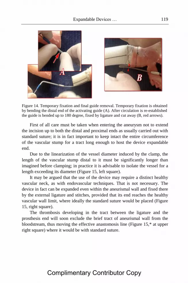
Expandable Devices … 119
Figure 14. Temporary fixation and final guide removal. Temporary fixation is obtained
by bending the distal end of the activating guide (A). After circulation is re-established
the guide is bended up to 180 degree, fixed by ligature and cut away (B, red arrows).
First of all care must be taken when entering the aneurysm not to extend
the incision up to both the distal and proximal ends as usually carried out with
standard suture; it is in fact important to keep intact the entire circumference
of the vascular stump for a tract long enough to host the device expandable
end.
Due to the linearization of the vessel diameter induced by the clamp, the
length of the vascular stump distal to it must be significantly longer than
imagined before clamping; in practice it is advisable to isolate the vessel for a
length exceeding its diameter (Figure 15, left square).
It may be argued that the use of the device may require a distinct healthy
vascular neck, as with endovascular techniques. That is not necessary. The
device in fact can be expanded even within the aneurismal wall and fixed there
by the external ligature and stitches, provided that its end reaches the healthy
vascular wall limit, where ideally the standard suture would be placed (Figure
15, right square).
The thrombosis developing in the tract between the ligature and the
prosthesis end will soon exclude the brief tract of aneurismal wall from the
bloodstream, thus moving the effective anastomosis line (Figure 15,* at upper
right square) where it would be with standard suture.
Complimentary Contributor Copy

Stefano Nazari 120
Figure 15. Expandable device operational details. Left square: When a clamp is applied
the linearization of the stump requires to keep its length significantly longer than usual
in order to leave the space necessary for full expansion of the expandable end. It is also
important to avoid full longitudinal vascular opening as carried out with current
technique and thus keeping intact the entire circumference of the vascular stump for a
tract long enough to host the whole device expandable end. Right square: The
expandable end also can be positioned against aneurismal wall, provided that its distal
end would reach the healthy vascular wall. Thrombosis of the tract between the
ligature and the prosthesis end will soon move the effective anastomosis line (upper
right square*) where it would be with standard suture (mod from 28 with permission).
Thus devices type I and II can be ideally used anywhere in descending
thoracic and abdominal aorta and allow to carry out any required additional
surgical maneuver on the tube graft, i.e. collateral branch anastomosis, as well
as its appropriate tailoring at the required length measured directly on the
operative field as in standard technique without obstacles or hampering
conditions. Its great simplicity of use allows the devices to be used also in
condition of suboptimal operative field exposure. Thus for example both
proximal and distal anastomosis in extended descending thoracic aorta
substitution can be easily and safely carried out though a single space
thoracotomy; moreover aortic prosthetic substitution via mini-access
thoracotomy or laparotomy video-assisted setting may be also predictably
possible and certainly more easy than standard suture technique.
2.1.2. Device Type III In this version of the device the external ligature is substituted by an
expandable sleeve, which is based essentially on the same working principle as
the inner sleeve, but activated contrariwise. Thus, the vascular stump is
compressed between two sleeves (Figure 16, upper left little squares), with
Complimentary Contributor Copy

Expandable Devices … 121
variable and controllable diameters, allowing full control of the pressure
(amount and surface of its application) actually applied to the vascular stump.
Operative technique for device type III is illustrated in Figure 16 in
ascending aorta swine “ex vivo” model and it is really very simple. First of all
both sleeves diameter is set at the predicted value of the aortic tract where the
device will be applied. Then by acting on its guide-wire the inner sleeve only
its diameter is reduced as much as an easy insertion into the vascular stump is
possible; the inner sleeve diameter reduction causes also the backwards
rotation and eversion of the outer sleeve thus greatly enhancing inner sleeve
visibility and then its easy positioning into the vascular stump. The inner
sleeve is then re-expanded against the vascular stump inner surface; at this
point the outer sleeve is gently retracted acting on its own activating guide to
compress the vascular stump. Temporary and final aperture of inner and outer
sleeves as well as guide removal is carried out with the same technique
indicated for type I and II devices at Figure 14.
Figure 16. Device type III. The wire-frame of the device is quite soft and compliant,
and can be easily compressed and widely deformed while maintaining perfect
reciprocal alignment of the internal and external sleeves (top left squares) and is
intended ideally for use in dissection cases or when the aortic wall may need some
form of buttressing. Lower squares: “ex vivo” ascending aorta swine model. First of all
both sleeves are set at predicted final aperture. The acting on the activating guide of the
inner sleeve only results in the backwards rotation and eversion of the outer sleeve,
bringing the retracted inner sleeve in full visibility, so that it can be easily inserted into
the vascular stump. Then inner ring is then expanded so that the vascular wall is
pushed against the outer sleeve inner surface. At this point, the outer sleeve is only
slightly retracted towards the aortic wall using its own guide. Temporary and final
aperture stabilization and guide removal is carried out in the same way as with type 1
or II devices (figure 14). Video at http://www.fondazionecarrel.org/nazarichaptervideo.
html.
Complimentary Contributor Copy

Stefano Nazari 122
Figure 17. Type III security fixation. A simple in and out 3 or 4-0 prolene in several
points linking the inner and outer device wireframe and aortic stump will greatly
increase coupling stability and can be carried out easily and quickly when circulation is
resumed. Video at http://www.fondazionecarrel.org/nazarichaptervideo.html.
Even though needles present on the outer surface may be sufficient to
provide stability when the aortic wall is compressed by the outer ring, we think
that polypropylene 3 or 4 0 running suture with several full thickness, in and
out, circumferential passages firmly linking aortic stump with inner and outer
expandable sleeves (Figure 17) may add absolute safety in an easy and quick
way that may be carried out when circulation is resumed.
The primary aim of this new version of the device is to make possible and
convenient to apply this coupling principle also to acute ascending aorta and
arch dissection, in order to simplify the technique, to reduce the ischemic time,
to improve hemostasis of the anastomosis line and to achieve reliable, stable
sealing of the dissection layers in this very complex surgical setting.
Type III device in fact allows to actually automate substantially the same
aortic wall sandwiching between two graft strips procedure usually carried out
in the dissection cases and to realize at once the tube prosthesis anastomosis,
being the tube graft (not shown in the figure 16) obviously previously sutured
to the inner sleeve. Interestingly enough the particular configuration of the
device allows full and easy compliance with aortic anatomy, perfectly
adapting also to the elliptic, asymmetric “oblique” stump resulting from
inclusion of the arch concavity in the anastomosis line.
In fact full and persistent air-tight sealing of the device-aortic wall
coupling was verified at endovascular pressures of up to 150 mmHg in “ex
vivo” swine aortic models, including those involving an elliptic, ‘oblique’
Complimentary Contributor Copy

Expandable Devices … 123
anastomosis (Figure 18, b) [32]. Interestingly enough standard vascular sutures
were not “air-tight” even at pressures below 10 mmHg (Figure 18, c).
The solution of positioning and stability problems of the 70ies
intraluminal prosthesis (Figure 10) allows for the first time the clinical
appreciation of the most important feature of the “Payr principle” of
anastomosis (vascular stumps compression against/between rigid structures i.e.
an inner rigid ring and external ligature/outer rigid ring) which is to achieve an
immediate hermetic seal (“air-tight” in fact) ensuring reliable haemostasis at
the anastomosis line, not dependent from coagulation. Interestingly enough
were just the positioning problems of Payr model as well as of its more recent
modification (intraluminal ringed prosthesis), that prevented the clinical
implementation of this coupling method, which is the most intuitive (and in
fact was the first to be attempted even in vascular surgery) and whose
application failed only in the surgical field, while was in use, and still is, in all
other technological fields where connectors between elastic/floppy tubing are
required.
Figure 18. Device type III seal test. The outer surface of the inner sleeve was wrapped
by a latex cuff (top squares) in order to overcome the problem of the porosity of the
Dacron graft and the requirement for the connection of the tube graft to the proximal
end of the inner sleeve as in its final clinical use. A) The air-tightness of the connection
was verified at endovascular pressures of up to 150 mmHg in a regular cylindrical
anastomosis of ascending aorta (white bars). (B) The same was verified when the
anastomosis is irregularly oriented, such as when involves the arch concavity. (C) As
easily predictable, a standard suture (4–0 prolene) of an approximately 3 cm incision of
the aortic wall cannot be proved airtight even at minor endovascular pressure (modify.
from 32 with permission). Video at http://www.fondazionecarrel.org/
nazarichaptervideo.html.
Complimentary Contributor Copy

Stefano Nazari 124
Of course clinical experience over more than a century has shown that the
standard suturing technique does not need to provide an “air-tight”
anastomosis to ensure perfect hemostasis in virtually all clinical
circumstances. In particular cases however (acute dissection, Marfan [16] and
other genetic disorders, cystic medial necrosis, etc.), structural impairments of
the aortic wall may necessitate additional maneuvers including graft strips
buttressing, gluing and a variety of accessory techniques, whose efficacy at
achieving haemostasis are not always fully predictable and that obviously
further significantly prolong the period of ischemia in this most critical area. A
coupling method that provides haemostasis by compression of the stumps’
vascular wall between two rigid structures (i.e. inner and outer expandable
rigid sleeves) without perforation may be then particularly useful in these
cases, not only because of its ease and quickness, but also because it offers the
best mechanical chance of immediate blood-tightness. The expected
advantages with regards to approximation of dissecting layers and false lumen
permanent sealing rely on the same mechanical concept.
Another important difference with 70-80ies intraluminal ringed devices is
that the expandable sleeve is much more thin and porous, being formed
substantially by a double layer of standard vascular Dacron fabric, and can,
therefore, be wholly and quickly colonized by fibroblasts and soon integrated
within the aortic wall. The nitinol wire-frame in fact forms a very thin and
wide mesh net that accounts only for a very small proportion of the device’s
volume and that offers no significant barrier to fibroblastic invasion of the
Dacron fabric and thus to stable biological integration of the device.
Type III device use can be then ideally indicated in dissection cases for
distal anastomosis sited at distal ascending aorta, including as much as
required of the concavity of the arch during a very brief circulatory arrest
phase; proximal anastomosis will be then carried out either by hand suture or
with the expandable device version most appropriated for the particular
anatomical condition, in normal CEC in no rush and after having performed
any additional procedure possibly required, for example on the valve.
Anastomosis at the distal arch/proximal descending aorta in case of full
arch substitution is also an ideal indication for type III device use in case of
dissection or whenever, for particular aortic wall fragility, graft strips
sandwiching buttressing may be advisable. Thus in all cases where
sandwiching buttressing is planned, the use of the device type III requires
exactly the same aortic stump external wall preparation required for hand
suture double graft strips application with the advantage of realizing it much
more quickly and at once with the tube graft anastomosis.
Complimentary Contributor Copy

Expandable Devices … 125
When compared with device I or II the double ring configuration of device
type III allows a very significant reduction of its length, thus greatly enhance
its positioning into the curved anatomy of the ascending aorta and arch without
any orientation conflict, even allowing an asymmetric disposition of the
expandable sleeves in different area of the anastomosis without losing “air-
tight” competence.
The great simplification and the very significant quickening of this
complex surgical part together with the higher accuracy and “mechanical”
reliability of this coupling method in comparison with manual suture could
potentially have impact that may exceed that strictly related to the
anastomosis. For example being possible to carry out even the entire arch re-
vascularization in very few minutes of circulatory arrest, the level of
hypothermia may be significantly reduced and even the type of cerebral
protection may be tailored to these very restricted time lapses, when supraortic
trunks anstomosis is carried out by graft assembled with appropriate devices’
combination (see below), just to say the firsts coming in mind.
2.2. Other Expandable Device Models and Combinations
We also realized a variety of modified versions of the device to better fit
with the anatomical configurations occurring in particular clinical
circumstances.
2.2.1. Type I with Single Outer Layer for Small Diameters In small vessel diameters device fabric wrapping can interfere more
significantly with effective lumen amplitude in their range of use. More in
particular the way of folding of the inner fabric layer when the device is
incompletely opened may reduce the lumen effectively available for the blood
flow to an extent that may be difficult to predict. Accordingly for diameters ≤
12 and ≥6 mm we prepared devices wrapped by un-crimped thin fabric
positioned only at external side of the nitinol wireframe. Three sizes were
prepared respectively for diameter from 6-8 mm, 8-10 and from 10 to 12 mm.
The device is previously prepared at the end of collateral branch of the main
tube graft cut at the expected appropriated length or directly on the main tube
graft (see Figure 20, 21, 22 and 23). The ideal evolution of this project
however would probably consider devices size appropriate to each currently
available small diameter tube graft size, mounted at an un-crimped part of one
of its end.
Complimentary Contributor Copy

Stefano Nazari 126
This device type may ideally fit for renal, celiac, mesenteric art. and
supraortic trunks as well as for any vessel with diameter ≥ 6 mm.
2.2.2. Type II Bendable and with Independent Opening This type II device modified version is intended for use in ascending aorta
substitution in absence of dissection. The two ends of the device can vary their
reciprocal axis up to 90° to better fit with the curvature of the ascending aorta;
their aperture can be independently controlled in order to comply with possible
differences in diameter of the proximal and distal stumps (Figure 19). In this
way the ascending aorta substitution in anatomically suitable cases can be
carried out very quickly by one single device.
In case of dissection however type III device previously connected with
graft tube should be better used at distal or both anastomosis. Various possible
combinations between device types can ideally fit any particular anatomical
occurrence. Anyway proximal anastomosis can be carried out with standard
suture, time being not here a critical factor, or by a second type III device if
suggested by the quality of the proximal vascular stump.
Figure 19. Device Type II Bendable and with Independent Opening. This type II
device version allows the bending up to a 90° degree angle of the axis of their ends,
whose aperture can be independently controlled. This allows to fit curved anatomy of
ascending aorta and possible variations in diameter of the vascular stumps. The lower
strip shows the device application in “ex-vivo” swine ascending aorta model. Video at:
http://www.fondazionecarrel.org/nazarichaptervideo.html.
Complimentary Contributor Copy

Expandable Devices … 127
Figure 20. Composite configuration for arch substitution (swine model). Distal
anastomosis is performed with type I or 3 devices according to the presence of
dissection or poor quality of aortic stump as well as distal aorta pathology; in case type
I is used full isolation of the distal anastomosis site can be as usually avoided and
device fixed by passing the ligature, totally or only in part from inside as shown in
figure 11. Type I devices with single outer layer is used if the supraortic trunk inner
diameter is less then 12 mm. The illustrated prototypes are shaped to fit with “ex vivo”
swine anatomy.
This device version can also be ideally suitable for isthmus rupture repair,
having care of entering the aorta through or close to the laceration in order to
preserve integrity of the vascular wall at site of device ends deployment;
obviously the left subclavian artery origin may not allow room for proximal
device deployment and then re-implant or by-pass can be considered if
deemed advantageous in time critical conditions.
2.2.3. Composite Configurations
2.2.3.1. Aortic Arch With expandable devices appropriately prepared on tube graft it is
possible to quickly connect supra-aortic trunks and proximal descending aorta
during a very short circulatory arrest phase. The ideal configuration of tube
graft include expandable device at its distal end for distal arch-proximal
descending aorta anastomosis, the type being selected on the basis of vascular
Complimentary Contributor Copy

Stefano Nazari 128
stump and descending aorta conditions (Figure 20). Moreover type I devices
appropriate to supraortic trunks size (single outer layer for inner diameters ≤
12 mm) are previously prepared ideally on branched tube graft whose length is
tailored according to the occurring anatomy before circulatory arrest. Elephant
trunk (original or frozen) procedure is also easily performed also when type III
device is to be used for distal arch anastomosis, here illustrated in “ex vivo”
swine anatomy compatible prototypes (Figure 21); in fact backwards
displacement of the outer sleeve when the inner one is retracted greatly
enhance insertion of the elephant trunk and appropriate vascular stump
positioning to be compressed between the device’s sleeves whose final
aperture can be adjusted to the most appropriate diameter. The particular
configuration of prototypes for human anatomy and size suitable for aortic
arch substitution (Figure 22) allows an easy and effective compensation of
even wide discrepancy between aortic diameter and elephant trunk.
Figure 21. Elephant trunk (swine model). Device type 3 (A) backwards displacement
of the outer sleeve after the inner one is retracted (C) greatly enhance insertion of the
elephant trunk and appropriate vascular stump positioning (D) to be compressed
between the device’s sleeves (E); the easy and quick supraortic trunks is then carried
out (F,F’) Video at: http://www.fondazionecarrel.org/nazarichaptervideo.html.
Complimentary Contributor Copy

Expandable Devices … 129
Figure 22. Human configuration prototypes for aortic arch. Figure illustrates
prototypes human anatomy and size (outer sleeve 55 mm, tube graft 36 mm) suitable
for arch substitution.
Figure 23. Thoraco-abdominal Aorta. Expandable device type I, ≧ 6 mm in diameter,
with single outer Dacron layer (red squares) can be prepared on appropriately tailored
branches of Coselli’s type tube graft for quick connections of main visceral aortic
collaterals. This version of device type I for small diameters was prepared with single
outer un-crimped fabric graft in order to prevent the possible significant interference
with vascular lumen of unpredictable way of folding of the inner layer when the device
cannot be fully expanded.
Complimentary Contributor Copy

Stefano Nazari 130
2.2.3.2. Thoraco-Abdominal Aorta Type I devices with graft single outer layer as small as 6 mm in diameter
can also be previously prepared on branches of Coselli’s tube (Figure 23)
tailored in length appropriated to the patient anatomy before clamping
allowing; this can significantly simplify and quickening this still very critical
clinical condition.
Figure 24. External ligature and scattered transfixion - a meeting point. Reliable
coupling stability, instead of simple external ligature is achieved by few prolene 3-0 or
4-0 encircling suture, transfixing full thickness the device and aortic wall stump at few
(2 or more approximately equidistant) points, passed and tied as shown. It may be then
speculate that expandable configuration of the prosthesis end made possible a quite
advantageous meeting point between the first endeavor (Payr) and current technique
(Carrel) for vascular-graft anastomosis. Video at
http://www.fondazionecarrel.org/nazarichaptervideo.html.
Conclusion and Perspectives
The extensive past experience provided full mechanical reliability of
devices in all their versions and proved their easy applicability to aortic stump
in all conditions in “ex vivo” models. Animal experience and successful
clinical application in few clinical case confirmed that expandable devices
provide very significant advantages over current hand suture anastomosis in
aortic prosthetic substitution.
While the expandable device allows a much easier, quicker and more
efficient (“airtight” seal) graft-aortic stump coupling than standard suture [32],
it may be argued however that the permanence of endovascular tubular
wireframe and external ligature could, in particular circumstances,
Complimentary Contributor Copy

Expandable Devices … 131
mechanically conflict with aortic wall, particularly at device ends, and/or with
confining tissues/organs.
Aorto-digestive fistula is an infrequent but well documented occurrence
after aortic open [14, 22] as well as after endovascular [37, 23, 10] repair.
While graft or/and suture line contamination/infection may occasionally be
suspected as the primary etiological factor, pure mechanical erosion from
systolic or other causes movements of graft and even from the suture line only
[40] may probably represent the first triggering factor in a fraction of cases
difficult to quantify. The pure mechanical effect of these movements on the
confining tissue/organ is predictably higher the harder/less compliant is the
prosthetic material as well as the more conflicting is its orientation in relation
to the confining tissue/organ. We then recently focused on optimal consistency
of the expandable wireframe and on means to provide external fixation with
the final aim of achieving mechanical forces of coupling as much as possible
similar to those taking place in standard hand suture anastomosis.
For this purpose, on one side, the wireframe consistency was decreased by
variably reducing the gauge of nitinol wires and their respective position
within wireframe and, on the other side, the external ligature was carried out
with the thinnest possible prolene (4-0 / 5-0) encircling suture, transfixing full
thickness the device and aortic wall at two or three approximately equidistant
points. The mechanical limit of the former was the ability to sustain the
external ligature without wireframe deformation/collapse at pressure providing
“airtight” seal; the limit of the later was the ability to provide stability of the
aortic stump-expandable device coupling at stretch test.
The present versions of the devices minimize the risk of mechanical
conflict and erosion with the confining tissue/organs; moreover wireframe
consistency was settled in such a way that is minimal (thinner nitinol wire) at
the device distal end where the possible conflict with aortic wall could be
higher. However a tighter surgical limit to reduction of nitinol wire diameter
and then overall wireframe consistency is related to the necessity to preserve
the ability of the device to actively expand the vascular stump enough to
achieve the expected diameter when blood perfusion is resumed. Ex-vivo
experiments showed that this goal requires nitinol wire diameters significantly
greater than those required for simple achieving haemostasis and stability by
external ligature.
Moreover the variable conditions of vascular stump may also suggest in
this regard different devices wireframe consistency ranging from the lowest,
appropriate for example in case of dissection, to the strongest in presence of
heavy calcification/atherosclerosis. Accordingly it seems important to provide
Complimentary Contributor Copy

Stefano Nazari 132
each device type and version in two or three different wireframe consistencies
(i.e. strong, standard, light) to adequately fit with all clinical conditions.
Concerning the external ligature and its many possible ways of
application, it may be useful to recall again that haemostatic seal and stability
of the expandable device anastomosis, contrariwise to standard suture, rely on
different mechanisms (Figure 8).
Thus even though external devices surface is provided with short needles
perpendicularly positioned around its circumference at 4-6 equidistant points,
reduction of the wireframe consistency may probably decrease their reliability
in keeping coupling stability. We then think that it is very important to
optimize coupling stability adding to the external ligature, transfixing full
thickness the device and aortic wall at least in two or three approximately
equidistant points.
The stability of this coupling in its many possible variations was checked
by a stretch test model consisting in a sequence of maneuvers carried out on
the anastomosis that includes: 1) complete compression of the anastomosis in
orthogonal directions and then 2) vigorous manual stretching of the
anastomosis in the coaxial plane separately at four points of its circumference.
The sequence was repeated three times and the anastomosis checked for any
significant vascular stump backwards dislocation on the expandable wireframe
throughout the entire circumference (Video at: http://www.fondazionecarrel.
org/nazarichaptervideo.html).
This allowed to refine details involved in this procedure (external ligature
+ 2/3 stitches fixation) that can be ideally carried out with polypropylene
suture, 3 to 5-0 depending on the vascular stump consistency and diameter,
positioned either in two steps, i.e. encircling ligature and vascular wall-device
transfixion stitches or with a single step the suture being passed in the way
indicated in Figure 23.
Device type III stability is also increased by further buttressing the inner
and outer Dacron wrapping by 4-0 prolene suture transfixing the
circumference in several points (Figure 17). In synthesis this method ideally
allows to convert any vascular-graft anastomosis ≥ 6 mm in diameter from the
current facing ends Carrel suture into a simpler, quicker and more efficient
(“airtight” seal) telescoping anastomosis, sealed and fixed by single thread
external ligature passed full thickness at two/three points (or more when
appropriate), in a sort of ideal optimized meeting point between Payr first and
Carrel current technique.
Complimentary Contributor Copy

Expandable Devices … 133
Table 1. Potential impact of expandable device aortic anastomosis
compared with current hand suture technique
The hypothesizable potential impact (table 1) may exceed that expected on
complication rate of open prosthetic substitution of all aortic tracts and in
particular in those higher risk conditions as acute dissection.
In fact the technical simplification with increased reliability of
anastomosis haemostasis and dissection layer approximation with false lumen
permanent seal has the logical direct consequence, for example, of enabling
also lesser expert cardiovascular surgeons to deal with these clinical cases very
often requiring immediate surgical attention, thus increasing surgical team
efficiency and hospital unit productivity.
Complimentary Contributor Copy

Stefano Nazari 134
References
[1] Aluffi A, Buniva P, Rescigno G and Nazari S. (2002) Improved device
for sutureless aortic anastomosis applied in a case of cancer. Tex. Heart
Inst. J. 29: 56–59.
[2] Antoniou GA, El Sakka K, Hamady M and Wolfe JH. (2010) Hybrid
treatment of complex aortic arch disease with supraaortic debranching
and endovascular stent graft repair. Eur. J. Vasc. Endovasc. Surg.
39:683-90.
[3] Berger RL, Romero L, Chaudhry AG and Dobnik DB.(1983) Graft
replacement of the thoracic aorta with a sutureless technique. Ann.
Thorac. Surg. 35:231–239.
[4] Brown LC, Thompson SG, Greenhalgh RM and Powell JT (2011):
Incidence of cardiovascular events and death after open or endovascular
repair of abdominal aortic aneurysm in the randomized EVAR trial 1.
British Journal of Surgery 98: 935–942.
[5] Buniva P, Rescigno G, Salvi S, Aluffi A, Berti A. Salsi F, Nazari S
(2002) Sutureless expandable end prosthesis for descending aorta
substitution invaded by bronchogenic cancer. Video, Proceedings Aortic
Surgery Symposium VIII, New York, May 2-3, 2002.
[6] Canaud L, Hireche K, Berthet JP et al.(2010): Endovascular repair of
aortic arch lesions in high-risk patients or after previous aortic surgery:
midterm results. J. Thorac. Cardiovasc. Surg. 140:52-8.
[7] Carrel A. (1902) La technique operatoire des anastomoses vasculaires et
la transplantation des visceres. Lyon Med. 98:859–864.
[8] CarrelA. (1907) The surgery of blood vessels. Bull. Johns Hopkins
Hosp.; 18: 18–27.
[9] Charnsangavej C, Wallace S, Wright KC, Carrasco CH and Gianturco C.
(1985) Endovascular stent for use in aortic dissection: an in vitro
experiment. Radiology.157:323-4.
[10] Chiesa R, Melissano G, Marone EM, Marrocco-Trischitta MM and
Kahlberg A (2010)Aorto- oesophageal and aortobronchial fistulae
following thoracic endovascular aortic repair: a national survey. Eur. J.
Vasc. Endovasc. Surg. 39:273-9.
[11] Crawford ES and Crawford JL, editors (1984). Diseases of the aorta.
Baltimore: Williams and Wilkins, 61–77.
[12] De Bruin JL, Baas AF, Buth J, Prinssen M, Verhoeven EL, Cuypers PW
et al.; DREAM Study Group.(2010) Long-term outcome of open or
Complimentary Contributor Copy

Expandable Devices … 135
endovascular repair of abdominal aortic aneurysm. N. Engl. J. Med. 362:
1881–1889.
[13] Di Eusanio M, Armaro A, Di Marco L, Pacini D, Savini C, Martin
Suarez S, Pilato E and Di Bartolomeo R (2011) Short- and midterm
results after hybrid treatment of chronic aortic dissection with the frozen
elephant trunk technique. Eur. J. Cardiothorac. Surg. 2011 Oct; 40(4):
875-80. doi: 10.1016/j.ejcts.2011.01.068. Epub 2011 Mar 5.
[14] Geraci G, Pisello F, Li Volsi F, Facella T, Platia L, Modica G and
Sciume C (2008) Secondary aortoduodenal fistula. World J.
Gastroenterol. 14:484-6.
[15] Gopaldas RR., Huh J, Dao TK, LeMaire SA, Chu D, Bakaeen FG and
Coselli JS (2010) Superior nationwide outcomes of endovascular versus
open repair for isolated descending thoracic aortic aneurysm in 11,669
patients. J. Thorac. Cardiovasc. Surg. 140:1001-10.
[16] Gott VL, Laschinger JC, Cameron DE, Dietz HC, Greene PS, Gillinov
AM, Pyeritz RE, Alejo DE, Fleischer KJ, Anhalt GJ, Stone CD and
McKusick VA:(1996) The Marfan syndrome and the cardiovascular
surgeon. Eur. J. Cardiothorac. Surg. 10:149-158.
[17] Kawaharada N, Ito T, Koyanagi T, Harada R, et al. (2010) Spinal cord
protection with selective spinal perfusion during descending thoracic and
thoracoabdominal aortic surgery. Interact. CardioVasc. Thorac. Surg.
10:986-991.
[18] Kolesov VI, Kolesov EV, Gurevich IY and Leosko VA.(1970)
Vasosuturing apparatuses in surgery of coronary arteries. Med. Tekhnika
6:24–8.
[19] Kolesov VI and Kolesov EV. (1991) Twenty years' results with internal
thoracic artery- coronary artery anastomosis [letter]. J. Thorac.
Cardiovasc. Surg. 101:360–1.
[20] Konstantinov I E. (2004): Vasilii I. Kolesov A Surgeon to Remember.
Tex. Heart Inst. J. 31: 349–358.
[21] Lemole GM, Strong MD, Spagna PM and Karilomicz MD (1982).
Improved results for dissecting aneurysms: intraluminal sutureless
prosthesis. J. Thorac. Cardiovasc. Surg. 83:249–255.
[22] Luo CY, Lai CH, Wen JS and Lin BW (2010) Secondary aortocolic
fistula: case report and review of the literature. Ann. Vasc. Surg. 24:
256.e5-12.
[23] Marone EM, Esposito G, Kahlberg A, Tshomba Y, Brioschi C, Zannini
P and Chiesa R. (2007) Surgical treatment of tracheoinnominate fistula
Complimentary Contributor Copy

Stefano Nazari 136
after stent- graft implantation. J. Thorac. CardioVasc. Surg. 133:1641–
1643.
[24] Nazari S, Manelli A, Gastaldo A, Rinaldo A, Barana L and Zonta A.
(1990) A new vascular stapler for pulmonary artery anastomosis in
experimental single lung transplantation. Video, Proceedings of the 4th
Annual Meeting of the Association for Cardio-Thoracic Surgery, Naples,
September 16–19, 1990.
[25] Nazari S., Mourad Z, Luzzana F. (1994) Prosthetic substitution of the
descending aorta without blood flow interruption. Proceedings of the IV
Aortic Surgery Symposium, New York, April 20-22, 1994.
[26] Nazari S, Luzzana F, Banfi C, Mourad Z, Salvi S, Gaspari A and Nazari-
Coerezza F.(1996) Expandable prosthesis for sutureless anastomosis in
thoracic aorta prosthetic substitution. Eur. J. Cardiothorac. Surg. 10:
1003– 1009.
[27] Nazari S, Mourad Z, Banfi C, Salvi S, Visconti E, Aluffi A. (1996)
Composite graft with expandable ends for total arch replacement. Video
and Poster, Proceedings Aortic Surgery Symposium V, New York, April
23-26, 1996.
[28] Nazari S, Salvi S, Aluffi A, Visconti E, Rescigno G and Buniva P (1997)
A new prosthesis for aortic arch substitution. Ann. Thorac. Surg. 64:
1339–1344.
[29] Nazari S, Salvi S, Visconti E, Caramella F, Rossi C, Rescigno G, Banfi
C and Buniva P. (1999) Descending aorta substitution using a prosthesis
with expandable ends. Case report. J. Cardiovasc. Surg. (Torino) 40:
417–420.
[30] Nazari S, Thoracic Aorta Substitution in less than 2 minutes.
(2006)Video, Proceedings 16th World Congress of World Society of
Cardio- Thoracic Surgeons, Ottawa, August 17-20, 2006.
[31] Nazari S. (2009) Modified Expandable Device for Easy and Realiable
Dissection Layers Approximation in Sutureless Aortic Anastomosis.
Video, proceedings of The Houston Aortic Symposium, Houston, March
26-28, 2009.
[32] Nazari S (2010) Expandable device type III for easy and reliable
approximation of dissection layers in sutureless aortic anastomosis. Ex
vivo experimental study. Interact. CardioVasc. Thorac. Surg. 10:161-
164; originally published online Nov 20, 2009; DOI: 10.1510/icvts.2009.
216291.
Complimentary Contributor Copy

Expandable Devices … 137
[33] Parodi JC, Palmaz JC and Barone HD (1991) Transfemoral intraluminal
graft implantation for abdominal aortic aneurysms. Ann. Vasc. Surg.
5:491-9.
[34] Parodi JC. (1994) Endovascular repair of abdominal aortic aneurysms
and other arterial lesions. Surg. Technol. Int. 3:431-6.
[35] Payr E. (1900) Beitrage zur Technik der Blutgefass und Nervennaht
nebst Mittheilungentiber die Verwendung eines resorbierbaren Metalles
in der Chirurgie. Arch. Klin. Chir.; 62:67–93.
[36] Rossella C, Rescigno G, Nazari S. (2008) Modified expandable device
for easy, quick and blood-tight sutureless anastomosis in ascending
aorta. Proceedings of The Houston Aortic Symposium, April 2008.
[37] Ruby BJ and Cogbill TH (2007) Aortoduodenal fistula 5 years after
endovascular abdominalaortic aneurysm repair with the Ancure stent
graft. J. Vasc. Surg. 45:834-6.
[38] Schermerhorn ML, O’Malley AJ, Jhaveri A, Cotterill P, Pomposelli F,
Landon BE. (2008) Endovascular vs. open repair of abdominal aortic
aneurysms in the Medicare population. N. Engl. J. Med. 2008; 358: 464–
474.
[39] Takata M, Watanabe G, Ohtake H, Ushijima T, Yamaguchi S, Kikuchi
Y and Yamamoto Y(2011): Automatic aortic anastomosis with an
innovative computer-controlled circular stapler for surgical treatment of
aortic aneurysm. J. Thorac. Cardiovasc. Surg. 141:1265-9.
[40] Tanaka S, Kameda N, Kubo Y, Obatake N, Wakasa T, Ohsawa M and
Wakasa K. (2009) Secondary aortoduodenal fistula caused on the suture
line of the wrapping. Pathology International 59: 598–600 doi:10.1111/
j.1440-1827.2009.02414.x.
[41] Tsagakis K, Pacini, D, Di Bartolomeo R et al. (2011).: Arch replacement
and downstream stent grafting in complex aortic dissection: first results
of an international registry. Eur. J. CardioThor. Surg. 39:87-94.
[42] Tesauro B. (1967 ) Impiego degli adesivi plastici e delle cucitrici
meccaniche in chirurgia vascolare. Atti del 698 Congresso della Societa`
Italiana di Chirurgia, Firenze 18– 20 Ottobre 1967. In: Fegiz G, editor.
Archivio ed atti della Societ`a Italiana di Chirurgia. Roma: EMES,
1:663–790.
[43] Tesauro B and Persico G. (1979) Valutazione critica delle suture
meccaniche in chirurgia vascolare. Atti dell’818 Congresso della
Societa` Italiana di Chirurgia, Catania 8–11 Ottobre 1979. In: Fegiz G,
editor. Archivio ed atti della Società Italiana di Chirurgia, Bologna:
CLUEB, 1:633– 652.
Complimentary Contributor Copy

Stefano Nazari 138
[44] The UK EVAR Trial Investigators, Greenhalgh RM, Brown LC, Powell
JT, Thompson SG, Epstein D, Sculpher MJ.(2010) Endovascular versus
open repair of abdominal aortic aneurysm. N. Engl. J. Med. 2010; 362:
1863–1871.
[45] Wright KC, Wallace S, Charnsangavej C, Carrasco CH and Gianturco C
(1985) Percutaneous endovascular stents: an experimental evaluation.
Radiology. 156:69-72.
[46] Yoshioka T, Wright KC, Wallace S, Lawrence DD Jr and Gianturco
C.(1988) Self-expanding endovascular graft: an experimental study in
dogs. AJR Am. J. Roentgenol. 151:673-6.
[47] Zannini G (1967) Introduzione storica. In Atti del 69° Congresso della
Societa` Italiana di Chirurgia, Firenze 18–20 Ottobre 1967. In: Fegiz G,
editor. Archivio ed atti della Società Italiana di Chirurgia. Roma:
EMES, 1:7-32.
Complimentary Contributor Copy

In: Anastomoses ISBN: 978-1-62618-657-6
Editors: F. King, McKineley A. Malloy © 2013 Nova Science Publishers, Inc.
Chapter V
Bowel Anastomosis: Types, Techniques/Procedures,
Clinical Outcomes and
Complications
Jair Santos-Torres, Jaime Ruiz-Tovar,
Antonio Arroyo and Rafael Calpena General University Hospital of Elche,
Department of General and Digestive Surgery,
Elche, Alicante, Spain
Abstract
Despite development of improved surgical techniques, advances in
perioperative and critical care and introduction of broad-spectrum
antibiotics, colorectal surgery continues to present with as a great
challenge. Postoperative complications are common, occurring in 18-57%
of patients after elective surgery and in 39.3-72% after emergency one.
Potential postoperative complications associated with the colorrectal
surgery are complications related to anastomosis.
There is nothing that provokes greater anxiety and consternation to
the gastrointestinal surgeon than the prospect of a leak from a colonic or
colorectal anastomosis. The consequences to the patient from such a
complication can be significant and not infrequently life-threatening. A
Complimentary Contributor Copy

Jair Santos-Torres, Jaime Ruiz-Tovar, Antonio Arroyo et al. 140
surgeon can only control some of the many variables in anastomotic
construction. The fundamental principles of preservation of an adequate
blood supply, total absence of tension on the suture line and healthy
bowel for both the proximal and distal ends without thickening or
inflammation have remained constant. The necessity of bowel preparation
is now a topic of considerable debate, defending many surgeons not to be
performed. The technical requirements include the creation of an airtight
suture line, in some circumstances protected by a proximal diverting
procedure, and/or omental wrap. Whether the anastomosis is hand-sewn
in one or two layers, performed with interrupted or running suture
technique, or constructed with a stapling device has no impact on leak
rates. Factors often beyond the surgeon’s control are immutable
comorbidities and the patient’s body habitus.
A safe anastomosis should include: not leak, cause no persistent
bleeding, cause no stricture of the lumen and create no risk for internal
hernia. An ideal anastomosis must be also easy to construct, consistently
reproducible, and easy to teach.
The aim of paper is to review types, techniques, procedures, clinical
outcomes and complications of colorectal anastomoses, including
mechanical and hand-sewn sutures, of the colonic and colorectal
anastomoses. We expect to help surgeons and surgical fellows to learn
about this topic with particular attention to the risk factors and procedure-
related complications.
Introduction
Despite the development of improved surgical techniques, advances in
perioperative and critical care and introduction of broad-spectrum antibiotics,
colorectal surgery continues presenting with as a great challenge.
Postoperative complications are common, occurring in 18-57% of patients
after elective surgery and in 39.3-72% after emergency colorectal surgery. A
number of prospective studies, both multicenter and single center, have
evaluated patient outcomes after colorectal surgery. The rate of major
morbidity ranged from 20 to 35% and the 30-day mortality rate ranged from 2
to 9 %. Potential postoperative complications associated with the colorrectal
surgery are complications related to anastomosis. This may be secondary to
ischemia, bleeding, leakage and inappropriate technique (i.e. inadequate tissue
approximation) [1-5].
Options for restoring bowel continuity after a resection include an end-to-
end, side-to-end or side-to-side colorectal, colocolic or ileocolic anastomosis,
which can be either performed with a stapling device or hand-sewn. Stapled
Complimentary Contributor Copy

Bowel Anastomosis 141
anastomosis requires less time to be performed and offers the potential for
reduced fecal contamination. An alternative anastomosis that can be used
between the proximal and distal colon (not rectum) is the functional end-to-
end anastomosis, which is technically a side-to-side approach. There is
insufficient evidence that any configuration is better functionally or less likely
to leak, or that a stapled colorectal anastomosis is superior to a hand-sewn in
ileocolic and colocolic anastomosis; the decision is based upon surgeon
experience, preference, and available equipment [6-8]. However, in colorectal
anastomosis, specially referring to lower rectal resections, though there is no
enough evidence of better results with stapled devices, some trials show better
result with this approach due to technical difficulty of the hand-sewn
procedures [9-10].
Over the past two decades, numerous different materials have been used to
join one bowel end to the other one, including catgut, stainless steel and newer
monofilamentous and absorbable sutures. In the past 30 years, stapling devices
have been embraced enthusiastically by the surgical community. The diffusion
in the 80’ decade of the stapler has modified the habits of the surgeons and the
possibility to operate cancer of lower rectum, but leaving unchanged the rates
of postoperative complications and bringing to the footlights some new
complications. However, the choice of either technique in fashioning
anastomosis is a matter of controversy among various schools [10].
The aim of paper is to review types, techniques, procedures, clinical
outcomes and complications of colorectal anastomoses, including mechanical
and hand-sewn sutures, of the colonic and colorectal anastomoses. We expect
to help surgeons and surgical fellows to learn about this topic with particular
attention to the risk factors and procedure-related complications.
Broadly speaking, there are three methods in the bowel anastomosis using
a single-layer hand sewn suture: (a) An end-to-end anastomosis can be used
when bowel ends are of similar diameter. (b) An end-to-side anastomosis can
be used when the proximal end presents a wider diameter than the distal end,
e.g. in small bowel obstruction. (c) A side-to-end anastomosis can be used
when the distal end is greater in diameter than the proximal end, e.g. in right
hemicolectomy. Anyway, the election of each technique depends on the
surgeons’ preferences. Figure 1.
Complimentary Contributor Copy

Jair Santos-Torres, Jaime Ruiz-Tovar, Antonio Arroyo et al. 142
Anastomotic Techniques
Figure 1. Methods of matching the diameter of the bowel ends to effect a safe
anastomosis. Source: modified from reference [11].
Figure 2. Lember sutures.
Complimentary Contributor Copy

Bowel Anastomosis 143
Figure 3. Side-to-side (functional end to end) anastomosis. Source: from reference
[12].
Side to Side Anastomosis (Functional End to End Anastomosis): Using a Stapler
This kind of anastomosis is frequently used in open right colon resection.
In this case, after the division of the bowel using a GIA 80 mm stapler, the
ileum and transverse colon are aligned side-to-side (a). The antimesenteric
(a) (b)
(c)
(d)
Complimentary Contributor Copy

Jair Santos-Torres, Jaime Ruiz-Tovar, Antonio Arroyo et al. 144
corners of the bowel are cut off just before the GIA 80 mm stapler (GIA 80
mm stapler, United States Surgical Corp.) (b). The bowel ends are aligned and
the stapler tines are inserted without tension on either small or large intestines
(c). The GIA 80 mm stapler is fired creating a side-to-side (functional end to
end) anastomosis. This technique is fast, and permits a wide union between the
ileum and colon regardless of discrepancy of diameters of the two bowel ends.
The remaining open section of intestine is closed either with a separate firing
of a linear stapler (PI 55 mm, United States Surgical Corp.) or using a hand
sewn technique of running 3/0 absorbable sutures. Some surgeons put several
interrupted 3/0 absorbable “Lembert” sutures (Figure 2) into the crotch of the
stapled anastomosis and any interesting staple lines. We prefer using a 80 mm
stapler device rather than a shorter one, trying to obtain a wide anastomosis,
avoiding future anastomotic strictures related with cicatricial fibrosis in some
cases. Figure 3.
Side-to-Side Anastomosis: Hand Sewn
Anastomosis when the gut is of very different diameter and end to end or
end to side anastomosis is difficult, as may happen if it is obstructed: (1) In the
new- born when the distal gut is small, because it has never contained anything
but meconium. (2) In older patients when end-to-end anastomosis is difficult
because of differences in diameter. (3) When gut is difficult to mobilize
because of adhesions, as sometimes when anastomosing the ileum to the
colon.
If gut has to be resected, first close the ends of both loops of the gut to be
anastomosed, as for the larger end of an end-to-side anastomosis described
above (A, to G, Figure 4). If there is no gut to be resected, leave the ends in
continuity.
Expel as much of the contents of both loops as you can, and apply non-
crushing clamps about 6 cm from the ends of each. Insert a posterior layer of
interrupted Lembert sutures or a running one including the seromuscular coats
of both of them, starting with stay sutures at each end about 1 cm from the line
of your proposed incision (A, in Figure 4).
Complimentary Contributor Copy

Bowel Anastomosis 145
Figure 4. Site-to-site anastomosis: hand sewn. A, if, as in this figure, gut has been
resected, close the ends of the two pieces of gut as in the previous figure. If, as is
usually the case, and you are merely doing a bypass operation, no gut has been
resected, leave the ends in continuity. Hold them with stay sutures and join them with
the Lembert sutures that will form the posterior layer of the anastomosis. B, open both
pieces of gut. C, start the posterior all-coats layer with a Connell stitch. D, the posterior
all coats layer has reached the other end, so now insert another Connell stitch. E, the
third and final Connell stitch. F, insert the anterior Lembert layer. G, test the stoma for
patency. Source: from reference [13].
Complimentary Contributor Copy

Jair Santos-Torres, Jaime Ruiz-Tovar, Antonio Arroyo et al. 146
Incise both pieces of gut for about 3 cm, in the line of a taenia in the case
of the colon (B). Starting with a Connell inverting stitch (C), use 2/0 or 3/0
(depending on the bowel wall thickness) absorbable suture to join the posterior
cut edges of the gut with an all coats continuous over-and-over suture (D).
When you reach the other end make another Connell inverting stitch. Then
continue the over-and-over continuous suture along the anterior layer of the
anastomosis. Finally, complete it with another Connell inverting stitch (E) and
tie the ends of the suture together, leaving 5 mm cut ends.
Insert an anterior layer of 2/0 or 3/0 absorbable Lembert or running
seromuscular sutures (F). Test the lumen of the stoma with your fingers (G)
and move the gut contents over the anastomosis to check for leaks.
Figure 5. The single Cheatle slit. Source: modified from reference [14].
End-to-End Anastomosis: Hand Sewn (Figure 6)
A standard end-to-end anastomosis can be performed between two
segments of small bowel, ileum and colon, or two segments of colon using a
single layer of interrupted sutures. Absorbable sutures 3/0 (polyglycolic
acid/polyglactin) with a tapered needle are frequently used. The discrepancy in
size of the lumens between ileum and large intestine can be accommodated
Complimentary Contributor Copy

Bowel Anastomosis 147
with a Cheatle slit on the antimesenteric side of the ileum for an end to end
anastomosis. (Figure 5).
Stapled end-to-end anastomosis are most frequently used in colorectal
anastomosis and are later on described.
Figura 6. End-to-end anastomosis: hand sewn.
End-to-Side Anastomosis: Hand Sewn
An end-to-side anastomosis is commonly used in the operations of right
hemicolectomy or subtotal colectomy. In an end-to-side anastomosis one
segment of the bowel must be closed. To close the bowel, a two-layer
inverting suture technique can be used (stapled and inverting suture of the
staple line); this technique could be done with a single-layer hand sewn suture.
The bowel end is held in a non-crushing clamp and starting at the
antimesenteric border a 3/0 absorbable suture is inserted as a continuous
Complimentary Contributor Copy

Jair Santos-Torres, Jaime Ruiz-Tovar, Antonio Arroyo et al. 148
horizontal mattress suture. This is then tied at the mesenteric border and the
stitch returned as a continouous over-and-over stitch incorporating the full
thickness of the bowel wall. The described method is a single-layer suture;
however, this anastomosis can also be performed as a two-layer technique. In
this way, a first seromuscular suture of the posterior face of the anastomosis is
performed from the mesenteric border to the antimesenteric one, followed by a
full thickness suture of the posterior and anterior face of the anastomosis,
similarly to the already described single-layer suture. Finally, in the two-layer
anastomosis a continuous seromuscular suture of the anterior face of the
anastomosis is performed invaginating the mucosa. Both layers are usually
sutured with absorbable material that can be mono or polifilamentous. (Figure
7).
Some studies show that two-layer intestinal anastomosis offers no definite
advantage over single-layer anastomosis in terms of postoperative leak.
Considering duration of the anastomosis procedure and medical expenses,
some authors defend single-layer intestinal anastomosis as the optimal choice
in most surgical situations [15].
Figure 7. End-to-side anastomosis: hand sewn.
Two layered technique is the classic teaching of GastroIntestinal (GI)
anastomosis. This technique produced serosal apposition and mucosal
inversion, and the inner layer believed to be haemostatic but also strangulates
Complimentary Contributor Copy

Bowel Anastomosis 149
mucosa. The single layered technique is the modern teaching of GI
anastomoses. This technique incorporates strong submucosal layer and
minimal damage to submucosal vascular plexus.
Colorectal Anastomosis
For colorectal or coloanal anastomoses, there are essentially six technical
alternatives:
1. Hand-sewn end to end
2. Circular stapled end to end
3. Circular stapled side to end
4. Circular stapled with colonic J-pouch
Figure 8. Hand-sewn anastomosis between the transected proximal colon and the
rectum after sigmoid colectomy. Source: from reference [16].
The choice of a given anastomosis is to some degree at the discretion and
preference of the surgeon, but also based on the level of transection of the
rectum and the anatomy of the patient. Most anastomoses above 7 to 8 cm can
Complimentary Contributor Copy
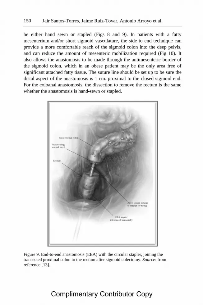
Jair Santos-Torres, Jaime Ruiz-Tovar, Antonio Arroyo et al. 150
be either hand sewn or stapled (Figs 8 and 9). In patients with a fatty
mesenterium and/or short sigmoid vasculature, the side to end technique can
provide a more comfortable reach of the sigmoid colon into the deep pelvis,
and can reduce the amount of mesenteric mobilization required (Fig 10). It
also allows the anastomosis to be made through the antimesenteric border of
the sigmoid colon, which in an obese patient may be the only area free of
significant attached fatty tissue. The suture line should be set up to be sure the
distal aspect of the anastomosis is 1 cm. proximal to the closed sigmoid end.
For the coloanal anastomosis, the dissection to remove the rectum is the same
whether the anastomosis is hand-sewn or stapled.
Figure 9. End-to-end anastomosis (EEA) with the circular stapler, joining the
transected proximal colon to the rectum after sigmoid colectomy. Source: from
reference [13].
Complimentary Contributor Copy
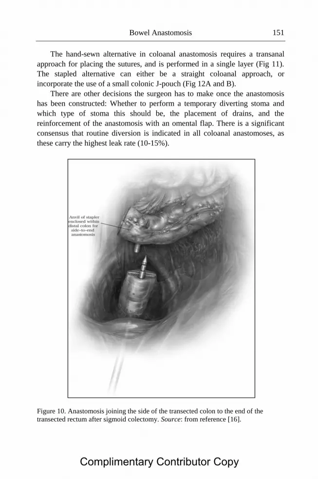
Bowel Anastomosis 151
The hand-sewn alternative in coloanal anastomosis requires a transanal
approach for placing the sutures, and is performed in a single layer (Fig 11).
The stapled alternative can either be a straight coloanal approach, or
incorporate the use of a small colonic J-pouch (Fig 12A and B).
There are other decisions the surgeon has to make once the anastomosis
has been constructed: Whether to perform a temporary diverting stoma and
which type of stoma this should be, the placement of drains, and the
reinforcement of the anastomosis with an omental flap. There is a significant
consensus that routine diversion is indicated in all coloanal anastomoses, as
these carry the highest leak rate (10-15%).
Figure 10. Anastomosis joining the side of the transected colon to the end of the
transected rectum after sigmoid colectomy. Source: from reference [16].
Complimentary Contributor Copy

Jair Santos-Torres, Jaime Ruiz-Tovar, Antonio Arroyo et al. 152
Figure 11. Single layer hand-sewn transanal anastomosis of colon to anus. Source:
from reference [16].
Since these low anastomoses can very rarely be revised or redone, a
coloanal reconstruction is essentially a “one shot” endeavor; all reasonable
measures to protect it are warranted. Most surgeons would also agree that
temporary diversion is reasonable for any anastomosis at or below 7 to 8 cm,
or when preoperative radiation therapy was administered. Diversion can be
accomplished with either a proximal loop colostomy or ileostomy. A
temporary loop ileostomy has the advantage of being easily delivered through
the abdominal wall, and being straightforward to close, usually through a peri-
stomal approach. This saves the patient from having a second major
laparotomy incision, and is typically a brief procedure with a short hospital
stay. Several recent studies have also demonstrated that a loop ileostomy
accomplishes essentially 100% fecal diversion. The long length of
defunctionalized colon has never been shown to generate an adverse outcome
[17].
Complimentary Contributor Copy

Bowel Anastomosis 153
Figure 12. (A) J pouch coloanal anastomosis. The linear stapler is used to create a
reservoir in the previously mobilized colon. (B) J pouch coloanal anastomosis. On
removal of the lineal stapler, the circular stapler is introducer through the rectum and
intocolostomy, where a side-to-end anastomosis is created between colon and rectum.
Source: from reference [16].
The use of pelvic drains has been shown in most studies to either have no
benefit or to actually increase the incidence of leaks. Nevertheless, most
surgeons are understandably uncomfortable with the notion of significant fluid
and debris accumulation in the dependent pelvis after a very low or coloanal
reconstruction, and the specific circumstances of the individual procedure
should determine whether drains are used or not. If the operation is technically
difficult and/or generates significant blood loss or oozing, a drain may be
reasonably used [18].
Complimentary Contributor Copy

Jair Santos-Torres, Jaime Ruiz-Tovar, Antonio Arroyo et al. 154
Employing an omental wrap around an anastomosis in the pelvis carries a
common sense appeal, provided there is sufficient omentum to reach the
pelvis. This is usually accomplished by creating an omental pedicle off of
either the right or left gastroepiploic arcade. The limited clinical studies
assessing this maneuver have shown results similar to what is accomplished
with a proximal diversion; the leak rate is not significantly reduced, but the
omental wrap confines the leak to the pelvis and diminishes the likelihood of a
more widespread peritonitis [19].
Colorectal Anastomotic Complications
Failure of an anastomosis with leakage of intestinal contents is one of the
most significant surgical complications. Reported leakage rates range from 4
to 26%, depending on what type of anastomosis was performed and whether
the operation was an elective or an emergency procedure. An anastomotic leak
increases the morbidity and mortality associated with the operation: it can
double the length of the hospital stay and increase the mortality as much as
10fold [20-21].
Signs and symptoms suggestive of an anastomotic leak include
postoperative (usually between days 4 to 7) abdominal pain or peritonitis,
fever, leukocytosis, elevation of de C-reactive protein and procalcitonin. An
abdominal X -ray showing free air or a CT scan with pneumoperitoneum and
significant free fluid or inflammatory changes around the anastomosis are
suggestive of an anastomotic leak. In the lower anastomosis of colon and
rectum, it is useful to perform an anal contrast-enhanced CT scan for a more
reliable diagnosis of the leakage.
A localized anastomotic leak that is not associated with peritonitis or
significant systemic sepsis can be managed with percutaneous or open
drainage of the abscess, however anastomotic leaks associated with peritonitis
or systemic manifestation of sepsis require a laparotomy and either revision of
the anastomosis if feasible or fecal diversion proximally or at the site of the
anastomosis.
Complimentary Contributor Copy

Bowel Anastomosis 155
Factors Contributing to Anastomotic Failure
Type and Location As a rule, for any given technique the location of the anastomosis does not
influence the overall leakage rate. There are two exceptions to this general
rule. First, low anterior rectal and ultra-low anterior anastomosis are associated
with leakage rates ranging from 4.5% to 8%, however an acceptable leak rate
is around 5% [22].
Bowel Preparation For elective anastomoses of the colon and rectum, it is traditional to
cleanse the large bowel prior to surgery. The rationale being that decreasing
the bacterial load in the colon facilitates anastomotic healing and decreases the
incidence and consequences of anastomotic leakage, is nowadays a
controversial issue. Recent studies have questioned this approach and there is
increasing evidence that a bowel preparation may not be essential and that it
may actually have even some disadvantages [23].
Associated Diseases and Systemic Factors Age, infection, hypotension and hypoperfusion states, intraoperative
transfusion, carcinoma at the line of reception, anemia, diabetes mellitus,
previous irradiation or chemotherapy, malnutrition with hypoalbuminemia,
vitamin deficiencies, chronic steroid use, smoking, and certain disease
conditions, like Crohn’s disease, are associated with poor anastomotic healing
and increased anastomotic leak rates.
In spite of the fact that leakage cannot be entirely prevented, principles of
good technique must be observed:
Anastomose only healthy colon
Avoid tension on the anastomosis
Ensure a good blood supply at the cut ends of both bowels. Good
color, bleeding at the cut edge, and pulsation of mesenteric vessels are
indicators of a good blood supply.
Avoid intramural hematoma. If a hematoma appears to be spreading,
do not hesitate to resect more colon. Good hemostasis is absolutely
mandatory; ligate all vessels.
Clean fat, epiploic appendices, and mesentery from both proximal and
distal edges of the anastomosis. Clean not more than 1 to 1.5 cm.
Complimentary Contributor Copy

Jair Santos-Torres, Jaime Ruiz-Tovar, Antonio Arroyo et al. 156
Ensure that these maneuvers do not compromise the blood supply to
the bowel ends.
Preserve an adequate lumen
Close mesenteric gaps if possible. If they cannot be closed, open them
as wide as possible to avoid postoperative internal hernias.
Adequate preioperative antibiotic systemic prophylaxis is mandatory.
If there is any doubt about the correct performance of the
anastomosis, do a proximal colostomy. This is a life-saving
procedure.
Anastomotic Bleeding
Anastomotic bleeding is common, varies in severity. In most cases,
bleeding is minor and is manifested by the passage of dark blood with the
patient’s first bowel movements after surgery. It is related to the choice of
surgical approach, location of anastomosis and in rectal surgery with or
without preventive colostomy. Some studies have shown that laparoscopic
surgery is a risk factor, and preventive colostomy is a protective factor in
rectal surgery. Most cases are mild and self-limited. More serious bleeding,
especially that from colonic or ileal pouches, can be successfully managed
with epinephrine (1:100,000) and saline retention enemas. If this fails, or
bleeding is massive and results in hemodynamic instability, the patient is best
returned to the operating room for surgical intervention. In cases of bleeding
small bowel or proximal colonic anastomoses, angiography with selective
infusion of vasopressin or embolization of the bleeding vessel may be
indicated [24, 25].
Anastomotic Strictures
Anastomotic stricture may be the end result of anastomotic leak or
ischemia. It typically presents 2–12 months after surgery with increasing
constipation and difficulty evacuating. If the initial resection was done for
malignancy, recurrence as a cause of the stricture must be excluded with a
combination of CT scan and fluorodeoxyglucose–positron emission
tomography (PET) scan. Biopsy is mandatory if a mass or abnormality is
identified. Low colorectal, coloanal, or ileal pouch-anal anastomotic strictures
may be successfully treated with repeated dilations using an examining finger
Complimentary Contributor Copy

Bowel Anastomosis 157
or rubber dilators. Dilation is more successful if initiated within the first few
weeks after surgery. In fact, almost all coloanal or ileoanal anastomoses will
stricture to some degree during the early postoperative period, especially if a
diverting stoma is present. All such anastomoses should undergo digital
examination at 4–6 weeks after surgery and just before stoma closure (usually
at 2–3 months). Strictures are usually soft and easily dilated during these
examinations. Higher colorectal, colocolic or ileocolic strictures may be
approached using endoscopic balloon dilation. If these measures fail, or if the
stricture is extremely tight or long, revision surgery may be required. These
are difficult operations, however, because of the pelvic fibrosis that develops
after anastomotic leak and complications are common. In some cases,
permanent fecal diversion is the only option [24-27].
Early obstruction at the anastomotic site can result from edema or
excessive inversion; late obstruction can be caused by recurrent carcinoma.
References
[1] Slieker JC, Komen N, Komen NA, et al. Long-term and perioperative
corticosteroids in anastomotic leakage: a prospective study of 259 left-
sided colorectal anastomoses. Arch Surg. 2012; 147:447-52.
[2] Alves A, Panis Y, Mathieu P, et al. Postoperative mortality and
morbidity in French patients undergoing colorectal surgery: results of a
prospective multicenter study. Arch Surg. 2005; 140:278-83.
[3] Fazio VW, Tekkis PP, Remzi F, Lavery IC. Assessment of operative risk
[4] Tekkis PP, Poloniecki JD, Thompson MR
[5] Ho YH, Ashour MA. Techniques for colorectal anastomosis. World J
Gastroeneterol 2010; 16:1610-21.
[6] Neutzling CB, Lutosa SA, Proenca IM, et al. Stapled versus handsewn
methods for colorectal anastomosis surgery. Cochrane Databased of
Systematic Reviews 2012; (2):CD003144.
[7] Leung E, Ferjani AM, Stellard N, Wong LS. Predicting post-operative
mortality
[8] Ruiz-Tovar J, Santos J, Arroyo A, et al. Microbiological spectrum of the
intraperitoneal surface after elective right-sided colon cancer: are there
differences in the peritoneal contamination after performing a stapled or
a handsewn anastomosis?. Int J Colorectal Dis 2012; 27:1515-9.
[9] Ishii Y, Hasegawa H, Nishibori H, et al. The application of a new
stapling device for open surgery (Contour TM Curved Cutter Stapler) in
Complimentary Contributor Copy

Jair Santos-Torres, Jaime Ruiz-Tovar, Antonio Arroyo et al. 158
the laparoscopic resection of the rectal cancer. Surg Endosc 2006;
20:1329-31.
[10] Neutzling CB, Lustosa SA, Proencia IM, et al. Stapled versus handsewn
methods for colorectal anastomosis surgery. Cochrane Database sys Rev
2012; 15;2CD003144.
[11] Elseiver. Burkitt et al: Essential Surgery 4e – www.studentconsult.com
[12] Milson JW. Hunt D. Open right colon resection. Operative Techniques
in General Surgery 2003; 5:190-8.
[13] http://ps.cnis.ca/wiki/index.php/Methods_for_abdominal_surgery
[14] The McGraw-Hill Companies. Skandalakis J. et al: Skandalakis’
Surgical Anatomy
[15] Shikata S, Yamagishi H, Taji Y, et al. Single- versus two-layer intestinal
anastomosis: meta-analysis of randomized control trials. BMC Surgery
2006;6:2.
[16] Stahl T. Technical considerations in the difficult colorectal anastomosis.
Operative Techniques in General Surgery 2007; 9:139-84.
[17] Marusch F, Kock A, Schmidt U, et al. Value of a protective stoma in low
anterior resections for rectal cancer. Dis Colon Rectum 2002; 45:1164-
71.
[18] Karliczek A, Jesus EC, Matos D, et al. Drainage or nondrainage in
elective colorectal anastomosis: a systematic review and meta-analysis.
Colorectal Dis 2006; 8:259-65.
[19] Agnifili A, Schietroma M, Carloni A, et al. The value of omentoplasty in
protecting colorectal anastomosis from leakage. A prospective
randomized study in 126 patients. Hepatogastroenterology 2004;
51:1694-7.
[20] Boccola MA, Lin J, Rozen WN, et al. Reducing anastomotic leakage in
oncologic colorectal surgery: an evidence- based review. Anticancer Res
2010; 30:601-7.
[21] Platell C, Barwood N, Dorfmann G, et al. The incidence of anastomotic
leaks in patients undergoing colorectal surgery. Colorectal Dis 2007;
9:71-9.
[22] Buchs NC, Gervaz P, Secic M, et al. Incidence, consequences, and risk
factors for anastomotic dehiscence after colorectal surgery: a prospective
monocentric study. Int J Colorectal Dis 2008; 23:265-70.
[23] Cao F, Li J, Li F. Mechanical bowel preparation for elective colorectal
surgery: updated systematic review and meta-analysis. Int J Colorectal
Dis 2012; 27:803-10.
Complimentary Contributor Copy

Bowel Anastomosis 159
[24] Davis B, Rivadeneira DE. Complications of colorectal anastomoses:
leaks, strictures, and bleeding. Surg Clin North Am 2013; 93:61-87.
[25] Dietz DW. Postoperative complications. In: The ASCRS textbook of
colon and rectal surgery, Wolff BG, Fleshman JW, Beck DE, et al. New
York, Springer Science +Business Media, 2011:pp 141-55.
[26] Di Giorgio P, De Luca L, Rivellini G, et al. Endoscopic dilation of
benign colorectal anastomotic stricture after low anterior resection: a
prospective comparison study of two balloon types. Gastrointest Endosc
2004; 60:347–50.
[27] Suchan KL, Muldner A, Manegold BC. Endoscopic treatment of
postoperative colorectal anastomotic strictures. Surg Endosc 2003;
17:1110–13.
Complimentary Contributor Copy

Complimentary Contributor Copy

Index
#
20th century, 78
A
abdominal wound, ix, 25, 78
access, x, 59, 104, 120
accessibility, 52
acid, 8, 35, 37, 40, 80, 84, 85, 146
AD, ix, 77
adaptation, 69
adenocarcinoma, 53
adhesion(s), 80, 84, 89, 98, 144
adults, 62, 83
advancements, 3
adventitia, 91
age, 25, 44, 50, 112
aggregation, 98
alcohol abuse, 72
alcohol consumption, 52
ALT, 32
ambidexterity, 56
amplitude, 125
anatomy, 2, 19, 22, 48, 58, 62, 63, 89, 90,
91, 122, 125, 126, 127, 128, 129, 130,
149
anemia, 52, 155
aneurysm, 112, 113, 119, 134, 135, 137,
138
angiography, 156
antibiotic, 156
anus, 84, 86, 152
anxiety, xi, 139
aorta, ix, x, 103, 104, 110, 112, 113, 115,
117, 120, 121, 122, 123, 124, 125, 126,
127, 134, 136, 137
apex, 88
arrest, ix, x, 103, 104, 114, 124, 125, 127
arteriosclerosis, 114
artery, 15, 91, 108, 109, 127, 135, 136
ascending colon, 91
ascites, 25
aseptic, 78
aspiration, 25
assessment, 50, 63, 67, 69, 76
assimilation, 87
asymptomatic, 29
atherosclerosis, 131
automate, 122
automation, 67
B
bacteria, 89
base, 53
BD, 74
Complimentary Contributor Copy

Index 162
BEA, vii, 1, 2, 3, 4, 6, 7, 8, 24, 25, 26, 27,
28, 29, 30, 31, 32, 33, 36
beef, 82
bending, 116, 117, 118, 119, 126
beneficial effect, 86
benign, vii, 1, 3, 6, 7, 26, 27, 30, 37, 39, 40,
42, 44, 45, 80, 159
bile, vii, 1, 3, 5, 6, 7, 8, 12, 15, 17, 19, 24,
25, 28, 29, 30, 32, 35, 36, 37, 38, 40, 41,
44, 45, 46
bile duct, vii, 1, 5, 6, 7, 12, 15, 17, 19, 24,
25, 28, 29, 32, 35, 36, 37, 38, 40, 41, 44,
45, 46
bile duct stricture, 5, 40, 41, 45
bile peritonitis, 25
biliary atresia, 3, 37
biliary cirrhosis, vii, 1, 3, 28
biliary fistula, viii, 2, 25, 30, 31
biliary obstruction, 3, 27, 32, 39, 42, 45
biliary stricture, vii, 1, 6, 26, 30, 40, 41, 42,
44, 45
biliary tract, vii, 1, 2, 3, 4, 6, 7, 15, 17, 19,
20, 21, 22, 23, 24, 28, 29, 33, 34, 35, 36,
37, 38, 41
bilioenteric anastomoses, vii, 2, 3, 8, 24, 31,
32, 34, 35
bilirubin, 25, 32
biomarkers, 32
bleeding, xi, 7, 19, 27, 36, 64, 140, 155,
156, 159
blood, x, xi, 50, 52, 62, 64, 69, 78, 80, 82,
83, 90, 104, 105, 111, 112, 113, 115,
118, 124, 125, 131, 134, 136, 137, 140,
153, 155, 156
blood flow, 52, 90, 111, 113, 118, 125, 136
blood pressure, 111, 112
blood stream, 112
blood supply, xi, 78, 80, 83, 140, 155, 156
blood vessels, 50, 82, 134
bloodstream, 119
body weight, 92
bone, ix, 78
bowel, vii, xi, 50, 51, 52, 53, 54, 71, 72, 73,
74, 76, 80, 84, 85, 88, 92, 93, 94, 96, 99,
140, 141, 142, 143, 146, 147, 155, 156,
158
bowel obstruction, 141
breakdown, 52, 97
C
cadaver, 50, 63
caecum, 88, 89, 90
calcification, 131
calibration, 83
cancer, 5, 37, 41, 50, 72, 73, 134, 141, 158
capillary, 99
capsule, 20, 23
carcinoma, 5, 7, 27, 86, 155, 157
cardiac surgery, 82
carnivores, 89, 90
catheter, 31, 32
cauterization, 91
CBD, 28
CEC, 124
cecum, 91
cellulose, 89
challenges, 79
chemotherapy, 52, 155
children, 40, 62
cholangiocarcinoma, vii, 1, 4, 7, 29, 40
cholangiography, 26, 31, 32, 33, 40, 41, 42
cholangiojejunostomy, vii, 2, 7
cholangitis, vii, 1, 3, 5, 6, 24, 25, 27, 28, 29,
30, 32, 34, 35, 38
cholecystectomy, 2, 29, 36, 39, 44, 45
choledochal cysts, vii, 1, 40
choledochoduodenostomy, vii, 2, 6, 38, 39,
42, 43, 44
choledochojejunostomy, vii, 2, 16, 36, 39,
43
choledocholithiasis, 43
cholelithiasis, 27, 43
chromium, 82
circulation, 108, 117, 118, 119, 122
City, 1
classification, 34
clinical application, 106, 130
clinical assessment, 26
Complimentary Contributor Copy

Index 163
clinical judgment, 49
clinical symptoms, 34
closure, viii, 47, 60, 76, 78, 79, 81, 82, 157
CNS, ix, x, 103, 104, 114
coagulation process, 64
coatings, 82
colectomy, 58, 147, 149, 150, 151
colic, 54, 58, 62
collagen, 50, 51, 59, 96, 97, 98
collateral, 117, 120, 125
colon, 12, 13, 15, 17, 60, 61, 74, 84, 85, 86,
88, 89, 90, 91, 92, 101, 141, 143, 144,
146, 149, 150, 151, 152, 153, 154, 155,
157, 158, 159
colon cancer, 157
color, 155
colorectal, x, xi, 52, 53, 72, 73, 74, 84, 86,
94, 95, 100, 139, 140, 141, 147, 149,
156, 157, 158, 159
colorectal cancer, 94
colostomy, 86, 152, 156
common bile duct, 5, 6, 7, 12, 14, 15, 17,
27, 28, 30, 38, 43
common sense, 154
communication, 66
compensation, 128
complexity, 56, 61
compliance, 122
complications, vii, viii, ix, x, xi, 2, 3, 6, 7,
24, 25, 26, 27, 28, 30, 32, 33, 40, 41, 42,
54, 79, 81, 82, 94, 98, 103, 104, 112,
114, 139, 140, 141, 154, 157, 159
comprehension, 66
compression, ix, 84, 103, 123, 124, 132
computer, 50, 137
conciliation, 49
concordance, 98
configuration, x, 94, 95, 104, 115, 122, 125,
127, 129, 130, 141
conflict, 125, 131
Congress, 101, 136
consensus, 151
conservation, 54, 63
constipation, 156
Constitution, 45
construction, ix, xi, 80, 83, 103, 114, 140
consumption, 67
containers, 63
contamination, 52, 131, 141, 157
controlled trials, 53, 74
controversial, 27, 36, 155
convergence, 113
coordination, 53, 67
coronary arteries, 135
corticosteroids, 157
cost, 58, 65, 71, 82, 83
creep, 95, 97
CT, 25, 29, 32, 154, 156
CT scan, 29, 154, 156
cure, 87, 112
curriculum, 50, 65
cyst, 29, 44
cystic duct, 5, 9, 15
D
Dacron fabric, ix, 103, 114, 115, 124
deaths, 113
deconstruction, 66
deficiencies, 52, 155
deformation, 64, 131
dehiscence, 24, 25, 158
dehydration, 89
delayed gastric emptying, 30
deposition, 97
depth, 56, 91
depth perception, 56
descending colon, 90
diabetes, 52, 70, 155
diet, 89
diffusion, 141
dilation, 24, 26, 29, 30, 32, 33, 35, 42, 157,
159
direct bilirubin, 32
direct observation, 68
directives, 49
disability, 87
diseases, vii, 1, 2, 3, 40, 78, 87
dislocation, 111, 112, 118, 132
disorder, 87
Complimentary Contributor Copy

Index 164
displacement, 118, 128
disposition, 125
diversity, 83
diverticulitis, 86
dogs, 62, 78, 79, 80, 85, 90, 98, 101, 138
DOI, 100, 136
drainage, vii, 1, 6, 7, 25, 30, 31, 32, 38, 154
duodenum, 5, 12, 13, 14, 28, 29
dura mater, 84
durability, 81
E
edema, 52, 83, 98, 157
editors, 134
education, vii, 50, 70
EEA, 150
Egypt, ix, 2, 77
elaboration, 105
election, 141
electrocautery, 80
electrolyte, 72
emboli, 156
embolization, 156
emergency, x, 80, 114, 139, 140, 154
emission, 63
endoscopic retrograde
cholangiopancreatography, 6
endoscopy, 28, 29, 45
endotracheal intubation, 90
enemas, 156
engineering, 37
England, 43, 65
environment(s), 49, 66, 76
epinephrine, 156
epithelial cells, 37
epithelium, 29, 44, 88, 98
equipment, 52, 56, 62, 141
ergonomics, 56
erosion, 110, 111, 131
esophagus, 51, 52, 57
ethanol, 41
etiology, 3
evacuation, 95
evidence, viii, 25, 32, 48, 50, 53, 57, 73, 81,
94, 95, 141, 155, 158
evolution, 105, 106, 112, 125
examinations, 157
exercise, 49
experimental design, 62
exposure, 12, 15, 17, 18, 19, 49, 80, 120
external fixation, 131
extraction, 83
extravasation, 85
exudate, 98
F
family life, 49
fat, 98, 155
FDA, 110
fear, 82
feces, 78, 80
fermentation, 89
fever, 34, 154
fibrin, 84
fibrinolytic, 98
fibroblasts, 97, 98, 124
fibrosis, 6, 144, 157
fidelity, 50, 62, 63
financial, 58
fistulas, 24, 25
fixation, ix, 62, 104, 107, 114, 117, 118,
119, 122, 132
flexibility, 59
fluid, 25, 32, 89, 153, 154
fluorescence, 86
food, 6, 28, 29, 88
force, 45, 96
formaldehyde, 63
formation, 8, 24, 29, 30, 51, 89, 94, 96, 98
fragility, 61, 116, 124
freezing, 60
friction, 110, 111
G
gallbladder, 5, 7, 9, 12, 13, 20, 37, 42, 89
Complimentary Contributor Copy

Index 165
gallstones, 2
gastrectomy, 58, 91
gastritis, 27
gastrointestinal tract, vii, 1, 3, 5, 8, 51, 74,
80, 83, 88, 90, 105
general surgeon, 49
general surgery, 49, 77
genes, 88
genetic disorders, 124
genetic engineering, 88
gland, 29
glucose, 60
glycol, 72
goblet cells, 29, 88
graft technique, 2
Greece, 2
Greeks, ix, 77
growth, 90
growth rate, 90
guidance, 42
H
haemostasis, 114, 117, 123, 124, 131, 133
healing, viii, 47, 51, 52, 72, 78, 80, 81, 84,
86, 91, 96, 98, 101, 155
health, 36
height, 94
hematoma, 155
hemorrhage, 6, 30, 39
hemostasis, 112, 122, 124, 155
hepaticojejunostomy, vii, 2, 28, 29, 39, 40,
44, 45
hepatojejunostomy, vii, 2, 5, 7, 18, 40
herbivorous, ix, 77
hernia, xi, 99, 140
histology, 29
history, ix, 3, 4, 77
HM, 72
hormones, 88
horses, 89
host, 119, 120
House, 37, 44
housing, 87, 90
human, 37, 50, 54, 56, 58, 59, 60, 62, 63,
64, 65, 75, 78, 79, 82, 84, 85, 87, 89, 90,
128, 129
human body, 65
hybrid, 112, 135
hyperplasia, 29
hypotension, 89, 155
hypothermia, x, 52, 104, 125
hypovolemia, 60
I
iatrogenic, vii, 1, 6, 7, 35, 36, 40, 41
ID, 41
ideal, xi, 8, 27, 53, 65, 67, 81, 106, 113,
124, 125, 127, 132, 140
identification, 15, 58
ileostomy, 152
ileum, 60, 143, 144, 146
immersion, 63
impairments, 124
improvements, 3, 106, 112
in vitro, 97, 134
in vivo, 116
incidence, 28, 29, 84, 85, 91, 94, 95, 114,
153, 155, 158
independence, 48
individuals, 71
infection, 24, 52, 81, 84, 88, 131, 155
inflammation, xi, 81, 91, 98, 140
inflammatory cells, 98
injury(ies), vii, 1, 6, 7, 15, 25, 30, 35, 36,
37, 40, 41, 44, 45, 46, 72
insertion, ix, x, 5, 9, 103, 104, 114, 115,
121, 128
integration, 124
integrity, x, 60, 96, 104, 115, 127
interface, 91
interference, 129
intervention, 87
intestinal anastomosis, vii, viii, 48, 54, 56,
57, 58, 61, 62, 63, 64, 65, 66, 67, 70, 74,
76, 78, 79, 80, 81, 83, 90, 94, 99, 148,
158
intestinal flora, 52
Complimentary Contributor Copy

Index 166
intestinal obstruction, 84
intestinal tract, 50
intestine, viii, ix, 47, 51, 54, 59, 60, 61, 72,
77, 78, 79, 80, 82, 89, 90, 144
intima, 105
intra-abdominal abscess, viii, 2, 25, 28
intussusception, 6, 27
invaginate, ix, 78
inversion, 79, 148, 157
iodine, 92
irradiation, 52, 155
irrigation, 17, 19
ischemia, 83, 95, 107, 108, 113, 114, 117,
124, 140, 156
isolation, 127
Italy, 37, 103
J
Japan, 101, 106
jaundice, 2, 3, 5, 27, 32, 34, 37, 45
jejunum, 5, 6, 9, 16, 20, 39, 60, 79, 91
K
knots, 8, 18, 54, 56, 78
L
laboratory tests, 34
laceration, 105, 127
laparoscopic cholecystectomy, 3, 36, 37, 41,
44, 45, 46, 70
laparoscopic surgery, 37, 56, 58, 60, 65, 69,
70, 71, 156
laparoscopy, 56, 57, 58
laparotomy, 99, 120, 152, 154
large intestine, 62, 72, 89, 90, 144, 146
lead, 6, 25, 29, 69, 81, 85, 87
leakage, 25, 28, 30, 31, 32, 41, 44, 52, 53,
72, 73, 83, 84, 85, 91, 94, 95, 96, 98,
100, 140, 154, 155, 157, 158
leaks, 25, 67, 69, 70, 94, 146, 153, 154, 158,
159
learning, viii, ix, 47, 48, 49, 56, 57, 58, 59,
63, 66, 67, 69, 73, 74, 76
learning process, 63, 66, 74
legs, 11
lesions, 41, 56, 80, 134, 137
leukocytosis, 154
life cycle, 88
life expectancy, 6, 7
ligament, 12, 13, 15, 17, 19, 20
light, 132
liver, 2, 3, 8, 19, 23, 32, 34, 35, 41, 42
liver damage, 32
liver function tests, 34, 35
liver transplant, 41, 42
liver transplantation, 41, 42
longitudinal study, 46
low temperatures, 60
lumen, xi, 8, 18, 22, 51, 54, 79, 83, 96, 114,
116, 118, 124, 125, 129, 133, 140, 146,
156
lung transplantation, 108, 136
Luo, 135
lymphocytes, 98
lymphoid tissue, 89
M
magnesium, 105
magnetic resonance, 42
magnitude, 98
majority, 82, 96, 114
malignancy, 27, 31, 156
malignant diseases, vii, 1
malnutrition, 52, 155
man, 87, 88, 90, 91
management, 30, 37, 39, 40, 41, 42, 43, 44,
45, 58, 92
manipulation, 24, 29, 32, 83, 108, 117
manufacturing, 82
Marfan syndrome, 114, 135
Marx, 42
mass, 156
materials, viii, ix, 48, 52, 57, 59, 62, 65, 77,
81, 82, 86, 99, 112, 141
matter, 91, 141
Complimentary Contributor Copy
Index 167
MB, 41, 70, 72
meat, 89
meconium, 144
medical, 34, 37, 48, 58, 59, 60, 70, 76, 148
Medicare, 137
medicine, 3, 50
memory, 55
mentor, 66, 67, 68
mesenteric vessels, 155
mesentery, 52, 80, 155
Mesopotamia, 2
meta-analysis, 53, 73, 74, 158
metabolism, 51
metabolized, 82
methodology, 68, 87
mice, 86
microscope, 88
migration, 98
models, viii, x, 48, 49, 50, 54, 56, 58, 59,
60, 62, 63, 65, 67, 69, 70, 71, 74, 87, 90,
104, 115, 122, 130
modifications, 2, 115
modules, 64
monolayer, 8
morbidity, 7, 25, 27, 28, 30, 36, 49, 52, 53,
84, 91, 140, 154, 157
morphology, 25, 90
mortality, ix, 25, 28, 30, 49, 52, 70, 84, 91,
103, 113, 140, 154, 157
mortality rate, 25, 91, 113, 140
Moscow, 51
MR, 157
mucosa, 18, 19, 29, 44, 51, 60, 79, 85, 88,
91, 92, 93, 99, 148, 149
mucous membrane, 50
muscular dystrophy, 87
N
necrosis, 83, 84, 95, 98, 124
neoangiogenesis, 99
nerve, 50
nutrients, 89, 90
nutrition, 90
O
obstacles, 120
obstruction, 3, 5, 6, 7, 27, 32, 37, 38, 81, 86,
157
occlusion, 24, 40, 80
oesophageal, 134
omentum, 98, 154
operations, 15, 40, 49, 63, 147, 157
organ(s), 11, 49, 60, 82, 84, 86, 88, 89, 107,
108, 131
organism, 64, 87, 88
P
pain, 27, 34, 52, 94, 154
palliative, 5, 6, 7, 27
pancreatic cancer, vii, 1, 37
pancreatitis, 6, 27, 28, 30
parallel, 9, 20, 51, 54, 81, 92, 116
parenchyma, 7, 34, 35
pathogenesis, 114
pathology, 37, 39, 48, 52, 62, 114, 127
pathophysiological, 87
pelvis, 150, 153, 154
perforation, ix, 5, 84, 96, 103, 124
perfusion, x, 62, 64, 104, 108, 113, 114,
131, 135
peritoneal cavity, 85, 95
peritonitis, 25, 52, 89, 98, 154
permission, 115, 118, 120, 123
PET, 156
Philadelphia, 39, 72
physiology, 2, 48, 90
pigs, 60, 62, 64, 85, 90
plants, 89
plexus, 83, 149
PM, 42, 45, 135
polymer, 82
polypropylene, 74, 82, 84, 106, 117, 118,
122, 132
polyurethane foam, 59
population, 40, 89, 137
porosity, 123
Complimentary Contributor Copy

Index 168
portal hypertension, 3, 7
portal vein, 27
positron emission tomography, 156
preparation, xi, 52, 72, 73, 81, 107, 124,
140, 155, 158
preservation, xi, 63, 140
prevention, 80
principles, viii, xi, 8, 47, 57, 78, 79, 80, 106,
140, 155
probability, 26
prognosis, 78
project, 125
proliferation, 98
proline, 51
prophylaxis, 156
prostheses, 109
prosthesis, vii, ix, x, 85, 103, 104, 110, 111,
112, 114, 115, 119, 120, 122, 123, 130,
134, 135, 136
protection, 125, 135
prototype(s), 51, 109, 127, 128, 129
pubis, 92
public concern, 90
pylorus, 29
pyogenic, 38
Q
quality of life, 45, 46
questionnaire, 59
R
radiation, 4, 152
radiation therapy, 152
rating scale, 67, 68
RE, 44, 45, 135
realism, 59, 65
reality, viii, 48, 50, 58, 71, 75
recall, 132
reception, 155
reconstruction, vii, 1, 3, 7, 30, 33, 35, 36,
91, 152, 153
rectosigmoid, 85
rectum, 51, 57, 72, 83, 90, 141, 149, 150,
151, 153, 154, 155
recurrence, 36, 44, 94, 156
reference system, 49
reinforcement, 84, 100, 151
relative size, 90
reliability, 125, 130, 132, 133
remodelling, 51
renal failure, 30
repair, 30, 36, 41, 44, 45, 72, 98, 105, 113,
127, 131, 134, 135, 137, 138
requirements, xi, 87, 90, 140
researchers, 81, 84, 88, 94
resection, 2, 7, 19, 20, 28, 63, 72, 73, 83, 85,
91, 99, 140, 143, 156, 158, 159
resistance, 58
response, 8, 52, 81
restenosis, 26
restoration, 52
RH, 70
rings, 84, 108
risk(s), ix, xi, viii, 5, 6, 7, 25, 27, 28, 32, 44,
45, 47, 55, 57, 72, 83, 84, 97, 103, 112,
114, 131, 133, 134, 140, 141, 156, 157,
158
risk factors, xi, 25, 44, 45, 72, 112, 140,
141, 158
rodents, 91
rubber, 85, 86, 105, 157
S
safety, 50, 53, 58, 83, 94, 122
salts, 82
scaling, 95
school, 48, 141
science, 87
scope, 11
security, 118, 122
sensation, viii, 47, 55
sensitivity, 26
sepsis, 30, 52, 80, 154
serum, 25, 30
serum albumin, 30
shape, ix, 103, 110, 112, 114, 115
Complimentary Contributor Copy

Index 169
sheep, ix, 54, 78, 82
shock, 100
showing, 93, 154
sigmoid colon, ix, 77, 90, 92, 150
signs, 34
silk, 78, 82, 83, 96
simulation, viii, 48, 49, 50, 53, 63, 64, 71,
75
Singapore, 37
Single graft, x, 104
skin, 76, 82, 92
sludge, 8, 16, 17
small intestine, 19, 62, 79, 89, 90, 98
smoking, 52, 155
smooth muscle cells, 97
social sciences, 87
society, 58
sodium, 92
solution, 60, 63, 72, 86, 92, 109, 123
Soviet Union, 79
Spain, 47, 139
specialists, 49
species, 87, 90
specific knowledge, 67
spending, 48
sphincter, 28, 29
spinal cord, 114
SS, 71, 74
stability, x, 37, 94, 97, 104, 110, 112, 115,
122, 123, 130, 131, 132
stabilization, 117, 121
standardization, 69, 107
stasis, 8, 24
state(s), 36, 65, 98, 155
steel, 82, 116, 118, 141
stenosis, 5, 7, 24, 26, 30, 35, 45, 57, 69, 84,
95
stent, 24, 35, 40, 45, 85, 86, 134, 136, 137
sterile, 59
stoma, 28, 72, 86, 145, 146, 151, 157, 158
stomach, 5, 50, 60, 62
stretching, 132
strictures, vii, 1, 6, 7, 26, 30, 35, 45, 95,
144, 156, 159
structure, 59, 72, 91, 107, 109
submucosa, 50, 78, 83, 88, 96, 100
substitutes, 116
substitution, ix, x, 103, 104, 109, 112, 113,
120, 124, 126, 127, 128, 129, 130, 133,
134, 136
success rate, 35
supervision, viii, 47, 66
suppression, 56
surgical intervention, 2, 90, 156
surgical procedure, vii, viii, 1, 3, 24, 38, 47,
49, 64, 80, 83
surgical removal, 2
surgical resection, 3
surgical technique, viii, x, 37, 48, 57, 65,
69, 78, 79, 80, 83, 106, 114, 139, 140
survival, 27, 49, 85, 94
suture, viii, ix, x, xi, 8, 11, 13, 18, 20, 47,
50, 52, 54, 56, 57, 58, 59, 63, 64, 65, 66,
67, 72, 74, 77, 78, 79, 81, 82, 83, 85, 95,
96, 98, 99, 104, 105, 109, 110, 114, 115,
118, 119, 120, 122, 123, 124, 125, 126,
130, 131, 132, 133, 137, 140, 141, 146,
147, 150
symptoms, 28, 29, 34, 52, 154
syndrome, 6, 27, 28, 39, 43, 52, 113
synthesis, 109, 132
T
tachycardia, 94
tactics, 37
teams, 107
techniques, vii, viii, xi, 2, 3, 8, 25, 26, 32,
53, 55, 56, 57, 58, 65, 69, 74, 75, 78, 79,
80, 83, 84, 91, 96, 97, 99, 101, 105, 112,
113, 119, 124, 140, 141
technological advances, 53
technology(ies), 49, 50, 64, 83
temperature, 64
tensile strength, 50, 81, 96
tension, viii, xi, 9, 11, 12, 13, 47, 52, 70, 78,
80, 140, 144, 155
territory, 113
testing, 86
textbook, 78, 159
Complimentary Contributor Copy

Index 170
texture, 63
therapy, 34, 37, 41, 44, 89
thoracotomy, 120
thrombosis, 119
tissue, 3, 7, 37, 51, 52, 54, 57, 58, 63, 64,
67, 78, 79, 80, 81, 83, 84, 131, 140, 150
tones, 43
torsion, 107
tourniquet, 116
toxicity, 82
toxicology, 88
trachea, ix, 78
trainees, 49, 50, 59, 62, 63, 65
training, vii, viii, ix, 47, 48, 49, 50, 53, 54,
55, 56, 58, 59, 60, 62, 63, 64, 65, 66, 68,
69, 70, 71, 74, 75
training programs, 49, 60
transducer, 96
transection, 19, 39, 149
transformation, 29
transfusion, 155
transplantation, 107, 108, 134
transverse colon, 90, 143
trauma, 53, 73, 98
treatment, 2, 5, 7, 26, 27, 28, 31, 35, 36, 37,
38, 39, 43, 45, 46, 78, 87, 113, 134, 135,
137, 159
tremor, 56
trial, 40, 71, 72, 73, 74, 134
triangulation, 105
tumor(s), 5, 6, 7, 9
twist, 81
U
UK, 76, 138
UL, 39
ulcer, 95
ultrasonography, 6, 38
ultrasound, 15, 25
United States (USA), 39, 70, 79, 106, 113,
144
V
vacuum, 109
validation, viii, 48, 75
valve, 89, 124
variables, xi, 140
variations, 69, 126, 132
vascular prostheses, 59
vascular surgery, 105, 112, 123
vascular wall, 105, 109, 110, 111, 119, 120,
121, 124, 127, 132
vascularization, viii, 47, 52, 70, 125
vasculature, 150
vasopressin, 156
versatility, 106
vessels, 155
videos, 54, 66, 68
viscera, viii, 4, 48
vision, 56
volvulus, ix, 77, 86
W
war, 62
water, 60, 89, 96
welding, 84
well-being, 36
West Africa, 99
wires, ix, 82, 103, 114, 131
wood, ix, 78
workers, 79
World Health Organization, 36, 45
World War I, 79
World Wide Web, 91
wound healing, 51, 81
wound infection, 25, 28, 30, 52
Complimentary Contributor Copy