Embed Size (px)
Citation preview
ues to anastomotic failure.
© 2006 The WJG Press. All rights reserved.
Key words: Mechanical behavior; Failure load; Colon; Anastomosis; Wound healing
Ekmektzoglou KA, Zografos GC, Kourkoulis SK, Dontas IA, Giannopoulos PK, Marinou KA, Poulakou MV, Perrea DN. Mechanical behavior of colonic anastomosis in experimental settings as a measure of wound repair and tissue integrity. World J Gastroenterol 2006; 12(35): 5668-5673
http://www.wjgnet.com/1007-9327/12/5668.asp
INTRODUCTIONInvestigating wound healing and attempting to improve its outcome necessitates process quantifi cation[1]. Parameters for anastomotic repair and adhesion formation[2] may be mechanical, biochemical, or histological. Histology is not a primary tool for quantifi cation when comparing various series of experimental anastomoses. Certainly it is very useful to describe the course and eventual result of the healing sequence at the tissue level. Also, the successive infiltration of various cells into the wound area may be followed, and obvious differences between anastomoses (e.g., ileal and colonic) will certainly be demonstrated this way. However, the measurement of choice to evaluate anastomotic repair and the effects of variations in surgical techniques, administration of drugs, or of any other modifi cation to establish procedures, will mostly be either mechanical or biochemical or both.
The developing mechanical strength is, without doubt, a meaningful parameter to follow while investigating anastomotic healing. For this purpose, two fundamentally different approaches can be chosen. First, one can choose bursting strength, which is expressed either as bursting pressure or bursting wall tension, which is the measure of the resistance of the intestinal wall to increasing intraluminal pressure. Second, one can choose breaking strength, which refl ects the resistance of the intestinal wall to forces exerted in a longitudinal direction[3-5].
While both of these methods used to evaluate anastomotic healing have been investigated in the international literature, no paper has so far described
BASIC RESEARCH
Mechanical behavior of colonic anastomosis in experimental settings as a measure of wound repair and tissue integrity
Konstantinos A Ekmektzoglou, Georgios C Zografos, Stavros K Kourkoulis, Ismene A Dontas, Panagiotis K Giannopoulos, Katerina A Marinou, Maria V Poulakou, Despina N Perrea
www.wjgnet.com
Konstantinos A Ekmektzoglou, Ismene A Dontas, Maria V Poulakou, Despina N Perrea, Laboratory of Experimental Sur-gery and Surgical Research “NS Christeas”, University of Athens, Medical School, Athens, GreeceGeorgios C Zografos, Panagiotis K Giannopoulos, 1st Univer-sity Department of General Surgery, Athens School of Medicine, Hippocration Hospital, Athens, GreeceStavros K Kourkoulis, Laboratory of Testing and Materials, De-partment of Mechanics, National Technical University of Athens, Greece Κaterina A Marinou, BVMS, MVM, Laboratory of Experimen-tal Surgery and Surgical Research “NS Christeas”, University of Athens, Medical School, Athens, Greece and Greek Ministry of Rural Development and Food, General Directorate of Veterinary ServicesCorrespondence to: Konstantinos A Ekmektzoglou, MD, 15 Zoodohou Pigis Street, Melissia, Athens 15127, Greece. [email protected]: +30-210-8033273 Fax: +30-210-8033273Received: 2006-07-17 Accepted: 2006-07-29
AbstractAIM: To determine the mechanical properties of anasto-motic colonic tissue in experimental settings and there-fore give a measure of wound healing.
METHODS: Thirty-six male Wistar rats were used as ex-perimental models of anastomotic tissue integrity. On the 5th post-operative day, the tensile strength was measured by application of an axial force, providing a quantitative measure of anastomotic dehiscence and leakage.
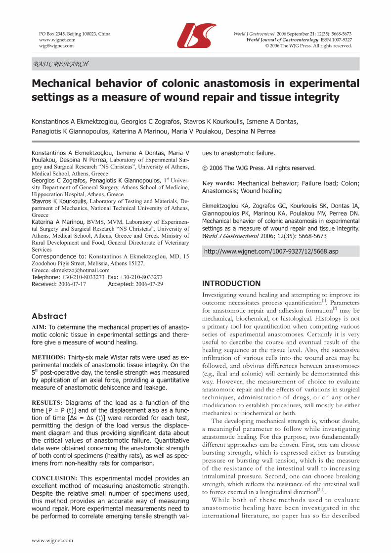
RESULTS: Diagrams of the load as a function of the time [P = P (t)] and of the displacement also as a func-tion of time [Δs = Δs (t)] were recorded for each test, permitting the design of the load versus the displace-ment diagram and thus providing signifi cant data about the critical values of anastomotic failure. Quantitative data were obtained concerning the anastomotic strength of both control specimens (healthy rats), as well as spec-imens from non-healthy rats for comparison.
CONCLUSION: This experimental model provides an excellent method of measuring anastomotic strength. Despite the relative small number of specimens used, this method provides an accurate way of measuring wound repair. More experimental measurements need to be performed to correlate emerging tensile strength val-
PO Box 2345, Beijing 100023, China World J Gastroenterol 2006 September 21; 12(35): 5668-5673www.wjgnet.com World Journal of Gastroenterology ISSN [email protected] © 2006 The WJG Press. All rights reserved.

in detail the process itself, analyzing its advantages, disadvantages and parameters taken into consideration, therefore establishing the need for an in-depth presentation of the mechanical apparatus used and presenting not only the theoretical background behind the measurements but also the technical diffi culties that arise.
MATERIALS AND METHODSThirty-six male Wistar rats weighing 300-350 g were used, and were housed two per cage. They were fed a standard diet and water ad libitum. All experiments were approved by the Athens Prefecture, Directorate of Veterinary Services (License No. K/355/27-1-2005), according to the Presidential Decree No. 160/1991 (Governmental Gazette A’ 64), with which Greece has conformed to the 86/609/EEC directive. Laparotomy[6] was performed through a midline 2 cm incision under anesthesia induced by ketamine (80 mg/kg) and xylazine (3 mg/kg). A colonic segment, 1 cm in length, 5 cm distal to the ileocecal junction was transected and the colon was re-anastomosed end-to-end using 5-0 Vicryl (Ethicon) sutures in single-layer interrupted fashion[7]. About 10 sutures were placed for each anastomosis to secure an inverted anastomosis without mucosal protrusion, which is regarded as a major cause of perianastomotic adhesions. The abdominal muscle wall was then closed with 5-0 Vicryl (Ethicon) sutures, followed by skin closure with 4-0 Silk (Medipac) sutures.
To obtain the test specimen, the rats were sacrificed with an overdose of ether, on the 5th post-operative day. The previous abdominal incision was reopened, and the anastomotic site identifi ed and inspected for possible ad-hesions and leakage. An 8 cm segment of the colon with the anastomosis in the middle was resected. Care was taken not to detach adhesions from the anastomosis, but to dissect the surrounding tissues. The resected specimen was gently irrigated with saline to remove feces and was mounted on a table.
The basic purpose of the present experimental pro-tocol is the determination of the mechanical behavior of intestinal anastomoses and more specifi cally the response to tensile loading and the determination of the respective tensile strength. One can defi ne the ratio of the applied force at the moment of failure, Fcr, over the surface, Α, upon which the force acts normally[8], as tensile strength (Figure 1). The ratio of the force over the respective area is known in engineering science as stress and therefore the tensile strength is the respective tensile stress at the mo-ment of failure.
In the international scientifi c literature the mechanical behavior under tension, of specimens like the ones of the present protocol, has been studied in two ways: (1) By applying an internal hydraulic pressure, p,[9-12]. In this case (and for points relatively far from the borders of the specimens) a stress state equivalent to the so-called biaxial tension appears on the surface of the concave cylindrical specimen. Assuming that the thickness of the specimens is much smaller in comparison to its diameter, the principal tensile stresses at the moment of failure are given by the
Ekmektzoglou KA et al. Mechanical behavior of colonic anastomosis 5669
www.wjgnet.com
relations (Figure 2)[13]:
L Tcr crp r p r,2t t
σ = σ =
where pcr is the value of the hydraulic pressure at the moment of the fi rst failure, r the radius of the intestine and t its wall thickness. The stress system is characterized as principal since no shear stresses can be generated by a pure hydraulic pressure.
(2) By applying directly an axial force, F,[14,15]. In this case the critical value of the tensile strength is expressed as:
cr
crF
2 rtσ =
πThe procedures described above present both
advantages and disadvantages. More specifically, the application of hydraulic pressure, although relatively easily realizable in the laboratory, creates a two-dimensional stress field, as shown in Figure 2. Therefore the conclusions drawn are not directly comparable to the respective ones of the experiments with uniaxial loading. Especially in the case of anisotropic materials (like the ones of the present study), it is impossible to define which one of the two stresses is responsible for the failure and therefore it is not possible to determine the critical value, since the direction at which failure will appear is not a priori known.
On the contrary, the application of an axial force is especially diffi cult from a practical point of view (as it will be seen in the next paragraph), but the results obtained are directly usable without reductions and additional assumptions.
Experimental diffi culties of the direct tension experiment In the present study the second procedure (direct tension) was adopted. The most important diffi culties stated above
Figure 1 Typical forces applied on a standard specimen.
F
P
A
σT
σT
σLσL
Figure 2 The application of hydraulic pressure creating a two-dimensional stress fi eld.

www.wjgnet.com
are summarized as following: (1) The nature of the mate-rials under study renders the gripping of the specimens, with the aid of conventional friction grips through com-pression loads, extremely diffi cult. In fact, since it is im-possible to form “gripping heads” to the specimens (“dog-bone” specimens), it is given that the failure will appear in the portion of the specimen which is inside the grips or in their immediate vicinity. However, in this area the stress fi eld is strongly triaxial and therefore the results obtained are invalid and should be rejected. On the other hand, the limited chances to obtain long specimens in combination with the low friction coeffi cient between the external sur-faces of the specimens (intestines) do not allow the use of pulley-shaped grips, in which the holding force emanates from the friction of a number of successive layers of the material rolled around the periphery of the pulley. (2) The extremely low force which is necessary for the fracture of even intact and healthy specimens, which according to international literature is estimated at the value of a few tenths of Newton[14], renders the conventional arrange-ments of applying axial tension practically useless. (3) The nature of the specimens under study, which are twisted and bended around different axes, due to adhesion forma-tion around the anastomotic area, renders the measure-ments of length changes and therefore of reduced defor-mations (strains, ε) almost impossible. (4) Finally, the na-ture of the intestines once again, which under torsion are “self-confi gured” into the form of plane plates, results in an interaction between the walls of the specimens, making diffi cult the reduction of the external loads into stresses (σ). Another factor making the situation more diffi cult is the non-constant thickness of the specimens throughout their length and their perimeter, which does not allow us to cal-culate the effective area of the loaded intestine.
In order to confront these diffi culties in the present ex-perimental study, the following procedures were adopted.
Gripping the specimensA specifi c gripping system was designed, consisting of a pair of light metallic pins of cylindrical cross-section of diameter equal to 5 mm, with rounded head which permits easy entrance of the intestine in the pin, without injuring the specimen walls, reducing thus the time required for the in-situ preparation of the specimens (Figure 3). The pins are grooved at their mid-length and a suture which holds the specimens in place is rolled up in this groove. Τhe up-per part of the pins is drilled through the thickness and the specimen is suspended through this hole from the upper plate of the loading frame. At the same time, the second pin is fi xed to the immobile plate of the frame.
The suspension and fixing of the pins is achieved with the aid of circular rings. In this way the maximum possible number of degrees of freedom is given to the specimen making possible the self-alignment and the “un-twisting” of the intestine during tension without external limitations and therefore without, as much as possible, the development of parasitic tensions and disfi gurations.
Despite the low total weight of the specimens grip-ping system (about 0.12 N), it was deemed appropriate to add the weight of the lower half, which is suspended
and therefore sustained by the specimen, at the value of the fi nal failure load, taken into consideration that these values are relatively comparable (the mean value of the failure load, as it was obtained from a series of preliminary experiments is equal to about ranges between 1.00 Ν and 2.20 Ν).
The load application systemAfter the rejection of loading through the application of dead weights (water or lead grains), due to the induction of vibrations and oscillations, a special load cell of capac-ity of 5 Ν and sensitivity of 10-3 Ν was used attached in a stiff electrical loading frame (Instron). This frame was selected, apart from its robustness, because it provides the ability of choosing the load application speed between wide ranges (from 0.5 mm/min to 500 mm/min). This characteristic of the frame is very important in case bio-logical materials are to be studied, since their mechanical behavior exhibits viscoelastic nature, which is strongly de-pendent on the strain rate induced (dε/dt).
In the first phase of the experimental project an especially low tension speed (1 mm/min) was selected, and therefore the loading can be considered as static or at least quasi-static. As a result the overall duration of each test usually exceeds 10 min. It is planned, in a second phase, to study the effect of loading rate by employing dynamic or quasi-dynamic protocols.
Calibration of the apparatusBefore starting the main series of experiments, a number of preliminary tests were carried out, in order to defi ne the range of the expected values of both the failure load and the elongation of typical specimens, and to calibrate the apparatus in the specifi c range of values.
The calibration of the loads was achieved with the safest method of the suspension of standardized (certifi ed) weights from the load cell. Both the absolute reading values of the load cell as well as their linearity at the range of the expected loads were checked. The deviations detected for the absolute values of the loads did not exceed in any case the limit of 0.2% set by the “Quality Assurance System” of the Laboratory of Testing and
Figure 3 The gripping system consisting of a pair of light metallic pins. The grooves at which the intestine is gripped are indicated by the arrows.
5670 ISSN 1007-9327 CN 14-1219/R World J Gastroenterol September 21, 2006 Volume 12 Number 35

Materials of the National Technical University of Athens (NTUA/LTM), as it is described in the respective “Quality Assurance Manual” according to ISO9000/2000 system.
On the other hand, the linearity of the values of the loading cell in relation to the respective ones of the standard weights exceeded 99.8% for the whole range of interest, as it was concluded from a linear interpolation in the experimental data, using the least square method.
The calibration of the readings of the load frame for the displacements was achieved with the aid of three LVDT’s (Linear Voltage Displacement Transducers), which have been verifi ed with a standard micrometric vernier of an accuracy of 1 μm. Apart from the absolute values of the displacements, the parallel of the motion of the load-ing frame was also checked. The deviations detected did not exceed in any case the limits set by “Quality Assurance System” of the NTUA/LTM. Finally, the time recording device of the data acquisition and storage system was also calibrated with the aid of a prototype chronometer. The deviations were not measurable.
Data acquisition and storage systemThe data to be recorded during the experiments include the values of the load as a function of the time [P = P (t)] and the values of the displacement of the moving plate of the loading frame also as a function of time [Δs = Δs (t)]. The data acquisition system includes a special multi chan-nel “bridge” (National Instruments, type SCXI-1000), with the ability of adjusting the sampling rate. The system in-cludes, also, a personal computer with suitable commercial software (LabVIEW-8). From the functions F = F (t) and Δs = Δs (t) recorded, one can eliminate the time obtain-ing the function of the applied force as a function of the displacement induced and therefore as a function of the elongation of the intestine, i.e. F = F (Δs).
After the preliminary experiments, it was deemed ap-propriate to add to the data acquisition system a video device, in order to monitor the specimen during the ex-periment in a mode synchronous to the recording of the values of the load and the displacement. This was consid-ered necessary, since the records of the load versus pre-sented oscillations, due to two different reasons: (1) The “un-twisting” of the twisted parts or the “un-folding” of the folded parts of the intestine, which lead to a sudden length increase of the specimen, and therefore to instan-taneous unloading, that is to a fall in the recorded load, as it is shown characteristically in the diagram of Figure 4A. (2) Local failures of parts of the specimen and especially in the case of anastomosed intestines failure of the anas-tomotic area itself or of directly neighboring areas, due to the tearing of the material from the anastomotic suture.
Since one cannot distinguish between these two discontinuities of the F = F (t) diagram, the synchronous video-recording of the experiment was considered necessar y. In this way i t is possible to locate the discontinuities of the diagram due to the “un-twisting” or to the “un-folding” of the specimen, until the discontinuity due to the anastomotic failure or failure of its immediate neighboring area. Therefore the loading corresponding to this discontinuity can be safely considered as the crucial anastomotic failure load.
1.5
1.0
0.5
0.00.00 1.00 2.00 3.00 4.00 5.00
Load
(N
)
Displacement (mm)
A
B
6004002000
3
2
1
0
t /s
Load
(N
)
t /s
0 250 500 750
2.0
1.5
1.0
0.5
0.0
Load
(N
)
C
D
t /(s x 10)
0 25 50 75
1.5
1.0
0.5
0.0
Load
(N
)
Figure 4 Load versus time and displacement diagrams for characteristic tests. A: Load versus displacement for a typical test of the preliminary series; B: Load versus time using intact specimens from healthy rats; C: Load versus time using specimens from healthy rats after colonic anastomosis; D: Load versus time using specimens from non-healthy rats after colonic anastomosis.
Discontinuity due to failure
Discontinuities due to un-winding
Ekmektzoglou KA et al. Mechanical behavior of colonic anastomosis 5671
www.wjgnet.com

RESULTSThree different classes of specimens were tested using the system described in the previous paragraphs.
The fi rst one included a number of “intact” specimens, i.e. specimens from healthy rats without anastomoses. The results of these tests are to be used as a measure that will permit the characterization of the quality of the anastomosis, at least from the point of view of mechanical strength. Two characteristic examples of these tests are shown in Figure 4B. The data obtained from these tests for the failure force exhibited very small scattering (as it was perhaps expected) and the average value was of the order of:
Fcrintact = 2.09 N ± 0.6 N
Taking into account that the thickness of the wall of the intestine of the rats after the 8th week of their life is stabilized to about 1.1 mm while its perimeter varies in the range 9-12 mm[16] , it is concluded that the tensile failure strength of the “intact” specimens ranges between:
160 kPa ≤ σfailintact ≤ 210 kPa
The second class of experiments included the control tests, namely it was carried out using specimens obtained from healthy rats but after having been subjected to colon anastomoses. The scattering of this series of tests was obviously higher compared to that of the “intact” specimens and it was considered necessary to study the acceptability of the results based on statistical experiments. The Chauvenet criterion was adopted and a number of tests were excluded from the analysis. In Figure 4C the results of three tests of this series are shown, corresponding to the ones with the lowest and highest acceptable failure forces and to a third one with failure force almost equal to the average value. The average value for the failure force was determined equal to:
Ffailcontrol = 1.35 N ± 0.42 N
Similarly the failure strength ranges between: 100 kPa ≤ σfail
control ≤ 135 kPaIt can be concluded that the present procedure for
the anastomotic operation results in a decrease of the mechanical strength of the colonic segments under study of the order of only 35% in comparison to the intact specimens.
As a fi nal step, a third series of tests was carried out with specimens obtained from non-healthy rats after having been subjected to colon anastomotic surgery. It was strange to observe that the scattering of the results was rather lower in this case and the application of the Chauvenet criterion yielded the exemption of only one test. A number of characteristic tests of this class experiments is shown in Figure 4D. The average value for the failure force, for this series of tests was determined equal to:
Ffailnon-healthy = 1.09 N ± 0.19 N
In this case failure strength ranges between: 82 kPa ≤ σfail
non-healthy ≤ 110 kPaThe decrease of the mechanical strength compared to
the intact specimens is of the order of about 50%, while if the comparison is carried out on the basis of the results of the control tests, is of the order of about 19%.
DISCUSSIONWound leakage, the major concern for every surgeon performing intestinal anastomosis, is considered a multifactorial process, upon which many factors act, accelerating or inhibiting its metabolic pathway[17,18]. Numerous clinical entities and metabolic abnormalities can alter the course of tissue repair. Amongst them diabetes mellitus, hypothyroidism, immunocompetence, infection and other diseases are proven to be detrimental to anastomotic healing, while other factors like the surgical technique, advanced age, malnutrition, obesity, inadequate perfusion and/or oxygenation are considered risk factors for impaired wound healing[19-22].
Taking the 5th post-operative day as a crucial time point upon which anastomotic failure is mostly recognized in clinical practice, the authors tried to give a measure of the anastomotic strength by taking advantage of its mechanical behavior. While both bursting pressure and tensile strength are used to describe the mechanical properties of viscoelastic materials like the ones under study, the authors preferred to evaluate the second and correlate it to the healing of colonic anastomosis. This is because tensile strength appears to be a better standard to evaluate the biological aspects of healing. Tensile strength is an important determinant of anastomotic strength, in contrast to the bursting pressure, which can evaluate the overall anastomotic integrity, but may refl ect healing less accurately.
The authors in this paper described not only a system for gripping the specimens, the load application, the data acquisition and storage system, but also a detailed view of the theoretical background behind the forces applied in the tissues under study, as well as the experimental diffi cul-ties of the direct tension experiment.
The values of the load as a function of the time [P = P (t)] and the values of the displacement of the moving plate of the loading frame also as a function of time [Δs = Δs (t)] were recorded, giving the load versus the displacement curve for each measurement and therefore providing the recorded discontinuities due to the anastomotic failure.
While the tests performed were used only for a prelimi-nary series of measurements, since the number of speci-mens was relatively small, significant conclusions can be made regarding wound strength and tissue regeneration. The decrease of the axial force required causing mechani-cal failure from 2.09 N in case of the “intact” specimens to about 1.35 N for the control tests and to 1.09 N for the specimens from non-healthy rats is an excellent index of the quality of the anastomotic operation. Of course, a larger number of measurements need to be carried out, so as to provide a more rigid approach to tissue leakage, its quantative expression through the tensile strength experi-ments, and its clinical correlations with pathological enti-ties that delay wound healing or with factors that promote anastomotic integrity and repair.
REFERENCES1 Zografos GC, Martis K, Morris DL. Laser Doppler fl owmetry
in evaluation of cutaneous wound blood fl ow using various
www.wjgnet.com
5672 ISSN 1007-9327 CN 14-1219/R World J Gastroenterol September 21, 2006 Volume 12 Number 35

suturing techniques. Ann Surg 1992; 215: 266-268 2 Zografos GC, Simeonidis KM, Parasi AS, Messaris EG, Mene-
nakos EE, Dontas IA, Marti KC, Androulakis GA. Adhesion formation: intraperitoneal catheters in surgical practice. J In-vest Surg 2002; 15: 37-43
3 Hendriks T, Mastboom WJ. Healing of experimental intestinal anastomoses. Parameters for repair. Dis Colon Rectum 1990; 33: 891-901
4 Ikeuchi D, Onodera H, Aung T, Kan S, Kawamoto K, Imamu-ra M, Maetani S. Correlation of tensile strength with bursting pressure in the evaluation of intestinal anastomosis. Dig Surg 1999; 16: 478-485
5 Koruda MJ, Rolandelli RH. Experimental studies on the heal-ing of colonic anastomoses. J Surg Res 1990; 48: 504-515
6 Komarek V. Gross anatomy. In: Krinke GJ. The laboratory rat. London: Academic Press, 2000: 253-264
7 Verhofstad MH, Bisseling TM, Haans EM, Hendriks T. Col-lagen synthesis in rat skin and ileum fibroblasts is affected differently by diabetes-related factors. Int J Exp Pathol 1998; 79: 321-328
8 Popov EP. Stress - axial loads - safety. In: Engineering Me-chanics of Solids. Popov EP ed. New Jersey: Prentice Hall, 1990: 1-30
9 Wheeless CR Jr, Zanagnolo V, Bowers D, Brenner MJ, Lilley R. The effect of growth hormone on the bursting strength of ileal anastomotic segments in radiation-injured rat bowel. Gynecol Oncol 1998; 70: 121-122
10 Sweeney T, Rayan S, Warren H, Rattner D. Intestinal anasto-moses detected with a photopolymerized hydrogel. Surgery 2002; 131: 185-189
11 Stoop MJ, Dirksen R, Hendriks T. Advanced age alone does not suppress anastomotic healing in the intestine. Surgery
1996; 119: 15-1912 Waninger J, Kauffmann GW, Shah IA, Farthmann EH. Infl u-
ence of the distance between interrupted sutures and the ten-sion of sutures on the healing of experimental colonic anasto-moses. Am J Surg 1992; 163: 319-323
13 Ugural AC. Concept of stress. In: Mechanics of Materials. Ugural AC ed. New York: McGraw Hill, 1991: 11-28
14 Jonsson K, Jiborn H, Zederfeldt B. Breaking strength of small intestinal anastomoses. Am J Surg 1983; 145: 800-803
15 Tani T, Tsutamoto Y, Eguchi Y, Araki H, Ebira Y, Ameno H, Fujino M, Oka H, Kodama M. Protease inhibitor reduces loss of tensile strength in rat anastomosis with peritonitis. J Surg Res 2000; 88: 135-141
16 Lu X, Zhao J, Gregersen H. Small intestinal morphometric and biomechanical changes during physiological growth in rats. J Biomech 2005; 38: 417-426
17 Stadelmann WK, Digenis AG, Tobin GR. Impediments to wound healing. Am J Surg 1998; 176: 39S-47S
18 Norris SO, Provo B, Stotts NA. Physiology of wound healing and risk factors that impede the healing process. AACN Clin Issues Crit Care Nurs 1990; 1: 545-552
19 Ekmektzoglou KA, Zografos GC. A concomitant review of the effects of diabetes mellitus and hypothyroidism in wound healing. World J Gastroenterol 2006; 12: 2721-2729
20 Silhi N. Diabetes and wound healing. J Wound Care 1998; 7: 47-51
21 Makela JT, Kiviniemi H, Laitinen S. Risk factors for anasto-motic leakage after left-sided colorectal resection with rectal anastomosis. Dis Colon Rectum 2003; 46: 653-660
22 Natori J, Shimizu K, Nagahama M, Tanaka S. The infl uence of hypothyroidism on wound healing. An experimental study. Nippon Ika Daigaku Zasshi 1999; 66: 176-180
S- Editor Liu Y L- Editor Zhu LH E- Editor Ma WH
Ekmektzoglou KA et al. Mechanical behavior of colonic anastomosis 5673
www.wjgnet.com