Embed Size (px)
Citation preview

Attached is the galley proof of your paper that will appear in the Bulletin of Marine Science. It must be returned promptly. This form can be completed using your computer. Return galley proof and PDF order form to:
The cost of the electronic (PDF) file is $25. Checks or postal money orders must be payable in U.S. dollars only and drawn on a U.S. bank. PDFs can be ordered anytime and will be sent via e-mail.
Are you ordering a PDF? Yes No
Article BMS number:
Author receiving PDF:
Article running head:
E-mail address for receipt of PDF:
Official PO No. is attached hereto.
For credit card purchases, return form with your galley and contact Geoffrey Shideler at (305) 421-4624, e-mail: [email protected] for payment.
• The Bulletin of Marine Science has adopted an automated online article reprint ordering system. As part of this system, author reprints will now be produced using digital technology and orders will be collected online.
• In addition to much faster order fulfillment, benefits include the ability to order reprints up to six months after the journal has been printed, without incurring penalty charges, and lower minimum quantity requirements (as few as 25 copies) on orders.
• No action on your part has to be taken at this time. Before your article goes to press, you will receive an e-mail with instructions on how to order your reprints. If ordering a PDF, please return this form with your galley proof.
Research from the tropical and subtropical waters of the world’s oceans.
Rafael J. Araújo, Assistant EditorBulletin of Marine ScienceRosenstiel School of Marine and Atmospheric Science4600 Rickenbacker CausewayMiami, Florida 33149-1031 [email protected]
Galley Proof and PDF Order FormOrdering reprints?

BULLETIN OF MARINE SCIENCE. 88(1):000–000. 2012http://dx.doi.org/10.5343/bms.2010.1086
1Bulletin of Marine Science© 2012 Rosenstiel School of Marine and Atmospheric Science of the University of Miami
MORPHOLOGICAL CHANGES IN POLYP STRUCTURE OF MASSIVE CORAL SPECIES IN CLEAR AND TURBID WATERS
C Pisapia, SJ Hennige, J Haapkylä, R Matteucci, and DJ Smith
ABSTRACT
We explored variations in polyp morphology of four ecologically important massive coral species, Porites lutea (Milne-Edwards and Haime, 1860), Diploastrea heliopora (Lamarck, 1816), Favia speciosa (Dana, 1846), and Favia matthaii (Vaughan, 1918), across a depth gradient and among sites of differing turbidity within the Wakatobi Marine National Park, Indonesia. Increased polyp area with decreased light availability was found in all target species except F. speciosa, whereas polyp density only varied across sites or depth in D. heliopora, P. lutea, and F. speciosa. Variability in polyp morphology may reflect both an optimization of light capture and an increase in heterotrophic efficiency of the host to compensate for decreased photosynthesis with depth. The morphological variability demonstrated in these key species, which acts to optimize environmental suitability, may be a crucial attribute that has contributed to the success and abundance of these species within the Wakatobi Marine National Park.
As the potential for environmental stress increases, it becomes increasingly im-portant to understand whether environmentally regulated coral morphological vari-ation, likely driven by phenotypic plasticity and/or genetic variability, plays a role in determining coral abundance across habitats. The present study focues on intraspe-cific morphological variation in massive scleractinian corals, which are a key compo-nent of many reefs worldwide (Kenyon et al. 2006, Hennige et al. 2008).
Light is a critical factor in the ecology, distribution, physiology, and morphology of these reef-building corals (Dubinsky and Stambler 2005), and affects several process-es, such as calcification rates (Foster 1977, Mass et al. 2007), settlement, and recruit-ment (Maida et al. 1994). It is known that symbiotic zooxanthellae, Symbiodinium, which reside within the gastrodermis of polyp tissue (Muscatine 1990), are capable of responding to changes in the light environment (Hennige et al. 2009). However, variation in the quantity of light received by a colony has consequences for both Symbiodinium and the host coral, which may be morphologically acclimated or adapted to maximize light collection (Foster 1977, Todd et al. 2001, 2004, Mass et al. 2007). Colony growth, morphology, and corallite structure are thus dependent upon incident light (Goreau 1959, Todd et al. 2004, Einbinder et al. 2009), which decreases with increasing depth, and is affected by water properties such as suspended sedi-ment that absorbs and scatters light (Rogers 1990). Hence, light (and the way that it is affected by water properties) is likely to be the primary factor affecting symbiotic scleractinian colony growth and morphology, and its attenuation can be measured with the vertical attenuation coefficient (Kd , Kirk 1994). Kd accounts first for the at-tenuation of light caused by the physical traits of water, then by other light-absorbing or scattering bodies like plankton and minerals. Acclimation and adaptation of corals
NOTE
I have proofed and made all necessary corrections to this galley
Signature:________________________
IMPORTANT NOTE: Do not be alarmed by the image resolution on this PDF proof! Due to the limitations of printer output, figures can appear different than they will actually be on the printed journal. This proof is for checking design, accuracy of all type, and general scheme. Also be aware any photos are shown at a much coarser screen (resolution) and may show some loss of detail.
Galley Proof

BULLETIN OF MARINE SCIENCE. VOL 88, NO 1. 20122
to different site turbidities is crucial to facilitate survival, as coral energetic budgets are heavily affected by changes in downwelling irradiance (Anthony and Fabricius 2000). Corals under different conditions maintain a positive energy balance by pho-totrophic and heterotrophic acclimation, which are constrained in part by the host (Anthony and Fabricius 2000).
In the present study, we assessed the distribution and morphological adaptations of four coral species: Porites lutea (Milne-Edwards and Haime, 1860), Diploastrea heliopora (Lamarck, 1816), Favia speciosa (Dana, 1846), and Favia matthaii (Vaughan, 1918). These four species have been previously characterized in terms of growth and photophysiology (Hennige et al. 2008) across a depth gradient and between sites of differing turbidity, thus our focus was on morphological changes in polyp structure. Our goal was to assess whether all four massive species respond to decreasing light availability by increasing (retracted) polyp size, and whether the most abundant coral species exhibited the greatest change.
Methods
Study Sites.—Sites were located in the Wakatobi Marine National Park off Southeast Sulawesi, Indonesia. We selected three sites of differing turbidity and light availability, which have been described previously in terms of sedimentation and light attenuation by Crabbe and Smith (2005) and Hennige et al. (2008). Kd used in our study was reported by Hennige et al. (2008), where light (Photosynthetically Available Radiation, PAR) was recorded between 0 and 13 m with a submersible PAR sensor (Walz) in June–August 2007 and need to calculate Kd using Beer-Lambert’s Law (see Kirk 1994, Hennige et al. 2008). Site 1, Sampela, was a rela-tively turbid and degraded reef adjacent to a local Bajo community (Kd (PAR) = 0.20 ± 0.02 m−1). Site 2, Kaledupa, was a “pristine” site, with low human use, high flow rates, and medium tur-bidity (Kd (PAR) = 0.18 ± 0.01 m−1). Site 3, Pak Kasim’s, was a fringing reef off Hoga Island, with intermediate turbidity and human use (Kd (PAR) = 0.13 ± 0.01 m−1, Fig. 1). The light attenuation coefficient and sample depth were used to assess optical depth (ζ) of corals, which accounts for water depth and turbidity following Kirk (1994) as ζ = Kd z, where z is depth (m), so that at an identical depth, corals at a turbid site are at a greater optical depth than those at a non-turbid site. Average water column (0–18 m) temperature was measured using HOBO data loggers (Onset, USA) and ranged between 25 and 28 °C during data collection (June–August 2007). The four coral species D. heliopora, F. speciosa, F. matthaii, and P. lutea were abundant at all sites.
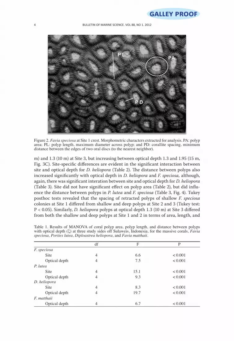
Sampling Methodology.—Presence of target coral species was recorded at each site along three replicate 30 × 2 m belt transects laid 5 m apart on the reef flat (5 m depth), crest (10 m depth), and slope (15 m depth). Five colonies of every coral species were photographed at each depth (with ~10 m between each replicate colony) for a total of 15 colonies of each species per site. Photographs of target corals were taken within a custom 10 × 10 cm graded grid using an Olympus camera FE-230. These photos were then used to assess (retracted) polyp area, polyp length, and the distance between polyps by processing digitized images in Image-Tool UTHSCSA (University of Texas Health Science Center, San Antonio, Texas; Fig. 2). In every image, 10 polyps were randomly chosen to assess polyp area and length. To assess polyp density, we recorded the distance between the edges of two oral discs (to the nearest neighbor; Fig. 2).
Data Analyses.—Since polyp area, length, and distance potentially may be correlated, multivariate analyses of variance (MANOVA) were performed. If MANOVA results were sig-nificant, then variation in polyp area, length, and distance with increasing optical depth and among sites was tested, using a two-way orthogonal analysis of variance (ANOVA) on each
Galley Proof

NOTES 3
species using optical depth and site as variables. A Tukey post hoc analysis was performed for each species to examine interactions between the two variables, site and optical depth. For F. matthaii, a one-way ANOVA was utilized with Tukey post hoc analysis, as data were only available for Site 1. Data for F. speciosa, F. matthaii, and D. heliopora were log-transformed to meet the assumptions of homogeneity of variance and normality. To assess the relationship between optical depth and polyp area in all four species, a Pearson’s correlation was also per-formed (n = 9). The colony mean was used for the MANOVA and ANOVA, while the transect mean was used for the correlation. All tests were performed with STATISTICA 7.0 (StatSoft) software.
Results
MANOVA results using polyp area, polyp length, and distance between polyps as variables for each coral species were significant, suggesting that there were multivar-iate differences between groups for D. heliopora, P. lutea, F. matthaii, and F. speciosa (Table 1). Subsequent ANOVA results indicate that polyp area and distance between polyps differed significantly with optical depth and between sites for some species (Tables 2, 3), while there was no significant change in polyp length among sites and with optical depth in all the coral species (ANOVA: P > 0.05). Specifically, polyp area increased significantly with increasing optical depth in P. lutea, D. heliopora, and F. matthaii (Table 2, Fig. 3). However, deviations from this trend were observed at spe-cific sites, such as D. heliopora polyp area decreasing between optical depth 0.65 (5
Figure 1. Study sites within the Wakatobi Marine National Park, southeast Sulawesi, Indonesia. Site 1 (5°28´S, 123°44´E) was situated next to a Bajo village, Site 2 (5°30´S, 123°45´E) was situated off Kaledupa, and Site 3 (5°28´S, 123°45´E) was situated on the Hoga reef. Adapted from Hennige et al. (2008).
BULLETIN OF MARINE SCIENCE. VOL 88, NO 1. 20124
m) and 1.3 (10 m) at Site 3, but increasing between optical depth 1.3 and 1.95 (15 m, Fig. 3C). Site-specific differences are evident in the significant interaction between site and optical depth for D. heliopora (Table 2). The distance between polyps also increased significantly with optical depth in D. heliopora and F. speciosa, although, again, there was significant interation between site and optical depth for D. heliopora (Table 3). Site did not have significant effect on polyp area (Table 2), but did influ-ence the distance between polyps in P. lutea and F. speciosa (Table 3, Fig. 4). Tukey posthoc tests revealed that the spacing of retracted polyps of shallow F. speciosa colonies at Site 1 differed from shallow and deep polyps at Site 2 and 3 (Tukey test: P < 0.05). Similarly, D. heliopora polyps at optical depth 1.3 (10 m) at Site 3 differed from both the shallow and deep polyps at Site 1 and 2 in terms of area, length, and
Table 1. Results of MANOVA of coral polyp area, polyp length, and distance between polyps with optical depth (ζ) at three study sides off Sulaweis, Indonesia, for the massive corals, Favia speciosa, Porites lutea, Diploastrea heliopora, and Favia matthaii.
df F PF. speciosa
Site 4 6.6 < 0.001Optical depth 4 7.5 < 0.001
P. luteaSite 4 15.1 < 0.001Optical depth 4 9.3 < 0.001
D. helioporaSite 4 8.3 < 0.001Optical depth 4 19.7 < 0.001
F. matthaiiOptical depth 4 6.7 < 0.001
Figure 2. Favia speciosa at Site 1 crest. Morphometric characters extracted for analysis. PA: polyp area; PL: polyp length, maximum diameter across polyp; and PD: corallite spacing, minimum distance between the edges of two oral discs (to the nearest neighbor).
Galley Proof

NOTES 5
spacing (Tukey test: P < 0.05). In addition to the previously mentioned interactions, site interacted with optical depth to polyp area in F. speciosa (Table 2, Fig. 3A) and polyp length (df = 4/36, F = 2.66, P = 0.04) in D. heliopora. Similarly, actual depth and site had a significant interactive effect on polyp area in F. speciosa (ANOVA: df = 4, 36; F = 2.56, P = 0.04) and on polyp area (ANOVA: df = 4, 36; F = 3.14, P = 0.02), length (ANOVA: df = 4, 36; F = 2.66, P = 0.04), and distance (ANOVA: df =4, 36; F = 3.12, P = 0.02) in D. heliopora.
Table 2. Results of ANOVA of coral polyp area with optical depth (ζ) at three study sites off Sulawesi, Indonesia, for the massive corals, Favia speciosa, Porites lutea, Diploastrea heliopora, and Favia matthaii. NS denotes non-significant.
df df Error F PF. speciosa
Site 2 36 2.65 NSOptical depth 2 36 2.65 NSSite × optical depth 4 36 2.65 0.04
P. luteaSite 2 36 0.64 NSOptical depth 2 36 3.56 0.03Site × optical depth 4 36 1.07 NS
D. helioporaSite 2 36 0.30 NSOptical depth 2 36 4.42 0.02Site × optical depth 4 36 3.14 0.02
F. matthaiiOptical depth 2 9 5.28 0.04
Table 3. Results of ANOVA of distance between coral polyps with optical depth at three study sites off Sulawesi, Indonesia, for the massive corals, Favia speciosa, Porites lutea, Diploastrea heliopora, and Favia matthaii. NS denotes non-significant.
df df Error F PF. speciosa
Site 2 36 7.21 0.02Optical depth 2 36 12.33 < 0.001Site × optical depth 4 36 1.89 NS
P. luteaSite 2 36 3.63 0.03Optical depth 2 36 2.65 NSSite × optical depth 4 36 1.2 NS
D. helioporaSite 2 36 0.73 NSOptical depth 2 36 3.95 0.02Site × optical depth 4 36 3.12 0.02
F. matthaiiOptical depth 2 9 2.45 NS

BULLETIN OF MARINE SCIENCE. VOL 88, NO 1. 20126
Discussion
Despite frequent site-specific responses, three massive coral species off Sulawesi, Indonesia, exhibited a general trend of increased polyp area with decreased light availability regardless of the amount of sedimentation. Increased polyp area could be a structural acclimation in low light situations to increase the potential for hetero-trophic efficiency (Anthony 2000, Anthony and Fabricius 2000). The trend observed across the massive species with regard to optical depth indicates that light (driven by depth or turbidity) is likely to be the driving factor behind these morphological changes. However, the additional effects of sedimentation may also be important (Rogers 1990).
Increased coral polyp area on reef slopes is likely a result of acclimation leading to changes in skeletal structure in a reduced light regime, driven by autotrophic or heterotrophic demands. Both Todd et al. (2001) and Caras et al. (2008) documented increased coral polyp sizes with increased sedimentation rates and\or decreased light levels. Importantly, sedimentation may have a dual role in polyp morphological adaptation: by affecting light attenuation and potentially as a source of food. It is un-known whether larger polyp areas and accompanying changes in skeletal reflectance primarily optimize light capture efficiency, or increase heterotrophic efficiency of the host to compensate for decreased photosynthesis at depth (Anthony 2000, Anthony and Fabricius 2000, Todd et al. 2001), but it is likely that changes in polyp sizes and
Figure 3. Mean area (mm2 ± SE) of polyps of (A) Favia speciosa, (B) Porites lutea, (C) Diploastrea heliopora, and (D) Favia matthaii from Sites 1, 2, and 3 vs optical depth (ζ, dimensionless).
Galley Proof

NOTES 7
spacing reflect components of both. Small and densely packed polyps may optimize light harvesting by increasing the surface area available for zooxanthellae (Anthony and Fabricius 2000, Einbinder et al. 2009). On the other hand, larger and fewer pol-yps may aid food capture (Einbinder et al. 2009). Some species are also capable of increasing feeding rates with depth (decreasing light availability, Palardy et al. 2005). However, other species, such as Stylophora pistillata (Esper, 1797), do not increase heterotrophic intake with depth (Alamaru et al. 2009) and may rely on decreasing polyp sizes with depth to maximize light harvesting (Einbinder et al. 2009). Larger polyps may also limit smothering stress by sediment settling, which potentially re-duces colony growth (Rogers 1990). Larger polyps, which may aid in sediment rejec-tion (Stafford-Smith and Ormond 1992), are therefore likely to be an advantage in turbid habitats.
Intraspecific morphological variation of our target corals suggests an ability to ac-climate across gradients, making P. lutea, D. heliopora, F. speciosa, and F. matthaii ecologically important corals within the Wakatobi Marine National Park. This mor-phological variation is consistent with the findings of previous studies which noted that the distribution of the four target coral species was attributable to host differ-ences and not to Symbiodinium acclimatory abilities [Hennige et al. 2008, although different Symbiodinium types can affect ecological and physiological host traits
Figure 4. Mean distance (mm ± SE) between polyps of (A) Favia speciosa, (B) Porites lutea, (C) Diploastrea heliopora, and (D) Favia matthaii from Sites 1, 2, and 3 vs optical depth (ζ, dimensionless).

BULLETIN OF MARINE SCIENCE. VOL 88, NO 1. 20128
including growth and tolerance to heat and light stress (LaJeunesse 2001, Abrego et al. 2008)]. The dominance of P. lutea (see Hennige et al. 2008) across sites examined here may be partially due to its ability to vary its polyp morphology to optimize envi-ronmental suitability. The relatively lower abundance of F. speciosa across sites, cou-pled with the narrower range of morphological change observed with regard to polyp area is consistent with this hypothesis. Consequently, although morphological varia-tion cannot solely determine coral distribution across environmental gradients, it is an important consideration in future studies, as without host morphological chang-es, acclimation of heterotrophy and photosynthesis would be severely constrained.
Acknowledgments
We thank Operation Wallacea, the Indonesian Institute of Sciences and J Jompa (UNHAS) for their support. We also thank M Flavell for help in editing figures and C Mattone for help with statistical analyses, and comments from MS Pratchett and the anonymous reviewers, which greatly improved this manuscript.
Literature Cited
Abrego D, Ulstrup KE, Willis BL, van Oppen MJH. 2008. Species-specific interactions between algal endosymbionts and coral hosts define their bleaching response to heat and light stress. Proc R Soc B. 275:2273–2282. PMid:18577506. PMCid:2603234. http://dx.doi.org/10.1098/rspb.2008.0180
Alamaru A, Loya Y, Brokovich E, Yam R, Shemesh A. 2009. Carbon and nitrogen utilization in two species of red sea corals along a depth gradient; insights from stable isotope analysis of total organic material and lipids. Geochim Cosmochim Ac. 73(18):5333–5342. http://dx.doi.org/10.1016/j.gca.2009.06.018
Anthony KRN. 2000. Enhanced particle-feeding capacity of corals on turbid reefs (Great Barrier Reef, Australia). Coral Reefs. 19:59–67. http://dx.doi.org/10.1007/s003380050227
Anthony KRN, Fabricius KE. 2000. Shifting roles of heterotrophy and autotrophy in coral ener-getics under varying turbidity. J Exp Mar Biol Ecol. 252:251–253. http://dx.doi.org/10.1016/S0022-0981(00)00237-9
Caras T, Bachar A, Pasternak Z. 2008. Morphological variation in the oral disc of the scler-actinian coral Favia speciosa (Dana) at Indonesia. Comput Biol Chem. 32:345–348. PMid:18657475. http://dx.doi.org/10.1016/j.compbiolchem.2008.06.003
Crabbe MJC, Smith DJ. 2005. Sediment impacts on growth rates of Acropora and Porites corals from fringing reefs of Sulawesi, Indonesia. Coral Reefs. 24:437–441. http://dx.doi.org/10.1007/s00338-005-0004-6
Dubinsky Z, Stambler N. 2005. Corals as light collectors: an integrating sphere approach. Coral Reefs. 24:1–9. http://dx.doi.org/10.1007/s00338-004-0452-4
Einbinder S, Mass T, Brokovich E, Dubinsky Z, Erez Y, Tchernov D. 2009. Changes in morphol-ogy and diet of the coral Stylophora pistillata along the depth gradient. Mar Ecol Prog Ser. 381:167–174. http://dx.doi.org/10.3354/meps07908
Foster AB. 1977. Patterns of small-scale variation of skeletal morphology within the scleractin-ian corals, Montastrea annularis and Siderastrea siderea. Proc 3rd Int Coral Reef Symp. 2:409–415.
Goreau TF. 1959. The physiology of skeleton formation in corals. 1. A method for measuring the rate of calcium deposition by corals under different conditions. Biol Bull (Woods Hole). 116: 59–75. http://dx.doi.org/10.2307/1539156
Hennige SJ, Smith DJ, Perkins R, Consalvey M, Paterson DM, Suggett DJ. 2008. Photoacclimation, growth and distribution of massive coral species in clear and turbid waters. Mar Ecol Prog Ser. 369:77–88. http://dx.doi.org/10.3354/meps07612
Galley Proof

NOTES 9
Hennige SJ, Suggett DJ, Warner ME, McDougall KE, Smith DJ. 2009. Photoacclimation of Symbiodinium revisited: bio-physical and bio-optical signatures. Coral Reefs. 28:179–195. http://dx.doi.org/10.1007/s00338-008-0444-x
Kenyon JC, Vroom PS, Page KN, Dunlap MJ, Wilkinson CB, Aeby GS. 2006. Community struc-ture of hermatypic corals at French frigate shoals, North-western Hawaiian Islands: capac-ity for resistance and resilience to selective stressors. Pac Sci. 60:153–175. http://dx.doi.org/10.1353/psc.2006.0006
Kirk JTO. 1994. Light and photosynthesis in aquatic ecosystems. 2nd ed. Cambridge: Cambridge University Press. http://dx.doi.org/10.1017/CBO9780511623370
LaJeunesse TC. 2001. Investigating the biodiversity, ecology and phylogeny of endosymbiotic dinoflagellates in the genus Symbiodinium using the ITS region: in search of a ‘species’ level marker. J Phycol. 37:866–880. http://dx.doi.org/10.1046/j.1529-8817.2001.01031.x
Maida M, Coll JC, Sammarco PW. 1994. Shedding new light on scleractinian coral recruitment. J Exp Mar Biol Ecol. 180:89–202. http://dx.doi.org/10.1016/0022-0981(94)90066-3
Mass T, Einbinder S, Brokovich E, Shashar N, Vago R, Erez J, Dubinsky Z. 2007. Photoacclimation of Stylophora pistillata to light extremes: metabolism and calcification. Mar Ecol Prog Ser. 334:93–102. http://dx.doi.org/10.3354/meps334093
Muscatine L. 1990. The role of symbiotic algae in carbon and energy flux in reef corals. In: Dubinsky Z, editor. Ecosystems of the world: coral reefs. Amsterdam: Elsevier. p. 75–87.
Palardy JE, Grottoli AG, Matthews KA. 2005. Effects of upwelling, depth, morphology and polyp size on feeding in three species of Panamanian corals. Mar Ecol Prog Ser. 300:79–89. http://dx.doi.org/10.3354/meps300079
Rogers CS. 1990. Responses of coral reefs and reef organisms to sedimentation. Mar Ecol Prog Ser. 62:185–202. http://dx.doi.org/10.3354/meps062185
Stafford-Smith MG, Ormond RFG. 1992. Sediment rejection mechanisms of 42 species of Australian scleractinian corals. Aust J Mar Freshwat Res. 43:683–705. http://dx.doi.org/10.1071/MF9920683
Todd PA, Ladle RJ, Lewin-Koh NJI, Chou LM. 2004. Genotype × environment interactions in transplanted clones of the massive corals Favia speciosa and Diploastrea heliopora. Mar Ecol Prog Ser. 271:167–182. http://dx.doi.org/10.3354/meps271167
Todd PA, Sanderson PG, Chou LM. 2001. Morphological variation in the polyps of the sclerac-tinian coral Favia speciosa (Dana) around Singapore. Hydrobiologia. 444:227–235. http://dx.doi.org/10.1023/A:1017570100029
Date Submitted: 5 October, 2010.Date Accepted: 2 June, 2011.Available Online:
Addresses: (CP, JH) ARC Centre of Excellence for Coral Reef Studies, James Cook University of North Queensland, Townsville, Queensland 4811, Australia. (SJH) Centre of Marine Biodiversity and Biotechnology, Heriot Watt University, Edinburgh, United Kingdom. (RM) Università Degli Studi Di Roma “La Sapienza,” Rome, 00185, Italy. (DJS) University of Essex, Wivenhoe Park, Essex, CO43SQ United Kingdom. Corresponding Author: (CP) E-mail: <[email protected]>.