Embed Size (px)
Citation preview
lable at ScienceDirect
Journal of Pharmaceutical Sciences 105 (2016) 1258e1268
Contents lists avai
Journal of Pharmaceutical Sciences
journal homepage: www.jpharmsci .org
Pharmaceutics, Drug Delivery and Pharmaceutical Technology
Preparation and Physical Characterization of a Diclofenac-RanitidineCo-precipitate for Improving the Dissolution of Diclofenac
Robertino O. Gaitano 1, Natalia L. Calvo 2, Griselda E. Narda 1, Teodoro S. Kaufman 2,Rub�en M. Maggio 2, *, Elena V. Brusau 1, *
1 Química Inorg�anica, Departamento de Química, Facultad de Química, Bioquímica y Farmacia, Universidad Nacional de San Luis and Instituto deInvestigaciones en Tecnología Química (INTEQUI, CONICET-UNSL), San Luis D5700BWQ, Argentina2 An�alisis Farmac�eutico, Departamento de Química Org�anica, Facultad de Ciencias Bioquímicas y Farmac�euticas, Universidad Nacional de Rosario andInstituto de Química Rosario (IQUIR, CONICET-UNR), Rosario S2002LRK, Argentina
a r t i c l e i n f o
Article history:Received 9 October 2015Revised 4 December 2015Accepted 5 January 2016Available online 9 February 2016
Keywords:ranitidinediclofenacco-crystalFTIRNMR spectroscopyphysical characterizationsolid-statedissolutionchemometrics
This article contains the supplementary materials FTvs. DRIFTS and physical mixture vs. DIC-RAN), chemicand 13C NMR spectra of RAN$HCl, RAN, DIC-Na, andauthors by request or via the Internet at http://dx.d001.* Correspondences to: Rub�en M. Maggio (Telephone
Elena V. Brusau (Telephone/Fax: þ54-266-4520300).E-mail addresses: [email protected] (R
edu.ar (E.V. Brusau).
http://dx.doi.org/10.1016/j.xphs.2016.01.0010022-3549/© 2016 American Pharmacists Association
a b s t r a c t
Mixing aqueous solutions of sodium diclofenac (DIC-Na) and ranitidine hydrochloride (RAN$HCl) affor-ded an off-white solid (DIC-RAN) that was investigated from the microscopic, thermal, diffractometric,spectroscopic, and functional (chemometrics-assisted dissolution) points of view. The solid has a 2:1(DIC:RAN) molar ratio according to 1H nuclear magnetic resonance spectroscopy. It is thermally stable,displaying a broad endothermic signal centered at 105�C in the thermogram, and its characteristic re-flections in the powder X-ray diffractogram remained unchanged after a 3-month aging period. Scanningelectron microscopy micrographs uncovered its morphology, whereas the spectral data suggested aninteraction between the carboxylic acid of DIC and the alkyldimethylamino moiety of RAN. The disso-lution of DIC-RAN was monitored at different pH values by an ultraviolet/chemometrics procedure, beingcomplete within 5 min at pH 6.8. This compares favorably with the dissolution of a DIC-Na sample of thesame particle size.
© 2016 American Pharmacists Association®. Published by Elsevier Inc. All rights reserved.
Introduction which is widely used in the long-term treatment of degenerative
The delivery of active pharmaceutical ingredients (APIs) withpoor aqueous solubility is a current challenge because the oralbioavailability depends on their dissolution rate in the absorptionsite. Various formulation or processing-based approaches havebeen devised to increase the solubility of poor water-solubledrugs; however, one of the most convenient strategies towardthis purpose is the improvement of their physicochemicalproperties.
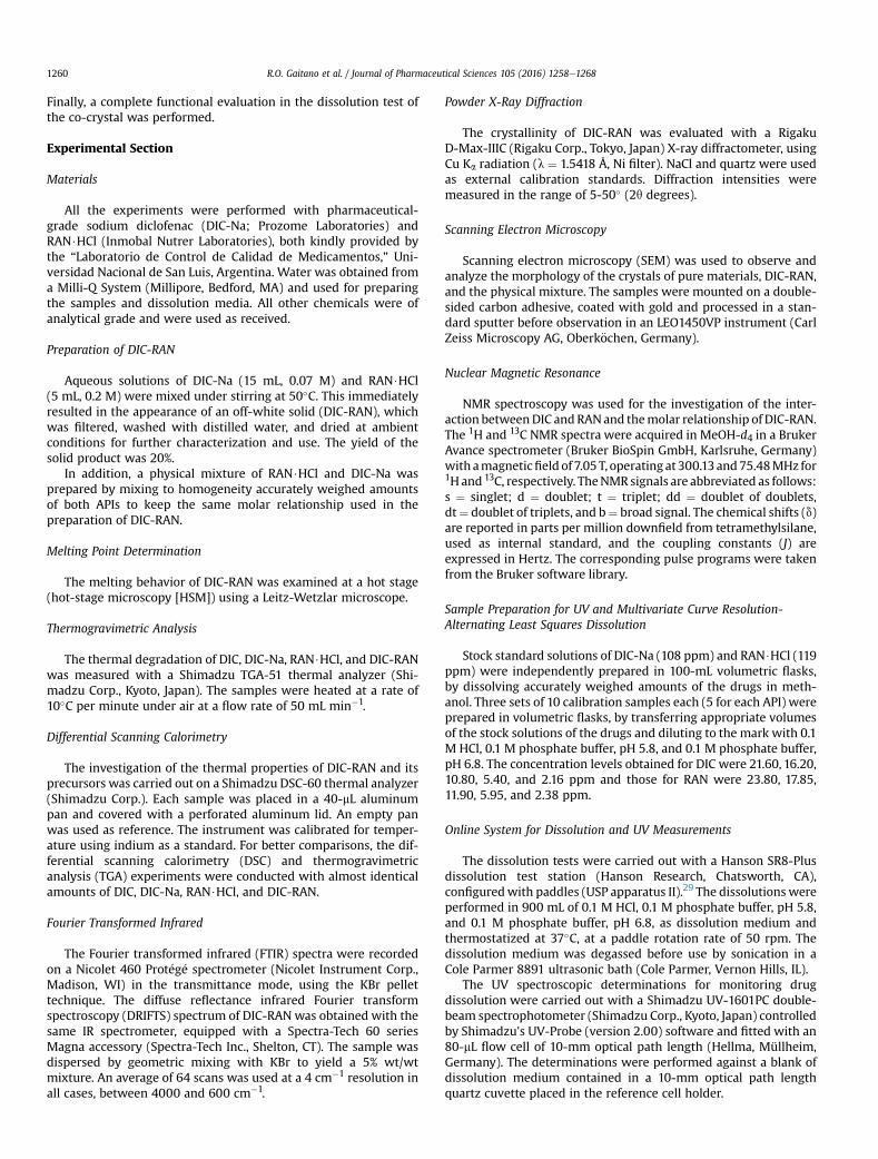
Diclofenac (DIC, Fig. 1) is 2-[(20,60-dichlorophenyl)amino]benzeneacetic acid, a potent nonsteroidal anti-inflammatoryagent, with pronounced antipyretic and analgesic properties,
IR spectral comparisons (KBral structures of DIC and RAN,DIC-RAN available from theoi.org/10.1016/j.xphs.2016.01.
/Fax: þ54-341-4370477) and
.M. Maggio), ebrusau@unsl.
®. Published by Elsevier Inc. All ri
joint diseases. DIC is an acidic drug, which has very low aqueoussolubility in its unionized form (6 � 10�5 M at 25�C),1 beingpractically insoluble in HCl solutions at pH 1.1, very slightly sol-uble in phosphate buffer at pH 6.8, and slightly soluble in pureH2O.2
DIC has 3 hydrogen bond acceptors, 2 hydrogen bond donors,and significant conformational flexibility; however, the carboxylicacid moiety and the secondary amino group of DIC are the sourcesof tight intra- and intermolecular H-bonds, which generate adimeric centrosymmetric structure, and cause the high meltingpoint of the drug. This dimeric form, where all the H-bonds involveinter- and intramolecular hydrophilic groups, represents thestructural unit of the solid state of DIC, which turns the drug lessavailable to intermolecular interactions with water, the result beingits poor aqueous solubility.
On the other side, ranitidine (RAN, Fig. 1) is N-{2-[[[5-[(dime-thylamino)methyl]-2-furanyl] methyl]thio]ethyl}-N0-methyl-2-nitro-1,1-ethenediamine. The drug is a histamine H2-receptorantagonist that inhibits the secretion of gastric acid induced byvarious stimuli, while lacking unwanted antiandrogenic andhepatic microsomal enzyme-inhibiting effects.
ghts reserved.
Figure 1. Chemical structures of DIC, the related compound A of DIC, and the tautomeric forms of RAN.
R.O. Gaitano et al. / Journal of Pharmaceutical Sciences 105 (2016) 1258e1268 1259
RAN is a dibasic drug, commercially available as the mono-hydrochloride salt (ranitidine hydrochloride [RAN$HCl]), which ishighly soluble in water. Theoretically, it can exist in 3 main tauto-meric forms (imine, enamine, and nitronic acid; see Fig. 1). Inaddition, the enamine could exist as 2 geometrical (E/Z) isomers,and the nitronic acid and imine forms could both exhibit furtherprototropic tautomerism within the amidine group and syn-anti-isomerism about the C-N bonds. This tautomer interconversionrequires 1,5- or 1,3-proton shifts,3 analogous to those proposed forcimetidine.4
RAN$HCl has 2 known polymorphs (forms 1 and 2). The struc-ture of form 2, solved by single crystal X-ray diffraction (XRD),exhibits molecular disorder in the nitroethenediamine moiety. Thestructural data suggest contributions, 50% each, from the nitronicacid and the enamine tautomers. On the other hand, nuclearmagnetic resonance (NMR) studies revealed that form 1 does notshow molecular disorder, hinting that a single tautomer is presentin the solid.5
It has been suggested that in aqueous solutions, the geometricalisomers are in rapid equilibrium, in the NMR time scale, affectingthe resonances of H-17 and H-18, which result in broad signals.6
However, on protonation of the diaminovinyl group, the intercon-version of the E and Z species is slowed down on account of theformation of intramolecularly H-bonded species.
It is well established that the solubility and the bioavailability ofpoorly water-soluble drugs can be improved by converting theminto the amorphous state.7 On the other hand, co-precipitate sys-tems resulting from the interaction of 2 chemical entities to form asingle-phase system usually exhibit improved physical stability andaqueous dissolution profile, compared with the individual drugs.8
It is also known that salt formation is a very simple andstraightforward chemical tool, commonly used for enhancing thesolubility and dissolution rate of poorly soluble drugs containingionizable functional groups.9 In this case, the nature of the coun-terion is relevant to both the processability of the product and itsphysicochemical characteristics, including solubility, dissolution
rate, and stability. In all cases, the intermolecular interactions be-tween the components of the system have been pointed out asresponsible for the observed improvements.
Because of its wide use, various strategies have been used toenhance the solubility and dissolution rate of DIC. One of themconsists in the elaboration of derivatives of the APIs, such assalts,10,11 co-crystals,12 ionic pairs,13 and complexes with cyclo-dextrins.14 Another alternative entails modifying the vehicle, in thisapproach, including the use of mixed solvency,15,16 formation ofliquisolids,17 solid dispersions,18 nanosuspensions,19 micellar dis-persions and microemulsions,20 and inclusion in supramolecularmatrices,21 among others.
Various salts of DIC are currently part of pharmaceutical prod-ucts, including sodium, potassium, and ammonium derivatives, anddiclofenac N-(2-hydroxyethyl)pyrrolidine and diclofenac diethyl-amine.22 In addition, a screening toward co-crystals of DIC has beenrecently reported.23
It has been shown that RAN is able to protect against the ul-cerogenic effect of nonsteroidal anti-inflammatory agents, such asDIC. Therefore, these active principles are frequently administered.Furthermore, the pharmacologic interaction between DIC and RANhas been studied in vivo, concluding that it is minimal and that theattained gastric pH range did not influence the oral absorption ofenteric-coated diclofenac.24
In addition, it has been observed that the concomitant intake ofRAN has no effect on the pharmacokinetics of DIC.25 Bilayer tabletscontaining a fixed-dose combination of DIC and RAN have recentlybeen evaluated,26 and the bioavailability of a formulation con-taining DIC and RAN has also been studied.27
Previous studies on the chemical interaction between DIC andRANwere also unable to detect any effect.28 As DIC is an acidic drugand RAN is a basic pharmaceutical ingredient, we were puzzledabout this intriguing observation; therefore, we decided to assess ifthis finding was correct. As a result, here we report the preparationand characterization (spectroscopic, thermal, structural, andmorphologic) of an off-white microcrystalline solid (DIC-RAN).
R.O. Gaitano et al. / Journal of Pharmaceutical Sciences 105 (2016) 1258e12681260
Finally, a complete functional evaluation in the dissolution test ofthe co-crystal was performed.
Experimental Section
Materials
All the experiments were performed with pharmaceutical-grade sodium diclofenac (DIC-Na; Prozome Laboratories) andRAN$HCl (Inmobal Nutrer Laboratories), both kindly provided bythe “Laboratorio de Control de Calidad de Medicamentos,” Uni-versidad Nacional de San Luis, Argentina. Water was obtained froma Milli-Q System (Millipore, Bedford, MA) and used for preparingthe samples and dissolution media. All other chemicals were ofanalytical grade and were used as received.
Preparation of DIC-RAN
Aqueous solutions of DIC-Na (15 mL, 0.07 M) and RAN$HCl(5 mL, 0.2 M) were mixed under stirring at 50�C. This immediatelyresulted in the appearance of an off-white solid (DIC-RAN), whichwas filtered, washed with distilled water, and dried at ambientconditions for further characterization and use. The yield of thesolid product was 20%.
In addition, a physical mixture of RAN$HCl and DIC-Na wasprepared by mixing to homogeneity accurately weighed amountsof both APIs to keep the same molar relationship used in thepreparation of DIC-RAN.
Melting Point Determination
The melting behavior of DIC-RAN was examined at a hot stage(hot-stage microscopy [HSM]) using a Leitz-Wetzlar microscope.
Thermogravimetric Analysis
The thermal degradation of DIC, DIC-Na, RAN$HCl, and DIC-RANwas measured with a Shimadzu TGA-51 thermal analyzer (Shi-madzu Corp., Kyoto, Japan). The samples were heated at a rate of10�C per minute under air at a flow rate of 50 mL min�1.
Differential Scanning Calorimetry
The investigation of the thermal properties of DIC-RAN and itsprecursors was carried out on a Shimadzu DSC-60 thermal analyzer(Shimadzu Corp.). Each sample was placed in a 40-mL aluminumpan and covered with a perforated aluminum lid. An empty panwas used as reference. The instrument was calibrated for temper-ature using indium as a standard. For better comparisons, the dif-ferential scanning calorimetry (DSC) and thermogravimetricanalysis (TGA) experiments were conducted with almost identicalamounts of DIC, DIC-Na, RAN$HCl, and DIC-RAN.
Fourier Transformed Infrared
The Fourier transformed infrared (FTIR) spectra were recordedon a Nicolet 460 Prot�eg�e spectrometer (Nicolet Instrument Corp.,Madison, WI) in the transmittance mode, using the KBr pellettechnique. The diffuse reflectance infrared Fourier transformspectroscopy (DRIFTS) spectrum of DIC-RAN was obtained with thesame IR spectrometer, equipped with a Spectra-Tech 60 seriesMagna accessory (Spectra-Tech Inc., Shelton, CT). The sample wasdispersed by geometric mixing with KBr to yield a 5% wt/wtmixture. An average of 64 scans was used at a 4 cm�1 resolution inall cases, between 4000 and 600 cm�1.
Powder X-Ray Diffraction
The crystallinity of DIC-RAN was evaluated with a RigakuD-Max-IIIC (Rigaku Corp., Tokyo, Japan) X-ray diffractometer, usingCu Ka radiation (l ¼ 1.5418 Å, Ni filter). NaCl and quartz were usedas external calibration standards. Diffraction intensities weremeasured in the range of 5-50� (2q degrees).
Scanning Electron Microscopy
Scanning electron microscopy (SEM) was used to observe andanalyze the morphology of the crystals of pure materials, DIC-RAN,and the physical mixture. The samples were mounted on a double-sided carbon adhesive, coated with gold and processed in a stan-dard sputter before observation in an LEO1450VP instrument (CarlZeiss Microscopy AG, Oberk€ochen, Germany).
Nuclear Magnetic Resonance
NMR spectroscopy was used for the investigation of the inter-action betweenDIC and RANand themolar relationship of DIC-RAN.The 1H and 13C NMR spectra were acquired in MeOH-d4 in a BrukerAvance spectrometer (Bruker BioSpin GmbH, Karlsruhe, Germany)with amagneticfield of 7.05T, operating at 300.13and75.48MHz for1Hand 13C, respectively. TheNMR signals are abbreviated as follows:s ¼ singlet; d ¼ doublet; t ¼ triplet; dd ¼ doublet of doublets,dt¼ doublet of triplets, and b¼ broad signal. The chemical shifts (d)are reported in parts per million downfield from tetramethylsilane,used as internal standard, and the coupling constants (J) areexpressed in Hertz. The corresponding pulse programs were takenfrom the Bruker software library.
Sample Preparation for UV and Multivariate Curve Resolution-Alternating Least Squares Dissolution
Stock standard solutions of DIC-Na (108 ppm) and RAN$HCl (119ppm) were independently prepared in 100-mL volumetric flasks,by dissolving accurately weighed amounts of the drugs in meth-anol. Three sets of 10 calibration samples each (5 for each API) wereprepared in volumetric flasks, by transferring appropriate volumesof the stock solutions of the drugs and diluting to the mark with 0.1M HCl, 0.1 M phosphate buffer, pH 5.8, and 0.1 M phosphate buffer,pH 6.8. The concentration levels obtained for DIC were 21.60, 16.20,10.80, 5.40, and 2.16 ppm and those for RAN were 23.80, 17.85,11.90, 5.95, and 2.38 ppm.
Online System for Dissolution and UV Measurements
The dissolution tests were carried out with a Hanson SR8-Plusdissolution test station (Hanson Research, Chatsworth, CA),configuredwith paddles (USP apparatus II).29 The dissolutions wereperformed in 900 mL of 0.1 M HCl, 0.1 M phosphate buffer, pH 5.8,and 0.1 M phosphate buffer, pH 6.8, as dissolution medium andthermostatized at 37�C, at a paddle rotation rate of 50 rpm. Thedissolution medium was degassed before use by sonication in aCole Parmer 8891 ultrasonic bath (Cole Parmer, Vernon Hills, IL).
The UV spectroscopic determinations for monitoring drugdissolution were carried out with a Shimadzu UV-1601PC double-beam spectrophotometer (Shimadzu Corp., Kyoto, Japan) controlledby Shimadzu's UV-Probe (version 2.00) software and fitted with an80-mL flow cell of 10-mm optical path length (Hellma, Müllheim,Germany). The determinations were performed against a blank ofdissolution medium contained in a 10-mm optical path lengthquartz cuvette placed in the reference cell holder.
R.O. Gaitano et al. / Journal of Pharmaceutical Sciences 105 (2016) 1258e1268 1261
The dissolution medium was continuously withdrawn from thedissolution vessel through the sampling probe at a 1.5 mL min�1
flow rate, by means of a Gilson Minipuls 3 peristaltic pump andreturned to the dissolution vessel after passing through the spec-trophotometer flow cell. Degassing of the dissolution media andonline sample filtration avoided potential interferences on accountof bubbles or undissolved particles.
The UV spectra of the dissolution samples were collected after astandard pre-established delay time of 1.0 min, corresponding tothe tubing dead volume. The dissolution experiments were moni-tored every 1.0 min during 2 h, and spectra were acquired at 1-nmintervals in the 245-400 nm range (156 data points per spectrum).The acquired data were saved as a matrix in comma-separatedvalue (CSV) format.
For quantitative purposes, the calibration solutions of DIC-Naand RAN$HCl were sequentially pumped through the flow cell forperiods of 5 min each, and 5 spectra per concentration level werecollected. The full calibration data of each analyte were saved inCSV format, read into MATLAB, and stored as matrices (DcDIC(25�156)and DcRAN(25�156)). The dissolution of the samples of DIC-RAN wasmonitored analogously.
Computational Methods
Chemometrics computations were carried out in MATLABR2010a (Mathworks, Natick, MA), using MCR-ALS routines.30 Thefull calibration data of each analyte were saved in CSV format,read into MATLAB, and stored as matrices (DcDIC(25�156) andDcRAN(25�156)). The dissolution data of the samples of DIC-RANwerealso stored in CSV format and read into MATLAB as a single matrix(Dt(120�156)) for each dissolution experiment. The calculations wereperformed without special data preprocessing, using non-negativity of absorbances and concentrations as MCR-ALS con-straints. Origin 8.5 (Origin Lab, Northampton, MA) was used forgraphics and statistical data analyses.
Spectroscopic Data
Powder X-Ray DiagramsFor DIC: 2q 10.9, 15.4, 17.9, 20.7, 21.7, 23.7, 24.6, 25.6, 26.1, 28.3,
and 28.7.For DIC-Na: 2q 6.6, 8.5, 11.1, 15.1, 17.1, 19.8, 23.1, 23.5, 23.8, 24.1,
24.9, 25.8, 26.9, and 27.8.For RAN$HCl: 2q 8.4, 14.6, 15.3, 16.5, 18.1, 19.3, 20.3, 20.9, 23.5,
24.1, 25.9, 26.7, 27.5, 28.7, 31.9, and 33.0.For DIC-RAN: 2q 7.6, 11.4, 15.3, 19.2, 20.2, 20.6, 21.4, 25.6, 30.9,
and 37.7.
Infrared AbsorptionFor DIC: 3323, 3080-2840, 2725-2563, 1694, 1587, 1577, 1568,
1508,1480,1454,1422,1413,1322,1305,1282,1273,1251,1200,1160,1093, 938, 890, 862, 837, 766, 752, 741, 710, 663, 630, and 610 cm�1.
For DIC-Na: 3387, 3257, 3079, 3065, 3036, 2971,1604,1587,1575,1557, 1508, 1499, 1469, 1453, 1400, 1305, 1283, 1250, 1234, 1193,1167, 1090, 1045, 952, 869, 845, 766, 747, 715, 670, and 636 cm�1.
For RAN$HCl: 3257, 3189, 3097, 3013, 2995, 2975, 2948,2910,2660, 2558, 2466, 1620, 1590, 1569, 1419, 1380, 1264, 1221,1164, 1075, 1046, 1022, 1006, 992, 880, 761, and 700 cm�1.
For DIC-RAN: 3285, 3197, 3155, 3135, 3067,1707, 1620, 1580,1509, 1453,1380, 1304, 1250, 1202, 1154, 1036, 1010, 946, 871, 836,793, 771, 757, 744, 715, 667, and 615 cm�1.
Nuclear Magnetic ResonanceFor DIC: (300 MHz, MeOH-d4) d 3.71 (s, 2H, ArCH2), 4.85 (s, 2H,
NH, and CO2H), 6.40 (d, 1H, J ¼ 7.5, H-3), 6.87 (dt, 1H, J ¼ 7.5, H-5),
7.03 (dt, 1H, J ¼ 2.5 and 7.5, H-4), 7.04 (t, 1H, J ¼ 8.0, H-40), 7.20 (dd,1H, J ¼ 2.5 and 7.5, H-6), and 7.39 (d, 2H, J ¼ 8.0, H-30, and H-50).
For RAN$HCl: (300 MHz, MeOH-d4) d 2.81 (t, 1H, J ¼ 6.3, H-12),2.86 (s, 6H, H-8, and H-9), 2.90 (bs, 3H, H-17), 3.44 (t, 2H, J ¼ 6.3,H-13), 3.84 (s, 2H, H-10), 4.36 (s, 2H, H-6), 4.87 (s, 3H, 3� NH), 6.38(d, 1H, J ¼ 3.2, H-3), and 6.64 (d, 1H, J ¼ 3.2, H-4). H-18 is missingdue to H-D exchange with MeOH-d4 during E/Z isomerization.31
For RAN: (300 MHz, MeOH-d4) d 2.25 (s, 6H, H-8, and H-9), 2.76(t, 1H, J ¼ 6.3, C-12), 2.89 (bs, 3H, H-17), 3.39 (t, 2H, J ¼ 6.3, C-13),3.51 (s, 2H, H-10), 3.79 (s, 2H, H-6), 4.86 (s, 3H, 3� NH), and 6.24 (s,2H, H-3, and H-4). H-18 is missing due to H-D exchange withMeOH-d4 during E/Z isomerization.
For DIC-RAN: (300 MHz, MeOH-d4) d 2.72 (s, 6H, H-8, and H-9,RAN), 2.78 (t, 1H, J ¼ 6.3, H-12, RAN), 2.88 (bs, 3H, H-17, RAN), 3.40(t, 2H, J¼ 6.3, H-13, RAN), 3.71 (s, 4H, ArCH2, DIC), 3.80 (s, 2H, H-10,RAN), 4.16 (s, 2H, H-6, RAN), 4.85 (s, 6H, NH, and OH), 6.33 (d, 1H,J ¼ 3.2, H-3, RAN), 6.39 (d, 2H, J ¼ 7.5, H-3, DIC), 6.52 (d, 1H, J ¼ 3.2,H-4, RAN), 6.88 (dt, 2H, J¼ 1.3 and 7.6, H-5, DIC), 7.03 (dt, 2H, J¼ 1.3and 7.6, H-4, DIC), 7.04 (t, 2H, J ¼ 7.8, H-40, DIC), 7.21 (dd, 2H, J ¼ 1.3and 7.6, H-6, DIC), and 7.38 (d, 4H, J ¼ 7.8, H-30 and H-50, DIC). H-18of RAN is missing due to H-D exchange with MeOH-d4 during E/Zisomerization.
Results and Discussion
Preparation of DIC-RAN
Despite the literature precedent indicating the lack of interac-tion between DIC and RAN,28 in our hands, the mixture of aqueoussolutions of DIC-Na and RAN$HCl resulted in the precipitation of anoff-white microcrystalline substance. The relative low yield of DIC-RAN obtained by the simple mixture of its precursors in water maybe the result of the high aqueous solubility of the product,compared with the low solubility of DIC-Na, used for its generation.Although no efforts were performed to increase product yields, it isworthwhile noting that previous reports stated that no interactiontook place when solutions of DIC-Na and RAN$HCl weremixed,28 asit was aforementioned. On the other hand, careful manipulation ofvolumes and concentrations of the starting solutions, using theprecursors in powder form and using antisolvents or co-solventsthat favor the solubility of DIC-Na, relative to DIC-RAN, mayresult in increased yields.32 However, these approaches should alsobear in mind that NaCl is concomitantly produced, and operatively,it would be desirable to keep it in solution.
Thermal Characterization of DIC-RAN
The association of different thermal methods, including DSC,TGA, and HSM, made it possible to unveil details of the thermalbehavior of DIC-RAN (Fig. 2). From the literature, it is known thatthe melting point of RAN is 69�C-70�C, whereas RAN$HCl melts at134�C-140�C (form 1) or about 140�C-144�C (form 2), withdecomposition.5 On the other side, DIC melts at 156�C-158�C,33
whereas DIC-Na has a melting point of 283�C-285�C.2
It was observed (Fig. 2a) that the melting point of DIC-RAN isassociated with an endothermic DSC signal centered at 105.36�C(DHm ¼ 67.76 J g�1). The event was also studied under an HSM,confirming its agreement with the melting of the sample and sug-gesting that a different phase was formed. The melt returned slowlyto the solid state once cooled to room temperature, and no recrys-tallization peak was observed in the corresponding cooling curve(Fig. 2b).
In an extended temperature scale (Fig. 2d), the DSC thermogramrevealed a complex pattern between 150�C and 375�C. In addition,
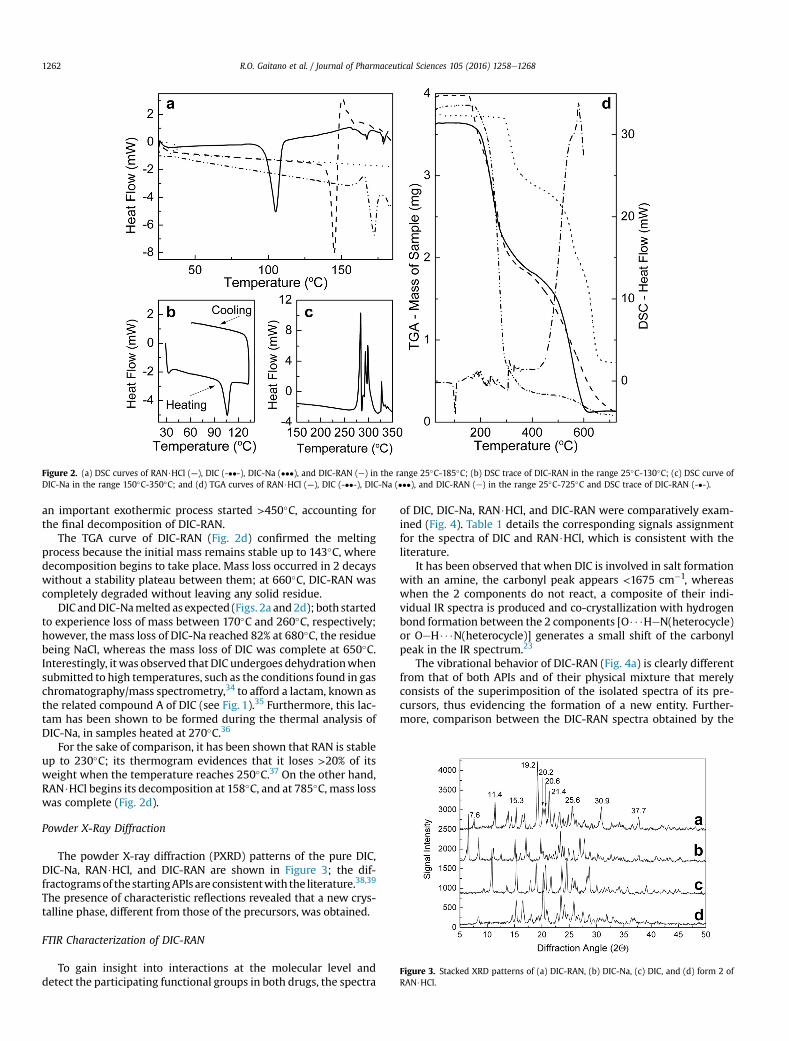
Figure 2. (a) DSC curves of RAN$HCl (—), DIC (-��-), DIC-Na (���), and DIC-RAN (e) in the range 25�C-185�C; (b) DSC trace of DIC-RAN in the range 25�C-130�C; (c) DSC curve ofDIC-Na in the range 150�C-350�C; and (d) TGA curves of RAN$HCl (—), DIC (-��-), DIC-Na (���), and DIC-RAN (e) in the range 25�C-725�C and DSC trace of DIC-RAN (-�-).
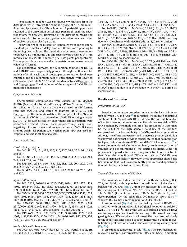
Figure 3. Stacked XRD patterns of (a) DIC-RAN, (b) DIC-Na, (c) DIC, and (d) form 2 ofRAN$HCl.
R.O. Gaitano et al. / Journal of Pharmaceutical Sciences 105 (2016) 1258e12681262
an important exothermic process started >450�C, accounting forthe final decomposition of DIC-RAN.
The TGA curve of DIC-RAN (Fig. 2d) confirmed the meltingprocess because the initial mass remains stable up to 143�C, wheredecomposition begins to take place. Mass loss occurred in 2 decayswithout a stability plateau between them; at 660�C, DIC-RAN wascompletely degraded without leaving any solid residue.
DIC andDIC-Namelted as expected (Figs. 2a and2d); both startedto experience loss of mass between 170�C and 260�C, respectively;however, the mass loss of DIC-Na reached 82% at 680�C, the residuebeing NaCl, whereas the mass loss of DIC was complete at 650�C.Interestingly, it was observed that DIC undergoes dehydrationwhensubmitted to high temperatures, such as the conditions found in gaschromatography/mass spectrometry,34 to afford a lactam, known asthe related compound A of DIC (see Fig. 1).35 Furthermore, this lac-tam has been shown to be formed during the thermal analysis ofDIC-Na, in samples heated at 270�C.36
For the sake of comparison, it has been shown that RAN is stableup to 230�C; its thermogram evidences that it loses >20% of itsweight when the temperature reaches 250�C.37 On the other hand,RAN$HCl begins its decomposition at 158�C, and at 785�C, mass losswas complete (Fig. 2d).
Powder X-Ray Diffraction
The powder X-ray diffraction (PXRD) patterns of the pure DIC,DIC-Na, RAN$HCl, and DIC-RAN are shown in Figure 3; the dif-fractogramsof thestartingAPIs are consistentwith the literature.38,39
The presence of characteristic reflections revealed that a new crys-talline phase, different from those of the precursors, was obtained.
FTIR Characterization of DIC-RAN
To gain insight into interactions at the molecular level anddetect the participating functional groups in both drugs, the spectra
of DIC, DIC-Na, RAN$HCl, and DIC-RAN were comparatively exam-ined (Fig. 4). Table 1 details the corresponding signals assignmentfor the spectra of DIC and RAN$HCl, which is consistent with theliterature.
It has been observed that when DIC is involved in salt formationwith an amine, the carbonyl peak appears <1675 cm�1, whereaswhen the 2 components do not react, a composite of their indi-vidual IR spectra is produced and co-crystallization with hydrogenbond formation between the 2 components [O$$$HeN(heterocycle)or OeH$$$N(heterocycle)] generates a small shift of the carbonylpeak in the IR spectrum.23
The vibrational behavior of DIC-RAN (Fig. 4a) is clearly differentfrom that of both APIs and of their physical mixture that merelyconsists of the superimposition of the isolated spectra of its pre-cursors, thus evidencing the formation of a new entity. Further-more, comparison between the DIC-RAN spectra obtained by the
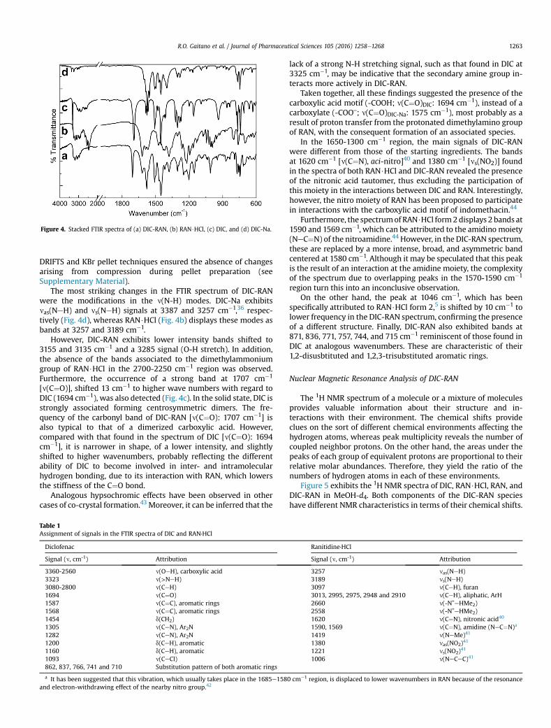
Figure 4. Stacked FTIR spectra of (a) DIC-RAN, (b) RAN$HCl, (c) DIC, and (d) DIC-Na.
R.O. Gaitano et al. / Journal of Pharmaceutical Sciences 105 (2016) 1258e1268 1263
DRIFTS and KBr pellet techniques ensured the absence of changesarising from compression during pellet preparation (seeSupplementary Material).
The most striking changes in the FTIR spectrum of DIC-RANwere the modifications in the n(N-H) modes. DIC-Na exhibitsnas(NeH) and ns(NeH) signals at 3387 and 3257 cm�1,36 respec-tively (Fig. 4d), whereas RAN$HCl (Fig. 4b) displays these modes asbands at 3257 and 3189 cm�1.
However, DIC-RAN exhibits lower intensity bands shifted to3155 and 3135 cm�1 and a 3285 signal (O-H stretch). In addition,the absence of the bands associated to the dimethylammoniumgroup of RAN$HCl in the 2700-2250 cm�1 region was observed.Furthermore, the occurrence of a strong band at 1707 cm�1
[n(C¼O)], shifted 13 cm�1 to higher wave numbers with regard toDIC (1694 cm�1), was also detected (Fig. 4c). In the solid state, DIC isstrongly associated forming centrosymmetric dimers. The fre-quency of the carbonyl band of DIC-RAN [n(C¼O): 1707 cm�1] isalso typical to that of a dimerized carboxylic acid. However,compared with that found in the spectrum of DIC [n(C¼O): 1694cm�1], it is narrower in shape, of a lower intensity, and slightlyshifted to higher wavenumbers, probably reflecting the differentability of DIC to become involved in inter- and intramolecularhydrogen bonding, due to its interaction with RAN, which lowersthe stiffness of the C¼O bond.
Analogous hypsochromic effects have been observed in othercases of co-crystal formation.43 Moreover, it can be inferred that the
Table 1Assignment of signals in the FTIR spectra of DIC and RAN∙HCl
Diclofenac
Signal (n, cm-1) Attribution
3360-2560 n(OeH), carboxylic acid3323 n(>NeH)3080-2800 n(CeH)1694 n(C¼O)1587 n(C¼C), aromatic rings1568 n(C¼C), aromatic rings1454 d(CH2)1305 n(CeN), Ar2N1282 n(CeN), Ar2N1200 d(CeH), aromatic1160 d(CeH), aromatic1093 n(CeCl)862, 837, 766, 741 and 710 Substitution pattern of both aromatic rings
a It has been suggested that this vibration, which usually takes place in the 1685e158and electron-withdrawing effect of the nearby nitro group.42
lack of a strong N-H stretching signal, such as that found in DIC at3325 cm�1, may be indicative that the secondary amine group in-teracts more actively in DIC-RAN.
Taken together, all these findings suggested the presence of thecarboxylic acid motif (-COOH; n(C¼O)DIC: 1694 cm�1), instead of acarboxylate (-COO‒; n(C¼O)DIC-Na: 1575 cm�1), most probably as aresult of proton transfer from the protonated dimethylamino groupof RAN, with the consequent formation of an associated species.
In the 1650-1300 cm�1 region, the main signals of DIC-RANwere different from those of the starting ingredients. The bandsat 1620 cm�1 [n(C¼N), aci-nitro]40 and 1380 cm�1 [ns(NO2)] foundin the spectra of both RAN$HCl and DIC-RAN revealed the presenceof the nitronic acid tautomer, thus excluding the participation ofthis moiety in the interactions between DIC and RAN. Interestingly,however, the nitro moiety of RAN has been proposed to participatein interactions with the carboxylic acid motif of indomethacin.44
Furthermore, the spectrumof RAN$HCl form2displays 2 bands at1590 and 1569 cm�1, which can be attributed to the amidino moiety(NeC¼N) of the nitroamidine.44 However, in the DIC-RAN spectrum,these are replaced by a more intense, broad, and asymmetric bandcentered at 1580 cm�1. Although it may be speculated that this peakis the result of an interaction at the amidine moiety, the complexityof the spectrum due to overlapping peaks in the 1570-1590 cm�1
region turn this into an inconclusive observation.On the other hand, the peak at 1046 cm�1, which has been
specifically attributed to RAN$HCl form 2,5 is shifted by 10 cm�1 tolower frequency in the DIC-RAN spectrum, confirming the presenceof a different structure. Finally, DIC-RAN also exhibited bands at871, 836, 771, 757, 744, and 715 cm�1 reminiscent of those found inDIC at analogous wavenumbers. These are characteristic of their1,2-disusbtituted and 1,2,3-trisubstituted aromatic rings.
Nuclear Magnetic Resonance Analysis of DIC-RAN
The 1H NMR spectrum of a molecule or a mixture of moleculesprovides valuable information about their structure and in-teractions with their environment. The chemical shifts provideclues on the sort of different chemical environments affecting thehydrogen atoms, whereas peak multiplicity reveals the number ofcoupled neighbor protons. On the other hand, the areas under thepeaks of each group of equivalent protons are proportional to theirrelative molar abundances. Therefore, they yield the ratio of thenumbers of hydrogen atoms in each of these environments.
Figure 5 exhibits the 1H NMR spectra of DIC, RAN$HCl, RAN, andDIC-RAN in MeOH-d4. Both components of the DIC-RAN specieshave different NMR characteristics in terms of their chemical shifts.
Ranitidine∙HCl
Signal (n, cm-1) Attribution
3257 nas(NeH)3189 ns(NeH)3097 n(CeH), furan3013, 2995, 2975, 2948 and 2910 n(CeH), aliphatic, ArH2660 n(-N+eHMe2)2558 n(-N+eHMe2)1620 n(C¼N), nitronic acid40
1590, 1569 n(C¼N), amidine (NeC¼N)a
1419 n(NeMe)41
1380 nas(NO2)41
1221 ns(NO2)41
1006 n(NeCeC)41
0 cme1 region, is displaced to lower wavenumbers in RAN because of the resonance
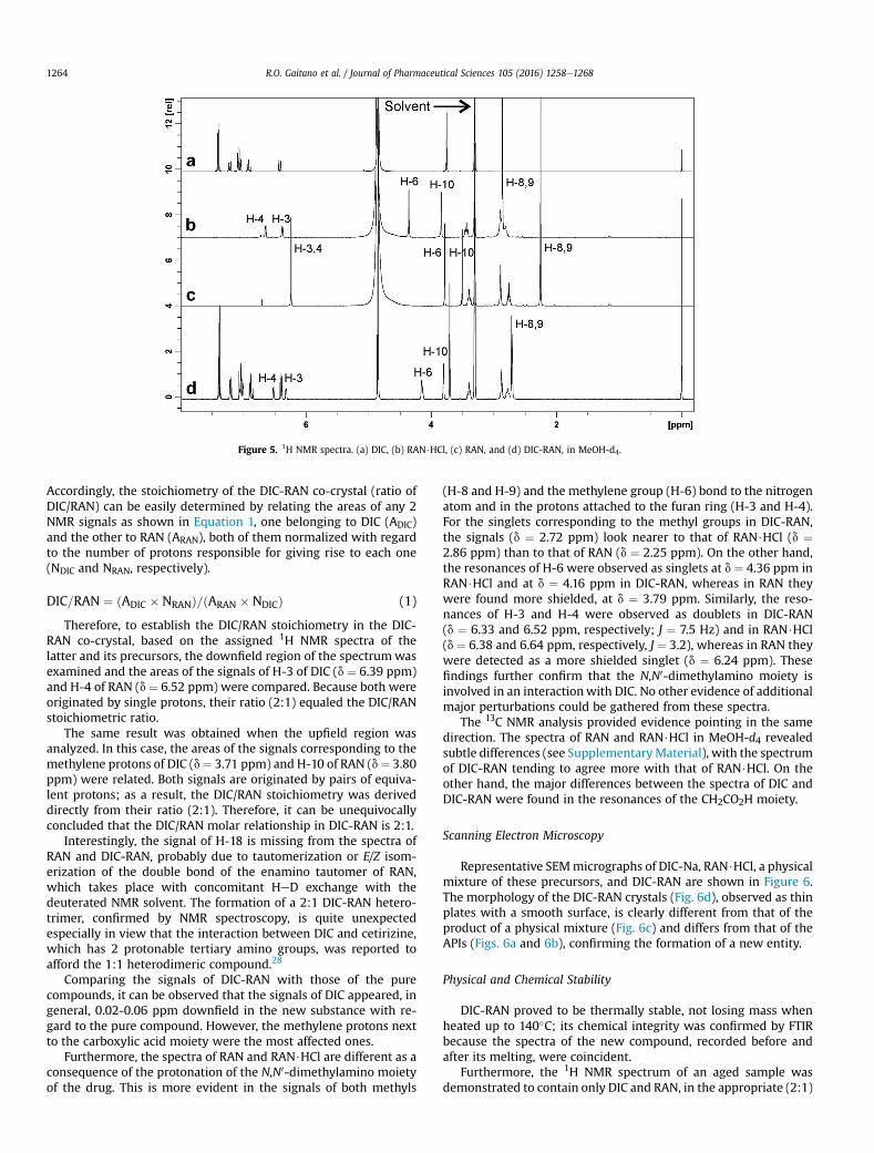
Figure 5. 1H NMR spectra. (a) DIC, (b) RAN$HCl, (c) RAN, and (d) DIC-RAN, in MeOH-d4.
R.O. Gaitano et al. / Journal of Pharmaceutical Sciences 105 (2016) 1258e12681264
Accordingly, the stoichiometry of the DIC-RAN co-crystal (ratio ofDIC/RAN) can be easily determined by relating the areas of any 2NMR signals as shown in Equation 1, one belonging to DIC (ADIC)and the other to RAN (ARAN), both of them normalized with regardto the number of protons responsible for giving rise to each one(NDIC and NRAN, respectively).
DIC=RAN ¼ ðADIC � NRANÞ=ðARAN � NDICÞ (1)
Therefore, to establish the DIC/RAN stoichiometry in the DIC-RAN co-crystal, based on the assigned 1H NMR spectra of thelatter and its precursors, the downfield region of the spectrumwasexamined and the areas of the signals of H-3 of DIC (d ¼ 6.39 ppm)and H-4 of RAN (d ¼ 6.52 ppm) were compared. Because both wereoriginated by single protons, their ratio (2:1) equaled the DIC/RANstoichiometric ratio.
The same result was obtained when the upfield region wasanalyzed. In this case, the areas of the signals corresponding to themethylene protons of DIC (d¼ 3.71 ppm) and H-10 of RAN (d¼ 3.80ppm) were related. Both signals are originated by pairs of equiva-lent protons; as a result, the DIC/RAN stoichiometry was deriveddirectly from their ratio (2:1). Therefore, it can be unequivocallyconcluded that the DIC/RAN molar relationship in DIC-RAN is 2:1.
Interestingly, the signal of H-18 is missing from the spectra ofRAN and DIC-RAN, probably due to tautomerization or E/Z isom-erization of the double bond of the enamino tautomer of RAN,which takes place with concomitant HeD exchange with thedeuterated NMR solvent. The formation of a 2:1 DIC-RAN hetero-trimer, confirmed by NMR spectroscopy, is quite unexpectedespecially in view that the interaction between DIC and cetirizine,which has 2 protonable tertiary amino groups, was reported toafford the 1:1 heterodimeric compound.28
Comparing the signals of DIC-RAN with those of the purecompounds, it can be observed that the signals of DIC appeared, ingeneral, 0.02-0.06 ppm downfield in the new substance with re-gard to the pure compound. However, the methylene protons nextto the carboxylic acid moiety were the most affected ones.
Furthermore, the spectra of RAN and RAN$HCl are different as aconsequence of the protonation of the N,N0-dimethylamino moietyof the drug. This is more evident in the signals of both methyls
(H-8 and H-9) and the methylene group (H-6) bond to the nitrogenatom and in the protons attached to the furan ring (H-3 and H-4).For the singlets corresponding to the methyl groups in DIC-RAN,the signals (d ¼ 2.72 ppm) look nearer to that of RAN$HCl (d ¼2.86 ppm) than to that of RAN (d ¼ 2.25 ppm). On the other hand,the resonances of H-6 were observed as singlets at d ¼ 4.36 ppm inRAN$HCl and at d ¼ 4.16 ppm in DIC-RAN, whereas in RAN theywere found more shielded, at d ¼ 3.79 ppm. Similarly, the reso-nances of H-3 and H-4 were observed as doublets in DIC-RAN(d ¼ 6.33 and 6.52 ppm, respectively; J ¼ 7.5 Hz) and in RAN$HCl(d ¼ 6.38 and 6.64 ppm, respectively, J ¼ 3.2), whereas in RAN theywere detected as a more shielded singlet (d ¼ 6.24 ppm). Thesefindings further confirm that the N,N0-dimethylamino moiety isinvolved in an interactionwith DIC. No other evidence of additionalmajor perturbations could be gathered from these spectra.
The 13C NMR analysis provided evidence pointing in the samedirection. The spectra of RAN and RAN$HCl in MeOH-d4 revealedsubtle differences (see SupplementaryMaterial), with the spectrumof DIC-RAN tending to agree more with that of RAN$HCl. On theother hand, the major differences between the spectra of DIC andDIC-RAN were found in the resonances of the CH2CO2H moiety.
Scanning Electron Microscopy
Representative SEMmicrographs of DIC-Na, RAN$HCl, a physicalmixture of these precursors, and DIC-RAN are shown in Figure 6.The morphology of the DIC-RAN crystals (Fig. 6d), observed as thinplates with a smooth surface, is clearly different from that of theproduct of a physical mixture (Fig. 6c) and differs from that of theAPIs (Figs. 6a and 6b), confirming the formation of a new entity.
Physical and Chemical Stability
DIC-RAN proved to be thermally stable, not losing mass whenheated up to 140�C; its chemical integrity was confirmed by FTIRbecause the spectra of the new compound, recorded before andafter its melting, were coincident.
Furthermore, the 1H NMR spectrum of an aged sample wasdemonstrated to contain only DIC and RAN, in the appropriate (2:1)
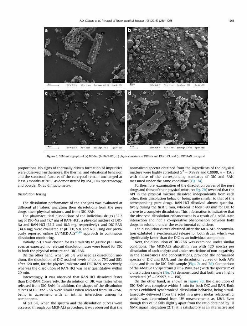
Figure 6. SEM micrographs of (a) DIC-Na, (b) RAN$HCl, (c) physical mixture of DIC-Na and RAN$HCl, and (d) DIC-RAN co-crystal.
R.O. Gaitano et al. / Journal of Pharmaceutical Sciences 105 (2016) 1258e1268 1265
proportions. No signs of thermally driven formation of impuritieswere observed. Furthermore, the thermal and vibrational behavior,and the structural features of the co-crystal remain unchanged atleast 3 months at 20�C, as demonstrated by DSC, FTIR spectroscopy,and powder X-ray diffractometry.
Dissolution Testing
The dissolution performance of the analytes was evaluated atdifferent pH values, analyzing their dissolutions from the puredrugs, their physical mixture, and from DIC-RAN.
The pharmaceutical dissolutions of the individual drugs (32.2mg of DIC-Na and 17.7 mg of RAN$HCl), a physical mixture of DIC-Na and RAN$HCl (33.2 and 18.3 mg, respectively), and DIC-RAN(34.4 mg) were evaluated at pH 1.0, 5.8, and 6.8, using our previ-ously reported online UV/MCR-ALS45,46 approach to continuousdissolution monitoring.
Initially, pH 1 was chosen for its similarity to gastric pH. How-ever, as expected, no relevant dissolution rates were found for DICin both the physical mixture and DIC-RAN.
On the other hand, when pH 5.8 was used as dissolution me-dium, the dissolution of DIC reached levels of about 75% and 85%after 120 min, for the physical mixture and DIC-RAN, respectively,whereas the dissolution of RAN$HCl was near quantitative within20 min.
Interestingly, it was observed that RAN$HCl dissolved fasterthan DIC-RAN. Conversely, the dissolution of DIC was faster whenreleased from DIC-RAN. In addition, the shapes of the dissolutioncurves of DIC and RAN were similar when released from DIC-RAN,being in agreement with an intimal interaction among itscomponents.
At pH 6.8, when the spectra and the dissolution curves wereaccessed through our MCR-ALS procedure, it was observed that the
normalized spectra obtained from the ingredients of the physicalmixture were highly correlated (r2 ¼ 0.9998 and 0.9999, n ¼ 156),with those of the corresponding standards of DIC and RAN,measured under the same conditions (Fig. 7a).
Furthermore, examination of the dissolution curves of the puredrugs and those of their physical mixture (Fig. 7b) revealed that theAPI in the physical mixture dissolved independently from eachother, their dissolution behavior being quite similar to that of thecorresponding pure drugs. RAN$HCl dissolved almost quantita-tively during the first 5 min, whereas it took >90 min for DIC toarrive to a complete dissolution. This information is indicative thatthe observed dissolution enhancement is a result of a solid-stateinteraction and not a co-operative phenomenon between bothdrugs in solution, under the experimental conditions.
The dissolution curves obtained after the MCR-ALS deconvolu-tion exhibited a synchronized release for both drugs, which wassignificantly faster than the DIC as an individual component.
Next, the dissolution of DIC-RAN was examined under similarconditions. The MCR-ALS algorithm, run with 120 spectra perdissolution of each analyte and under restrictions of non-negativityin the absorbances and concentrations, provided the normalizedspectra of DIC and RAN, and the dissolution curves of both APIsalone and from the DIC-RAN sample (Figs. 7c and 7d). Comparisonof the additive UV spectrum (DICþ RAN, 2þ1) with the spectrum ofa dissolution sample (Fig. 7c) demonstrated that both were highlycorrelated (r2 ¼ 0.9997, n ¼ 156).
On the other hand, as shown in Figure 7d, the dissolution ofDIC-RAN was complete within 5 min for both DIC and RAN. Bothcurves exhibited synchronized dissolution behavior, being simul-taneously delivered from the solid in a given molar relationship,which was determined from UV measurements as 1.9:1. Eventhough this value falls slightly apart from the ratio obtained by 1HNMR signal integration (2:1), it is satisfactory as an alternative and
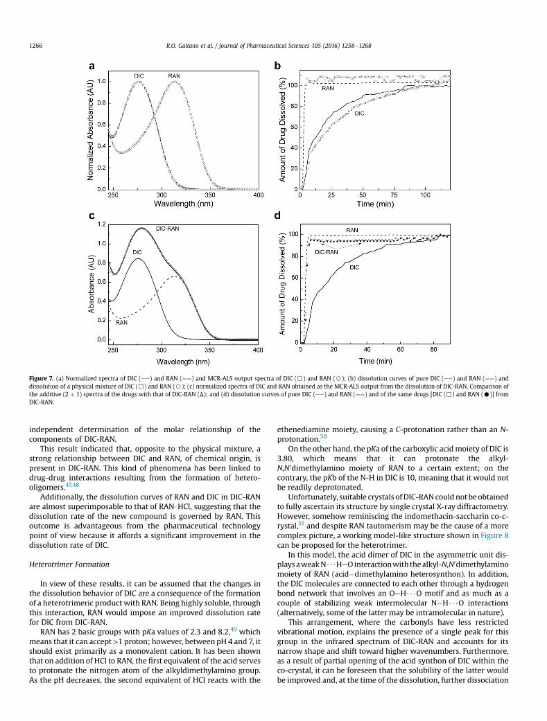
Figure 7. (a) Normalized spectra of DIC (ee) and RAN (——) and MCR-ALS output spectra of DIC (,) and RAN (B); (b) dissolution curves of pure DIC (ee) and RAN (——) anddissolution of a physical mixture of DIC (,) and RAN (B); (c) normalized spectra of DIC and RAN obtained as the MCR-ALS output from the dissolution of DIC-RAN. Comparison ofthe additive (2 þ 1) spectra of the drugs with that of DIC-RAN (D); and (d) dissolution curves of pure DIC (ee) and RAN (——) and of the same drugs [DIC (,) and RAN (C)] fromDIC-RAN.
R.O. Gaitano et al. / Journal of Pharmaceutical Sciences 105 (2016) 1258e12681266
independent determination of the molar relationship of thecomponents of DIC-RAN.
This result indicated that, opposite to the physical mixture, astrong relationship between DIC and RAN, of chemical origin, ispresent in DIC-RAN. This kind of phenomena has been linked todrug-drug interactions resulting from the formation of hetero-oligomers.47,48
Additionally, the dissolution curves of RAN and DIC in DIC-RANare almost superimposable to that of RAN$HCl, suggesting that thedissolution rate of the new compound is governed by RAN. Thisoutcome is advantageous from the pharmaceutical technologypoint of view because it affords a significant improvement in thedissolution rate of DIC.
Heterotrimer Formation
In view of these results, it can be assumed that the changes inthe dissolution behavior of DIC are a consequence of the formationof a heterotrimeric product with RAN. Being highly soluble, throughthis interaction, RAN would impose an improved dissolution ratefor DIC from DIC-RAN.
RAN has 2 basic groups with pKa values of 2.3 and 8.2,49 whichmeans that it can accept>1 proton; however, between pH 4 and 7, itshould exist primarily as a monovalent cation. It has been shownthat on addition of HCl to RAN, the first equivalent of the acid servesto protonate the nitrogen atom of the alkyldimethylamino group.As the pH decreases, the second equivalent of HCl reacts with the
ethenediamine moiety, causing a C-protonation rather than an N-protonation.50
On the other hand, the pKa of the carboxylic acidmoiety of DIC is3.80, which means that it can protonate the alkyl-N,N0dimethylamino moiety of RAN to a certain extent; on thecontrary, the pKb of the N-H in DIC is 10, meaning that it would notbe readily deprotonated.
Unfortunately, suitable crystals ofDIC-RANcouldnotbeobtainedto fully ascertain its structure by single crystal X-ray diffractometry.However, somehow reminiscing the indomethacin-saccharin co-c-rystal,51 and despite RAN tautomerism may be the cause of a morecomplex picture, a working model-like structure shown in Figure 8can be proposed for the heterotrimer.
In this model, the acid dimer of DIC in the asymmetric unit dis-playsaweakN$$$HeOinteractionwith thealkyl-N,N0dimethylaminomoiety of RAN (acid�dimethylamino heterosynthon). In addition,the DIC molecules are connected to each other through a hydrogenbond network that involves an OeH$$$O motif and as much as acouple of stabilizing weak intermolecular N�H$$$O interactions(alternatively, some of the latter may be intramolecular in nature).
This arrangement, where the carbonyls have less restrictedvibrational motion, explains the presence of a single peak for thisgroup in the infrared spectrum of DIC-RAN and accounts for itsnarrow shape and shift toward higher wavenumbers. Furthermore,as a result of partial opening of the acid synthon of DIC within theco-crystal, it can be foreseen that the solubility of the latter wouldbe improved and, at the time of the dissolution, further dissociation
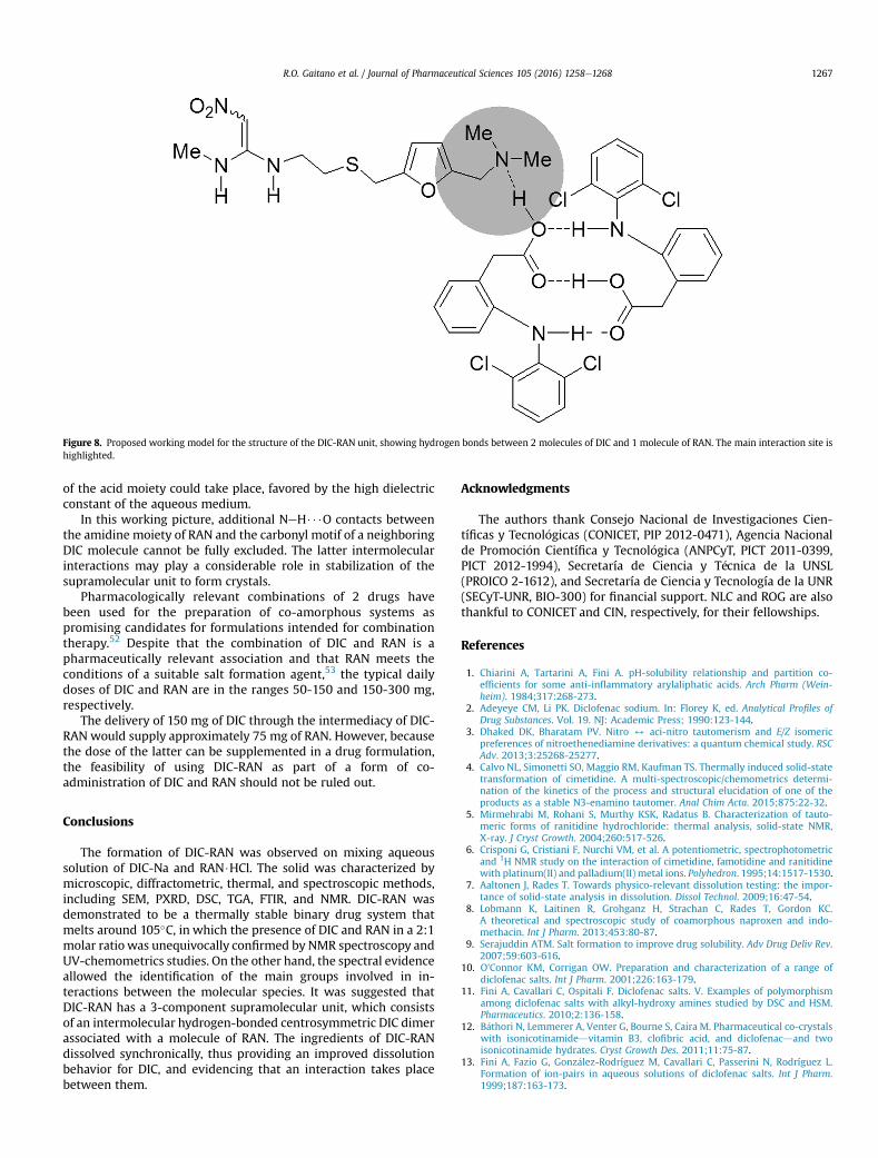
Figure 8. Proposed working model for the structure of the DIC-RAN unit, showing hydrogen bonds between 2 molecules of DIC and 1 molecule of RAN. The main interaction site ishighlighted.
R.O. Gaitano et al. / Journal of Pharmaceutical Sciences 105 (2016) 1258e1268 1267
of the acid moiety could take place, favored by the high dielectricconstant of the aqueous medium.
In this working picture, additional NeH$$$O contacts betweenthe amidine moiety of RAN and the carbonyl motif of a neighboringDIC molecule cannot be fully excluded. The latter intermolecularinteractions may play a considerable role in stabilization of thesupramolecular unit to form crystals.
Pharmacologically relevant combinations of 2 drugs havebeen used for the preparation of co-amorphous systems aspromising candidates for formulations intended for combinationtherapy.52 Despite that the combination of DIC and RAN is apharmaceutically relevant association and that RAN meets theconditions of a suitable salt formation agent,53 the typical dailydoses of DIC and RAN are in the ranges 50-150 and 150-300 mg,respectively.
The delivery of 150 mg of DIC through the intermediacy of DIC-RAN would supply approximately 75 mg of RAN. However, becausethe dose of the latter can be supplemented in a drug formulation,the feasibility of using DIC-RAN as part of a form of co-administration of DIC and RAN should not be ruled out.
Conclusions
The formation of DIC-RAN was observed on mixing aqueoussolution of DIC-Na and RAN$HCl. The solid was characterized bymicroscopic, diffractometric, thermal, and spectroscopic methods,including SEM, PXRD, DSC, TGA, FTIR, and NMR. DIC-RAN wasdemonstrated to be a thermally stable binary drug system thatmelts around 105�C, in which the presence of DIC and RAN in a 2:1molar ratio was unequivocally confirmed by NMR spectroscopy andUV-chemometrics studies. On the other hand, the spectral evidenceallowed the identification of the main groups involved in in-teractions between the molecular species. It was suggested thatDIC-RAN has a 3-component supramolecular unit, which consistsof an intermolecular hydrogen-bonded centrosymmetric DIC dimerassociated with a molecule of RAN. The ingredients of DIC-RANdissolved synchronically, thus providing an improved dissolutionbehavior for DIC, and evidencing that an interaction takes placebetween them.
Acknowledgments
The authors thank Consejo Nacional de Investigaciones Cien-tíficas y Tecnol�ogicas (CONICET, PIP 2012-0471), Agencia Nacionalde Promoci�on Científica y Tecnol�ogica (ANPCyT, PICT 2011-0399,PICT 2012-1994), Secretaría de Ciencia y T�ecnica de la UNSL(PROICO 2-1612), and Secretaría de Ciencia y Tecnología de la UNR(SECyT-UNR, BIO-300) for financial support. NLC and ROG are alsothankful to CONICET and CIN, respectively, for their fellowships.
References
1. Chiarini A, Tartarini A, Fini A. pH-solubility relationship and partition co-efficients for some anti-inflammatory arylaliphatic acids. Arch Pharm (Wein-heim). 1984;317:268-273.
2. Adeyeye CM, Li PK. Diclofenac sodium. In: Florey K, ed. Analytical Profiles ofDrug Substances. Vol. 19. NJ: Academic Press; 1990:123-144.
3. Dhaked DK, Bharatam PV. Nitro 4 aci-nitro tautomerism and E/Z isomericpreferences of nitroethenediamine derivatives: a quantum chemical study. RSCAdv. 2013;3:25268-25277.
4. Calvo NL, Simonetti SO, Maggio RM, Kaufman TS. Thermally induced solid-statetransformation of cimetidine. A multi-spectroscopic/chemometrics determi-nation of the kinetics of the process and structural elucidation of one of theproducts as a stable N3-enamino tautomer. Anal Chim Acta. 2015;875:22-32.
5. Mirmehrabi M, Rohani S, Murthy KSK, Radatus B. Characterization of tauto-meric forms of ranitidine hydrochloride: thermal analysis, solid-state NMR,X-ray. J Cryst Growth. 2004;260:517-526.
6. Crisponi G, Cristiani F, Nurchi VM, et al. A potentiometric, spectrophotometricand 1H NMR study on the interaction of cimetidine, famotidine and ranitidinewith platinum(II) and palladium(II) metal ions. Polyhedron. 1995;14:1517-1530.
7. Aaltonen J, Rades T. Towards physico-relevant dissolution testing: the impor-tance of solid-state analysis in dissolution. Dissol Technol. 2009;16:47-54.
8. Lobmann K, Laitinen R, Grohganz H, Strachan C, Rades T, Gordon KC.A theoretical and spectroscopic study of coamorphous naproxen and indo-methacin. Int J Pharm. 2013;453:80-87.
9. Serajuddin ATM. Salt formation to improve drug solubility. Adv Drug Deliv Rev.2007;59:603-616.
10. O'Connor KM, Corrigan OW. Preparation and characterization of a range ofdiclofenac salts. Int J Pharm. 2001;226:163-179.
11. Fini A, Cavallari C, Ospitali F. Diclofenac salts. V. Examples of polymorphismamong diclofenac salts with alkyl-hydroxy amines studied by DSC and HSM.Pharmaceutics. 2010;2:136-158.
12. B�athori N, Lemmerer A, Venter G, Bourne S, Caira M. Pharmaceutical co-crystalswith isonicotinamidedvitamin B3, clofibric acid, and diclofenacdand twoisonicotinamide hydrates. Cryst Growth Des. 2011;11:75-87.
13. Fini A, Fazio G, Gonz�alez-Rodríguez M, Cavallari C, Passerini N, Rodríguez L.Formation of ion-pairs in aqueous solutions of diclofenac salts. Int J Pharm.1999;187:163-173.
R.O. Gaitano et al. / Journal of Pharmaceutical Sciences 105 (2016) 1258e12681268
14. Dias MMR, Raghavan SL, Pellett MA, Hadgraft J. The effect of b-cyclodextrins onthe permeation of diclofenac from supersaturated solutions. Int J Pharm.2003;263:173-181.
15. Khan MA, Chourasia A. Mixed solvency approach- boon for solubilization ofpoorly water soluble drug diclofenac sodium. Res J Pharm Biol Chem Sci. 2012;3:865-868.
16. Khan MA. Enhancement of solubility of poorly water soluble drugs diclofenacsodium bymixed solvency approach. Int J Res Dev Pharm Life Sci. 2013;2:580-582.
17. Vajir S, Sahu V, Ghuge N, Bang P, Bakde BV. Enhancement of dissolution rate ofpoorly water soluble diclofenac sodium by liquisolid technique. Int J PharmChem Sci. 2012;1:1338-1349.
18. Pavan Kumar KH, Chandramouli Y, Prakash Reddy CS, Sreekanth K, Murali D,Chakravarthi RN. Enhancement of solubility and dissolution rate of diclofenacsodium by solid dispersion method. Int J Adv Pharm. 2012;2:110-118.
19. Lai F, Sinico C, Ennas G, Marongiu F, Marongiu G, Fadda AM. Diclofenacnanosuspensions: influence of preparation procedure and crystal form on drugdissolution behavior. Int J Pharm. 2009;373:124-132.
20. Fanun M. Conductivity, viscosity, NMR and diclofenac solubilization capacitystudies of mixed nonionic surfactants microemulsions. J Mol Liq. 2007;135:5-13.
21. Gubbins RH, O0Driscoll CM, Corrigan OI. The effects of casein on diclofenacrelease from hydroxypropylmethylcellulose (HPMC) compacts. Int J Pharm.2003;260:69-76.
22. O0Connor KM, Corrigan OI. Comparison of the physicochemical properties ofthe N-(2-hydroxyethyl)pyrrolidinediethylamine and sodium salt forms ofdiclofenac. Int J Pharm. 2001;222:281-293.
23. Aaker€oy CB, Grommet AB, Desper J. Co-crystal screening of diclofenac. Phar-maceutics. 2011;3:601-614.
24. Alioth C, Blum RA, D0Andrea DT, et al. Application of dual radiotelemetrictechnique in studying drug-drug interaction between diclofenac sodium andranitidine HCl in volunteers. Pharm Res. 1993;10:1688-1692.
25. Leucuta A, Vlase L, Farcau D, Nanulescu M. No effect of short term ranitidineintake on diclofenac pharmacokinetics. Roman J Gastroenterol. 2004;13:305-308.
26. Shirse P. Formulation and evaluation of bilayer tablets of diclofrenac sodiumwith ranitidine HCl for sustained and immediate release. J Appl Pharm Sci.2012;2:136-141.
27. Carrasco-Portugal MC, Aguilar-Cota ME, P�erez-Urlzar J, Cabrera O, Herrera JE,Flores-Murrieta FJ. Bioavailability of a formulation containing a diclofenac-ranitidine combination. Proc West Pharmacol Soc. 2002;45:8-10.
28. Kenawi IM, Barsoum BN, Youssef MA. Drugedrug interaction between diclo-fenac, cetirizine and ranitidine. J Pharm Biomed Anal. 2005;37:655-661.
29. The United States Pharmacopeia, USP35eNF30. Dissolution Procedure: Devel-opment and Validation, U. S. Rockville, MD, USA: Pharmacopeial ConventionInc.; 2011 Chapter 1092.
30. Jaumot J, Gargallo R, de Juan A, Tauler RA. Graphical user-friendly interface forMCR-ALS: a new tool for multivariate curve resolution in MATLAB. ChemomIntell Lab Syst. 2005;76:101-110.
31. Gaggelli E, Marchettini N, Sega A, Valensin G. Conformation and dynamics ofranitidine in solution as detected by lH and 13C NMR spectroscopy. Magn ResonChem. 1988;26:1041-1046.
32. Lee M-J, Chun N-H, Wang I-C, Liu JJ, Jeong M-Y, Choi GJ. Understanding theformation of indomethacinesaccharin cocrystals by anti-solvent crystalliza-tion. Cryst Growth Des. 2013;13:2067-2074.
33. Goh CF, Lane ME. Formulation of diclofenac for dermal delivery. Int J Pharm.2014;473:607-616.
34. El Haj BM, Al Ainri AM, Hassan MH, Bin Khadem RK, Marzouq MS. The GC/MSanalysisof somecommonlyusednon-steroidal anti-inflammatorydrugs (NSAIDs)in pharmaceutical dosage forms and in urine. Forens Sci Int. 1999;105:141-153.
35. Vignaduzzo SE, Castellano PM, Kaufman TS. Experimentally designed, validatedHPLC simultaneous determination of pridinol and diclofenac in their combinedpharmaceutical formulations, which allows limiting diclofenac related com-pound A. J Liq Chromatogr Relat Technol. 2010;33:1720-1732.
36. Tudja P, Khan MZI, Mestrovic E, Horvat M, Golja P. Thermal behaviour ofdiclofenac sodium: decomposition and melting characteristics. Chem PharmBull. 2001;49:1245-1250.
37. Novoa De Armas H, Peeters OM, Blaton N, et al. Solid state characterization andcrystal structure from X-ray powder diffraction of two polymorphic forms ofranitidine base. J Pharm Sci. 2009;98:146-158.
38. Palomo ME, Ballesteros MP, Frutos P. Analysis of diclofenac sodium derivatives.J Pharm Biomed Anal. 1999;21:83-94.
39. Madan T, Kakkar AP. Preparation and characterization of ranitidine-HCl crys-tals. Drug Dev Ind Pharm. 1994;20:1571-1588.
40. Hohnjec M, Kuftinec J, Malnar M, et al. Ranitidine. In: Florey K, ed. AnalyticalProfiles of Drug Substances. Vol. 15. NewYork, NY: Academic Press; 1986:533-562.
41. Sovilj SP, Dzambaski A, Jovanovic T. Novel Cu(II) complexes with ranitidine andnizatidine. Asian J Chem. 2003;15:19-24.
42. Colthup NB, Daly LH, Wiberley SE. Introduction to Infrared and Raman Spec-troscopy. 3rd ed. San Diego, CA: Academic Press; 1990.
43. Basavoju S, Bostr€om D, Velaga SP. Indomethacinesaccharin cocrystal: Design,synthesis and preliminary pharmaceutical characterization. Pharm Res.2008;25:530-541.
44. Chieng N, Aaltonen J, Saville D, Rades T. Physical characterization andstability of amorphous indomethacin and ranitidine hydrochloride binarysystems prepared by mechanical activation. Eur J Pharm Biopharm. 2009;71:47-54.
45. Calvo NL, Maggio RM, Kaufman TS. An eco-friendly strategy, using on-linemonitoring and dilution coupled to a second-order chemometric method, forthe construction of dissolution curves of combined pharmaceutical associa-tions. J Pharm Biomed Anal. 2014;89:213-220.
46. Maggio RM, Rivero MA, Kaufman TS. Simultaneous acquisition of the dissolu-tion curves of two active ingredients in a binary pharmaceutical association,employing an on-line circulation system and chemometrics-assistance. J PharmBiomed Anal. 2013;72:51-58.
47. Alleso M, Chieng N, Rehder S, Rantanen J, Rades T, Aaltonen J. Enhanceddissolution rate and synchronized release of drugs in binary systems throughformulation: amorphous naproxenecimetidine mixtures prepared by me-chanical activation. J Control Rel. 2009;136:45-53.
48. L€obmann K, Laitinen R, Grohganz H, Gordon KC, Strachan C, Rades T. Coa-morphous drug systems: enhanced physical stability and dissolution rate ofindomethacin and naproxen. Mol Pharm. 2011;8:1919-1928.
49. Dumanovic D, Juranic I, Dzeletovic D, Vasic VM, Jovanovic J. Protolytic con-stants of nizatidine, ranitidine and N,N-dimethyl-2-nitro-1,1-ethenediamine,spectrophotometric and theoretical investigation. J Pharm Biomed Anal.1997;15:1667-1678.
50. Cholerton TJ, Hunt JH, Klinkert G, Smith MM. Spectroscopic studies on raniti-dine: Its structure and the influence of temperature and pH. J Chem Soc PerkinTrans. 1984;2:1761-1766.
51. Qiao N, Li M, Schlindwein W, Malek N, Davies A, Trappitt G. Pharmaceuticalcocrystals: an overview. Int J Pharm. 2011;419:1-11.
52. L€obmann K, Strachan C, Grohganz H, Rades T, Korhonen O, Laitinen R. Co-amorphous simvastatin and glipizide combinations show improved physicalstability without evidence of intermolecular interactions. Eur J Pharm Bio-pharm. 2012;81:159-169.
53. Saal C, Becker A. Pharmaceutical salts: a summary on doses of salt formers fromthe Orange Book. Eur J Pharm Sci. 2013;49:614-623.