Embed Size (px)
Citation preview
QATAR UNIVERSITY
COLLEGE OF ARTS AND SCIENCES
PREPARATION AND CHARACTERIZATION OF MULTILAYERED
ELECTROSPUN COMPOSITE SCAFFOLDS FOR BIOMEDICAL APPLICATIONS..
BY
HANA MUHAMMED KAYYAL KADAVIL
A Thesis Submitted to the Faculty of
the College of Arts and Sciences
in Partial
Fulfillment of the
Requirements for
the Degree of
Masters of Science
in
Material Science and Technology
January 2019
© 2019. Hana Muhammed. All Rights Reserved.
ii
COMMITTEE PAGE
The members of the Committee approve the Thesis of Hana Muhammed
Kadavil defended on 2/12/2018
Dr. Talal Al tahtamouni
Thesis/Dissertation Supervisor
Dr. Ahmed Elzatahry
Committee Member
Dr. Morana Jaganjac
Committee Member
Dr. Aboubakr M. Abdullah
Committee Member
Approved:
Rashid Al-Kuwari, Dean, College of College of Arts and Sciences
iii
ABSTRACT
KADAVIL, HANA , MUHAMMED ., Masters :January:2019, Material Science and Technology
Title: Preparation and Characterization of multilayered electrospun composite scaffolds for
biomedical applications.
Supervisor of Thesis: Dr. Talal Al tahtamouni.
Electrospinning has gained wide attention recently in biomedical applications.
Electrospun biocompatible scaffolds are well-known for biomedical applications such as
drug delivery, wound dressing and tissue engineering applications. In this study,
composites based electrospun polymer fibers Polycaprolactone (PCL) and polyvinyl
alcohol (PVA) was produced by using electrospinning technique in which multilayered
structure (PCL-PVA-PCL) loaded with gentamicin sulphate (GS) (drug) in the middle
layer with PVA. Formerly, metal silver particles were deposited on the surface of
electrospun fibers using plasma sputtering technology. The electrospun scaffolds were
characterized by scanning electron microscope (SEM), Energy Dispersive X-ray
Spectroscopy (EDX), Fourier Transform Infrared Spectroscopy (FTIR), Water contact
angle measurement, X-ray Diffraction (XRD) and Thermogravimetric Analysis (TGA).
The drug delivery of gentamicin sulphate from the multilayered electrospun PCL-PVA-
PCL was investigated by using calorimetric method. The outcomes prove that, initial
burst release of the drug could be reduced with the increase in the drug loading from 1%
to 4%GS in the multilayered structure. Furthermore, the antibacterial properties of the
sample were examined. The multilayered electrospun fiber loaded with drug and sputter
iv
coated with Ag has enhanced the antibacterial efficiency from the control. In addition,
Biological performance such as cell cytotoxicity was investigated on the cell line
fibroblast (Wi38) and human keratinocyte (HaCaT) by using alamar blue assay. The
presence of Ag has revealed certain toxic effect with both cell line, and also the increase
in the gentamicin concentration has also exhibited particular toxicity. Finally, In-vitro
release of silver from the surface of the scaffold was inspected. The results show a
uniform release of silver from the surface rather than initial burst release. Therefore, the
formulated scaffolds are suitable candidate for biomedical application such as wound
healing for prolonged antibacterial Inhibition through uniform release of drug as well as
silver particles from the surface of the scaffolds.
.

v
DEDICATION
I dedicate this work to my better half, Shajil Kareem for being a good supporter and
strength provider in my life. In addition, to my son Ewaan Wildan and my upcoming
baby (In Sha Allaah).
And also I dedicate to my parents for their prayers, love, affection, guidance and brought
up me to a successful human being. Moreover, I would like to devote to my parent in-laws
for their prayers, love and support towards me during my studies.
Most importantly, to my dear sister Hiba Muhammed who was there with me to take
care of my son more than me during the difficult period.
vi
ACKNOWLEDGMENTS
I would like to express my sincere gratitude to my thesis supervisor, Dr. Talal Al
Tahtamouni. His motivation and enthusiasm have guided me throughout the whole period
of research activities and writing of this thesis. I highly appreciate Dr. Talal‘s kind
encouragement and patience all the time.
I‘m contented to my committee member Dr. Morana Jaganjac for her patience, guidance
and support during the research work as well as granting me to work under her
supervision in Qatar anti-doping laboratory. And also, I am thankful to my thesis
committee members: Dr. Ahmed Elzatahry and Dr. Aboubakr Ali, for their fruitful
comments and productive feedback at all times.
My gratitude also goes to the Dr. Mohammad Al-sayrafi general manager of Anti-doping
laboratory Qatar, Prof. Aisha Latiff (Director of toxicity and multipurpose lab) and IREC
committee members for permitting me to access to conducting research using high-
technological laboratory facilities in ADLQ-Qatar.
I thankful to Dr Samir Jaoua and Dr Zahoor Ul Hassan from department of biological and
environmental science for giving permission to use the microbiology lab for my
antibacterial studies.
I acknowledge the efforts of the staff in the Center of advanced materials (CAM) at Qatar
Univeristy Mr. Abdullah al Ashraf, Mr. Abdul Jaleel, Dr. Peter Kasak, and
Mr.Moinuddin Mohammed Yusuf. Special thanks to Dr. Nasser Alnuaimi, the director of
the Center for Advanced Materials, for giving me the opportunity to use the CAM
facilities
vii
And also my grace to Central Laboratory Unit (CLU) at Qatar University for testing my
samples in their labs Dr. Mohammed Yousuf, Mr. Ahmed Easa, Mr. Maath Arar and Mr.
Fathy Atia..
I‘d like to express my great appreciation to Prof. Mariam Al-Maadeed, the VP of research
and graduate studies at Qatar University, for accepting me as a Master student in the
Materials Science and Technology program and as Graduate Assistant, and for her
encouragement to maintain an exceptional academic record.
I would like to thanks the entire faculty, students and staffs in Material science and
technology, Dr. Khaled Yousef and Mr. Mustafa Zagho for their support during the
academic years
Finally, I would like to express my deepest gratitude to my beloved family and friends for
their encouragement and support throughout my life.
Last, but by no means least, I thank Allah.
viii
TABLE OF CONTENTS
DEDICATION ................................................................................................................... V
ACKNOWLEDGMENTS ................................................................................................ VI
LIST OF TABLES ............................................................................................................. xi
LIST OF FIGURES .......................................................................................................... xii
Introduction ......................................................................................................................... 1
1.1 Objectives ............................................................................................................. 4
Literature Review................................................................................................................ 6
1.2 Introduction: ......................................................................................................... 6
1.3 Drug delivery........................................................................................................ 8
1.4 Drug loading type ................................................................................................. 8
1.5 Drug loading materials ....................................................................................... 12
1.6 Type of drugs ..................................................................................................... 17
1.7 Mechanisms of drug release: .............................................................................. 20
1.8 Tissue Engineering ............................................................................................. 23
1.8.1 Skin tissue engineering/ wound healing ..................................................... 23
1.8.2 Bone tissue engineering applications: ......................................................... 27
1.8.3 Skeletal muscle regeneration: ..................................................................... 28
1.8.4 Nerve tissue engineering: ............................................................................ 30
1.9 Sputtering technology for biomedical applications:........................................... 31
1.9.1 Antibacterial coatings: ................................................................................ 33
1.9.2 Tissue engineering: ..................................................................................... 39
Experimental works .......................................................................................................... 43
1.10 Materials ............................................................................................................. 43
1.10.1 Poly(caprolactone) (PCL) ........................................................................... 43
1.10.2 Poly vinyl alcohol ....................................................................................... 44
1.10.3 Gentamicin sulphate: .................................................................................. 44
1.10.4 Solvents: ...................................................................................................... 46
1.11 Scaffolds preparation equipment:....................................................................... 46
1.11.1 Electrospinning: .......................................................................................... 46
ix
1.11.2 Magnetron Sputtering: ................................................................................ 48
1.12 Fiber preparation methods and electrospinning conditions: .............................. 51
1.12.1 Optimization of PVA and PCL individual fibers and multilayered structure.
52
1.12.2 PCL sputter coated with Silver (Ag) ........................................................... 52
1.12.3 PCL-PVA-PCL sputter coated with Silver (Ag) ......................................... 53
1.13 Characterization: ................................................................................................ 53
1.13.1 Scanning Electron Microscopy (SEM) and EDX. ...................................... 53
1.13.2 The Fourier Transform Infrared Spectroscopy ( FT-IR) ............................. 55
1.13.3 Thermogravimetric analysis (TGA) ............................................................ 56
1.13.4 UV-VIS spectrophotometry: ....................................................................... 57
1.13.5 Inductively coupled plasma mass spectrometry ICP-MS: ......................... 58
1.14 In-vitro drug delivery of gentamicin Sulphate ................................................... 59
1.14.1 Gentamicin-ninhydrin assay: ...................................................................... 59
1.15 In vitro release of Ag:......................................................................................... 63
1.16 In-vitro bio-compatibility studies: ...................................................................... 64
1.16.1 Cell lines: .................................................................................................... 64
1.16.2 Cell seeding ................................................................................................. 65
1.16.3 In-vitro quantitative cell viability assay:..................................................... 65
1.17 Antibacterial Studies: ......................................................................................... 67
1.17.1 Medium preparation : .................................................................................. 67
1.17.2 Antibacterial testing and spreading the plates. ........................................... 67
Results and Discussions .................................................................................................... 70
1.18 Optimization of PVA and PCL individual fibers, and multilayered structure. .. 70
1.18.1 Poly (vinyl alcohol) PVA: .......................................................................... 70
1.18.2 Polycaprolactone (PCL): ............................................................................. 71
1.19 PCL coated with Ag ........................................................................................... 73
1.19.1 Scanning Electron Microscope (SEM) & EDX: ......................................... 74
1.19.2 X-ray diffraction (XRD). ............................................................................ 76
1.19.3 Cytotoxicity of Ag: ..................................................................................... 77
1.19.4 Antibacterial study: ..................................................................................... 78
x
1.20 Multilayer PCL-PVA-PCL loaded with GS and sputter coated with Ag: ......... 81
1.20.1 Fiber characterization: ................................................................................ 81
1.20.2 Encapsulation efficiency of the GS in the fiber scaffolds: ......................... 86
1.20.3 In-vitro release of drug................................................................................ 87
1.20.4 In-vitro release of silver. ............................................................................. 89
1.20.5 Cytotoxicity of the fibers: ........................................................................... 90
1.20.6 Antibacterial Study: .................................................................................... 91
CONCLUSION ................................................................................................................. 96
References ......................................................................................................................... 97
xi
LIST OF TABLES
TABLE 1: PHYSICAL PROPERTIES OF GENTAMICIN SULPHATE ............................................. 45
TABLE 2: SOLVENTS USED FOR ELECTROSPINNING ............................................................. 46
TABLE 3: PREPARATION OF STANDARD SOLUTIONS PROTOCOL .......................................... 60
TABLE 4: ELECTROSPINNING PARAMETERS APPLIED FOR OPTIMIZATION OF PCL FIBERS. .. 72
TABLE 5: AVERAGE FIBER DIAMETER OF THE PREPARED PCL MATS IN DIFFERENT WEIGHT
PERCENTAGE. .............................................................................................................. 72
TABLE 6: ELECTROSPINNING CONDITION FOR MULTILAYERED PCL-PVA-PCL................. 82
xii
LIST OF FIGURES
FIGURE 1: SCHEMATIC REPRESENTATION OF ELECTROSPINNING APPARATUS. ...................... 7
FIGURE 2: SCHEMATIC REPRESENTATION OF DIFFERENT TYPE OF ELECTROSPINNING
PROCESS[23]. .............................................................................................................. 10
FIGURE 3: DRUG LOADING AND RELEASE (DESORPTION AND DIFFUSION) FROM POLYMERIC
MICRO/NANOFIBERS FABRICATED BY (A) SURFACE MODIFICATION, (B) BLENDING, (C)
COAXIAL AND (D) EMULSION ELECTROSPINNING. THE GREEN COLOR STANDS FOR
POLYMER, BLUE FOR DRUGS AND MAROON FOR SURFACTANT. THE RED ARROWS
REPRESENT THE DIRECTION OF THE DRUG RELEASE [58]. ............................................ 22
FIGURE 4: SCHEMATIC REPRESENTATION OF ELECTROSPINNING AND SPUTTERING OF
ELECTROSPUN FOR MUSCLE TISSUE ENGINEERING APPLICATIONS [98]. ....................... 42
FIGURE 5: CHEMICAL EQUATION FOR THE SYNTHESIS OF PCL ........................................... 43
FIGURE 6: CHEMICAL STRUCTURE OF GENTAMICIN SULPHATE .......................................... 45
FIGURE 7: ELECTROSPINNING APPARATUS CONSISTS OF A) FLOW METER FOR SYRINGE PUMP
B) POWER SUPPLY C) NEEDLE CONNECTED D) ROTATING COLLECTOR DRUM E)
CHAMBER PROTECT FROM THE HIGH VOLTAGE............................................................ 47
FIGURE 8: PLASMA SPUTTERING TECHNOLOGY .................................................................. 49
FIGURE 9: SCHEMATIC REPRESENTATION OF SPUTTERING TECHNOLOGY [127] .................. 50
FIGURE 10: FACTORS DEPENDS ON THE PLASMA SPUTTERING TECHNIQUES. ...................... 51
FIGURE 11: FEI QUANTA 200 SCANNING ELECTRON MICROSCOPY (SEM). ........................ 54
FIGURE 12: TGA FTIR SET UP (FT-IR SPECTROMETER FRONTIER/ TGA 4000 – PERKIN
ELMER) ....................................................................................................................... 55
FIGURE 13: THERMOGRAVIMETRIC ANALYZER - PYRIS 6 (PERKIN-ELMER)....................... 56
xiii
FIGURE 14: UV-VIS SPECTROSCOPY (AGILENT TECHNOLOGIES CARY 60 UV-VIS) ........... 57
FIGURE 15: ICP-MS NEXION 300D ................................................................................. 58
FIGURE 16: CALIBRATION CURVE FOR NINHYDRIN - GENTAMICIN SULPHATE ASSAY .......... 61
FIGURE 17: SCHEMATIC PROCEDURE TO STUDY THE DRUG RELEASE .................................. 62
FIGURE 18: SEQUENTIAL REPRESENTATION OF AG RELEASE FROM DEIONIZED WATER ...... 64
FIGURE 19: SCHEMATIC REPRESENTATION OF IN-VITRO CELL TOXICITY TEST .................... 66
FIGURE 20: SCHEMATIC PROCEDURE FOR ANTIBACTERIAL TESTING .................................. 69
FIGURE 21: A) SEM IMAGE OF 10WT%/V% PVA (ELECTROSPINNING PARAMETERS ARE
APPLIED VOLTAGE 15KV, FLOW RATE OF 1ML/H, AND NEEDLE TIP TO COLLECTOR
DISTANCE IS 10 CM) (MAGNIFICATION: M). B) NORMAL DISTRIBUTION OF FIBER
DIAMETER ................................................................................................................... 70
FIGURE 22: SEM IMAGES OF ELECTROSPUN PCL IN 1:1 (WT/WT%) DMF:CF AT DIFFERENT
WEIGHT PERCENTAGE OF PCL A) 8.5WT%PCL, B)10WT%PCL, C) 11WT%PCL, D)
12WT%PCL E) AVERAGE FIBER DIAMETER AND F) FIBER DIAMETER DISTRIBUTION OF
11WT%PCL (MAGNIFICATION M). ....................................................................... 73
FIGURE 23: SEM IMAGES OF PCL COATED WITH AG A)PCL/AG 30S B) PCL/AG 60S AND C)
PCL/AG 120S (MAGNIFICATION M) ..................................................................... 75
FIGURE 24: XRD SPECTRA FOR ELECTROSPUN PCL SPUTTER COATED WITH AG AT
DIFFERENT TIME A) PCL PEAKS B) INDEXING THE PRESENCE OF SILVER IN PCL ONLY,
PCL/AG 30S, PCL/AG 60S AND PCL/ AG 120S. ......................................................... 76
FIGURE 25: : IN VITRO CELL VIABILITY TEST OF A)WI38 B) HACAT CELL LINE PCL,
PCL/AG 30S, PCL/AG 60S AND PCL/AG 120S (N=5). ***P<0.05. ............................. 78
xiv
FIGURE 26: ANTIBACTERIAL ACTIVITIES OF THE ELECTROSPUN FIBERS COATED WITH AG
AGAINST GRAM POSITIVE BACILLUS PUMILUS (A) CONTROL, B) PCL ONLY, C) PCL/AG
30S, D) PCL/AG 60S AND E) PCL/AG 120S AND ALSO I) ROW BACILLUS PUMILUS AFTER
2H OF INCUBATION AND II) 6H OF INCUBATION. .......................................................... 80
FIGURE 27: ANTIBACTERIAL ACTIVITIES OF ELECTROSPUN PCL SPUTTER COATED WITH AG
WITH A) NUMBER OF COLONIES (CFU/ML) AG AND B) ANTIBACTERIAL INHIBITION (%)
WAS PLOTTED FOR BACILLUS PUMILUS. ....................................................................... 80
FIGURE 28: SEM IMAGES OF A) PCL OUTER LAYER B) PVA LOADED WITH DRUG (MIDDLE
LAYER) C) PCL FIBER (OUTER LAYER) ........................................................................ 82
FIGURE 29: FTIR SPECTROSCOPY OF PURE GS, PURE PCL AND PURE PVA, 0 WT% GS ML,
1 WT% GS ML, 2 WT% GS ML AND 4 WT% GS ML. ................................................. 83
FIGURE 30: COMPARATIVE TGA RESULTS OF ALL THE PREPARED FIBER SAMPLES.
INCLUDING PURE PCL, PURE PVA, PCL-PVA-PCL MULTILAYERED ELECTROSPUN
MATS LOADED WITH 1WT% GS, 2WT% GS AND 4WT% GS RESPECTIVELY. ............... 85
FIGURE 31: ENCAPSULATION EFFICIENCY OF THE GS IN THE FIBER SCAFFOLDS ................. 86
FIGURE 32: CUMULATIVE RELEASE OF GS FROM ELECTROSPUN SCAFFOLDS A) CUMULATIVE
% OF DRUG LOADED 1,2,4 WT% IN MULTILAYERED STRUCTURE B) CUMULATIVE
RELEASE OF GS PER MG OF SCAFFOLDS OF CORE SHELL, AND MULTILAYERED
STRUCTURE. ................................................................................................................ 88
FIGURE 33: CUMULATIVE RELEASE OF AG (PPB/MG OF THE FIBER SCAFFOLD) FOR PCL/AG
30S, PCL/AG 60S, PCL/AG 120S AND PCL/AG 240S RESPECTIVELY. ........................ 89
xv
FIGURE 34: IN VITRO CELL VIABILITY TEST OF A)WI38 B) HACAT CELL LINE IN PRISTINE
PVA/PCL CORE SHELL WITHOUT DRUG AND 1MG/ML OF GS, MULTILAYERED FIBER
SCAFFOLD WITHOUT AND WITH GS (1MG/ML AND 4MG/ML) (N=5). ***P<0.05. ....... 91
FIGURE 35: ANTIBACTERIAL ACTIVITIES OF THE MULTILAYERED ELECTROSPUN FIBER MATS
LOADED WITH DRUG AND SPUTTER COATED WITH AG AGAINST GRAM POSITIVE
BACILLUS PUMILUS WITH COLUMN (A) CONTROL, B)ML 0% GS, C) ML 1% GS, D) ML
AG AND E) ML 1%GS/AG AND ALSO I) ROW BACILLUS PUMILUS AFTER 2H OF
INCUBATION AND II) 6H OF INCUBATION. .................................................................... 93
FIGURE 36: ANTIBACTERIAL ACTIVITIES OF MULTILAYERED ELECTROSPUN FIBER MATS
LOADED WITH DRUG AND SPUTTER COATED WITH AG AGAINST GRAM POSTIVE
BACILLUS PUMILUS A) NUMBER OF COLONIES (CFU/ML) B) ANTIBACTERIAL INHIBITION
(%) ............................................................................................................................. 93
FIGURE 37: ANTIBACTERIAL ACTIVITIES OF THE MULTILAYERED ELECTROSPUN FIBER MATS
LOADED WITH DRUG AND SPUTTER COATED WITH AG AGAINST GRAM NEGATIVE E.COLI
WITH COLUMN (A) CONTROL, B)ML 0% GS, C) ML 1% GS, D) ML AG AND E) ML
1%GS/AG AND ALSO I) ROW AFTER 2H OF INCUBATION AND II) 6H OF INCUBATION. .. 95
FIGURE 38: ANTIBACTERIAL ACTIVITIES OF MULTILAYERED ELECTROSPUN FIBER MATS
LOADED WITH DRUG AND SPUTTER COATED WITH AG AGAINST GRAM NEGATIVE E.COLI
A) NUMBER OF COLONIES (CFU/ML) B) ANTIBACTERIAL INHIBITION (%) ................... 95
1
INTRODUCTION
Electrospinning is a technique used for the preparation of polymer Nano-micro fibers
which have great attention in the biomedical industry. The fibers prepared by
electrospinning process have high-surface volume ratio, adjustable porosity, tailored
composition and properties. Therefore, it can electrospun wide varieties of polymers such
as natural polymers, synthetic polymers and biodegradable polymers. These micro/ nano
fibrous polymers have several advantages such as the fiber scaffold mimics extracellular
matrix. Therefore, it enhances the cell adhesion, proliferation, migration and
differentiation; the scaffolds show higher volume to surface ration, higher porosity with
customized pore size. All this opens for the release of biofactors such as drug, protein,
genes as well as promote the nutrients and oxygen diffusion plus waste removal. In
addition, morphology of electrospun nanofibers such as core-shell, hollow, nanowire-
microtubers and three-dimensional fiber scaffold could be modified by changing the
parameters of electrospinning process. Thus, these precious factors make the electrospun
nanofibers suitable for biomedical applications such as drug delivery, tissue engineering
and wound healing.
Drug delivery idea emerged in the 1970‘s for the controlled release of drug for
therapeutic treatments [1]. The high surface area and porosity of polymer fibers grabbed
great attention in recent years to use as a drug carrier. The polymer fibers morphologies
and bulk properties can be modified by the use of electrospinning technique. In this
process, polymer nanofibers loaded with drugs are synthesized for drug delivery. Drugs
ranging from antibiotic and anticancer agents to proteins, aptamer, DNA[2] and RNA[3]
2
have been incorporated into the nanofiber. The release mechanism of drugs in polymer
fiber can be altered by the change in the type of drug loadings, such as co-axial
electrospinning, emulsion electrospinning, multiple layers, blended electrospinning and
co electrospinning etc. However, multi-layered electrospun fibers have shown a great
application in drug delivery due to the sustained release of the drug rather than initial
burst release of drug from the fiber scaffolds. Innovative design for the controlled release
of drug is layer-by-layer nanofibers stacking sandwich with drug loaded in between. This
type of design is very simple, easily controllable, and readily fabrication process as
compared to core/shell. The drug release mechanism of multilayers fibrous mats could be
controlled by adjustment in the thickness of the outer layer, amount of drug loaded,
porosity of the scaffolds etc. One of the methods for regulating the delivery of drugs from
an electrospun fiber mats loaded with drug system and altering the release behavior over
a specific time is to use hydrophilic and hydrophobic polymers [4].
PVA known as polyvinyl alcohol is a semicrystalline hydrophilic polymer which is easily
soluble in water. The solubility in water makes the PVA a great application in drug
delivery. PVA is a biocompatible, biodegradable and easily electrospinnable polymer.
However, individual PVA cannot be used for drug delivery due to its water solubility. As
it will lead to burst release of the drug. PVA is fused with chitson to improve its
biocompatibility and cell attachment [5][6]. On the other hand, PCL, known as
polycaprolactone, is a hydrophobic polyester polymer widely studied in electrospinning.
PCL has a great biomedical application due to its biocompatibility, biodegradation,
mechanical property, non-toxicity, low cost and low melting point. PCL properties like
biodegradability, cytotoxicity and degradation rate were studied elaborately for short and
3
long duration in implantations [7], [8]. Degradation of PCL is non-enzymatic which
means by hydrolysis. PCL fibers are well studied in as a drug carrier in drug delivery. In
a study, the release of model drug tetracycline hydrochloride (TC-HCL) and phenytoin
sodium from the PVA-PCL-PVA electrospun nanofibers was reported. The hydrophilic
and hydrophobic polymer was prepared layer by layer by incorporating multiple drugs
such as PHT-Na to OVA and TC-HCL to PCL, respectively.
Other than drug delivery, electrospinning techniques has gained much consideration in
tissue engineering. The electrospun fibers structure possesses many characters suitable
for tissue engineering such as mechanical properties, high surface to volume ratio and
adjustable porosity. Tissue engineering is an advance field of science which merge with
applied engineering and bioscience for constructing biomaterials that recover, sustain or
improve the biological activities of injured tissues [9]. Wound healing or skin tissue
engineering has been studied very well in recent years. The wound healing process takes
place in four different steps: hemostasis, inflammation, proliferation and remodeling [10].
The wound healing is stabilized by different cells, growth factors and cytokines. In the
first process, the host cells and bacteria are removed by the macrophages in the
inflammation step. These macrophages also secret some factors eligible for the
angiogenesis, fibroplasia and extracellular matrix production [11]. The endothelial cells
proliferation, migration and remodeling are important factors that contribute to the
angiogenesis [40]. Finally, fibroblast proliferation leads to the rebuilding of the function
and structure in the injured site[13]. During these all stages, antibacterial protection is an
important factor. In electrospun fiber antibacterial agents such as antibiotics and silver
are incorporated. Silver is a transition metal in the periodic table. The silver related
4
compounds or nanoparticles have a biocidal effect on around 16 specious bacterial
because of its toxic effect on microorganisms [14][15]. The silver nanoparticles were
added to the electrospun fibers via Ag ions through the wetting process[16][17], silver
sulfadiazine [18], etc. The wetting process for the preparation Ag to the matrix have
many disadvantages such as uneven distribution of nanoparticles, using reducing agents
that are toxic, and controlling the size of nanoparticles is a difficult task depending on the
strong and weak reducing agents used[19]. However, the most efficient way of
introducing nanoparticles to the surface of polymer or fabrics could be done by using
plasma technology. Plasma technology provides a uniform deposition, less use of
resources as well as a simple process for the coating of an antibacterial material such as
Ag, Si, Cu etc, to the surface of the polymer than wetting process.
In this work, drug delivery mechanism of multilayered electrospun fiber mats of PVA
and PCL loaded with Gentamicin sulfate sputter coated with silver was studied. As well
as cell toxicity of the fiber mats was studied. Simultaneously, sputter coated silver
nanoparticles on the PCL electrospun fiber was studied. And we investigate the effect of
cytotoxicity of silver with the human cell line (HaCaat and Wi38). Finally, in-vitro
release of silver from the fiber scaffold was studied for investigating the longevity of
antibacterial property of the material. Antibacterial effect was also demonstrated in this
work for wound dressing applications.
1.1 Objectives
To optimize the individual fibers of PCL, PVA and multilayer electrospun fiber
mats.
5
To prepare electrospun fiber mats in multilayer (PCL-PVA-PCL) design loaded
with gentamicin sulfate to investigate the effect on drug release behavior on
scaffold loaded with drug at different percentage
To investigate the release pattern of Ag from the composite scaffold for
longitivity of antibacterial properties.
To investigate the cytotoxicity of the prepared fiber mats for biomedical
applications.
To introduce sputter coating technology to the fiber mats for antibacterial activity.
Study the cytotoxicity of the prepared scaffolds.
To investigate the antibacterial property of the prepared fiber mats
6
LITERATURE REVIEW
1.2 Introduction:
In the last 20 years, emergence of nanotechnology has gained much attention for
electrospinning process. This process is used for the preparation of polymer Nano-micro
fibers and it has great importance in biomedical industry due to its cost effectiveness,
scalability, versatility and simplicity. The process was invented in 1901 by JF cooley and
WJ morton but had slower development over 100 years. Later in the years, Reneker [20]
invented the fiber preparation from organic polymer, which created a new field of science
for the formulation of fibers ranging between nm – m.
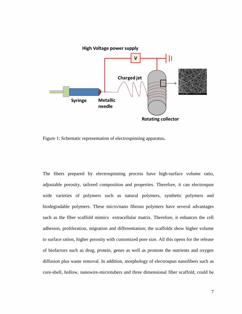
Electrospinning device includes four main components Figure (1) high power supply,
syringe pump, syringe needle with solutions and a collector for fiber deposition. The
electric field passes through the polymer solution, where the positive electrode is
connected to the needle and the negative electrode to the collector. Hence, when the
voltage is applied, the repulsive charge accumulates at the tip of the needle which is
shaped in the form of a hemisphere[21]. When the electric filed is applied the solution at
the tip of the needle should possess high surface energy and surface tension. When the
repulsive charge overcomes the surface tension, it leads to the formation of Taylor cone.
This will direct the polymer solution towards the negative electrode as collector and
produce fibers. The solvent from the polymer solution is evaporated and the polymer
solution is deposited on the collector as dry fibers ranging from nanometers to
micrometers[22].
7
Figure 1: Schematic representation of electrospinning apparatus.
The fibers prepared by electrospinning process have high-surface volume ratio,
adjustable porosity, tailored composition and properties. Therefore, it can electrospun
wide varieties of polymers such as natural polymers, synthetic polymers and
biodegradable polymers. These micro/nano fibrous polymers have several advantages
such as the fiber scaffold mimics extracellular matrix. Therefore, it enhances the cell
adhesion, proliferation, migration and differentiation; the scaffolds show higher volume
to surface ration, higher porosity with customized pore size. All this opens for the release
of biofactors such as drug, protein, genes as well as promote the nutrients and oxygen
diffusion plus waste removal. In addition, morphology of electrospun nanofibers such as
core-shell, hollow, nanowire-microtubers and three dimensional fiber scaffold, could be
8
modified by changing the parameters of electrospinning process. Thus, these precious
factors make the electrospun nanofibers suitable for biomedical applications such as drug
delivery, tissue engineering and wound healing.
1.3 Drug delivery
Drug delivery idea emerged in the 1970‘s for the controlled release of drug for
therapeutic treatments [1]. The high surface area and porosity of polymer fibers grabbed
great attention in recent years to use as drug carrier. The polymer fibers morphologies
and bulk properties can be modified by the use of electrospinning technique. In this
process, polymer nanofibers loaded with drugs are synthesized for drug delivery. Drugs
ranging from antibiotic and anti- cancer agents to proteins, aptamer, DNA[2] and RNA[3]
have been incorporated into nanofiber. The release mechanism of drugs in polymer fiber
can be altered by the change in the type of drug loading, such as co-axial electrospinning,
emulsion electrospinning, multiple layer, blended electrospinning and co electrospinning
etc. However, co-axial electrospinning and multi layered electrospun fibers have shown
great application in drug delivery due to the sustained release of the drug rather than
initial burst release of drug from the fiber scaffolds. Therefore, recent advancement in the
field of electrospinning for drug delivery will be discussed in the proceeding paragraphs.
Electrospinning in drug delivery is categorized into the drug loading type, drug loading
materials and type of drugs.
1.4 Drug loading type
The electrospinning have different drug loading type which determines the diverse
structure and different drug release kinetics. The drug loading procedure in
9
electrospinning could be executed in different ways such as co-electrospinning, multi
electrospinning, side by side electrospinning, co-axial, surface immobilization and
emulsion electrospinning Figure (2). The co-electrospinning drug molecules are mixed
with the polymer solution prior to electrospinning. These electrospun fibers provide
uniform distribution of drugs/biomolecules and high drug/biomolecules loading.
However, the biomolecules properties could be damaged when they face the high voltage
directly. On the other hand, blend electrospinning or side by side electrospinning help to
sort the issue with the drug and molecules solubility in common solvents. Moreover, the
multi-jets with more than two spinnerets exhibit a way to protection for the bioactivity of
drug. Furthermore, surface immobilization is another method where drug molecules are
covalently bonded with the scaffolds via chemical or physical immobilization method. In
this chemical methods, the surface of nanofibers is changed by introducing amines,
carboxyl, hydroxyl or thiol; the physical method includes the incorporation of
vaderwaals, electrostatics and hydrophobic interactions. This immobilization retains the
biomolecule activity, but all the electrospinning process show burst release kinetics of the
drug molecules. In this situation, co-axial and emulsion electrospinning process are
introduced. In co-axial and emulsion electrospinning is where the biomolecules are
incorporated with the core protected by shell molecules from environmental defects.
They also provide sustained release of drug by minimizing initial burst release by
controlling the thickness and composition of the shells. Another advantage of drug
loading for sustained release of drug is layer by layer via addition of drug in between the
electrospun scaffolds; this controlled release is promoted by the shield provided by the
layer polymer scaffolds. Therefore, co-axial electrospinning and multilayer
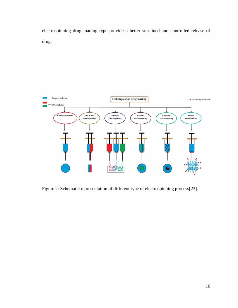
10
electrospinning drug loading type provide a better sustained and controlled release of
drug.
Figure 2: Schematic representation of different type of electrospinning process[23].
11
1.4.1.1 Multiple layered fiber mats.
Multilayered fiber mats provide the control release of drug is layer-by-layer nanofibers
stacking sandwich with drug loaded in between. This type of design is very simple, easily
controllable, and readily fabrication process as compared to core/shell. The drug release
mechanism of multilayers fibrous mats could be controlled by adjusting the thickness of
the outer layer, the amount of drug loaded, porosity of the scaffolds etc. The designing of
core/shell structure is a difficult process in one sense due to the diverse properties of
electrical and rheological properties such as conductivity and surface tension of the core
and shell polymer materials [24] . Hence, due to difficulty in fabricating core/shell,
electrospun fiber mats could not achieve a sustained repeatability, and also controlled
release of drug from the structure is difficult to study efficiently. There are many fibers
incorporated with drug molecules that were electrospun with varied thickness and studied
its drug delivery mechanism. Geun Hyung Kim [25] prepared PCL-PEO-PCL layered
fiber mats and drug delivery was studied with various thickness of PCL outer layer. It is
shown that, burst release can be erased by increasing the thickness of the PCL layer as
well as by incorporating antimicrobial peptide HPA3NT3 which doesn‘t lose its
biological activities. On the other hand, sustained release of drug haloperidol was
investigated by changing the hydrophobicity of the scaffolds. Herein, PVA-methylated b-
cyclodextrin was incorporated with PLA and PLGA. Addition of b-cyclodextrin reduces
the fiber degradation rate of PVA [26]. It is noted that, as the hydrophobicity of the
scaffold increases the release of hydrophilic drug is sustained in a controlled manner.
Where, the polyester polymers release the drug via hydrolysis manner. The blending of
hydrophobic and hydrophilic drug will minimize the toxicity caused by the burst release
12
of drug. This type of combination can be applied in hydrophobic and heat sensitive drug
due to the simplicity of the process.
The drug delivery of ibuprofen from sandwich layered fibers mates was studied and its
mathematical modeling was elaborated by using power law, higuchi equation and Fick‘s
second law was applied [27]. The mathematical modeling suggests that, thickness of the
fiber mats concern more on drug delivery than the concentration of the loaded drug. Here,
PLA was electrospun successfully by incorporating ibuprofen drug in between the two
layers of PLA. Finally, according to the type of treatment the drug loading can be
changed in accordance with the thickness of the layers for the controlled release of drug.
Dave Wei-Chih Chen [28] has studied the drug delivery of vancomycin, gentamicin, and
lidocaine for wound healing applications. Here in, they successfully mixed
PLGA/collagen on the outer layer and PLGA loaded with drug in the middle layer. The
drug vancomycine and gentamicin has released in high concentration from biodegradable
polymer scaffold. However, lidocaine has shown release upto 3 weeks. The bioactivity of
drug was shown 40-100% efficiency and it was concluded that this scaffold was suitable
for boosting the wound healing process at initial stage of wound.
1.5 Drug loading materials
Varieties of polymers can be electrospun into diverse design for drug delivery
applications in accordance with polymer-drug compatibility and ability to be molded to
fit for variety of delivery routes. While designing an optimized drug delivery system,
there are many polymer factors to be considered. For instance, biocompatibility,
biodegradability, mechanical properties and hydrophilicity [29]. There are many polymer
varieties such as natural and synthetic polymers that are used for designing drug delivery
13
system. A diverse range of drug have been loaded into the system such as growth factors,
DNA, proteins, inhibitors and antibiotics [30][31][32].
Electrospinning process can be easily applied for synthetic polymers with great
flexibility. However, synthetic polymers affect cell affinity due to the hydrophobic nature
and smooth surface for the cell recognition sites. On the other hand, natural polymers
show enhanced biocompatibility, some exhibit antibacterial properties and better clinical
functionality. The natural polymer include cellulose, chitosan, chitin, dextrose, collagen,
silk, gelatin etc [33]. For instance, lee et al. concluded the features of diverse
polysaccharide on electrospinning and their biomedical applications such as drug
delivery, wound dressings and enzyme immobilization[34]. The studied polysaccharides
are such as cellulose, chitosan, alginate, chitin, starch, hyaluronic acid, dextran and
heparin. Chitosan polymer had the properties of anticancer due to the polycationic nature.
Quartininized form of chitosan is well known for their better in vitro anticancer ability
against Hep3B, HeLa and SW480 cells [35]. However, the natural polymers lack better
mechanical strength as well as relatively sudden degradation rate due to its hydrophilic
nature which inhibit their use in long term drug delivery process. And also chances for
immunogenicity, batch to batch differences, their limited availability, expensive
productions and vulnerability to cross-contamination, these all demerits make their
clinical application to limited form [36].
On the other hand, the limitation of natural polymers could be minimized in the
application by the use of synthetic polymers:mainly biodegradable polymers such as poly
caprolcatone (PCL), polyvinyl alcohol (PVA), polylactic acid (PLA), Poligylcolic acid
(PLGA). These synthetic polymers can be degraded via enzymolysis or hydrolysis.
14
Therefore, these materials have great importance in drug delivery because the drug
delivery for tissue regeneration process could take time, also the tissue regeneration can
occur [37]. The rate of degradation depends on the sustained release of drug so that the
degradation rate can be controlled by changing the parameters such as rate of the ratio of
amorphous to crystalline segments of polymers and polymer blend compositions
[38][39]. Synthetic polymers has many advantages in comparison to natural polymers
because of its non-expensive, good mechanical properties, tunable degradation as well as
durability. However, they have demerits such as lack of cell specific recognition site due
to the hydrophobic and smooth surface.
The production of novel composite fibers in combination of synthetic and natural
polymer could reduce the disadvantages. The combination of natural and synthetic
polymers would help for the formation of fiber same as extracellular matrix, outstanding
mechanical properties and adjustable biodegradability. For example, the PLGA-gelatin
was fabricated in blending electrospinning for the drug delivery of fenbufen (FBF) [40].
These blend scaffolds have optimized mechanical properties, degradation rate and
bioactivity. However, the drug release profile could be controlled by increasing the
PLGA in the blend. It will make the scaffolds more hydrophobic and slower degradation
rate. In another paper, blend of PCL-gelatin composite scaffolds was prepared and PCL
being a hydrophobic polymer gave rise to tunable hydrophobicity, degradation rate and
mechanical properties. Consequently, gelatin provided cellular attachment and adhesion
of bone marrow derived from human mesenchymal stem cells (hMSCs). Thus, these type
of tunable properties could engender the promising scaffolds for drug delivery
application and tissue engineering system [41]. While designing a system for sustained
15
release of drug, there are many factors contributing for the efficient release of drug from
the polymer scaffolds. The factors are degradation and wettability of the polymer
scaffolds, type of drug and drug loading type.
For sustained release of the drug, the most important factor is the drug loading type.
There are many type of loading including co-axial electrospinning and multilayer
electrospinning which shows controlled release of drug for long term. The sustained
release of the drug depends on the following factors, in coaxial electrospinning such as
thickness of the shell layer, porosity, degradation rate of the shell fiber, hydrophobicity of
the scaffolds etc. On the other hand, in multilayered electrospinning the drug release
kinetics depends on the scaffolds porosity, thickness of the outer layer and hydrophilicity
of the scaffolds etc.
1.5.1.1 Polycaprolactone (PCL)
PCL, known as polycaprolactone, is a hydrophobic polyester polymer widely studied in
electrospinning. PCL is having great biomedical application due to its biocompatibility,
biodegradation, mechanical property, non-toxicity, low cost, low melting point.
Commercially available PCL ranges from 3000 to 85000g/mol molecular weight. PCL is
a hydrophobic molecule. Hence, it dissolves in solvents like chloroform, acetone, acetic
acid, dichloromethane, toluene, methanol, benzene and tetrachloride [42]. PCL properties
like biodegradability, cytotoxicity and degradation rate has been studied elaborately for
short and long duration in implantations [7], [8]. Degradation of PCL is non-enzymatic
by hydrolysis. PCL fibers are well studied as drug carrier in drug delivery.
.
16
1.5.1.2 Polyvinyl alcohol (PVA)
PVA known as polyvinyl alcohol is a semicrystalline hydrophilic polymer which is easily
soluble in water. The solubility in water makes the PVA a great application in drug
delivery. PV is a biocompatible, biodegradable and easily electrospinnable polymer. PVA
has been used for sacrificing template for the preparation of non-electrospinnable
polymers. However, individual PVA cannot be used for drug delivery due to its water
solubility. As it will lead to burst release of drug. PVA was fused with chitson to improve
its biocompatibility and cell attachment [5][6]. Gelatin electrospun with PVA as template
for better fiber of gelatin[43]. PVA has been used for sacrificing template for the
preparation of non-electrospinnable polymers[43]. However, PVA have poor mechanical
properties. Therefore, many scientists have tried to study the composite material which
could enhance the mechanical properties of the PVA[43]. PVA embodiment with PCL
polymer has gained much attention recently because its mechanical character could be
enhanced by the addition of PCL. Therefore, PCL/PVA as multilayers scaffolds for
sustained and controlled release of drug was studied below.
1.5.1.3 PCL/PVA:
The multilayered structure has gained much attention due to its versatility and controlled
release of drugs. The drug was studied as middle layer and outer layer would controlled
release of antibiotics. For instance, Yuan-Yuan Liu [44] prepared a novel scaffold by
integrating 3D bioprinting platform and electrospinning for studying the multiple drug
delivery. Here, PVA blended with gentamicin sulfate and co-axial PVA-DFO/PCL was
fused like layer by layer to form a 3D scaffold for osteointegration and sustained drug
release. Burst release was noted for gentamicin sulfate, but sustained and controlled
17
release for DFO due to the presence of vertical gradient of sodium alginate/gelatin in the
scaffold makes the DFO release as gradient- mode. Therefore, combination of 3D
bioprinting and electrospinning can be used for preparing functional gradient scaffolds. In
another study, release of the model drug tetracycline hydrochloride (TC-HCL) and
phenytoin sodium from the PVA-PCL-PVA multilayered electrospun nanofibers was
reported[45]. The hydrophilic and hydrophobic polymer was prepared layer by layer by
incorporating multiple drugs such as PHT-Na to OVA and TC-HCL to PCL, respectively.
The 87% of TC-HCL was released from single fiber and only 47% was released from
multilayer scaffolds. The release kinetics mechanism was fickian diffusion as well as
release profile corresponded to korsmeyer-peppas equation. These materials had great
application in wound dressing mats. The multilayered electrospun fiber scaffolds have
great importance in drug delivery.
1.6 Type of drugs
1.6.1.1 Antibacterial and antibacterial agents
Antibiotics and antibacterial agents were incorporated for the enhancement of scaffold
properties. Iganatova.et al. has studied the use of diverse antibiotics in the electrospun
scaffolds and their application of wound dressing [46]. The antibiotics are tetracycline
hydrochloride, ciprofloxacin, moxifloxacin, levofloxacin and antibacterial agents (for
example, 8-hydroquioline derivatives, bezalkonium chloride, itraconazole, fusidic acid or
silver nanoparticles). Gentamicin sulphate loaded PLGA and gelatin was also studied for
the continuous release of drug [47]. The results showed that PLGA/gelatin 70:30
nanofibers scaffolds exhibited a systematic release in the drug during first 15h rather than
burst release effect. This indicates that this is a promising scaffold for wound healing
18
applications. On the other hand, the drug release profile was studied for polyethylene
covinyl acetate and PLA blend scaffold in which TC-HCl was the model drug[48]. The
release of the drug delivery depends on the type of fiber and percentage of drug content.
The result stated that the 5% drug loaded 50/50 blend solutions had shown drug release
for 5 days without burst release. In addition, 25wt% has indicated rapid release than the
5wt% due to the more surface-partition effect of the drug in the former case.
1.6.1.2 Anticancer:
Not only antibiotics but also many other types of drugs, such as anticancer drugs, are
applied to the scaffolds of electrospun mats for chemotherapy. Diverse anticancer drugs,
such as docorubucin (Dox), paclitaxel (PTX), dicholoroacetate and platinum complexes
are incoparated into the electrospun fibers for localized postoperative chemotherapy
session. For instance, Xu et al. fabricated PEG-PLLA loaded electrospun fibers via
emulsion water-in-oil emulsion method in which aqueous phase was hydrophilic drug
and oily phase was chloroform solution of PEG-PLLA. The results showed that, PLLA
was completely immersed in the electrospun fibers[49]. In the same way they
successfully incorporated hydrophobic Pacitaxel (PTX) and DOX, simultaneously added
to the nanofibers scaffolds via emulsion electrospinning method, and then multiple drug
delivery was studied [50]. In contrast, Xe et al prepared electrospun scaffold of
PLA/PLGA (30/70) blend fiber added with cisplatin, and the results showed a 90%
encapsulation efficiency; the sustained release of drug was noted for 75days to treat in
vitro of glioma [51].
19
1.6.1.3 Protein, DNA, RNA and other growth factors
Overtime, electrospinning has improved; thereby propagating many new innovative ideas
for biomedical applications. Blend electrospinning and co-axial electrospinning has been
developed with the addition of protein, DNA, RNA and growth factors combined with
the electrospun fiber mats for biomedical applications. The main challenges faced in this
type of design is the loss in the bioactivity of the drug incorporated. Therefore, it is
mandatory to optimize the material and electrospinning parameters for efficient results.
Hence, the process of blend electrospinning and co-axial electrospinning has acquired
more interest in this specific type of drug addition. Co-axial electrospinning is more
efficient for protecting the bioactivity of the drug than blend. Chew et al, encapsulated
the human nerve growth factor with the BSA as a carrier into the polymer such as PCL
and poly(ethyl ethylene phosphate)[52]. The results showed that, there was a partial
bioactive retention of the hNGF when the PC12 cell line was introduced to the scaffolds.
There was a consistent release of hNGF around 3 month without burst release. The same
group studied the release of small interfering RNA (snRNA) and transfection reagent
(TKO) on electrospun fibers of copolymer caprolactone and ethyl ethylene phosphate
(PCLEEP) [53]. The results showed a sustained release of siRNA around 28 days. The
copolymerization of ethyl ethylene phosphate with PCL has induced the SiRNA delivery
rate as well as gene knock down efficiency than PCL alone. In co-axial electrospinning
the bioactive components are incorporated inside the core and protected by shell polymer.
Hence, bioactivity can be protected to the electrospinning environment and biological
environment. Saraf and co-workers have studied the addition of plasmid DNA (pDNA)
in to the core and shell polymers with non-viral gene carrier poly(ethleamine)-hyalouric
20
acid (PEI-HA)[54]. The gene release was notified around 60 days by diverging the
parameters such as concentration of pDNA and molecular weight of the core for
controlling the transfer efficiency of the pDNA. The Bioactivity of the drug could be
controlled by the new design suggested by Mickova et al [55]. He proposed the addition
of liposomes to the core which can hold the bioactive ingredients and protect its activity
for efficient action due to the shielding of lipid sphere from the electrospinning process.
1.7 Mechanisms of drug release:
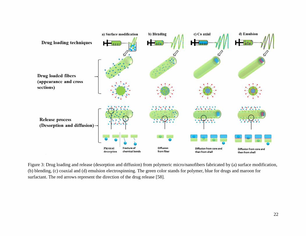
The drug release mechanism from the scaffolds are completed in three process such as
desorption from the surface, diffusion through the fibers and fiber degradation[56]. These
three processes could occur together which impacts the release kinetics throughout the
entire process. The Figure (3) illustrates the schematic representation of drug release
behavior from different types of dug loading. When the fiber is immersed in the aqueous
media desorption mechanism happened to the drug on the surface as well as drug present
inside the nanopores of the nanofibers [57] . In these three mechanisms, desorption is
considered for the drug on the surface of the polymer, therefore burst release is noted.
This burst release is due to the direct interaction of the medium with the polymer surface.
Hence, burst release of the drug is not useful so surface modification is obtained, i.e. The
main physical modification for the controlled and sustained release of the drug to the
environment.
The second type of kinetics is diffusion mechanism, in which the concentration gradient
makes the release of drug to the medium. Herein, diffusion process reduces the effect of
initial burst release and exhibit controlled and sustained release of drug. The co-axial and
emulsion electrospinning model can showcase this type of release kinetics. Finally, the
21
third type of release mechanism is degradation of the outer surface. For instance, low
degradable polymer used as the shell showed sustained release of drug due to low
degradation rate. Therefore, the mechanism of drug release kinetics was optimized using
the polymer incorporated and the type of electrospinning process. PCL is a low
biodegradable polymer however; PVA is a highly biodegradable polymer. Therefore,
combination of these two polymers could provide a better drug release profile.
22
Figure 3: Drug loading and release (desorption and diffusion) from polymeric micro/nanofibers fabricated by (a) surface modification,
(b) blending, (c) coaxial and (d) emulsion electrospinning. The green color stands for polymer, blue for drugs and maroon for
surfactant. The red arrows represent the direction of the drug release [58].
23
1.8 Tissue Engineering
Electrospinning techniques have gained much consideration in tissue engineering. The
electrospun fibers structure possesses many characters suitable for tissue engineering
such as mechanical properties, high surface to volume ratio and adjustable porosity.
Tissue engineering is an advance field of science which merges with applied engineering
and bioscience for constructing biomaterials that recover, sustain or improve the
biological activities of injured tissues [9]. For efficient tissue engineering process, three
parameters are considered such as, seeding and attachments of cells, biomaterial scaffolds
and addition of cell signaling factors. In this, biomaterial scaffolds is a major parameter
where they should mimic natural extra cellular matrix, sufficient mechanical properties,
biocompatibility, hydrophilic surface, biodegradability, high surface area and high
interpore connectivity. These criterions which contribute to cell proliferation,
differentiation and migration. In this, electrospun fibers can be prepared in cost-effective
and efficient manner to produce suitable candidate scaffold for tissue engineering.
Nowadays, several electrospun fiber mats are prepared and studied for tissue engineering
with or without addition of biological agents or growth factors for wound healing, bond
construction and nerve tissue regeneration.
1.8.1 Skin tissue engineering/ wound healing
In recent years, wound healing or skin tissue engineering has been researched very often .
Generally, the wound healing process takes place in four different steps: hemostasis,
inflammation, proliferation and remodeling [10]. The wound healing is stabilized by
different cells, growth factors and cytokines. In the first process, the host cells and
24
bacteria are removed by the macrophages in the inflation step. These macrophages also
secrete some factors eligible for the angiogenesis, fibroplasisa and extracellular matrix
production [11]. The proliferation, migration and remodeling of endothelial cells is an
important factor that contributes to angiogenesis [40]. Finally, fibroblast proliferation
leads to the rebuilding of the function and structure in the injured site[13]. Therefore,
efficient wound dressing material is mandatory for proper treatment of the wound.
Wound healing scaffold should have good biocompatibility, mechanical properties and
capability to prevent the fluid evaporation from the injured site. Furthermore, it should
provide the site for cell epithelization and inhibit the infections[59]. Hence, the ability of
cell attachment to the electrospun fiber scaffolds plays an important role in the efficiency
of engineered wound dressing scaffolds. Material and manufacturing process are
important for the preparation of an ideal wound dressings mats. Electrospinnning is an
ideal manufacturing process for wound dressing mats due to the above advantages such
as it has biocompatibility, biodegradability, hydrophilic surface, porosity and so on.
Moreover, as nanofibers scaffolds grant a better clearing of exudates from the injures site,
manages the loss of water and also oxygen diffusion in and out of the wound site [60].
There are natural (like collagen, gelatin, chitosan) and synthetic biodegradable polymers
(PCL,PLGA,PGA,PLA, PVA etc) that are molded together to form scaffolds.
Electrospun scaffold are prepared in combinations of different natural and synthetic
biodegradable polymers loaded with antibacterial and wound healing factors. The
polymers are co-electrospinned, blended, co-axial and multilayers electrospinning. Syed
Mahdi Saeed [61], has studied prepared a multilayered fiber mat loaded with curcumin as
an antibacterial active component from the novel PCL-PVA-PCL multilayered
25
electrospun fibers. The results show that, multilayered PCL-PVA curcumin-PCL has
illustrated better exudate absorbance than pristine dressing in the incision. In the same
vein, it summarize that, 16% loaded curcumin display antibacterial activity without
killing the cell viability. Antibacterial properties can be built up in the scaffold with the
addition of antibacterial agents or antibiotics. Silver is a well-known antibacterial agent
because it can damage the DNA replication of bacteria [18]. Fatemeh Khodkar [62] has
successfully prepared PVA/PCL core/shell loaded with silver nanoparticles in core for
wound dressing applications. Fiber loaded with silver shown lower porosity as well a
water vapor transmission rate(WVTR) and also greater contact angle. These scaffolds are
suitable for long term antibacterial activity (Escherichia coli and Staphylococcus aureus)
because of the sustained and controlled release of the silver nanoparticles in core/shell
structure. On the other hand, PCL/PVA was co-electrospinned by loading silver
sulfadiazine(SSD) as drug for wound dressing mats [18]. PCL and PVA loaded with
SSD was prepared successfully. The effect of different weight % of SSD on mechanical
and cell toxicity and antibacterial properties was studied. The better SSD concentration
was identified with antibacterial ability and cellular attachment as well as proliferation
was notified. Where, addition f SSD increases the fiber diameter as well as hydrophilic
properties but reduces the mechanical properties of the scaffold. Fibronectin coating can
improve the biocompatibility of the scaffolds loaded with SSD. Therefore, 5% SSD
loaded co-electrospun PVA-PCL show better antibacterial and reasonable cell
proliferation and differentiations. Recently, L. Du has fabricated PVA merged with
monodisperse AgNPs and PCL loaded with Ascorbyl palmitate (AP) by duel spinneret
electrospinning [63]. The NIH-3T3 fibroblast cells are seeded on the scaffold mats and
26
showed that, AP inhibits the toxic effects of AgnPs on cell proliferation. It should also be
noted that, antibacterial tests validate the inhibition towards gram negative and gram
positive Escherichia coli (E. coli) and Staphylococcus aureus (S. aureus) respectively.
Wound healing test and histological observation conclude that, this material provide a
promising for future biomedical applications.
[64] Porosity and surface wettability is an important parameter which determines the
healing process. Xin Liu has electrospun PVA,PCL,PAN and PVdF-HFP incorporating
wool protein and Ag for wound dressing mats. Thus, it can be concluded that, other than
nanofiber diameter and antibacterial property the porosity and hydrophilicity is an
important factor that enhances the wound healing process. The Hydrophilic membrane
showed efficient remedy for wounds in comparison to the hydrophobic membrane.
Porosity for oxygen diffusion also leads to better wound healing process. However,
wound healing process for diabetic ulcers are time consuming due to the lack of efficient
blood supply because of higher amount of sugar in the blood. These processes leads to
long inflammatory stage, defected angiogenesis and blocked fibroblast proliferation.
Adeleh Gholipour-Kanani has prepared novel electrospun blended fibers of Chitosan-
poly (vinyl alcohol) (Cs: PVA) (2:3) and poly (caprolactone)-chitosan-poly (vinyl
alcohol) (PCL: Cs: PVA) (2:1:1.5) for wound healing in diabetic patients. The above
fabricated scaffolds wound healing prate was studied by applying to diabetic dorsum skin
wounds and diabetic foot wounds on a rat model (n-16). The scaffolds had far effective
healing process than control ones. More granulation tissues are presence in the scaffolds
treated wounds than the control ones and within 20 days the scaffolds provided better
27
wound repair than controls. Hence, PVA:CS:PCL is a considerable material for diabetic
wound healing.
1.8.2 Bone tissue engineering applications:
Bone is the strong rigid organ which plays an essential role in our body. It protects our
vital interior organs, movement, manufacturing of white blood cells and red blood cells,
and also storage of minerals[65]. In bone extra cellular matrix mainly consist or organic
and inorganic components such as collagen and hydroxyapatite (HAp). Incorporation of
these components make suitable scaffolds for bone tissue engineering applications. The
electrospun scaffolds architecture play an important parameter for successful bone
regeneration. They are microstructure of the scaffolds, its porosity and surface
properties[66]. The electrospun fiber should provide better mechanical properties to
support the structure and arrange a space for the osteocondral adhesion, proliferation and
differentiation. Hence, the development of an ideal scaffold for tissue regeneration could
be achieved by using a porous ceramic material, lamellar material and a fiber matric
material for better biological and physical properties. Subramanian Uma Maheshwari has
developed a scaffold comprise of polymer –ceramic combination of PCL/PVA bilayer
scaffold blended with HAp nanoparticles[67]. (PVA-PCL)-HAp has improved its
porosity around 64% as well as hydrophilicity around 141%. Also MTT assay studies
with MG-63 osteoblast cells had better cell adhesion and proliferation which show a
promising application for tissue regeneration. However, incorporation of growth factors
(GF) or drug to the scaffold is also important for enhancing the regrowth of broken
bones. There are many GF such as bone morphogenetic protein-2 (BMP-2) and VEGF
added to the electrospun scaffolds for the long lasting sustained release of GF to mimic
28
the natural healing process. For instance, co-axial electrospun of collegen-PCL
incorporated with BMP-2 and dexamethasone (DEX) has shown better controlled release
of GF, thereby encouraging the osteogenic expression of human mesenchymal stromal
cells hMSCs [68]. In this design the shell layer was loaded with DEX and the core was
incorporated with BMP-2. The dual drug release was exhibited, in which DEX shown
fast release. However, BMP-2 demonstrated sustained release over 22 days. This scaffold
provide healing process as well as osteogeneration effieciency.
On the other hand, incorporation of stem cells into the biomaterials is also a novel
approach for tissue regeneration of the cells. For instance, Abbas Shafie has studied in
vitro and vivo of the cartilage tissue regeneration from rabbit bone marrow mesenchymal
stem cells (BM-MSC) seeded on electrospun scaffold of PVA/PCL nanofibers[69]. In
vitro, the MTT assay shown that, the scaffolds backing the chondrogenic differentiation
of MSC. In vivo, the scaffold with and without MSC loaded were implanted on a rabbit
full-thickness cartilage defects. To study the cartilage regeneration, histological and semi-
quantitative grading was executed. The results shown that, scaffold seeded with MSC has
enhanced the healing process as compared to non-seeded scaffolds. These results show
that, PVA/PCL scaffold seeded with MSC is suitable for graft for articular cartilage
repair.
1.8.3 Skeletal muscle regeneration:
Skeletal muscle made up of around 40% of human body. Skeletal muscle is made of
various fibers with diameters ranging from 10 to 80mm[70]. These fibers are
unidirectional and produce enormous amount of force during contraction [71]. If a
muscle cell gets injured or wounded, it will not be possible to contract, satellite cells are
29
switched on its activity for muscle cells regeneration. However, this healing process
would create a scar in the tissue and block the muscle function [71]. There are many
efforts took place to study the initial steps for muscle regeneration such as autologous
muscle transplant, satellite cells, exogenousmyogenic cells and myoblasts but these
methods met finite success[72]. Therefore, long term denervation and severe injuries can
leads to the loss of skeletal muscle activities.
Muscle tissue engineering materials desire better contracting ability and mechanical
properties [73]. Muscle cell adhesion and proliferation has been studied using both
mechanical properties and electric stimulus in the cell culture. Mckeon-fischer K D has
prepared co-axial electrospun fibers with the core as PCL and Multiwaled carbon
nanotubes (MWCNT) and blend of (83/17, 60/40, 50/50, and 40/60) poly(acrylic
acid/poly(vinly alcohol) (PAA/PVA) as the modified outer shell layer [74]. All the four
components were electrically conductive, although, the scaffold didn‘t show a actuated
when electric field is applied. The best result was shown at 20V. The MTA assay by
Soleus and vastus lateralis (VL) muscles extracted from rats, result shown that, 0, 0.14%
and 0.7% of concentration of MWCNT in the scaffold has not toxic for the cells over 4
weeks period. According to different percentages of blend solutions PAA/PVA in the
outerlayer 40/60 has illustrated higher number cells than other scaffolds. Scaffold has
tensile properties that are higher than the skeletal muscle‘s. More modification of these
scaffolds for contraction rather than bending can lead to promising scaffolds for artificual
muscle applications.
30
1.8.4 Nerve tissue engineering:
Electro-conducting polymers such as polypyrrole (PPy), polyaniline (PANI), polythio-
phene (PT), poly(3,4-ethylenedioxythiophene) (PEDOT)) show attractive electrical and
optical phenomena. Thus, they have been researched in the past few decades for various
applications such as microelectronics, actuators and polymer batteries[75]. The electro-
conducting polymers which hold the specialization of biocompatibility and good
conductibility can be applied as biosensors and tissue engineering scaffolds [76]. The
electrospun electro-conducting polymer have great deal for electrically stimulate neurons
and nerve tissue engineering as well as neural prostheses application for therapeutical
function [76][77]. Schmidt et al has firstly studied the PC12 cells through polypyrrole
(PPy) electrosconducting polymer, recognize the growth of PC12 cells on the thin film of
PPy these enhance the neurite outgrowth from the cells, these results suggest the greatest
application of these type of scaffolds for nerve tissue regeneration [78]. Many studies
have been suggested improvement of the electro conducting polymer for nerve tissue
regeneration application by adding cell adhesive[79], neurotrophins[80] and
topographical features[81]. Jae Y Lee has prepared electrospun nanofibers coated with
conductive polymer PPy for nerve tissue engineering applications[82]. The PPy-PLGA
improved the growth of rat pheochromocytoma 12 PC12 cells and hippocampal neurons
than that of non-coated PLGA as control. This suggest that PPy-PLGA would use for the
nerve tissue engineering application. Simultaneously, electrical stimulus studies on the
scaffold suggest that, at stimulus of 10mV/cm has improved the neurties to 40-50%
longer as well as increment of 40-90% more neurons formation compared to non-
stimulus at same scaffolds. Moreover, aligned scaffolds show more neurites elongation
31
and formation than that of randomly oriented PPy-PLGA fiber scaffolds. These results
suggest that, good response for electric stimulus and biocompatible polymers prepared by
electrospinning have great advantages for biomedical applications such as nerve tissue
engineering.
1.9 Sputtering technology for biomedical applications:
Plasma technology is a polymer that has improved the surface properties of the polymers
without changing their bulk characters. The plasma treated polymer had found great
application in diverse fields such as automobiles, microelectronics, chemical and
biomedical industries[83]. The polymer surface properties such as hydrophobicity,
roughness, chemical structure, conductivity etc. can be molded for various applications.
The plasma treatment could affect the polymer surfaces by micro-etching, organic
contamination, cross-linking ,surface chemistry modification and surface coating with
specific target material[84]. The biomaterials should possess a good mechanical and
surface characters eligible for biological environment. For instance, for cell adhesion, the
polymer surface should have low surface free energy, surface roughness and
hydrophilicity matters. The plasma treatment via magnetron sputtering technology has
been reimagined by coating the surface of polymer to form biomaterial suitable for
biomedical applications such as antibacterial activities, biocompatibility and tissue
engineering.
There are thermal and non-thermal deposition process in plasma sputtering technology.
However, non-thermal deposition procedure is highly recommended for polymers
because it will not damage the bulk properties of the polymer. Magnetron sputtering is
the technique used for coating the polymer surface. Magnetron sputtering is a technology
32
developed during 1970‘s it is a high-speed and low temperature technique for preparing
uniform and strong adhesion film on the surface of polymer, ceramics and composite
materials [85]. Sputtering is simply a call as momentum transfer between the particles
[86]. Wherein, when an positive ion from sputtering gas (plasma) with high speed strike
on the surface of target materials and surface atoms is ejected from the target and
deposited on the substrate[87][88] when the environment is electric and magnetic field..
Argon, nitrogen and oxygen are the sputtering gas. However, argon gas is commonly
used because they don‘t damage the target due to its nobility. The ejected atoms are
accumulated on the substrate via adjusting the distance between the target and substrate.
This process leads to the formation of thin film on the surface of the polymer. The full
process is very fast and requires only low temperature, and high film forming rate as well
as strong film adhesion[89]. For instance, the composite microfibers of
Poly(methylmethacrylate/ organically modified montmorrillonite (O-MMT) was
manufactured by electrospinning incorporation with emulsion polymerization[90]. Here
the prepared composite microfibers of PMMA—O-MMT was magnetron sputter coated
with Titanium dioxide (TiO2). The results showed that, increased deposited anatase-TiO2
and rutile-TiO2 has shown better wettability of the surface without damaging the
PMMA—O-MMT compound. These composite fibers have UV absorption 254nm.
Therefore, it has the tendency for photocatalytic degradation of model compound
methylene blue. Thus, these materials provide a promising application in dye waste water
treatment.
The polymer microsphere[91] , thin film[92] and fibers[93] are sputter coated with
Ag[93][94], Cu[95], Ti[96], TiO2 [97], gold[98], Thydroxyapatite (HAP), tricalcium
33
phosphate (TCP)[99], amorphous calcium pyrophosphate (CPP) [99] and dicalcium
phosphate dihydrate (DCPD) [99] for various polymers in microsphere, thin film and
electrospun fibers for different biomedical applications such as antibacterial and tissue
engineering applications are discussed below in details.
1.9.1 Antibacterial coatings:
Attachment of bacteria to the surface of a polymer can lead to the formation of biofilm.
Therefore, biofilm resistant polymers for medical field are an important factor. Biofilm
resistant could be an assistance to the polymer by the addition of antibacterial agents on
the surface of polymer to prevent the bacterial adhesion. The materials such as medical
textile, wound dressing, prosthesis and implant materials should display antibacterial
activity for efficient biological activity. Antibacterial properties are an important
parameter to take into account for wound dressing. The antibacterial activities are
promoted by the addition of some antibacterial components to the fabrics. There are
many components such as inorganic and organic (drugs) as well as metal. The inorganic
agents include titanium dioxide, carbon nanotubes, silver, zinc, zinc oxide, copper,
gallium and gold nanoparticles [100]. Organic includes some antibiotics [101].
Among the inorganic antibacterial components silver nanoparticles was well studied
[102]. The silver nanoparticles was added in the electrospun fibers via Ag ions through
wetting process[16][17], silver sulfdiazide [18], etc. The wetting process for the
preparation of Ag to the matrix has many disadvantages such as uneven distribution of
nanoparticles, using reducing agents that are toxic, and controlling the size of
nanoparticles is a difficult task depending on the strong and weak reducing agents
used[19]. However, the most efficient way of introducing nanoparticles to the surface of
34
polymer or fabrics could be done by using plasma technology. Plasma technology
provide uniform deposition, less use of resources as well as simple process for the
coating of antibacterial material such as Ag, Si, Cu etc, to the surface of the polymer than
wetting process. Sputtering known as physical vapor deposition has been effectively used
in coating diverse thin film in electronics applications. Therefore, sputter coating of
metals to enhancing the antibacterial properties are contributed by addition of many
target materials such as Ag, Ag/Si, Cu, Ti etc..). Therefore, more studies are required to
compare the antibacterial properties of various materials incorporated with bioresorbable
polymers..
1.9.1.1 Silver (Ag):
Silver is a transition metal in the periodic table. Silver related compounds or
nanoparticles have biocidal effect on around 16 specious bacterial because of its toxic
effect on microorganisms [14][15]. Thus, silver is coated on medical devices for
antibacterial properties [103]. At low concentration silver nanoparticles show good
antibacterial efficiency [104]. Moreover, the in-toxicity effect of AgNP to human
monocytes cell lines show the application of Ag to the fabrication of medical devices.
The mechanism of antibacterial activity of Ag on microorganism are not studied well yet.
It has been shown that in E.Coli the AgNP treated bacteria has shown some pits on the
cell wall and accumulation of Ag in the cellular membrane. This type of membrane has
shown increase in the permeability. Bacterial DNA drops its replication ability as well as
cellular protein and becomes denatured by binding Ag ions or nanoparticles to the
functional group of the protein[105]. This results in the cellular death of bacteria. On the
other hand, some reported that Ag nanoparticles will denatures cellular protein suitable
35
for the cellular nutrient transport and damage the cell membrane or cell wall, enhanced
cell permeability and followed by cell death [15]. It is also noted that, the antibacterial
efficiency of Ag depends on its shape. Ag Nps with {111} plan lattice basal plane show
strongest antibacterial action than spherical and rod-shaped nanoparticles and silver ions
[106].
Silver has good antibacterial properties. Silver treatment is well known for wound
dressing materials. The silver is incorporated as silver nanoparticles by the introduction
through AgNO3 by using reducing agent. However, this type of silver incorporation leads
to burst release of silver from the material with very high concentration of Ag in the
wound. These is followed by the sudden reduction of silver because of both bacterial
consumption as well as reaction with other compounds present in the wound beds such as
phosphates, chlorine and proteins. Therefore, silver release in the wound should be in a
controlled manner. In addition, silver nitrate present is an hypotonic and hence it can
effect strong electrolyte imbalance which could damage the wound site, produce gross
systematic imbalance which could kill the patients with heavy burns who require large
doses of silver. But silver sulfadiazide was developed in accordance to minimize the side
effect of using silver nitrate. However, the removal of silver sulfadiazide cream from the
wound surface is by scrapping, it could proclaim the patients painful dressing procedure.
Moreover, sulfadiazide does not show any hypotonic effect. Therefore, it is mandatory to
have a better procedure for delivering silver. The efficient way of introducing silver is via
prolonged period, act against many range of bacteria, require only few changing wound
dressings, and never interfere with the wound healing process. Thence, sputtering is a
new field of surface coating of wound dressing materials for prolonged release of silver
36
with efficient antibacterial properties. The optimization of sputtering process is an
important criterion for better antibacterial properties.
It is also noted that, silver is the best candidate for wound curing application because it
will reduce the inflammation [107] and impedes the contraction and improve cell
epithelialization [108]. The silver nanoparticles are toxic to effect on cell viability these
leads to decrease in biocompatibility of scaffolds [109]. However, the amount of Ag to
the scaffolds can be optimized with the antibacterial effects and cellular toxicity of Ag.
Antibacterial coating on medical textiles is an important criterion to avoid infections
during surgical process[110]. The silver is coated on textiles via different techniques. But
it has also reduced the durability of the coatings, which were very poor, hence limiting
their usage. Therefore, strong adhesion of silver on fabrics is required via sputtering
coating technique [110]. Cotton fabric with antibacterial properties could have various
applications. Silver is the most commonly used material to enhance the antibacterial
properties in cellulosic fibers [110]. Silver incorporation could be made wet or dry
process. The wet process is change the bulk properties of textiles and also has negative
impact to the environments. However, dry process such as sputter coating is an
ecofriendly process which only changes the surface of the matrix. Therefore, Ag was
cooperated into the cotton matrix in various thickness and studied the antibacterial,
release of the Ag in water etc. The results suggested that, Ag has shown antibacterial
activity against Staphylococcus aureus, Escherichia coli and Candida albicans [110]. And
also sputter coating has improved the water contact angle of the cotton fabrics. Thus,
these ideas sputtering could be added to nanofibers for antibacterial activity.
Simultaneously, Ying-Hung Chen has coated sputter coated the PET fabrics in high
37
power impulse magnetron sputtering which provide highly concentrated plasma so these
will support strong adhered films[111]. These results shown that, the sputter coated more
than 1 min films has shown great antibacterial performance via bacteriostatic (>2.0) and
bactericidal (>0) effects, based on JIS standards. Furthermore, the coated fabrics have
shown capability to hold the antibacterial properties over 20 cycles of washing, indicating
long-term durability for the fabrics.
The wound dressing mats of polymer sheet and electrospun fiber scaffolds was sputter
coated with Ag and studied its antibacterial properties. Xiuju Liu has studied the
influence of magnetron sputter coating of nano-silver on polyetheretherketone (PEEK)
and studied its cytotoxicity and antibacterial properties[112]. PEEKs were sputter coated
with Ag 3nm, 6nm, 9nm and 12nm. The antibacterial properties and bacterial adhesion to
the surface was studied. The homogeneous Nano silver was coated on the surface and it
shown that, water contact angle was increased as well as there was no cytotoxicity for the
CCK-8. And also the coating provides good adhesion of bacterial towards PEEK and also
improved antibacterial activities towards Streptococcus mutans and
Straphylococcusaureus.
The combination of electrospinning and sputtering technology can make many novel
composite fibers with diverse applications in biomedical such as wound dressing mats
with good biocompatibility and antibacterial properties. The electrospun microfibers was
coated with Ag by DC magnetron sputtering [94]. The electrospun scaffolds of
poly(glycerol sebacate)/poly(3-caprolactone) (PGS/PCL) was coated with Ag and its
antibacterial properties and silver release behavior was studied. PGS/PCL shown good
mechanical and thermal behavior due to increase in fiber diameter and decrease in fiber
38
pore size when sputter coated with Ag. The fiber scaffolds has shown gradual release of
Ag which contribute for antibacterial activity. Therefore, this material could possess good
application in wound dressing and bandages. Moreover, implant prosthesis also requires
antibacterial properties to avoid infection after surgery. Incorporation of Ag with SiO2
was used in implantic prosthesis after hernia repair can leads to infections in the
abdominal area, therefore prevention is an important goal. Giuliana Muzio has studied the
effect of polypropylene prostheses coated with silver-silica composite (Ag/SiO2) layer by
sputter coating technique for improving the substrate antibacterial and biocompatibility of
the prosthesis [113]. The prepare meshes hernia prostheses (CMC) consist of two layers
microporous light mesh and thin transparent film of polypropylene. They have sputter
coated the Ag/SiO2 composite on CMC meshes and microporous mesh layer alone. The
sputtering process was optimized via addition testing biocompatibility and antibacterial
properties. And it is noted that, sputter coated with CMC improve antibacterial property
but reduced biocompatibility. However, sputter coated meshes alone show good
antibacterial and biocompatibility. And also, fiber meshes coated with Ag/SiO2 enhance
the growth of seeded fibroblast without creating apoptosis or necrosis of the fibroblast in
addition meshes exhibited good antibacterial property as well. .
1.9.1.2 Copper (Cu):
Other than Ag the electrospun scaffolds was sputter coated with Cu for antibacterial
property [114][115]. Cu is cheaper than Ag therefore Cu coating can provide economical
wound dressing mats. PLA scaffold was DC magnetron sputter coated with Copper (Cu)
[95]. The PLLA scaffold has increased the hydrophobicity in proportion to the plasma
treatment time. The antibacterial testing concludes that, modified composite scaffold
39
have bacteriostatic effect in which 30 and 50% of bacterial has reduced. And also it is
found that copper have strong antibacterial effect than copper oxide. Therefore, this type
of composite material can be used for economically cheap wound dressing mats for
having antibacterial effect.
1.9.2 Tissue engineering:
Plasma technology has also emerged recently on tissue engineering application such as
vascular grafting, stem cell therapy, artificial muscle sputter coated with conductive gold.
Therefore, the good biocompatible polymer had provided a platform for cell adhesion,
proliferation and differentiation. Therefore, if stem cells are added to the polymer
scaffold which can provide better tissue regeneration environment. Stem cell therapy is a
new platform for substituting many complicated surgical procedures. Stems cells loaded
materials has gained much attention recently [91]. Here in, PCL microsphere
incorporated with sputter coated Au has used as a platform for the differentiation of
cardiomyogenic cells from human embryonic stem cells. Presently, here they
demonstrated the PCL microsphere sputter coated with gold particle for 5min and without
coating [91]. Then they added human embryonic stem cells (hESCs) to the microsphere.
And it noted that, AuMS shown higher cardiac differentiation in because Au play as
mediator for expressing genes on day 4 and day 14 which are suitable for the
cardiomyogenic differentiation than MS. These papers also suggest AuMS is non-
cytotoxic material.
Moreover, The PLLA and PEG fibers was electrospun and sputter coated with calcium
phosphate for bone tissue engineering applications. Here, they just combined
40
electrospinning and sputtering technique are feasible for fabricating biopolymer scaffolds
for biomedical applications[116].
There are many novel composite material has emerged due to the fusing of two precious
technique such as electrospinning and sputtering technique[117]. A novel material for
vascular tissue engineering was studied. These composite materials was prepared by
electrospinning and sputtering technique. In this, beresosable polymer PCL and PHBV
was incorporated in 1:2 (v/v) and sputter coated with Ti. Firstly, they optimized the
sputtering process which would not damage the macro structure of the scaffolds. The
biocompatibility of the prepared composite mats was studied with hybridoma of the
endothelial cells of human umlical vein and human lungs carcinoma (EA.hy.926 cell
line). The results from cell adhesion, viability and secrection of IL8 interleukin-8 (IL8),
interleukin-6 (IL6) ang vasculat endothelial growth factors (VEGF) demonstrate that cell
adhesion has improved for ti coated scaffolds as well as it determine that novel scaffolds
have better proangiogenic activity.
And also a novel approach was incorporated to make a fiber scaffold suitable for
artificial muscle or smart devices for human body was synthesized by using
electrospinning and sputtering technology[98]. A core-shell structure was made via first
electrospinning the PMMA in optimized form to get a uniform fiber and later PMMA
was coated with gold for inducing the conductivity and mechanical properties of the
scaffolds. Later on, the polyaniline (PANI) was coated on the scaffolds via in situ
electrochemical polymerization starting with aniline and using sulfuric acid as oxidizing
agent Figure (4). PANI coated metalized fiber scaffolds in similar to core-shell structure
has shown fascinating electrochromic properties where color changes occur when applied
41
voltage was simultaneously switched from 0 to 1V and vice versa. In vitro
biocompatibility test reveal good cell adhesion tested on human amniotic fluid stem cells
show a better for eukaryotic cells. Moreover, PANI coated exhibit high cell adhesion
property. Therefore, this type of web could be used for preparing smart artificial muscle
devices via simple and versatile preparations technique such as electrospinning and
sputtering.
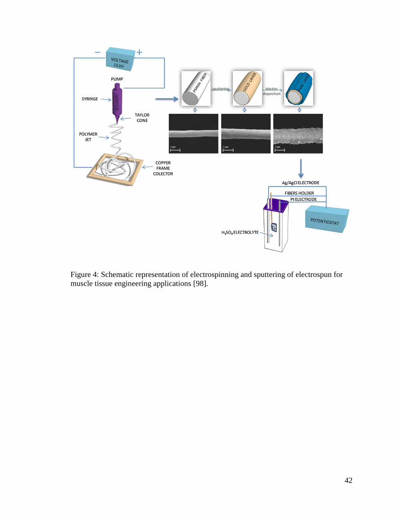
42
Figure 4: Schematic representation of electrospinning and sputtering of electrospun for
muscle tissue engineering applications [98].
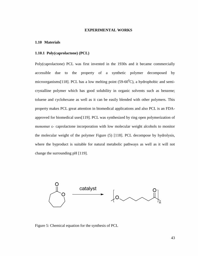
43
EXPERIMENTAL WORKS
1.10 Materials
1.10.1 Poly(caprolactone) (PCL)
Poly(caprolactone) PCL was first invented in the 1930s and it became commercially
accessible due to the property of a synthetic polymer decomposed by
microorganisms[118]. PCL has a low melting point (59-600C), a hydrophobic and semi-
crystalline polymer which has good solubility in organic solvents such as benzene;
toluene and cyclohexane as well as it can be easily blended with other polymers. This
property makes PCL great attention in biomedical applications and also PCL is an FDA-
approved for biomedical uses[119]. PCL was synthesized by ring open polymerization of
monomer ε- caprolactone incorporation with low molecular weight alcohols to monitor
the molecular weight of the polymer Figure (5) [118]. PCL decompose by hydrolysis,
where the byproduct is suitable for natural metabolic pathways as well as it will not
change the surrounding pH [119].
Figure 5: Chemical equation for the synthesis of PCL
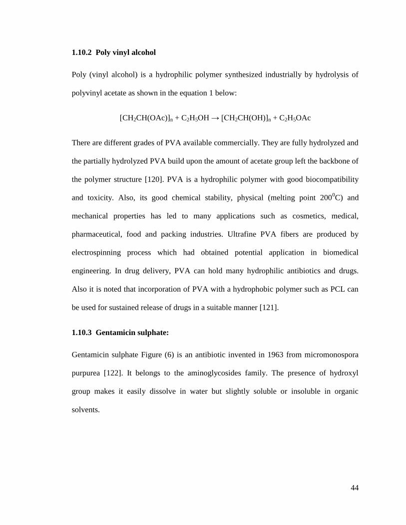
44
1.10.2 Poly vinyl alcohol
Poly (vinyl alcohol) is a hydrophilic polymer synthesized industrially by hydrolysis of
polyvinyl acetate as shown in the equation 1 below:
[CH2CH(OAc)]n + C2H5OH → [CH2CH(OH)]n + C2H5OAc
There are different grades of PVA available commercially. They are fully hydrolyzed and
the partially hydrolyzed PVA build upon the amount of acetate group left the backbone of
the polymer structure [120]. PVA is a hydrophilic polymer with good biocompatibility
and toxicity. Also, its good chemical stability, physical (melting point 2000C) and
mechanical properties has led to many applications such as cosmetics, medical,
pharmaceutical, food and packing industries. Ultrafine PVA fibers are produced by
electrospinning process which had obtained potential application in biomedical
engineering. In drug delivery, PVA can hold many hydrophilic antibiotics and drugs.
Also it is noted that incorporation of PVA with a hydrophobic polymer such as PCL can
be used for sustained release of drugs in a suitable manner [121].
1.10.3 Gentamicin sulphate:
Gentamicin sulphate Figure (6) is an antibiotic invented in 1963 from micromonospora
purpurea [122]. It belongs to the aminoglycosides family. The presence of hydroxyl
group makes it easily dissolve in water but slightly soluble or insoluble in organic
solvents.
45
Figure 6: Chemical structure of Gentamicin Sulphate Gentamicin quantification was done by using ninhydrin as a reaction compound [123].
The ninhydrin reacts with primary and secondary amines to give a purple color. These
color changes can be studied calorimetrically. The gentamicin ninhydrin mixture gives
linear relationship ranges from 1-80ug/mL at 400nm wavelength [124].
Table 1: Physical properties of gentamicin sulphate
Physical properties of gentamicin sulphate
Physical appearance Amorphous powder
Solubility Soluble in water and phosphate buffer
solution at pH 7.4 and insoluble at
acetone and ethanol
Chemical properties
Reaction with ninhydrin Give rise to purple color
46
1.10.4 Solvents:
Table 2: Solvents used for electrospinning
Solvents Suppliers Specifications Item No
Chloroform BDH laboratory
suppliers
99.8% purity 152834E
Dimethyl
formamide (DMF)
Riedel-de Haen
Germany
99% purity
Absolute Ethanol SISCO research
laboratory PVT,
Mumbai India
99% purity 11526689
1.11 Scaffolds preparation equipment:
1.11.1 Electrospinning:
1.11.1.1 Setup-operating principles:
The electrospinning is a simple technology exists of four main components Figure (7): 1)
high power supply 2) syringe pump 3) syringe needle with solutions 4) a collector for
fiber deposition. The electric field is applied to the polymer solution where the positive
pole is connected to the needle and negative electrode to the collector. Hence, when the
voltage is applied the repulsive charge is accumulated at the tip of the needle which exist
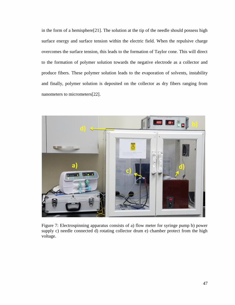
47
in the form of a hemisphere[21]. The solution at the tip of the needle should possess high
surface energy and surface tension within the electric field. When the repulsive charge
overcomes the surface tension, this leads to the formation of Taylor cone. This will direct
to the formation of polymer solution towards the negative electrode as a collector and
produce fibers. These polymer solution leads to the evaporation of solvents, instability
and finally, polymer solution is deposited on the collector as dry fibers ranging from
nanometers to micrometers[22].
Figure 7: Electrospinning apparatus consists of a) flow meter for syringe pump b) power
supply c) needle connected d) rotating collector drum e) chamber protect from the high
voltage.
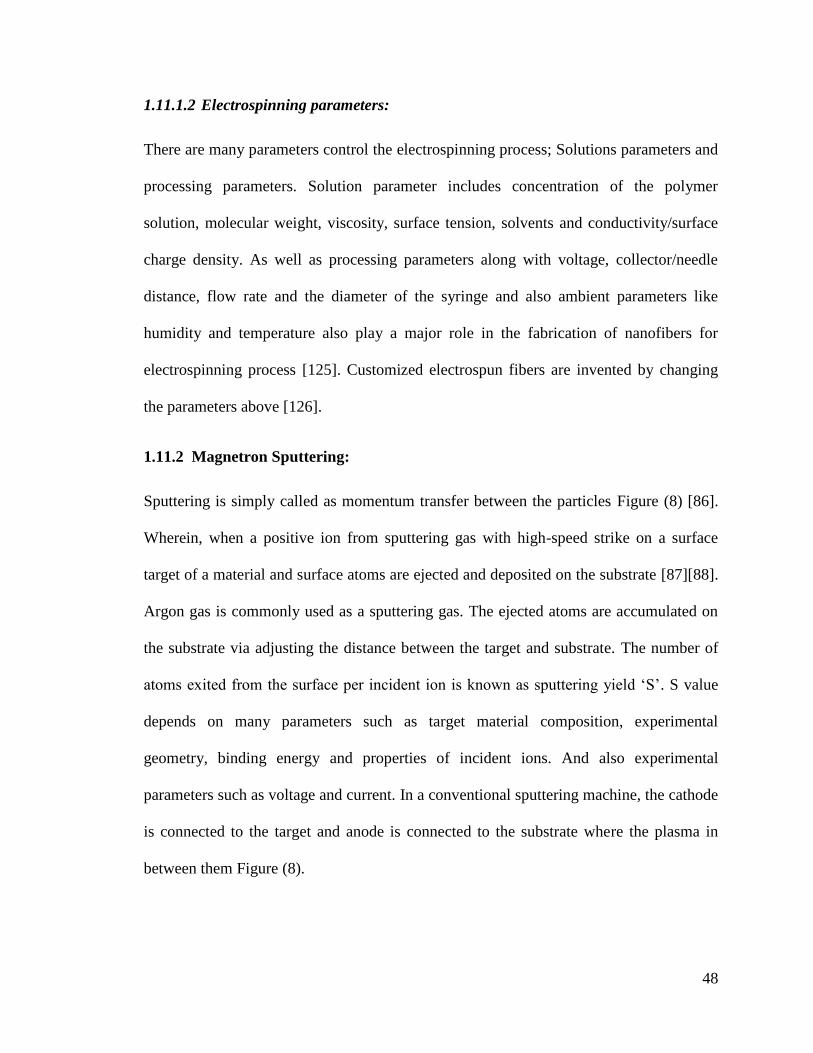
48
1.11.1.2 Electrospinning parameters:
There are many parameters control the electrospinning process; Solutions parameters and
processing parameters. Solution parameter includes concentration of the polymer
solution, molecular weight, viscosity, surface tension, solvents and conductivity/surface
charge density. As well as processing parameters along with voltage, collector/needle
distance, flow rate and the diameter of the syringe and also ambient parameters like
humidity and temperature also play a major role in the fabrication of nanofibers for
electrospinning process [125]. Customized electrospun fibers are invented by changing
the parameters above [126].
1.11.2 Magnetron Sputtering:
Sputtering is simply called as momentum transfer between the particles Figure (8) [86].
Wherein, when a positive ion from sputtering gas with high-speed strike on a surface
target of a material and surface atoms are ejected and deposited on the substrate [87][88].
Argon gas is commonly used as a sputtering gas. The ejected atoms are accumulated on
the substrate via adjusting the distance between the target and substrate. The number of
atoms exited from the surface per incident ion is known as sputtering yield ‗S‘. S value
depends on many parameters such as target material composition, experimental
geometry, binding energy and properties of incident ions. And also experimental
parameters such as voltage and current. In a conventional sputtering machine, the cathode
is connected to the target and anode is connected to the substrate where the plasma in
between them Figure (8).
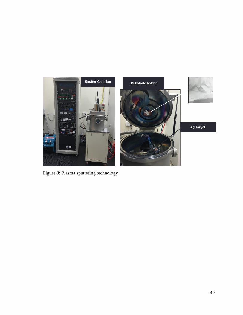
49
Figure 8: Plasma sputtering technology
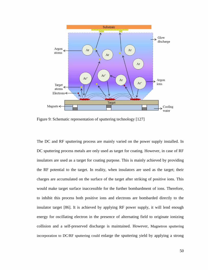
50
Figure 9: Schematic representation of sputtering technology [127]
The DC and RF sputtering process are mainly varied on the power supply installed. In
DC sputtering process metals are only used as target for coating. However, in case of RF
insulators are used as a target for coating purpose. This is mainly achieved by providing
the RF potential to the target. In reality, when insulators are used as the target; their
charges are accumulated on the surface of the target after striking of positive ions. This
would make target surface inaccessible for the further bombardment of ions. Therefore,
to inhibit this process both positive ions and electrons are bombarded directly to the
insulator target [86]. It is achieved by applying RF power supply, it will lend enough
energy for oscillating electron in the presence of alternating field to originate ionizing
collision and a self-preserved discharge is maintained. However, Magnetron sputtering
incorporation to DC/RF sputtering could enlarge the sputtering yield by applying a strong
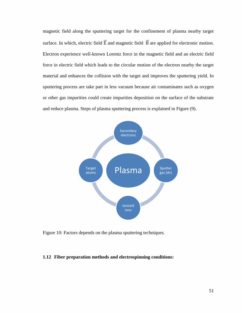
51
magnetic field along the sputtering target for the confinement of plasma nearby target
surface. In which, electric field ⃗⃗ and magnetic field ⃗⃗ are applied for electronic motion.
Electron experience well-known Lorentz force in the magnetic field and an electric field
force in electric field which leads to the circular motion of the electron nearby the target
material and enhances the collision with the target and improves the sputtering yield. In
sputtering process are take part in less vacuum because air contaminates such as oxygen
or other gas impurities could create impurities deposition on the surface of the substrate
and reduce plasma. Steps of plasma sputtering process is explained in Figure (9).
Figure 10: Factors depends on the plasma sputtering techniques.
1.12 Fiber preparation methods and electrospinning conditions:
Plasma
Secondary electrons
Sputter gas (Ar)
Ionized ions
Target atoms
52
1.12.1 Optimization of PVA and PCL individual fibers and multilayered structure.
Polyvinyl alcohol (PVA): 10 wt%/v% PVA was dissolved in deionized water and stirred
for 3h at 80oC. The dissolved solution was electrospun to produce PVA fibers.
Electrospinning parameters are voltage 15kV, distance 10cm, needle size 21G, and flow
rate 1mL/h.
Polycaprolactone (PCL): 5.5 to 11 wt% of PCL was dissolved in 9:1 (wt%/wt%)
CF:EtOH and stir for 1h. And also 8.5 to 12 (wt%) PCL solution was prepared in 1:1
(wt%/wt%) DMF:CF and stirred for overnight. Electrospinning parameters are voltage 7-
15kV, distance 15cm, needle size 21G, and flow rate 1mL/h Table (4).
Multilayered (PCL-PVA-PCL) electrospun fiber loaded with drug: 10% (wt%/v%)
PVA was dissolved in deionized water with 0mg/mL, 1mg/mL (1% wt/wt%), 2mg/mL
(2% wt/wt%) and 4mg/mL (4% wt/wt%) of gentamicin sulphate (GS) and stir for
overnight at 800C. On the other hand, 11 wt% of PCL was prepared in 1:1 (wt%/wt%)
DMF and CF and stirred for overnight. The electrospinning parameters are voltage 15kV,
distance 15cm, needle size 21G, and flow rate 1mL/h, in which, the outer layers was
electrospun for 2 hours each and middle layer loaded with drugs was electrospun for 1h
respectively Table (6).
1.12.2 PCL sputter coated with Silver (Ag)
The PCL nanofibers were sputter coated with 99.9% of Silver target (Plasma Technology
Limited 83 Tat Chee Avenue, Kowloon Tong, Hong Kong) and applied base pressure
1*10-3
Pa and working pressure 0.7Pa sputter coated for 30s, 60 and 120s. The applied
voltage was 340-350 V and the current was 0.09A.
53
1.12.3 PCL-PVA-PCL sputter coated with Silver (Ag)
The PCL-PVA-PCL nanofibers with and without gentamicin sulphate were sputter coated
with Ag with base pressure of 1*10-3
Pa and working pressure 0.7Pa and coating for 30s..
The applied voltage was 340-350 V and the current was 0.09A.
1.13 Characterization:
1.13.1 Scanning Electron Microscopy (SEM) and EDX.
The fiber morphology was characterized using the SEM images. The SEM images were
obtained by using an SEM, LEO 1550 equipped with Schottky field emission gun (10
kV) and Robinson backscatter detector and FEI Quanta 200 scanning electron
microscopy Figure (11). The all-fiber scaffolds were sputter coated with gold for 1min to
minimize the charging effect. The entire fiber diameter was characterized by using
imageJ an imaging processing software. All fiber diameters were measured and
statistically analyzed.
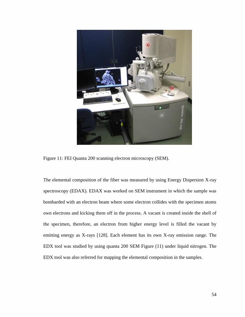
54
The elemental composition of the fiber was measured by using Energy Dispersion X-ray
spectroscopy (EDAX). EDAX was worked on SEM instrument in which the sample was
bombarded with an electron beam where some electron collides with the specimen atoms
own electrons and kicking them off in the process. A vacant is created inside the shell of
the specimen, therefore, an electron from higher energy level is filled the vacant by
emitting energy as X-rays [128]. Each element has its own X-ray emission range. The
EDX tool was studied by using quanta 200 SEM Figure (11) under liquid nitrogen. The
EDX tool was also referred for mapping the elemental composition in the samples.
Figure 11: FEI Quanta 200 scanning electron microscopy (SEM).
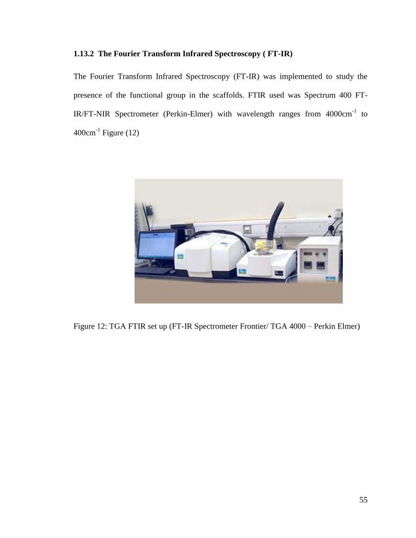
55
1.13.2 The Fourier Transform Infrared Spectroscopy ( FT-IR)
The Fourier Transform Infrared Spectroscopy (FT-IR) was implemented to study the
presence of the functional group in the scaffolds. FTIR used was Spectrum 400 FT-
IR/FT-NIR Spectrometer (Perkin-Elmer) with wavelength ranges from 4000cm-1
to
400cm-1
Figure (12)
Figure 12: TGA FTIR set up (FT-IR Spectrometer Frontier/ TGA 4000 – Perkin Elmer)
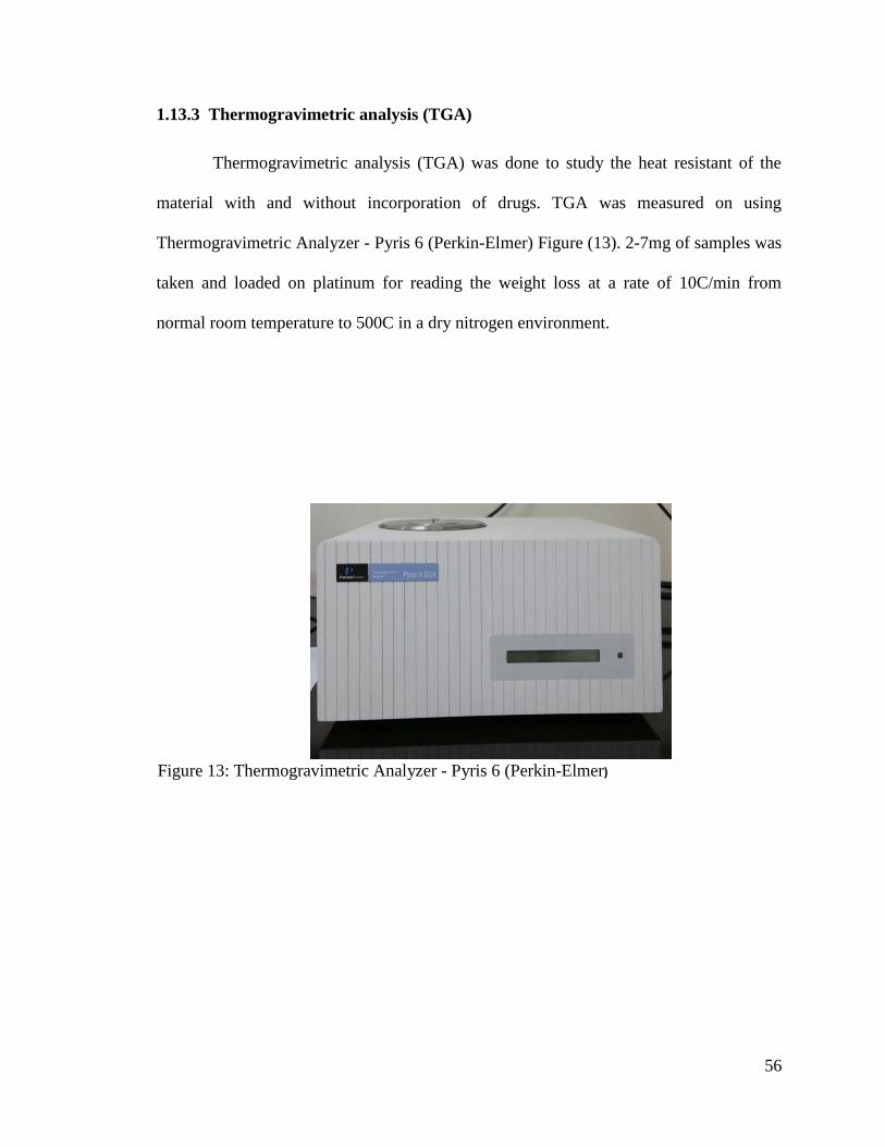
56
1.13.3 Thermogravimetric analysis (TGA)
Thermogravimetric analysis (TGA) was done to study the heat resistant of the
material with and without incorporation of drugs. TGA was measured on using
Thermogravimetric Analyzer - Pyris 6 (Perkin-Elmer) Figure (13). 2-7mg of samples was
taken and loaded on platinum for reading the weight loss at a rate of 10C/min from
normal room temperature to 500C in a dry nitrogen environment.
Figure 13: Thermogravimetric Analyzer - Pyris 6 (Perkin-Elmer)
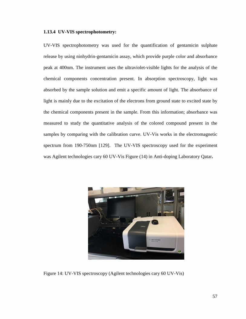
57
1.13.4 UV-VIS spectrophotometry:
UV-VIS spectrophotometry was used for the quantification of gentamicin sulphate
release by using ninhydrin-gentamicin assay, which provide purple color and absorbance
peak at 400nm. The instrument uses the ultraviolet-visible lights for the analysis of the
chemical components concentration present. In absorption spectroscopy, light was
absorbed by the sample solution and emit a specific amount of light. The absorbance of
light is mainly due to the excitation of the electrons from ground state to excited state by
the chemical components present in the sample. From this information; absorbance was
measured to study the quantitative analysis of the colored compound present in the
samples by comparing with the calibration curve. UV-Vis works in the electromagnetic
spectrum from 190-750nm [129]. The UV-VIS spectroscopy used for the experiment
was Agilent technologies cary 60 UV-Vis Figure (14) in Anti-doping Laboratory Qatar.
Figure 14: UV-VIS spectroscopy (Agilent technologies cary 60 UV-Vis)

58
1.13.5 Inductively coupled plasma mass spectrometry ICP-MS:
Inductively coupled plasma mass spectrometry (ICP-MS) is a metal and non-metals at a
concentration less than 1015
(part per quadrillion, ppq) on non-interfered low background
isotopes. This method mainly works by ionizing the sample via inductive couple plasma
and then the mass spectrometry categorizes ions based on their mass and quantify them.
The ICP-MS was used to quantify the presence of Ag release in deionized water. It was
done by using ICP-MS NexION 300D model Figure (15).
Figure 15: ICP-MS NexION 300D
59
1.14 In-vitro drug delivery of gentamicin Sulphate
1.14.1 Gentamicin-ninhydrin assay:
Gentamicin quantification was done by using ninhydrin (Biochemical Prod 440605V) as
a reaction compound [123]. The ninhydrin reacts with primary and secondary amines to
give a purple color. These color changes can be studied calorimetrically.
1.14.1.1 Preparation of Phosphate buffered solution (PBS):
One PBS tablets (MDL; MFCD00131855) was mixed with 200mL (as per the instruction
on the PBS tablets bottle) of distilled water (pH 7.4) at room temperature to obtain 0.01
M phosphate buffer, 0.0027 M potassium chloride and 0.137 M sodium chloride and pH
7.4.
1.14.1.2 Preparation of Ninhydrin reagent:
Ninhydrin reagent (1.25wt/v%) was prepared by dissolving 0.0622g of ninhydrin in 5mL
PBS solution (pH 7.4) to obtain concentration of 12.5mg/mL of ninhydrin solution. The
mixture was vortexed for 10min followed by sonication for 30min. The prepared
ninhydring should be consumed within 3 days if the solution was kept at room
temperature
1.14.1.3 Preparation of calibration curve:
The GS standard solution were prepared by serial dilution of 50mg/mL GS to obtain a
ranges of 0-80ug/mL of gentamicin solution according to the Table (3) and used for the
calibration curve Figure (16).
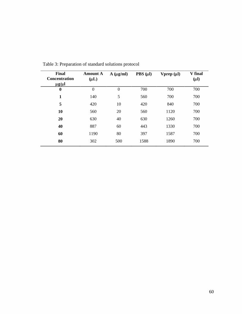
60
Table 3: Preparation of standard solutions protocol
Final
Concentration
g/l
Amount A
(L)
A (g/ml) PBS (l) Vprep (l) V final
(l)
0 0 0 700 700 700
1 140 5 560 700 700
5 420 10 420 840 700
10 560 20 560 1120 700
20 630 40 630 1260 700
40 887 60 443 1330 700
60 1190 80 397 1587 700
80 302 500 1588 1890 700
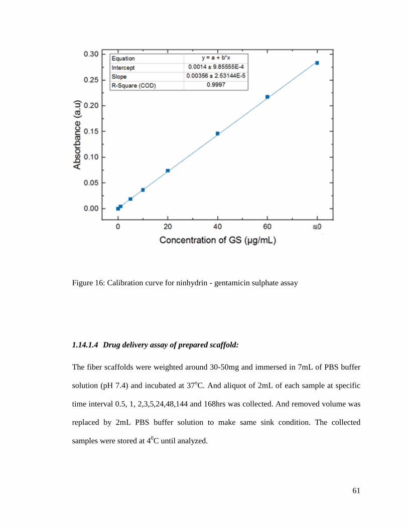
61
Figure 16: Calibration curve for ninhydrin - gentamicin sulphate assay
1.14.1.4 Drug delivery assay of prepared scaffold:
The fiber scaffolds were weighted around 30-50mg and immersed in 7mL of PBS buffer
solution (pH 7.4) and incubated at 37oC. And aliquot of 2mL of each sample at specific
time interval 0.5, 1, 2,3,5,24,48,144 and 168hrs was collected. And removed volume was
replaced by 2mL PBS buffer solution to make same sink condition. The collected
samples were stored at 40C until analyzed.
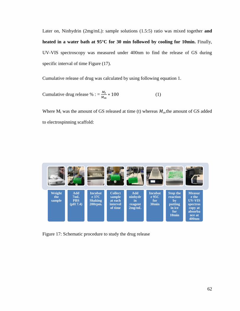
62
Later on, Ninhydrin (2mg/mL): sample solutions (1.5:5) ratio was mixed together and
heated in a water bath at 95°C for 30 min followed by cooling for 10min. Finally,
UV-VIS spectroscopy was measured under 400nm to find the release of GS during
specific interval of time Figure (17).
Cumulative release of drug was calculated by using following equation 1.
Cumulative drug release % : =
(1)
Where Mt was the amount of GS released at time (t) whereas the amount of GS added
to electrospinning scaffold:
Figure 17: Schematic procedure to study the drug release
Weight the
sample
Add 7mL PBS
(pH 7.4)
Incubate 37C
Shaking 200rpm.
Collect sample at each intervel of time
Add ninhydr
in reagent 2mg/mL
Incubate 95C
for 30min
Stop the reaction
by putting in ice for
10min
Measure the
UV-VIS spectroscopy at absorbance at 400nm
63
1.14.1.5 Encapsulation efficiency:
Firstly 5-8mg of electrospun scaffolds was mixed with 1 mL of chloroform and vortexed
for 5min until the PCL layer mix well. Henceforth, 1mL of PBS (pH 7.4) was added to
the solution and mix for 30min by shaking at 1000rpm. Hereafter, the solution mixture
was centrifuge at 6000rpm for 5min. Then, 800ul of supernatant was collected and
blended with 240uL of ninhydrin solution (2mg/mL) (1.5:5 rations). Then for the
complex formation of gentamicin-ninhydrin the solution was heat at 950C for 30min and
later on to stop the ninhydrin-gentamicin sulphate reaction the solution was cooled for
10min. Subsequently, UV-VIS absorbance was measurement at 400nm for the solution.
The gentamicin concentration in the scaffold was calculated from the calibration curve.
Encapsulation efficiency was calculated by using the following Equation (2).
(2)
1.15 In vitro release of Ag:
In vitro release of Ag was carried out as in Figure (18). 7-10mg of the PCL-PVA-
PCL/Ag fiber scaffold in a 15mL test tube and 5 mL of deionized water were added to
the test tube. Incubate the test-tube in water-bath at 37oC. At each interval of time, collect
1 ml of the deionized water in the test tube and refill with 1ml to establish same sink
conditions. Finally, to validate the amount in parts per billion (ppb) of silver present in
the solution was studied by using ICP-OES.
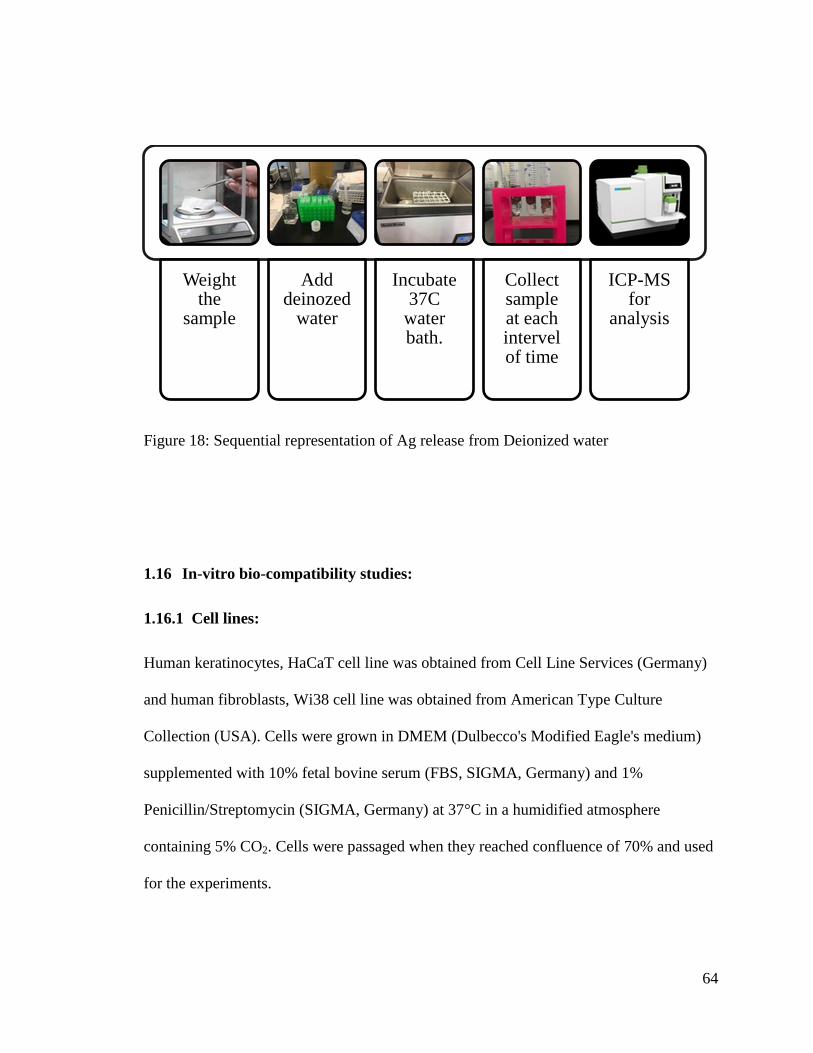
64
Figure 18: Sequential representation of Ag release from Deionized water
1.16 In-vitro bio-compatibility studies:
1.16.1 Cell lines:
Human keratinocytes, HaCaT cell line was obtained from Cell Line Services (Germany)
and human fibroblasts, Wi38 cell line was obtained from American Type Culture
Collection (USA). Cells were grown in DMEM (Dulbecco's Modified Eagle's medium)
supplemented with 10% fetal bovine serum (FBS, SIGMA, Germany) and 1%
Penicillin/Streptomycin (SIGMA, Germany) at 37°C in a humidified atmosphere
containing 5% CO2. Cells were passaged when they reached confluence of 70% and used
for the experiments.
Weight the
sample
Add deinozed
water
Incubate 37C
water bath.
Collect sample at each intervel of time
ICP-MS for
analysis
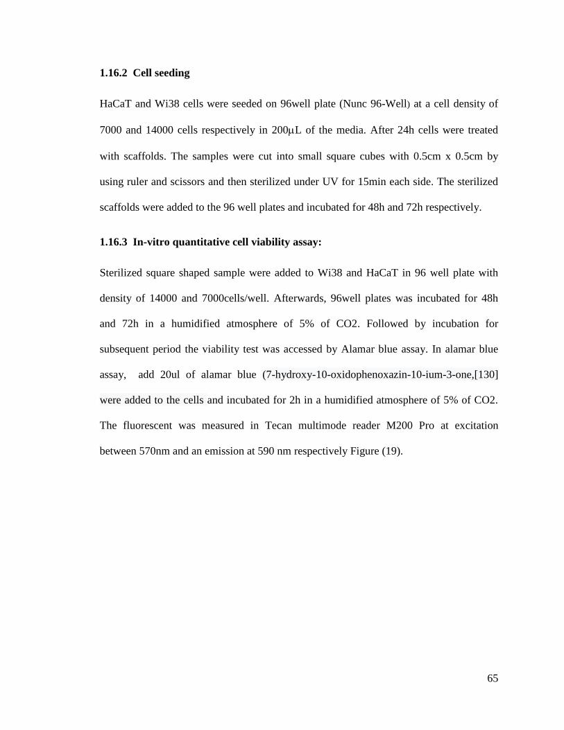
65
1.16.2 Cell seeding
HaCaT and Wi38 cells were seeded on 96well plate (Nunc 96-Well) at a cell density of
7000 and 14000 cells respectively in 200L of the media. After 24h cells were treated
with scaffolds. The samples were cut into small square cubes with 0.5cm x 0.5cm by
using ruler and scissors and then sterilized under UV for 15min each side. The sterilized
scaffolds were added to the 96 well plates and incubated for 48h and 72h respectively.
1.16.3 In-vitro quantitative cell viability assay:
Sterilized square shaped sample were added to Wi38 and HaCaT in 96 well plate with
density of 14000 and 7000cells/well. Afterwards, 96well plates was incubated for 48h
and 72h in a humidified atmosphere of 5% of CO2. Followed by incubation for
subsequent period the viability test was accessed by Alamar blue assay. In alamar blue
assay, add 20ul of alamar blue (7-hydroxy-10-oxidophenoxazin-10-ium-3-one,[130]
were added to the cells and incubated for 2h in a humidified atmosphere of 5% of CO2.
The fluorescent was measured in Tecan multimode reader M200 Pro at excitation
between 570nm and an emission at 590 nm respectively Figure (19).
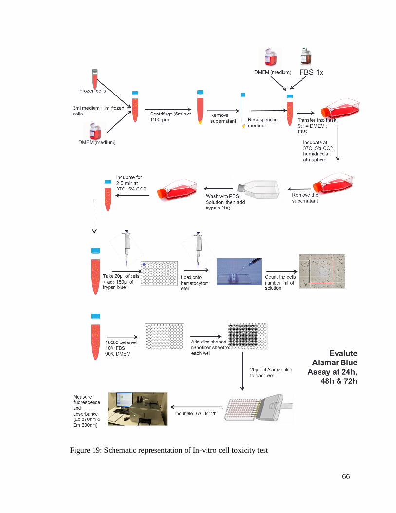
66
Figure 19: Schematic representation of In-vitro cell toxicity test
67
1.17 Antibacterial Studies:
1.17.1 Medium preparation :
Luria broth (LB) medium was prepared by taking 28g of nutrient agar (HiMedia
laboratories Pvt.Ltd. M001-500G) was mixed with 1L of distilled water. The prepared
mixture was autoclaved at 121oC for 1h. Followed by sterilization the LB agar medium
was pour into the pteridishes to cool down and solidify. All the above procedure was
done under laminar to avoid contamination.
Nutrient broth (NB) medium was processed by suspending 25g in 1000 ml
purified/distilled water. The prepared mixture was heated autoclaved at 121oC for
1h[131]. The NB medium was used for the preparation of bacterial suspension
incorporation with the samples.
Phosphate saline solution was prepared by mixing 0.91% NaCl in distilled water.
1.17.2 Antibacterial testing and spreading the plates.
The antibacterial testing was carried out for the PCL only, PCL fibers sputter coated with
Ag at 30s, 60s and 120s sputtering time, multilayered (ML) structure of PCL-PVA-PCL
electro spun fibers incorporated with gentamicin sulphate as well as sputter coated with
Ag. They are labeled as PCL only, PCL/Ag 30s, PCL/Ag 60s, PCL/Ag 120s, ML only,
ML/GS 1%, ML/GS4%, ML /Ag 30s and ML/GS/Ag respectively with 3 repetition to
evaluate standard error.
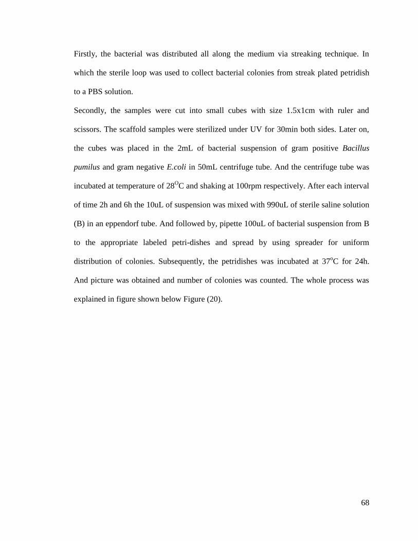
68
Firstly, the bacterial was distributed all along the medium via streaking technique. In
which the sterile loop was used to collect bacterial colonies from streak plated petridish
to a PBS solution.
Secondly, the samples were cut into small cubes with size 1.5x1cm with ruler and
scissors. The scaffold samples were sterilized under UV for 30min both sides. Later on,
the cubes was placed in the 2mL of bacterial suspension of gram positive Bacillus
pumilus and gram negative E.coli in 50mL centrifuge tube. And the centrifuge tube was
incubated at temperature of 28OC and shaking at 100rpm respectively. After each interval
of time 2h and 6h the 10uL of suspension was mixed with 990uL of sterile saline solution
(B) in an eppendorf tube. And followed by, pipette 100uL of bacterial suspension from B
to the appropriate labeled petri-dishes and spread by using spreader for uniform
distribution of colonies. Subsequently, the petridishes was incubated at 37oC for 24h.
And picture was obtained and number of colonies was counted. The whole process was
explained in figure shown below Figure (20).
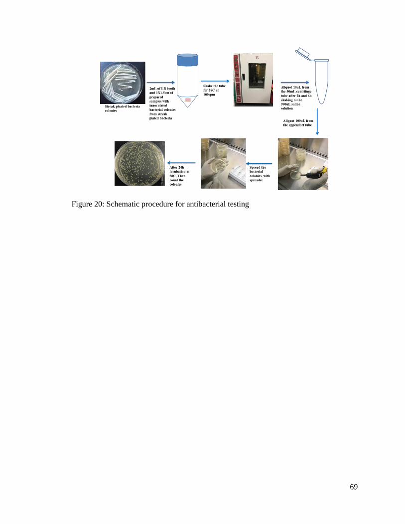
69
Figure 20: Schematic procedure for antibacterial testing
70
RESULTS AND DISCUSSIONS
1.18 Optimization of PVA and PCL individual fibers, and multilayered structure.
1.18.1 Poly (vinyl alcohol) PVA:
PVA 10wt%/v% solution was electrospun to prepare nanofibrous mats, where the
electrospinning parameters applied are voltage 15kV, flow rate of 1ml/h, and needle tip
to collector distance of 10 cm. Scanning electron microscope of the electrospun
nanofibers are given in the Figure (21). A beadles uniform fibers with an average
diameter of 107.25±21.07 nm is obtained.
Figure 21: a) SEM image of 10wt%/v% PVA (electrospinning parameters are applied
voltage 15kV, flow rate of 1ml/h, and needle tip to collector distance is 10 cm)
(magnification: 1m). b) Normal distribution of fiber diameter
71
1.18.2 Polycaprolactone (PCL):
PCL solution was prepared in 1:1 (wt/wt%) DMF:CF at different weight percentage of
8.5wt%, 10wt%, 11wt% and 12wt% respectively. The electrospinning parameters are
described in Table (4). SEM results show that, at 8.5wt% and 10wt% of PCL, it has
shown some beads on the scaffold structure. However, at 11wt% and 12wt% uniform
fiber with diameter around 407.04±112.1nm and 901.14±350nm respectively Figure (22)
The optimization of polycaprolactone was done by using different solvents at various
weight percentages to obtain uniform fibers with smaller diameters Table (4). PCL is a
hydrophobic polymer which is easily soluble in chloroform. However, chloroform has
lower electric conductivity [42]. As a result, insulating polymer solution could not beat
the surface tension in high electrostatic force. Therefore, introduction of conductive
polymer solution is essential for improved electrospinning process. So, DMF show
moderate conductivity due to its dielectric constant [132].
There is critical minimum concentration (Ce) required for the formation of beaded fibers.
The concentration above Ce shows a reduction in the beads as well as increase in the
fiber diameter and uniformity. Concentrations lower than Ce shows drops like
electrospraying. The value of Ce depends on polymer molecular weight, solution and
chemical properties of the polymer. Polymer solvent plays an important role in selecting
proper Ce and also morphology of the fiber. Hence, selection of solvent is an important
factor for better fiber production in electrospinning process. Therefore, the critical
concentration for 1:1 wt%/wt% of DMF: CF is 10% of PCL Figure (22). 11wt% of PCL
was used for the further production of electrospun fibers due to more uniform beadles
fiber with average diameter of 407.04±112nm Figure (22e)
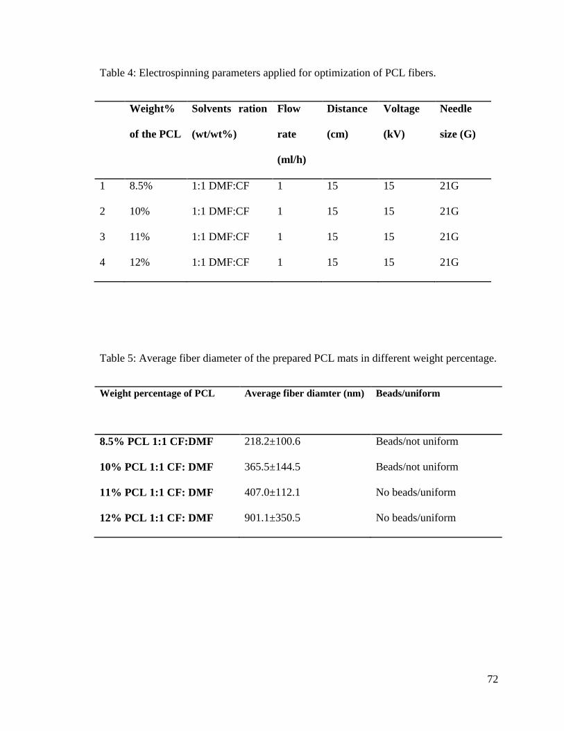
72
Table 4: Electrospinning parameters applied for optimization of PCL fibers.
Weight%
of the PCL
Solvents ration
(wt/wt%)
Flow
rate
(ml/h)
Distance
(cm)
Voltage
(kV)
Needle
size (G)
1 8.5% 1:1 DMF:CF 1 15 15 21G
2 10% 1:1 DMF:CF 1 15 15 21G
3 11% 1:1 DMF:CF 1 15 15 21G
4 12% 1:1 DMF:CF 1 15 15 21G
Table 5: Average fiber diameter of the prepared PCL mats in different weight percentage.
Weight percentage of PCL Average fiber diamter (nm) Beads/uniform
8.5% PCL 1:1 CF:DMF 218.2±100.6 Beads/not uniform
10% PCL 1:1 CF: DMF 365.5±144.5 Beads/not uniform
11% PCL 1:1 CF: DMF 407.0±112.1 No beads/uniform
12% PCL 1:1 CF: DMF 901.1±350.5 No beads/uniform
73
Figure 22: SEM images of electrospun PCL in 1:1 (wt/wt%) DMF:CF at different weight
percentage of PCL a) 8.5wt%PCL, b)10wt%PCL, c) 11wt%PCL, d) 12wt%PCL e)
Average fiber diameter and f) Fiber diameter distribution of 11wt%PCL (magnification
10m).
1.19 PCL coated with Ag
The individual PCL fibers was sputter coated with silver at different sputtering time 30s,
60s and 120s with base pressure of 1x10⁻³
pa, working pressure 0.7Pa, voltage 340-350 V
and the current was 0.09A. The prepared electrospun sputter coated fibers are labeled as
PCL only, PCL/Ag 30s , PCL/ Ag 60s and PCL/ Ag 120s.
The morphology of the fibers was characterized by SEM, elemental analysis by EDX and
XRD. Moreover, cytotoxicity and antibacterial properties was studied for the optimized
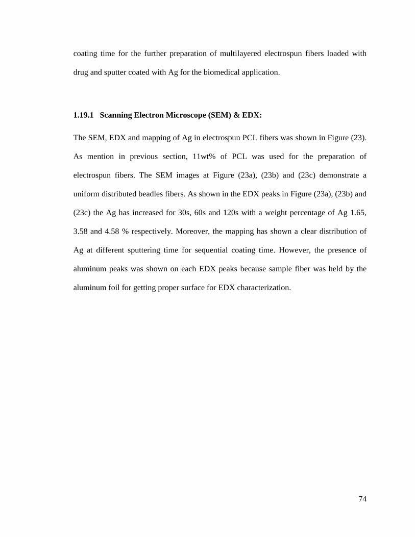
74
coating time for the further preparation of multilayered electrospun fibers loaded with
drug and sputter coated with Ag for the biomedical application.
1.19.1 Scanning Electron Microscope (SEM) & EDX:
The SEM, EDX and mapping of Ag in electrospun PCL fibers was shown in Figure (23).
As mention in previous section, 11wt% of PCL was used for the preparation of
electrospun fibers. The SEM images at Figure (23a), (23b) and (23c) demonstrate a
uniform distributed beadles fibers. As shown in the EDX peaks in Figure (23a), (23b) and
(23c) the Ag has increased for 30s, 60s and 120s with a weight percentage of Ag 1.65,
3.58 and 4.58 % respectively. Moreover, the mapping has shown a clear distribution of
Ag at different sputtering time for sequential coating time. However, the presence of
aluminum peaks was shown on each EDX peaks because sample fiber was held by the
aluminum foil for getting proper surface for EDX characterization.
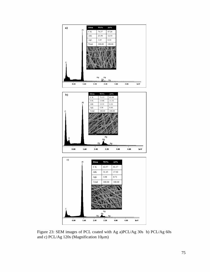
75
Figure 23: SEM images of PCL coated with Ag a)PCL/Ag 30s b) PCL/Ag 60s
and c) PCL/Ag 120s (Magnification 10m)
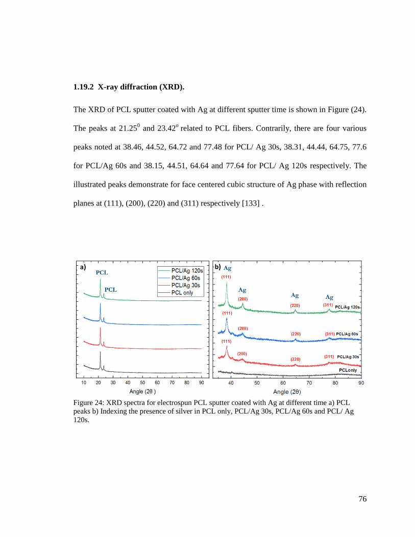
76
1.19.2 X-ray diffraction (XRD).
The XRD of PCL sputter coated with Ag at different sputter time is shown in Figure (24).
The peaks at 21.250 and 23.42
o related to PCL fibers. Contrarily, there are four various
peaks noted at 38.46, 44.52, 64.72 and 77.48 for PCL/ Ag 30s, 38.31, 44.44, 64.75, 77.6
for PCL/Ag 60s and 38.15, 44.51, 64.64 and 77.64 for PCL/ Ag 120s respectively. The
illustrated peaks demonstrate for face centered cubic structure of Ag phase with reflection
planes at (111), (200), (220) and (311) respectively [133] .
Figure 24: XRD spectra for electrospun PCL sputter coated with Ag at different time a) PCL
peaks b) Indexing the presence of silver in PCL only, PCL/Ag 30s, PCL/Ag 60s and PCL/ Ag
120s.
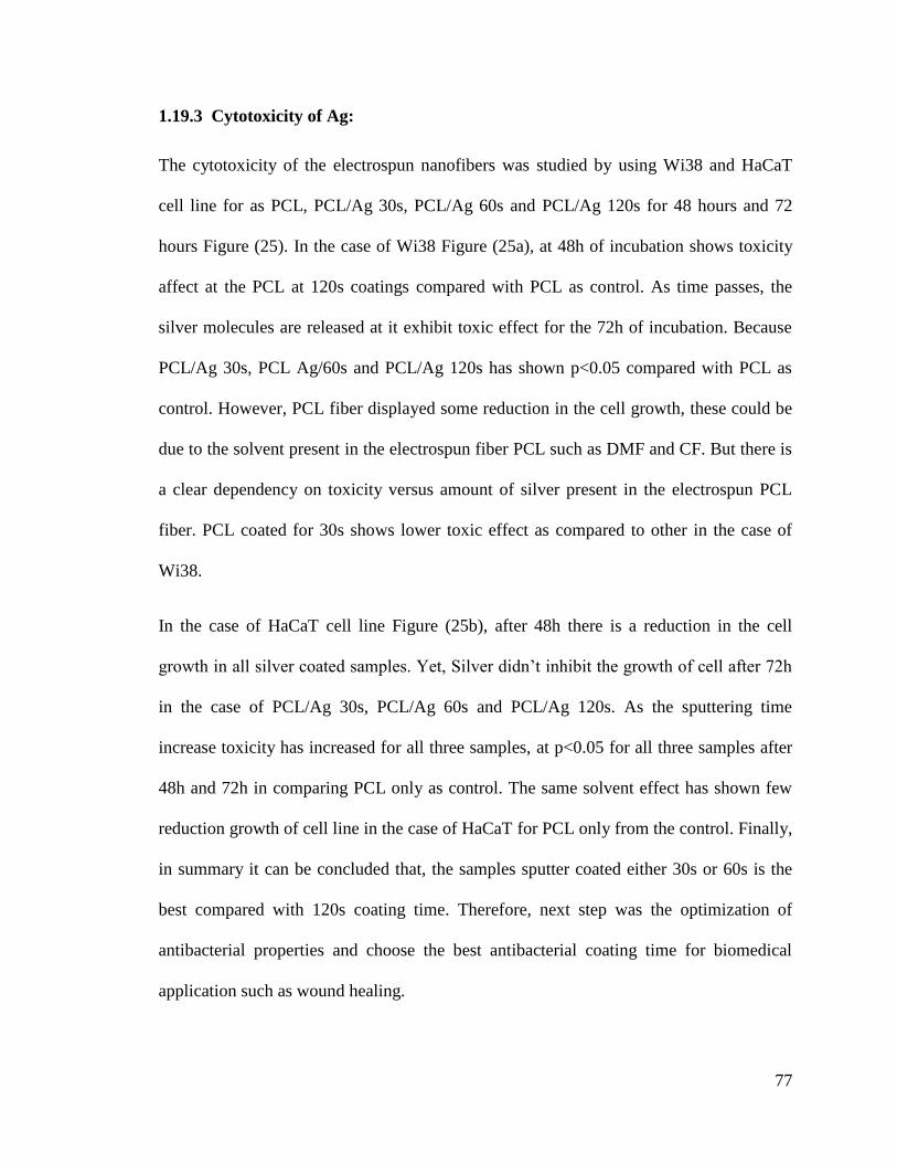
77
1.19.3 Cytotoxicity of Ag:
The cytotoxicity of the electrospun nanofibers was studied by using Wi38 and HaCaT
cell line for as PCL, PCL/Ag 30s, PCL/Ag 60s and PCL/Ag 120s for 48 hours and 72
hours Figure (25). In the case of Wi38 Figure (25a), at 48h of incubation shows toxicity
affect at the PCL at 120s coatings compared with PCL as control. As time passes, the
silver molecules are released at it exhibit toxic effect for the 72h of incubation. Because
PCL/Ag 30s, PCL Ag/60s and PCL/Ag 120s has shown p<0.05 compared with PCL as
control. However, PCL fiber displayed some reduction in the cell growth, these could be
due to the solvent present in the electrospun fiber PCL such as DMF and CF. But there is
a clear dependency on toxicity versus amount of silver present in the electrospun PCL
fiber. PCL coated for 30s shows lower toxic effect as compared to other in the case of
Wi38.
In the case of HaCaT cell line Figure (25b), after 48h there is a reduction in the cell
growth in all silver coated samples. Yet, Silver didn‘t inhibit the growth of cell after 72h
in the case of PCL/Ag 30s, PCL/Ag 60s and PCL/Ag 120s. As the sputtering time
increase toxicity has increased for all three samples, at p<0.05 for all three samples after
48h and 72h in comparing PCL only as control. The same solvent effect has shown few
reduction growth of cell line in the case of HaCaT for PCL only from the control. Finally,
in summary it can be concluded that, the samples sputter coated either 30s or 60s is the
best compared with 120s coating time. Therefore, next step was the optimization of
antibacterial properties and choose the best antibacterial coating time for biomedical
application such as wound healing.
78
Figure 25: : In vitro cell viability test of a)Wi38 b) HaCaT cell line PCL, PCL/Ag 30s,
PCL/Ag 60s and PCL/Ag 120s (n=5). ***p<0.05.
1.19.4 Antibacterial study:
The antibacterial properties of the PCL only, PCL/Ag 30s, PCL/Ag 60s and PCL/Ag
120s were carried out with two kinds of microorganism: Bacillus pumilus (Gram
positive). The release of Ag from the sample with respect to time of incubation for 2h and
6h was studied. The results were summarized in Figure (26) and Figure (27). In Figure
(26) it clearly demonstrate that, the presence of silver has reduced the growth of bacterial
colonies. The Figure (26) was plotted graphically in Figure (27). The PCL fiber itself has
never shown any antibacterial properties. However, in the case of PCL/Ag 30s there was
not any antibacterial character during first 2h in gram positive, but has improved the
79
antibacterial activity after 6h of incubation by 96% from the control for gram positive.
The PCL/Ag 60s has exhibited antibacterial inhibition from 1.85% to 95% for Bacillus
pumilus for 2h and 6h incubation respectively. Moreover, PCL/Ag 120s displayed
antibacterial inhibition from 48% to 92.45% for 2h and 6h incubation respectively. From
these results we can conclude that, for gram positive bacteria as the time passes the
release of Ag inhibit the growth. In addition, the sputtering time has not shown much
change in the antibacterial properties. Only difference is in the release of silver to the
media. Statistically, after 6h of incubation has shown much more variation in
antibacterial inhibition compared with PCL as control (p<0.05).
The antibacterial properties of silver are by various methods [134], [135]. For instance,
the physical contact of silver particles to the cellular membrane of the bacteria could leafs
to cellular damage, silver ion interact with cellular activity and DNA replication and
Inhibit the growth of bacterial cells [136]. These results depict that the release of silver
coated on the surface of electrospun fiber mats has exhibited antibacterial inhibition with
time. Therefore, this type of coating could provide a better antibacterial inhibition for
long term for biomedical application such as wound healing process. Hence, PCL sputter
coated with Ag for 30s and 60s was more precise in antibacterial properties and cell
cytotoxicity. Therefore, multilayered structure was sputter coated for 30s for better
antibacterial and reduced cell cytotoxicity.
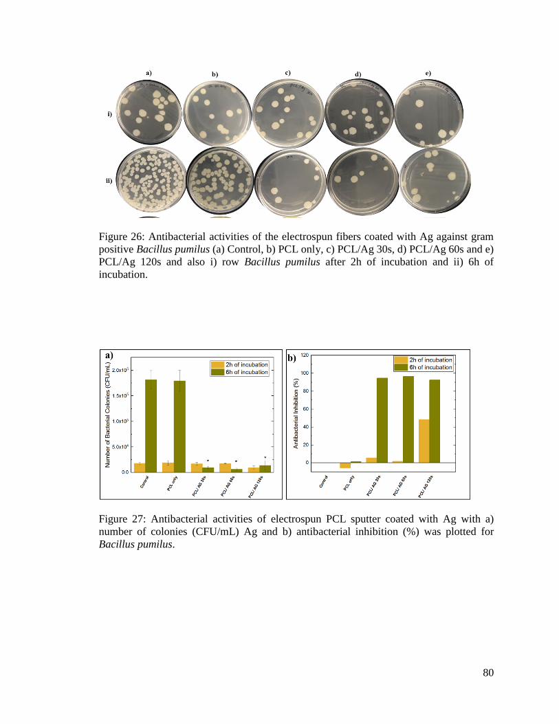
80
Figure 26: Antibacterial activities of the electrospun fibers coated with Ag against gram
positive Bacillus pumilus (a) Control, b) PCL only, c) PCL/Ag 30s, d) PCL/Ag 60s and e)
PCL/Ag 120s and also i) row Bacillus pumilus after 2h of incubation and ii) 6h of
incubation.
Figure 27: Antibacterial activities of electrospun PCL sputter coated with Ag with a)
number of colonies (CFU/mL) Ag and b) antibacterial inhibition (%) was plotted for
Bacillus pumilus.
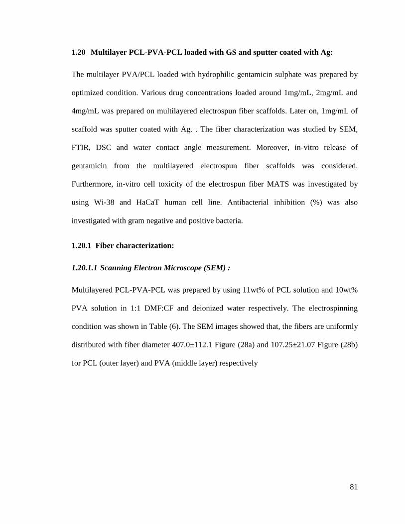
81
1.20 Multilayer PCL-PVA-PCL loaded with GS and sputter coated with Ag:
The multilayer PVA/PCL loaded with hydrophilic gentamicin sulphate was prepared by
optimized condition. Various drug concentrations loaded around 1mg/mL, 2mg/mL and
4mg/mL was prepared on multilayered electrospun fiber scaffolds. Later on, 1mg/mL of
scaffold was sputter coated with Ag. . The fiber characterization was studied by SEM,
FTIR, DSC and water contact angle measurement. Moreover, in-vitro release of
gentamicin from the multilayered electrospun fiber scaffolds was considered.
Furthermore, in-vitro cell toxicity of the electrospun fiber MATS was investigated by
using Wi-38 and HaCaT human cell line. Antibacterial inhibition (%) was also
investigated with gram negative and positive bacteria.
1.20.1 Fiber characterization:
1.20.1.1 Scanning Electron Microscope (SEM) :
Multilayered PCL-PVA-PCL was prepared by using 11wt% of PCL solution and 10wt%
PVA solution in 1:1 DMF:CF and deionized water respectively. The electrospinning
condition was shown in Table (6). The SEM images showed that, the fibers are uniformly
distributed with fiber diameter 407.0±112.1 Figure (28a) and 107.25±21.07 Figure (28b)
for PCL (outer layer) and PVA (middle layer) respectively
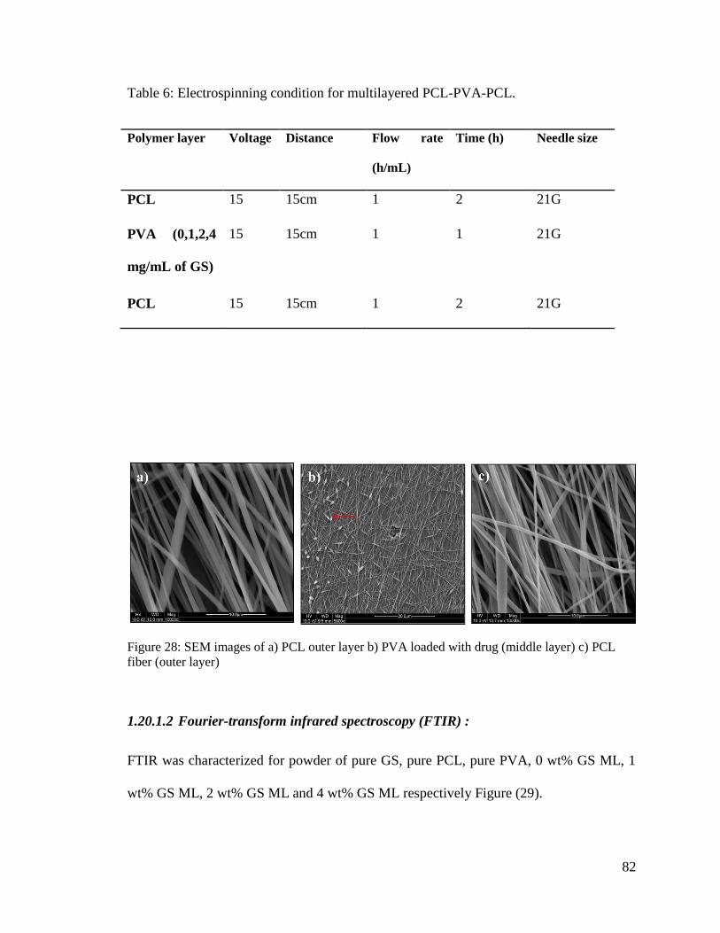
82
Table 6: Electrospinning condition for multilayered PCL-PVA-PCL.
Polymer layer Voltage Distance Flow rate
(h/mL)
Time (h) Needle size
PCL 15 15cm 1 2 21G
PVA (0,1,2,4
mg/mL of GS)
15 15cm 1 1 21G
PCL 15 15cm 1 2 21G
Figure 28: SEM images of a) PCL outer layer b) PVA loaded with drug (middle layer) c) PCL
fiber (outer layer)
1.20.1.2 Fourier-transform infrared spectroscopy (FTIR) :
FTIR was characterized for powder of pure GS, pure PCL, pure PVA, 0 wt% GS ML, 1
wt% GS ML, 2 wt% GS ML and 4 wt% GS ML respectively Figure (29).
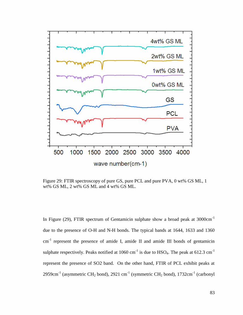
83
Figure 29: FTIR spectroscopy of pure GS, pure PCL and pure PVA, 0 wt% GS ML, 1
wt% GS ML, 2 wt% GS ML and 4 wt% GS ML.
In Figure (29), FTIR spectrum of Gentamicin sulphate show a broad peak at 3000cm-1
due to the presence of O-H and N-H bonds. The typical bands at 1644, 1633 and 1360
cm-1
represent the presence of amide I, amide II and amide III bonds of gentamicin
sulphate respectively. Peaks notified at 1060 cm-1
is due to HSO4. The peak at 612.3 cm-1
represent the presence of SO2 band. On the other hand, FTIR of PCL exhibit peaks at
2959cm-1
(asymmetric CH2 bond), 2921 cm-1
(symmetric CH2 bond), 1732cm-1
(carbonyl
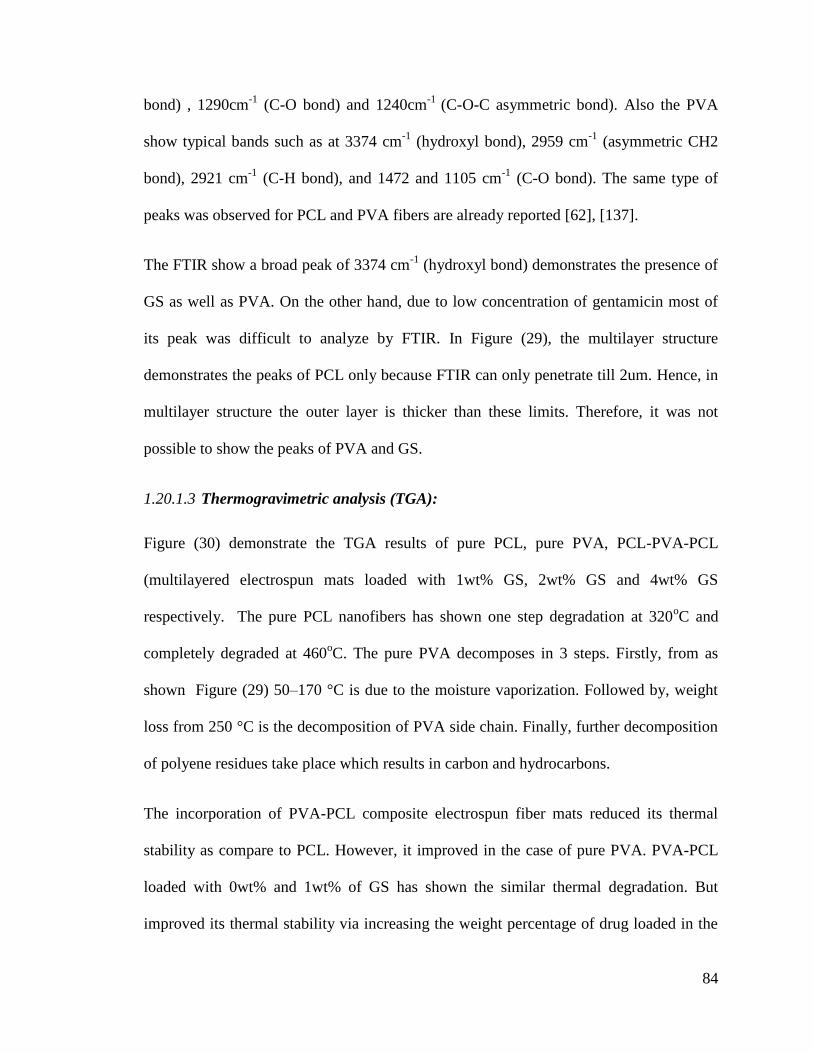
84
bond) , 1290cm-1
(C-O bond) and 1240cm-1
(C-O-C asymmetric bond). Also the PVA
show typical bands such as at 3374 cm-1
(hydroxyl bond), 2959 cm-1
(asymmetric CH2
bond), 2921 cm-1
(C-H bond), and 1472 and 1105 cm-1
(C-O bond). The same type of
peaks was observed for PCL and PVA fibers are already reported [62], [137].
The FTIR show a broad peak of 3374 cm-1
(hydroxyl bond) demonstrates the presence of
GS as well as PVA. On the other hand, due to low concentration of gentamicin most of
its peak was difficult to analyze by FTIR. In Figure (29), the multilayer structure
demonstrates the peaks of PCL only because FTIR can only penetrate till 2um. Hence, in
multilayer structure the outer layer is thicker than these limits. Therefore, it was not
possible to show the peaks of PVA and GS.
1.20.1.3 Thermogravimetric analysis (TGA):
Figure (30) demonstrate the TGA results of pure PCL, pure PVA, PCL-PVA-PCL
(multilayered electrospun mats loaded with 1wt% GS, 2wt% GS and 4wt% GS
respectively. The pure PCL nanofibers has shown one step degradation at 320oC and
completely degraded at 460oC. The pure PVA decomposes in 3 steps. Firstly, from as
shown Figure (29) 50–170 °C is due to the moisture vaporization. Followed by, weight
loss from 250 °C is the decomposition of PVA side chain. Finally, further decomposition
of polyene residues take place which results in carbon and hydrocarbons.
The incorporation of PVA-PCL composite electrospun fiber mats reduced its thermal
stability as compare to PCL. However, it improved in the case of pure PVA. PVA-PCL
loaded with 0wt% and 1wt% of GS has shown the similar thermal degradation. But
improved its thermal stability via increasing the weight percentage of drug loaded in the
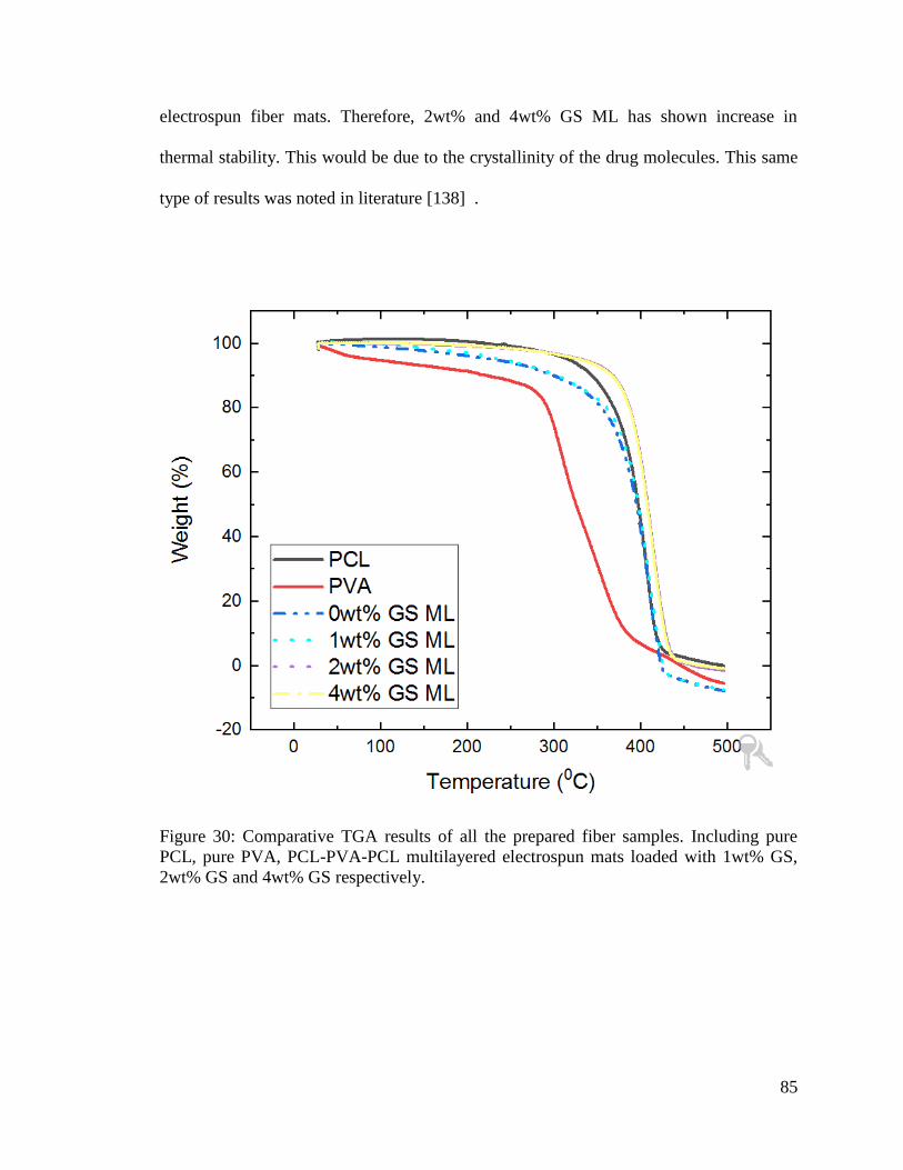
85
electrospun fiber mats. Therefore, 2wt% and 4wt% GS ML has shown increase in
thermal stability. This would be due to the crystallinity of the drug molecules. This same
type of results was noted in literature [138] .
Figure 30: Comparative TGA results of all the prepared fiber samples. Including pure
PCL, pure PVA, PCL-PVA-PCL multilayered electrospun mats loaded with 1wt% GS,
2wt% GS and 4wt% GS respectively.
86
1.20.2 Encapsulation efficiency of the GS in the fiber scaffolds:
The encapsulation efficiency of the Gentamicin sulphate in the fiber scaffolds was
calculated Figure (31) and results show that, multilayered scaffolds of 1wt%, 2wt% and
4wt% of gentamicin sulphate have encapsulation efficiency of 54.8±22.9%, 54.4±13.46%
and 64.39±13.99% respectively. This is due to the fact that, PVA loaded with GS is
electrospun separately in multilayered therefore, there is chance for GS accumulation in
the syringe and reduction in the encapsulation efficiency. On the other hand, as the drug
concentration increases in the multilayered structure its encapsulation efficiency is
improved in the case of 4wt% of GS in multilayered structure.
Figure 31: Encapsulation efficiency of the GS in the fiber scaffolds
87
1.20.3 In-vitro release of drug.
The in vitro release of GS from the electrospun fiber mats was studied using ninhydrin-
gentamicin assay. The set of standards was prepared from gentamicin sulphate with
ranging concentrations of 0 to 80ug/mL according to the Table (3). The calibration curve
is shown in Figure (16) with regression 0.9997. The calibration curve was used to
analyses the concentration of gentamicin sulphate release from the fiber scaffold into the
PBS medium at each interval of time.
After plotting the calibration curve the drug release of the scaffold was studied for
multilayered structure for 7 days. The cumulative drug release per mg of scaffold was
investigated with taking the percentage of drug present in solution at particular interval of
time with total encapsulated drug from encapsulation efficiency. The results are shown in
Figure (32). Multilayered has shown initial burst release over 5h was around 65%. After
initial burst release its shown slower release of drug over 7 days where only 80% of
loaded drug was released. In the case of multilayered structure, there are many factors
contribute for the sustained release of drug, they are thickness of the fiber, amount of
drug loaded, fiber diameters and porosity [27][139]. The initial burst release suggests that
a good amount of GS was released during first 5h. Succeeding initial burst, the outer
layer of multilayer scaffold will swells and stabilize and trap the drug from further
release. These phenomena makes the continuous and sustained release of GS in ML for
over 7 days where only 79% was released [27].
The different weight percentage of GS was loaded into the fiber scaffolds and studied
release kinetics. The results showed that, 1wt% GS ML has showed 65% of intial relase
of drug from the scaffold. Also, in the 2wt% GS ML has only 53% of drug initial burst
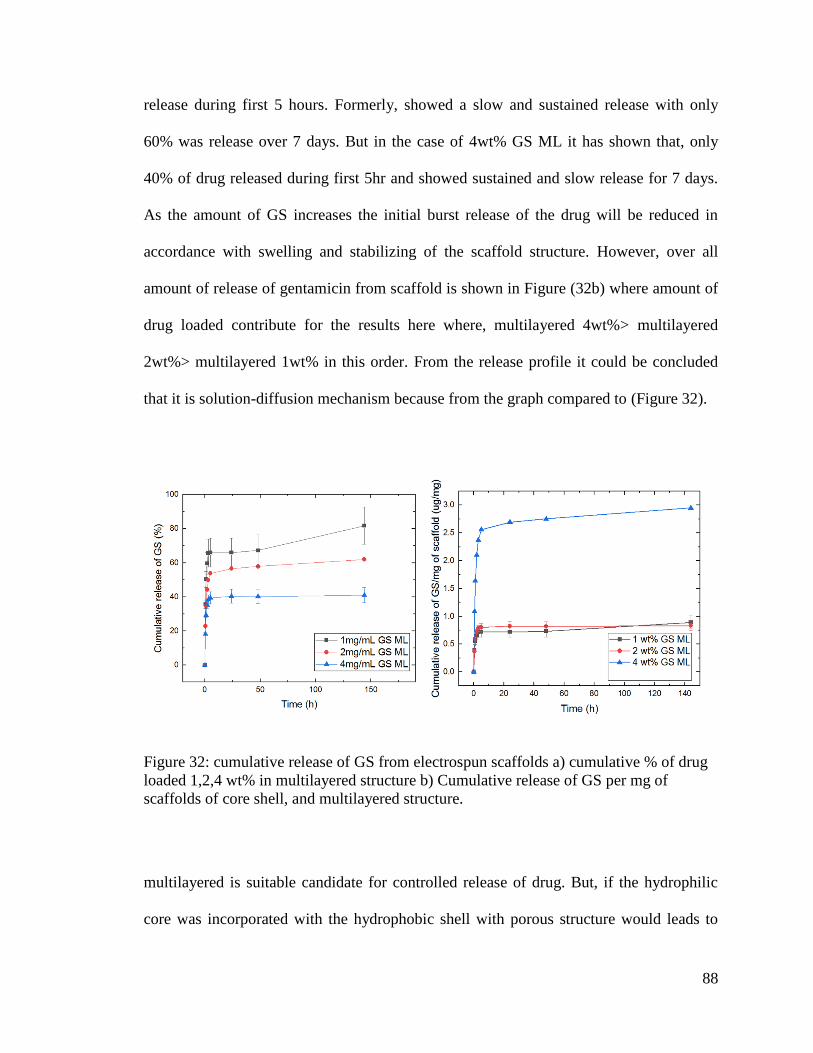
88
release during first 5 hours. Formerly, showed a slow and sustained release with only
60% was release over 7 days. But in the case of 4wt% GS ML it has shown that, only
40% of drug released during first 5hr and showed sustained and slow release for 7 days.
As the amount of GS increases the initial burst release of the drug will be reduced in
accordance with swelling and stabilizing of the scaffold structure. However, over all
amount of release of gentamicin from scaffold is shown in Figure (32b) where amount of
drug loaded contribute for the results here where, multilayered 4wt%> multilayered
2wt%> multilayered 1wt% in this order. From the release profile it could be concluded
that it is solution-diffusion mechanism because from the graph compared to (Figure 32).
Figure 32: cumulative release of GS from electrospun scaffolds a) cumulative % of drug
loaded 1,2,4 wt% in multilayered structure b) Cumulative release of GS per mg of
scaffolds of core shell, and multilayered structure.
multilayered is suitable candidate for controlled release of drug. But, if the hydrophilic
core was incorporated with the hydrophobic shell with porous structure would leads to
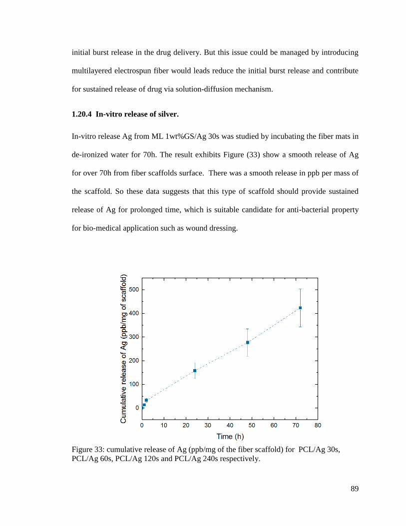
89
initial burst release in the drug delivery. But this issue could be managed by introducing
multilayered electrospun fiber would leads reduce the initial burst release and contribute
for sustained release of drug via solution-diffusion mechanism.
1.20.4 In-vitro release of silver.
In-vitro release Ag from ML 1wt%GS/Ag 30s was studied by incubating the fiber mats in
de-ironized water for 70h. The result exhibits Figure (33) show a smooth release of Ag
for over 70h from fiber scaffolds surface. There was a smooth release in ppb per mass of
the scaffold. So these data suggests that this type of scaffold should provide sustained
release of Ag for prolonged time, which is suitable candidate for anti-bacterial property
for bio-medical application such as wound dressing.
Figure 33: cumulative release of Ag (ppb/mg of the fiber scaffold) for PCL/Ag 30s,
PCL/Ag 60s, PCL/Ag 120s and PCL/Ag 240s respectively.
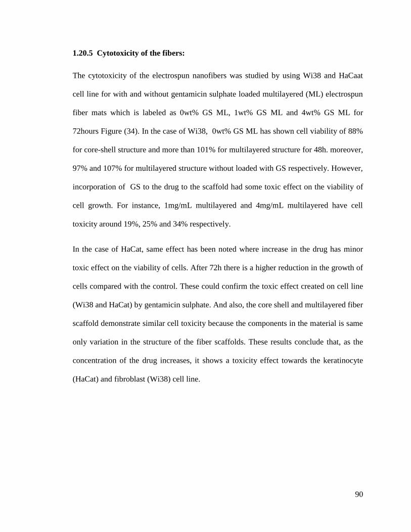
90
1.20.5 Cytotoxicity of the fibers:
The cytotoxicity of the electrospun nanofibers was studied by using Wi38 and HaCaat
cell line for with and without gentamicin sulphate loaded multilayered (ML) electrospun
fiber mats which is labeled as 0wt% GS ML, 1wt% GS ML and 4wt% GS ML for
72hours Figure (34). In the case of Wi38, 0wt% GS ML has shown cell viability of 88%
for core-shell structure and more than 101% for multilayered structure for 48h. moreover,
97% and 107% for multilayered structure without loaded with GS respectively. However,
incorporation of GS to the drug to the scaffold had some toxic effect on the viability of
cell growth. For instance, 1mg/mL multilayered and 4mg/mL multilayered have cell
toxicity around 19%, 25% and 34% respectively.
In the case of HaCat, same effect has been noted where increase in the drug has minor
toxic effect on the viability of cells. After 72h there is a higher reduction in the growth of
cells compared with the control. These could confirm the toxic effect created on cell line
(Wi38 and HaCat) by gentamicin sulphate. And also, the core shell and multilayered fiber
scaffold demonstrate similar cell toxicity because the components in the material is same
only variation in the structure of the fiber scaffolds. These results conclude that, as the
concentration of the drug increases, it shows a toxicity effect towards the keratinocyte
(HaCat) and fibroblast (Wi38) cell line.
91
Figure 34: In vitro cell viability test of a)Wi38 b) HaCat cell line in pristine PVA/PCL core
shell without drug and 1mg/mL of GS, multilayered fiber scaffold without and with GS
(1mg/mL and 4mg/mL) (n=5). ***p<0.05.
1.20.6 Antibacterial Study:
The antibacterial properties of the electrospun multilayered fibers loaded with gentamicin
sulphate and silver was carried out two kinds of microorganism: Bacillus pumilus (Gram
positive) and Escherichia coli (Gram negative). They are labeled as ML 0% GS, ML 1%
GS, ML 4% GS, ML Ag and ML 1%GS/Ag. The results were summarized in Figure (35)
and Figure (36) for gram positive Bacillus pumilus and Figure (37) and Figure (38) for
gram negative E.coli. The presence of gentamicin sulphate in the middle layer of
multilayered electrospun fibers make it controlled release and sustain the antibacterial
activity. Gentamicin is a bacteriocidal antibiotics which intrude the protein synthesis by
binding irreversibly with the 30s subunits of ribosomes. These process is alike with other
aminoglycosides. Gentamicin are active against many bacterial infection caused mainly
by gram negative such as Pseudomonas, Proteus, Escherichia coli, Klebsiella
pneumoniae, Enterobacter aerogenes, Serratia, and the Gram-positive Staphylococcus.
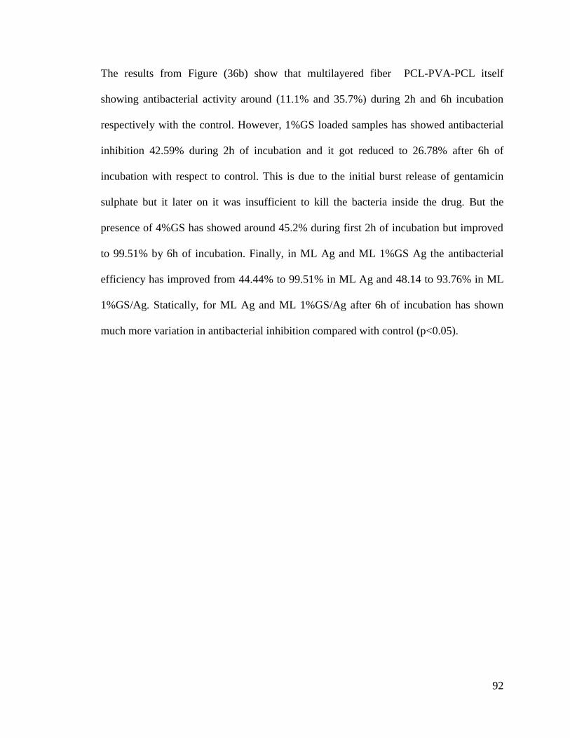
92
The results from Figure (36b) show that multilayered fiber PCL-PVA-PCL itself
showing antibacterial activity around (11.1% and 35.7%) during 2h and 6h incubation
respectively with the control. However, 1%GS loaded samples has showed antibacterial
inhibition 42.59% during 2h of incubation and it got reduced to 26.78% after 6h of
incubation with respect to control. This is due to the initial burst release of gentamicin
sulphate but it later on it was insufficient to kill the bacteria inside the drug. But the
presence of 4%GS has showed around 45.2% during first 2h of incubation but improved
to 99.51% by 6h of incubation. Finally, in ML Ag and ML 1%GS Ag the antibacterial
efficiency has improved from 44.44% to 99.51% in ML Ag and 48.14 to 93.76% in ML
1%GS/Ag. Statically, for ML Ag and ML 1%GS/Ag after 6h of incubation has shown
much more variation in antibacterial inhibition compared with control (p<0.05).
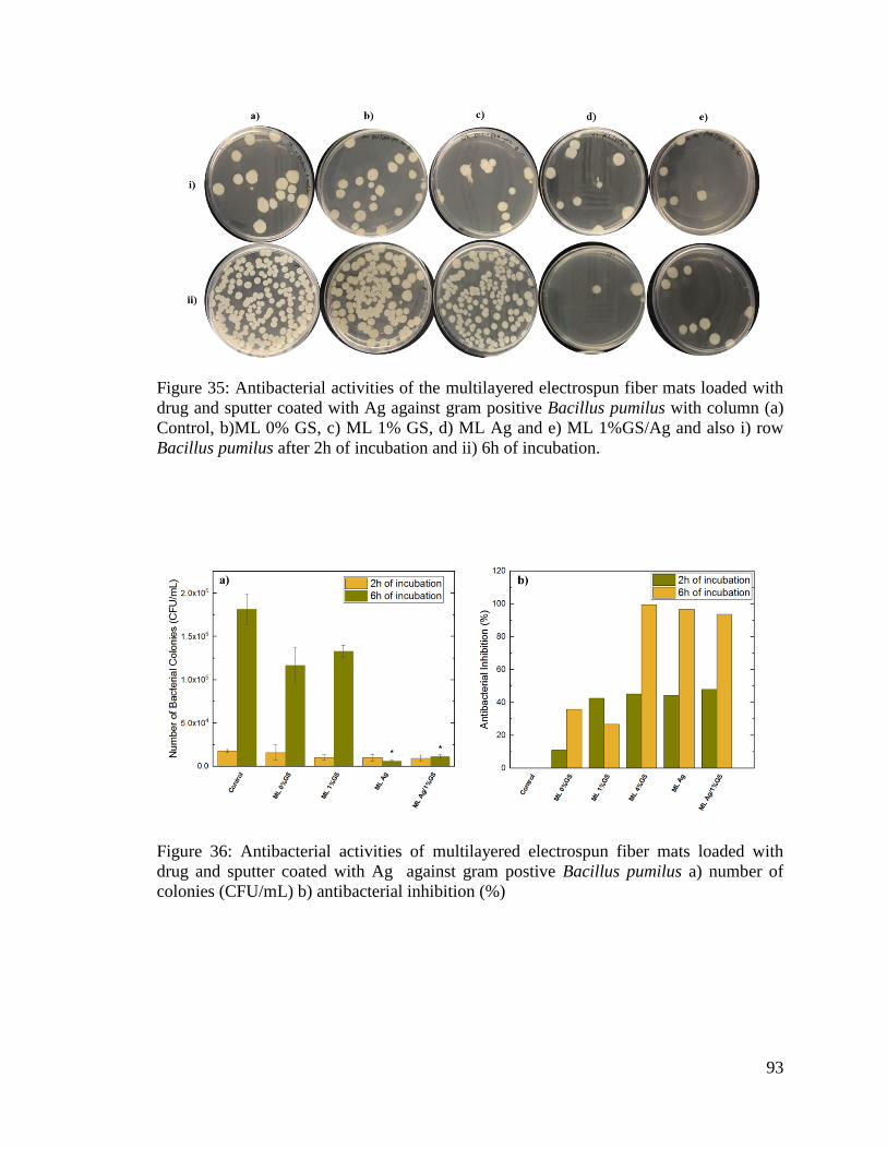
93
Figure 35: Antibacterial activities of the multilayered electrospun fiber mats loaded with
drug and sputter coated with Ag against gram positive Bacillus pumilus with column (a)
Control, b)ML 0% GS, c) ML 1% GS, d) ML Ag and e) ML 1%GS/Ag and also i) row
Bacillus pumilus after 2h of incubation and ii) 6h of incubation.
Figure 36: Antibacterial activities of multilayered electrospun fiber mats loaded with
drug and sputter coated with Ag against gram postive Bacillus pumilus a) number of
colonies (CFU/mL) b) antibacterial inhibition (%)

94
On the other hand in the case Figure (37) &Figure (38) gram negative bacteria (E.coli),
ML 0%GS (11.7%), ML 1%GS (33.85%) , ML 4%GS (54.44%), ML Ag (63.47%) and
ML 1%GS/Ag (53.71%) has shown antibacterial inhibition (%) for first 2h of incubation.
However, after 6h of incubation the ML 0%GS (34.97%), ML 1%GS (30.98%) , ML
4%GS (90.33%), ML Ag (82.49%) and ML 1%GS/Ag (71.39%) antibacterial inhibition
has been observed.
These results recommend that, multilayered structure loaded with gentamicin sulphate
sputter coated with Ag has improved the antibacterial properties of the material.
Moreover, the samples antibacterial properties could be improved by addition of
antibacterial drug into the samples. And it can give long antibacterial by reducing the
initial burst release of the drug as well as consistent release of Ag from the surface of the
electrospun fiber.
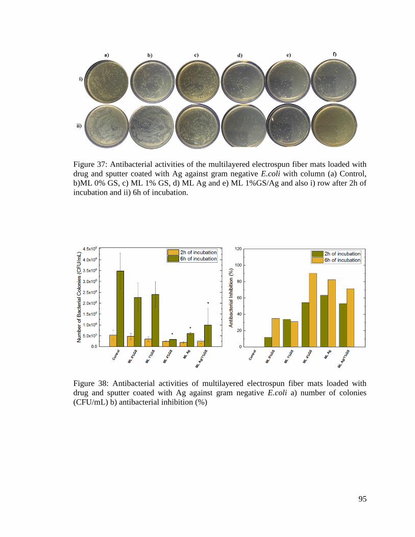
95
Figure 37: Antibacterial activities of the multilayered electrospun fiber mats loaded with
drug and sputter coated with Ag against gram negative E.coli with column (a) Control,
b)ML 0% GS, c) ML 1% GS, d) ML Ag and e) ML 1%GS/Ag and also i) row after 2h of
incubation and ii) 6h of incubation.
Figure 38: Antibacterial activities of multilayered electrospun fiber mats loaded with
drug and sputter coated with Ag against gram negative E.coli a) number of colonies
(CFU/mL) b) antibacterial inhibition (%)
96
CONCLUSION
In this work preparation and characterization of multilayed fiber electrospun fiber mats of
PVA/PCL polymers loaded with gentamicin sulphate was studied. Uniform fibers were
prepared by electrospinning process. In-vitro release of GS suggest that, multilayered
fiber mats reduces initial burst release of drug from the scaffolds. Moreover, intial burst
release in multilayer could be reduced by improving the amount of loaded GS in the
scaffolds. The cytotoxicity study illustrate conclude that, there is a toxic effect for the
increase in the drug concentration for Wi38 and Hacaat cell line. However, the
component PCL and PVA is biocompatabile because it has improved the growth of cells.
On the other hand PCL was sputter coated with Ag to investigate the in-vitro release of
Ag and cytotoxicity of the scaffolds. It is summarized that, Ag coated scaffolds have
toxicity effect towards the cell line. However, it possess a gradual release of Ag for the
antibacterial activity. Antibacterial inhibition (%) has improved with the addition of drug
as well as silver particles to the surface of the outer layer of the electrospun fiber.
Therefore, it is concluded that, the prepared fiber scaffold could be promising material
for sustained protection from antibacterial for wound dressing applications.
97
REFERENCES
M. Liu, X. Duan, Y. Li, D. Yang, and Y. Long, ―Electrospun nano fi bers for wound healing,‖
vol. 76, pp. 1413–1423, 2017.
J. Zhang, Y. Duan, D. Wei, L. Wang, H. Wang, Z. Gu, and D. Kong, ―Co-electrospun fibrous
scaffold–adsorbed DNA for substrate-mediated gene delivery,‖ J. Biomed. Mater. Res.
Part A, vol. 96A, no. 1, pp. 212–220, 2011.
H. Cao, X. Jiang, C. Chai, and S. Y. Chew, ―RNA interference by nanofiber-based siRNA
delivery system,‖ J. Control. Release, vol. 144, no. 2, pp. 203–212, 2010.
F. Siepmann, J. Siepmann, M. Walther, R. J. MacRae, and R. Bodmeier, ―Polymer blends for
controlled release coatings,‖ Journal of Controlled Release, vol. 125, no. 1. pp. 1–15,
2008.
S. N. Alhosseini, F. Moztarzadeh, M. Mozafari, S. Asgari, M. Dodel, A. Samadikuchaksaraei, S.
Kargozar, and N. Jalali, ―Synthesis and characterization of electrospun polyvinyl alcohol
nanofibrous scaffolds modified by blending with chitosan for neural tissue engineering,‖
Int. J. Nanomedicine, vol. 7, pp. 25–34, 2012.
K. Mojtaba and M. Hamid, ―Electrospinning, mechanical properties, and cell behavior study of
chitosan/PVA nanofibers,‖ J. Biomed. Mater. Res. Part A, vol. 103, no. 9, pp. 3081–3093,
Jul. 2015.
X. Zhu, S. Ni, T. Xia, Q. Yao, H. Li, B. Wang, J. Wang, X. Li, and W. Su, ―Anti-neoplastic
cytotoxicity of SN-38-loaded PCL/Gelatin electrospun composite nanofiber scaffolds
against human glioblastoma cells in vitro,‖ J. Pharm. Sci., vol. 104, no. 12, pp. 4345–
4354, 2015.
98
S. R. Gomes, G. Rodrigues, G. G. Martins, M. A. Roberto, M. Mafra, C. M. R. Henriques, and J.
C. Silva, ―In vitro and in vivo evaluation of electrospun nanofibers of PCL, chitosan and
gelatin: A comparative study,‖ Mater. Sci. Eng. C, vol. 46, pp. 348–358, 2015.
J. P. Vacanti and R. Langer, ―Tissue engineering: the design and fabrication of living replacement
devices for surgical reconstruction and transplantation,‖ Lancet, vol. 354, pp. S32–S34,
1999.
F. Diegelmann, Robert, ―Wound healing: an overview of acute, fibrotic and delayed healing,‖
Front. Biosci., vol. 9, no. 1–3, p. 283, 2004.
T. J. Koh and L. A. DiPietro, ―Inflammation and wound healing: the role of the macrophage.,‖
Expert Rev. Mol. Med., vol. 13, no. July 2011, pp. 1–12, 2011.
H.-J. Park, Y. Zhang, S. P. Georgescu, K. L. Johnson, D. Kong, and J. B. Galper, ―Human
umbilical vein endothelial cells and human dermal microvascular endothelial cells offer
new insights into the relationship between lipid metabolism and angiogenesis,‖ Stem Cell
Rev., vol. 2, no. 2, pp. 93–101, 2006.
R. A. Clark, ―Fibrin and wound healing.,‖ Ann. N. Y. Acad. Sci., vol. 936, pp. 355–367, 2001.
R. M. Slawson, M. I. Van Dyke, H. Lee, and J. T. Trevors, ―Germanium and silver resistance,
accumulation, and toxicity in microorganisms,‖ Plasmid, vol. 27, no. 1, pp. 72–79, 1992.
G. Zhao and S. E. Stevens, ―Multiple parameters for the comprehensive evaluation of the
susceptibility of Escherichia coli to the silver ion,‖ BioMetals, vol. 11, no. 1, pp. 27–32,
1998.
S. Ren, L. Dong, X. Zhang, T. Lei, F. Ehrenhauser, K. Song, M. Li, X. Sun, and Q. Wu,
―Electrospun nanofibers made of silver nanoparticles, cellulose nanocrystals, and
99
polyacrylonitrile as substrates for surface-enhanced raman scattering,‖ Materials (Basel).,
vol. 10, no. 1, 2017.
L. Ge, Q. Li, M. Wang, J. Ouyang, X. Li, and M. M. Q. Xing, ―Nanosilver particles in medical
applications: Synthesis, performance, and toxicity,‖ International Journal of
Nanomedicine, vol. 9, no. 1. pp. 2399–2407, 2014.
M. Mohseni, A. Shamloo, Z. Aghababaei, M. Vossoughi, and H. Moravvej, ―Antimicrobial
Wound Dressing Containing Silver Sulfadiazine With High Biocompatibility : In Vitro
Study,‖ vol. 40, no. 8, pp. 765–773, 2016.
J. Yip, S. Jiang, and C. Wong, ―Characterization of metallic textiles deposited by magnetron
sputtering and traditional metallic treatments,‖ Surf. Coatings Technol., vol. 204, no. 3,
pp. 380–385, 2009.
J. Doshi and D. H. Reneker, ―Electrospinning process and applications of electrospun fibers,‖ J.
Electrostat., vol. 35, no. 2, pp. 151–160, 1995.
T. Geoffrey, ―Electrically driven jets,‖ Proc. R. Soc. London. A. Math. Phys. Sci., vol. 313, no.
1515, p. 453 LP-475, Dec. 1969.
A. Greiner and J. H. Wendorff, ―Electrospinning: a fascinating method for the preparation of
ultrathin fibers.,‖ Angew. Chem. Int. Ed. Engl., vol. 46, no. 30, pp. 5670–5703, 2007.
A. Balaji, M. V. Vellayappan, A. A. John, A. P. Subramanian, S. K. Jaganathan, E. Supriyanto,
and S. I. A. Razak, ―An insight on electrospun-nanofibers-inspired modern drug delivery
system in the treatment of deadly cancers,‖ RSC Adv., vol. 5, no. 71, pp. 57984–58004,
2015.
G. H. K. and T. M. and S. A. P. and W. D. Kim, ―Coaxially electrospun micro/nanofibrous
100
poly(ε-caprolactone)/eggshell-protein scaffold,‖ Bioinspir. Biomim., vol. 3, no. 1, p.
16006, 2008.
G. Kim, H. Yoon, and Y. Park, ―Drug release from various thicknesses of layered mats consisting
of electrospun polycaprolactone and polyethylene oxide micro/nanofibers,‖ Appl. Phys. A
Mater. Sci. Process., vol. 100, no. 4, pp. 1197–1204, 2010.
A. Fathi-Azarbayjani and S. Y. Chan, ―Single and multi-layered nanofibers for rapid and
controlled drug delivery.,‖ Chem. Pharm. Bull. (Tokyo)., vol. 58, no. 2, pp. 143–6, 2010.
A. P. S. Immich, M. L. Arias, N. Carreras, R. L. Boemo, and J. A. Tornero, ―Drug delivery
systems using sandwich configurations of electrospun poly(lactic acid) nanofiber
membranes and ibuprofen,‖ Mater. Sci. Eng. C, vol. 33, no. 7, pp. 4002–4008, 2013.
D. W. C. Chen, J. Y. Liao, S. J. Liu, and E. C. Chan, ―Novel biodegradable sandwich-structured
nanofibrous drug-eluting membranes for repair of infected wounds: An in vitro and in
vivo study,‖ Int. J. Nanomedicine, vol. 7, pp. 763–771, 2012.
P. Ahlin Grabnar and J. Kristl, ―The manufacturing techniques of drug-loaded polymeric
nanoparticles from preformed polymers,‖ J. Microencapsul., vol. 28, no. 4, pp. 323–335,
Jun. 2011.
M. Zamani, M. P. Prabhakaran, and S. Ramakrishna, ―Advances in drug delivery via electrospun
and electrosprayed nanomaterials,‖ Int. J. Nanomedicine, vol. 8, pp. 2997–3017, 2013.
D.-G. G. Yu, J. Zhou, N. P. Chatterton, Y. Li, J. Huang, and X. Wang, ―Polyacrylonitrile
nanofibers coated with silver nanoparticles using a modified coaxial electrospinning
process.,‖ Int. J. Nanomedicine, vol. 7, pp. 5725–32, 2012.
S. Chen, G. Wang, T. Wu, X. Zhao, S. Liu, G. Li, W. Cui, and C. Fan, ―Silver
101
nanoparticles/ibuprofen-loaded poly(L-lactide) fibrous membrane: Anti-infection and anti-
adhesion effects,‖ Int. J. Mol. Sci., vol. 15, no. 8, pp. 14014–14025, 2014.
E. Mele, ―Electrospinning of natural polymers for advanced wound care: towards responsive and
adaptive dressings,‖ J. Mater. Chem. B, vol. 4, no. 28, pp. 4801–4812, 2016.
K. Y. Lee, L. Jeong, Y. O. Kang, S. J. Lee, and W. H. Park, ―Electrospinning of polysaccharides
for regenerative medicine,‖ Adv. Drug Deliv. Rev., vol. 61, no. 12, pp. 1020–1032, 2009.
R. Toshkova, N. Manolova, E. Gardeva, M. Ignatova, L. Yossifova, I. Rashkov, and M.
Alexandrov, ―Antitumor activity of quaternized chitosan-based electrospun implants
against Graffi myeloid tumor,‖ Int. J. Pharm., vol. 400, no. 1–2, p. 221—233, Nov. 2010.
T. Garg, G. Rath, and A. K. Goyal, ―Biomaterials-based nanofiber scaffold: targeted and
controlled carrier for cell and drug delivery,‖ J. Drug Target., vol. 23, no. 3, pp. 202–221,
2015.
J. Gunn and M. Zhang, ―Polyblend nanofibers for biomedical applications: perspectives and
challenges,‖ Trends Biotechnol., vol. 28, no. 4, pp. 189–197, Apr. 2018.
K. Kim, M. Yu, X. Zong, J. Chiu, D. Fang, Y.-S. Seo, B. S. Hsiao, B. Chu, and M. Hadjiargyrou,
―Control of degradation rate and hydrophilicity in electrospun non-woven poly(d,l-lactide)
nanofiber scaffolds for biomedical applications,‖ Biomaterials, vol. 24, no. 27, pp. 4977–
4985, 2003.
A. M. Behrens, J. Kim, N. Hotaling, J. E. Seppala, P. Kofinas, and W. Tutak, ―Rapid fabrication
of poly(DL-lactide) nanofiber scaffolds with tunable degradation for tissue engineering
applications by air-brushing,‖ Biomed. Mater., vol. 11, no. 3, p. 35001, Apr. 2016.
Z. X. Meng, X. X. Xu, W. Zheng, H. M. Zhou, L. Li, Y. F. Zheng, and X. Lou, ―Preparation and
102
characterization of electrospun PLGA/gelatin nanofibers as a potential drug delivery
system,‖ Colloids Surfaces B Biointerfaces, vol. 84, no. 1, pp. 97–102, 2011.
N. S. Binulal, A. Natarajan, D. Menon, V. K. Bhaskaran, U. Mony, and S. V Nair, ―PCL-gelatin
composite nanofibers electrospun using diluted acetic acid-ethyl acetate solvent system for
stem cell-based bone tissue engineering.,‖ J. Biomater. Sci. Polym. Ed., vol. 25, no. 4, pp.
325–340, 2014.
X. Qin and D. Wu, ―Effect of different solvents on poly(caprolactone)(PCL) electrospun
nonwoven membranes,‖ J. Therm. Anal. Calorim., vol. 107, no. 3, pp. 1007–1013, 2012.
L. N. T. Ba, M. Y. Ki, S. Ho‐Yeon, and L. Byong‐Taek, ―Fabrication of polyvinyl alcohol/gelatin
nanofiber composites and evaluation of their material properties,‖ J. Biomed. Mater. Res.
Part B Appl. Biomater., vol. 95B, no. 1, pp. 184–191, Aug. 2010.
Y. Liu, H. Yu, Y. Liu, G. Liang, T. Zhang, and Q. Hu, ―Dual Drug Spatiotemporal Release From
Functional Gradient Scaffolds Prepared Using 3D Bioprinting and Electrospinning,‖ 2016.
P. Askari, P. Zahedi, and I. Rezaeian, ―Three-layered electrospun PVA / PCL / PVA nanofibrous
mats containing tetracycline hydrochloride and phenytoin sodium : A case study on
sustained control release , antibacterial , and cell culture properties,‖ vol. 43309, pp. 1–9,
2016.
М. Ignatova, I. Rashkov, and N. Manolova, ―Drug-loaded electrospun materials in wound-
dressing applications and in local cancer treatment,‖ Expert Opin. Drug Deliv., vol. 10,
no. 4, pp. 469–483, Apr. 2013.
C. Dwivedi, H. Pandey, A. C. Pandey, and P. W. Ramteke, ―Fabrication and assessment of
gentamicin loaded electrospun nanofibrous scaffolds as a quick wound healing dressing
material,‖ Curr. Nanosci., vol. 11, no. 2, pp. 222–228, 2015.
103
E. R. Kenawy, G. L. Bowlin, K. Mansfield, J. Layman, D. G. Simpson, E. H. Sanders, and G. E.
Wnek, ―Release of tetracycline hydrochloride from electrospun poly(ethylene-co-
vinylacetate), poly(lactic acid), and a blend,‖ J. Control. Release, vol. 81, no. 1–2, pp. 57–
64, 2002.
F. Zamani, F. Jahanmard, F. Ghasemkhah, S. Amjad-Iranagh, R. Bagherzadeh, M. Amani-
Tehran, and M. Latifi, Chapter 7 – Nanofibrous and nanoparticle materials as drug-
delivery systems, no. September. 2017.
X. Xu, X. Chen, Z. Wang, and X. Jing, ―Ultrafine PEG-PLA fibers loaded with both paclitaxel
and doxorubicin hydrochloride and their in vitro cytotoxicity,‖ Eur. J. Pharm. Biopharm.,
vol. 72, no. 1, pp. 18–25, 2009.
J. Xie, S. T. Ruo, and C. H. Wang, ―Biodegradable microparticles and fiber fabrics for sustained
delivery of cisplatin to treat C6 glioma in vitro,‖ J. Biomed. Mater. Res. - Part A, vol. 85,
no. 4, pp. 897–908, 2008.
S. Y. Chew, J. Wen, E. K. F. Yim, and K. W. Leong, ―Sustained release of proteins from
electrospun biodegradable fibers,‖ Biomacromolecules, vol. 6, no. 4, pp. 2017–2024,
2005.
P. on Rujitanaroj, Y. C. Wang, J. Wang, and S. Y. Chew, ―Nanofiber-mediated controlled release
of siRNA complexes for long term gene-silencing applications,‖ Biomaterials, vol. 32, no.
25, pp. 5915–5923, 2011.
A. Saraf, L. S. Baggett, R. M. Raphael, F. K. Kasper, and A. G. Mikos, ―Regulated non-viral
gene delivery from coaxial electrospun fiber mesh scaffolds,‖ J. Control. Release, vol.
143, no. 1, pp. 95–103, 2010.
A. Mickova, M. Buzgo, O. Benada, M. Rampichova, and Z. Fisar, ―Core/Shell Nanofibers with
104
Embedded Liposomes as a Drug Delivery System,‖ 2012.
V. Pillay, C. Dott, Y. E. Choonara, C. Tyagi, L. Tomar, P. Kumar, L. C. Du Toit, and V. M. K.
Ndesendo, ―A review of the effect of processing variables on the fabrication of
electrospun nanofibers for drug delivery applications,‖ Journal of Nanomaterials, vol.
2013. 2013.
V. Leung and F. Ko, ―Biomedical applications of nanofibers,‖ Polymers for Advanced
Technologies, vol. 22, no. 3. pp. 350–365, 2011.
Q. Zhang, Y. Li, Z. Y. (William) Lin, K. K. Y. Wong, M. Lin, L. Yildirimer, and X. Zhao,
―Electrospun polymeric micro/nanofibrous scaffolds for long-term drug release and their
biomedical applications,‖ Drug Discov. Today, vol. 22, no. 9, pp. 1351–1366, 2017.
A. S. Halim, T. L. Khoo, and S. J. Mohd. Yussof, ―Biologic and synthetic skin substitutes: An
overview,‖ Indian J. Plast. Surg., vol. 43, no. Suppl, pp. S23–S28, Sep. 2010.
M.-S. Khil, D.-I. Cha, H.-Y. Kim, I.-S. Kim, and N. Bhattarai, ―Electrospun nanofibrous
polyurethane membrane as wound dressing.,‖ J. Biomed. Mater. Res. B. Appl. Biomater.,
vol. 67, no. 2, pp. 675–679, Nov. 2003.
S. M. Saeed, H. Mirzadeh, M. Zandi, and J. Barzin, ―Designing and fabrication of curcumin
loaded PCL/PVA multi-layer nanofibrous electrospun structures as active wound
dressing,‖ Prog. Biomater., vol. 6, no. 1–2, pp. 39–48, 2017.
F. Khodkar and N. G. Ebrahimi, ―Preparation and properties of antibacterial , biocompatible core
– shell fibers produced by coaxial electrospinning,‖ vol. 44979, no. 14115, pp. 1–9, 2017.
V. A. Online, L. Du, H. Z. Xu, T. Li, Y. Zhang, and F. Y. Zou, ―Fabrication of ascorbyl palmitate
loaded poly(caprolactone)/silver nanoparticle embedded poly(vinyl alcohol) hybrid
105
nanofibre mats as active wound dressings via dual-spinneret electrospinning†,‖ pp.
31310–31318, 2017.
X. Liu, T. Lin, J. Fang, G. Yao, H. Zhao, M. Dodson, and X. Wang, ―In vivo wound healing and
antibacterial performances of electrospun nanofibre membranes,‖ pp. 499–508, 2010.
M. Fröhlich, W. L. Grayson, L. Q. Wan, D. Marolt, M. Drobnic, and G. Vunjak-Novakovic,
―Tissue engineered bone grafts: biological requirements, tissue culture and clinical
relevance.,‖ Curr. Stem Cell Res. Ther., vol. 3, no. 4, pp. 254–64, 2008.
P. Nooeaid, V. Salih, J. P. Beier, and A. R. Boccaccini, ―Osteochondral tissue engineering:
Scaffolds, stem cells and applications,‖ J. Cell. Mol. Med., vol. 16, no. 10, pp. 2247–2270,
2012.
S. Uma Maheshwari, V. K. Samuel, and N. Nagiah, ―Fabrication and evaluation of
(PVA/HAp/PCL) bilayer composites as potential scaffolds for bone tissue regeneration
application,‖ Ceram. Int., vol. 40, no. 6, pp. 8469–8477, 2014.
Y. Su, Q. Su, W. Liu, M. Lim, J. R. Venugopal, X. Mo, S. Ramakrishna, S. S. Al-Deyab, and M.
El-Newehy, ―Controlled release of bone morphogenetic protein 2 and dexamethasone
loaded in core-shell PLLACL-collagen fibers for use in bone tissue engineering,‖ Acta
Biomater., vol. 8, no. 2, pp. 763–771, 2012.
A. Shafiee, M. Soleimani, G. A. Chamheidari, E. Seyedjafari, M. Dodel, A. Atashi, and Y.
Gheisari, ―Electrospun nanofiber-based regeneration of cartilage enhanced by
mesenchymal stem cells,‖ pp. 467–478, 2011.
A. Guyton and J. Hall, ―Textbook of medical physiology, 11th,‖ in Elsevier Saunders, 2006, pp.
802–804.
106
A. D. Bach, J. P. Beier, J. Stern-Staeter, and R. E. Horch, ―Skeletal muscle tissue engineering,‖ J.
Cell. Mol. Med., vol. 8, no. 4, pp. 413–422, 2004.
W. Bian and N. Bursac, ―Engineered skeletal muscle tissue networks with controllable
architecture,‖ Biomaterials, vol. 30, no. 7, pp. 1401–1412, 2009.
H. Liao and G.-Q. Zhou, ―Development and Progress of Engineering of Skeletal Muscle Tissue,‖
Tissue Eng. Part B Rev., vol. 15, no. 3, pp. 319–331, 2009.
K. D. Mckeon-fischer, D. H. Flagg, and J. W. Freeman, ―Poly ( acrylic acid )/ poly ( vinyl alcohol
) compositions coaxially electrospun with poly ( 3 -caprolactone ) and multi-walled
carbon nanotubes to create nanoactuating scaffolds,‖ Polymer (Guildf)., vol. 52, no. 21,
pp. 4736–4743, 2011.
K. Gurunathan, A. V. Murugan, R. Marimuthu, U. . Mulik, and D. . Amalnerkar,
―Electrochemically synthesised conducting polymeric materials for applications towards
technology in electronics, optoelectronics and energy storage devices,‖ Mater. Chem.
Phys., vol. 61, no. 3, pp. 173–191, 1999.
N. K. Guimard, N. Gomez, and C. E. Schmidt, ―Conducting polymers in biomedical
engineering,‖ Prog. Polym. Sci., vol. 32, no. 8–9, pp. 876–921, 2007.
D. D. Ateh, H. a Navsaria, and P. Vadgama, ―Polypyrrole-based conducting polymers and
interactions with biological tissues.,‖ J. R. Soc. Interface, vol. 3, no. 11, pp. 741–52, 2006.
C. E. Schmidt, V. R. Shastri, J. P. Vacanti, and R. Langer, ―Stimulation of neurite outgrowth
using an electrically conducting polymer.,‖ Proc. Natl. Acad. Sci. U. S. A., vol. 94, no. 17,
pp. 8948–8953, 1997.
S. Joo, K. Kang, and Y. Nam, ―In vitro neurite guidance effects induced by polylysine pinstripe
107
micropatterns with polylysine background,‖ J. Biomed. Mater. Res. - Part A, vol. 103, no.
8, pp. 2731–2739, 2015.
J. Y. Lee, C. A. Bashur, C. A. Milroy, L. Forciniti, A. S. Goldstein, and C. E. Schmidt, ―Nerve
growth factor-immobilized electrically conducting fibrous scaffolds for potential use in
neural engineering applications,‖ IEEE Trans. Nanobioscience, vol. 11, no. 1, pp. 15–21,
2012.
N. Gomez, J. Y. Lee, J. D. Nickels, and C. E. Schmidt, ―Micropatterned polypyrrole: A
combination of electrical and topographical characteristics for the stimulation of cells,‖
Adv. Funct. Mater., vol. 17, no. 10, pp. 1645–1653, 2007.
J. Y. Lee, C. A. Bashur, A. S. Goldstein, and C. E. Schmidt, ―Biomaterials Polypyrrole-coated
electrospun PLGA nanofibers for neural tissue applications,‖ Biomaterials, vol. 30, no. 26,
pp. 4325–4335, 2009.
N. Gomathi, A. Sureshkumar, and S. Neogi, ―RF plasma-treated polymers for biomedical
applications,‖ Current Science, vol. 94, no. 11. pp. 1478–1486, 2008.
E. M. Liston, ―Plasma treatment for improved bonding: A review,‖ J. Adhes., vol. 30, no. 1–4, pp.
199–218, 1989.
A. Gao, R. Hang, X. Huang, L. Zhao, X. Zhang, L. Wang, B. Tang, S. Ma, and P. K. Chu, ―The
effects of titania nanotubes with embedded silver oxide nanoparticles on bacteria and
osteoblasts,‖ Biomaterials, vol. 35, no. 13, pp. 4223–4235, 2014.
J. J. George, ―Preparation of Thin Films.‖ Marcel Dekker. INC, New york, 1992.
D. Depla, S. Mahieu, and J. E. Greene, ―Chapter 5 - Sputter Deposition Processes A2 - Martin,
Peter M. BT - Handbook of Deposition Technologies for Films and Coatings (Third
108
Edition),‖ Boston: William Andrew Publishing, 2010, pp. 253–296.
M. Ohring, ―Chapter 5 - Plasma and Ion Beam Processing of Thin Films BT - Materials Science
of Thin Films (Second Edition),‖ San Diego: Academic Press, 2002, pp. 203–275.
H.-L. Huang, Y.-Y. Chang, H.-J. Chen, Y.-K. Chou, C.-H. Lai, and M. Y. C. Chen, ―Antibacterial
properties and cytocompatibility of tantalum oxide coatings with different silver content,‖
J. Vac. Sci. Technol. A Vacuum, Surfaces, Film., vol. 32, no. 2, p. 02B117, 2014.
Q. Wang, X. Wang, X. Li, Y. Cai, and Q. Wei, ―Surface modification of PMMA/O-MMT
composite microfibers by TiO2coating,‖ Appl. Surf. Sci., vol. 258, no. 1, pp. 98–102,
2011.
T.-J. Lee, S. Kang, G.-J. Jeong, J.-K. Yoon, S. H. Bhang, J. Oh, and B.-S. Kim, ―Incorporation of
Gold-Coated Microspheres into Embryoid Body of Human Embryonic Stem Cells for
Cardiomyogenic Differentiation,‖ Tissue Eng. Part A, vol. 21, no. 1–2, pp. 374–381,
2015.
E. N. Bolbasov, M. Rybachuk, A. S. Golovkin, L. V. Antonova, E. V. Shesterikov, A. I.
Malchikhina, V. A. Novikov, Y. G. Anissimov, and S. I. Tverdokhlebov, ―Surface
modification of poly(l-lactide) and polycaprolactone bioresorbable polymers using RF
plasma discharge with sputter deposition of a hydroxyapatite target,‖ Mater. Lett., vol.
132, pp. 281–284, 2014.
A. Majumdar, B. S. Butola, and S. Thakur, ―Development and performance optimization of
knitted antibacterial materials using polyester-silver nanocomposite fibres,‖ Mater. Sci.
Eng. C, vol. 54, pp. 26–31, 2015.
P. Kalakonda, M. A. Aldhahri, M. S. Abdel-wahab, A. Tamayol, K. M. Moghaddam, F. Ben
Rached, A. Pain, A. Khademhosseini, A. Memic, and S. Chaieb, ―Microfibrous silver-
109
coated polymeric scaffolds with tunable mechanical properties,‖ RSC Adv., vol. 7, no. 55,
pp. 34331–34338, 2017.
A. D. Badaraev, A. L. Nemoykina, E. N. Bolbasov, and S. I. Tverdokhlebov, ―PLLA scaffold
modification using magnetron sputtering of the copper target to provide antibacterial
properties,‖ Resour. Technol., vol. 3, no. 2, pp. 204–211, 2017.
L. S. Barbarash, E. N. Bolbasov, L. V. Antonova, V. G. Matveeva, E. A. Velikanova, E. V.
Shesterikov, Y. G. Anissimov, and S. I. Tverdokhlebov, ―Surface modification of poly-ε-
caprolactone electrospun fibrous scaffolds using plasma discharge with sputter deposition
of a titanium target,‖ Mater. Lett., vol. 171, pp. 87–90, 2016.
G. Q. Blantocas, A. S. Alaboodi, and A. baset H. Mekky, ―Synthesis of Chitosan–
TiO2Antimicrobial Composites via a 2-Step Process of Electrospinning and Plasma
Sputtering,‖ Arab. J. Sci. Eng., vol. 43, no. 1, pp. 389–398, 2018.
M. Beregoi, C. Busuioc, A. Evanghelidis, E. Matei, F. Iordache, M. Radu, A. Dinischiotu, and I.
Enculescu, ―Electrochromic properties of polyaniline-coated fiber webs for tissue
engineering applications,‖ Int. J. Pharm., vol. 510, no. 2, pp. 465–473, 2016.
S. I. Goreninskii, N. N. Bogomolova, A. I. Malchikhina, A. S. Golovkin, E. N. Bolbasov, T. V.
Safronova, V. I. Putlyaev, and S. I. Tverdokhlebov, ―Biological Effect of the Surface
Modification of the Fibrous Poly(L-lactic acid) Scaffolds by Radio Frequency Magnetron
Sputtering of Different Calcium-Phosphate Targets,‖ Bionanoscience, vol. 7, no. 1, pp.
50–57, 2017.
R. Dastjerdi and M. Montazer, ―A review on the application of inorganic nano-structured
materials in the modification of textiles: Focus on anti-microbial properties,‖ Colloids and
Surfaces B: Biointerfaces, vol. 79, no. 1. pp. 5–18, 2010.
110
K. Murugesh Babu and K. B. Ravindra, ―Bioactive antimicrobial agents for finishing of textiles
for health care products,‖ J. Text. Inst., vol. 106, no. 7, pp. 706–717, 2015.
W. Zhong, ―Efficacy and toxicity of antibacterial agents used in wound dressings,‖ Cutaneous
and Ocular Toxicology, vol. 34, no. 1. pp. 61–67, 2015.
J. M. Schierholz, J. Beuth, and G. Pulverer, ―Silver coating of medical devices for catheter-
associated infections? [3],‖ American Journal of Medicine, vol. 107, no. 1. pp. 101–102,
1999.
V. K. Mishra and A. Kumar, ―Impact of Metal Nanoparticles on the Plant Growth Promoting
Rhizobacteria,‖ Dig. J. Nanomater. Biostructures, vol. 4, no. 3, pp. 587–592, 2009.
T. Hamouda and J. R. Baker, ―Antimicrobial mechanism of action of surfactant lipid preparations
in enteric gram-negative bacilli,‖ J. Appl. Microbiol., vol. 89, no. 3, pp. 397–403, 2000.
S. Pal, Y. K. Tak, and J. M. Song, ―Does the antibacterial activity of silver nanoparticles depend
on the shape of the nanoparticle? A study of the gram-negative bacterium Escherichia
coli,‖ J. Biol. Chem., vol. 290, no. 42, pp. 1712–1720, 2015.
J. Tian, K. K. Y. Wong, C. M. Ho, C. N. Lok, W. Y. Yu, C. M. Che, J. F. Chiu, and P. K. H.
Tam, ―Topical delivery of silver nanoparticles promotes wound healing,‖ ChemMedChem,
vol. 2, no. 1, pp. 129–136, 2007.
R. H. Demling and M. D. Leslie DeSanti, ―The rate of re-epithelialization across meshed skin
grafts is increased with exposure to silver,‖ Burns, vol. 28, no. 3, pp. 264–266, 2002.
V. K. M. Poon and A. Burd, ―In vitro cytotoxity of silver: Implication for clinical wound care,‖
Burns, vol. 30, no. 2, pp. 140–147, 2004.
M. Irfan, S. Perero, M. Miola, G. Maina, A. Ferri, M. Ferraris, and C. Balagna, ―Antimicrobial
111
functionalization of cotton fabric with silver nanoclusters/silica composite coating via RF
co-sputtering technique,‖ Cellulose, vol. 24, no. 5, pp. 2331–2345, 2017.
Y. H. Chen, C. C. Hsu, and J. L. He, ―Antibacterial silver coating on poly(ethylene terephthalate)
fabric by using high power impulse magnetron sputtering,‖ Surf. Coatings Technol., vol.
232, pp. 868–875, 2013.
X. Liu, K. Gan, H. Liu, X. Song, T. Chen, and C. Liu, ―Antibacterial properties of nano-silver
coated PEEK prepared through magnetron sputtering,‖ Dent. Mater., vol. 33, no. 9, pp.
e348–e360, 2017.
G. Muzio, M. Miola, S. Perero, M. Oraldi, M. Maggiora, S. Ferraris, E. Vernè, V. Festa, F. Festa,
R. A. Canuto, and M. Ferraris, ―Polypropylene prostheses coated with silver
nanoclusters/silica coating obtained by sputtering: Biocompatibility and antibacterial
properties,‖ Surf. Coatings Technol., vol. 319, pp. 326–334, 2017.
G. Grass, C. Rensing, and M. Solioz, ―Metallic copper as an antimicrobial surface,‖ Applied and
Environmental Microbiology, vol. 77, no. 5. pp. 1541–1547, 2011.
C. Castro, R. Sanjines, C. Pulgarin, P. Osorio, S. A. Giraldo, and J. Kiwi, ―Structure-reactivity
relations for DC-magnetron sputtered Cu-layers during E. coli inactivation in the dark and
under light,‖ J. Photochem. Photobiol. A Chem., vol. 216, no. 2–3, pp. 295–302, 2010.
V. M. Pantojas and E. Velez, ―Initial Study on Fibers and Coatings for the Fabrication of
Bioscaffolds,‖ vol. 28, no. 3, 2009.
E. N. Bolbasov, L. V. Antonova, K. S. Stankevich, Ashrafov, V. G. Matveeva, E. A. Velikanova,
Y. I. Khodyrevskaya, Y. A. Kudryavtseva, Y. G. Anissimov, S. I. Tverdokhlebov, and L.
S. Barbarash, ―The use of magnetron sputtering for the deposition of thin titanium
coatings on the surface of bioresorbable electrospun fibrous scaffolds for vascular tissue
112
engineering: A pilot study,‖ Appl. Surf. Sci., vol. 398, pp. 63–72, 2017.
M. A. Woodruff and D. W. Hutmacher, ―The return of a forgotten polymer - Polycaprolactone in
the 21st century,‖ Prog. Polym. Sci., vol. 35, no. 10, pp. 1217–1256, 2010.
E. S. Place, J. H. George, C. K. Williams, and M. M. Stevens, ―Synthetic polymer scaffolds for
tissue engineering,‖ Chem. Soc. Rev., vol. 38, no. 4, p. 1139, 2009.
I. Vroman and L. Tighzert, ―Biodegradable polymers,‖ Materials (Basel)., vol. 2, no. 2, pp. 307–
344, 2009.
F. Ko, V. Leung, R. Hartwell, H. Yang, and A. Ghahary, ―Nanofibre based biomaterials:
Bioactive nanofibres for wound healing applications,‖ Proc. - 2012 Int. Conf. Biomed.
Eng. Biotechnol. iCBEB 2012, vol. 1, no. April, pp. 389–392, 2012.
B. E. Rosenkrantz, J. R. Greco, J. G. Hoogerheide, and E. M. Oden, ―Gentamicin Sulfate,‖ Anal.
Profiles Drug Subst. Excipients, vol. 9, no. C, pp. 295–340, 1981.
P. Frutos, S. Torrado, M. E. Perez-Lorenzo, and G. Frutos, ―A validated quantitative colorimetric
assay for gentamicin,‖ J. Pharm. Biomed. Anal., vol. 21, no. 6, pp. 1149–1159, 2000.
A. F. H. Ismail, F. Mohamed, L. M. M. Rosli, M. A. M. Shafri, M. S. Haris, and A. B. Adina,
―Spectrophotometric determination of gentamicin loaded PLGA microparticles and
method validation via ninhydrin-gentamicin complex as a rapid quantification approach,‖
J. Appl. Pharm. Sci., vol. 6, no. 1, pp. 007-014, 2016.
O. Landau, A. Rothschild, and E. Zussman, ―Processing-Microstructure-Properties Correlation of
Ultrasensitive Gas Sensors Produced by Electrospinning,‖ Chem. Mater., vol. 21, no. 1,
pp. 9–11, Jan. 2009.
W. E. Teo, R. Inai, and S. Ramakrishna, ―Technological advances in electrospinning of
113
nanofibers,‖ Sci. Technol. Adv. Mater., vol. 12, no. 1, 2011.
D. Maurya, A. Sardarinejad, and K. Alameh, ―Recent Developments in R.F. Magnetron Sputtered
Thin Films for pH Sensing Applications—An Overview,‖ Coatings, vol. 4, no. 4, pp. 756–
771, 2014.
J. Goldstein, D. E. Newbury, D. C. Joy, C. E. Lyman, P. Echlin, E. Lifshin, L. Sawyer, and J. R.
Michael, Scanning Electron Microscopy and X-ray Microanalysis, vol. 44, no. 0. 2003.
H.-H. Perkampus, UV-VIS Spectroscopy and Its Applications, vol. 12. 1992.
T. Scientific, ―https://www.thermofisher.com/order/catalog/product/DAL1025,‖
https://www.thermofisher.com/order/catalog/product/DAL1025, 2018. [Online].
Available: https://www.thermofisher.com/order/catalog/product/DAL1025.
Q. Control, ―Nutrient Broth Medium.‖
I. M. Smallwood, ―Handbook of Organic Solvent Properties,‖ Butterworth-Heinemann, 1996.
S. Bykkam, M. Ahmadipour, S. Narisngam, V. R. Kalagadda, and S. C. Chidurala,
―RETRACTED: Extensive Studies on X-Ray Diffraction of Green Synthesized Silver
Nanoparticles,‖ Adv. Nanoparticles, 2015.
Y. Qing, L. Cheng, R. Li, G. Liu, Y. Zhang, X. Tang, J. Wang, H. Liu, and Y. Qin, ―Potential
antibacterial mechanism of silver nanoparticles and the optimization of orthopedic
implants by advanced modification technologies,‖ Int. J. Nanomedicine, vol. 13, pp.
3311–3327, Jun. 2018.
K. J. Woo, C. K. Hye, W. K. Ki, S. Shin, H. K. So, and H. P. Yong, ―Antibacterial activity and
mechanism of action of the silver ion in Staphylococcus aureus and Escherichia coli,‖
Appl. Environ. Microbiol., vol. 74, no. 7, pp. 2171–2178, 2008.
114
B. Le Ouay and F. Stellacci, ―Antibacterial activity of silver nanoparticles: A surface science
insight,‖ Nano Today. 2015.
A. Doustgani, E. Vasheghani-Farahani, M. Soleimani, and S. Hashemi-Najafabadi, ―Preparation
and characterization of aligned and random nanofibrous nanocomposite scaffolds of poly
(vinyl alcohol), poly (e-Caprolactone) and nanohydroxyapatite,‖ Int. J. Nanosci.
Nanotechnol., vol. 7, no. 3, pp. 127–132, 2011.
A. R. Unnithan, N. A. M. Barakat, P. B. Tirupathi Pichiah, G. Gnanasekaran, R. Nirmala, Y. S.
Cha, C. H. Jung, M. El-Newehy, and H. Y. Kim, ―Wound-dressing materials with
antibacterial activity from electrospun polyurethane-dextran nanofiber mats containing
ciprofloxacin HCl,‖ Carbohydr. Polym., vol. 90, no. 4, pp. 1786–1793, 2012.
T. Okuda, K. Tominaga, and S. Kidoaki, ―Time-programmed dual release formulation by
multilayered drug-loaded nanofiber meshes,‖ J. Control. Release, vol. 143, no. 2, pp. 258–
264, 2010.


