Embed Size (px)
Citation preview
Dental Calculus as Predisposing Factor in Periodontal Disease
Prepared by Widowati Witjaksono
For year 3
Predisposing factor
• Any conditioning factor that influences both the type and the amount of resources that the individual can elicit to cope with stress. It may be biologic, psychologic, genetic, or sociocultural.
• Mosby's Medical Dictionary, 8th edition. © 2009, Elsevier.
10/23/22 2
Predisposing Factors in Periodontal Disease
• Calculus• Iatrogenic factors• Malocclusion• Periodontal complication associated w/ orthodontic therapy
• Extraction of impacted third molars• Habits and self-inflicted injuries• Tobacco use• Radiation therapy10/23/22 3
Calculus
• Calculus consists of mineralized bacterial plaque that forms on the surfaces of natural teeth and dental prostheses
• It is classified as supragingival or subgingival, according to its relation to the gingival margin
10/23/22 4
Supragingival Calculus
• It is located coronal to the gingival margin and therefore is visible in the oral cavity
• It is usually white or whitish yellow in color, hard with claylike consistency, and easily detached from the tooth surface
• After removal, it may rapidly recur, especially in the lingual area of the mandibular incisors
• The color is influenced by contact with such substances as tobacco and food pigments. It may localize on a single tooth or group of teeth, or it may be generalized through out the mouth
10/23/22 5
Continue..supragingival ….• The two most common locations for supragingival calculus to develop are the buccal surfaces of the maxillary molars and the lingual surfaces of the mandibular anterior teeth. Saliva from the parotid gland flows over the facial surfaces of upper molars through Stensen’s duct
• Whereas the orifices of Wharton’s duct and Bartholins’duct empty onto the lingual surfaces of the lower incisors from the submaxillary and sublingual glands, respectively
• In extreme cases, calculus may form a bridgelike structure over the interdental papilla of adjacent teeth or cover the occlusal surface of teeth without functional antagonists.
10/23/22 6
Subgingival calculus• It is located below the crest of marginal gingiva
and therefore is not visible on routine clinical examination
• The location and extent of subgingival calculus may be evaluated by carefull tactile perception with a delicate dental instrument such as an explorer
• Subgingival calculus is typically hard and dense and frequently appears darkbrown and greenish black while being firmly attached to the tooth surface
• Supra and subgingival calculus generally occur together, but one may be present without the other
• Microscopic studies deposits of subgingival calculus extend nearly to the base of periodontal pockets in chronic periodontitis but do not reach the junctional epithelium
10/23/22 7
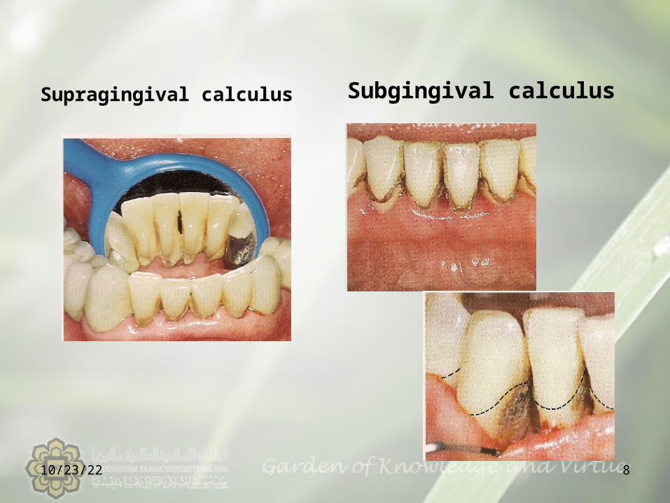
Supragingival calculus Subgingival calculus
10/23/22 8
Composition Inorganic Content • Supragingival calculus consist of inorganic (70 %-90%)
and organic components• The percentage of inorganic constituents in calculus is
similar to that in other calcified tissues of the body• At least two thirds of the inorganic component is
crystalyne in structure four (4)main crystal forms; Hydroxyapatite, Mg whitlockite, Octacalcium phosphate, Brushite
• Generally, two or more crystal forms are typically found in a sample of calculus. Hydroxyapatite and octacalcuim phosphate are detected most frequently (in 97 % to 100% of all supragingival calculus) and constitute the bulk of the specimen
• Brushite is more common in the mandibular anterior region and magnesium whitlockite in the posterior areas
• The incidence of the four crystal forms varies with the age of the deposit
10/23/22 9
Organic content• The organic component of calculus consist of a mixture of
protein-polysaccharide complexes, desquamated epitheliel cells,leucocytes, and various of microorganisms
• Between 1.9% and 9.1 % of the organic component is carbohydrate, which consists of galactose, glucose, rhamnose, mannose, glucuronic acid, galactosamine, and sometimes arabinose, galacturonic acid and glucosamine all are present in salivary glycoprotein,with the exception of arabinose and rhamnose
• The composition of subgingival calculus is similar to that of supragingival calculus, with some difference;
Subgingival calculus has the same hydroxyapatite content, more mg whitlockite, and less brushite and octacalcium phosphate than supragingival calculus
10/23/22 10
The composition of…• The ratio of calcium to phosphate is higher subgingivally, and the sodium content increases w/ the depth of periodontal pockets
• Salivary proteins present in supragingival calculus are not found subgingivally
• Dental calculus, salivary duct calculus, and calcified dental tissues are similar in inorganic composition
10/23/22 11
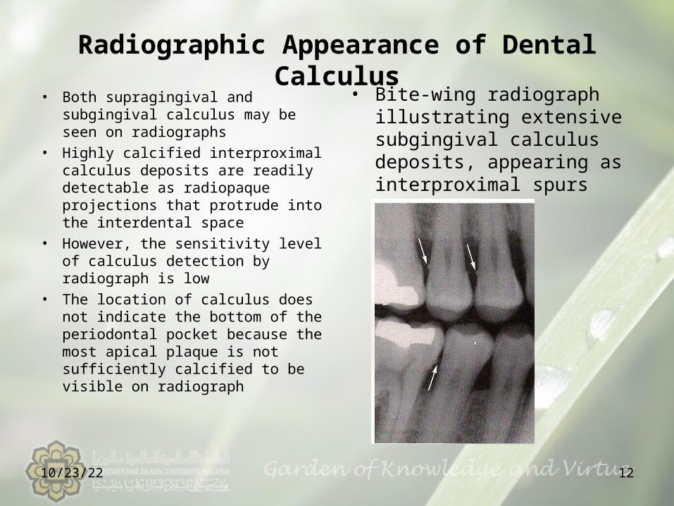
Radiographic Appearance of Dental Calculus
• Both supragingival and subgingival calculus may be seen on radiographs
• Highly calcified interproximal calculus deposits are readily detectable as radiopaque projections that protrude into the interdental space
• However, the sensitivity level of calculus detection by radiograph is low
• The location of calculus does not indicate the bottom of the periodontal pocket because the most apical plaque is not sufficiently calcified to be visible on radiograph
• Bite-wing radiograph illustrating extensive subgingival calculus deposits, appearing as interproximal spurs
10/23/22 12
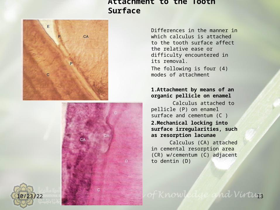
Attachment to the Tooth Surface
Differences in the manner in which calculus is attached to the tooth surface affect the relative ease or difficulty encountered in its removal.The following is four (4) modes of attachment
1.Attachment by means of an organic pellicle on enamel Calculus attached to pellicle (P) on enamel surface and cementum (C )2.Mechanical locking into surface irregularities, such as resorption lacunae Calculus (CA) attached in cemental resorption area (CR) w/cementum (C) adjacent to dentin (D)
10/23/22 13
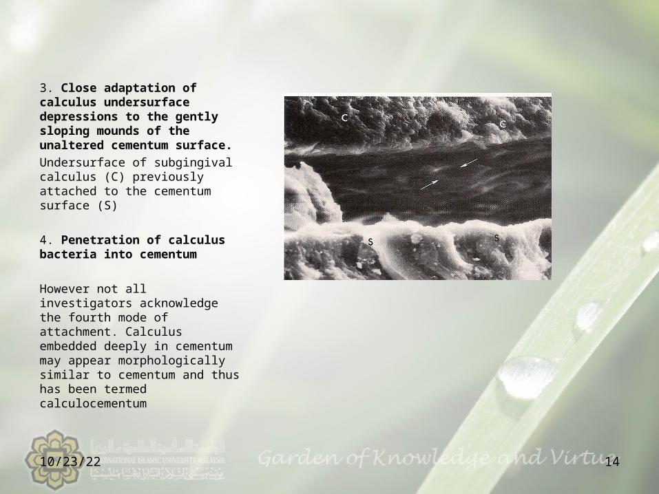
3. Close adaptation of calculus undersurface depressions to the gently sloping mounds of the unaltered cementum surface.Undersurface of subgingival calculus (C) previously attached to the cementum surface (S)
4. Penetration of calculus bacteria into cementum
However not all investigators acknowledge the fourth mode of attachment. Calculus embedded deeply in cementum may appear morphologically similar to cementum and thus has been termed calculocementum
10/23/22 14
Formation of dental calculus• Calculus is dental plaque that has undergone mineralization. The
soft plaque is hardened by the precipitation of mineral salts, which usually starts between the first and fourteenth days of plaque formation
• However, calcification has been reported to occur as soon as 4 to 8 hours. Calcifying plaques may become 50% mineralized in 2 days and 60% to 90% mineralized in 12 days
• All plaque does not necessarily undergo calcification• Microorganisms are not always essential in calculus
formation because calculus occurs readily in germ-free rodents
• Saliva is the source of mineralization for supragingival calculus, whereas the serum transudate called gingival crevicular fluid furnishes the minerals for subgingival calculus
10/23/22 15
formation of…• Early plaque of patients who are heavy calculus formers
contains more calcium, three times more phosphorus, and less potassium than that of non-calculus former, suggesting that phosphorus may be more critical than calcium in plaque mineralization
• Calcification begins along the inner surface of the supragingival plaque and in the attach component of subgingival plaque adjacent to the tooth. Calcification may be accompanied by alterations in the bacterial content and staining qualities of the plaque
• As calcification progresses, the number of filamentous bact.increases, and foci of calcification change from basophilic to eosinophilic
• Calculus is formed in layers, which are often separated by a thin cuticle that becomes embedded in the calculus as calcification progresses
10/23/22 16
formation of… • The initiation of calcification and the rate of
calculus accumulation vary from persons to persons, for different teeth, and at different times in the same persons may be classified as heavy, moderate or slight calculus formers or as noncalculus formers
• Calculus formation continues until it reaches a maximum, after which it may be reduced in amount. The time to reach the maximal level has been reported as 10 weeks and 6 months
• The decline from maximal calculus accumulation, referred to as reversal phenomenon, may be explained by the vulnerability of bulky calculus to mechanical wear from food and the cheecks, lips and tongue
10/23/22 17
Role of microorganisms in mineralization of calculus
- Mineralization of plaque starts extracellularly around both gram neg.organisms and gram pos.organisms, it may also start intracellularly, filamentous organisms, diphteroids, and bacterionema and veilonella sp have the ability to form intracellular apatite crystals. Calculus formation spreads until the matrix and bacteria are calcified
- Bacterial plaque may actively participate in the mineralization of calculus by forming phosphatases, which changes the pH of the plaque and induces mineralization, but the prevalent opinion is that these bacteria are only passively involved and are simply calcified with other plaque components.
10/23/22 18
Etiologic significance• Calculus is always covered with a nonmineralized layer of
plaque difficult to distinguishing the effects of both• A positive correlation between the presence of calculus and
the prevalence of gingivitis are exists, but this correlation is not as great as that between plaque and gingivitis
• In young persons, periodontal conditions are more closely related to plaque accumulation than to calculus, but situation is reversed with age
• The incidence of calculus, gingivitis, and periodontal disease increases with age
• The non mineralized plaque on the calculus surface is the principal irritant, but the underlying calcified portion may be a significant contributing factorit does not irritate the gingiva directly but provides a fixed nidus for the continued accumulation of plaque and retains it close to the gingiva
10/23/22 19
• Although the bacterial plaque that coats the teeth is the main etiologic factor in the development of periodontal disease, the removal of subgingival plaque and calculus constitutes the cornerstone of periodontal therapy
10/23/22 20
Reference:• Newman, Takei, Klokkevold, Carranza 2006 in Carranza’s Clinical Periodontology, 10th ed.
10/23/22 21 Snow white
Thank You