Embed Size (px)
Citation preview
PEDIATRIC ORAL HEALTH 0031-3955/00 $15.00 + .OO
DENTAL CARIES An Infectious and Transmissible Disease
Page W. Caufield, DDS, PhD, and Ann L. Griffen, DDS, MS
Of the infectious diseases that affect humans, dental caries may be the most prevalent, according to a 1996 bulletin from the Centers for Disease Control and Prevention.% Many children with caries do not have access to treatment for their disease (discussed in the article by Edelstein later in this issue), and appropriate early preventive services are not always available even to affluent children (discussed in the article by Nowak and Warren later in this issue). Among children in the United States, dental care is the largest unmet health care need, as reported in a large-scale study based on National Health Survey data.” Despite substantial unmet need, in the United States alone, more than $40 billion per year is spent on the treatment or sequelae of dental caries.
RECOGNIZING CARIES
Pediatric primary medical care providers are usually the first health care providers to examine the oral cavity of children and so must be able to recognize suspicious dental lesions. With early diagnosis and referral to a dental practitioner trained to manage infants, conservative management of incipient caries can be instituted. If the disease process can be interrupted in its early stages, the cosmetic burden and pain of caries, and costly and involved treatment can be avoided.
From the Department of Oral Biology, University of Alabama School of Dentistry, Bir- mingham, Alabama (PWC); and the Department of Pediatric Dentistry, The Ohio State University College of Dentistry, Columbus, Ohio (ALG)
PEDIATRIC CLINICS OF NORTH AMERICA
VOLUME 47 NUMBER 5 OCXOBER 2000 1001
1002 CAUFIELD & GRIFFEN
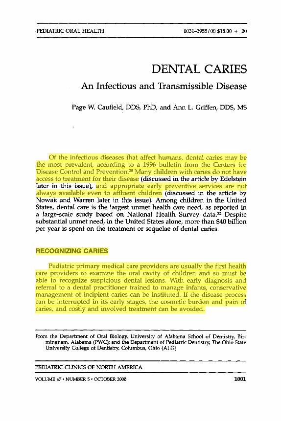
White Spot Stage
The acids produced by microorganisms in dental plaque dissolve the mineral matrix of teeth. In the earliest stages, dental caries appears as a chalky white spot on the tooth (Fig. 1; Fig. 2 in the article by Adair later in this issue; Fig. 2C in the article by Nowak and Warren later in this issue). At this stage, the surface is intact, and the subsurface lesion is reversible. In children less than 3 years of age, incipient caries most commonly is seen on the front surface of the front teeth (Fig. 2C in the article by Nowak and Warren later in this issue), so a simple "lift-the- lip" examination detects most caries. White spots resulting from incipi- ent caries can be difficult to distinguish from developmental hypocalci- fications, but any white spots warrant referral to a dentist for evaluation.
Cavity Stage
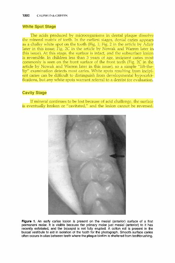
If mineral continues to be lost because of acid challenge, the surface is eventually broken or "cavitated," and the lesion cannot be reversed.
Figure 1. An early caries lesion is present on the mesial (anterior) surface of a first permanent molar. It is visible because the primary molar just mesial (anterior) to it has recently exfoliated, and the bicuspid is not fully erupted. A cotton roll is present in the buccal vestibule to aid in isolation of the tooth for the photograph. Smooth surface caries often occurs in sites between teeth where the plaque biofilm is sheltered from toothbrushing.
DENTAL CARIES 1003
Figure 2. Typical clinical presentation of severe early childhood caries (ECC) secondary to prolonged bottle feeding. A, The preferential involvement of the upper incisors from a frontal view. 13, An occlusal view of the same dentition revealing the involvement of the first primary molars, which are the next teeth to erupt after the incisors. (Courtesy of Dennis J. McTigue, DDS, MS, Columbus, OH.)
Early cavitation is shown in Figure 2C in the article by Nowak and Warren later in this issue. If the lesion progresses, large areas of the tooth can be lost, as seen in Figure 2. Active, cavitated lesions are usually golden brown. Long-standing lesions are darker, sometimes nearly black. Depth of color is not a good indicator of the severity of the lesion because arrested decay is often the darkest. Stain on the enamel surface, particularly in the fissures, can be difficult to distinguish from caries, but any discoloration or irregularity in the enamel surface warrants referral to a dentist for evaluation.
1004 CAUFIELD & GRIFFEN
EPIDEMIOLOGY
Prevalence in the Permanent Dentition
The most comprehensive information on the caries status of children in the United States comes from the large-scale National Health and Nutrition Examination Surveys (NHANES)2z, 23 and National Institute of Dental Research surveys8, 33 conducted over the past three decades. Much attention has been given to the decrease in caries prevalence that has been observed over this time, and the finding that more than half of all children aged 5 to 17 years are now caries free in the permanent denti- tion has been reported widely. This represents a significant improvement over the one fourth of children found to be caries free in the 1970s, but the impression it gives of overall caries prevalence being low is some- what misleading.21 Caries is a cumulative disease that is not initiated until teeth erupt into the oral cavity, and it may not be obvious until some time, perhaps years, after teeth emerge. The last permanent tooth usually emerges around 12 years of age, so caries rates among the oldest cohort examined, 17 years of age, are probably the best measure of how many children are caries free. These detailed data were not reported for the most recent NHANES, but in the 1986-1987 survey, only 15% of 17- year-olds were caries free.8 Although the fraction is probably higher today, it still represents a small minority of US children.
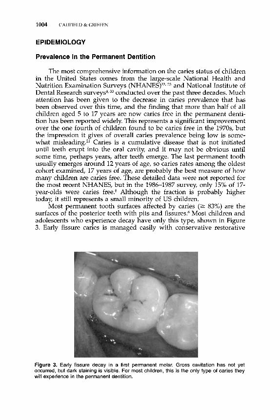
Most permanent tooth surfaces affected by caries (2 83%) are the surfaces of the posterior teeth with pits and fissures.'j Most children and adolescents who experience decay have only this type, shown in Figure 3. Early fissure caries is managed easily with conservative restorative
Figure 3. Early fissure decay in a first permanent molar. Gross cavitation has not yet occurred, but dark staining is visible. For most children, this is the only type of caries they will experience in the permanent dentition.
DENTAL CARIES 1005
treatment or prevented by sealing of the fissures (described in the article by Schafer and Adair later in this issue). This form of decay is self- limiting when all susceptible fissured surfaces are sealed or restored. Obtunding the fissures with sealants prevents cariogenic bacteria from accumulating in these retentive areas. Moreover, bacteria not removed mechanically before sealant placement become metabolically inactive after sealant placement because they are denied access to substrates.
Approximately one quarter of children and adolescents experience more extensive caries that extends to the less susceptible smooth surfaces of the teeth. The 25% of children who experience this pattern of decay account for 80% of the total number of permanent teeth affected by caries.6 If the disease process is uninterrupted in these children (dis- cussed in the article by Schafer and Adair later in this issue), damage to the dentition can be severe.
The prevalence of dental caries is decreasing in most industrialized countries, including the United States5 No clear consensus exists as to the reason for this decline. Some experts believe that the presence of fluoride in municipal water and topically applied dentifrices are the major reasons for the decrease; however, countries such as Japan, where use of fluorides has been minimal, also are experiencing a rapid decrease, so the effect of fluoride may not be a satisfactory explanation. Alterna- tively, some investigators have proposed that the decrease in dental caries in industrialized nations is correlated with an increased prevalence of antibiotic use, particularly among young children.29 The latter hypoth- esis is plausible in that the oral biota, especially streptococci, are sensitive to the most commonly prescribed antibiotics, the cell wall inhibitors, such as penicillin.
On the other hand, the prevalence of caries is increasing rapidly in developing nations, which is of concern because dental caries is mostly a childhood disease, and 80% of the worlds children live in the devel- oping countries. The most likely reasons for this increase in developing countries is a combination of poor nutrition accompanied by the ready availability of inexpensive, sugar-rich foods and beverages. Few areas of the world exist beyond the reach of the sugar-laden symbol of Western culture, Coca-Cola.
Prevalence in the Primary Dentition
Unlike the dramatic decrease in the prevalence of caries observed for permanent teeth, caries rates in the primary dentition have remained unchanged among children of low-income families over the past three decades, and only a small decrease has been observed for children living above the poverty level.7 The NHANES I11 measured caries in the primary incisors of 1-year-old children with a “lift-the-lip” visual inspection, and 2% of infants were found to have abnormal appearing incisors, probably indicative of caries.= Because dental caries is corre-
1006 CAUFJELD & GRIFFEN
lated with infant feeding practices, prevalence varies among cultural and ethnic groups. It has been reported in more than half of all children in some Native American communities, and in more than 10% of chil- dren in Head Start programs.35 Clearly, early childhood caries remains a severe problem and has not been significantly impacted by water fluori- dation or the use of fluoridated dentifrice.
Early Childhood Caries
Early childhood caries (ECC) is the most recent nomenclature for a particular pattern of caries in young children.40 Unlike caries in the permanent dentition, ECC preferentially affects the primary anterior teeth, as shown in Figure 2. It has previously been termed nursing curies or baby bottle tooth decay. This form of caries is associated with frequent and prolonged feeding with a bottle, although it can be associated with training cups, breastfeeding on demand, or the use of sweetening agents on a pacifier. The risk for ECC also may be determined by preexisting developmental defects of the enamel called hypoplasia. Hypoplasia can be difficult to visualize in young children and can be masked by caries. One studyz8 showed that hypoplasia predisposes teeth to early coloniza- tion by the caries pathogen Streptococcus mutans. In this same study, malnutrition was correlated with hypoplasia.
ECC exhibits a characteristic pattern related to the emergence se- quence of the teeth and the tongue position during feeding,35 as seen in Figure 2. The lower teeth are protected from exposure to ingested liquids by the tongue during feeding and by the pooling of saliva and so usually are not affected. The incisors are the first upper teeth to emerge and are most affected by ECC. Depending on how long the caries process is active, the upper first primary molars often are involved, followed by the upper second molars and canines, and, in severe cases, the lower teeth. Treatment of ECC often requires hospitalization with the patient under general anesthesia, which is discussed in the article by Wilson later in this issue.
ODONTOGENIC INFECTIONS
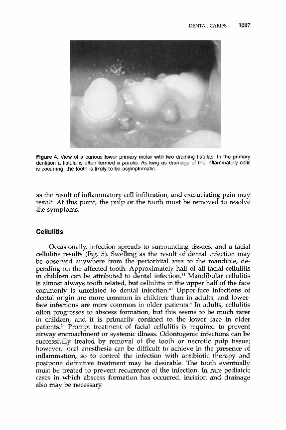
If decay progresses to the vital pulp of a tooth, it results in sensitiv- ity to cold or osmotic stimulation from sugary foods. If the decay is controlled by prompt treatment, the prognosis for survival of a vital pulp is good. If the decay is allowed to progress and a bacterial infection is established in the pulp chamber, spontaneous tooth pain often results. The immune response usually can contain bacteria within the pulp chamber, but a fistula is sometimes established between the tooth apex and the oral cavity, through which pus drains, as shown in Figure 4. Only rarely, an extraoral fistula develops. As long as a patent fistula is present, the tooth is usually asymptomatic. Without drain- age, pressure in the pulp chamber and periapical area may build
DENTAL CARIES 1007
Figure 4. View of a carious lower primary molar with two draining fistulas. In the primary dentition a fistula is often termed a parulis. As long as drainage of the inflammatory cells is occurring, the tooth is likely to be asymptomatic.
as the result of inflammatory cell infiltration, and excruciating pain may result. At this point, the pulp or the tooth must be removed to resolve the symptoms.
Cellulitis
Occasionally, infection spreads to surrounding tissues, and a facial cellulitis results (Fig. 5). Swelling as the result of dental infection may be observed anywhere from the periorbital area to the mandible, de- pending on the affected tooth. Approximately half of all facial cellulitis in children can be attributed to dental infectionA1 Mandibular cellulitis is almost always tooth related, but cellulitis in the upper half of the face commonly is unrelated to dental infectionA1 Upper-face infections of dental origin are more common in children than in adults, and lower- face infections are more common in older patient^.^ In adults, cellulitis often progresses to abscess formation, but this seems to be much rarer in children, and it is primarily confined to the lower face in older patients.20 Prompt treatment of facial cellulitis is required to prevent airway encroachment or systemic illness. Odontogenic infections can be successfully treated by removal of the tooth or necrotic pulp tissue; however, local anesthesia can be difficult to achieve in the presence of inflammation, so to control the infection with antibiotic therapy and postpone definitive treatment may be desirable. The tooth eventually must be treated to prevent recurrence of the infection. In rare pediatric cases in which abscess formation has occurred, incision and drainage also may be necessary.
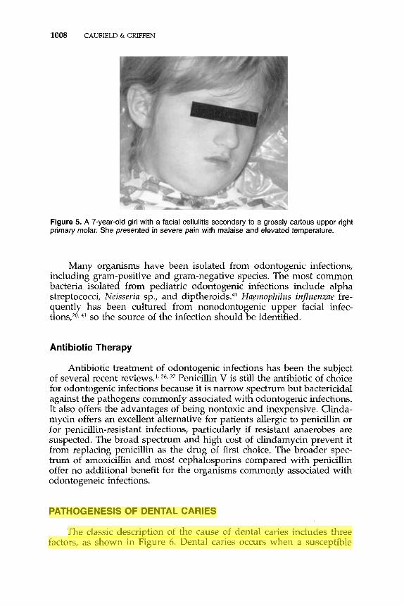
1008 CAUFIELD & GRIFFEN
Figure 5. A 7-year-old girl with a facial cellulitis secondaty to a grossly carious upper right primary molar. She presented in severe pain with malaise and elevated temperature.
Many organisms have been isolated from odontogenic infections, including gram-positive and gram-negative species. The most common bacteria isolated from pediatric odontogenic infections include alpha streptococci, Neisseriu sp., and diptheroid~.~~ Huemophilus inf2uenzae fre- quently has been cultured from nonodontogenic upper facial infec- tions,20, 41 so the source of the infection should be identified.
Antibiotic Therapy
Antibiotic treatment of odontogenic infections has been the subject of several recent reviews.', 36, 37 Penicillin V is still the antibiotic of choice for odontogenic infections because it is narrow spectrum but bactericidal against the pathogens commonly associated with odontogenic infections. It also offers the advantages of being nontoxic and inexpensive. Clinda- mycin offers an excellent alternative for patients allergic to penicillin or for penicillin-resistant infections, particularly if resistant anaerobes are suspected. The broad spectrum and high cost of clindamycin prevent it from replacing penicillin as the drug of first choice. The broader spec- trum of amoxicillin and most cephalosporins compared with penicillin offer no additional benefit for the organisms commonly associated with odontogeneic infections.
PATHOGENESIS OF DENTAL CARIES
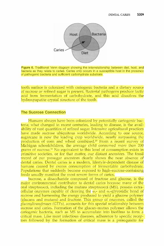
The classic description of the cause of dental caries includes three factors, as shown in Figure 6. Dental caries occurs when a susceptible
DENTAL CARIES 1009
Figure 6. Traditional Venn diagram showing the interrelationship between diet, host, and bacteria as they relate to caries. Caries only occurs in a susceptible host in the presence of pathogenic bacteria and sufficient carbohydrate substrate.
tooth surface is colonized with cariogenic bacteria and a dietary source of sucrose or refined sugar is present. Bacterial pathogens produce lactic acid from fermentation of carbohydrate, and this acid dissolves the hydroxyapatite crystal structure of the tooth.
The Sucrose Connection
Humans always have been colonized by potentially cariogenic bac- teria; what changed in recent centuries, leading to disease, is the avail- ability of vast quantities of refined sugar. Intensive agricultural practices have made sucrose ubiquitous worldwide. According to one source, sugarcane is now the leading crop worldwide, nearly surpassing the production of corn and wheat ~0mbined.l~ From a recent survey of Michigan schoolchildren, the average child consumed more than 200 grams of sucrose.l0 No equivalent to this level of consumption exists in primitive societies, or for that matter, our distant ancestors. The fossil record of our presugar ancestors clearly shows the near absence of dental caries. Dental caries is a modern, lifestyle-dependent disease of humans caused by excess consumption of fermentable carbohydrate. Populations that suddenly become exposed to high-sucrose-containing foods usually manifest the most severe forms of caries.
Sucrose, a disaccharide composed of fructose and glucose, is the major environmental contributor to dental caries because many of the oral streptococci, including the mutans streptococci (MS), possess extra- cellular enzymes capable of cleaving the al- and au,-glycosidic bond of sucrose and harnessing the energy produced to yield a glucose polymer (glucans and mutans) and fructose. This group of enzymes, called the gZucosyZtransferases (GTFs), accounts for this special relationship between sucrose and caries. Formation of the glucan-mutan polymer allows the cariogenic bacteria, such as MS to accumulate into biofilms to form a critical mass. Like most infectious diseases, adherence to specific recep- tors followed by the formation of critical mass is a prerequisite for

1010 CAUFIELD At GRIFFEN
disease. Without formation of critical mass, cariogenic bacteria would colonize the oral cavity but not be massed so as to cause destruction of the enamel surface. Accordingly, the formation of critical mass that is uniquely associated with GTF and sucrose is the biological reason for man's recent affliction with caries. Clearly, the reduction of refined sugar, especially sucrose, would be the most direct way of controlling dental caries. This prospect, although straightforward, approaches impractical- ity because sucrose is nearly ubiquitous among most human popula- tions. Moreover, the reduction would be not only costly but also must be dramatic to be effective,'O especially on a global basis.
Dental Caries: An Infectious Disease
Because bacteria cause dental caries, it is, by definition, an infectious disease. Unlike most infectious diseases encountered by pediatricians that are caused by exogenous pathogens, however, the source of bacteria responsible for caries arises from the bacterial populations indigenous to the oral cavity or from the so-called "normal flora." Caries also is considered a transmissible disease, but perhaps not in the traditional sense of the more familiar infectious diseases of children, such as measles and chickenpox. In the case of dental caries, the bacteria responsible for disease, together with the other indigenous biota, generally are transmit- ted vertically, from mother to childz7 compared with the other more familiar infectious diseases of childhood, which are transmitted horizon- tally from infected to uninfected people. Epidemics arise from horizon- tally transmitted diseases. So, although dental caries is an infectious disease, measures to prevent and control its spread and pathogenic potential are limited to and distinct from measures developed for pre- venting the transmission and disease potential of the horizontally trans- ferred infectious diseases. Because mothers are the major source of cariogenic bacteria to their children and sucrose consumption modulates the expression of disease, approaches aimed at interfering or preventing mother-child transmission hold promise. Such methods have been tested and continue to be tested, with varying degrees of success.'8, 26.
Bacterial Cause of Dental Caries
Historically, many theories have been invoked as to the cause of dental caries. The anciently derived Chinese character for dental caries depicts a worm, reflecting the notion that worms were the cause of dental caries. The bacterial cause of caries was definitively recognized following demonstration of two principles. The first demonstration was in the 1950s with the development of germ-free laboratory animals.% These germ-free rodents that were fed various diets, including those high in sugar and carbohydrates known to promote dental caries, re- mained caries free in the absence of bacteria. These same germ-free
DENTAL CARIES 1011
animals were then inoculated with certain oral bacteria and developed canes. The second demonstration of the relationship between dental caries and bacteria was preformed in 1960 by Keyes at the National Institutes of In fulfillment of Koch's postulate, Keyes inoculated pups with bacteria found in feces from caries-free or caries-active dams, showing caries was the result of specific infection by an unrecognized streptococcus. Keyes then showed that transmission could be interrupted and a reduced caries state ensued by administration of antibiotics active against the gram-positive biota.
In keeping with what was demanded for other infectious diseases, the satisfaction of Koch's postulates was, until recently, considered the sine qua non in showing that a disease is caused by a specific infectious agent. Holding dental caries to this standard of proof, that is, the "one bug, one disease, one bullet" approach, dental scientists have concen- trated their efforts on understanding the natural history of only a few oral microbes from among the 1000 or more genotypes present in the oral cavity. The leading candidates are among the lactic-acid producers of the plaque biofilm because the elaboration of acid leads to demineral- ization of the tooth substance.
The Mutans Streptococci
From these pioneering experiments of Keyes and others, the organ- ism associated with caries was identified as a member of the genus Streptococcus, similar or identical to a Streptococcus sp. described in litera- ture in 1924 by a British physician, Clarke, named S. mutuns.16 Clarke isolated his strain of S. mutans from the cavity of a caries-active child. The term mutuns was applied because the cocci underwent changes in morphology and retention of Gram stain as the cultures aged. Clarke erroneously attributed this to mutational events, hence the name mutans. Taxonomists then classified cariogenic streptococci into various subspe- cies, then genospecies, based on the increasing number of distinctive characteristics, including G + C content, DNA-DNA hybridization, and serotyping, among others. Collectively, this phenotypically similar group of bacteria was called the mutans streptococci (MS). Only two species of the MS, S. mutans and S. sobrinus, are associated with human caries, however.
As more information becomes available, Koch's postulates seem to apply only to a special subset of human pathogens, and the assumptions seem to be valid only under certain conditions. For example, the notion that a microbe must be isolated from diseased tissue and then "pure cultured applies to only a few obligate human parasites. The spirochete T. pallidurn, responsible for syphilis, for example, has yet to satisfy Koch's postulates because of its noncultivability. Because S. mutans could be pure cultured and caused disease in laboratory animals, investigators pronounced S. mutuns the causative agent of caries, and for the past 30 years, the mutans streptococci have been designated the principal agents
of caries. The selection of the MS as the causative agent of caries is not without substantial support, both in vivo and in vitro. As once stated by one proponent of the MS school of caries, “S. mutuns was intentionally designed from the bottom up to be a cariogenic ~rganism.”’~ What properties, u priovi, make S. mutuns a good candidate as the causative agent of caries? The ability to metabolize sucrose with GTFs and to elaborate acid likely would be of primary importance because of the association with sucrose and the process of demineralization, respec- tively. The putative etiologic agent should be found at the disease site, that is, the tooth surfaces, and be readily isolated from disease sites. For these and several other reasons, the MS seemed a good fit for the Koch model of infectious disease.
The cause of dental caries likely is not confined to a single organism but rather to a constellation of organisms and interactions within the plaque biofilm. The concept of dental plaque, comprised of more than 500 distinct genotypes, as a multicellular organism, may supplant the Koch “one bug, one disease” concept of these types of endogenous infections. Considering a genomic size for bacteria of approximately lo6 nucleotides, lo3 different genotypes collectively contain lo9 nucleotides, a close approximation of the human genome. With such a large reper- toire of DNA-coding segments present in plaque, the metabolic events responsible for cariogenicity likely resides with more than those confined to a single bacterial type. Modern molecular genetic tools allow research- ers to narrow down the collection of genetic loci within the plaque biofilm associated with caries irrespective of the specific bacterial geno- types harboring those loci. Risk may then be assessed based on genetic, rather than microbiological markers.
Acquisition in lnfants
Longitudinal studies show that MS colonize the oral cavity of in- fants sometime after the emergence of their first set of teeth. Typically, MS colonize the child’s dentition at approximately 2 years of age during a period called the window of infecti~ity.’~ This period of colonization is correlated with the surface area of the primary teeth because teeth are a prerequisite for colonization. Primary teeth emerge from 7 to 24 months of age; at 24 months, all 20 primary teeth usually are present. When teeth have emerged into the oral cavity, they are colonized not only by MS but also by other members of the oral biota. Because MS are a poor colonizer of tooth surfaces compared with other oral bacteria, its “window of infectivity” relies on the newly emerged, virgin teeth to gain an initial colonization. As infant’s teeth acquire a stable biofilm, the ability of MS to colonize becomes greatly reduced. Hence, the window of infectivity for acquiring MS is limited to the period of newly emerging teeth, after which the window closes. For a review, see the article by Caufield.lz A second window may open when permanent teeth begin emerging at 6 years of age, but the source of MS may be from reservoirs
DENTAL CARIES 1013
already established in the primary dentition. The time to colonization in infants varies depending on environmental factors, such as diet, level of exposure from other infected individuals, and tooth composition. As mentioned previously, infants experiencing malnutrition in utero often exhibit hypoplasia of tooth enameLZ8 MS more readily colonize these teeth because of their rough surfaces, so initial colonization occurs at an earlier age. Colonization of infants by MS preceding the 2-year median age of infectivity is not uncommon among some populations of infants in poverty. These infants may well have subclinical hypoplasia of enamel as a preexisting condition, and this, coupled with unfavorable dietary intake, leads to earlier colonization.
Fidelity of Transmission
Children’s caries experience is better correlated with their mothers’ experience than to their fathers’, which may be explained, in part, by the fact that mothers usually are responsible for nurturing infants for the first few years of life. Also, the mother is also the most likely source of oral bacteria because infants acquire their own complement of indigenous biota as they develop. Twenty-four to 36 hours after birth, the infant’s oral cavity harbors adult levels of oral bacteria in saliva, approximately lo8 bacteria per miililiter of saliva (Mongomery and Cau- field, unpublished data, 1994). Although MS are not detected until after teeth emerge, other oral bacteria that colonize the surfaces of the tongue and buccal mucosa become established early. Later, when teeth emerge, the oral environment becomes receptive to MS colonization. Where do these MS come from? Ultimately, the source is usually, but not always, from the mother. DNA fingerprinting shows that infants’ genotypes of MS match their mothers’ in more than 70% of cases. The way to demon- strate that two or more strains are the same is by using a genotyping method, such as chromosomal DNA fingerprinting or arbitrarily primed polymerase chain reaction. If an isolate from the mother has the same DNA fingerprint as that from the child, then the two strains are identical and come from the same source. Speculation as to how the MS are transferred during this window period (median, 26 mo of age) include contact with mother’s saliva or droplets, but direct proof is lacking. Also likely, but also unproven, is that MS are transmitted from mother to infant at birth, but the MS reside undetected in low levels in reservoirs such as the tonsils or tongue dorsum. When the right environmental conditions present, with the emergence of primary teeth, MS grow and reach detectable levels. Detection of MS before tooth emergence has been observed, and more sensitive techniques using PCR will undoubtedly show the presence of MS and all other of the indigenous biota present just after birth, which makes evolutionary sense because the orderly transfer of indigenous biota from mother to offspring is a recurring theme in the lower invertebrates in which such studies are available.” 31
1014 CAUFTELD & GRIFFEN
C/ona/ity Among Strains of Streptococcus mutans
A recent study using sequence comparisons from both 16s rDNA and the hypervariable region of a cryptic plasmid suggests that MS strains are clonal, at least among plasmid-containing strains.” If this is true for all MS, then MS may differ in their ability to initiate and promote caries, and only certain members of the MS family may be associated with disease. Coupled with the concept of timing to acquisi- tion (window of infectivity), and source of acquisition (fidelity), a child’s caries experience, at least from a microbiological perspective, may be a function of all three (or more) of these factors. If so, diagnosis and preventive strategies will require additional information concerning the natural history of acquisition to assess caries risk.
27 have shown that strains of S. mutans cluster within familial and racial cohorts, suggesting that strains are conserved within maternal lineage. Using sequence data from a cryptic plasmid present in approximately 5% of the strains of MS, they have been able to show that certain strains of MS arising out of Africa are stably conserved and follow the migration of their human host, in this case, individuals of African descent in the United States.” Because all three individuals were of African descent, it appears that the original strain arose out of Africa and was transmitted to descendants, some of whom immigrated to the Americas as slaves. Plasmid strains from around the world cluster according to their host. That is, the group I1 plasmid strains are found exclusively in whites, and the group I are found in Asian and black populations. This observation supports the notion that MS are transferred along maternal lines. In these instances, and likely among the nonplasmid strains, MS are clonal within maternal lineage. Certain strains, hence certain maternal lineages may be more virulent, which might explain in part the mosaic caries experience seen among different cultural and ethnic groups worldwide.
Caufield et
Open Questions on Bacterial Cause
The MS story, although compelling, is incomplete. It seems reason- able that, if a particular bacteria were the principal cause, its presence should be predictive of disease, that is, early colonization and, in relative abundance, should predate disease. However, longitudinal studies show that MS presence or levels predict caries only in approximately 20% of cases. As concluded by Burt et a1,9 “S. mutans is necessary, but not sufficient.” Children harboring high and low levels of MS experience the entire range of caries, from none to many. All humans (with teeth) harbor this organism, regardless of caries status, but not all humans get cavities. How then, can the specific association between a microbe and a disease be explained? One explanation is that strains of MS vary in their disease-causing potential (virulence; see previous section on clon- ality) and that humans who manifest disease are colonized by a virulent
DENTAL CARIES 1015
strain. Another explanation is that MS are only one of several bacteria responsible for dental caries. Because of limitations in the ability to detect and culture bacteria from the plaque biofilm, many putative candidates likely have been overlooked. Advances in genetic typing and detection of "uncultivable" bacteria now are being applied to examining the caries oral biota, and additional species appear to be consistently present in caries-producing biofilms.3 Caries risk assessment in the future may be composed of detection of genetic markers associated with caries potential within the plaque biofilm rather than just isolating single or groups of individual bacterial entities.
CHEMOTHERAPEUTICS
Fluoride
Fluoride from small quantities in the drinking water or in the form of other, more concentrated delivery systems, such as mouthwashes, toothpastes, and oral supplements, is well established as effective in reducing caries increment (discussed in the article by Adair later in this issue). The way in which fluoride effects its anticaries action remains controversial. Most oral health professionals believe that fluoride ions interact with the mineral of the tooth surface, rendering it less suscepti- ble to acid demineralization by cariogenic bacteria. That fluoride accu- mulates in the mineralized tissues, including bone, is not questioned; biopsies of mineralized tissue clearly indicate that fluoride accumulates in these tissues. How the alteration in mineralized tissues, including the teeth, results in less caries is debatable. The alternative theory holds that fluoride is concentrated in the surface of the teeth and then released into the plaque biomass as acid demineralizes the tooth surface. Fluoride has been well established as a potent inhibitor of enolase enzyme, a key enzyme on the glycolytic pathway of virtually all forms of life. Inhibition of enolase, in turn, results in less acid production among oral strepto- cocci, including S. mutans. Fluoride leached from the tooth surface, therefore, acts as a classic example of feedback inhibition.
Other Antimicrobial Agents
Shortly after the introduction of penicillin and, in later years, vanco- mycin, the notion that dental caries could be treated by antibiotics was tested in laboratory animals. This idea was not pursued, mainly because of the growing fear that pathogenic bacteria may become tolerant to the regular use of sublethal doses of antibiotic. Interestingly, and supportive of the bacterial cause of dental caries, children receiving regular doses of antibiotics for amelioration of symptoms associated with cystic fibrosis exhibited significantly less tooth decay compared with the nonmedicated population.
1016 CAUFIELD & GRIFFEN
Chlorhexidine and Other Chemicals
Chlorhexidine was one of the first antiseptic agents proposed for use against dental caries and has proved to be the most effective. Administered by oral lavages or by gels in custom mouthpieces, chlor- hexidine effects modest reductions in caries-approximately 30% in vari- ous populations. Because cariogenic bacteria are thought to be trans- ferred from mother to child, several groups have attempted to reduce the levels of cariogenic bacteria so that lesser amounts are transmitted to the infant.1E,25,42 The strategy is appealing in that it constitutes primary prevention, attacking the source and nature of the cause.
DENTAL CARIES VACCINES
For the past 30 years, the prospect of developing a vaccine against dental caries has had tremendous appeal. The use of vaccines against other infectious diseases afflicting children has profoundly changed the practice of medicine and has led to a marked improvement of life expectancy and quality of life compared with the last century. Enthusi- asm for developing a vaccine against caries was high after identifying the MS as the prime causes of dental caries. Identification of specific antigenic targets for stimulating immunologic response ensued, as did various methods for inducing the immune response (for a review, see references 30 and 39). Vaccine approaches for eliciting an IgA or IgG response are being debated. Results using animal models have been promising, especially coupled with newer forms of antigen delivery, such as liposomes, but safety and efficacy in humans have not yet been demonstrated. Whether MS alone are the appropriate target, or if other bacteria must be included, is still an open question. Moreover, targeting members of the indigenous flora, as opposed to exogenous pathogens, requires that immunoglobulins distinguish self from non-self antigens so as not to induce an autoimmune reaction. Despite some difficulties that must be overcome, the development of an inexpensive and effective vaccine would be a tremendous tool for combating dental caries, espe- cially in developing countries.
SURGICAL MANAGEMENT OF CARIES
Cavity formation results from demineralization and subsequent cav- itation of the tooth surface as the result of bacterial fermentation of carbohydrates, particularly refined sugar. Once cavitation occurs, it is irreversible and provides an ecologic niche that favors caries progression. When dental treatment is available, the process involves removal of the diseased tissue and replacement of the lost enamel or dentin with precious metals or the newer, tooth-colored resins. If caries has pro- gressed until the tooth becomes irreversibly affected by bacterial necrosis of the pulp, pain results. Tooth extraction is the method of treatment
DENTAL CARIES 1017
for many individuals, although more expensive restorative therapies involving cleaning of the pulp chamber and filling of the empty canals (i.e.; "root canal") are available. Although the lesion is restored by these approaches, the etiologic agent, that is, the specific bacteria of the plaque biofilm, are left basically intact, available for initiating new lesions should the right environment present. This surgical management of dental caries has been the standard for more than 100 years and consti- tutes the main approach to the treatment of caries.
Treatment of dental caries as an infectious disease is in its infancy. Most effective interventions rely on patient adherance to recommenda- tions that often require significant lifestyle changes (discussed in the article by Schafer and Adair later in this issue) and often are ignored. Highly effective therapies that can be used by health care providers are not yet available. Debridement of caries, followed by disinfecting the lesion before placement of bonded restorative material, coupled with comprehensive sealant application, may prove effective in reducing the cariogenic bacterial reservoirs, hence preventing future caries, but fol- low-up studies are needed to validate this appr0a~h.I~~
ROLE OF THE PEDIATRICIAN
Most children in the United States experience tooth decay, and the appropriate use of dental sealants and systemic and topical fluorides would prevent caries in most of these children. Referral of all children for routine dental care will help to limit the destruction associated with the milder forms of caries. In addition to making referrals, pediatricians can play an important role in caries prevention by seeing that infants receive optimal fluoride exposure from drinking water or in fluoride- deficient areas, by supplementation (see article by Schafer and Adair later in this issue). They also can identify high-risk feeding practices and provide appropriate diet counseling to reduce the risk for caries (dis- cussed in the article by Nowak and Warren later in this issue). Family history of significant decay and low socioeconomic status are risk indica- tors for caries and should trigger a careful examination. Early referral of children for dental assessment around the time of tooth eruption is probably the most effective measure to reduce caries risk (discussed in the article by Nowak and Warren later in this issue).
Most caries in US children are found in just 25% of the population. These children will benefit from identification by their primary health care providers and referral for appropriate care as soon as possible. Pediatric primary health care providers see many more children than are examined by dental colleagues and can play a major role in securing early intervention for children with caries. Simply "lifting the lip" and examining the anterior maxillary incisors for signs of caries (described in the previous section on recognizing caries and in the article by Nowak and Warren later in this issue) should be included in every infant pediatric examination. A brief visual dental inspection of anterior and posterior teeth should be a part of every routine physical assessment for older children.
1018 CAUFIELD & GRIFFEN
References
1. Baker KA, Fotos PG: The management of odontogenic infections: A rationale for appropriate chemotherapy. Dent Clin North Am 38:689-706, 1994
2. Baumann P, Baumann L, Lai CY, et al: Genetics, physiology, and evolutionary relation- ships of the genus Buchnera: Intracellular symbionts of aphids. Annu Rev Microbiol 49:55-94, 1995
3. Becker MR, Griffen AL, Leys EJ, et al: Checkerboard analysis of bacterial species associated with early childhood caries. J Dent Res 89(special issue):620, 2000
4. Biederman GR, Dodson TB: Epidemiologic review of facial infections in hospitalized pediatric patients. J Oral Maxillofac Surg 521042-1045, 1994
5. Bratthall D, Hansel-Petersson G, Sundberg H: Reasons for the caries decline: What do the experts believe? Eur J Oral Sci 104(4 Pt 2):41&422; discussion 42%425, 430- 432, 1996
6. Brown LJ, Kaste LM, Selwitz RH, et al: Dental caries and sealant usage in US. children, 1988-1991: Selected findings from the Third National Health and Nutrition Examination Survey. J Am Dent Assoc 127335343,1996
7. Brown LJ, Wall TP, Lazar V: Trends in total caries experience: Permanent and primary teeth. J Am Dent Assoc 131223-231, 2000
8. Brunelle J A Oral health of United States Children: The National Survey of Dental Caries in US School Children: 1986-1987. N M Publication 89-2247. National Institute of Dental Research, Epidemiology and Disease Prevention Program, 1989
9. Burt BA, Loesche WJ, Eklund SA, et al: Stability of Streptococcus mutuns and its relationship to caries in a child population over 2 years. Caries Res 17532-542, 1983
10. Burt BA, Szpunar SM The Michigan study: The relationship between sugars intake and dental caries over three years. Int Dent J M.230-240, 1994
11. Caufield PW, Li Y, Zou ZH: Evidence of clonality among African-derived strains of S. mutans. J Dent Res 78:345, 1999.
12. Caufield P W Dental caries: A transmissible and infectious disease revisited: A posi- tion paper. Pediatr Dent 19:491498, 1997
13. Caufield PW, Cutter GR, Dasanayake AP: Initial acquisition of mutans streptococci by infants: Evidence for a discrete window of infectivity. J Dent Res 72:3745, 1993
13a. Caufield PW, Mitchell JD, Ruby Y, et al: Alabama antimicrobial restorative treatment (AART): A novel approach to prevention and treatment of dental caries. J Dent Res 79(abstract):593, 2000
14. Caufield PW, Ratanapridakul K, Allen DN, et al: Plasmid-containing strains of Strepto- coccus mutuns cluster within family and racial cohorts: Implications for natural trans- mission. Infect Immun 56:3216-3220, 1988
15. Caufield PW, Wannemuehler YM, Hansen JB: Familial clustering of the Streptococcus mutuns cryptic plasmid strain in a dental clinic population. Infect Immun 38:785- 787, 1982
16. Clarke JK On the bacterial factor in the aetiology of dental caries. Br J Exp Pathol
17. Coykendall AL (ed): On the Evolution of Streptococcus mutuns and Dental Caries, vol 111. Information Retrieval, Inc., Washington DC, 1976
18. Dasanayake AP, Caufield PW, Cutter GR, et al: Transmission of mutans streptococci to infants following short term application of an iodine-NaF solution to mothers’ dentition. Community Dent Oral Epidemiol 21:136-142, 1993
19. Diamond J: Guns, Germs and Steel. New York, WW Norton, 1997 20. Dodson TB, Perrot DH, Kaban LB: Pediatric maxillofacial infections: A retrospective
study of 113 patients. J Oral Maxillofac Surg 47327-330, 1989 21. Edelstein BL, Douglass CW Dispelling the myth that 50 percent of U.S. schoolchildren
have never had a cavity. Public Health Rep 110:522-530; discussion 521,531-533,1995 22. Kaste LM, Selwitz RH, Oldakowski RJ, et al. Coronal caries in the primary and
permanent dentition of children and adolescents 1-17 years of age: United States, 1988-1991. J Dent Res 75:631441, 1996
23. Kelly JE, Harvey CR Basic data on dental examination findings of persons 1-74 years: United States, 1971-1974. Vital Health Stat 1l:l-33, 1979
5~141-147, 1924
DENTAL CARIES 1019
24. Keyes PH: The infectious and transmissible nature of experimental dental caries. Arch Oral Biol 1:304-320, 1960
25. Kohler B, Andreen I Influence of caries-preventive measures in mothers on cariogenic bacteria and caries experience in their children. Arch Oral Biol39907-911, 1994
26. Kohler 8, Bratthall D, Krasse B: Preventive measures in mothers influence the estab- lishment of the bacterium Streptococcus mutans in their infants. Arch Oral Biol28:225- 231, 1983
27. Li Y, Caufield P W The fidelity of initial acquisition of mutans streptococci by infants from their mothers. J Dent Res 74:681-685, 1995
28. Li Y, Navia J, Caufield PW. Colonization by mutans streptococci in the oral cavity of 3- and 4-year-old Chinese children with or without enamel hypoplasia. Arch Oral Biol 391057-1062, 1995
29. Loesche WJ, Eklund SA, Mehliscl DF, et al: Possible effect of medically administered antibiotics on the mutans streptococci: Implications for reduction in decay. Oral Microbiol Immunol477-81, 1989
30. Michalek SM, Childers N K Development and outlook for a caries vaccine. Crit Rev Oral Biol Med 1:37-54, 1990
31. Moran NA: Accelerated evolution and Muller’s rachet in endosymbiotic bacteria. Proc Natl Acad Sci USA 93:2873-2878, 1996
32. Newacheck PW, Hughes DC, Hung YY, et al: The m e t health needs of America’s children. Pediatrics 105(4 Pt 2):989-997, 2000
33. NIDR The prevalence of dental caries in United States children: The National Dental Caries Prevalence Survey: 1979-1980. NIH Publication 82-2245. National Institute of Dental Research, National Caries Program, 1981
34. Orland FJ, Blayney JR, Harrison RW: Use of the germfree animal technic in the study of experimental dental caries: I. Basic observations on rats reared free of all microorganisms. J Dent Res 33147-174, 1934
35. Ripa LW Nursing caries: A comp-rehensive review. Pediatr Dent 1026kL282, 1988 36. Sandor GK, Low DE, Judd PL, et ak Antimicrobial treatment options in the manage-
ment of odontogenic infections. J Can Dent Assoc 64:508-514, 1998 37. Sands T, Pynn BR, Katsikeris N: Odontogenic infections: 11. Microbiology, antibiotics
and management. Oral Health 85:ll-14, 17-21, 23 passim, 1995 38. Service PH: Healthy people 2000 National health promotion and disease prevention
objectives. Full report, with commentary. DHHS Publication # (PHS) 91-50212. US Department of Health and Human Services, Public Health Service, 1991
39. Smith DJ, Taubmar MA: Potential for glucosyltransferase-based synthetic peptides in a dental caries vaccine. Adv Exp Med Biol9:1157-1159, 1995
40. Tinanoff N, OSullivan D M Early childhood caries: Overview and recent findings. Pediatr Dent 1912-16, 1997
41. Unkel JH, McKibben DH, Fenton SJ, et ak Comparison of odontogenic and nonodon- togenic facial cellulitis in a pediatric hospital population. Pediatr Dent 19:47&479, 1997
42. Wright JT, Cutter GR, Dasanayake AP, et al: Effect of conventional dental restorative treatment on bacteria in saliva. Community Dent Oral Epidemiol20:138-143, 1992
Address reprint requests to Page W. Caufield, DDS, PhD
University of Alabama School of Dentistry Box 13
1919 Seventh Avenue, South Birmingham, AL 35294
e-mail: [email protected]


![[Dental caries and socioeconomic conditions in the State of Paraná, Brazil, 1996]](https://img.dokumen.tips/doc/110x75/635d1f62095e4caf22059111/dental-caries-and-socioeconomic-conditions-in-the-state-of-parana-brazil-1996.jpg)