Embed Size (px)
Citation preview
INTRODUCTION
Since the late 19th century, it has been recognized that detection and classificationof dental caries are not easy tasks. The problems of misdiagnosis of caries lesions
and "hidden caries" are not new phenomena (Knapp, 1868; Anonymous, 1869). Thefollowing account, reported in 1869 by a dentist from Missouri, USA (Anonymous,1869), clearly shows the dilemmas that faced and still face dentists. The authorreported:
"A few months ago a young lady called on me for an examination of her teeth. Iendeavored to make it thorough. Seventeen cavities were found and so reported toher. Her astonishment was very great for she had just come from one who had madean examination of her teeth, and reported four cavities. In a couple of weeks I hadfinished operations on her teeth and plugged eighteen cavities."
That dentist's solution to the problem, however, has also been the subject ofdebate and study (and controversy) during at least the last 20 years. He reported:
"There are some cases of failure in diagnosis of dental decay, even when oneintends to be very thorough. First and foremost is the large size of the excavator usedfor examination. The...excavator should be of the very smallest kind, and hatchetshaped..... This excavator should be made for diagnosis alone, and not for cuttingenamel or dentine. The mouth mirror is another cause of defective diagnosis. Onethat magnifies two diameters should be used, and not the ordinary natural mirror.Saliva often obscures slight decay, especially in the fissures of the bicuspids."
Interestingly, the same dentist proclaimed that "once the teeth are separated, agood eye, experienced in this kind of diagnosis, and the mirror will usually besufficient" (Anonymous, 1869). Visual detection of dental caries is not a newsuggestion!
The concept that dental caries is a process rather than a categorical disease with"cavitated" and "not cavitated" states was also reported over 100 years ago. Magitot(1886) divided the diseases into three stages: caries of enamel, caries of dentin, anddeep caries. Morsman, in 1888, stressed the importance of diagnosis as the "firststep" in the management of dental caries—a goal that is yet to be universallyachieved and supported.
In the early decades of the 20th century, the technical foundation of restorativedentistry was developed. In the USA, a pioneering and inquisitive dentist, dentalteacher, and researcher, Dr. G.V. Black, developed a system for restoring decayedteeth. Dr. Black was surprisingly well aware of the limitation of the restorativeapproach to management of dental caries (Black, 1880, 1910, 1922, 1924). A recentdiscovery of a speech presented in 1910 confirmed that he did recognize theimportance of caries in enamel. In a visionary lecture before the Philadelphia dentalsociety, Dr. Black (1910) stated:
"Studies of [the] beginning caries should be continuously made, as it appears inthe teeth of patients in the chair from day to day, with the view of becoming morefamiliar with its tendencies to spread on the surface of the enamel and the positionsand directions of spreading.
"The whole subject of caries of the enamel is a most important one in its relationto everyday practice..."
The dilemma is that while several solutions have been proposed, we still do nothave consistent and valid systems for clinical caries detection. Hence, this paper aimsto evaluate the content validity of published visual and visuo-tactile caries detectionsystems. Content validation refers to the comprehensiveness of a system used tomeasure a phenomenon (Feinstein, 1987).
Content validity, in contrast to criterion validity (correlational or predictivevalidity), is not judged by statistical analyses (e.g., sensitivity and specificity or ROCanalysis). While research of caries diagnostic methods has focused exclusively oncriterion validity, the content validity of existing and proposed systems has not yet
ABSTRACTThe objective of this review is to describe and discuss thecontent validity of a sample of caries detection criteriareported in the literature between January 1, 1966, and May1, 2000. Using filters to locate randomized or controlledclinical trials on dental caries, fluorides, sealants, and"restorative" care, I identified a total of 171 documentsfrom MEDLINE and the Cochrane Collaboration's OralHealth Group (CC-OHG) special register. These articlesmet the following inclusion criteria: (1) Data had beencollected from samples of patients or populations; and (2)dental caries was assessed clinically, and criteria wereeither published or described in the paper. From theselected articles, evidence tables were prepared describingeach caries detection criterion. Analysis of the contentvalidity of the criteria systems was based on evaluation ofthe disease process, exclusion of non-caries lesions,subjectivity, use of explorers, and drying of teeth prior toexamination. This review included 29 unique criteriasystems. Of those, 13 originated from the UK, 3 from theUSA, 4 from Denmark, and others from the World HealthOrganization (WHO), Sweden, Switzerland, Norway,Netherlands, and Canada. Thirteen of the criteria systemseither measured active and inactive early and cavitatedlesions or defined separate criteria for smooth and occlusaltooth surfaces. Nine systems measured early as well ascavitated stages of the caries process, and 7 measuredcavitation only. Eleven of the criteria systems providedexplicit descriptions of the disease process measured orinformation on how to exclude non-caries from carieslesions. The use of explorers and drying and cleaning ofteeth varied widely among the criteria. The majority of thenewly developed criteria systems originated from Europe.In conclusion, this review of the content validity of the 29criteria systems found substantial variability in diseaseprocesses measured, inclusion and exclusion criteria, andexamination conditions.
KEY WORDS: dental caries, diagnosis, detection,validity, criteria, measurement.
Presented at the International Consensus Workshop onCaries Clinical Trials, Glasgow, Scotland, January 7-10,2002
Visual and Visuo-tactile Detection of Dental Caries
A.I. Ismail
Department of Cariology, Restorative Sciences, andEndodontics, School of Dentistry, University of Michigan,Ann Arbor, MI 48109-1078, USA; [email protected]
J Dent Res 83(Spec Iss C):C56-C66, 2004
PROCEEDINGSClinical
C56 at PENNSYLVANIA STATE UNIV on April 8, 2016 For personal use only. No other uses without permission.jdr.sagepub.comDownloaded from
International and American Associations for Dental Research
J Dent Res 83(Spec Iss C) 2004 Clinical Caries Detection C57
been thoroughly evaluated. The most recent comprehensive review ofthe sensitivity and specificity of clinical diagnostic systems (criterionvalidity), conducted by the Research Triangle Institute/University ofNorth Carolina, investigated the evidence on the correlational validityof caries diagnostic systems (Bader, 2001). That review found that thevisual and visuo-tactile methods have low sensitivity and moderate tohigh specificity in detecting cavitated lesions. The correlational validityin detecting enamel caries on occlusal surfaces was lower than thedesired 80% (Bader, 2001). The gold standard in the studies included inthat review was histological examination of extracted teeth.
The objective of this paper is to review the content validity ofselected caries criteria system based on the perspective that dentalcaries is:
(1) a disease process that is caused by an imbalance, in favor ofdemineralization, in the demineralization-remineralizationcycle in the oral cavity;
(2) a disease process that may manifest itself first by minorchanges in the enamel structure that may lead, if it continues, tothe destruction of tooth structure and cavitation; and
(3) a disease process that may reverse or stop, resulting in completehealing of the demineralized dental tissue or in preservation ofminutely damaged tissue.
MATERIALS & METHODSThis review is not a systematic search for all evidence ever published onvisual and visuo-tactile methods of caries detection. Rather, the reviewfocuses on the content validity of a sample of caries detection criteriareported in literature published in MEDLINE and the CochraneCollaboration's Oral Health Group (CC-OHG) special register ofrandomized or controlled clinical trials. Papers included in this reviewmet the following inclusion criteria: (1) Data were collected fromsamples of patients or populations; and (2) dental caries assessedclinically in the study and criteria were either published or described inthe paper. The review focused on papers published in English. Due totime constraints, only the author read, selected, and abstracted therelevant studies.
To sample relevant studies, in May, 2001, I conducted thefollowing searches of the two databases. In the first search, thefollowing filters were used to identify relevant documents publishedbetween January 1, 1966, and May 1, 2001, in MEDLINE:
exp Tooth demineralization/ or demineralization.mp. or caries.mp.or caires.mp. or craies.mp. or careis.mp. or "tooth cavit:".mp. or "teethcavit:".mp.or "dental cavit:".mp. or "tooth decay:".mp. or "teethdecay:".mp. or "active decay".mp. or "white spots".mp. or "enameldecay".mp. or "rampant decay".mp. or carious.mp. or "non-cavitatedlesion:".mp. or "noncavitated lesion:".mp. or "precavitat:".mp. or Toothremineralization/ or "dental fissure:".mp. or "tooth fissure:".mp. or"teeth fissure:".mp. or "oral fissure:".mp. or "cariesfree".mp. or "caries-free".mp. or "cariogenic:".mp. or Cariogenic agents/ or "filledteeth".mp. or "filled tooth".mp. or dft.mp. or dfs.mp. or dmf:.mp.
This search found 37,397 citations.The search also located citations classified under "sensitivity and
specificity" (explode "sensitivity and specificity") or those classifiedunder diagnostic errors (explode diagnostic errors) or predictive valuein the title (predictive value$.tw.). This search resulted in 154,996citations. Finally, the terms "diagnosis, differential" and "diagnosticcriteria" ("diagnostic criteria".mp.) were searched. A total of 222,496citations was classified as such or had these words. When these lasttwo searches were combined (with "or"), 366,519 citations wereidentified. This group of citations was cross-matched with the "caries"filter (the first 37,397 citations), resulting in 1022 citations. Of those,997 were classified as "human" studies. Based upon a review of thetitles of the 977 citations, 136 articles were selected for photocopyingbecause the titles or abstracts of these articles indicated that the studiesmay include criteria for the detection of dental caries.
In the second search, the CC-OHG was searched for articles orabstracts with the following key words: "caries and prevent" or"fluoride*" or "sealant*" or "xylitol*" or "chlorhexidine" or "prevent".A total of 3486 citations was located. A review of the titles of thesecitations identified 123 relevant articles. After reading the abstracts ofthe 123 articles, I photocopied 35 full reports.
Additionally, other key reviews, documents describing diagnosticcriteria used around the world, and papers published in the 19thcentury or early part of the 20th century were included in this review.The search methods used to locate these articles have been described ina previous paper (Ismail et al., 2001).
Hence, 171 articles were photocopied, and the articles or abstracts(for those documents presented only as abstracts) were read (a copy ofthe citations of the 171 documents can be obtained from the author).From these articles, 29 were selected for inclusion because they includedetailed description of unique criteria for caries detection. From theseincluded articles, one evidence table was prepared. The content validityof each criteria system described in Table 1 was evaluated according tothe following characteristics and scoring system:
(A) Disease process(1) Measures only one stage of an active disease process(2) Measures at least two stages of an active disease process(3) Measures active and inactive stages of the disease processor defines separate criteria for measuring the stages of theactive disease processes on different tooth surfaces
(B) Exclusion of non-caries lesions(1) Inclusion of signs not related to caries or no differentiationbetween dental caries and other changes caused by staining ordevelopmental enamel defects(2) Focuses only on signs related to the caries process anddifferentiates between caries and staining or developmentalenamel defects
(C) Subjectivity(1) Criteria contain vague terms that may increase examinersubjectivity.(2) Criteria clearly define the terms used to measure the cariesprocess.
Additionally, given concern about the use of sharp explorers(Ismail et al., 2001), the criteria were scored for use of an explorer (0 =Yes, 1 = No exploring or gentle exploring only, or explorer was usedto clean the teeth) and drying or cleaning of teeth (0 = No, 1 = Yes).Based on this evaluation system, the possible score ranged between aminimum of 3 and a maximum of 9.
RESULTSThe criteria systems included in this review are described in Table 1.Thirteen of the 29 criteria systems were published in the UK (Englandand Scotland), 3 were from the USA, 2 from the Netherlands, 2 fromthe World Health Organization, 4 from Denmark, 2 from Sweden, 1from Norway, 1 from Switzerland, and 1 from Canada. The criteriavaried in definitions of dental caries, content, details on use ofexplorers, drying of teeth, and other examination conditions.
The evaluation of the criteria systems, presented in Table 2, showsthat 13 of the criteria systems measured both active and inactive stagesof the disease process or defined separate criteria for measuring thestages of the active disease process on different tooth surfaces; 9measured only the carious process at the cavitation stage, and 7measured non-cavitated as well as cavitated active caries lesions. Only7 of the 29 criteria systems differentiated between caries and non-caries lesions (e.g., fluorosis or developmental defects). Subjectivityrating found that 11 of the criteria systems defined the terms used tomeasure the caries process in ways that may reduce subjectivity of theexaminers. Eleven of the 29 criteria systems relied on either visualinspection or the use of "gentle exploring" or dull-ended explorer orperiodontal probes for detection of caries. Fourteen out of the 29criteria systems required examination of dried teeth. Overall, the
(Text continues on p. C63.)
at PENNSYLVANIA STATE UNIV on April 8, 2016 For personal use only. No other uses without permission.jdr.sagepub.comDownloaded from
International and American Associations for Dental Research
C58 Ismail J Dent Res 83(Spec Iss C) 2004
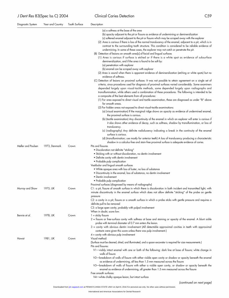
Table 1. Criteria for Detection of Dental Caries
Diagnostic System Year and Country Tooth Surface Description
Jackson 1950, UK Crown There should be positive and clear evidence of enamel dissolution before the diagnosis of caries is made.(1) A clear mirror and sharp probes must be used for examination. For the detection of pit and fissure lesions,
Ash's Sickle Probe No. 54 is to be used, and for approximal lesions, Ash's Probe No. 12 is to be used.(2) Each tooth must be dried thoroughly, and every surface examined.(3) Before each examination, the sickle probe is to be sharpened lightly with a medium sandpaper disc to
make a conical point.(4) A pit or fissure is counted as carious if, with a little pressure, the point sticks without doubt and requires a
definite pull to be removed. Anything that is at all doubtful is not included.(5 Stained pits or fissures are not counted as carious unless they satisfy this test.(6 The probe must be used in all fissures and pits in several angles.(7 Approximal lesions are considered detectable if Ash's No. 12 probe catches a roughened surface or a
definite cavity.(8) Stained or opaque light areas on other smooth surfaces are not called carious unless there is positive
and undoubted evidence of enamel dissolution.(9) Arrested caries is counted as carious, and exposed dentin in hypoplastic teeth is counted as carious,
only if there is positive evidence of softening.Parfitt 1954, UK Crown The first sign of caries as a slight discoloration, with loss of luster on the enamel surface.
Grade 1 = slight discoloration with loss of luster of the enamel surfaceGrade 2 = Surface is roughened and pitted, a condition which can be detected by explorer point.Grade 3 = further penetration and loss of tissue, causing pitting to reach the dentinGrade 4 = loss of dentin and cavitation
Backer-Dirks et al. 1961, Netherlands Crown For approximal caries, the clinical examination by mirror and explorer was completely abandoned, becauseof poor accuracy which makes it almost impossible to standardize diagnosis.Pits and fissures were cleaned with a new sharp explorer and dried with compressed air. The diagnosis wasmade with a small hand light of high intensity. Incident and transmitted light was used. Caries was estimatedin 4 different grades. Caries I signifies a minute black line at the bottom of the fissure. In caries II, there is alsoa white zone along the margins of the fissure. Caries III denotes the smallest perceptible break in the continuityof the enamel (cavity) with or without undermined margins. Caries IV is a large cavity more than 3 mm wide.
McHugh et al. 1964, UK Crown Teeth were counted as carious if a probe stuck definitely in a pit or fissure on being applied with gentlepressure, or if there were other signs of caries. Only limited pressure could be applied with the replaceableprobe points without causing breakage, and this helped in the standardization of "sticky fissures". In addition,each carious cavity was given a "penetration score" on the following basis:
1 = sticky fissure2 = fissure or free-surface cavity with softness at base and staining or opacity of the enamel3 = cavity with obvious dentin involvement (All detectable approximal cavities in teeth with approximal
contacts were given this score unless there was pulp involvement.)4 = cavity with obvious pulp involvement
Marthaler 1966, Switzerland Crown First look! Probe only when doubtful.Grade 1: slightly brown narrow line or [on smooth surfaces Class V] white spot with hard surface, smallest
extent not exceeding 2 mmGrade 2: clearly brown or black line or [or on Class V lesions] white spot, smallest extent exceeding 2 mm.
For Class III lesions [proximal of anterior teeth], the lesion has a dark brown discolored surface.Grade 3: cavity, discontinuity of the enamel surfaceGrade 4: cavity with the narrowest extent of the entrance broader than 2 mm
Møller 1966, Denmark Crown Buccal and lingual smooth surfaces:Grade I: a white opaque spot that keeps its luster after a short (3 sec) period of dryingGrade II: After being dried, the area appears white and chalky.
Caries in pits and fissures:Grade I: Area is dark by incident as well as transmitted light; the lesion is confined to a small dark line.Grade II: In addition to Grade 1, a white zone can be seen along the margins of the fissure, which
appears dark in transmitted light.Grade III: There is smallest perceptible break in the continuity of the enamel.
Radike 1968, USA Crown (I) Frank lesions—The detection of these lesions on the basis of gross cavitation usually does not present aproblem in diagnosis. When cavitation is present, the diagnosis is positive.
(A) Cavitation in this context can be defined as a discontinuity of the enamel surface caused by loss of toothsurfaces.
(B) Cavitation which is the result of the caries process must be distinguished from fractures and smoothlesions or erosion and abrasion.
(II) Lesions not showing cavitation—The most difficult part of the examiner's task is the detection of lesionswithout frank cavitation. These are lesions close to the decision point between carious and sound. The criteriafor detection of these lesions are summarized in three categories, each presenting its special problems.
(A) Detection of pit and fissure lesions of the occlusal, facial, and lingual surfaces.(1) Area is carious when the explorer "catches" or resists removal after insertion into a pit or fissure with
moderate to firm pressure and when accompanied by one or more of the following signs of caries:
at PENNSYLVANIA STATE UNIV on April 8, 2016 For personal use only. No other uses without permission.jdr.sagepub.comDownloaded from
International and American Associations for Dental Research
J Dent Res 83(Spec Iss C) 2004 Clinical Caries Detection C59
Diagnostic System Year and Country Tooth Surface Description
(a) a softness at the base of the area(b) opacity adjacent to the pit or fissure as evidence of undermining or demineralization(c) softened enamel adjacent to the pit or fissure which may be scraped away with the explorer
(2) Area is carious if there is loss of the normal translucency of the enamel, adjacent to a pit, which is incontrast to the surrounding tooth structure. This condition is considered to be reliable evidence ofundermining. In some of these cases, the explorer may not catch or penetrate the pit.
(B) Detection of lesions on smooth area[s] of facial and lingual surfaces(1) Area is carious if surface is etched or if there is a white spot as evidence of subsurface
demineralization, and if the area is found to be soft by:(a) penetration with explorer(b) enamel can be scraped away with explorer
(2) Area is sound when there is apparent evidence of demineralization (etching or white spots) but noevidence of softness.
(C) Detection of lesions on proximal surfaces: It was not possible to attain agreement on a single set ofcriteria, since procedures used for diagnosis of proximal surfaces varied considerably. Some examinersdepended largely upon visual-tactile methods, some depended largely upon radiographs andtransillumination, while others used a combination of these procedures. The following is intended to bea composite of the best elements from all procedures:(1) For area exposed to direct visual and tactile examination, these are diagnosed as under "B" above
for smooth areas.(2) For hidden areas not exposed to direct visual-tactile examinations:
(a) (visual examination) If the marginal ridge shows an opacity as evidence of undermined enamel,the proximal surface is carious.
(b) (tactile examination) Any discontinuity of the enamel in which an explorer will enter is carious ifit also shows other evidence of decay, such as softness, shadow by transillumination, or loss oftranslucency.
(c) (radiography) Any definite radiolucency indicating a break in the continuity of the enamelsurface is carious.
(d) (transillumination; use mostly for anterior teeth) A loss of translucency producing a characteristicshadow in a calculus-free and stain-free proximal surface is adequate evidence of caries.
Møller and Poulsen 1973, Denmark Crown Pits and fissures• Discoloration not definite "sticking"• Sticking with or without discoloration, no dentin involvement• Definite cavity with dentin involvement• Probable pulp complication
Vestibular and lingual smooth surfaces• White opaque area with loss of luster, no loss of substance• Discontinuity in the enamel, loss of substance, no dentin involvement• Dentin involvement• Probable pulp complication
Proximal surfaces (diagnosed by means of radiographs)Murray and Shaw 1975, UK Crown C1: a pit, fissure of smooth surface in which there is discoloration in both incident and transmitted light, with
minute discontinuity in the enamel surface which does not allow definite "sticking" of the probe on gentlepressureC2: a cavity in a pit, fissure or a smooth surface in which a probe sticks with gentle pressure and requires adefinite pull to be removedC3: a large open cavity, probably with pulpal involvementWhen in doubt, score low.
Bennie et al. 1978, UK Crown 1 = sticky fissure2 = fissure or free-surface cavity with softness at base and staining or opacity of the enamel. A blunt sickle
probe with terminal diameter of 0.7 mm enters the lesion.3 = cavity with obvious dentin involvement (All detectable approximal cavities in teeth with approximal
contacts were given this score unless there was pulp involvement.)4 = cavity with obvious pulp involvement
Howat 1981, UK Crown Visual method:(Surface must be cleaned, dried, and illuminated, and a spoon excavator is required for size measurements.)Pits and fissures:
V1—visibly intact enamel with one or both of the following: dark line at base of fissure; white change inwalls of fissure
V2—breakdown of walls of fissure with either visible open cavity or shadow or opacity beneath the enamelas evidence of undermining, all less than 1.5 mm measured across the fissure
V3—breakdown of walls of fissure with either a visible open cavity, or shadow or opacity beneath theenamel as evidence of undermining, all greater than 1.5 mm measured across the fissure
Free smooth surfaces:V4—white chalky opaque lesion, but intact surface
(continued on next page) at PENNSYLVANIA STATE UNIV on April 8, 2016 For personal use only. No other uses without permission.jdr.sagepub.comDownloaded from
International and American Associations for Dental Research
C60 Ismail J Dent Res 83(Spec Iss C) 2004
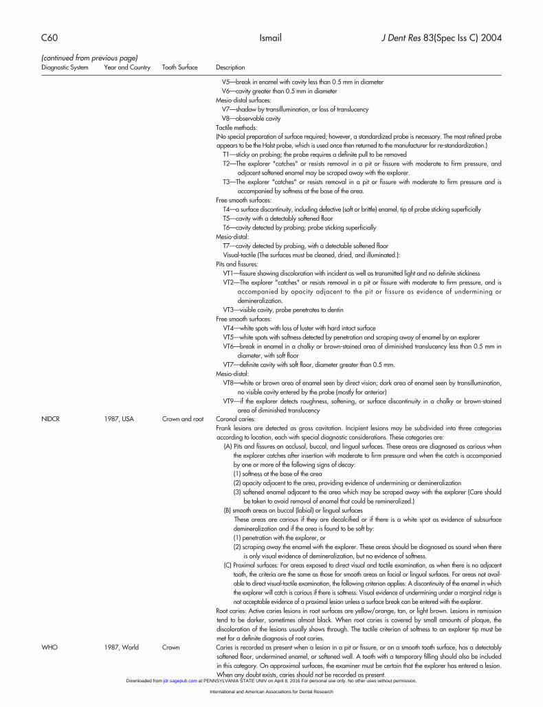
Diagnostic System Year and Country Tooth Surface Description
V5—break in enamel with cavity less than 0.5 mm in diameterV6—cavity greater than 0.5 mm in diameter
Mesio-distal surfaces:V7—shadow by transillumination, or loss of translucencyV8—observable cavity
Tactile methods:(No special preparation of surface required; however, a standardized probe is necessary. The most refined probeappears to be the Holst probe, which is used once then returned to the manufacturer for re-standardization.)
T1—sticky on probing; the probe requires a definite pull to be removedT2—The explorer "catches" or resists removal in a pit or fissure with moderate to firm pressure, and
adjacent softened enamel may be scraped away with the explorer.T3—The explorer "catches" or resists removal in a pit or fissure with moderate to firm pressure and is
accompanied by softness at the base of the area.Free smooth surfaces:
T4—a surface discontinuity, including defective (soft or brittle) enamel, tip of probe sticking superficiallyT5—cavity with a detectably softened floorT6—cavity detected by probing; probe sticking superficially
Mesio-distal:T7—cavity detected by probing, with a detectable softened floorVisual-tactile (The surfaces must be cleaned, dried, and illuminated.):
Pits and fissures:VT1—fissure showing discoloration with incident as well as transmitted light and no definite stickinessVT2—The explorer "catches" or resists removal in a pit or fissure with moderate to firm pressure, and is
accompanied by opacity adjacent to the pit or fissure as evidence of undermining ordemineralization.
VT3—visible cavity, probe penetrates to dentinFree smooth surfaces:
VT4—white spots with loss of luster with hard intact surfaceVT5—white spots with softness detected by penetration and scraping away of enamel by an explorerVT6—break in enamel in a chalky or brown-stained area of diminished translucency less than 0.5 mm in
diameter, with soft floorVT7—definite cavity with soft floor, diameter greater than 0.5 mm.
Mesio-distal:VT8—white or brown area of enamel seen by direct vision; dark area of enamel seen by transillumination,
no visible cavity entered by the probe (mostly for anterior)VT9—if the explorer detects roughness, softening, or surface discontinuity in a chalky or brown-stained
area of diminished translucencyNIDCR 1987, USA Crown and root Coronal caries:
Frank lesions are detected as gross cavitation. Incipient lesions may be subdivided into three categoriesaccording to location, each with special diagnostic considerations. These categories are:
(A) Pits and fissures on occlusal, buccal, and lingual surfaces. These areas are diagnosed as carious whenthe explorer catches after insertion with moderate to firm pressure and when the catch is accompaniedby one or more of the following signs of decay:(1) softness at the base of the area(2) opacity adjacent to the area, providing evidence of undermining or demineralization(3) softened enamel adjacent to the area which may be scraped away with the explorer (Care should
be taken to avoid removal of enamel that could be remineralized.)(B) smooth areas on buccal (labial) or lingual surfaces
These areas are carious if they are decalcified or if there is a white spot as evidence of subsurfacedemineralization and if the area is found to be soft by:(1) penetration with the explorer, or(2) scraping away the enamel with the explorer. These areas should be diagnosed as sound when there
is only visual evidence of demineralization, but no evidence of softness.(C) Proximal surfaces: For areas exposed to direct visual and tactile examination, as when there is no adjacent
tooth, the criteria are the same as those for smooth areas on facial or lingual surfaces. For areas not avail-able to direct visual-tactile examination, the following criterion applies: A discontinuity of the enamel in whichthe explorer will catch is carious if there is softness. Visual evidence of undermining under a marginal ridge isnot acceptable evidence of a proximal lesion unless a surface break can be entered with the explorer.
Root caries: Active caries lesions in root surfaces are yellow/orange, tan, or light brown. Lesions in remissiontend to be darker, sometimes almost black. When root caries is covered by small amounts of plaque, thediscoloration of the lesions usually shows through. The tactile criterion of softness to an explorer tip must bemet for a definite diagnosis of root caries.
WHO 1987, World Crown Caries is recorded as present when a lesion in a pit or fissure, or on a smooth tooth surface, has a detectablysoftened floor, undermined enamel, or softened wall. A tooth with a temporary filling should also be includedin this category. On approximal surfaces, the examiner must be certain that the explorer has entered a lesion.When any doubt exists, caries should not be recorded as present.
(continued from previous page)
at PENNSYLVANIA STATE UNIV on April 8, 2016 For personal use only. No other uses without permission.jdr.sagepub.comDownloaded from
International and American Associations for Dental Research
J Dent Res 83(Spec Iss C) 2004 Clinical Caries Detection C61
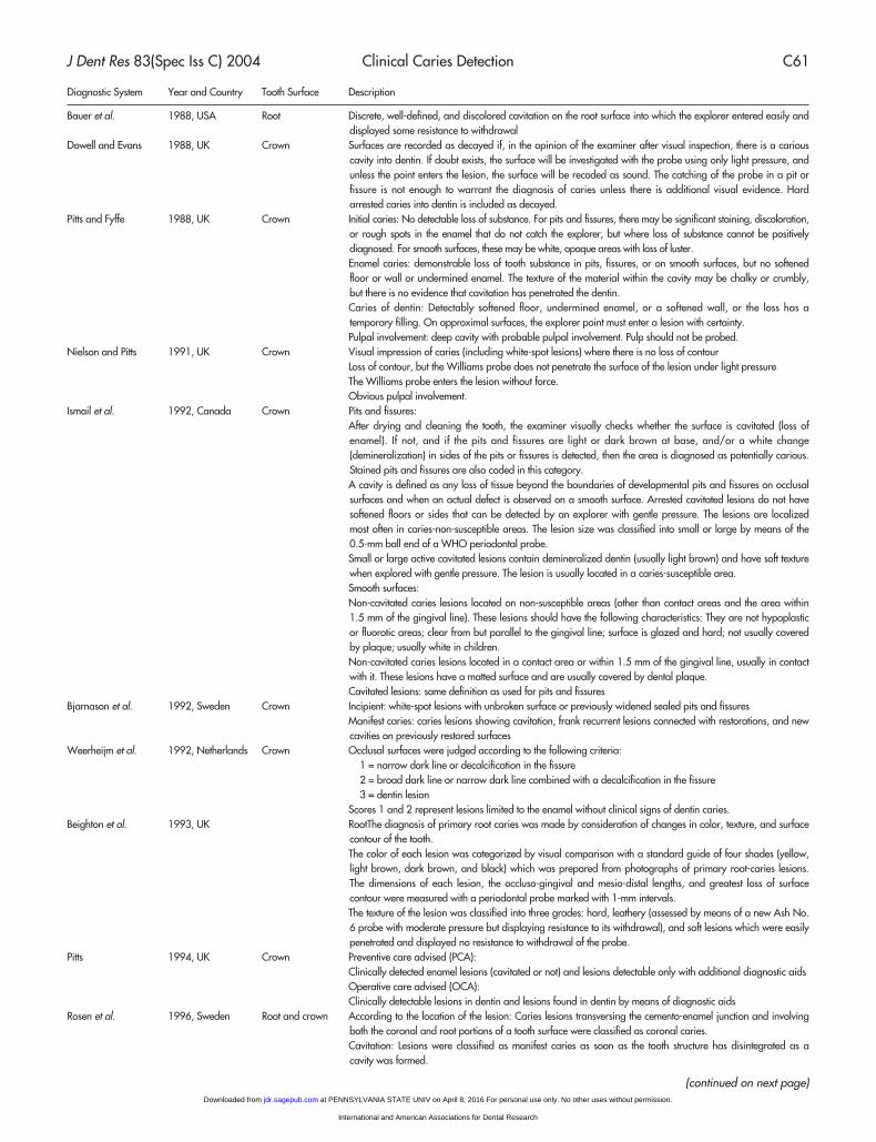
Diagnostic System Year and Country Tooth Surface Description
Bauer et al. 1988, USA Root Discrete, well-defined, and discolored cavitation on the root surface into which the explorer entered easily anddisplayed some resistance to withdrawal
Dowell and Evans 1988, UK Crown Surfaces are recorded as decayed if, in the opinion of the examiner after visual inspection, there is a cariouscavity into dentin. If doubt exists, the surface will be investigated with the probe using only light pressure, andunless the point enters the lesion, the surface will be recoded as sound. The catching of the probe in a pit orfissure is not enough to warrant the diagnosis of caries unless there is additional visual evidence. Hardarrested caries into dentin is included as decayed.
Pitts and Fyffe 1988, UK Crown Initial caries: No detectable loss of substance. For pits and fissures, there may be significant staining, discoloration,or rough spots in the enamel that do not catch the explorer, but where loss of substance cannot be positivelydiagnosed. For smooth surfaces, these may be white, opaque areas with loss of luster.Enamel caries: demonstrable loss of tooth substance in pits, fissures, or on smooth surfaces, but no softenedfloor or wall or undermined enamel. The texture of the material within the cavity may be chalky or crumbly,but there is no evidence that cavitation has penetrated the dentin.Caries of dentin: Detectably softened floor, undermined enamel, or a softened wall, or the loss has atemporary filling. On approximal surfaces, the explorer point must enter a lesion with certainty.Pulpal involvement: deep cavity with probable pulpal involvement. Pulp should not be probed.
Nielson and Pitts 1991, UK Crown Visual impression of caries (including white-spot lesions) where there is no loss of contourLoss of contour, but the Williams probe does not penetrate the surface of the lesion under light pressureThe Williams probe enters the lesion without force.Obvious pulpal involvement.
Ismail et al. 1992, Canada Crown Pits and fissures:After drying and cleaning the tooth, the examiner visually checks whether the surface is cavitated (loss ofenamel). If not, and if the pits and fissures are light or dark brown at base, and/or a white change(demineralization) in sides of the pits or fissures is detected, then the area is diagnosed as potentially carious.Stained pits and fissures are also coded in this category.A cavity is defined as any loss of tissue beyond the boundaries of developmental pits and fissures on occlusalsurfaces and when an actual defect is observed on a smooth surface. Arrested cavitated lesions do not havesoftened floors or sides that can be detected by an explorer with gentle pressure. The lesions are localizedmost often in caries-non-susceptible areas. The lesion size was classified into small or large by means of the0.5-mm ball end of a WHO periodontal probe.Small or large active cavitated lesions contain demineralized dentin (usually light brown) and have soft texturewhen explored with gentle pressure. The lesion is usually located in a caries-susceptible area.Smooth surfaces:Non-cavitated caries lesions located on non-susceptible areas (other than contact areas and the area within1.5 mm of the gingival line). These lesions should have the following characteristics: They are not hypoplasticor fluorotic areas; clear from but parallel to the gingival line; surface is glazed and hard; not usually coveredby plaque; usually white in children.Non-cavitated caries lesions located in a contact area or within 1.5 mm of the gingival line, usually in contactwith it. These lesions have a matted surface and are usually covered by dental plaque.Cavitated lesions: same definition as used for pits and fissures
Bjarnason et al. 1992, Sweden Crown Incipient: white-spot lesions with unbroken surface or previously widened sealed pits and fissuresManifest caries: caries lesions showing cavitation, frank recurrent lesions connected with restorations, and newcavities on previously restored surfaces
Weerheijm et al. 1992, Netherlands Crown Occlusal surfaces were judged according to the following criteria:1 = narrow dark line or decalcification in the fissure2 = broad dark line or narrow dark line combined with a decalcification in the fissure3 = dentin lesion
Scores 1 and 2 represent lesions limited to the enamel without clinical signs of dentin caries.Beighton et al. 1993, UK RootThe diagnosis of primary root caries was made by consideration of changes in color, texture, and surface
contour of the tooth.The color of each lesion was categorized by visual comparison with a standard guide of four shades (yellow,light brown, dark brown, and black) which was prepared from photographs of primary root-caries lesions.The dimensions of each lesion, the occluso-gingival and mesio-distal lengths, and greatest loss of surfacecontour were measured with a periodontal probe marked with 1-mm intervals.The texture of the lesion was classified into three grades: hard, leathery (assessed by means of a new Ash No.6 probe with moderate pressure but displaying resistance to its withdrawal), and soft lesions which were easilypenetrated and displayed no resistance to withdrawal of the probe.
Pitts 1994, UK Crown Preventive care advised (PCA):Clinically detected enamel lesions (cavitated or not) and lesions detectable only with additional diagnostic aidsOperative care advised (OCA):Clinically detectable lesions in dentin and lesions found in dentin by means of diagnostic aids
Rosen et al. 1996, Sweden Root and crown According to the location of the lesion: Caries lesions transversing the cemento-enamel junction and involvingboth the coronal and root portions of a tooth surface were classified as coronal caries.Cavitation: Lesions were classified as manifest caries as soon as the tooth structure has disintegrated as acavity was formed.
(continued on next page) at PENNSYLVANIA STATE UNIV on April 8, 2016 For personal use only. No other uses without permission.jdr.sagepub.comDownloaded from
International and American Associations for Dental Research
C62 Ismail J Dent Res 83(Spec Iss C) 2004
Diagnostic System Year and Country Tooth Surface Description
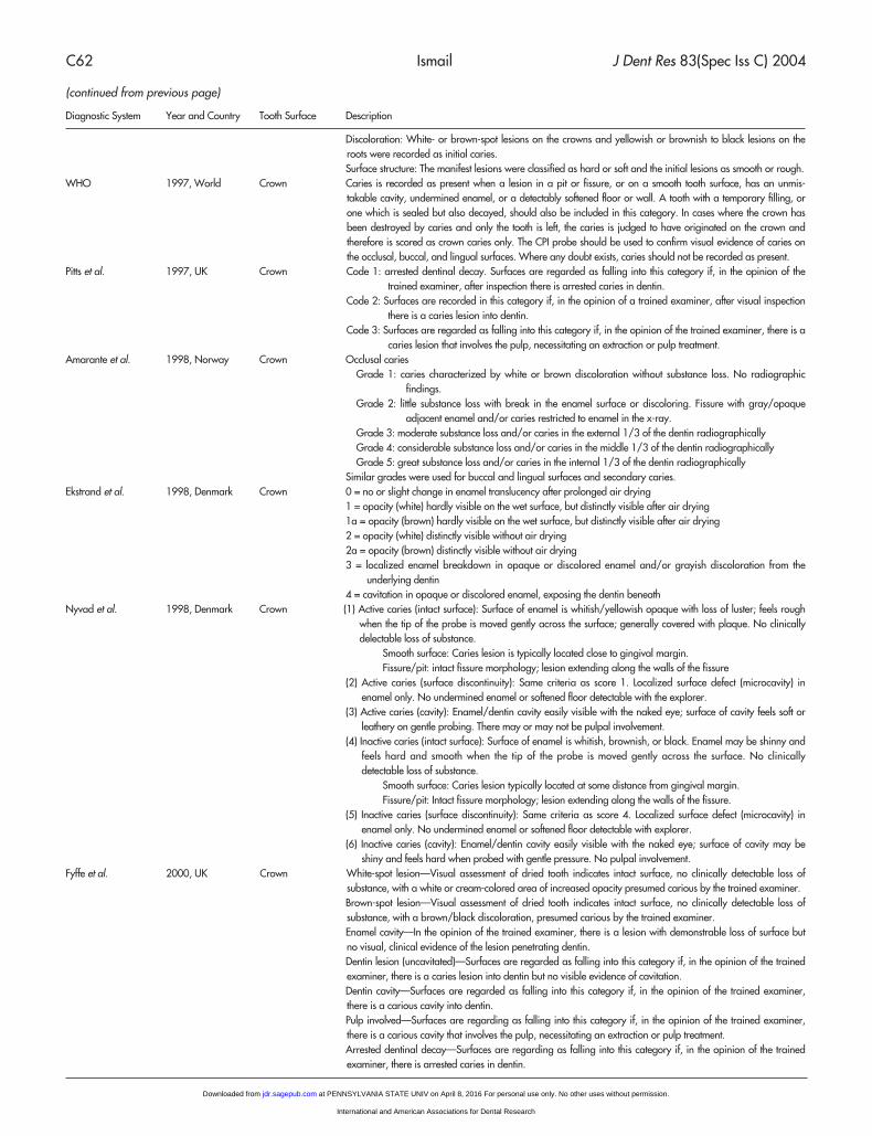
Discoloration: White- or brown-spot lesions on the crowns and yellowish or brownish to black lesions on theroots were recorded as initial caries.Surface structure: The manifest lesions were classified as hard or soft and the initial lesions as smooth or rough.
WHO 1997, World Crown Caries is recorded as present when a lesion in a pit or fissure, or on a smooth tooth surface, has an unmis-takable cavity, undermined enamel, or a detectably softened floor or wall. A tooth with a temporary filling, orone which is sealed but also decayed, should also be included in this category. In cases where the crown hasbeen destroyed by caries and only the tooth is left, the caries is judged to have originated on the crown andtherefore is scored as crown caries only. The CPI probe should be used to confirm visual evidence of caries onthe occlusal, buccal, and lingual surfaces. Where any doubt exists, caries should not be recorded as present.
Pitts et al. 1997, UK Crown Code 1: arrested dentinal decay. Surfaces are regarded as falling into this category if, in the opinion of thetrained examiner, after inspection there is arrested caries in dentin.
Code 2: Surfaces are recorded in this category if, in the opinion of a trained examiner, after visual inspectionthere is a caries lesion into dentin.
Code 3: Surfaces are regarded as falling into this category if, in the opinion of the trained examiner, there is acaries lesion that involves the pulp, necessitating an extraction or pulp treatment.
Amarante et al. 1998, Norway Crown Occlusal cariesGrade 1: caries characterized by white or brown discoloration without substance loss. No radiographic
findings.Grade 2: little substance loss with break in the enamel surface or discoloring. Fissure with gray/opaque
adjacent enamel and/or caries restricted to enamel in the x-ray.Grade 3: moderate substance loss and/or caries in the external 1/3 of the dentin radiographicallyGrade 4: considerable substance loss and/or caries in the middle 1/3 of the dentin radiographicallyGrade 5: great substance loss and/or caries in the internal 1/3 of the dentin radiographically
Similar grades were used for buccal and lingual surfaces and secondary caries.Ekstrand et al. 1998, Denmark Crown 0 = no or slight change in enamel translucency after prolonged air drying
1 = opacity (white) hardly visible on the wet surface, but distinctly visible after air drying1a = opacity (brown) hardly visible on the wet surface, but distinctly visible after air drying2 = opacity (white) distinctly visible without air drying2a = opacity (brown) distinctly visible without air drying3 = localized enamel breakdown in opaque or discolored enamel and/or grayish discoloration from the
underlying dentin4 = cavitation in opaque or discolored enamel, exposing the dentin beneath
Nyvad et al. 1998, Denmark Crown (1) Active caries (intact surface): Surface of enamel is whitish/yellowish opaque with loss of luster; feels roughwhen the tip of the probe is moved gently across the surface; generally covered with plaque. No clinicallydelectable loss of substance.
Smooth surface: Caries lesion is typically located close to gingival margin.Fissure/pit: intact fissure morphology; lesion extending along the walls of the fissure
(2) Active caries (surface discontinuity): Same criteria as score 1. Localized surface defect (microcavity) inenamel only. No undermined enamel or softened floor detectable with the explorer.
(3) Active caries (cavity): Enamel/dentin cavity easily visible with the naked eye; surface of cavity feels soft orleathery on gentle probing. There may or may not be pulpal involvement.
(4) Inactive caries (intact surface): Surface of enamel is whitish, brownish, or black. Enamel may be shinny andfeels hard and smooth when the tip of the probe is moved gently across the surface. No clinicallydetectable loss of substance.
Smooth surface: Caries lesion typically located at some distance from gingival margin.Fissure/pit: Intact fissure morphology; lesion extending along the walls of the fissure.
(5) Inactive caries (surface discontinuity): Same criteria as score 4. Localized surface defect (microcavity) inenamel only. No undermined enamel or softened floor detectable with explorer.
(6) Inactive caries (cavity): Enamel/dentin cavity easily visible with the naked eye; surface of cavity may beshiny and feels hard when probed with gentle pressure. No pulpal involvement.
Fyffe et al. 2000, UK Crown White-spot lesion—Visual assessment of dried tooth indicates intact surface, no clinically detectable loss ofsubstance, with a white or cream-colored area of increased opacity presumed carious by the trained examiner.Brown-spot lesion—Visual assessment of dried tooth indicates intact surface, no clinically detectable loss ofsubstance, with a brown/black discoloration, presumed carious by the trained examiner.Enamel cavity—In the opinion of the trained examiner, there is a lesion with demonstrable loss of surface butno visual, clinical evidence of the lesion penetrating dentin.Dentin lesion (uncavitated)—Surfaces are regarded as falling into this category if, in the opinion of the trainedexaminer, there is a caries lesion into dentin but no visible evidence of cavitation.Dentin cavity—Surfaces are regarded as falling into this category if, in the opinion of the trained examiner,there is a carious cavity into dentin.Pulp involved—Surfaces are regarding as falling into this category if, in the opinion of the trained examiner,there is a carious cavity that involves the pulp, necessitating an extraction or pulp treatment.Arrested dentinal decay—Surfaces are regarding as falling into this category if, in the opinion of the trainedexaminer, there is arrested caries in dentin.
(continued from previous page)
at PENNSYLVANIA STATE UNIV on April 8, 2016 For personal use only. No other uses without permission.jdr.sagepub.comDownloaded from
International and American Associations for Dental Research
J Dent Res 83(Spec Iss C) 2004 Clinical Caries Detection C63
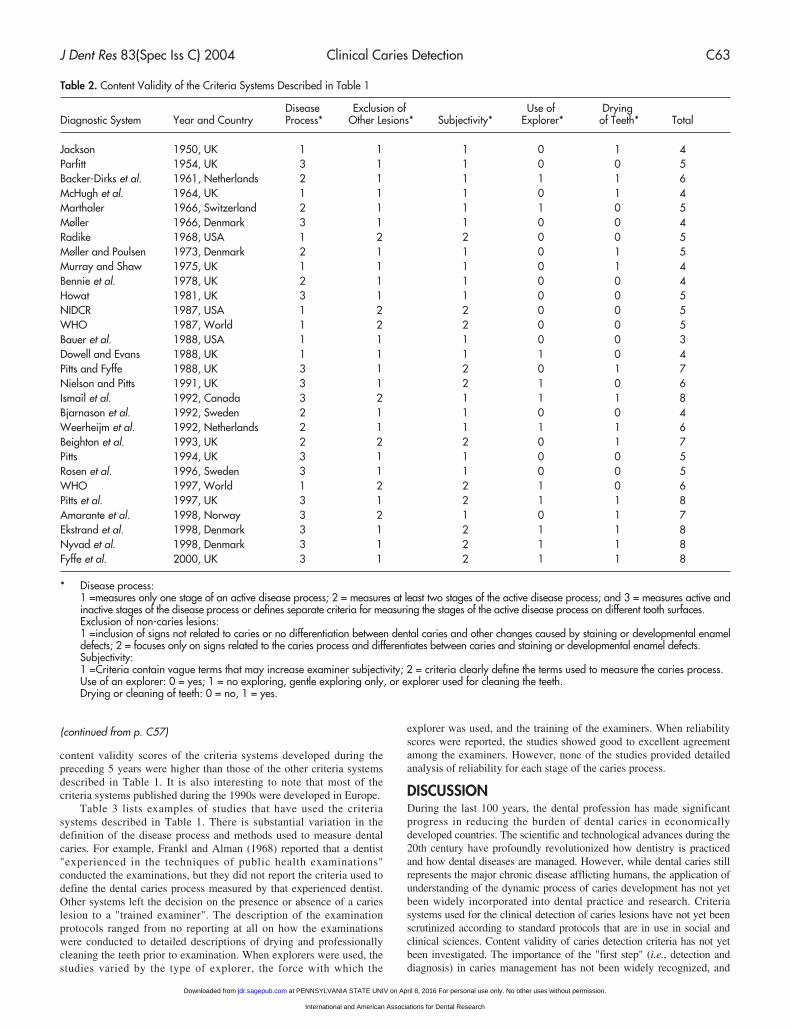
content validity scores of the criteria systems developed during thepreceding 5 years were higher than those of the other criteria systemsdescribed in Table 1. It is also interesting to note that most of thecriteria systems published during the 1990s were developed in Europe.
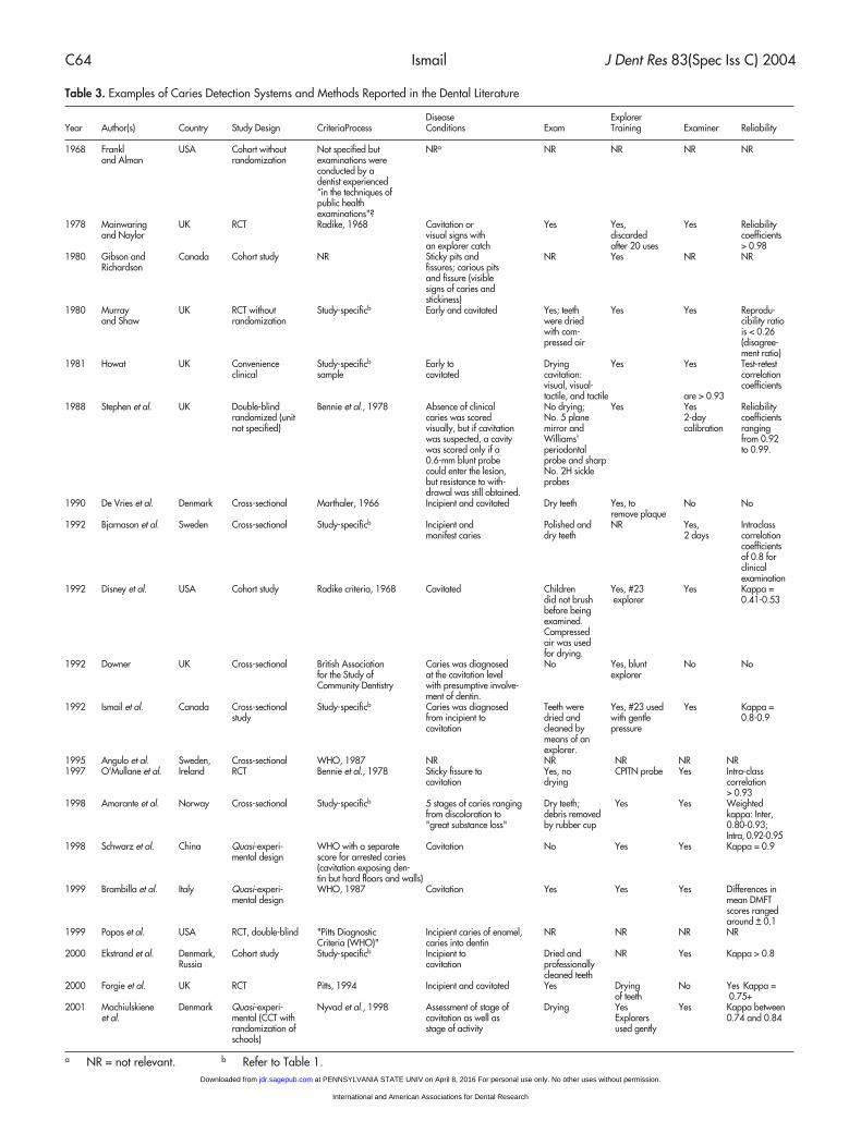
Table 3 lists examples of studies that have used the criteriasystems described in Table 1. There is substantial variation in thedefinition of the disease process and methods used to measure dentalcaries. For example, Frankl and Alman (1968) reported that a dentist"experienced in the techniques of public health examinations"conducted the examinations, but they did not report the criteria used todefine the dental caries process measured by that experienced dentist.Other systems left the decision on the presence or absence of a carieslesion to a "trained examiner". The description of the examinationprotocols ranged from no reporting at all on how the examinationswere conducted to detailed descriptions of drying and professionallycleaning the teeth prior to examination. When explorers were used, thestudies varied by the type of explorer, the force with which the
explorer was used, and the training of the examiners. When reliabilityscores were reported, the studies showed good to excellent agreementamong the examiners. However, none of the studies provided detailedanalysis of reliability for each stage of the caries process.
DISCUSSIONDuring the last 100 years, the dental profession has made significantprogress in reducing the burden of dental caries in economicallydeveloped countries. The scientific and technological advances during the20th century have profoundly revolutionized how dentistry is practicedand how dental diseases are managed. However, while dental caries stillrepresents the major chronic disease afflicting humans, the application ofunderstanding of the dynamic process of caries development has not yetbeen widely incorporated into dental practice and research. Criteriasystems used for the clinical detection of caries lesions have not yet beenscrutinized according to standard protocols that are in use in social andclinical sciences. Content validity of caries detection criteria has not yetbeen investigated. The importance of the "first step" (i.e., detection anddiagnosis) in caries management has not been widely recognized, and
Table 2. Content Validity of the Criteria Systems Described in Table 1
Disease Exclusion of Use of DryingDiagnostic System Year and Country Process* Other Lesions* Subjectivity* Explorer* of Teeth* Total
Jackson 1950, UK 1 1 1 0 1 4Parfitt 1954, UK 3 1 1 0 0 5Backer-Dirks et al. 1961, Netherlands 2 1 1 1 1 6McHugh et al. 1964, UK 1 1 1 0 1 4Marthaler 1966, Switzerland 2 1 1 1 0 5Møller 1966, Denmark 3 1 1 0 0 4Radike 1968, USA 1 2 2 0 0 5Møller and Poulsen 1973, Denmark 2 1 1 0 1 5Murray and Shaw 1975, UK 1 1 1 0 1 4Bennie et al. 1978, UK 2 1 1 0 0 4Howat 1981, UK 3 1 1 0 0 5NIDCR 1987, USA 1 2 2 0 0 5WHO 1987, World 1 2 2 0 0 5Bauer et al. 1988, USA 1 1 1 0 0 3Dowell and Evans 1988, UK 1 1 1 1 0 4Pitts and Fyffe 1988, UK 3 1 2 0 1 7Nielson and Pitts 1991, UK 3 1 2 1 0 6Ismail et al. 1992, Canada 3 2 1 1 1 8Bjarnason et al. 1992, Sweden 2 1 1 0 0 4Weerheijm et al. 1992, Netherlands 2 1 1 1 1 6Beighton et al. 1993, UK 2 2 2 0 1 7Pitts 1994, UK 3 1 1 0 0 5Rosen et al. 1996, Sweden 3 1 1 0 0 5WHO 1997, World 1 2 2 1 0 6Pitts et al. 1997, UK 3 1 2 1 1 8Amarante et al. 1998, Norway 3 2 1 0 1 7Ekstrand et al. 1998, Denmark 3 1 2 1 1 8Nyvad et al. 1998, Denmark 3 1 2 1 1 8Fyffe et al. 2000, UK 3 1 2 1 1 8
* Disease process:1 =measures only one stage of an active disease process; 2 = measures at least two stages of the active disease process; and 3 = measures active andinactive stages of the disease process or defines separate criteria for measuring the stages of the active disease process on different tooth surfaces.Exclusion of non-caries lesions:1 =inclusion of signs not related to caries or no differentiation between dental caries and other changes caused by staining or developmental enameldefects; 2 = focuses only on signs related to the caries process and differentiates between caries and staining or developmental enamel defects.Subjectivity:1 =Criteria contain vague terms that may increase examiner subjectivity; 2 = criteria clearly define the terms used to measure the caries process.Use of an explorer: 0 = yes; 1 = no exploring, gentle exploring only, or explorer used for cleaning the teeth.Drying or cleaning of teeth: 0 = no, 1 = yes.
(continued from p. C57)
at PENNSYLVANIA STATE UNIV on April 8, 2016 For personal use only. No other uses without permission.jdr.sagepub.comDownloaded from
International and American Associations for Dental Research
C64 Ismail J Dent Res 83(Spec Iss C) 2004
Table 3. Examples of Caries Detection Systems and Methods Reported in the Dental Literature
Disease ExplorerYear Author(s) Country Study Design CriteriaProcess Conditions Exam Training Examiner Reliability
1968 Frankl USA Cohort without Not specified but NRa NR NR NR NRand Alman randomization examinations were
conducted by adentist experienced “in the techniques of public health examinations"?
1978 Mainwaring UK RCT Radike, 1968 Cavitation or Yes Yes, Yes Reliability and Naylor visual signs with discarded coefficients
an explorer catch after 20 uses > 0.981980 Gibson and Canada Cohort study NR Sticky pits and NR Yes NR NR
Richardson fissures; carious pits and fissure (visible signs of caries and stickiness)
1980 Murray UK RCT without Study-specificb Early and cavitated Yes; teeth Yes Yes Reprodu-and Shaw randomization were dried cibility ratio
with com- is < 0.26 pressed air (disagree-
ment ratio)1981 Howat UK Convenience Study-specificb Early to Drying Yes Yes Test-retest
clinical sample cavitated cavitation: correlation visual, visual- coefficients tactile, and tactile are > 0.93
1988 Stephen et al. UK Double-blind Bennie et al., 1978 Absence of clinical No drying; Yes Yes Reliability randomized (unit caries was scored No. 5 plane 2-day coefficients not specified) visually, but if cavitation mirror and calibration ranging
was suspected, a cavity Williams' from 0.92 was scored only if a periodontal to 0.99.0.6-mm blunt probe probe and sharp could enter the lesion, No. 2H sickle but resistance to with- probesdrawal was still obtained.
1990 De Vries et al. Denmark Cross-sectional Marthaler, 1966 Incipient and cavitated Dry teeth Yes, to No Noremove plaque
1992 Bjarnason et al. Sweden Cross-sectional Study-specificb Incipient and Polished and NR Yes, Intraclass manifest caries dry teeth 2 days correlation
coefficients of 0.8 for clinical examination
1992 Disney et al. USA Cohort study Radike criteria, 1968 Cavitated Children Yes, #23 Yes Kappa = did not brush explorer 0.41-0.53before being examined.Compressedair was usedfor drying.
1992 Downer UK Cross-sectional British Association Caries was diagnosed No Yes, blunt No Nofor the Study of at the cavitation level explorerCommunity Dentistry with presumptive involve-
ment of dentin.1992 Ismail et al. Canada Cross-sectional Study-specificb Caries was diagnosed Teeth were Yes, #23 used Yes Kappa =
study from incipient to dried and with gentle 0.8-0.9cavitation cleaned by pressure
means of an explorer.
1995 Angulo et al. Sweden, Cross-sectional WHO, 1987 NR NR NR NR NR1997 O'Mullane et al. Ireland RCT Bennie et al., 1978 Sticky fissure to Yes, no CPITN probe Yes Intra-class
cavitation drying correlation > 0.93
1998 Amarante et al. Norway Cross-sectional Study-specificb 5 stages of caries ranging Dry teeth; Yes Yes Weighted from discoloration to debris removed kappa: Inter, "great substance loss" by rubber cup 0.80-0.93;
Intra, 0.92-0.951998 Schwarz et al. China Quasi-experi- WHO with a separate Cavitation No Yes Yes Kappa = 0.9
mental design score for arrested caries (cavitation exposing den-tin but hard floors and walls)
1999 Brambilla et al. Italy Quasi-experi- WHO, 1987 Cavitation Yes Yes Yes Differences in mental design mean DMFT
scores ranged around + 0.1
1999 Papas et al. USA RCT, double-blind "Pitts Diagnostic Incipient caries of enamel, NR NR NR NRCriteria (WHO)" caries into dentin
2000 Ekstrand et al. Denmark, Cohort study Study-specificb Incipient to Dried and NR Yes Kappa > 0.8Russia cavitation professionally
cleaned teeth2000 Forgie et al. UK RCT Pitts, 1994 Incipient and cavitated Yes Drying No Yes Kappa =
of teeth 0.75+2001 Machiulskiene Denmark Quasi-experi- Nyvad et al., 1998 Assessment of stage of Drying Yes Yes Kappa between
et al. mental (CCT with cavitation as well as Explorers 0.74 and 0.84randomization of stage of activity used gentlyschools)
a NR = not relevant. b Refer to Table 1. at PENNSYLVANIA STATE UNIV on April 8, 2016 For personal use only. No other uses without permission.jdr.sagepub.comDownloaded from
International and American Associations for Dental Research
J Dent Res 83(Spec Iss C) 2004 Clinical Caries Detection C65
dentists are usually underpaid for this activity.This review focused on the content validation of caries detection
systems. As stated before in this paper, calls to study and detect earlycaries lesions were made in the 19th century and by G.V. Black in1910. Over the last 20 years, there have been many attempts to expandthe methods used to detect and diagnose the presence of caries lesions.However, the narrow focus on "drilling and filling" and themisconception that early caries lesions cannot be reliably measuredmay have led to the development of criteria systems that have skewedthe understanding of dental caries epidemiology, prevention, andmanagement.
In a previous review, I discussed the reasons why early non-cavitated lesions should be included in new diagnostic systems ofdental caries (Ismail, 1997). First, there is evidence—even fromstudies published in the 1940s, 1970s, 1980s, and 1990s—that non-cavitated caries lesions are more prevalent than cavitated lesions ineconomically developed countries (Ismail, 1997; Amarante et al.,1998). Second, non-cavitated caries lesions are more likely to berestored compared with sound tooth surfaces (Ismail and Gagnon,1995; Ismail et al., 1997). Third, non-cavitated lesions, especially onsmooth tooth surfaces in young children, may serve as indicators ofcaries activity (Domoto et al., 1994; Grindefjord et al., 1995; Imfeld etal., 1995). Fourth, inclusion of non-cavitated lesions may provide abetter understanding of the mechanism of action of fluoride, sealants,and other preventive agents (Ismail, 1997). Fifth, inclusion of earlysigns of the caries process improves the precision of clinical trials ofpreventive agents (Howat et al., 1981).
Analysis of the data presented in Tables 1 and 2 shows that thereis a gulf between researchers in Europe and those in the USA.European researchers, as early as the 1960s, have included early signsof dental caries in their criteria systems. By contrast, criteria developedin the USA have focused on measuring the cavitated stage of caries orthe stage when an explorer sticks in teeth with visual signs of cariesdemineralization. The sensibility of the European criteria systemsfavors the disease process, while the sensibility of the USA systemsfavors reliability and comparability. The guiding principle for any newcaries diagnostic system in the 21st century should be its contemporarycontent validity, provided that it is accompanied by a detailed protocolfor calibration of examiners. Research studies should be conducted toidentify scientifically based protocols that can lead to achieving a highdegree of reliability among examiners. Any newly proposed protocolshould define the tools, methods of use, and length and frequency oftraining of examiners in studies of dental caries.
This review shows the lack of consistency regarding the use ofexplorers. The first account for the need of a sharp instrument to detectcaries lesions was reported by a dentist in the USA who attributed widevariation in diagnosis to the "...large size of the excavator used forexamination. The...excavator should be of the very smallest kind, andhatchet shaped.....This excavator should be made for diagnosis aloneand not for cutting enamel or dentine" (Anonymous, 1869). During the20th century, the use of the term "explorer catch" became part of thetradition of caries diagnosis (Sognnaes, 1940). The 2001 NIHConsensus Development Conference on Dental Caries Diagnosis andManagement Throughout Life concluded that "...the use of sharpexplorers in the detection of primary occlusal caries appears to add littlediagnostic information to other modalities and may be detrimental"(http://odp.od.nih.gov/consensus/cons/115/115_statement.htm#1).
Similarly, there seems to be a variation among the criteria systemsdescribed in Table 1 regarding whether teeth should be cleaned ordried before an examination. Some criteria stipulated that teeth shouldbe cleaned with a toothbrush or professionally, while othersrecommended cleaning by means of an explorer. The majority of thecriteria listed in Table 1 did not report on whether the teeth werecleaned or dried before examination. While no data are available tocompare the accuracy and reliability of examiners of clean vs. uncleanor dry vs. wet teeth, the detection of early signs of caries cannot beachieved unless the teeth are clean and dry.
In conclusion, analysis of the data summarized in this reviewpaper underscores the need to define one criteria system for visual andvisuo-tactile detection of dental caries that has content validity basedupon current scientific evidence and the consensus of experts in thefields of cariology and restorative sciences. There is also a need toinitiate a research program to test key constructs in caries detection anddevelop examination protocols that enable researchers to achieve ahigh degree of reliability. In achieving these goals, the workshopshould address the following questions:
(1) What stage of the caries process should be measured in clinicaltrials?
(2) What are the definitions for each stage of the caries process?(3) What is the best approach, in terms of objectivity and
consistency, which should be used to detect each stage of thecaries process for different tooth surfaces?
(4) What is the consensus on examiners' training protocols thatcan provide the highest degree of examiner reliability?
All answers to these questions should be based on scientificevidence. If evidence does not exist, the participants in this conferenceshould define the research questions that must be answered to advancethe field of caries detection, diagnosis, and management.
REFERENCESAmarante E, Raadal M, Espelid I (1998). Impact of diagnostic criteria on
the prevalence of dental caries in Norwegian children aged 5, 12 and18 years. Community Dent Oral Epidemiol 26:87-94.
Angulo M, Zinernanas E, Piyel L, Jorysz, Casamayou R, Krasse B (1995).Caries incidence, effect of preventive measures, and caries predictionin Uruguayan children. Acta Odontol Scand 53:1-6.
Anonymous (1869). Operative and surgical dentistry. Missouri Dent J1:399-403.
Backer-Dirks OB, Houwink B, Kwant GW (1961). The results of 6 1/2years of artificial fluoridation of drinking water in the Netherlands.The Tiel-Culemburg experiment. Arch Oral Biol 5:284-300.
Bader J (2001). Diagnosis and management of dental caries. Number36. AHRQ Publication No.01-E055, February 2001. Rockville,MD: Agency for Healthcare Research and Quali ty.http://www.ahrq.gov/clinic/dentsumm.htm.
Bauer JG, Cretin S, Schweitzer SO, Hunt RJ (1988). The reliability of diag-nosing root caries using oral examinations. J Dent Educ 52:622-629.
Beighton D, Lynch E, Heath MR (1993). A microbiological study ofprimary root-caries lesions with different treatment needs. J Dent Res72:623-629.
Bennie AM, Tullis JI, Stephen KW, MacFadyen EE (1978). Five years ofcommunity preventive dentistry and health education in the County ofSutherland, Scotland. Community Dent Oral Epidemiol 6:1-5.
Bjarnason S, Kohler B, Ranggard L (1992). Dental caries in a group of 15to 16-year-olds from Göteborg. Swed Dent J 16:143-149.
Black G (1880). Some points in the natural history of caries of the teeth,and the value of fillings for its arrest. Am J Dent Sci 14:289-308.
Black G (1910). A plea for greater earnestness in the study of caries of theenamel in its relation to the practice of dentistry. Dent Brief 15:161-178.
Black AD (1922). Preventive dentistry. Illinois State Dental Society Fifty-Eighth Annual Meeting. Springfield: The American Dental JournalPublishers, pp. 32-38.
Black G (1924). Operative dentistry. Chicago: Med-Dent Publ. Co., pp. 1-319.
Brambilla E, Gagliani M, Felloni A, Garcia Godoy F, Strohmenger L(1999). Caries-preventive effect of topical amine fluoride in childrenwith high and low salivary levels of mutans streptococci. Caries Res33:423-427.
de Vries HC, Ruiken HM, König KG, van't Hof MA (1990). Radiographicversus clinical diagnosis of approximal carious lesions. Caries Res24:364-370.
Disney JA, Abernathy JR, Graves RC, Mauriello SM, Bohannan HM, ZackDD (1992). Comparative effectiveness of visual/tactile and simplified
at PENNSYLVANIA STATE UNIV on April 8, 2016 For personal use only. No other uses without permission.jdr.sagepub.comDownloaded from
International and American Associations for Dental Research
C66 Ismail J Dent Res 83(Spec Iss C) 2004
screening examinations in caries risk assessment. Community DentOral Epidemiol 20:326-332.
Domoto P, Weinstein P, Leroux B, Koday M, Ogura S, Iatridi-Roberson I(1994). White spot caries in Mexican-American toddlers and parentalpreferences for various strategies. J Dent Child 61:342-346.
Dowell TB, Evans DJ (1988). The dental caries experience of 14-year oldchildren in England and Wales. A survey coordinated by the BritishAssociation for the Study of Community Dentistry. Community DentHealth 5:395-410.
Downer MC (1992). The quality of caries data from the national andBASCD surveys. Community Dent Health 9:107-108.
Ekstrand KR, Ricketts DNJ, Kidd EAM, Qvist V, Schou S (1998).Detection, diagnosing, monitoring, and logical treatment of occlusalcaries in relation to lesion activity and severity: an in vivo examinationwith histological validation. Caries Res 32:247-254.
Ekstrand KR, Kuzmina IN, Kuzmina E, Christiansen ME (2000). Two anda half-year outcome of caries-preventive programs offered to groups ofchildren in the Solntsevsky district of Moscow. Caries Res 34: 8-19.
Feinstein A (1987). Clinimetrics. New Haven, CT: Yale University Press,pp. 141-146.
Forgie AH, Paterson M, Pine CM, Pitts NB, Nugent ZJ (2000). Arandomized controlled trial of the caries-preventive efficacy of achlorhexidine-containing varnish in high-caries-risk adolescents.Caries Res 34:432-439.
Frankl SN, Alman JE (1968). Report of a three-year clinical trialcomparing a toothpaste containing sodium monofluorophosphate withtwo marketed products. J Oral Therap Pharmacol 4:443-450.
Fyffe HE, Deery C, Nugent ZJ, Nuttall NM, Pitts NB (2000). Effect ofdiagnostic threshold on the validity and reliability of epidemiologicalcaries diagnosis using the Dundee Selectable Threshold Method forcaries diagnosis (DSTM). Community Dent Oral Epidemiol 28:42-51.
Gibson GB, Richardson AS (1980). Sticky fissure management. 30-monthreport. J Can Dent Assoc 46:255-258.
Grindefjord M, Dahlöf G, Modéer T (1995). Caries development inchildren from 2.5 to 3.5 years of age: a longitudinal study. Caries Res29:449-454.
Howat AP (1981). A comparison of the sensitivity of caries diagnosticcriteria. Caries Res 15:331-337.
Howat AP, Holloway PJ, Brandt RS (1981). The effect of diagnosticcriteria on the sensitivity of dental epidemiological data. Caries Res15:117-123.
Imfeld TN, Steiner M, Menghini GD, Marthaler TM (1995). Prediction offuture caries increments for children in a school dental service, and inprivate practice. J Dent Educ 59:941-944.
Ismail AI (1997). Clinical diagnosis of precavitated carious lesions.Community Dent Oral Epidemiol 25:13-23.
Ismail AI, Gagnon P (1995). A longitudinal evaluation of fissure sealantsapplied in dental practices. J Dent Res 74:1583-1590.
Ismail AI, Brodeur JM, Gagnon P, Payette M, Picard D, Hamalian T, et al.(1992). Prevalence of non-cavitated and cavitated carious lesions in arandom sample of 7-9-year-old schoolchildren in Montreal, Quebec.Community Dent Oral Epidemiol 20:250-255.
Ismail AI, Brodeur JM, Gagnon P, Payette M, Picard D, Hamalian T, et al.(1997). Restorative treatments received by children covered by auniversal, publicly financed, dental insurance plan. J Public HealthDent 57:11-18.
Ismail AI, Hasson H, Sohn W (2001). Dental caries in the secondmillennium. J Dent Educ 65:953-959.
Jackson D (1950). The clinical diagnosis of dental caries. Br Dent J88:207-213.
Knapp J (1868). Hidden dental caries. Am Dent Assoc Trans 8:108-112.Machiulskiene V, Nyvad B, Baelum V (1999). A comparison of clinical
and radiographic caries diagnoses in posterior teeth of 12-year-oldLithuanian children. Caries Res 33:340-348.
Magitot E (1886). Therapeutic indications in dental caries. Br J Dent Sci29:405-410.
Mainwaring PJ, Naylor MN (1978). A three-year clinical study todetermine the separate and combined caries-inhibiting effects ofsodium monofluorophosphate toothpaste and an acidulated phosphate-fluoride gel. Caries Res 12:202-212.
Marthaler TM (1966). A standardized system of recording dentalconditions. Helv Odontol Acta 10:1-19.
McHugh WD, McEwen JD, Hitchin AD (1964). Dental disease and relatedfactors in 13-year-old children in Dundee. Br Dent J 117:246-253.
Møller IJ (1966). Clinical criteria for the diagnosis of the incipient cariouslesion. Adv Fluor Res Dent Caries Prev 4:67-72.
Møller IJ, Poulsen S (1973). A standardized system for diagnosing,recording and analyzing dental caries data. Scand J Dent Res 81:1-11.
Morsman A (1888). The diagnosis of dental caries and its sequelae. DentAdvert 19:49-53.
Murray JJ, Shaw L (1975). Errors in diagnosis of approximal caries onbitewing radiographs. Community Dent Oral Epidemiol 3:276-282.
Murray JJ, Shaw L (1980). A 3-year clinical trial into the effect of fluoridecontent and toothpaste abrasivity on the caries inhibitory properties ofa dentifrice. Community Dent Oral Epidemiol 8:46-51.
National Institute for Dental and Craniofacial Research (1987). Oral healthof United States adults. Bethesda, MD: NIDCR, pp. 161-165.
Neilson A, Pitts NB (1991). The clinical behavior of free smooth surfacecarious lesions monitored over 2 years in a group of Scottish children.Br Dent J 171:313-318.
Nyvad B, Machiulskiene V, Baelum V (1998). Reliability of a new cariesdiagnostic system differentiating between active and inactive carieslesions. Caries Res 33:252-260.
Papas A, Russell D, Singh M, Stack K, Kent R, Triol C, et al. (1999).Double blind clinical trial of a remineralizing dentifrice in theprevention of caries in a radiation therapy population. Gerodontology16:2-10.
Parfitt GJ (1954). A standard clinical examination of the teeth. Br Dent J96:296-300.
Pitts N (1994). Discovering dental public health: from Fisher to the future.Community Dent Health 11:172-178.
Pitts NB, Fyffe HE (1988). The effect of varying diagnostic thresholdsupon clinical caries data for a low prevalence group. J Dent Res67:592-596.
Pitts NB, Evans DJ, Pine CM (1997). British Association for the Study ofCommunity Dentistry (BASCD) diagnostic criteria for caries prevalencesurveys—1996/97. Community Dent Health 14(Suppl 1):6-9.
Radike AW (1968). Criteria for diagnosing dental caries. In: Proceedingsof the Conference on the Clinical Testing of Cariostatic Agents.Chicago: American Dental Association, pp. 87-88.
Rosen B, Birkhed D, Nilsson K, Olavi G, Egelberg J (1996).Reproducibility of clinical caries diagnoses on coronal and rootsurfaces. Caries Res 30:1-7.
Schwarz E, Lo EC, Wong MC (1998). Prevention of early childhoodcaries—results of a fluoride toothpaste demonstration trial on Chinesepreschool children after three years. J Public Health Dent 58:12-18.
Sognnaes RF (1940). The importance of a detailed clinical examination ofcarious lesions. J Dent Res 19:11-15.
Stephen KW, Creanor SL, Russell JI, Burchell CK, Huntington E, DownieCF (1988). A 3-year oral health dose-response study of sodiummonofluorophosphate dentifrices with and without zinc citrate: anti-caries results. Community Dent Oral Epidemiol 16:321-325.
Weerheijm K, Gruythuysen RJM, van Amerongen WE (1992). Prevalenceof hidden caries. J Dent Child 59:408-412.
World Health Organization (1987). Oral health surveys. Basic methods.Geneva, Switzerland: WHO, pp. 35-36.
World Health Organization (1997). Oral health surveys. Basic methods.Geneva, Switzerland: WHO, pp. 41-42.
at PENNSYLVANIA STATE UNIV on April 8, 2016 For personal use only. No other uses without permission.jdr.sagepub.comDownloaded from
International and American Associations for Dental Research