Embed Size (px)
Citation preview
DENTAL RADIOLOGY
DR. NUHAD
-------------------------------------------------------- Extra-oral Radiographic Technique
Extra-oral radiograph is defined as examination made of the head and
facial region using film located outside the mouth.
• They allow the dentist to view large area of the jaw and skull on a
single radiograph not covered by intraoral film.
:soral radiograph-of extraMain indications
• Examine large areas of the jaws and Skull.
• Study growth and development of bone and teeth
• Detect fractures and evaluate trauma
• Detect pathological lesions and Diseases of the jaws
• Detect and evaluate impacted teeth.
• Evaluate TMJ Disorders.
Uses of extra-oral radiographs:
Orthodontists : Uses lateral cephalometric radiograph to:
Measure and compare changes in growth and development of bone and
teeth through pre & progress and post treatment records.
Prosthodontists: Use Facial profile radiographs (lateral cephalometric)
to record :
• The contour of the lips and the face
• The relationship of the teeth before removal, this will help them
construct prosthetic appliances that look natural.
Oral surgeons: use extra-oral radiographs extensively to:
• Evaluate trauma.
• Determine the location and extent of fractures.
• Locate impacted teeth & abnormalities.
• Malignancies.
• Injuries to TMJ
Extra-oral Radiographic techniques:
Lateral jaw projection
• Known also as lateral oblique projection
• It has been largely replaced by panoramic radiographs but still taken
when image details is needed.
Main indications:
• Examine the posterior region of the mandible.
• Valuable for use in children, and in patients who have difficulty in
stabilizing or tolerating intraoral film placement
• Patients with limited jaw opening due to a fracture or swelling .
• Evaluate the condition of the bone and to locate impacted teeth or large
lesions.
The film in this extraoral projection technique is positioned lateral to
the jaw during exposure.
• Body of mandible projection.
Purpose:The purpose of this film is to evaluate impacted teeth, fractures
and lesions located in the body of the mandible.This projection
demonstrates the mandibular premolar and molar regions as well as the
inferior border of the mandible.
• Ramus of mandible projection.
This gives a view of the ramus from the angle to the condyle. It is also
useful for examining the impacted maxillary and mandibular third
molar regions.
Technique:
• The film is kept first against the cheek of the required side and is
centered over the body for body projection or over ramus for ramus
projection of mandible . The patients head is titled 15 degree to the side
being imaged and the chin is elevated and extended upwards.
• The central x-ray beam is directed perpendicular to the horizontal
plane of the cassette with a vertical angulation of -15 to -20 degrees.
X-ray beam is directed posterio- anteriorly from the opposite side
A- Cassette and X-ray tubehead position for the RIGHT mandibular
molars. Note the upward angulation of the X-ray tubehead and its
position beneath the left body of the mandible.
LATERAL OBLIQUE RADIOGRAPH
Skull Radiography
Skull radiography is used to examine the bones of the face and skull and
is most often used in oral surgery and orthodontics. Although some skull
films can be exposed using a standard intraoral X-ray machine, most
require the use of an extraoral unit and cephalostat .
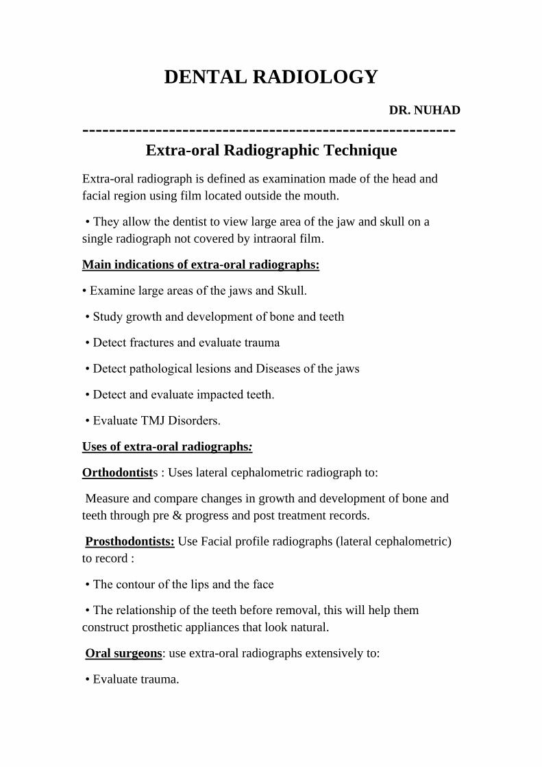
The most common skull radiographs used in dentistry are:
• Cephalometric projection.
• Posteroanterior projection.
• Occipitomental (Waters view) projection.
• Submentovertex projection.
• Reverse Towne’s projection
• TMJ Projections
Cephalometric Radiographs
• may be either frontal (posteroanterior) or lateral skull projections
• Device called cephalostats have ear rods that stabilizes the patient’s
head parallel to the film and at right angle to the direction of the beam
• The cephalometer allows the Exposure to be taken several times for the
same patient in the same head position.
Lateral skull (cephalometric ) projection:
• It shows the entire skull from the side and the X-ray passes from the
lateral side
Purposes:
• Orthodontic purpose :
1.Pre and post treatment records.
2.Evaluate the growth and development
3.Facial soft tissue profile of the face
• Surgeons also use it for pre and post treatment records
• Trauma • Pathology • Developmental Abnormalities
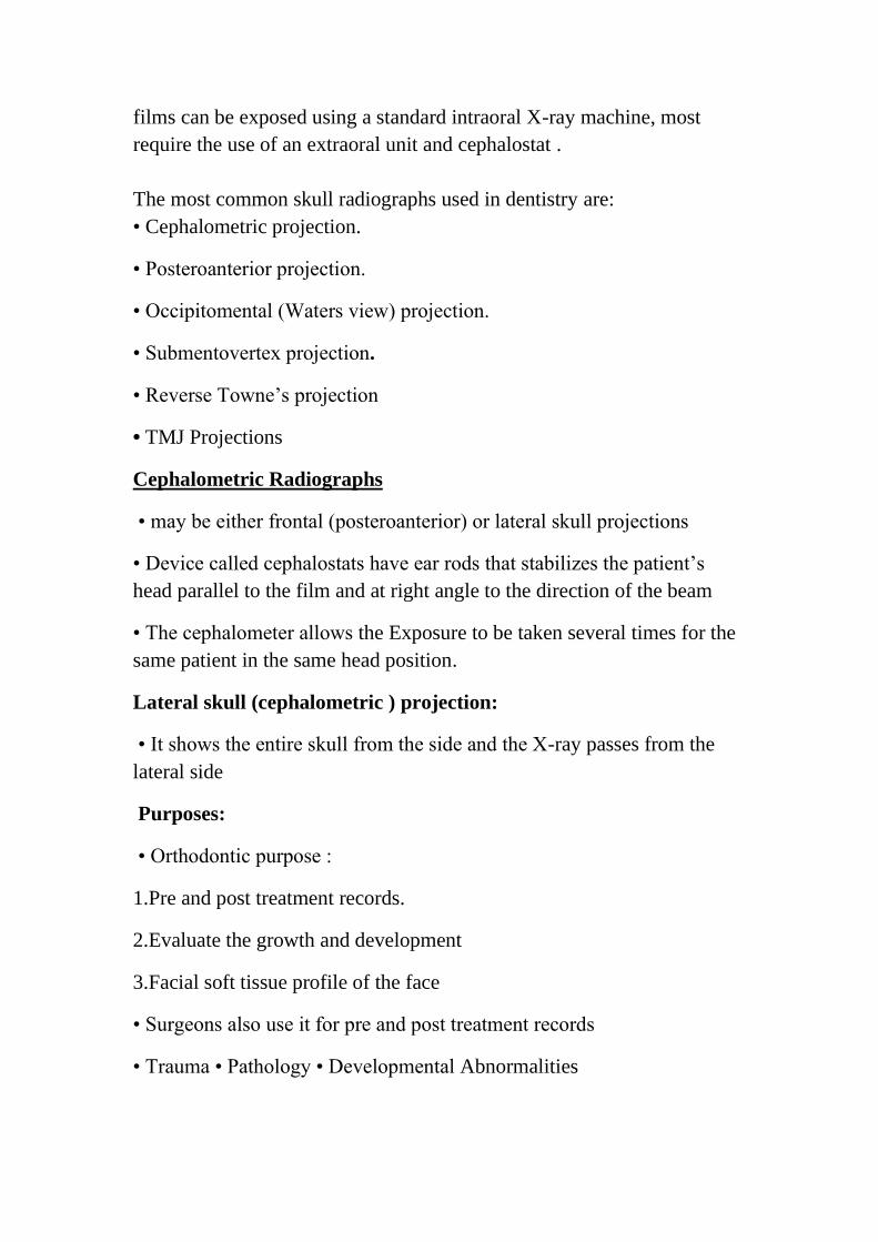
Technique
• In this projection, the head is positioned so that the midsagittal plane is
parallel to the film and a line connecting the external auditory meatus is
perpendicular to the film.
•The central ray is perpendicular to the midsagittal plane and
perpendicular to the plane of the film and is centered over the external
auditory meatus.
• If the facial soft tissue profile is desired , a wedge filter is placed over
the anterior side of the beam at the tube head so that filter will absorbs
some of the x-rays in the anterior region.
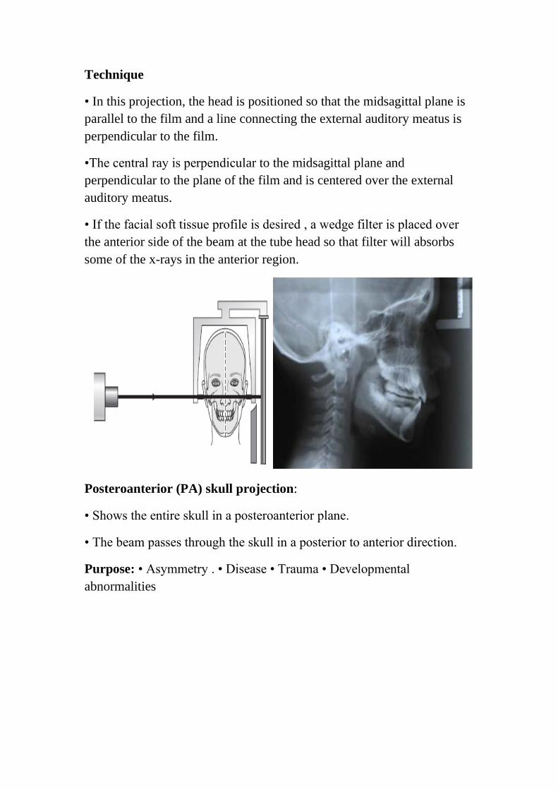
Posteroanterior (PA) skull projection:
• Shows the entire skull in a posteroanterior plane.
• The beam passes through the skull in a posterior to anterior direction.
Purpose: • Asymmetry . • Disease • Trauma • Developmental
abnormalities
The PA radiograph with the major anatomical features drawn in.
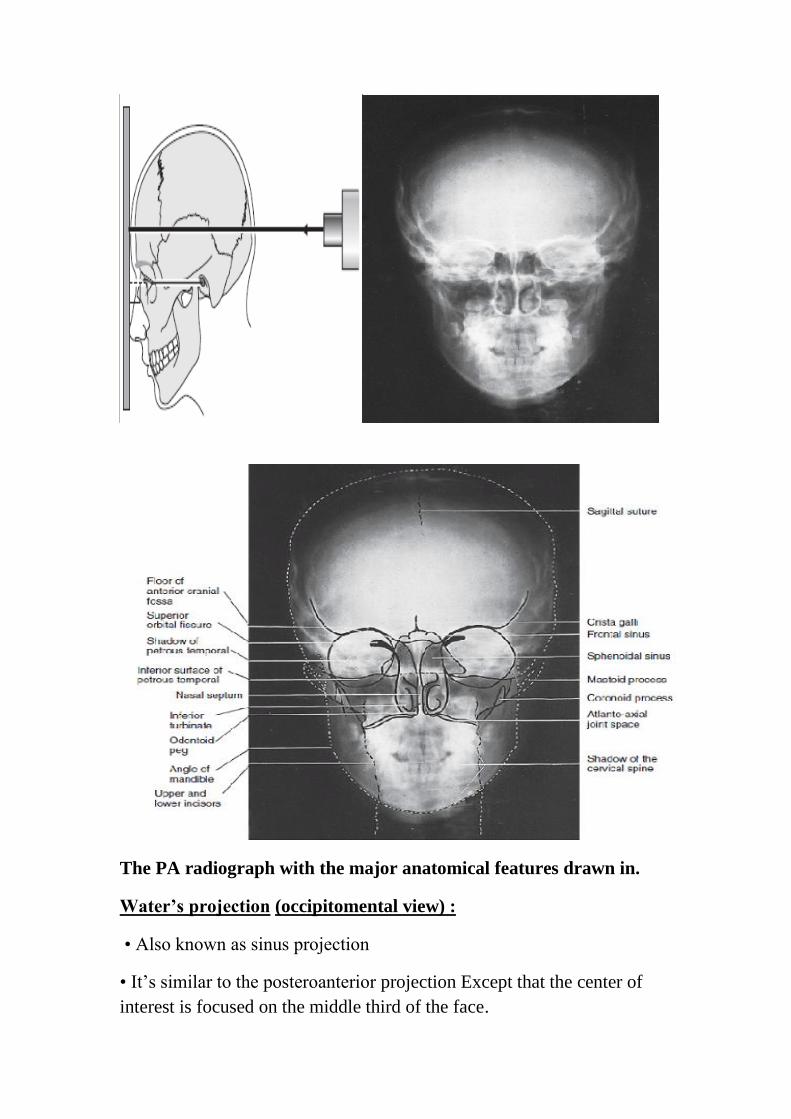
Water’s projection (occipitomental view) :
• Also known as sinus projection
• It’s similar to the posteroanterior projection Except that the center of
interest is focused on the middle third of the face.
Purpose. The purpose of the Waters projection is to evaluate the
maxillary sinus , the frontal and ethmoid sinuses.
The standard occipitomental radiograph with the major anatomical
features drawn in.
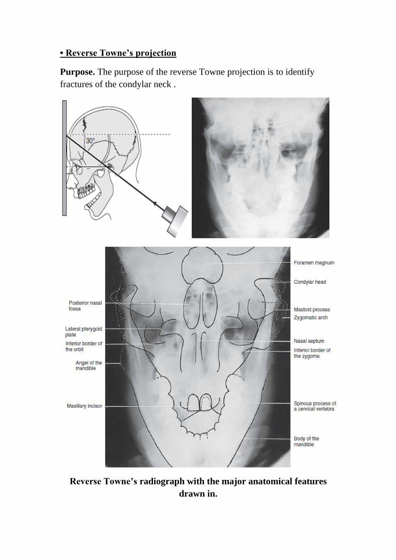
• Reverse Towne’s projection
Purpose. The purpose of the reverse Towne projection is to identify
fractures of the condylar neck .
Reverse Towne’s radiograph with the major anatomical features
drawn in.
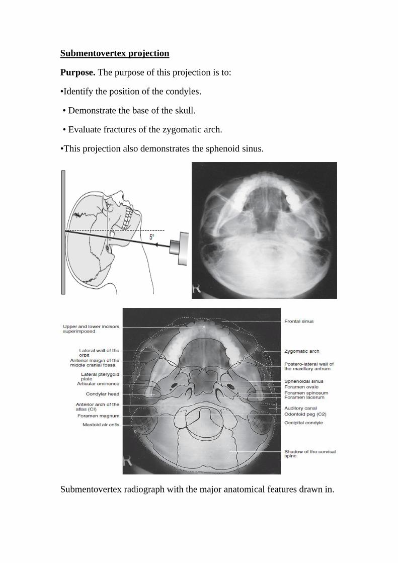
Submentovertex projection
Purpose. The purpose of this projection is to:
•Identify the position of the condyles.
• Demonstrate the base of the skull.
• Evaluate fractures of the zygomatic arch.
This projection also demonstrates the sphenoid sinus.•
Submentovertex radiograph with the major anatomical features drawn in.
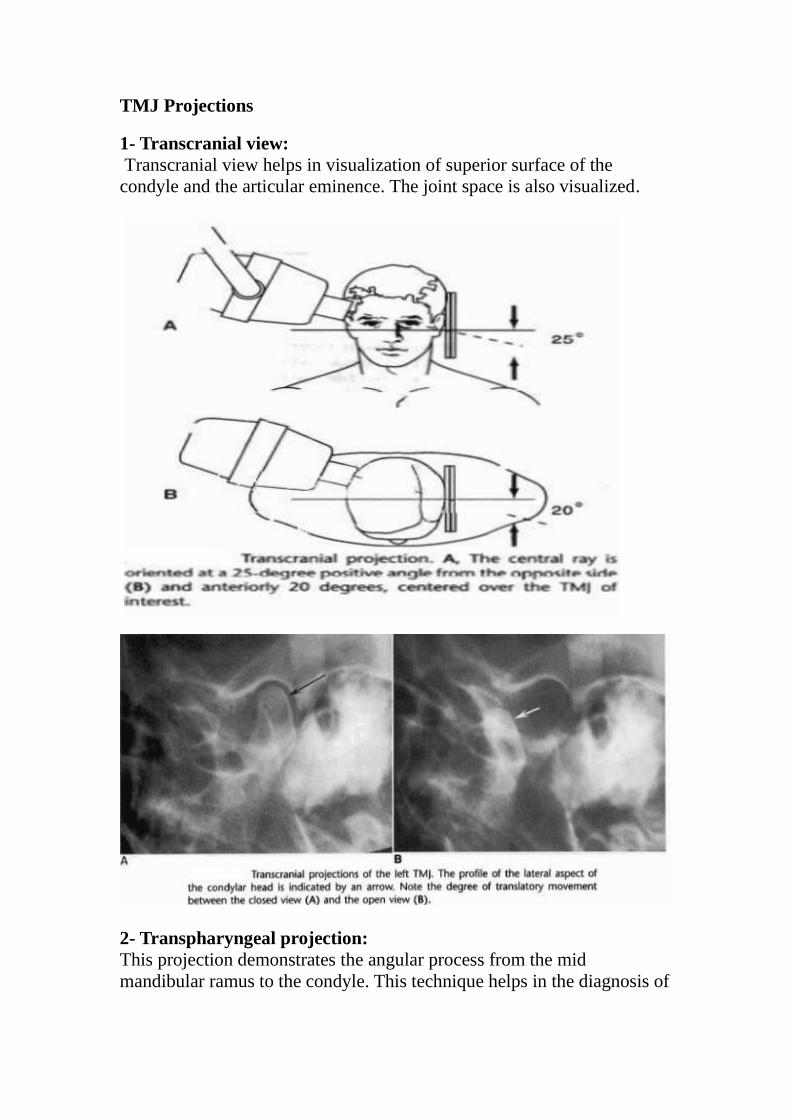
TMJ Projections
1- Transcranial view:
Transcranial view helps in visualization of superior surface of the
condyle and the articular eminence. The joint space is also visualized.
2- Transpharyngeal projection:
This projection demonstrates the angular process from the mid
mandibular ramus to the condyle. This technique helps in the diagnosis of
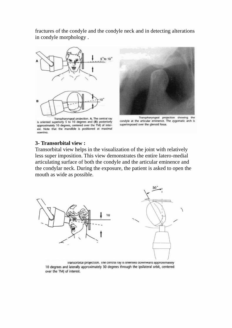
fractures of the condyle and the condyle neck and in detecting alterations
in condyle morphology .
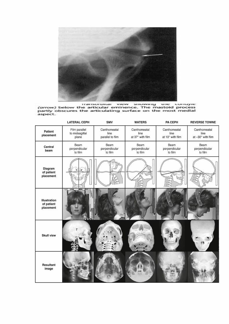
3- Transorbital view :
Transorbital view helps in the visualization of the joint with relatively
less super imposition. This view demonstrates the entire latero-medial
articulating surface of both the condyle and the articular eminence and
the condylar neck. During the exposure, the patient is asked to open the
mouth as wide as possible.