Embed Size (px)
Citation preview
Radiology in forensic expert team operations
Juha Rainio*, Kaisa Lalu, Helena Ranta, Antti PenttilaÈ
Department of Forensic Medicine, P.O. Box 40 (KytoÈsuontie 11), 00014 University of Helsinki, Helsinki, Finland
Received 28 December 2000; accepted 16 January 2001
Abstract
Radiological methods are widely used in forensic pathology. Their most common applications are in complementing human
identi®cation, particularly in investigations of mass disasters and decomposed bodies, and in searching for foreign material
inside corpses. A team of Finnish forensic experts investigated human skeletal remains in Bosnia and Herzegovina (1996) and
in Kosovo, the Federal Republic of Yugoslavia (1998). It also investigated more recently deceased victims in Kosovo (1999). In
these investigations, the bene®t of X-ray was in the detection of foreign material inside victims and their remains. For
identi®cation purposes, X-rays were mainly used to provide the best evidence possible of any pathological changes, physical
characteristics, and injuries present. q 2001 Elsevier Science Ireland Ltd. All rights reserved.
Keywords: Forensic pathology; Forensic radiology; Skeletal remains; Human identi®cation; Mass graves; Gunshot wounds
1. Introduction
Tasks of forensic scientists in investigation of mass
disasters and alleged mass graves include identi®ca-
tion of victims, determination of cause and manner of
death, and assembly of ®ndings that may help the
assertion of the causes of the incident and the
sequence of events [1±4]. For these purposes, multi-
disciplinary forensic teams collect data about the
victims using complementary methods [1,5±11].
Radiology is widely used to visualize and document
®ndings. It is recommended for use by the United
Nations in investigation of mass graves [12], by Inter-
pol in disaster victim identi®cation [13], and by the
American Board of Forensic Odontology in body
identi®cation [14].
Radiography is used in forensic pathology for
human identi®cation, especially in cases of decom-
posed, fragmented, or burned victims [5,15±20]. X-
rays can be used for anthropological determination of
sex, age, and stature [17±19,21,22]. For personal iden-
ti®cation, radiographs of nasal accessory or mastoid
sinuses [23±25], the thorax, or other areas typically
X-rayed [16,26±28] can be used to identify individual
anatomic features. Ante-mortem and post-mortem
radiographs can be compared concerning pathological
changes to bones, old traumas, prostheses, or other
orthopedic operations [5,8,15,16,21]. Radiology is
also used for assessment and documentation of dental
status [14,29,30]. Another application in forensic prac-
tice is in the determination and documentation of the
existence, number, localization, and identi®cation of
foreign material inside victims [17,22,27,31±33].
Legal Medicine 3 (2001) 34±43
1344-6223/01/$ - see front matter q 2001 Elsevier Science Ireland Ltd. All rights reserved.
PII: S1344-6223(01)00009-8
www.elsevier.nl/locate/legalmed
* Corresponding author. Tel.: 1358-9-19127473; fax: 1358-9-
19127518.
E-mail address: juha.rainio@helsinki.® (J. Rainio).
2. Materials and methods
2.1. Bosnia and Herzegovina 1996
Under the mandate of the United Nations, a team of
Finnish forensic experts (UN-FET) carried out an
investigation of human remains in Srebrenica, in
Bosnia and Herzegovina (BiH) in July 1996 [34].
The victims had allegedly been killed in July 1995.
In total, 64 samples of human remains were recovered
by the UN-FET on the battle®eld. The samples
included single bones and collections of bones and
soft tissue remnants from different parts of the
human body. Some of the human remains were joined
together by remnants of clothing.
X-ray examination for identi®cation purposes and
for indication and documentation of foreign material
was carried out for each bone or collection of bones in
52 of the 64 samples. The skull was X-rayed in lateral
and anterior-posterior views. Femora and tibias were
pictured for anthropological purposes. Other bones
were X-rayed when gunshot wounds, other injuries,
or pathological changes were noted. For tentative
identi®cation, including eventual age assessment,
dentition was X-rayed using periapical and occlusal
®lms. The objects of X-ray examination are presented
in Table 1.
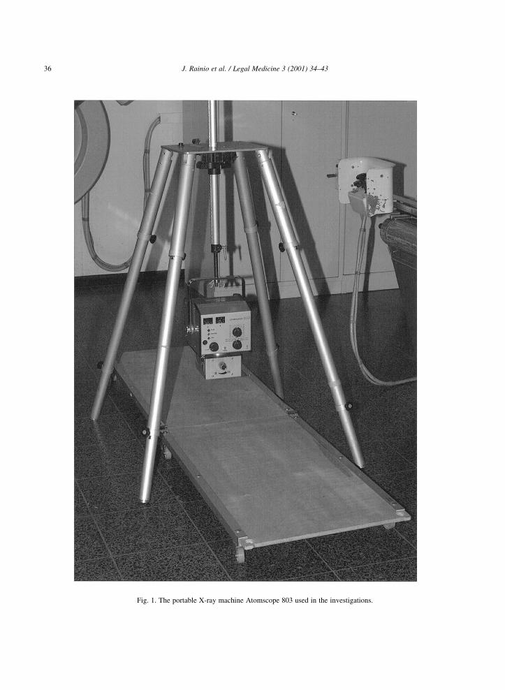
For X-ray examination, the portable Atomscope
803 machine (Fig. 1) and Kodak TML1, TME1,
and ENB1 ®lms were used. The developing machine
was the Flat Level 365. For forensic odontological
examination, the Philips Oralix 65S machine, Kodak
DF50 and DF58 ®lms, and the Periomat 1304 devel-
oping machine were used. In addition to the afore-
mentioned, all other equipment was brought from
Finland. The same equipment for medical and dental
X-ray examinations was used in the subsequent
operation in Kosovo. The radiological examination
was carried out by two specially trained autopsy
technicians, both of whom also participated in the
Kosovo operation. The X-rays were analyzed by
the presiding forensic pathologists and forensic odon-
tologist, and the ®ndings were recorded in the
autopsy protocols.
2.2. Kosovo 1998
In December 1998, the Finnish forensic expert team
under the mandate of the European Union (EU-FET)
investigated human skeletal remains from two alleged
mass graves in Volujak and Klecka, in Kosovo, the
Federal Republic of Yugoslavia [35]. The victims had
reportedly been killed some months earlier.
The investigated material from Volujak comprised
three almost complete human skeletons, along with
individual bones and bone fragments. The three skele-
tons were X-rayed selectively, and all of the individual
bones were X-rayed. The total number of medical X-
rays was 70. Dental X-rays were taken in four cases.
The objects X-rayed are presented in Table 1.
The Klecka material consisted of 90 human bone
samples. Most samples included one to ten bones,
which were partly burned and fragmented, with one
sample containing 108 small, burned bone fragments.
An X-ray examination was carried out for all bones,
except the sample with 108 fragments (see Table 1).
The number of medical X-rays was 95. Dental X-rays
were taken in six cases.
J. Rainio et al. / Legal Medicine 3 (2001) 34±43 35
Table 1
X-ray examination in investigations of human skeletal remains
Srebrenica Volujak Klecka
(n� 64)a (n� 6)b (n� 4)c
Skull 16 5 4
Maxilla 1 ± 1
Mandible 4 2 3
Cervical vertebrae 4 1 2
Thoracic vertebrae 9 2 3
Lumbar vertebrae 10 2 2
Scapula 6 3 2
Clavicle 2 2 2
Sternum 2 1 1
Ribs 8 3 3
Humerus 14 4 3
Ulna 7 3 4
Radius 4 4 4
Bones of hand 2 1 1
Pelvis 9 2 3
Femur 21 5 3
Tibia 20 3 2
Fibula 3 3 2
Bones of foot ± 4 1
a Remains of 30 to 35 victims.b Five adult male victims and nine bone samples which could not
be connected with the ®ve victims by DNA analysis.c Three adult male victims and nine mostly burned bone samples
which could not be connected with the three victims by DNA analy-
sis.
J. Rainio et al. / Legal Medicine 3 (2001) 34±4336
Fig. 1. The portable X-ray machine Atomscope 803 used in the investigations.
2.3. Kosovo 1999
In January 1999, the EU-FET participated in foren-
sic investigation of 40 victims who were killed during
an incident at the village of Racak, Kosovo [36],
approximately ten days before the investigation. The
EU-FET performed an autopsy in ten cases, moni-
tored 14 autopsies performed by Yugoslavian forensic
experts, and carried out an external examination of 16
corpses, which had been autopsied before the arrival
of the EU-FET.
The purpose of the X-ray examination was to loca-
lize X-ray-positive foreign material, with objects of
the examination speci®ed after the external examina-
tion of victims. The head was X-rayed in 21 autopsies,
the thorax in 25, the abdomen in 14, the pelvis in nine,
the neck in six, the upper limb or part thereof in ®ve,
and the lower limb or part thereof in ten cases. The
total number of medical X-rays taken in all 40 autop-
sies was 157; in the ten autopsies performed by
Finnish experts, the number was 73, in 14 of the
autopsies performed by the Yugoslavian experts 60.
X-ray examination of the bodies, which had been
autopsied before the arrival of the EU-FET, was
performed after the external examination carried out
by the EU-FET. Totally 24 X-rays were taken in seven
cases of the 16. In nine cases, X-ray examination
could not be performed after breakage of the X-ray
machine. For identi®cation purposes, dental X-rays
were carried out in 37 cases. A summary of X-ray
examination during the Racak investigation is
presented in Table 2.
3. Results
3.1. Bosnia and Herzegovina 1996
During the investigation in BiH, the 64 samples of
human skeletal remains, part of which had remnants
of soft tissue attached, yielded a minimum number of
30±35 individuals, estimated morphologically.
In seven cases, skeletal anomalies useful in identi-
®cation were noted: one skull was asymmetrical and
another had no frontal sinuses but did have a metopic
suture, in another case, the clavicles were unusually
straight and the right clavicle was also unusually
short, two tibias presented with a radiologically brigh-
tened area, one scapula presented with a small defor-
mation on the inner edge, and one lumbar vertebra had
a groove-like defect. In eight cases, degenerative
changes were present. Furthermore, in one case,
signs of a maxillary operation, and in another case,
an old fracture were noted. Prostheses were not
observed.
Dental radiographs were included in the autopsy
reports together with the Interpol post-mortem forms
of dental ®ndings. Unfortunately, this information
J. Rainio et al. / Legal Medicine 3 (2001) 34±43 37
Table 2
X-ray examination in the Racak investigation
Autopsy Monitoring Veri®cation Total
(n� 10) (n� 14) (n� 16) (n� 40)
Medical X-ray examination performed 10 14 7a 31
Number of medical X-rays 73 60 24 157
Object of X-ray examination
Head 10 7 4 21
Thorax 9 10 6 25
Abdomen 8 6 0 14
Pelvis 5 3 1 9
Neck 3 2 1 6
Upper limb or part thereof 4 1 0 5
Lower limb or part thereof 4 4 2 10
Dental X-ray examination performed 10 13b 14c 37
Number of dental X-rays 80 104 112 296
a In nine cases, examination could not be performed after breakage of the X-ray machine.b In one case, the victim had no teeth.c In one case, the victim had no teeth and in another case, the head was fragmented.
could not be used at the time of investigations due to
the lack of reliable ante-mortem information. The
®ndings including severe periodontal destruction and
periapical translucencies indicate deteriorating dental
health among the population of the enclave.
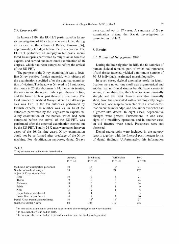
Gunshot injuries to bones were noted in 21 cases
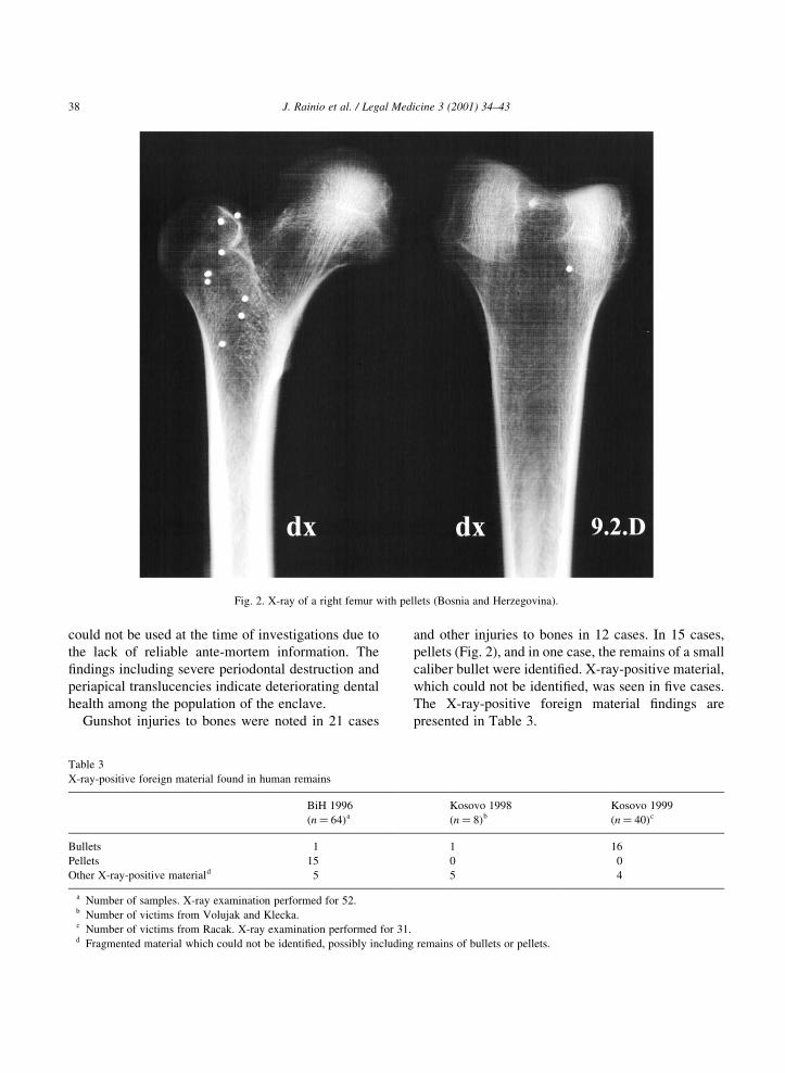
and other injuries to bones in 12 cases. In 15 cases,
pellets (Fig. 2), and in one case, the remains of a small
caliber bullet were identi®ed. X-ray-positive material,
which could not be identi®ed, was seen in ®ve cases.
The X-ray-positive foreign material ®ndings are
presented in Table 3.
J. Rainio et al. / Legal Medicine 3 (2001) 34±4338
Fig. 2. X-ray of a right femur with pellets (Bosnia and Herzegovina).
Table 3
X-ray-positive foreign material found in human remains
BiH 1996 Kosovo 1998 Kosovo 1999
(n� 64)a (n� 8)b (n� 40)c
Bullets 1 1 16
Pellets 15 0 0
Other X-ray-positive materiald 5 5 4
a Number of samples. X-ray examination performed for 52.b Number of victims from Volujak and Klecka.c Number of victims from Racak. X-ray examination performed for 31.d Fragmented material which could not be identi®ed, possibly including remains of bullets or pellets.
3.2. Kosovo 1998
During the investigations of Volujak and Klecka in
1998, the skeletal remains of most likely eight adult
males, ®ve in the Volujak material and three in the
Klecka material, were examined.
Skeletal anomalies which could be used for identi-
®cation were noted in three cases: in the ®rst, there
was a sutural bone of the skull and lumbar vertebra V
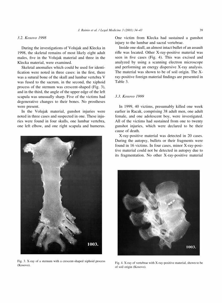
was fused to the sacrum, in the second, the xiphoid
process of the sternum was crescent-shaped (Fig. 3),
and in the third, the angle of the upper edge of the left
scapula was unusually sharp. Five of the victims had
degenerative changes to their bones. No prostheses
were present.
In the Volujak material, gunshot injuries were
noted in three cases and suspected in one. These inju-
ries were found in four skulls, one lumbar vertebra,
one left elbow, and one right scapula and humerus.
One victim from Klecka had sustained a gunshot
injury to the lumbar and sacral vertebrae.
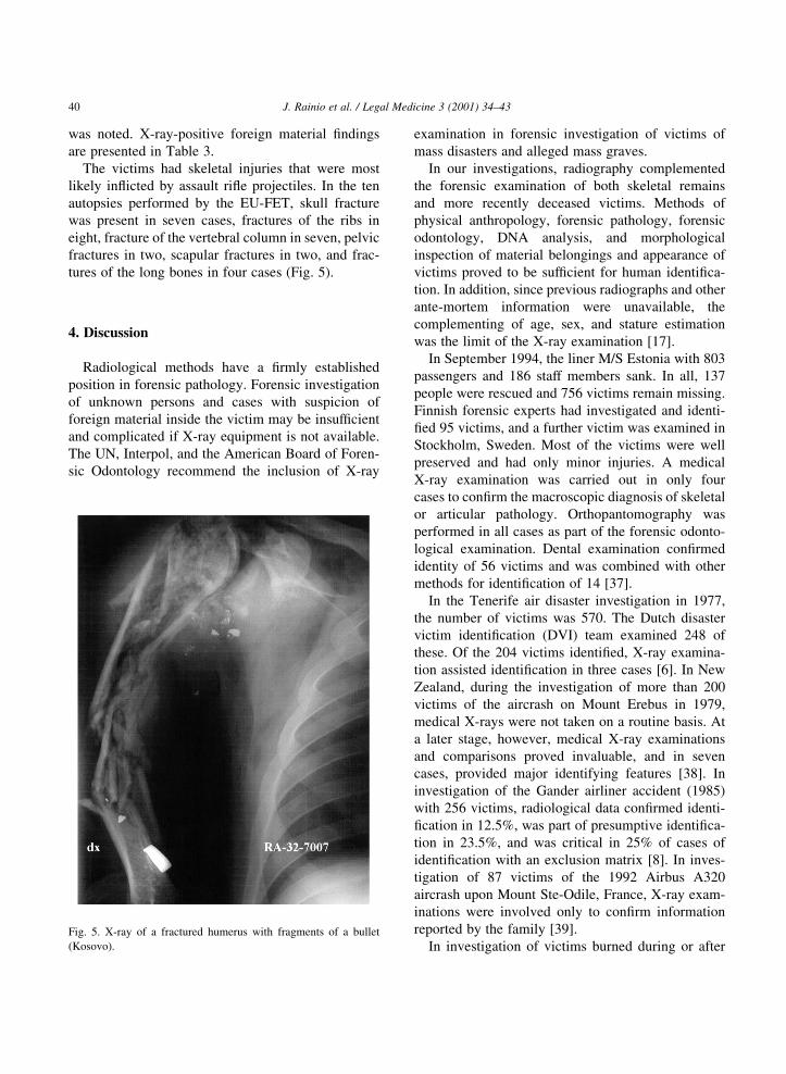
Inside one skull, an almost intact bullet of an assault
ri¯e was located. Other X-ray-positive material was
seen in ®ve cases (Fig. 4). This was excised and
analyzed by using a scanning electron microscope
and performing an energy dispersive X-ray analysis.
The material was shown to be of soil origin. The X-
ray-positive foreign material ®ndings are presented in
Table 3.
3.3. Kosovo 1999
In 1999, 40 victims, presumably killed one week
earlier in Racak, comprising 38 adult men, one adult
female, and one adolescent boy, were investigated.
All of the victims had sustained from one to twenty
gunshot injuries, which were declared to be their
cause of death.
X-ray-positive material was detected in 20 cases.
During the autopsy, bullets or their fragments were
found in 16 victims. In four cases, minor X-ray-posi-
tive material could not be detected in autopsy due to
its fragmentation. No other X-ray-positive material
J. Rainio et al. / Legal Medicine 3 (2001) 34±43 39
Fig. 3. X-ray of a sternum with a crescent-shaped xiphoid process
(Kosovo).Fig. 4. X-ray of vertebrae with X-ray-positive material, shown to be
of soil origin (Kosovo).
was noted. X-ray-positive foreign material ®ndings
are presented in Table 3.
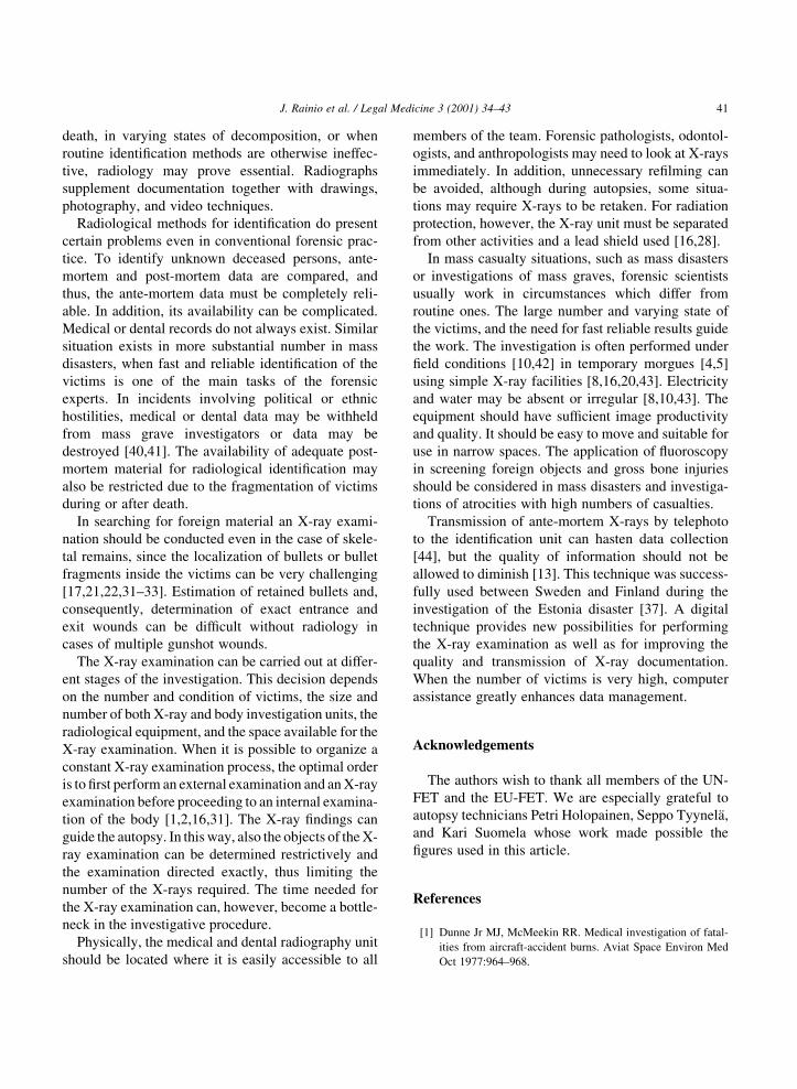
The victims had skeletal injuries that were most
likely in¯icted by assault ri¯e projectiles. In the ten
autopsies performed by the EU-FET, skull fracture
was present in seven cases, fractures of the ribs in
eight, fracture of the vertebral column in seven, pelvic
fractures in two, scapular fractures in two, and frac-
tures of the long bones in four cases (Fig. 5).
4. Discussion
Radiological methods have a ®rmly established
position in forensic pathology. Forensic investigation
of unknown persons and cases with suspicion of
foreign material inside the victim may be insuf®cient
and complicated if X-ray equipment is not available.
The UN, Interpol, and the American Board of Foren-
sic Odontology recommend the inclusion of X-ray
examination in forensic investigation of victims of
mass disasters and alleged mass graves.
In our investigations, radiography complemented
the forensic examination of both skeletal remains
and more recently deceased victims. Methods of
physical anthropology, forensic pathology, forensic
odontology, DNA analysis, and morphological
inspection of material belongings and appearance of
victims proved to be suf®cient for human identi®ca-
tion. In addition, since previous radiographs and other
ante-mortem information were unavailable, the
complementing of age, sex, and stature estimation
was the limit of the X-ray examination [17].
In September 1994, the liner M/S Estonia with 803
passengers and 186 staff members sank. In all, 137
people were rescued and 756 victims remain missing.
Finnish forensic experts had investigated and identi-
®ed 95 victims, and a further victim was examined in
Stockholm, Sweden. Most of the victims were well
preserved and had only minor injuries. A medical
X-ray examination was carried out in only four
cases to con®rm the macroscopic diagnosis of skeletal
or articular pathology. Orthopantomography was
performed in all cases as part of the forensic odonto-
logical examination. Dental examination con®rmed
identity of 56 victims and was combined with other
methods for identi®cation of 14 [37].
In the Tenerife air disaster investigation in 1977,
the number of victims was 570. The Dutch disaster
victim identi®cation (DVI) team examined 248 of
these. Of the 204 victims identi®ed, X-ray examina-
tion assisted identi®cation in three cases [6]. In New
Zealand, during the investigation of more than 200
victims of the aircrash on Mount Erebus in 1979,
medical X-rays were not taken on a routine basis. At
a later stage, however, medical X-ray examinations
and comparisons proved invaluable, and in seven
cases, provided major identifying features [38]. In
investigation of the Gander airliner accident (1985)
with 256 victims, radiological data con®rmed identi-
®cation in 12.5%, was part of presumptive identi®ca-
tion in 23.5%, and was critical in 25% of cases of
identi®cation with an exclusion matrix [8]. In inves-
tigation of 87 victims of the 1992 Airbus A320
aircrash upon Mount Ste-Odile, France, X-ray exam-
inations were involved only to con®rm information
reported by the family [39].
In investigation of victims burned during or after
J. Rainio et al. / Legal Medicine 3 (2001) 34±4340
Fig. 5. X-ray of a fractured humerus with fragments of a bullet
(Kosovo).
death, in varying states of decomposition, or when
routine identi®cation methods are otherwise ineffec-
tive, radiology may prove essential. Radiographs
supplement documentation together with drawings,
photography, and video techniques.
Radiological methods for identi®cation do present
certain problems even in conventional forensic prac-
tice. To identify unknown deceased persons, ante-
mortem and post-mortem data are compared, and
thus, the ante-mortem data must be completely reli-
able. In addition, its availability can be complicated.
Medical or dental records do not always exist. Similar
situation exists in more substantial number in mass
disasters, when fast and reliable identi®cation of the
victims is one of the main tasks of the forensic
experts. In incidents involving political or ethnic
hostilities, medical or dental data may be withheld
from mass grave investigators or data may be
destroyed [40,41]. The availability of adequate post-
mortem material for radiological identi®cation may
also be restricted due to the fragmentation of victims
during or after death.
In searching for foreign material an X-ray exami-
nation should be conducted even in the case of skele-
tal remains, since the localization of bullets or bullet
fragments inside the victims can be very challenging
[17,21,22,31±33]. Estimation of retained bullets and,
consequently, determination of exact entrance and
exit wounds can be dif®cult without radiology in
cases of multiple gunshot wounds.
The X-ray examination can be carried out at differ-
ent stages of the investigation. This decision depends
on the number and condition of victims, the size and
number of both X-ray and body investigation units, the
radiological equipment, and the space available for the
X-ray examination. When it is possible to organize a
constant X-ray examination process, the optimal order
is to ®rst perform an external examination and an X-ray
examination before proceeding to an internal examina-
tion of the body [1,2,16,31]. The X-ray ®ndings can
guide the autopsy. In this way, also the objects of the X-
ray examination can be determined restrictively and
the examination directed exactly, thus limiting the
number of the X-rays required. The time needed for
the X-ray examination can, however, become a bottle-
neck in the investigative procedure.
Physically, the medical and dental radiography unit
should be located where it is easily accessible to all
members of the team. Forensic pathologists, odontol-
ogists, and anthropologists may need to look at X-rays
immediately. In addition, unnecessary re®lming can
be avoided, although during autopsies, some situa-
tions may require X-rays to be retaken. For radiation
protection, however, the X-ray unit must be separated
from other activities and a lead shield used [16,28].
In mass casualty situations, such as mass disasters
or investigations of mass graves, forensic scientists
usually work in circumstances which differ from
routine ones. The large number and varying state of
the victims, and the need for fast reliable results guide
the work. The investigation is often performed under
®eld conditions [10,42] in temporary morgues [4,5]
using simple X-ray facilities [8,16,20,43]. Electricity
and water may be absent or irregular [8,10,43]. The
equipment should have suf®cient image productivity
and quality. It should be easy to move and suitable for
use in narrow spaces. The application of ¯uoroscopy
in screening foreign objects and gross bone injuries
should be considered in mass disasters and investiga-
tions of atrocities with high numbers of casualties.
Transmission of ante-mortem X-rays by telephoto
to the identi®cation unit can hasten data collection
[44], but the quality of information should not be
allowed to diminish [13]. This technique was success-
fully used between Sweden and Finland during the
investigation of the Estonia disaster [37]. A digital
technique provides new possibilities for performing
the X-ray examination as well as for improving the
quality and transmission of X-ray documentation.
When the number of victims is very high, computer
assistance greatly enhances data management.
Acknowledgements
The authors wish to thank all members of the UN-
FET and the EU-FET. We are especially grateful to
autopsy technicians Petri Holopainen, Seppo TyynelaÈ,
and Kari Suomela whose work made possible the
®gures used in this article.
References
[1] Dunne Jr MJ, McMeekin RR. Medical investigation of fatal-
ities from aircraft-accident burns. Aviat Space Environ Med
Oct 1977:964±968.
J. Rainio et al. / Legal Medicine 3 (2001) 34±43 41
[2] McMeekin RR. An organizational concept for pathologic
identi®cation in mass disaster. Aviat Space Environ Med
Sept 1980:999±1003.
[3] Hill IR, Howell RD, Jarmulowicz M. Identi®cation in the
Manchester air disaster. Br Dent J Dec 1988;24:445±446.
[4] Hooft PJ, Noji EK, van de Voorde HP. Fatality management in
mass casualty incidents. Forensic Sci Int 1989;40:3±14.
[5] Fisher RS. Aircraft crash investigation. In: Spitz WU, Fisher
RS, editors. Medicolegal investigation of death. Spring®eld,
IL: Charles C Thomas, 1973. pp. 347±359.
[6] van den Bos A. Mass identi®cation: a multidisciplinary opera-
tion. Am J Forensic Med Path 1980;1:265±270.
[7] Skinner M. Planning the archaeological recovery of evidence
from recent mass graves. Forensic Sci Int 1987;34:267±287.
[8] Mulligan ME, McCarthy MJ, Wippold FJ, Lichtenstein JE,
Wagner GN. Radiologic evaluation of mass casualty victims:
lessons from the Gander, Newfoundland, accident. Radiology
1988;168:229±233.
[9] Primorac D, Andelinovic S, De®nis-Gojanovic M, Drmic I,
Rezic B, Baden MM, Kennedy MA, Shan®eld MS, Skakel
SB, Lee HC. Identi®cation of war victims from mass graves
in Croatia, Bosnia and Herzegovina by the use of standard
forensic methods and DNA typing. J Forensic Sci
1996;41:891±894.
[10] Chandrasiri N. Experiences of a forensic pathologist in the
examination of a mass grave in former Yugoslavia. Ceylon
Med J 1997;42:98±102.
[11] Blewitt GT. The role of forensic investigations in genocide
prosecutions before an International Criminal Tribunal. Med
Sci Law 1997;37:284±288.
[12] United Nations, Of®ce of Legal Affairs. Guidelines for the
conduct of United Nations inquiries into allegations of
massacres, 1995. pp. 1±108.
[13] International Criminal Police Organization (Interpol). Disas-
ter victim identi®cation guide, 1998. pp. 1±66.
[14] American Board of Forensic Odontology, Inc. Body identi®-
cation guidelines. JADA 1994;125:1244±1254.
[15] Cornwell WS. Radiography and photography in problems of
identi®cation: a review. Med Radiography Photography
1956;32(1):34±35.
[16] Lichtenstein JE, Madewell JE, McMeekin RR, Feigin DS,
Wolcott JH. Role of radiology in aviation accident investiga-
tion. Aviat Space Environ Med Sept 1980:1004±1014.
[17] Evans KT, Knight B. Forensic radiology. Oxford: Blackwell
Scienti®c, 1981.
[18] Murphy WA, Gantner GE. Radiologic examination of
anatomic parts and skeletonized remains. J Forensic Sci
1982;27:9±18.
[19] Krogman WM, IsËcan MY. The human skeleton in forensic
medicine. Spring®eld, IL: Charles C Thomas, 1986.
[20] Lichtenstein JE. Radiology in mass casualty situations. In:
Brogdon BG, editor. Forensic radiology. Boca Raton, FL:
CRC Press, 1998. pp. 189±205.
[21] Dutra FR. Identi®cation of person and determination of cause
of death from skeletal remains. Arch Path 1944;38:339±349.
[22] Fatteh AV, Mann GT. The role of radiology in forensic pathol-
ogy. Med Sci Law 1969;9:27±30.
[23] SchuÈller A. Das RoÈntgenogram der StirnhoÈhle: Ein Hilfsmittel
fuÈr die IdentitaÈtsbestimmung von SchaÈdeln. Monatenschrift
fuÈr Ohrenheilkunde 1921;55:1617±1620.
[24] Culbert WL, Law FM. Identi®cation by comparison of roent-
genograms of nasal accessory sinuses and mastoid processes.
JAMA 1927;88:1634±1636.
[25] Law FM. Roentgenograms as a means of identi®cation. Am J
Surg 1934;26:195±198.
[26] Murphy WA, Spruill FG, Gantner GE. Radiologic identi®ca-
tion of unknown human remains. J Forensic Sci 1980;25:727±
735.
[27] Schmidt Gg, Kallieris D. Use of radiographs in the forensic
autopsy. Forensic Sci Int 1982;19:263±270.
[28] Fischman SL. The use of medical and dental radiographs in
identi®cation. Int Dental J 1985;35:301±306.
[29] Clark DH. An analysis of the value of forensic odontology in
ten mass disasters. Int Dental J 1994;44:241±250.
[30] Bowers CM, Bell GL, editors. Manual of forensic odontology,
3rd ed. Montpelier: American Society of Forensic Odontol-
ogy, 1995.
[31] Di Maio VJM. Gunshot wounds. New York: Elsevier, 1985.
pp. 257±265.
[32] Dodd GDIII, Budzik Jr RF. Identi®cation of retained ®rearm
projectiles on plain radiographs. Am J Roentgenol
1990;154:471±475.
[33] Messmer JM. Radiology of gunshot wounds. In: Brogdon BG,
editor. Forensic radiology, Boca Raton, FL: CRC Press, 1998.
pp. 209±223.
[34] Ranta H, PenttilaÈ A. Finnish forensic expert team in Bosnia
and Herzegovina. In: Koskenniemi M, editor. The Finnish
Yearbook of International Law. Kluwer Law International
and Ius Gentium Association, 1999. pp. 420±431.
[35] Rainio J, Hedman M, Karkola K, Lalu K, Peltola P, Ranta H,
Sajantila A, SoÈderholm N, PenttilaÈ A. Forensic osteological
investigations in Kosovo. Forensic Sci Int 2001 in press.
[36] Rainio J, Lalu K, PenttilaÈ A. Independent forensic autopsies in
an armed con¯ict ± investigation of the victims from Racak,
Kosovo. Forensic Sci Int 2001;116(2,3):171±185.
[37] PenttilaÈ A, Ranta H. Medicolegal examination and identi®ca-
tion of victims of M/S Estonia mass disaster. Report to the
Joint Accident Investigation Commission of Estonia, Finland
and Sweden. Helsinki 1996:1±29.
[38] Clairns FJ, Herdson PB, Hitchcock GC, Koelmeyer TD,
Smeeton WMI, Synek BJL. Aircrash on Mount Erebus. Med
Sci Law 1981;21:184±188.
[39] Ludes B, Tracqui A, P®tzinger H, Kintz P, Levy F, Disteldorf
M, Hutt JM, Kaess B, Haag R, Memheld B, Kaempf C, Frie-
derich F, Evenot E, Mangin P. Medico-legal investigations of
the Airbus A320 crash upon Mount Ste-Odile, France. J
Forensic Sci 1994;39:1147±1152.
[40] Brkic H, Strinovic D, Slaus M, Skavic J, Zecevic D, Milicevic
M. Dental identi®cation of war victims from Petrinja in Croa-
tia. Int J Legal Med 1997;110:47±51.
[41] Strinovic D, Kostovic I, Henigsberg N, Judas M, Clark D.
Identi®cation of war victims in Croatia. Med Sci Law
1994;34:207±212.
[42] Stankovic Z. Sudsko-medicinska ekspertiza dvadeset cÏetvoro
J. Rainio et al. / Legal Medicine 3 (2001) 34±4342

ubijenih gradana iz Gospic a i okoline grada (Forensic-medi-
cal expertise of twenty four murdered citizens from GospicÂ
and its surroundings). Vojnosanitetski predleg 1992;49:143±
170.
[43] Grif®ths CJG, Oettle THG. Forensic odontology in war graves
exhumation in the Ukraine. J Forensic Odonto-Stomat
1993;11:63±69.
[44] Churton MC. Disaster victim identi®cation - the transmission
of antemortem dental records by telephoto. Med Sci Law
1982;22:79±90.
J. Rainio et al. / Legal Medicine 3 (2001) 34±43 43


