Embed Size (px)
Citation preview
Chirinos-Saldaña et al. BMC Ophthalmology 2013, 13:54http://www.biomedcentral.com/1471-2415/13/54
RESEARCH ARTICLE Open Access
Clinical and microbiological profile of infectiouskeratitis in childrenPatricia Chirinos-Saldaña1, Victor Manuel Bautista de Lucio2, Julio Cesar Hernandez-Camarena1, Alejandro Navas1,Arturo Ramirez-Miranda1, Lizet Vizuet-Garcia2, Mariana Ortiz-Casas2, Nadia Lopez-Espinosa2, Carolina Gaona-Juarez2,Luis Antonio Bautista-Hernandez2 and Enrique O Graue-Hernandez1*
Abstract
Background: Infectious keratitis is a sight-threatening condition for children. The purpose of this study was todescribe the clinical profile, risk factors and microbiological profile of infectious keratitis in children.
Methods: Retrospective review of clinical records of patients under 16 years of age with history of microbialkeratitis seen at a tertiary referral center. Clinical characteristics, risk factors, visual and surgical outcomes as well asthe microbiological profile are analyzed.
Results: Forty-one eyes of 41 patients. Mean age was 8.7 years. Time between the onset of symptoms andophthalmological examination was 12.7 days. Predisposing factors were found in 78%; ocular trauma was the mostcommon (25%). Visual acuity equal or worse than 20/200 at admission correlated positively with a poorer visualoutcome, p=0.002. Positivity of cultures was 34%. Gram-positive bacteria were isolated in 78.5%; Staphylococcusepidermidis (28.6%) was the most common microorganism.
Conclusions: Our study emphasizes the importance of a prompt diagnosis and treatment of infectious cornealulcers in children. Trauma and contact lenses were the main predisposing factors. Gram-positive organisms wereisolated in the vast majority of cases and visual outcomes are usually poor.
Keywords: Paediatrics, Children, Drug-resistance, Microbial, Risk factors
BackgroundWorldwide, infectious corneal disease is an importantcause of visual impairment and blindness, with reportedannual incidence between 1.5 to 8 million [1], beingmore prevalent in developing countries. Although, infec-tious keratitis is an uncommon event in paediatric pa-tients, amblyopia is of concern, since altered cornealtransparency during infancy prevents normal neuro-physiological development [2].According to the World Health Organization (WHO),
approximately 700,000 children annually develop cornealpathology that permanently affects their vision [3]. Thisfact is significant because the eventual blind-years aregreater when compared to adults, and so its incrementalcost to healthcare systems. Incidence of blindness caused
* Correspondence: [email protected] and Refractive Surgery Department, Institute of Ophthalmology“Fundación de Asistencia Privada Conde de Valenciana”, Mexico City, MexicoFull list of author information is available at the end of the article
© 2013 Chirinos-Saldaña et al.; licensee BioMeCreative Commons Attribution License (http:/distribution, and reproduction in any medium
by keratitis in children is 20 times higher in tropical de-veloping countries with poor healthcare when comparedto developed countries [4]. Ocular trauma, the main pre-disposing factor for infectious keratitis in children, isreported in 26–58.8% of cases [5].Corneal infections in pediatric patients differ from
adult disease in the risk factors, evolution, treatmentcompliance and complications. These differences usuallyresult in a poorer visual prognosis [2,6,7].The purpose of this study is to describe the clinical,
microbiological and predisposing factors of infectiouskeratitis in pediatric patients to improve the diagnosis,treatment and visual prognosis of this unique set ofpatients.
MethodsThe study was approved by the ethics committee ofthe Institute of Ophthalmology “Conde de Valenciana”,Mexico City. This is a retrospective review of clinical
d Central Ltd. This is an open access article distributed under the terms of the/creativecommons.org/licenses/by/2.0), which permits unrestricted use,, provided the original work is properly cited.
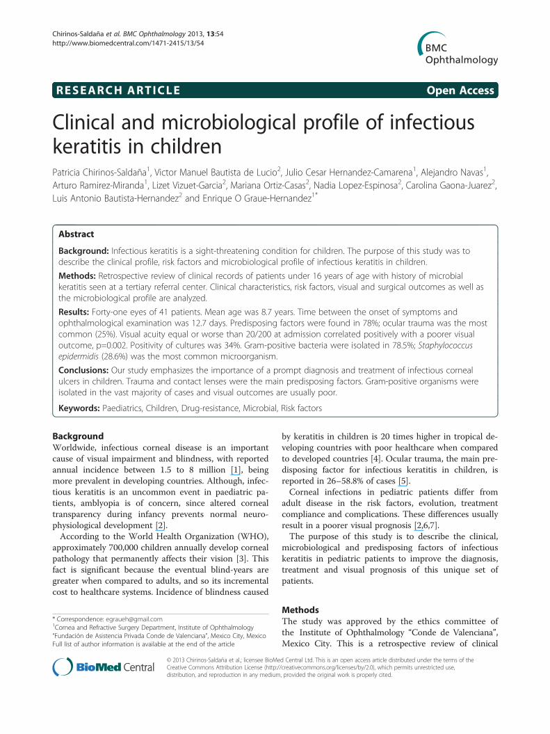
Table 1 Predisposing factors for infectious keratitis inchildren
Predisposing factors No. of patients %
Trauma 8 25%
Contact lenses 5 15.6%
Steroid treatment 4 12.5%
Ocular rosacea 3 9.4%
Previous ocular surgery 3 9.4%
Systemic immunodeficiency 3 9.4%
Congenital facial paralysis 3 9.4%
Congenital anomalies of anterior chamber 2 6.3%
Previous herpetic infection 1 3.1%
Chirinos-Saldaña et al. BMC Ophthalmology 2013, 13:54 Page 2 of 6http://www.biomedcentral.com/1471-2415/13/54
records of patients younger than 16 years with diagnosisof infectious keratitis seen at the Cornea and ExternalDisease Unit at Institute of Ophthalmology “Conde deValenciana”, in Mexico City between January 2006 toDecember 2011. Microbiological data were obtained fromthe Department of Microbiology and Ocular Proteomicsof the same institution.The studied variables included demographic data,
medical history, risk factors (history of ocular trauma,use of contact lenses, associated eye diseases, systemicdiseases, previous ocular surgery), clinical presentation,initial and final visual acuity, medication use before andafter diagnosis, and need for surgical therapy.
Ophthalmic examinationAll patients had a detailed clinical evaluation followedby corneal scrapings. The material obtained on scrapingwas subjected to standard microbiology evaluation. Ini-tial medical treatment was based on fourth-generationfluoroquinolones monotherapy and modified in accord-ance with clinical response, culture and antibiotic sus-ceptibility results.Every patient underwent a comprehensive ophthalmic
examination, including but not limited to, presentinguncorrected distance visual acuity (UDVA) and pinholecorrected distance visual acuity (CDVA), slit-lampbiomicroscopy examination, fundoscopy and intraocularpressure. With respect to corrected visual acuity, pa-tients were classified according to the revised ICD-10[8]. The location, depth and size of the ulcer, as well asinfiltrate appearance, presence or absence of lysis,hypopyon or neovascularization were documented ateach visit. The longest diameter of the ulcer at presenta-tion was defined as the size of the ulcer. The ulcer wasdefined, as being central if it involved the pupillary areaotherwise it was recorded as peripheral. Follow-up wasdone as needed.
Microbiology workupIn all patients, corneal scrapings were obtained, smearswere prepared for standard microbiologic evaluation in-cluding Gram and Giemsa stains. The sample was sowedin Columbia agar + 5% sheep, chocolate agar + PolyViteX(PVX) and Brain-Heart Infusion (BHI), those were incu-bated at 37°C and 5% CO2; and Sabouraud dextrose agar,which was incubated at 28°C and 5% CO2. The bacteriawere identified using the Vitek 2 Compact system(bioMérieux, France) with GP-test Vitek card. The drugsensitivity was determined by the Kirby-Baüer methodusing the following antibiotic discs: polymyxin, oxacillin,neomycin, sulfamethoxazole, vancomycin, gentamicin,ciprofloxacin, ofloxacin, cephalothin, cephazolin andceftazidime and according to Clinical and LaboratoryStandards Institute guidelines [9]. Criteria for culture
positivity were growth of the organism at the site of inocu-lation on two or more solid phase cultures, or growth atthe site of inoculation on one solid phase media of anorganism consistent with microscopy, or confluent growthon one media.
Statistical evaluationThe statistical analysis was performed with SPSS 17.0software (SPSS Inc, Chicago, IL, USA). Descriptive sta-tistics were obtained to determine the frequency andproportions. One-way analysis of variance (ANOVA)and linear regression model were used to evaluate thechange of visual acuity from admission to discharge.
ResultsPatient characteristicsBetween 2006 and 2011, 41 corneal samples from 41children with infectious keratitis were identified. Twenty-one cases were male (51%), and 20 were female (49%).The mean patient age was 8.7 years ± 5.1 (range, 3 monthsto 15 years). The mean time from the onset of symptomsto the ophthalmological examination was 12.7 days ± 18.7(range, 1–60 days).Predisposing factors were identified in 78% of cases,
with 2 or more factors occurring in 26%. The most com-mon predisposing factor was ocular trauma (25%),followed by wearing contact lenses and prolonged ster-oid treatment (Table 1). In cases associated with oculartrauma, pencils were the most common cause (10.6%);other was associated with fireworks, cat scratch, rope,soil and toys.Patients with CDVA ≥ 20/60 at admission showed a
statistically non-significant improvement of their visionat discharge, but happened inversely when CDVA was <20/60 (p=0.003). A worsening of visual acuity was morepronounced when CDVA was less than 20/200 at admis-sion (mean increment of logMar 1.04, p = 0.002). Linearregression analysis showed for lower visual acuity at

Chirinos-Saldaña et al. BMC Ophthalmology 2013, 13:54 Page 3 of 6http://www.biomedcentral.com/1471-2415/13/54
admission, lower visual acuity at discharge (p<0.0001)[Figure 1].The ophthalmological examination revealed a mean
epithelial defect size of 2.74 ± 1.6 mm, with visual axisinvolvement in 63.2% of cases, anterior chamber reac-tion in 31.6% and hypopyon in 15.8%.
TreatmentAs mentioned previously initial medical therapy wasbased on fourth generation fluoroquinolones and modi-fied according to clinical response or antibiogram. In 26patients (63.4%) this therapeutic regimen remained(0.5% moxifloxacin or 0.3% gatifloxacin); 6 (14.6%) wereswitched to macrolides (0.5% erythromycin); 5 (12.2%)to third generation cephalosporins (5% ceftazidime); 3(7.3%) to third generation fluoroquinolones (0.3% cipro-floxacin) and 1 (2,4%) to topical 0.15% amphotericin B to-gether with 1% natamycin and systemic oral itraconazole.Medical therapy achieved remission in 39 cases (95%).
One case developed endophthalmitis and was success-fully treated with intravitreal antibiotics (vancomycinand ceftazidime). Perforation occurred in a single caseand was treated with tectonic keratoplasty.
MicrobiologyRegarding the microbiological results, 66% (n=27) werenegative, 26% of them (n=7) were previously treated withtopical antibiotics. Cultures were positive in only 34%(n= 14), which identified 7 different microorganisms andno polymicrobial infections. Bacteria were responsible forinfection in 93% (13) and fungi (Microsporum gypseum) in7% (n=1).
Figure 1 Linear regression model: Estimation of change ofvisual acuity at discharge for every unit of visual acuity atadmission. Legend: An mean increment of logMAR 0.55 atdischarge was observed for every increasing unit of logMAR atadmission, p < 0.0001.
Gram-positive bacteria were isolated in 79% (n=11) ofthe positive cultures. Staphylococcus epidermidis wasthe most common isolate followed by equal frequenciesof Streptococcus spp., Corynebacterium spp. andPseudomonas aeruginosa. No significant associationbetween risk factors and culture positivity was encoun-tered (Table 2).The antibiogram of Staphylococcus spp. isolates re-
vealed that all isolates were sensitive to gentamicin, 80%(n=4) were sensitive to vancomycin and ciprofloxacin.Eighty percent (n=4) of these demonstrated resistance tosulfamethoxazole, and 75% (n=3) to cefazolin, oxacillinand polymyxin B. Eighty percent (n=4) of these revealedresistance to multiple antibiotics.All Streptococcus spp. isolates were sensitive to cipro-
floxacin, cefazolin, ofloxacin and ceftriaxone, while 75%(n=3) were also sensitive to sulfamethoxazole, vanco-mycin and gentamicin. Seventy five percent (n=3) wereresistant to polymyxin B.Both Pseudomonas aeruginosa isolates were sensitive to
gentamicin and resistant to ciprofloxacin and ceftazidime.
DiscussionAlthough uncommon, infectious keratitis in children is acondition that leads to an imminent risk of amblyopiaand/or permanent visual loss and because of this thecost per case is very high [10].Children may be poor historians and/or may not com-
plain of ocular pain. Keratitis diagnosis and treatmentmay be delayed by parents, or by primary care physiciansconfusing keratitis with the less severe conjunctivitis, es-pecially if the cornea is not severely affected and the in-filtrate not macroscopically obvious. This may bereflected in our study where the mean time to diagnosiswas almost 2 weeks. This fact may also explain the poor
Table 2 Microorganisms isolated from corneal ulcers inchildren
Microorganisms Nº of positivecultures
% of positivecultures
Gram-positivebacteria
Staphylococcusepidermidis
4 28.6%
Staphylococcusaureus
1 7.1%
Streptococcusviridans
2 14.3%
Streptococcuspneumoniae
2 14.3%
Corynebacterium sp. 2 14.3%
Gram-negativebacteria
Pseudomonasaeruginosa
2 14.3%
Fungi Microsporumgypseum
1 7.1%
No growth 27 66%

Chirinos-Saldaña et al. BMC Ophthalmology 2013, 13:54 Page 4 of 6http://www.biomedcentral.com/1471-2415/13/54
visual outcomes and highlights the importance to edu-cate parents and primary care personnel in the import-ance of immediate referral whenever the cornea may beinvolved.In our series, a history of trauma was the major pre-
disposing factor, present in approximately one quarter ofthe cases (25%). This result is consistent with multiplemicrobial keratitis studies involving children where ocu-lar trauma has been associated in up to two thirds of thecases. (26–58.8%) [2,11-13]. Corneal trauma disrupts theprotective mechanism of the corneal epithelium, facili-tating bacterial adhesion and accelerating penetrationand replication of microorganisms [12,13]. Children areless careful than adults and do not understand the harmthat is associated with dangerous objects. Plants, metals,plastic parts, fireworks and pencils may cause oculartrauma [2].In our study, 15.6% of patients had a history of wear-
ing contact lenses. Interactions between contact lens andocular surface generated by chronic or improper contactlens wear such as overnight wear, may produce epithelialdefects that predispose the wearer to bacterial adhesion[14,15]. Exposure to contaminated disinfectant solutionsand biofilm formation are additional mechanisms thatcan cause corneal infection [16,17]. Although statisticsare lacking, pediatric contact lens wear may be morecommon in populations where high myopia is preva-lent and/or orthokeratology is popular. In our popula-tion, orthokeratology is rarely used and but its useshould be cautiously advised specially in children withother risk factors for infections (pediatric rosacea, re-current blepharitis) [6,18].The influence of systemic diseases and malnutrition
on the wound healing process should be considered as apredisposing factor for microbial keratitis in children[19,20]. A wide variety of factors such as low socioeco-nomic status, incomplete immunization profile andsystemic diseases, including hypoxic encephalopathy,pulmonary stenosis, protein-energy malnutrition, mul-tiple congenital anomalies and prematurity have been as-sociated with severe microbial keratitis in children[2,12,13,21,22]. Jhanji and co-authors recently reviewedthe role of immunization and malnutrition in corneal ul-cers in children 5 years or younger [22]. The severity ofprotein-energy malnutrition was related significantly tothe occurrence of bilateral infection, to an incompleteimmunization scheme and poor socioeconomic status,however this study did not address the confounding be-tween these variables. In our study, we found 3 caseswith associated systemic diseases. Two children had psy-chomotor retardation and malnutrition, and the otherhad chronic cardiopulmonary disease.Local predisposing factors for infectious keratitis were
found in 16 cases (50%), which included chronic steroid
use, ocular rosacea and previous ocular surgeries, con-genital facial paralysis and previous herpetic infection.These factors, as well as dry eye, exposure keratopathyand eyelid abnormalities act as facilitators of cornealinfection [11,13].Regarding microbiological profile, we found that cul-
tures were positive in 34% of cases, a value that waslower than those reported in other studies (48% to 87%)[20,21,23]. Self-prescribed antibiotic, microorganismswith slow growth on culture media, viral causes of kera-titis, improper corneal sampling, and the inherent diffi-culty in getting corneal samples from pediatric patientsmay account for the low positivity rate observed in ourstudy. The higher rates of culture positivity reported inthe other studies, may also be explained by their use ofgeneral anesthesia or deep sedation for corneal scrapingsin uncooperative patients [20,21,23].As shown previously by other authors, Gram-positive
microorganisms are the main etiological agents of infec-tious keratitis in children [2,7]. Over the years, anincreased incidence of keratitis caused by coagulase-negative Staphylococcus has been reported, [12,13,20-23]and many studies consider coagulase-negative Staphylo-coccus as an important cause of endophthalmitis [24-26].The antibiotic susceptibility of coagulase-negativeStaphylococcus isolates is unpredictable, and that multi-resistance to antibiotics is common. Therefore, an anti-biogram should be performed in the clinically significantocular infections that arise from these organisms [24].We observed a high resistance of Staphylococcus spp.
to the antibiotics known for their action against gram-positive organisms, including sulfamethoxazole, first-generation cephalosporins and oxacillin; the latter usedas a surrogate marker for methicillin-resistant organ-isms. A hundred percent of these cases were susceptibleto gentamicin, and 80% to ciprofloxacin and vanco-mycin. Alternatively; Streptococcus spp. isolates weresensitive to the majority of antibiotics. Resistance tomultiple antibiotics was seen in 80% of Staphylococcusspp. and in 25% of Streptococcus spp. isolates. Prolongedantibiotic therapy is known to promote the adaptation oforganisms and development of specific cross-resistancemechanisms although knowledge of the local resistancetrends in ophthalmic specimens is mandatory to providea prompt and effective treatment [27,28].In general, fluoroquinolones susceptibility profile was
good across our series of positive cultures, making thisclass of antibiotic suitable for empiric treatment thatmay be modified according to the antibiogram results.Although in our setting resistance is uncommon, siteswhere it’s use is widespread in healthcare, resistance is aconcern [29,30].Finally, as previously reported [31], vancomycin is an
effective anti-staphylococcal drug that is rarely associated

Chirinos-Saldaña et al. BMC Ophthalmology 2013, 13:54 Page 5 of 6http://www.biomedcentral.com/1471-2415/13/54
with resistance, hence, it is important that this drug be re-served for treating infections that are resistant to otheranti-staphylococcal antibiotics or in cases of severecorneal infections. In this report we encountered aStaphylococcus epidermidis strain that was resistant tovancomycin, which suggests the existence of some strainswith complex resistance mechanisms in our environment.The findings of this study should be interpreted cau-
tiously. This study is limited by its small sample size andis subject to selection bias since it was performed at atertiary referral eye care center, so the results herepresented cannot be extrapolated to the general popula-tion. Nevertheless, our results strengthen the body ofknowledge around infectious keratitis in children andcontribute to a better understanding of microbial cor-neal ulcers in the pediatric patient, in the hope of im-proving their visual outcome.
ConclusionIn conclusion, trauma and contact lenses were the mainpredisposing factors for infectious keratitis in patients16 years or younger. The frequent involvement of thecentral cornea, the delay in reaching specialty care andlow positivity of cultures may all account for the poorvisual outcomes conveyed in our study.
Competing interestsThe authors declare that they have no competing interests.
Authors’ contributionsPCS, VMBL, EOGH conceived, designed and drafted the manuscript. LVG,MOC, NLLE, CGJ, LABH performed the data collection. JCHC, ANP, ARMcontributed to review and to the revision of the manuscript. All authors readand approved the final manuscript.
AcknowledgmentsThe authors would like to thank Dr Mark Mannis, for his support and the“Fundación de Asistencia Privada Conde de Valenciana” OphthalmologyInstitute.
Author details1Cornea and Refractive Surgery Department, Institute of Ophthalmology“Fundación de Asistencia Privada Conde de Valenciana”, Mexico City, Mexico.2Microbiology and Ocular Proteomics, Research Unit, Institute ofOphthalmology “Fundación de Asistencia Privada Conde de Valenciana”,Mexico City, Mexico.
Received: 12 January 2013 Accepted: 6 October 2013Published: 16 October 2013
References1. Whitcher JP, Srinivasan M: Cornel ulceration in the developing world a
silent epidemic. Br J Ophthalmol 1997, 8:622–631.2. Parmar P, Salman A, Kalavathy CM, Kaliamurthy J, Thomas PA, Jesudasan CA:
Microbial keratitis at extremes of age. Cornea 2006, 25(2):153–158.3. Underwood BA: Update: xerophthalmia, keratomalacia, and child
mortality including measles. In World Blindness and Its Prevention. Volume4. Edited by Kupfur C, Gillen T. Oxford: Oxford University Press;1990:171–175.
4. Maurin JF, Renard JP, Ahmedou O, Bidaux F, Dordain Y, Pariselle J, FroussartF, Dot C, Rigal-Sastourne JC: [Corneal blindness in tropical areas]. MedTrop (Mars) 1995, 55(4 Pt 2):445–449.
5. Ormerod LD, Murphree AL, Gomez DS, Schanzlin DJ, Smith RE: Microbialkeratitis in children. Ophthalmology 1986, 93(4):449–455.
6. Young AL, Leung AT, Cheng LL, Law RW, Wong AK, Lam DS:Orthokeratology lens-related corneal ulcers in children: a case series.Ophthalmology 2004, 111(3):590–595.
7. Satpathy G, Vishalakshi P: Ulcerative keratitis: Microbial profile andsensitivity pattern: A five year study. Ann Ophthalmol 1995,27(5):301–306.
8. Dandona L, Dandona R: Revision of visual impairment definitions in theInternational Statistical Classification of Diseases. BMC Med 2006, 16:4–7.
9. Clinical and Laboratory Standards Institute: Performance standards forantimicrobial susceptibility testing: Seventeenth informational supplement.CLSI document M100-S17. 940 West Valley Road, Suite 1400, Wayne,Pennsylvania: Clinical and Laboratory Standards Institute;2007:19087–1898. ISBN 1-56238-625-5.
10. Carlton J, Karnon J, Czoski-Murray C, Smith KJ, Marr J: The clinicaleffectiveness and cost-effectiveness of screening programmes foramblyopia and strabismus in children up to the age of 4–5 years: asystematic review and economic evaluation. Health Technol Asess 2008,12(25):194. iii, xi.
11. Cruz OA, Sabir SM, Capo H, Alfonso EC: Microbial keratitis in childhood.Ophthalmology 1993, 100:192–196.
12. Al Otaibi AG, Allam K, Damri AJ, Shamri AA, Kalantan H, Mousa A:Childhood microbial keratitis. Oman J Ophthalmol 2012, 5(1):28–31.
13. Song X, Xu L, Sun S, Zhao J, Xie L: Pediatric microbial keratitis: a tertiaryhospital study. Eur J Ophthalmol 2012, 22(2):136–141.
14. Dart JK, Radford CF, Minassian D, Verma S, Stapleton F: Risk factors formicrobial keratitis with contemporary contact lenses: a case–controlstudy. Ophthalmology 2008, 115(10):1647–1654.
15. Stapleton F, Keay L, Edwards K, Naduvilath T, Dart JK, Brian G, Holden BA:The incidence of contact lens-related microbial keratitis in Australia.Ophthalmology 2008, 115(10):1655–1662.
16. Behlau I, Gilmore MS: Microbial biofilms in ophthalmology and infectiousdisease. Arch Ophthalmol 2008, 126(11):1572–1581.
17. Shovlin JP, Argüeso P, Carnt N, Chalmers RL, Efron N, Fleiszig SM, Nichols JJ,Polse KA, Stapleton F, Wiley L, Willcox M, Bright FV, Efron N, Jones LW, KeirN, Peterson RC, Stapleton F: 3. Ocular surface health with contact lenswear. Cont Lens Anterior Eye 2013, 36(1):14–21.
18. Watt K, Swarbrick HA: Microbial keratitis in overnight orthokeratology:review of the first 50 cases. Eye Contact Lens 2005, 31(5):201–208.
19. Wong VWY, Lai TYY, Chi SCC, Lam DSC: Pediatric ocular surface infections:a 5-year review of demographics, clinical features, risk factors,microbiological results, and treatment. Cornea 2011, 30:995–1002.
20. Emery PW, Sanderson P: The effects of dietary restriction on proteinsynthesis and wound healing after surgery in the rat. Clin Sci 1995,89(4):383–388.
21. Kunimoto DY, Sharma S, Reddy MK, Gopinathan U, Jyothi J, Miller D, RaoGN: Microbial keratitis in children. Ophthalmology 1998, 105(2):252–257.
22. Vajpayee RB, Ray M, Panda A, Sharma N, Taylor HR, Murthy GV, Satpathy G,Pandey RM: Risk factors for pediatric presumed microbial keratitis: acase–control study. Cornea 1999, 18(5):565–569.
23. Jhanji V, Naithani P, Lamoureux E, Agarwal T, Sharma N, Vajpayee RB:Immunization and nutritional profile of cases with atraumaticmicrobial keratitis in preschool age group. Am J Ophthalmol 2011,151(6):1035–1040.
24. Singh G, Palanisamy M, Madhavan B, Rajaraman R, Narendran K, Kour A,Venkatapathy N: Multivariate analysis of childhood microbial keratitis inSouth India. Ann Acad Med Singapore 2006, 35(3):185–189.
25. Pinna A, Zanetti S, Sotgiu M, Sechi LA, Fadda G, Carta F: Identification andantibiotic susceptibility of coagulase negative staphylococci isolated incorneal/external infections. Br J Ophthalmol 1999, 83(7):771–773.
26. Kattan HM, Flynn HW Jr, Pflugfelder SC, Robertson C, Forster RK:Nosocomial endophthalmitis survey. Current incidence of infection afterintraocular surgery. Ophthalmology 1991, 98(2):227–238.
27. Speaker MG, Milch FA, Shah MK, Eisner W, Kreiswirth BN: Role of externalbacterial flora in the pathogenesis of acute postoperativeendophthalmitis. Ophthalmology 1991, 98(5):639–649. discussion 650.
28. Asbell PA, Colby KA, Deng S, McDonnell P, Meisler DM, Raizman MB,Sheppard JD Jr, Sahm DF: Ocular TRUST: nationwide antimicrobialsusceptibility patterns in ocular isolates. Am J Ophthalmol 2008,145(6):951–958.

Chirinos-Saldaña et al. BMC Ophthalmology 2013, 13:54 Page 6 of 6http://www.biomedcentral.com/1471-2415/13/54
29. Chalita MR, Hofling-Lima AL, Paranhos A Jr, Schor P, Belfort R Jr: Shiftingtrends in in vitro antibiotic susceptibilities for common ocular isolatesduring a period of 15 years. Am J Ophthalmol 2004, 137(1):43–51.
30. Graves A, Henry M, O'Brien TP, Hwang DG, Van Buskirk A, Trousdale MD: Invitro susceptibilities of bacterial ocular isolates to fluoroquinolones.Cornea 2001, 20(3):301–305.
31. Guzmán Lista MDC, Lozada Oca RA: Detección de Staphylococcus aureusmeticilino-resistentes aislados de pacientes con infeccionesnosocomiales y adquiridas en la comunidad. Rev Soc Ven Microbiol 2007,27:349–363.
doi:10.1186/1471-2415-13-54Cite this article as: Chirinos-Saldaña et al.: Clinical and microbiologicalprofile of infectious keratitis in children. BMC Ophthalmology 2013 13:54.
Submit your next manuscript to BioMed Centraland take full advantage of:
• Convenient online submission
• Thorough peer review
• No space constraints or color figure charges
• Immediate publication on acceptance
• Inclusion in PubMed, CAS, Scopus and Google Scholar
• Research which is freely available for redistribution
Submit your manuscript at www.biomedcentral.com/submit