Embed Size (px)
Citation preview
IMMUNOLOGY AND HERPETIC KERATITIS
LUC MISSOTTEN
Leuven. Belgium
The immune defence against herpes virus in keratitis requires an intricate interplay of macrophages, B-lymphocytes, cytotoxic T- and helper T-lymphocytes, sometimes by direct contact, sometimes by exchange of mediators. In most diseases immunological interactions take place in lymphoid organs, where lymphocytes, macrophages and other cells are tightly packed in great numbers, facilitating the exchanges. This is the site where the first encounter with herpes virus leads to immunisation. However, in
I clinical practice we usually treat dendritic keratitis in adults who have already been immunised to herpes virus, a long time ago. Specific lymphocytes have already been activated previously, they have moved out of the lymph nodes and are present as memory cells in all tissues.
There are many good arguments to support the view that in keratitis the immune response to this new challenge occurs in the ocular tissues, in full view of the biomicroscope, and not in distant lymph nodes. Although the optical resolution of a slit lamp is insufficient for a detailed observation of leucocytes, a skilled ophthalmologist is able to recognise their presence and activity.
The immune defences rest primarily on the action of a few classes of specialised cells, among them the B-lymphocytes and the T-Iymphocytes.
B-lymphocytes
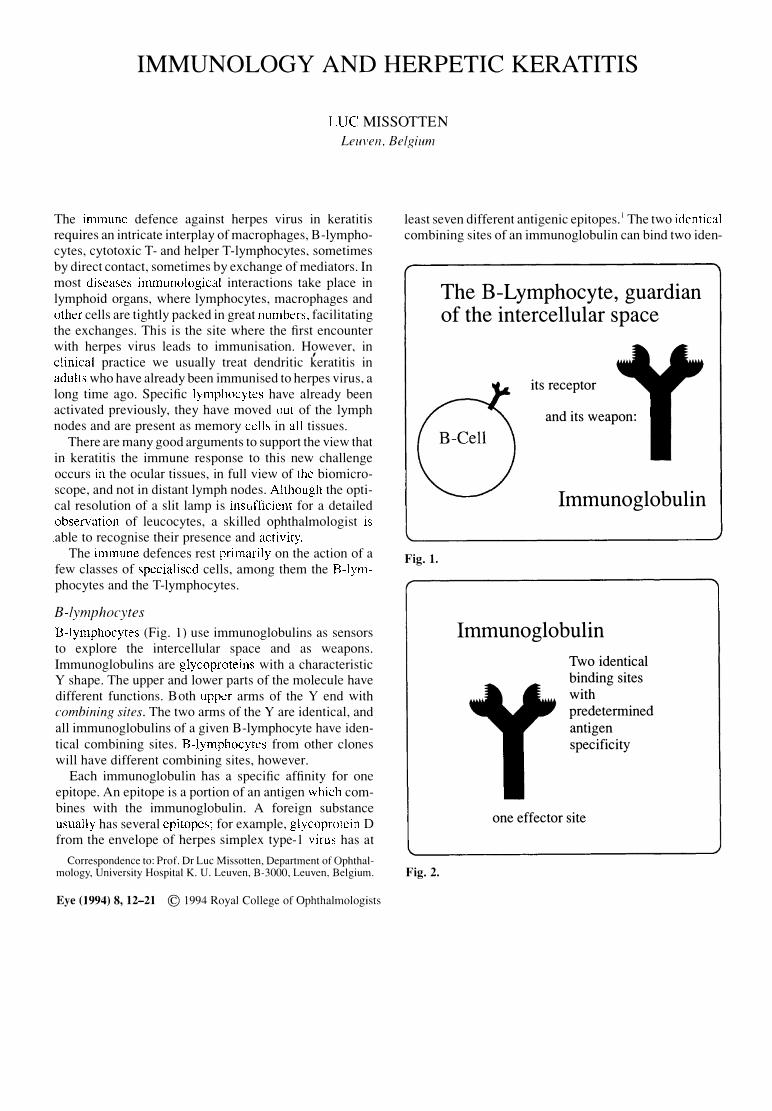
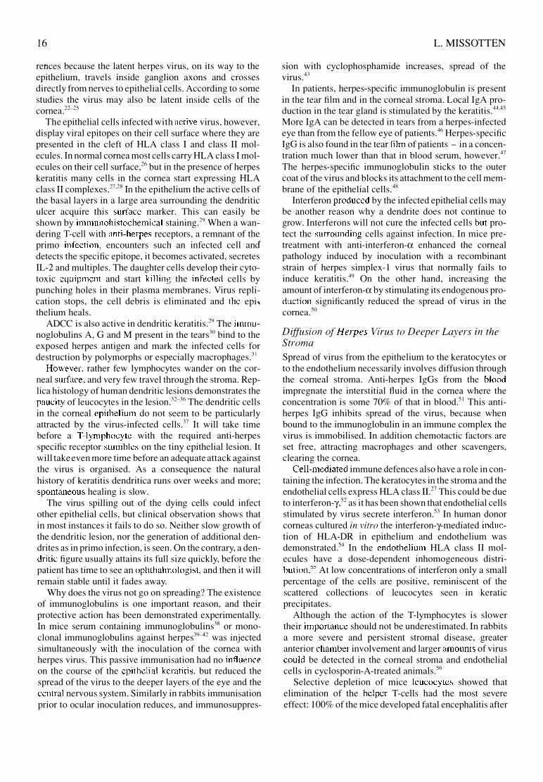
B-lymphocytes (Fig. 1) use immunoglobulins as sensors to explore the intercellular space and as weapons. Immunoglobulins are glycoproteins with a characteristic Y shape. The upper and lower parts of the molecule have different functions. Both upper arms of the Y end with combining sites. The two arms of the Yare identical, and all immunoglobulins of a given B-lymphocyte have identical combining sites. B-lymphocytes from other clones will have different combining sites, however.
Each immunoglobulin has a specific affinity for one epitope. An epitope is a portion of an antigen which combines with the immunoglobulin. A foreign substance usually has several epitopes; for example, glycoprotein D from the envelope of herpes simplex type-I virus has at
Correspondence to: Prof. Dr Luc Missotten, Department of Ophthalmology, University Hospital K. U. Leuven, B-3000, Leuven. Belgium.
Eye (1994) 8, 12-21 © 1994 Royal College of Ophthalmologists
least seven different antigenic epitopes.1 The two identical combining sites of an immunoglobulin can bind two iden-
Fig. 1.
Fig. 2.
The B-Lymphocyte, guardian of the intercellular space
its receptor
and its weapon:
Immunoglobulin
Immunoglobulin
Two identical binding sites with predetermined antigen specificity
one effector site
IMMUNOLOGY AND HERPETIC KERATITIS
tical epitopes, and in this way link two antigens together (Fig. 12).
The stem of the Y is the effector part of the immunoglobulin. It can have different shapes and perform different functions.
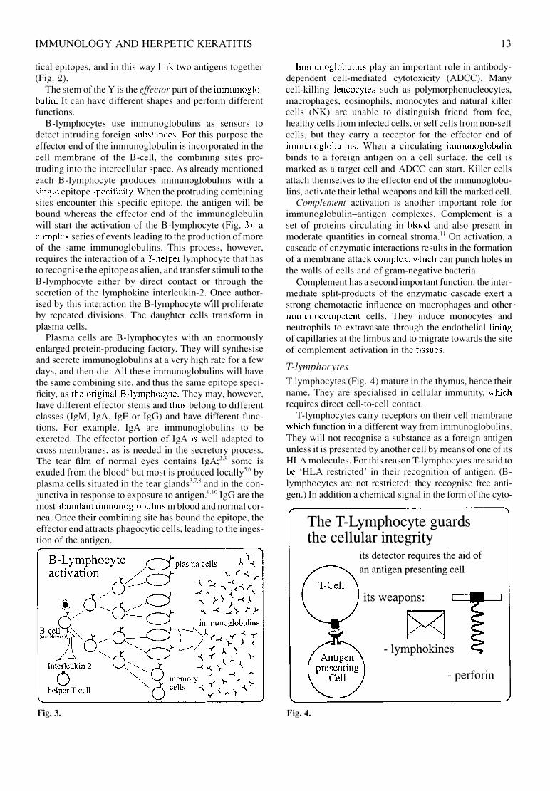
B-Iymphocytes use immunoglobulins as sensors to detect intruding foreign substances. For this purpose the effector end of the immunoglobulin is incorporated in the cell membrane of the B-cell, the combining sites protruding into the intercellular space. As already mentioned each B-Iymphocyte produces immunoglobulins with a single epitope specificity. When the protruding combining sites encounter this specific epitope, the antigen will be bound whereas the effector end of the immunoglobulin will start the activation of the B-Iymphocyte (Fig. 3). a complex series of events leading to the production of more of the same immunoglobulins. This process, however. requires the interaction of a T-helper lymphocyte that has to recognise the epitope as alien, and transfer stimuli to the B-Iymphocyte either by direct contact or through the secretion of the lymphokine interleukin-2. Once authorised by this interaction the B-lymphocyte will proliferate by repeated divisions. The daughter cells transform in plasma cells.
Plasma cells are B-Iymphocytes with an enormously enlarged protein-producing factory. They will synthesise and secrete immunoglobulins at a very high rate for a few days. and then die. All these immunoglobulins will have the same combining site, and thus the same epitope specificity, as the original B-lymphocyte. They may. however. have different effector stems and thus belong to different classes (IgM, IgA, IgE or IgG) and have different functions. For example. IgA are immunoglobulins to be excreted. The effector portion of IgA is well adapted to cross membranes, as is needed in the secretory process. The tear film of normal eyes contains IgA;2.3 some is exuded from the blood4 but most is produced locally"·6 by plasma cells situated in the tear glands3.7.8 and in the conjunctiva in response to exposure to antigen.9.IO IgG are the most abundant immunoglobulins in blood and normal cornea. Once their combining site has bound the epitope. the effector end attracts phagocytic cells. leading to the ingestion of the antigen.
Fig. 3.
13
Immunoglobulins play an important role in antibodydependent cell-mediated cytotoxicity (ADCC). Many cell-killing leucocytes such as polymorphonucleocytes, macrophages, eosinophils, monocytes and natural killer cells (NK) are unable to distinguish friend from foe, healthy cells from infected cells, or self cells from non-self cells, but they carry a receptor for the effector end of immunoglobulins. When a circulating immunoglobulin binds to a foreign antigen on a cell surface, the cell is marked as a target cell and ADCC can start. Killer cells attach themselves to the effector end of the immunoglobulins, activate their lethal weapons and kill the marked cell.
Complement activation is another important role for immunoglobulin-antigen complexes. Complement is a set of proteins circulating in blood and also present in moderate quantities in corneal stroma. I I On activation, a cascade of enzymatic interactions results in the formation of a membrane attack complex, which can punch holes in the walls of cells and of gram-negative bacteria.
Complement has a second important function: the intermediate split-products of the enzymatic cascade exert a strong chemotactic influence on macrophages and other' immunocompetent cells. They induce monocytes and neutrophils to extravasate through the endothelial lining of capillaries at the limbus and to migrate towards the site of complement activation in the tissues.
T-lymphocytes
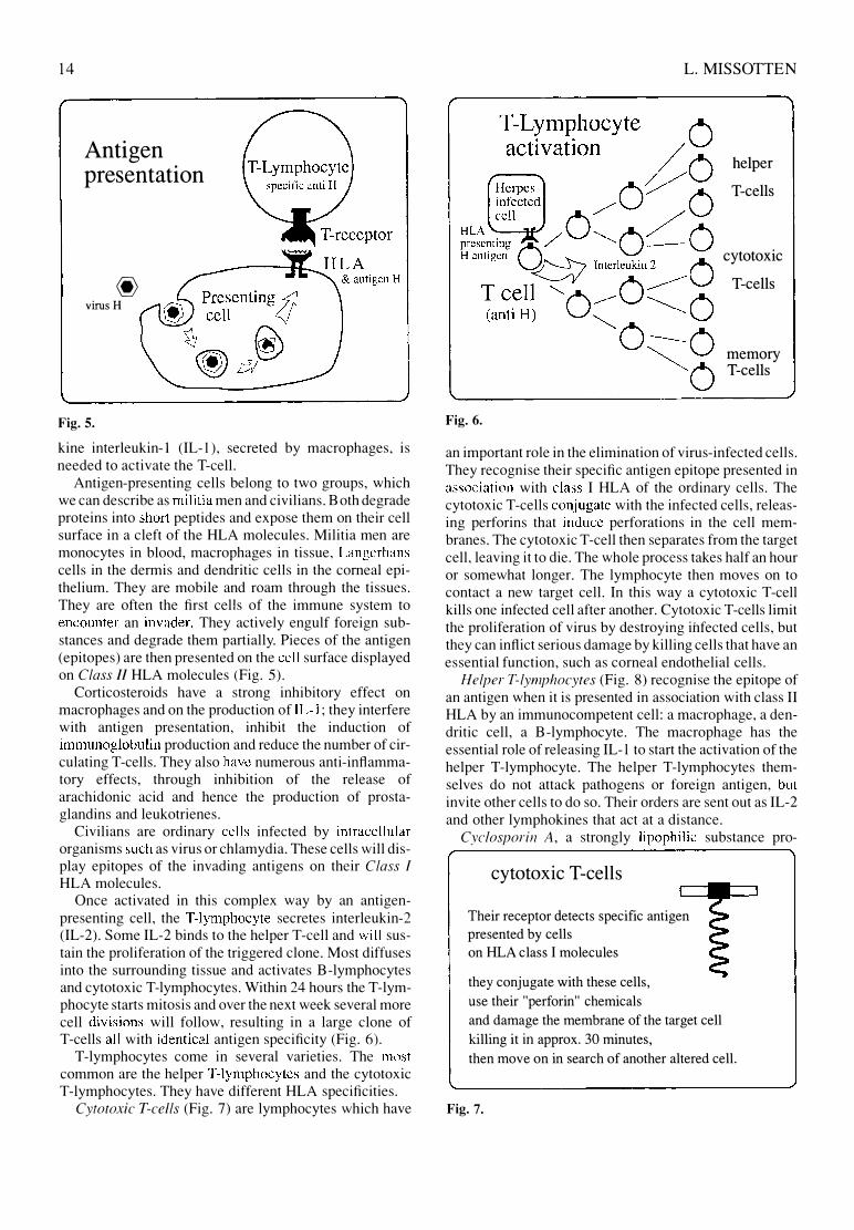
T-Iymphocytes (Fig. 4) mature in the thymus, hence their name. They are specialised in cellular immunity, which requires direct cell-to-cell contact.
T-Iymphocytes carry receptors on their cell membrane which function in a different way from immunoglobulins. They will not recognise a substance as a foreign antigen unless it is presented by another cell by means of one of its HLA molecules. For this reason T-Iymphocytes are said to be 'HLA restricted' in their recognition of antigen. (Blymphocytes are not restricted: they recognise free antigen.) In addition a chemical signal in the form of the cyto-
The T-Lymphocyte guards the cellular integrity
Fig. 4.
its detector requires the aid of
an antigen presenting cell
its weapons:
- lymphokines
- perforin
14
Antigen presentation
@ virus H
Fig. S.
kine interleukin-l (IL-l ), secreted by macrophages, is needed to activate the Tcell.
Antigen-presenting cells belong to two groups, which we can describe as militia men and civilians. Both degrade proteins into short peptides and expose them on their cell surface in a cleft of the HLA molecules. Militia men are monocytes in blood, macrophages in tissue, Langerhans cells in the dermis and dendritic cells in the corneal epithelium. They are mobile and roam through the tissues. They are often the first cells of the immune system to encounter an invader. They actively engulf foreign substances and degrade them partially. Pieces of the antigen (epitopes) are then presented on the cell surface displayed on Class II HLA molecules (Fig. 5).
Corticosteroids have a strong inhibitory effect on macrophages and on the production of IL-l ; they interfere with antigen presentation, inhibit the induction of immunoglobulin production and reduce the number of circulating T-cells. They also have numerous anti-inflammatory effects, through inhibition of the release of arachidonic acid and hence the production of prostaglandins and leukotrienes.
Civilians are ordinary cells infected by intracellular organisms such as virus or chlamydia. These cells will display epitopes of the invading antigens on their Class I
HLA molecules. Once activated in this complex way by an antigen
presenting cell, the T-lymphocyte secretes interleukin-2 (IL-2). Some IL-2 binds to the helper T-cell and will sustain the proliferation of the triggered clone. Most diffuses into the surrounding tissue and activates B-lymphocytes and cytotoxic T-lymphocytes. Within 24 hours the T-lymphocyte starts mitosis and over the next week several more cell divisions will follow, resulting in a large clone of T-cells all with identical antigen specificity (Fig. 6).
T-lymphocytes come in several varieties. The most common are the helper T-lymphocytes and the cytotoxic T-lymphocytes. They have different HLA specificities.
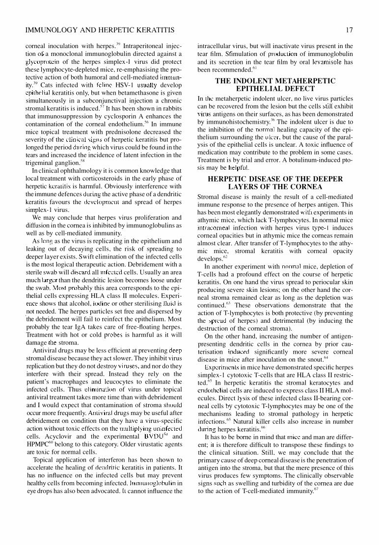
Cytotoxic T-cells (Fig. 7) are lymphocytes which have
L. MISSOTTEN
Fig. 6.
helper
T-cells
cytotoxic
T-cells
memory T-cells
an important role in the elimination of virus-infected cells. They recognise their specific antigen epitope presented in association with class I HLA of the ordinary cells. The cytotoxic T-cells conjugate with the infected cells, releasing perforins that induce perforations in the cell membranes. The cytotoxic T-cell then separates from the target cell, leaving it to die. The whole process takes half an hour or somewhat longer. The lymphocyte then moves on to contact a new target cell. In this way a cytotoxic T-cell kills one infected cell after another. Cytotoxic T-cells limit the proliferation of virus by destroying infected cells, but they can inflict serious damage by killing cells that have an essential function, such as corneal endothelial cells.
Helper T-lymphocytes (Fig. 8) recognise the epitope of an antigen when it is presented in association with class II HLA by an immunocompetent cell: a macrophage, a dendritic cell, a B-lymphocyte. The macrophage has the essential role of releasing IL-I to start the activation of the helper T-lymphocyte. The helper T-lymphocytes themselves do not attack pathogens or foreign antigen, but invite other cells to do so. Their orders are sent out as IL-2 and other lymphokines that act at a distance.
Cyclosporin A, a strongly lipophilic substance pro-
cytotoxic T-cells
Their receptor detects specific antigen
presented by cells
on HLA class I molecules
they conjugate with these cells,
use their "perforin" chemicals
and damage the membrane of the target cell
killing it in approx. 30 minutes,
then move on in search of another altered cell.
Fig. 7.
IMMUNOLOGY AND HERPETIC KERATITIS
helper T-cells
Detection of their specific antigen
presented by a HLA Class II molecule
induces the secretion of Iymphokines (chemical messages)
such as: INTERLEUKIN 2.
It -authorizes B-cells to proliferate
-supports the multiplication of T-cells
Cyciosporin-A inhibits the production of interleukin 2
Fig. 8.
duced by the fungus Trichoderma polysporum. has a specific action on T-lymphocytes. It interferes with the transcription of the genes encoding IL-2. interleukin-4 and interferon-yo By inhibiting the production of IL-2. cyclosporin prevents the T-helper cell from activating itself and from instructing other cells such as the cytotoxic T-cell or the immunoglobulin-producing B-celr Cyclosporin A reduces the T-helper cells to silence.
The term inte7j"erol1 is used for several unrelated classes of proteins with antiviral effects. Insight into their mode of action and their classification is constantly changing. Interferons diffuse from the site of infection. Some influence neighbouring infected cells to prevent the synthesis of viral nucleic acids. Others increase the expression of class I and class II HLA glycoproteins. facilitating recognition of viral antigens by the immune system. Another effect is the activation of cells with the ability to destroy virus-infected cells; these include natural killer cells and macrophages.
IMMUNOLOGY IN KERATITIS DUE TO HERPES SIMPLEX TYPE 1
Numerous papers on the immunology of experimental herpes keratitis are found in the literature. It is. however. difficult to judge in which way these observations are significant for clinical ophthalmology. There are many different strains of herpes simplex- l virus. Mice and man react very differently to the same strain of virus.12 Even in mice small differences in the strain of the herpes virus. or in the genome of the experimental animal. may change the pathophysiology of the disease. 1.1
Moreover. many experiments simulate either primo infections or re-infections of already-immunised animals. In clinical practice, however. neither of these situations occurs. We usually treat patients with reactivation of latent virus. Only recently have animal models been proposed of recurrent herpes simplex- l keratitis. either spontaneous 14
or induced by ultraviolet radiation 15 or iontophoresis. 1 6 17
Translating the results of animal experiments to clinical ophthalmology therefore calls for some elaborate guesswork to detect the information that applies to human herpetic keratitis. However. a few conclusions are obvious.
15
First, the strain of the virus and the genetic structure of the animal are both very important for the course of the disease. Most probably this is also true for herpes in patients. We all harbour herpes virus, but most of us have a favourable distinction between our virus and our 'self'. The unlucky patient with an unharmonious combination is condemned to lifelong misery. Second, various stages of the disease call into action different immune responses, and our therapeutic approach has to take this into account.
Primo Injection by Herpes Virus Type I
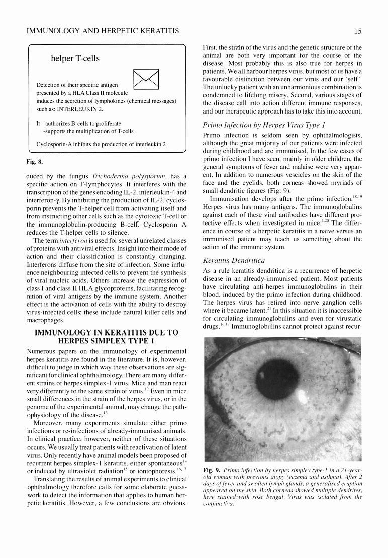
Primo infection is seldom seen by ophthalmologists, although the great majority of our patients were infected during childhood and are immunised. In the few cases of primo infection I have seen. mainly in older children, the general symptoms of fever and malaise were very apparent. In addition to numerous vescicles on the skin of the face and the eyelids. both corneas showed myriads of small dendritic figures (Fig. 9).
Immunisation develops after the primo infection.IR.19
Herpes virus has many antigens. The immunoglobulins against each of these viral antibodies have different protective effects when investigated in mice.120 The difference in course of a herpetic keratitis in a naive versus an immunised patient may teach us something about the action of the immune system.
Keratitis Dendritica
As a rule keratitis dendritica is a reCUITence of herpetic disease in an already-immunised patient. Most patients have circulating anti-herpes immunoglobulins in their blood. induced by the primo infection during childhood. The herpes virus has retired into nerve ganglion cells where it became latent. 2 I In this situation it is inaccessible for circulating immunoglobulins and even for virustatic d 1 617 I I b I'
. rugs.' mmunog 0 U InS cannot protect agamst recur-
Fig. 9. Primo infection hy he/pes simplex type-1 in a 21-yearold woman with previous atopy (ec::ema and asthma). After 2 days offel'('/" and swol/en lymph glands, a generalised eruption appeared Oil the skin. Both corneas showed multiple dendrites, here stained . with rose hengal. Virus was isolated from the conjunctiva.
16
rences because the latent herpes virus, on its way to the epithelium, travels inside ganglion axons and crosses directly from nerves to epithelial cells. According to some studies the virus may also be latent inside cells of the cornea.22-25
The epithelial cells infected with active virus, however, display viral epitopes on their cell surface where they are presented in the cleft of HLA class I and class II molecules. In normal cornea most cells carry HLA class I molecules on their cell surface,26 but in the presence of herpes keratitis many cells in the cornea start expressing HLA class II complexes.27,28 In the epithelium the active cells of the basal layers in a large area surrounding the dendritic ulcer acquire this surface marker. This can easily be shown by immunohistochemical staining.29 When a wandering T-cell with anti-herpes receptors, a remnant of the primo infection, encounters such an infected cell ane!' detects the specific epitope, it becomes activated, secretes IL-2 and multiples. The daughter cells develop their cytotoxic equipment and start killing the infected cells by punching holes in their plasma membranes. Virus replication stops, the cell debris is eliminated and the epi .. thelium heals.
ADCC is also active in dendritic keratitis.29 The immunoglobulins A, G and M present in the tears30 bind to the exposed herpes antigen and mark the infected cells for destruction by polymorphs or especially macrophages.31
However, rather few lymphocytes wander on the corneal surface, and very few travel through the stroma. Replica histology of human dendritic lesions demonstrates the paucity of leucocytes in the lesion.32-.36 The dendritic cells in the corneal epithelium do not seem to be particularly attracted by the virus-infected cells.37 It will take time before a T-Iymphocyte with the required anti-herpes specific receptor stumbles on the tiny epithelial lesion. It will take even more time before an adequate attack against the virus is organised. As a consequence the natural history of keratitis dendritica runs over weeks and more; spontaneous healing is slow.
The virus spilling out of the dying cells could infect other epithelial cells, but clinical observation shows that in most instances it fails to do so. Neither slow growth of the dendritic lesion, nor the generation of additional dendrites as in primo infection, is seen. On the contrary, a dendritic figure usually attains its full size quickly, before the patient has time to see an ophthalmologist, and then it will remain stable until it fades away.
Why does the virus not go on spreading? The existence of immunoglobulins is one important reason, and their protective action has been demonstrated experimentally. In mice serum containing immunoglobulins38 or monoclonal immunoglobulins against herpes39-42 was injected simultaneously with the inoculation of the cornea with herpes virus. This passive immunisation had no influence on the course of the epithelial keratitis, but reduced the spread of the virus to the deeper layers of the eye and the central nervous system. Similarly in rabbits immunisation prior to ocular inoculation reduces, and immunosuppres-
L. MISSOTTEN
slOn with cyclophosphamide increases, spread of the virus.43
In patients, herpes-specific immunoglobulin is present in the tear film and in the corneal stroma. Local IgA production in the tear gland is stimulated by the keratitis.44,45
More IgA can be detected in tears from a herpes-infected eye than from the fellow eye of patients.46 Herpes-specific IgG is also found in the tear film of patients - in a concentration much lower than that in blood serum, however.47 The herpes-specific immunoglobulin sticks to the outer coat of the virus and blocks its attachment to the cell membrane of the epithelial cells.48
Interferon produced by the infected epithelial cells may be another reason why a dendrite does not continue to grow. Interferons will not cure the infected cells but protect the surrounding cells against infection. In mice pretreatment with anti-interferon-a enhanced the corneal pathology induced by inoculation with a recombinant strain of herpes simplex-l virus that normally fails to induce keratitis.49 On the other hand, increasing the amount of interferon-a by stimulating its endogenous production significantly reduced the spread of virus in the cornea. 50
D(ffusion of Herpes Virus to Deeper Layers in the Stroma
Spread of virus from the epithelium to the keratocytes or to the endothelium necessarily involves diffusion through the corneal stroma. Anti-herpes IgGs from the blood impregnate the interstitial fluid in the cornea where the concentration is some 70% of that in blood.51 This antiherpes IgG inhibits spread of the virus, because when bound to the immunoglobulin in an immune complex the virus is immobilised. In addition chemotactic factors are set free, attracting macrophages and other scavengers, clearing the cornea.
Cell-mediated immune defences also have a role in containing the infection. The keratocytes in the stroma and the endothelial cells express HLA class 11.27 This could be due to interferon-y,S2 as it has been shown that endothelial cells stimulated by virus secrete interferon.53 In human donor corneas cultured in vitro the interferon-y-mediated induction of HLA-DR in epithelium and endothelium was demonstrated.54 In the endothelium HLA class II molecules have a dose-dependent inhomogeneous distribution.55 At low concentrations of interferon only a small percentage of the cells are positive, reminiscent of the scattered collections of leucocytes seen in keratic precipitates.
Although the action of the T-Iymphocytes is slower their importance should not be underestimated. In rabbits a more severe and persistent stromal disease, greater anterior chamber involvement and larger amounts of virus could be detected in the corneal stroma and endothelial cells in cyclosporin-A-treated animals.56
Selective depletion of mice leucocytes showed that elimination of the helper T-cells had the most severe effect: 100% of the mice developed fatal encephalitis after
IMMUNOLOGY AND HERPETIC KERATITIS
corneal inoculation with herpes.39 Intraperitoneal injection o� a monoclonal immunoglobulin directed against a glycoprotein of the herpes simplex -1 virus did protect these lymphocyte-depleted mice, re-emphasising the protective action of both humoral and cell-mediated immunity.39 Cats infected with feline HSV-I usually develop epithelial keratitis only, but when betamethasone is given simultaneously in a subconjunctival injection a chronic stromal keratitis is induced.57 It has been shown in rabbits that immunosuppression by cyclosporin A enhances the contamination of the corneal endothelium.56 In immune mice topical treatment with prednisolone decreased the severity of the clinical signs of herpetic keratitis but prolonged the period during which virus could be found in the tears and increased the incidence of latent infection in the trigeminal ganglion.5R
In clinical ophthalmology it is common knowledge that local treatment with corticosteroids in the early phase of herpetic keratitis is harmful. Obviously interference with the immune defences during the active phase of a dendritic keratitis favours the development and spread of herpes simplex -I virus.
We may conclude that herpes virus proliferation and diffusion in the cornea is inhibited by immunoglobulins as well as by cell-mediated immunity.
As long as the virus is replicating in the epithelium and leaking out of decaying cells, the risk of spreading to deeper layer exists. Swift elimination of the infected cells is the most logical therapeutic action. Debridement with a sterile swab will discard all infected cells. Usually an area much larger than the dendritic lesion becomes loose under the swab. Most probably this area corresponds to the epithelial cells expressing HLA class II molecules. Experience shows that alcohol, iodine or other sterilising fluid is not needed. The herpes particles set free and dispersed by the debridement will fail to reinfect the epithelium. Most probably the tear IgA takes care of free-floating herpes. Treatment with hot or cold probes is harmful as it will damage the stroma.
Antiviral drugs may be less efficient at preventing deep stromal disease because they act slower. They inhibit virus replication but they do not destroy viruses, and nor do they interfere with their spread. Instead they rely on the patient's macrophages and leucocytes to eliminate the infected cells. Thus elimination of virus under topical antiviral treatment takes more time than with debridement and I would expect that contamination of stroma should occur more frequently. Antiviral drugs may be useful after debridement on condition that they have a virus-specific action without toxic effects on the multiplying uninfected cells. Acyclovir and the experimental BVDU59 and HPMPC60 belong to this category. Older virustatic agents are toxic for normal cells.
Topical application of interferon has been shown to accelerate the healing of dendritic keratitis in patients. It has no influence on the infected cells but may prevent healthy cells from becoming infected. Immunoglobulin in eye drops has also been advocated. It cannot influence the
17
intracellular virus, but will inactivate virus present in the tear film. S6mulation of production of immunoglobulin and its secretion in the tear film by oral levamisole has been recommended.6!
THE INDOLENT METAHERPETIC EPITHELIAL DEFECT
In the metaherpetic indolent ulcer, no live virus particles can be recovered from the lesion but the cells still exhibit virus antigens on their surfaces, as has been demonstrated by immunohistochemistry.36 The indolent ulcer is due to the inhibition of the normal healing capacity of the epithelium surrounding the ulcer, but the cause of the paralysis of the epithelial cells is unclear. A toxic influence of medication may contribute to the problem in some cases. Treatment is by trial and error. A botulinum-induced ptosis may be helpful.
HERPETIC DISEASE OF THE DEEPER LAYERS OF THE CORNEA
Stromal disease is mainly the result of a cell-mediated immune response to the presence of herpes antigen. This has been most elegantly demonstrated with experiments in athymic mice, which lack T-Iymphocytes. In normal mice intracorneal infection with herpes virus type-I induces corneal opacities but in athymic mice the corneas remain almost clear. After transfer of T-Iymphocytes to the athymic mice, stromal keratitis with corneal opacity develops.62
In another experiment with normal mice, depletion of T-cells had a profound effect on the course of herpetic keratitis. On one hand the virus spread to periocular skin producing severe skin lesions; on the other hand the corneal stroma remained clear as long as the depletion was continued.63 These observations demonstrate that the action of T-Iymphocytes is both protective (by preventing the spread of herpes) and detrimental (by inducing the destruction of the corneal stroma).
On the other hand, increasing the number of antigenpresenting dendritic cells in the cornea by prior cauterisation induced significantly more severe corneal disease in mice after inoculation on the snout.64
Experiments in mice have demonstrated specific herpes simplex- l cytotoxic T-cells that are HLA class II restricted.6) In herpetic keratitis the stromal keratocytes and endothelial cells are induced to express class II HLA molecules. Direct lysis of these infected class II-bearing corneal cells by cytotoxic T-Iymphocytes may be one of the mechanisms leading to stromal pathology in herpetic infections.65 Natural killer cells also increase in number during herpes keratitis.66
It has to be borne in mind that mice and man are different; it is therefore difficult to transpose these findings to the clinical situation. Still, we may conclude that the primary cause of deep corneal disease is the penetration of antigen into the stroma, but that the mere presence of this virus produces few symptoms. The clinically observable signs such as swelling and turbidity of the cornea are due to the action of T-cell-mediated immunity."7
18
Immunosuppression by topical or general treatment has an ambiguous effect on herpes keratitis. In most experiments, inhibiting the immobilisation and elimination of the virus by immunosuppression enhanced the contamination of the deeper layers and resulted in uncontrollable inflammation. However, when the proliferation of the virus is kept under control, suppression of the T-cell immune response may be useful. For example, a combined trifluridine/cyclosporin A treatment in rabbits bad a beneficial effect on the stromal disease.o8 In the late stages of stromal disease, when virus multiplicatlon has stopped and only viral antigen keeps the immune response active, topical therapy with cyclosporin A alone may achieve a slowdown of the inflammation.69
The most common clinical forms of herpes-induced disease of the deeper corneal layers are disciform keratitis, corneal melting and immune rings.
Immune Rings
Immune rings in the corneal stroma,711 also called Wessely rings, form when locally produced herpes antigen diffuses into the cornea of an already-immunised patient. They are due to immunoglobulin-antigen complex deposition.71
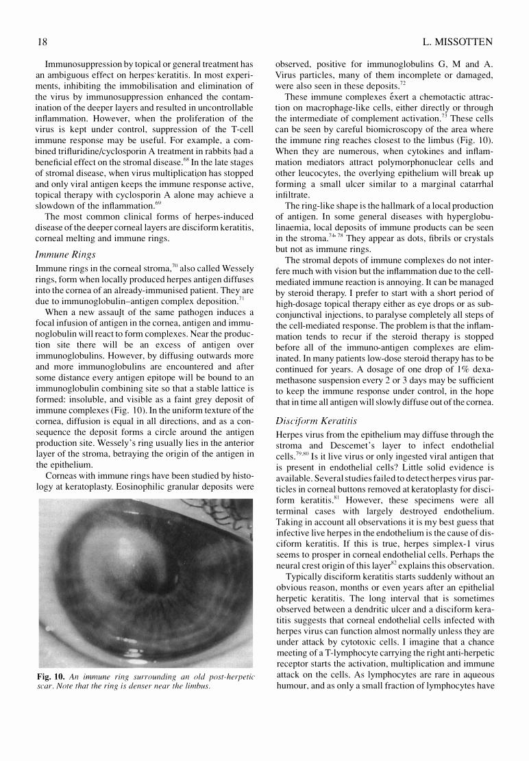
When a new assauJt of the same pathogen induces a focal infusion of antigen in the cornea, antigen and immunoglobulin will react to form complexes. Near the production site there will be an excess of antigen over immunoglobulins. However, by diffusing outwards more and more immunoglobulins are encountered and after some distance every antigen epitope will be bound to an immunoglobulin combining site so that a stable lattice is formed: insoluble, and visible as a faint grey deposit of immune complexes (Fig. 10). In the uniform texture of the cornea, diffusion is equal in all directions, and as a consequence the deposit forms a circle around the antigen production site. Wessely's ring usually lies in the anterior layer of the stroma, betraying the origin of the antigen in the epithelium.
Corneas with immune rings have been studied by histology at keratoplasty. Eosinophilic granular deposits were
Fig. 10. An immune ring surrounding an old post-herpetic scar. Note that the ring is denser near the limbus.
L. MISSOTTEN
observed, pOSitIve for immunoglobulins G, M and A. Virus particles, many of them incomplete or damaged, were also seen in these deposits.72
These immune complexes exert a chemotactic attraction on macrophage-like cells, either directly or through the intermediate of complement activation.73 These cells can be seen by careful biomicroscopy of the area where the immune ring reaches closest to the limbus (Fig. 10). When they are numerous, when cytokines and inflammation mediators attract polymorphonuclear cells and other leucocytes, the overlying epithelium will break up forming a small ulcer similar to a marginal catarrhal intiltrate.
The ring-like shape is the hallmark of a local production of antigen. In some general diseases with hyperglobulinaemia, local deposits of immune products can be seen in the stroma.74 7S They appear as dots, fibrils or crystals but not as immune rings.
The stromal depots of immune complexes do not interfere much with vision but the inflammation due to the cellmediated immune reaction is annoying. It can be managed by steroid therapy. I prefer to start with a short period of high-dosage topical therapy either as eye drops or as subconjunctival injections. to paralyse completely all steps of the cell-mediated response. The problem is that the inflammation tends to recur if the steroid therapy is stopped before all of the immuno-antigen complexes are eliminated. In many patients low-dose steroid therapy has to be continued for years. A dosage of one drop of 1 % dexamethasone suspension every 2 or 3 days may be sufficient to keep the immune response under control, in the hope that in time all antigen will slowly diffuse out of the cornea.
Disciform Keratitis
Herpes virus from the epithelium may diffuse through the stroma and Descemet's layer to infect endothelial cells.79.xo Is it live virus or only ingested viral antigen that is present in endothelial cells? Little solid evidence is available. Several studies failed to detect herpes virus particles in corneal buttons removed at keratoplasty for disciform keratitis.xl However, these specimens were all terminal cases with largely destroyed endothelium. Taking in account all observations it is my best guess that infective live herpes in the endothelium is the cause of disciform keratitis. If this is true, herpes simplex-l virus seems to prosper in corneal endothelial cells. Perhaps the neural crest origin of this layerX2 explains this observation.
Typically disciform keratitis starts suddenly without an obvious reason, months or even years after an epithelial herpetic keratitis. The long interval that is sometimes observed between a dendritic ulcer and a disciform keratitis suggests that corneal endothelial cells infected with herpes virus can function almost normally unless they are under attack by cytotoxic cells. I imagine that a chance meeting of a T-Iymphocyte carrying the right anti-herpetic receptor starts the activation, multiplication and immune attack on the cells. As lymphocytes are rare in aqueous humour, and as only a small fraction of lymphocytes have
IMMUNOLOGY AND HERPETIC KERATITIS
anti-herpes specificity, it may take months before such an encounter takes place.
Careful slit lamp examination of a disciform keratitis shows the lymphocytes as precipitates on the endothelium adherent only to the area of the disciform lesion. Their grouping in clusters ("precipitates') may reflect the social behaviour of immune-competent cells. or may be a reflection of the heterogenous induction of HLA class II antigens under the influence of interferon-y." Little evidence is available on the nature of the cells forming the precipitates. They may be either macrojJhages or natural killer cells attracted by immunoglobulin sticking on the cell surfaces of the infected cells, or they may be cytotoxic T-cells which recognise the viral epitope presented in association with the HLA complexes. The latter seems more likely.
The therapeutic approach poses a di lemma. The spontaneous evolution of the disease leads to the elimination of all infected endothelial cells. This may take several weeks. The remaining endothelial cells, though fewer in number. will restore the normal hydratation of the cornea in most patients. A permanent cure is to be expected. unless a new bout of epitl)elial viral keratitis disease brings in new VIruS.
Patients, however, do not like to wait for weeks to recover sight and ophthalmologists are loth to lose so many endothelial cells. Suppressing the lymphocyte aggression by corticosteroids wi II quickly restore the normal function of the endothelial cells and clear the cornea. The herpes virus, however, remains in the endothelial cells, which continue to display the antigen on their surface. Recurrences are to be expected when the local steroid therapy is stopped.
The optimal therapy most probably is a compromise between inaction and overreaction. One should inhibit the production of new herpes virus by a virustatic agent with good penetration in the cornea, and simultaneously reduce the lymphocyte reaction to such a low level that the transport activity of the endothelium remains good enough for corneal clarity while some virus removal is still going on, so that the cure obtained after a year or more could be permanent. This delicate balancing act is part of the art of ophthalmology.
Post-helpetic Corneal Melting
Corneal perforation as a complication of dendritic kenltitis was almost unknown before corticosteroids were introduced in ophthalmology. Immunosuppression by these drugs during the epithelial keratitis enhances the spread of herpes virus.57 The more the stroma is loaded with antigen the more severe the cell-mediated immune response will be. This is shown by histopathology of corneal buttons obtained at keratectomy. A study of 215 keratectomy specimens of patients with herpes stromal keratitis showed that in many of the specimens herpes antigen could be detected in keratocytes, endothelial cells and foci of epithelioid cells and multinucleated giant cells around Descemet's membrane. Both granulomatous reac-
19
tion and herpes antigen were identified significantly more often in specimens with ulcerative necrotising stromal keratitis.7Y
Corneal oedema predisposes to corneal melting. It disturbs the epithelial barrier and renders it more permeable to infection. Oedema also strongly reduces the amount of IgG in the stroma.s, Both factors favour superinfection by bacteria - one additional cause of ulceration and destruction.
The lysis of the tissue is due to proteases and collagenases. Most are produced by invading polymorphonuclear leucocytes84 that extravasate from blood vessels at the limbus and proceed at great speed to the site of inflammation, guided by chemotaxis. Some proteases could come from resident stromal cells. Once the epithelium breaks down, once an ulcer develops with melting of the stroma. steroid therapy will usually fail to stop the process, and so will most other therapeutic measures. The presently available protease inhibitors lack potency and arrive too late in most cases. The disease will progress until all antigen-loaded stroma has been eliminated. When medical therapy fails, surgery should be considered.
Tarsorrhaphy hides the problem but rarely brings a cure. At present it should be replaced by botulinum-toxininduced ptosis, which gives the same protection but permits better inspection of the eye, and avoids the problem of late trichiasis.
Keratoplasty a clzaud is probably the best therapy for post-herpetic corneal melting. Cutting away the antigenloaded stroma removes the cause of the problem. Although transplantation immunology limits the number of successes. the results can be surprisingly good and are almost invariably better than the outcome of spontaneous healing. When time permits, a few days of intensive steroid therapy before transplantation is useful to clear the corneal periphery of leucocytes and to reduce the risk of immunorejection of the graft.
CONCLUSION
In herpetic keratitis the immune response is chiefly localised onsite, in the transparent media of the eye. It is the privilege of the ophthalmologist to be able to observe the action of the immune system directly with his biomicroscope. We need to refine our observations and to improve our interpretation of what we see in order to treat our patients better.
Key words: Herpes keratitis. Herpes simplex type I. Leucocytes.
REFERENCES I. Lausch R, Staats H, Oakes 1. Cohen G, Eisenberg R. Preven
tion of herpes keratitis by monoclonal antibodies specific for discontinuous and continuous epitopes on glycoprotein D. Invest Ophthalmol Vis Sci 199 1 ; 3 2 :2735 .
2. Coyle P, Sibony P. Tear immunoglobulins measured by ELISA. Invest Ophthalmol Vis Sci 1 986;27 : 622.
3 . Coyle P, S ibony P. Viral antibodies in normal tears . Invest Ophthalmol Vis Sci 1 98 8;29: 1 55 2 .
4 . Wilhelmus K , Darougar S . Forsey T , Treharne J . Sequential antibody changes following ulcerative herpetic keratitis . Br J Ophthalmol 1 986;70 : 354.
20
5. Pedersen B , Moller Andersen S , Klauber A, Ottovay E , Prause J, Zhong C, Norrild B . Secretory IgA specific for herpes simplex virus in lacrimal fluid from patients with herpes keratitis: a possible diagnostic parameter. Br J Ophthalmol 1 982; 66:648.
6. Peppard J, Montgomery P. S tudies on the origin and composition of IgA in rat tears. Immunology 1 98 7 ; 62: 1 93.
7. Franklin R , Shepard K. T-cell adherence to lacrimal gland: the event responsible for IgA plasma cell predominance in lacrimal gland. Reg Immunol 1 990- 1 ; 3:2 1 3.
8. Friedman M. Antibodies in human tears during and after infection. SUfV Ophthalmol 1 990; 3 5 : 1 5 l.
9. Allansmith MR, Gudmundsson OG, Hann LE. Keys C, B lock KJ, Taubman MA, Sullivan DA. The immune response of the lacrimal gland to antigenic exposure. CUff Eye Res 1 987; 6:92l.
1 0. Hazlett L , B erk R. Kinetic s of immunoglobulin appearance at the ocular surface. Reg Immunol 1 989; 2:294.
ll. Mondino B , Brady Bl. Distribution of hemolytic complement in the normal cornea. Arch OphthalmoI 1 98 1 ; 99: 1 430.
1 2. Rinne J, Abghari SZ, Stulting R. The severi ty of herpes simplex viral keratitis in mice does not reflect the severity of disease in humans. Invest Ophthalmol Vis Sci 1 992;3 3:268.
1 3. Hendricks R, Tumpey T. Contribution of virus and immune factors to herpes simplex virus type I-induced corneal pathology. Invest Ophthalmol Vis Sci 1 990; 3 1 : 1 929.
1 4. Mitsui K. Recurrent herpetic keratitis in mice. Nippon Ganka Gakkai Zasshi 1 99 1 ;95:5 3 0.
1 5 . Laycock K, Lee S , Brady R, Pepose J. Characterisation of a murine model of recurrent herpes simplex viral keratiti s induced by ultraviolet B radiation. Invest Ophthalmol Vis Sci 1 99 1 ; 3 2:274l.
1 6. Nesburn A, Willey D. Trousdale M. Effect of intensive acyclovir therapy during artificial reactivation of latent herpes simplex virus. Proc Soc Exp BioI Med 1 98 3 ; 1 7 2:3 1 6.
1 7. Trousdale M, Robin J, Willey D, De Clercq E. Intentional reactivation of latent ocular herpes infection during BVDU therapy. Curr Eye Res 1 98 7 ; 6: 1 47 1 .
1 8. Meyers-Elliott R , Chitjian P. Induction of cell-mediated immunity in herpes simplex virus keratitis: kinetics of lymphocyte transformation and the effect of antiviral antibody. Invest Ophthalmol Vis Sci 1 980; 1 9:920.
1 9. Carter C, Easty D. Experimental ulcerative herpetic keratiti s. 1. Systemic immune responses and resistance to corneal infection. Br J Ophthalmol 1 9 8 1 ; 65:77.
20. Rector J, Lausch R, Oakes J. Use of monoclonal antibodies for analysis of antibody-dependent immunity to ocular herpes simplex virus type 1 infection. Infect Immunol 1 98 2 ; 3 8: 1 68.
2l. Green M, Dunkel E, Courtney R. Detection of herpes simplex virus induced polypeptides in rabbit trigeminal ganglia. Invest Ophthalmol Vis Sci 1 984;25: 1 43 6.
22. Crouse C, Pflugfelder S , Pereira I, Cleary T, Rabinowitz S , Atherton S. Detection o f herpes viral genomes i n normal and diseased corneal epithelium. Curr Eye Res 1 990;9:569.
23. Cook S, Hill 1. Herpes simplex virus: molecular biology and the possibility of corneal latency. Surv Ophthalmol 1 99 1 ; 3 6: 1 40.
24. Cook S, Hill J, Lynas C, Maitland N. Latency-associated transcripts in corneas and ganglia of HSV-l infected rabbits. Br J Ophthalmol 1 99 1 ; 7 5 :644.
25. Kaye S, Lynas C, Patterson A, Risk J, McCarthy K, Hart C. Evidence for herpes simplex viral latency in the human cornea. Br J Ophthalmol 1 99 1 ; 7 5 : 1 95.
26. B audouin C, Fredj-Reygrobellet D , Gastaud P, Lapalus P. HLA DR and DQ distribution in normal human ocular structures. Curr Eye Res 1 98 8 ; 7:903.
27. McBride B, McGill J, Smith J. MHC class I and class II anti gen expression in normal human corneas and in corneas from cases of herpetic keratitis. Immunology 1 98 8 ; 65 :5 83.
L. MISSOTTEN
28. Abu EI-Asrar AM, Geboes K, Missotten L, Emarah MH, Desmet V. Expression of MHC class II antigens and immunoglobulin M by the corneal epithelial cells in herpetic keratitis. Int Ophthalmo I 1 990; 1 4:23 3.
29. Hill J, Kwon B , Colborn G, Shimomura Y, Gapgarosa LS. Antibody-dependent cellular cytotoxicity mediated by polymorphonuclear leukocytes and mononuclear cells against HSV-l infected primary cultures of rabbit corneal epithelium. Curr Eye Res 1 984;3: 1 203.
30. Centifanto Y, Norrild B . Andersen S . Karcioglu Z. Porretta E, Caldwell D. Herpes simplex virus-specific antibodies present in tears during herpes keratitis. Proc Soc Exp B ioI Med 1 98 9 ; 1 92:87.
3l. Smith J, Sheppard A. Activity of rabbit monocytes, macrophages, and neutrophils in antibody-dependent cellular cytotoxicity of herpes simplex virus-infected corneal cells. Infect Immunol 1 98 2:36:685.
32. Maudgal P . Missotten L. La replique de la cornee appliquee a I 'etude de la keratite herpetique. Bull S oc B eige Ophthalmol 1 97 7 ; 1 75:26.
33. Maudgal P, Missotten L. Histopathology of the human herpes simplex keratitis. Br J Ophthalmol 1 97 8 ; 62:46.
34. Maudgal P, Missotten L. Histopathology and histochemistry of the corneal epithelium in experimental herpes simplex keratitis. Graeffes Arch Clin Exp Ophthalmol 1 979; 209:239-48.
35 . Maudgal P. Missotten L. Histopathological study of the human herpes simplex dendritic and punctate keratitis by replica technique. Doc Ophthalmol Proc Ser 1 979; 20:2 1 I.
36. Maudgal P, Missotten L. Superficial keratitis. Monographs in Ophthalmology. The Hague: Junk, 1 980: 1 92.
37. Pepose J. The relationship of corneal Langerhans cells to herpes simplex antigens during dendritic keratotis. Curr Eye Res 1 989;8:85 1 .
3 8. Shimeld C , Hill T. Blyth W. Easty D. Passive immunisation protects the mouse eye from damage after herpes simplex virus infection by limiting spread of virus in the nervous system. J Gen Virol 1 990; 7 1 :68l.
39. Staats H. Oakes J. Lausch R. Anti-glycoprotein D monoclonal antibody protects against herpes s implex virus type I -induced diseases in mice functionally depleted of selected T-cell subsets or asialo GM 1 + cells. J Virol 1 99 1 ;65 :6008.
40. Metcalf J, Koga J, Chatterjee S, Whitley R. Passive immunization with monoclonal antibodies against herpes simplex virus glycoproteins protects mice against herpetic ocular disease. Curr Eye Res 1 98 7 ; 6: 1 73.
4 1 . Metcalf 1. Chatterjee S , Koga J, Whitley R. Protection against herpetic ocular disease by immunotherapy with monoclonal antibodies to herpes simplex virus glycoproteins. Intervirology 1 98 8 ; 29:39.
42. Lausch R. Oakes J. Metcalf J, Scimeca 1. Smith L. Robertson S. Quantitation of purified monoclonal antibody needed to prevent HSV-l induced stromal keratitis in mice. Curr Eye Res 1 989; 8:499.
43. Green M. Dunkel E, Pavan-Langston D. Effect of immunization and immunosuppression on induced ocular shedding and recovery of herpes simplex virus in infected rabhits. Exp Eye Res 1 987 ;45:375.
44. Krichev skaya G . Zaitseva N. Kainarbaeva K , B asova N, Vinogradova V. The use of a passive hemagglutination test ( PHA) in the diagnosis of viral eye diseases: investigation of lacrimal fluid for the presence of antibody to herpes simplex virus (HSV). Graefes Arch Clin Exp Ophthalmol 1 980;2 1 4:239.
45. Fox P. Khaw P, McBride B, McGill J, Ward K. Tear and serum antibody levels in ocular herpetic infection: diagnostic preci sion of secretory IgA. Br J Ophthalmol 1 986; 70:5 84.
46. Shani L, Szanton E, David R. Yassur Y. Sarov 1. Studies on HSV specific IgA antibodies in lacrimal fluid from patients
IMMUNOLOGY AND HERPETIC KERATITIS
with herpes keratitis by solid phase radioimmunoassay. Curr Eye Res 1 98 5 ; 4 : 1 03 .
47 . McBride B , Ward K . Herpes simplex-specific IgG subclass response in herpetic keratiti s . J Med Virol 1 98 7 ; 2 1 : 1 79.
48. Malaty R, Gebhardt B, Franklin R. HSV-specific IgA from tears blocks virus attachment to the cell membrane . Curr Eye Res 1 98 8 ; 7 : 3 1 3 .
49 . Lausch R, Su Y, Ritchie M, Oake s J. Evidence endogenous interferon production contributed to the lack of ocular virulence of an HSV intertypic recombinant. Curr Eye Res 1 99 1 ; 1 0 : 39 .
5 0 . Hendricks R, Weber P, Taylor J, Koumbis A, Tumpey T. Glorioso J. Endogenously produced interferon alpha protects mice from herpes simplex virus type I corneal di sease . J Gen ViroI 1 99 1 ; 7 2 : 1 60 1 .
5 1 . Verhagen C , Breebaart A , Kijlstra A. Diffusion of immunoglobulin G from the vascular compartment into the normal rabbit cornea. Invest Ophthalmol Vis Sci 1 990; 3 1 : 1 5 1 9 .
52. Young E, Pepose J. Class II induction of human corneal fibroblasts by cell-free supernatants from HSV stimulated lymphocytes . Curr Eye Res 1 98 7 ; 6 : 1 4 1 .
5 3 . Einhorn S , Eldor A , Vlodavsky L Fuks Z , Panet A . Produc tion and characterization of interferon from endothelial cells . J Cell Physiol 1 98 5 ; 1 22 : 200.
54. Abu EI-Asrar AM, Van den Oord ]] , Bill iau A, Desmet V, Emarah MH, Missotten L . Recombinant interferon-gamma induces HJ-,A-DR expression on human corneal epithelial and endothelial cells in vitro : a preliminary report . Br J Ophthalmol 1 989;7 3 : 5 87 .
5 5 . Foets B , Van den Oord J , B ill iau A, Van Damme J , Missotten L. Heterogeneous induction of major histocompatibility complex class II antigens on corneal endothelium by interferon-yo Invest Ophthalmol Vis Sci 1 99 1 ; 3 2 : 34 1 .
56. Meyers-Elliott R . Chitjian p, B illups C . Effect of cyclosporine A on the corneal inflammatory response in herpes simplex virus keratitis. Exp Eye Res 1 987 ;45 : 2 8 1 .
57. Nasisse M, Guy J, Davidson M, Sussman W, Fairley N. Experimental ocular herpes virus infection in the cat: sites of virus replication, clinical features and effects of corticosteroids administration. Invest Opthalmol Vis Sci 1 989;30 : 1 75 8 .
5 8 . Easty D L , Tullo AB , Shimeld C. Hill TJ, B lyth WA . The influence of prednisolone on external eye disease. virus proliferation and latent infection in an animal model of herpe s simplex keratitis . In : Herpetic eye diseases . Leuven: Junk. 1 984.
59. Maudgal Pc. De Clercq E, Descamps J. Missotten L. Comparative evaluation of B VDU (E)-5- (bromovinyl)-2' -deoxyuri dine and IDU ( 5 -iodo-2' -deoxyuridine ) in the treatment of experimental herpes simplex keratitis in rabbits. Bull Soc Beige d ' Ophtalmol 1 979; 1 86 : 1 09 .
60. Maudgal PC, De Clercq E. (S)- 1 -0 -hydroxy-2-phosphonylmethoxypropyl) cytosine in the therapy of thymi dine kinase-positive and deficient herpe s simplex virus experimental keratitis . Invest Ophthalmol Vis Sci 1 99 1 ; 3 2 : 1 8 1 6 .
6 1 . Maichuk I, Mikuli S . Effect of levamisole on immunoglobulin secretion into lacrimal fluid in experimental herpetic keratitis . Vopr Virusol 1 98 1 ; I : 1 03 .
62. Kumano Y, Yamamoto M, Iwasaki M. Ishibashi T. lnomata H, Mori R. Participation of T lymphocyte in corneal edema in the early stage of herpetic stromal keratitis . Ophthalmologica 1 9 8 8 ; 1 96 : 1 1 3 .
63 . Hendricks R , Tumpey T. Concurrent regeneration o f T lymphocytes and susceptibility to HSV- l corneal stromal disease. Curr Eye Res 1 99 1 ; 1 0 :47 .
64. Jager M , Atherton S , Bradley D , Streilein J. Herpetic stromal keratitis in mice : less reversibil ity in the presence of
2 1
Langerhans cells in the central cornea. Curr Eye Res 1 99 1 ; 1 0 : 69.
65 . Doymaz M. Foster C . Destephano D , Rouse B. MHC IIrestricted, CD4+ cytotoxic T lymphocytes specific for herpes simplex virus- I : implications for the development of herpetic stromal keratitis in mice. Clirr Immunol ImmunopathoI 1 99 1 ; 6 1 : 3 9 8 .
66. Brandt C . Salkowski C. Activation of NK cells in mice following corneal i nfection with herpes simplex virus type- I . Invest Ophthalmol Vis Sci 1 992; 3 3 : 1 1 3 .
67 . Hendricks R , Epstein R, Tumpey T. The effect o f cellular immune tolerance to HSV- l antigens on the immunopathology of HSV- l keratitis . Invest Ophthalmol Vis Sci 1 98 9 ; 3 0 : 1 05 .
6 8 . Boisjoly HM. Woog JJ, Pavan-Langston D, Park NH. Prophylactic topical cyclosporine in experimental herpetic stromal keratiti s . Arch Ophthalmol 1 984; 1 02 : 1 804.
69. Goichot-B onnat L, De Beauregard C . Saragoussi J; Pouliquen Y. Use of eyclosporin A eyedrops in the prevention of corneal graft rejection in man. I. Preoperative development of 4 eyes with metaherpetic keratitis . J Fr Ophtalmol 1 98 7 ; 1 0 : 207 .
70. Breebaart AC, James Witte J. Studies on experimental corneal allergy. Am J Ophthalmol 1 959;48 : 3 7 .
7 1 . Verhagen C , Den Heyer R. Broersma L. Breebaart A. Kijlstra A. Analysis of corneal inflammation following the injection of heterologous serum in the rat cornea. Invest Ophthalmol Vis Sci 1 99 1 ; 3 2 : 3 2 3 8 .
72 . Meyers-Elliott R, Pettit T. Maxwell W. Viral antigens in the immune ring of herpes simplex stromal kerqtitis . Arch Ophthalmol 1 980;98 : 897.
73. Verhagen C . Den Heyer R, Klooster J. Breebaart A. Kijlstra A. Elimination of immune precipitates from the rat corneal stroma: a hi stological study. Exp Eye Res 1 99 1 ; 5 3 :47 1 .
74. Kremer I . Wright P . Merin S . Weiss J, Pick A . Kaufman H. Corneal subepithelial monoclonal kappa IgG deposits in essential cyroglobulinaemia. Br J OphthalmoI 1 989;73 : 669.
75. Hill J. Mull igan G. Subepithelial corneal deposits in IgG lambda myeloma. Br J Ophthalmol 1 989; 7 3 : 55 2 .
76. Yassa N . Font R. Fine B . Koffler B. Corneal immunoglobulin deposition in the posterior stroma: a case report including immunohistochemical and ultrastructural observations. Arch Ophthalmol 1 98 7 ; 1 05 :99.
7 7 . Miller K. Green W, Stark W, Wells H. Mendelsohn G. Kanhofer H. Immunoprotein deposition in the cornea. Ophthalmology 1 980;87 :944.
78. Hylkema H . Rathman W. Kijlstra A. Deposition of immune complexes in the mouse eye. Exp Eye Res 1 98 3 ; 3 7 :257 .
79. Holbach L. Font R, Naumann G . Herpes simplex stromal and endothelial keratitis : granulomatous cell reactions a t the level of Descemet ' s membrane. the stroma, and Bowman ' s layer. Ophthalmology 1 990;97 :722.
80. Ohashi Y, Yamanoto S. Nishida K. Okamoto S, Kinoshita S . Hayashi K. Manabe R. Demonstration o f herpes simplex virus DNA in idiopathic corneal endotheliopathy. Am J Ophthalmol 1 99 1 ; 1 1 2 :4 1 9 .
8 1 . Ahonen R, Vannas A. Makitie J. Virus particles and leukocytes in herpes simplex keratitis . Cornea 1 984; 3 :43 .
8 2 . Foets B, Van den Oord J. Desmet V, Missotten L. Cytoskeletal filament typing of human corneal endothelial cells. Cornea 1 990; 9 : 3 1 2 .
8 3 . Waldrep 1 . Noe R . Stulting R. Analysis of human corneal IgG by i s oelectric focusing. Invest Ophthalmol Vis Sci 1 98 8 ; 29 : 15 3 8 .
8 4 . Prause J . Jensen O. PAS -positive polymorphonuclear leucocytes in corneal ulcers . Acta Ophthalmol (Copenh) 1 980; 5 8 : 5 5 6 .


![[Visiting immunology]](https://img.dokumen.tips/doc/110x75/63453fbb38eecfb33a06782b/visiting-immunology.jpg)


