Embed Size (px)
Citation preview

RESEARCH ARTICLE Open Access
Pigmentary keratitis in pugs in the UnitedKingdom: prevalence and associatedfeaturesS. Maini1* , R. Everson2, C. Dawson3, Y. M. Chang4, C. Hartley1 and R. F. Sanchez5
Abstract
Background: Pigmentary keratitis (PK) is commonly recognised in Pugs, but its aetiology is not completelyunderstood. The aim of this study was to determine the prevalence and associated features of PK in Pugs in theUnited Kingdom (UK).
Results: A total of 210 Pugs (420 eyes) were recruited from 12 UK dog shows and social events. The median age ofPugs recruited was 2.50 years (range 0.25–16.25 years). Pigmentary keratitis was detected in 369/420 (87.8%) eyesand in at least one eye 193/210 (91.9%) Pugs, of which 17/193 (8.8%) were affected unilaterally and 176/193 (91.2%)bilaterally. Pigmentary keratitis was typically mild to moderate (46.3 and 49.9% of eyes, respectively). Detection of PKwas significantly associated with increased age (P = 0.002) and the presence of medial entropion of the lowereyelid (MELE) (P = 0.001). Severity of PK was significantly associated with the grade of MELE (P < 0.001). There wasalso a correlation between the presence of limbal pigment and PK (P = 0.036) that warrants further study.
Conclusions: This study estimated a high disease prevalence of PK in UK Pugs, and demonstrated significantassociations with age and the presence of MELE. These associations, which have not been previously reported, offeran insight into the underlying pathophysiology of this condition in Pugs. The results encourage further populationresearch, such as prospective longitudinal studies. These findings also support the development of clinical andbreeding strategies based on the reduction of MELE and, possibly, limbal pigment.
Keywords: Entropion, Brachycephalic, Canine, Cornea, Pigment, Keratitis, Corneal pigmentation, Medial entropion
BackgroundPigmentary keratitis is a term used to describe the devel-opment of corneal pigmentation associated with chronicinflammation [1]. If PK encroaches upon the visual axis,it can cause significant visual impairment and, in severecases, blindness [2, 3]. Pigmentary keratitis occurs dueto centripetal migration of melanocytes from the limbaland perilimbal region and subsequent deposition ofmelanocytic pigment within the corneal epithelium andanterior stroma [1, 3–6]. Corneal pigmentation is alsofrequently reported as a feature of inflammatory cornealpathology, such as keratoconjunctivitis sicca (KCS),chronic superficial keratitis (pannus) and chronic, ul-cerative/nonulcerative keratitis [2, 7–12]. Pigmentary
keratitis appears to develop more rapidly and readily insome brachycephalic breeds and it has been shown to bewidespread within the Pug breed in two studies based inthe United States of America (USA) and one study fromAustria, that reported estimated prevalence rates of 82.4,71.8 and 70%, respectively [1, 11, 13]. Reputed causativeor contributory factors of PK in Pugs include chronicirritation from distichiasis, nasal fold trichiasis, medialentropion, and macroblepharon [14, 15]; however, sup-porting evidence for their influence on the developmentof PK has so far proven elusive [11, 13]. Additional sug-gestions have been made of possible primary compo-nents in the development of PK in the Pug breed, suchas a limbal stem cell deficiency or genetic factors [13, 16,17]. Pugs are a popular breed in the UK, with the numberof Pugs registered with the UK Kennel Club (KC) havingdoubled between 2009 and 2015; figures plateaued at ap-proximately 10,000 Pugs per year between 2014 and 2018.
© The Author(s). 2019 Open Access This article is distributed under the terms of the Creative Commons Attribution 4.0International License (http://creativecommons.org/licenses/by/4.0/), which permits unrestricted use, distribution, andreproduction in any medium, provided you give appropriate credit to the original author(s) and the source, provide a link tothe Creative Commons license, and indicate if changes were made. The Creative Commons Public Domain Dedication waiver(http://creativecommons.org/publicdomain/zero/1.0/) applies to the data made available in this article, unless otherwise stated.
* Correspondence: [email protected] Veterinary Services, University of Bristol, Langford, Bristol, UKFull list of author information is available at the end of the article
Maini et al. BMC Veterinary Research (2019) 15:384 https://doi.org/10.1186/s12917-019-2127-y

The aim of this study was to contribute to the body ofresearch on this poorly understood but widespread con-dition by estimating the prevalence of PK in Pugs in theUK, and determining if there were any statistical associa-tions with ocular, adnexal or facial features.
ResultsStudy populationTwo hundred and ten dogs, with a total of 420 eyes,were included in the study. Individuals were recruited
from one of 12 national events and, collectively, repre-sented a large area of the UK (Fig. 1). Sex, neuter status,age, coat colour, UK KC registration and show statuswere as follows for those dogs where this informationwas provided/recorded. Sex was known for 208/210(99.0%) dogs; the sample population included 120/208(57.7%) females and 88/208 (42.3%) males. Neuter statuswas provided for 206/210 (98.1%) dogs; a total of 66/206(32.0%) dogs were neutered and 140/206 (68.0%) wereentire. Age was known for 203/210 (96.7%) Pugs; the
Fig. 1 Recruitment of Pugs within the United Kingdom. Map created using MapChart https://mapchart.net (licensed under a Creative CommonsAttribution-Share Alike 4.0 International License)
Maini et al. BMC Veterinary Research (2019) 15:384 Page 2 of 11

median age was 2.50 years (interquartile range 2.92 years,range 0.25–16.25 years). Coat colour was recorded for207/210 (98.6%) cases. Colour variations were cate-gorised into three groups: fawn (146/207; 70.5%), black(50/207; 24.2%) and other (11/207; 5.3%). Most dogs forwhom registration status was provided (173/210; 82.4%)were registered with the UK KC (158/173; 91.3%); 15/173 (8.7%) were not registered. Show status was pro-vided for 131/210 (62.4%) dogs; a total of 81/131 (61.8%)were described as ‘show dogs’ and 50/131 (38.2%) werenot routinely entered into shows.
General ocular, adnexal and facial features (excluding PK)Features that were not associated with PK detection orseverity included medial entropion of the upper eyelid(MEUE), craniofacial index (CFI), over-nose-wrinkle(ONW), nasal fold width, palpebral fissure length, iris-to-iris persistent pupillary membranes (IIPPMs) and dis-tichia (Table 1). MELE, which was associated with PKseverity, could be assessed in 374/420 (89.0%) eyes. Atotal of 352/374 (94.1%) eyes exhibited MELE and 22/374 (5.9%) did not. Of the 374 eyes that were assessed,276/374 (73.8%) were categorised as grade 1 MELE and76/374 (20.3%) as grade 2; and comprised 78.4 and21.6% of the eyes with MELE, respectively. Length (i.e. %)of lower eyelid affected by MELE ranged from 0.0–50.0%,with a mean (+/−sd) of 22.4 (+/− 9.9) %. IIPPMs were de-tected in 198/420 (47.1%) eyes and were not detected in222/420 (52.9%) eyes; they were detected in at least oneeye of 117/210 (55.7%) dogs, leaving 93/210 (44.3%) thatwere unaffected. Distichiasis was detected in 30/420(7.1%) eyes and was not detected in 390/420 (92.9%) eyes.At least one eye of 25/210 (11.9%) dogs was affected bydistichiasis, with most dogs being unaffected (185/210;88.1%). A total of 170/210 (81.0%) dogs were examined tosee if they had an ONW; an ONW was present in 59/170(34.7%) dogs and was absent in 111/170 (65.3%) dogs.Medial entropion of the upper eyelid was present in 28/410 (6.7%) eyes. Craniofacial index was assessed in 206/210 (98.1%) dogs; values ranged from 0.05 to 0.33, with amean of 0.18 (+/− 0.04). Nasal fold width was assessed in399/410 (95.0%) eyes; values ranged from 1.0 to 24.0mm,with a mean of 5.83 (+/− 2.06) mm. Palpebral fissurelength was measured in 363/410 (86.4%) eyes; valuesranged from 18.0 to 30.0mm, with a mean of 23.96 (+/−2.17) mm. All Pugs in this study (420/420 eyes) had aSchirmer tear test 1 (STT1) test performed to rule outwith certainty that there was no KCS; measurementsranged from 15.0 to 33.0mm/min of wetting, with a meanof 21.10 (+/− 3.24) mm/min. A total of 102/420 eyes hadSTT1 readings for less than 1 min (15–35 s); 94/102 hadSTT1 readings of at least 30 s. All 102 eyes were includedin the study as they measured at or above 15mm ofwetting.
Pigmentary keratitis – descriptive statisticsPigmentary keratitis was detected in at least one eye of193/210 (91.9%) dogs. Seventeen out of 193 (8.8%) hadunilateral PK and 176/193 (91.2%) exhibited bilateral PK.A total of 369/420 (87.9%) eyes were affected by PK. Ofthose affected eyes, 171/369 (46.3%) were classified asmild, 184/369 (49.9%) as moderate, and 14/369 (3.8%) assevere.
Pigmentary keratitis – detectionDetection of PK was not significantly associated withsignalment, breed club registration, ophthalmic, or anyof the adnexal and facial predictors other than age andMELE (Table 1) in the univariable analysis. There wasinsufficient data to assess coat colour as a predictor forPK detection. Both age (OR = 1.76, 95%CI 1.31–2.36, P <0.001) and presence of MELE (grade 1 vs 0: OR = 9.98,
Table 1 Univariable analysis of detection and severity of PK andassociation with predictors
Predictor Detection of PK Severity of PK
OR (95% CI), P OR (95% CI), P
Age (per year) 1.53 (1.16–2.00), 0.002 1.08 (0.98–1.20), 0.132
Sex (male vs. female) 1.44 (0.66–3.15), 0.362 0.96 (0.59–1.56), 0.878
Neuter status(neutered vs. entire)
0.70 (0.31–1.52), 0.355 1.02 (0.60–1.73), 0.947
KC registration(registered vs. no)
1.20 (0.32–4.53), 0.792 0.84 (0.31–2.32), 0.738
Show dog status(yes vs. no)
0.50 (0.18–1.40), 0.188 0.63 (0.34–1.2), 0.162
STT1 measurement 1.00 (0.93–1.07), 0.919 0.98 (0.92–1.05), 0.573
Limbal pigmentation 0.95 (0.87–1.04), 0.277 1.04 (0.98–1.11), 0.183
MELE (grade)
Grade 1 vs. MELEnot present
6.31 (2.18–18.26), 0.001 0.62 (0.21–1.81), 0.380
Grade 2 vs. MELEnot present
13.97 (3.44–56.81),< 0.001
1.94 (0.59–6.41), 0.279
Grade MELE notpresent vs. 2
0.07 (0.02–0.29), < 0.001 0.52 (0.16–1.71), 0.279
Grade 1 vs.Grade 2
0.45 (0.17–1.20), 0.112 0.32 (0.18–0.58), < 0.001
MELE (% length) 1.02 (0.98–1.06), 0.279 1.02 (0.99–1.04), 0.135
MEUE (% length) 1.01 (0.97–1.05), 0.694 1.02 (0.99–1.04), 0.168
CFI (< 0.18 vs ≥0.18) 1.45 (0.-3.27), 0.370 0.91 (0.55–1.49), 0.707
ONW (yes vs no) 1.75 (0.60–5.07), 0.306 1.27 (0.74–2.16), 0.386
Nasal fold width(per mm)
1.27 (1.03–1.56), 0.023 1.07 (0.88–1.30), 0.482
Palpebral fissurelength (per mm)
1.08 (0.92–1.27), 0.343 1.08 (0.95–1.21), 0.235
IIPPMs (presencevs absence)
0.69 (0.39–1.22), 0.206 0.96 (0.63–1.46), 0.853
Distichia (presencevs absence)
0.49 (0.23–1.06), 0.069 1.22 (0.61–2.44), 0.580
Maini et al. BMC Veterinary Research (2019) 15:384 Page 3 of 11
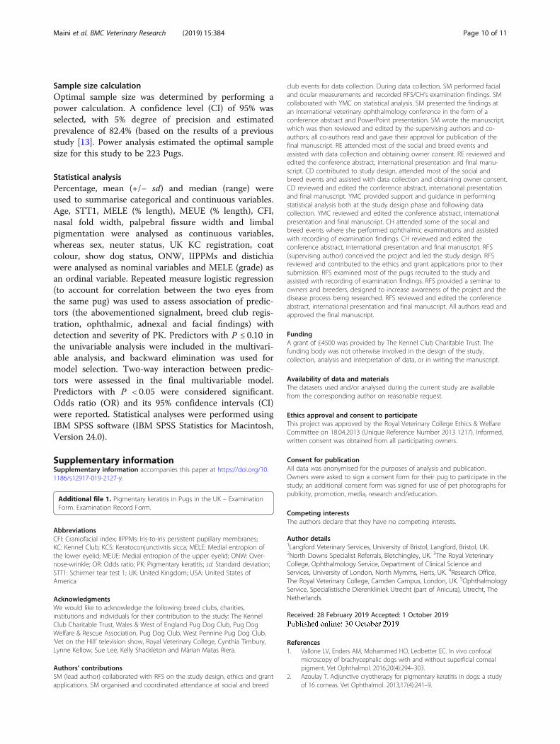
95%CI 3.12–31.94, P < 0.0001; grade 2 vs 0: OR = 13.19,95% CI 3.30–52.76, P < 0.001) remained significant in themultivariable analysis. No significant interaction betweenMELE and age on PK detection was observed (P = 0.09).
Pigmentary keratitis – severityDue to the relatively low number of severe cases, themoderate and severe groups were amalgamated to createa combined moderate/severe group, for the purposes ofrepeated measures logistic regression analysis (mild vs.moderate/severe). Severity of PK was not associated withsignalment, breed club registration, or any of the oph-thalmic, adnexal or facial predictors other than MELE(Table 1). Increasing PK severity was significantly associ-ated with higher grade MELE (OR = 0.32, 95% CI: 0.18–0.58, P < 0.001, when comparing grade 1 to grade 2). Nosignificant interaction between MELE and limbal pig-mentation on PK severity was observed (P = 0.666).As an association of PK with limbal pigment was not
found (P = 0.183), the authors included an additionalgroup of 16 Pugs from outside the study population totest if a larger population would help increase statisticalpower of this calculation, enough to find statistical sig-nificance; the results showed that the inclusion of theadditional cases offered enough statistical power to dem-onstrate a statistically significant association between theseverity of PK and increased limbal pigmentation (OR =1.07, 95% CI: 1.00–1.15, P = 0.036) in the multivariableanalysis.
DiscussionThis study suggests there is a high prevalence of PK inPugs living within the UK. The authors selected a repre-sentative sample of the national population by attendinga wide variety of breed shows and social events. Theprevalence of PK within the sample population washigher than reported in similar studies performed in theUSA and Austria [1, 11, 13]. This may reflect differencesin population, breeding patterns, genetic background,previous veterinary attention or other as yet unknownfactors.The significant association of PK with the presence of
MELE and its severity is, to the authors’ knowledge, thefirst report confirming an association between PK inPugs and this conformational abnormality, which haslong been suspected to be a contributory factor in thisdisease. This is in contrast with a previous study, whichdid not demonstrate a statistically significant influenceof MELE on PK in Pugs [13]. This disparity may be at-tributed to differences in population and/or in study de-sign, or to type I or II statistical errors. It is interestingto view the results of the present study in the context ofa recent publication which demonstrated microscopicinflammatory changes within corneas affected by PK [1].
The significant associations between PK and MELEfound in the current study could be explained by the ir-ritative effect of MELE on the medial cornea of Pugs.This study also found that PK was significantly associ-ated with increased age. The association with age wasnot entirely surprising as, clinically, PK is known to pro-gress over time [16]. It seems logical to conclude that ifcorneas with PK have inflammatory changes [1] and ifsevere entropion is associated with PK (which worsenswith age), that surgical correction of severe MELE wouldhave a positive effect on the long-term corneal health ofaffected Pugs. It would also seem logical to consider thedevelopment of breeding strategies that focus on redu-cing the presence of severe MELE in Pugs. A large-scaleprospective cohort study tracking the progression of PKin the presence of MELE, and in Pugs that have under-gone surgical MELE correction would undoubtedly fur-ther clarify the impact of the associated featuresidentified in the present study. Early, prototype studieshave hinted that the correction of MELE, possibly withadditional medical therapy, is an area of interest for fur-ther research [9, 18, 19]. However, until the results offuture investigative studies are available, owners of Pugswith PK and severe MELE should be informed of the as-sociations found to date, as they may wish to consideroptions that decrease ocular surface inflammation, suchas surgical MELE correction.When the authors realized that an association of PK
with limbal pigment was not present, they included agroup of 16 dogs from outside the study populationsolely to test if a larger population would help increasestatistical power of this calculation. In doing so the au-thors found there was indeed a statistically significant as-sociation between the severity of PK and increasedlimbal pigmentation, which was not entirely surprising.This additional group could not be included in the largeranalysis because they did not have an STT1 measuredand therefore KCS could not be ruled out as the causeof PK. Pigmentary keratitis in Pugs is associated with thepresence of MELE, as shown in the present study, but itcan also be associated with KCS [11]. It seems logical tosuspect an association between PK and the presence oflimbal pigment in Pugs in the absence of KCS. However,the statistical strength of this association warrants fur-ther study if we are to determine how important thepresence of limbal pigmentation is as a risk factor forthe development of PK in Pugs. This study demonstratesthat the population size needs to be large and offers theapproximate number of Pugs needed with a normalSTT1 reading and a clinical history free of KCS.Previous population studies of Pugs reported on the
presence of distichiasis as well as IIPPMs and CFI [11,13, 20]. The present study corroborates the results ofthose papers in which no significant association between
Maini et al. BMC Veterinary Research (2019) 15:384 Page 4 of 11

distichiasis and PK in Pugs was found [11, 13]. Withregards to IIPPMs, one study reported a prevalence of83.8% in the left eye and 85.3% in the right eye [13],while another study reported a much lower prevalenceof 8.46% [11]. The prevalence of IIPPMs in this studywas between the two previously published results. Avariance in prevalence rates may be due to populationdifferences, as each of the three studies was performedin a different country. The mean CFI of the Pugs in-cluded in the current study was higher than in a previ-ous paper, which reported a CFI of 0.08 (+/− 0.01) [20].Again, this could reflect differences between populationsalthough it is possible that the presence of a large nasalfold hindered the accurate measurement of muzzlelength in some cases, which may account in part for thedifference between the two studies.A limbal stem cell deficiency has been proposed as a
possible cause of PK in Pugs [13, 17]. A confocal micros-copy study of PK-affected Pug corneas supported therole of inflammation as opposed to a limbal stem celldeficiency [1]. However, the authors of that paper agreedthat further studies were required to definitively rule outa possible role of stem cell deficiency in Pugs with PK[1]. Microscopic evaluation of affected Pug corneas toprove or disprove this theory was well beyond the scopeof the present study. Genetic analysis has also been sug-gested as a potentially rewarding area of research [13].However, due to the low number of unaffected Pugs (i.e.controls) identified in the present study, genetic analysiswould appear to be challenging to pursue in the UKpopulation.Limitations of this study include failure to reach the
ideal sample size as proposed by power analysis. Add-itionally, there was a low number of unaffected dogs. Tocompound this, all unaffected individuals were less than5 years of age, likely because a large proportion of Pugswere recruited from shows. This has two important im-plications: a) the importance of this condition in the Pugpopulation may be underestimated and b) it is unknownif young, unaffected dogs will remain free of PKthroughout their lives. It is possible that the inclusion ofPugs that had previously had ulcerative keratitis mighthave had an impact on the estimated prevalence, as cor-neal pigmentation is known to develop secondary tochronic inflammation [2, 4, 7–12, 21], and Pugs havebeen shown to exhibit a high prevalence of corneal ul-ceration [22]. However, PK in Pugs starts classically inthe medial cornea and has a centripetal progression inthe shape of a triangle or wedge [3, 13] and, in the au-thors’ experience, corneal ulceration has a tendency tolead to a less predictable pigmentation pattern, asamorphous pigmented or non-pigmented scars tend todevelop in the spot where a cornea was ulcerated. Yet,distinguishing between the two might not be possible
and this is a potential problem of every PK study ofPugs. Even the exclusion of dogs with previously knownulcerative keratitis might not necessarily remove all casesthat have had ulcerative keratitis because a small ulcermight go unnoticed by the owner, heal and scar. The ex-aminers of this study made every effort to distinguishbetween the two presentations although it is acknowl-edged that the PK data collected might have been af-fected by pigmentation caused by previous ulceration.
ConclusionsThe prevalence of PK in UK Pugs in this study popula-tion was very high. Pigmentary keratitis was more likelyto be detected in older Pugs and in those with limbalpigmentation and MELE, especially if MELE was severe.The results of this study offer an insight into the under-lying pathophysiology of PK in Pugs and encouragefurther population research, such as prospective longitu-dinal studies, to further inform these findings. Moreover,these results support the development of clinical andbreeding strategies on the reduction of MELE and limbalpigment.
MethodsStudy designA cross-sectional study design was selected, with thefollowing objectives: to estimate prevalence and reportdescriptive statistics, and to investigate the presence orabsence of statistical associations with suspected riskfactors.
Study population and methodsAll study methods were approved by the Royal VeterinaryCollege (UK) Ethics & Welfare Committee. Informed,written consent was obtained from all participatingowners. Pugs were enrolled and examined at one of 12events (three breed club dog shows and nine social eventse.g. ‘Pug parties’, dog charity ‘garden parties’, breeder/owner social gatherings) in nine different countiesacross the UK (East Sussex, Northamptonshire,Lincolnshire, Cheshire, Hertfordshire, London, Wiltshire,Gloucestershire and South Yorkshire) between July 2014and October 2017. All Pugs that were presented to theexaminer at these events were enrolled to the study, unlessthey had already been examined at a prior event. Pugswith a history of KCS were excluded from the study dueto the documented link between KCS and cornealpigmentation [8, 11]. Signalment and ophthalmic historywere obtained from owners by use of owner-completedquestionnaires prior to examination. Examinations wereperformed free of charge and comprised slit-lamp biomi-croscopy of ocular adnexa and the anterior segment,STT1, and ocular and facial morphometrics. An examin-ation form (Additional file 1) was used to record
Maini et al. BMC Veterinary Research (2019) 15:384 Page 5 of 11

examination findings. Ocular and facial measurementswere collected using previously defined measuring proto-cols [23]. Craniofacial index was then calculated and usedas a measure of brachycephaly [20]. The presence orabsence of an ONW, MELE and MEUE were recorded.Additionally, unstretched palpebral fissure width (i.e.
distance in millimetres between medial and lateral can-thus), was measured using a blunt-ended Jameson caliper(Fig. 2), as was the nasal fold width (i.e. width in milli-metres of nasal fold skin that could be grasped by the cali-per). Ophthalmic examination was performed by one oftwo board-certified veterinary ophthalmologists (RFS &
Fig. 3 Assessment of corneal ‘clock hours’. Six clock hours were affected by corneal pigmentation (6 points). Two additional points were allocatedas the pigment extended beyond the resting pupil edge. Total = 8 points
Fig. 2 Measurement of unstretched palpebral fissure width with a blunt-ended Jameson caliper
Maini et al. BMC Veterinary Research (2019) 15:384 Page 6 of 11

CH), primarily for assessment of PK. The presence of dis-tichiasis and IIPPMs was also recorded. Ocular and facialmorphometrics were collected by the same observer (SM).
Schirmer tear test 1The authors specifically inquired about an ophthalmichistory of KCS and attempted to measure STT1 readingsin every Pug; Pugs that did not tolerate STT1
measurement were excluded from the study. Inclusionin the study required that the Pug exhibited a moist ocu-lar surface on ophthalmic examination. Obvious clinicalsigns of KCS (except for the presence of corneal pigmen-tation) were grounds for immediate exclusion, includingone or more than one of the following: the presence of adull ocular surface in the non-pigmented part of the cor-nea, presence of ulcerative keratitis, presence of mucous
Fig. 5 Assessment of limbal brush border. Corneal pigmentation extends just beyond the limbus creating a ‘limbal brush border’ affecting threeclock hours (3 points). Total = 3 points
Fig. 4 Assessment of pigment lines. Single lines of pigmentation were allocated half a point; two additional points were allotted as thepigmentation extended beyond the resting pupil edge. Total = 2.5 points
Maini et al. BMC Veterinary Research (2019) 15:384 Page 7 of 11

discharge that adhered to the ocular surface and/ormoderate, marked or severe conjunctival hyperaemia.
Assessment of pigmentary keratitisA grading system for PK was developed by one of theauthors (RS) for this study and was used for the assess-ment of all participants’ eyes. The corneal surface wasdivided into 12 sectors or ‘clock hours’. The extent ofcorneal pigmentation was assessed according to thenumber of ‘clock hours’ that were affected; one pointwas awarded per clock hour (Fig. 3). Single ‘lines’ ofpigmentation were allocated a half-point (Fig. 4). Oneadditional point was given if the pigmentation extendedto the resting pupil edge; two additional points were
assigned if the pigmentation extended beyond the rest-ing pupil edge, encroaching upon the visual axis (Figs. 3and 4). In cases where the corneal pigmentation ex-tended only just beyond the limbus, this was termed a‘limbal brush border’ and one point was allocated perclock hour affected (Fig. 5). Grey-white corneal lesionsthat presented medially and/or axially, often times witha centripetal ‘swirl’ appearance [3, 6], were considered aprecursor to corneal pigmentation. These grey-white le-sions were allotted points as though they representedcorneal pigmentation i.e. one point was awarded perclock hour, with an additional 1–2 points if the swirl ex-tended to or beyond the resting pupil edge (Fig. 6). Theoverall point score was then calculated and eyes were
Fig. 7 Severity of PK. Examples of mild (a), moderate (b) and severe (c) PK
Fig. 6 Assessment of grey/white corneal lesions and swirls. The swirl was allotted points as though it represented corneal pigmentation; onepoint per clock hour plus an additional two points for encroaching upon the visual axis. Laterally, a limbal brush border affects two corneal clockhours (2 points). Total = 5 points
Maini et al. BMC Veterinary Research (2019) 15:384 Page 8 of 11
assigned to one of three groups; mild (0.5–4.5 points),moderate (5.0–9.5 points) and severe (10.0–14.0 points)PK (Fig. 7).
Assessment of MELEThree methods were used to record this data:
i) Presence or absence of MELE, which was recordedas a binary measure.
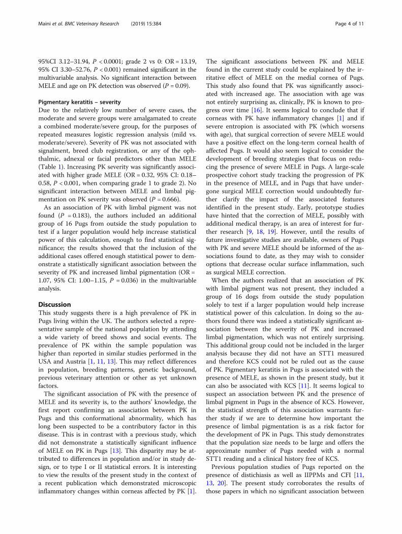
ii) Length of MELE. This was the proportion of eyelidlength affected by MELE expressed as a percentageof lid length e.g. 25, 50% (Fig. 8). Medial entropion
of the upper eyelid was also recorded in thismanner.
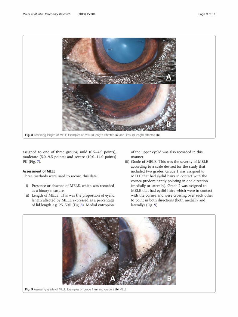
iii) Grade of MELE. This was the severity of MELEaccording to a scale devised for the study thatincluded two grades. Grade 1 was assigned toMELE that had eyelid hairs in contact with thecornea predominantly pointing in one direction(medially or laterally). Grade 2 was assigned toMELE that had eyelid hairs which were in contactwith the cornea and were crossing over each otherto point in both directions (both medially andlaterally) (Fig. 9).
Fig. 9 Assessing grade of MELE. Examples of grade 1 (a) and grade 2 (b) MELE
Fig. 8 Assessing length of MELE. Examples of 25% lid length affected (a) and 33% lid length affected (b)
Maini et al. BMC Veterinary Research (2019) 15:384 Page 9 of 11
Sample size calculationOptimal sample size was determined by performing apower calculation. A confidence level (CI) of 95% wasselected, with 5% degree of precision and estimatedprevalence of 82.4% (based on the results of a previousstudy [13]. Power analysis estimated the optimal samplesize for this study to be 223 Pugs.
Statistical analysisPercentage, mean (+/− sd) and median (range) wereused to summarise categorical and continuous variables.Age, STT1, MELE (% length), MEUE (% length), CFI,nasal fold width, palpebral fissure width and limbalpigmentation were analysed as continuous variables,whereas sex, neuter status, UK KC registration, coatcolour, show dog status, ONW, IIPPMs and distichiawere analysed as nominal variables and MELE (grade) asan ordinal variable. Repeated measure logistic regression(to account for correlation between the two eyes fromthe same pug) was used to assess association of predic-tors (the abovementioned signalment, breed club regis-tration, ophthalmic, adnexal and facial findings) withdetection and severity of PK. Predictors with P ≤ 0.10 inthe univariable analysis were included in the multivari-able analysis, and backward elimination was used formodel selection. Two-way interaction between predic-tors were assessed in the final multivariable model.Predictors with P < 0.05 were considered significant.Odds ratio (OR) and its 95% confidence intervals (CI)were reported. Statistical analyses were performed usingIBM SPSS software (IBM SPSS Statistics for Macintosh,Version 24.0).
Supplementary informationSupplementary information accompanies this paper at https://doi.org/10.1186/s12917-019-2127-y.
Additional file 1. Pigmentary keratitis in Pugs in the UK – ExaminationForm. Examination Record Form.
AbbreviationsCFI: Craniofacial index; IIPPMs: Iris-to-iris persistent pupillary membranes;KC: Kennel Club; KCS: Keratoconjunctivitis sicca; MELE: Medial entropion ofthe lower eyelid; MEUE: Medial entropion of the upper eyelid; ONW: Over-nose-wrinkle; OR: Odds ratio; PK: Pigmentary keratitis; sd: Standard deviation;STT1: Schirmer tear test 1; UK: United Kingdom; USA: United States ofAmerica
AcknowledgmentsWe would like to acknowledge the following breed clubs, charities,institutions and individuals for their contribution to the study: The KennelClub Charitable Trust, Wales & West of England Pug Dog Club, Pug DogWelfare & Rescue Association, Pug Dog Club, West Pennine Pug Dog Club,‘Vet on the Hill’ television show, Royal Veterinary College, Cynthia Timbury,Lynne Kellow, Sue Lee, Kelly Shackleton and Màrian Matas Riera.
Authors’ contributionsSM (lead author) collaborated with RFS on the study design, ethics and grantapplications. SM organised and coordinated attendance at social and breed
club events for data collection. During data collection, SM performed facialand ocular measurements and recorded RFS/CH’s examination findings. SMcollaborated with YMC on statistical analysis. SM presented the findings atan international veterinary ophthalmology conference in the form of aconference abstract and PowerPoint presentation. SM wrote the manuscript,which was then reviewed and edited by the supervising authors and co-authors; all co-authors read and gave their approval for publication of thefinal manuscript. RE attended most of the social and breed events andassisted with data collection and obtaining owner consent. RE reviewed andedited the conference abstract, international presentation and final manu-script. CD contributed to study design, attended most of the social andbreed events and assisted with data collection and obtaining owner consent.CD reviewed and edited the conference abstract, international presentationand final manuscript. YMC provided support and guidance in performingstatistical analysis both at the study design phase and following datacollection. YMC reviewed and edited the conference abstract, internationalpresentation and final manuscript. CH attended some of the social andbreed events where she performed ophthalmic examinations and assistedwith recording of examination findings. CH reviewed and edited theconference abstract, international presentation and final manuscript. RFS(supervising author) conceived the project and led the study design. RFSreviewed and contributed to the ethics and grant applications prior to theirsubmission. RFS examined most of the pugs recruited to the study andassisted with recording of examination findings. RFS provided a seminar toowners and breeders, designed to increase awareness of the project and thedisease process being researched. RFS reviewed and edited the conferenceabstract, international presentation and final manuscript. All authors read andapproved the final manuscript.
FundingA grant of £4500 was provided by The Kennel Club Charitable Trust. Thefunding body was not otherwise involved in the design of the study,collection, analysis and interpretation of data, or in writing the manuscript.
Availability of data and materialsThe datasets used and/or analysed during the current study are availablefrom the corresponding author on reasonable request.
Ethics approval and consent to participateThis project was approved by the Royal Veterinary College Ethics & WelfareCommittee on 18.04.2013 (Unique Reference Number 2013 1217). Informed,written consent was obtained from all participating owners.
Consent for publicationAll data was anonymised for the purposes of analysis and publication.Owners were asked to sign a consent form for their pug to participate in thestudy; an additional consent form was signed for use of pet photographs forpublicity, promotion, media, research and/education.
Competing interestsThe authors declare that they have no competing interests.
Author details1Langford Veterinary Services, University of Bristol, Langford, Bristol, UK.2North Downs Specialist Referrals, Bletchingley, UK. 3The Royal VeterinaryCollege, Ophthalmology Service, Department of Clinical Science andServices, University of London, North Mymms, Herts, UK. 4Research Office,The Royal Veterinary College, Camden Campus, London, UK. 5OphthalmologyService, Specialistische Dierenkliniek Utrecht (part of Anicura), Utrecht, TheNetherlands.
Received: 28 February 2019 Accepted: 1 October 2019
References1. Vallone LV, Enders AM, Mohammed HO, Ledbetter EC. In vivo confocal
microscopy of brachycephalic dogs with and without superficial cornealpigment. Vet Ophthalmol. 2016;20(4):294–303.
2. Azoulay T. Adjunctive cryotherapy for pigmentary keratitis in dogs: a studyof 16 corneas. Vet Ophthalmol. 2013;17(4):241–9.
Maini et al. BMC Veterinary Research (2019) 15:384 Page 10 of 11

3. Sanchez RF. The Cornea. In: Gould D, GJ ML, editors. BSAVA Manual ofCanine and Feline Ophthalmology. 3rd ed. Gloucester: British Small AnimalVeterinary Association. 2014; p. 200–31.
4. Bellhorn RW, Henkind P. Superficial pigmentary keratitis in the dog. JAVMA.1966;149:173–5.
5. McCracken JS, Klintworth GK. Ultrastructural observations on experimentallyproduced melanin pigmentation of the corneal epithelium. Am J Pathol.1976;85:167–82.
6. Kim S, Thomasy SM, Ramsey D, Zhao M, Mannis MJ, Murphy CJ. Whorlpattern keratopathies in veterinary and human patients. Vet Ophthalmol.2018;21(6):661–7.
7. Slatter DH, Lavach JD, Severin GA, Young S. Uberreiter’s syndrome (chronicsuperficial keratitis) in dogs in the Rocky Mountain area--a study of 463cases. J Small Anim Pract. 1977;18(12):757–72.
8. Kaswan RL, Salisbury MA, Ward DA. Spontaneous canine KeratoconjunctivitisSicca. Arch Ophthalmol. 1989;107:1210–6.
9. Yi NY, Park SA, Jeong MB, Kim MS, Lim JH, Nam TC, et al. MedialCanthoplasty for Epiphora in dogs: a retrospective study of 23 cases. J AmAnim Hosp Assoc. 2006;42(6):435–9.
10. Ledbetter EC, Marfurt CF, Dubielzig RR. Metaherpetic corneal disease in adog associated with partial limbal stem cell deficiency and neurotrophickeratitis. Vet Ophthalmol. 2012;16(4):282–8.
11. Krecny M, Tichy A, Rushton J, Nell B. A retrospective survey of ocularabnormalities in pugs: 130 cases. J Small Anim Pract. 2014;56(2):96–102.
12. Ledbetter EC, Gilger BC. Canine Ophthalmology. In: Gelatt KN, Gilger BC,Kern TJ, editors. Veterinary Ophthalmology. 5th ed. Iowa: Wiley-Blackwell;2018. p. 980–1. Corneal pigmentation; vol. 1.
13. Labelle AL, Dresser CB, Hamor RE, Allender MC, Disney JL. Characteristics of,prevalence of, and risk factors for corneal pigmentation (pigmentarykeratopathy) in pugs. JAVMA. 2013;243(5):667–74.
14. van der Woerdt A. Adnexal surgery in dogs and cats. Vet Ophthalmol. 2004;7(5):284–90.
15. Ledbetter EC, Gilger BC. Canine Ophthalmology. In: Gelatt KN, Gilger BC,Kern TJ, editors. Veterinary Ophthalmology. 5th ed. Iowa: Wiley-Blackwell;2013. p. 010–2. Nonulcerative keratitis; vol. 1.
16. Nicholas E. The Cornea. In: Gray H, editor. Veterinary and ComparativeOphthalmology. London: H & W Brown. 1914; pp. 161–163.
17. Sanchez RF, Daniels JT. Mini-review: Limbal stem cells deficiency incompanion animals: time to give something Back? Curr Eye Res.2015;41(4):425–32.
18. Allgoewer I, Sahr S, Neumann K. Abstracts: American College of VeterinaryOphthalmologists, Monterey, CA October 26–29, 2016. Abstract 75: Resultsof the evaluation of the long-term effect of different therapies onpigmentary keratitis (PK) of the Pug. Vet Ophthalmol. 2016;19(6):E21–43.
19. Steinmetz A, Markert C, Bernick J. Abstracts: Annual Scientific Meeting of theEuropean College of Veterinary Ophthalmologists, Estoril, Portugal May 18–21, 2017. Poster 48: Owner satisfaction after surgical therapy of the ocularbrachycephalic syndrome - a survey. Vet Ophthalmol. 2017;20(4):E1–E14.
20. Packer R, Hendricks A, Burn CC. Do dog owners perceive the clinical signsrelated to conformational inherited disorders as “normal” for the breed? Apotential constraint to improving canine welfare. Anim Welf.2012;21(1):81–93.
21. Bernays ME, Flemming D, Peiffer RL. Primary corneal papilloma andsquamous cell carcinoma associated with pigmentary keratitis in four dogs.JAVMA. 1999;214(2):215–7.
22. O’Neill DG, Lee MM, Brodbelt DC, Church DB, Sanchez RF. Corneal ulcerativedisease in dogs under primary veterinary care in England: epidemiologyand clinical management. Canine Genetics and Epidemiology 2017 4:1.Canine Genet Epidemiol. 2017;4(1):1–12.
23. Sutter NB, Mosher DS, Gray MM, Ostrander EA. Morphometrics within dogbreeds are highly reproducible and dispute Rensch’s rule. Mamm GenomeSpringer-Verlag. 2008;19(10–12):713–23.
Publisher’s NoteSpringer Nature remains neutral with regard to jurisdictional claims inpublished maps and institutional affiliations.
Maini et al. BMC Veterinary Research (2019) 15:384 Page 11 of 11


