Embed Size (px)
Citation preview
Essential Clinical Immunology
The ways in which we can better understand cancer, HIV, and other autoimmune diseases through clinical immunology are of great interest to all scientists, from students to post-graduate practitioners. Designed as an introduction to science students (MD, MD–PhD, PhD) as well as to more advanced PhDs and medical practitioners, this book focuses on the importance of immunological concepts in disease states. Essential Clinical Immunology begins with the basic concepts and then details the immuno-logical aspects of various disease states involving major organs of the body. The book explores how we can better understand disease and its treatment through clinical immunology. Looking forward, each chapter concludes with patterns for future research.
John B. Zabriskie (M.D., Columbia College of Physicians and Surgeons) is Professor Emeritus and former head of the Laboratory of Clinical Micro-biology and Immunology at The Rockefeller University, New York, New York. He has written numerous articles on immunology, micro biology, and neurology.

Essential Clinical
Immunology
Edited by
John B. ZabriskieThe Rockefeller University
CAMBRIDGE UNIVERSITY PRESS
Cambridge, New York, Melbourne, Madrid, Cape Town, Singapore, São Paulo
Cambridge University Press
The Edinburgh Building, Cambridge CB2 8RU, UK
First published in print format
ISBN-13 978-0-521-51681-5
ISBN-13 978-0-521-70489-2
ISBN-13 978-0-511-46529-1
© Cambridge University Press 2009
2008
Information on this title: www.cambridge.org/9780521516815
This publication is in copyright. Subject to statutory exception and to the
provision of relevant collective licensing agreements, no reproduction of any part
may take place without the written permission of Cambridge University Press.
Cambridge University Press has no responsibility for the persistence or accuracy
of urls for external or third-party internet websites referred to in this publication,
and does not guarantee that any content on such websites is, or will remain,
accurate or appropriate.
Published in the United States of America by Cambridge University Press, New York
www.cambridge.org
paperback
eBook (NetLibrary)
hardback
List of Contributors vii
1 Basic Components of the Immune System 1
John B. Zabriskie, M.D.
2 Immunological Techniques 21
John B. Zabriskie, M.D.
3 Immune Regulation 33
Nina Bhardwaj, M.D., Ph.D., David O’Neill, M.D., and Thomas Waldmann, M.D.
4 Immunological Aspects of Infection 45
Kumar Visvanathan, M.D., Ph.D., Christopher M. MacIsaac, M.D., Ph.D.,
William W. Hall, M.D., Ph.D., and Vincent A. Fischetti, Ph.D.
5 Immunological Aspects of Immunodefi ciency Diseases 61
Dinakantha S. Kumararatne, M.D.
6 Autoimmunity 91
Haoyang Zhuang, Ph.D., Matthew Kosboth, M.D., Jennifer A. Sipos, M.D.,
Minoru Satoh, M.D., Ph.D., Lijun Yang, M.D., and Westley H. Reeves, M.D.
7 Chronic Lymphocytic Leukemia 119
Nicholas Chiorazzi, M.D., and Manlio Ferrarini, M.D.
8 Immunology of HIV Infections 131
Anders G. Vahlne, M.D., Ph.D.
9 Immunological Aspects of Allergy and Anaphylaxis 145
Paul M. Ehrlich, M.D., and Jonathan D. Field, M.D., F.A.A.A.I.
10 Immunological Aspects of Skin Diseases 163
James G. Krueger, M.D., Ph.D., and Lisa Zaba, M.D., Ph.D.
v
Contents
vi Contents
11 Experimental Approaches to the Study of Autoimmune Rheumatic Diseases 175
Dalit Ashany, M.D., and Mary K. Crow, M.D.
12 Immunological Aspects of Cardiac Disease 199
John B. Zabriskie, M.D., Allan Gibofsky, M.D., J.D., Wesley C. Van Voorhis,
M.D., Ph.D., Frederick S. Buckner, M.D., and Noel R. Rose, M.D., Ph.D.
13 Immunological Aspects of Chest Diseases: The Case of Tuberculosis 231
Ernesto Muñoz-Elías, Ph.D., and Robert J. Wilkinson, Ph.D., FRCP
14 Immunological Aspects of Gastrointestinal and Liver Disease 251
Christine Moung, M.D., and Lloyd Mayer, M.D.
15 Immunological Aspects of Endocrine Disease 277
Jean-François Bach, M.D.
16 Immune-Mediated Neurological Syndromes 293
Jacqueline Friedman, M.D.
17 Immunological Aspects of Renal Disease 313
Gil Cu, M.D., and John B. Zabriskie, M.D.
18 Immunological Aspects of Transplantation 331
Jeffrey M. Venstrom, M.D., and James W. Young, M.D.
Index 349
Dalit Ashany, M.D.Assistant Attending Physician,
Hospital for Special SurgeryAssistant Professor of Medicine,
Weill Medical College of Cornell University
Hospital for Special SurgeryNew York, NY
Jean-François Bach, M.D.Professor of Immunology,
Necker Hospital, ParisSecrétaire perpétuelAcadémie des sciencesParis, France
Nina Bhardwaj, M.D., Ph.D.Director of Tumor Vaccine CenterNew York Cancer InstituteNew York, NY
Frederick S. Buckner, M.D.Associate ProfessorDepartment of Medicine, Division of
Infectious DiseasesUniversity of WashingtonSeattle, WA
Nicholas Chiorazzi, M.D.Investigator and Director, Laboratory
of Experimental ImmunologyFeinstein Institute for Medical
ResearchManhasset, NY
Mary K. Crow, M.D.Senior Scientist
Department of RheumatologyHospital for Special SurgeryNew York, NY
Gil Cu, M.D.Assistant Professor and Medical
DirectorDepartment of Medicine, Division of
Nephrology and HypertensionUniversity of Florida, JacksonvilleJacksonville, FL
Paul M. Ehrlich, M.D.Clinical Assistant Professor Department of PediatricsNew York University School of MedicineNew York, NY
Manlio Ferrarini, M.D.Istituto Nazionale per La Ricerca sul
Cancro, ISTGenova, Italy
Jonathan D. Field, M.D., F.A.A.A.I.Director, Pediatric Allergy and
Immunology Clinic New York University/Bellevue
Medical CenterNew York, NY
Vincent A. Fischetti, Ph.D.Head, Laboratory of Bacterial
PathogenesisRockefeller UniversityNew York, NY
vii
Contributors
Jacqueline Friedman, M.D.Clinical ProfessorDepartment of NeurologyNew York Harbor
VA Medical CenterNew York, NY
Allan Gibofsky, M.D.ProfessorDepartment of Medicine and Public
HealthWeill Medical College of Cornell
UniversityNew York, NY
William W. Hall, M.D., Ph.D.ProfessorDepartment of Medical
MicrobiologyUniversity College DublinDublin, Ireland
Matthew Kosboth, M.D.Rheumatology FellowDivision of Rheumatology and Clinical
ImmunologyUniversity of FloridaGainesville, FL
James G. Krueger, M.D., Ph.D.Professor/Medical DirectorRockefeller UniversityNew York, NY
Dinakantha S. Kumararatne, M.D.Consultant in ImmunologyDepartment of Clinical Biochemistry
and ImmunologyAddenbrooke’s HospitalCambridge, UK
Christopher M. MacIsaac, M.D., Ph.D. Candidate
Centre for Infl ammatory DiseasesMonash Institute of Medical ResearchClayton, Victoria, Australia
Lloyd Mayer, M.D.Professor and Chairman,
Immunobiology CenterDorothy and David Merksamer
Professor of MedicineChief, Division of Clinical
ImmunologyChief, Division of GastroenterologyMount Sinai Medical CenterNew York, NY
Christine Moung, M.D.Department of PathologyMount Sinai Medical CenterNew York, NY
Ernesto Muñoz-Elías, Ph.D.Postdoctoral FellowDepartment of MicrobiologyTufts UniversityBoston, MA
David O’Neill, M.D.Assistant Professor of PathologyDirector, NYUCI Vaccine and Cell
Therapy Core FacilityNew York University School of
MedicineNew York, NY
Westley H. Reeves, M.D.Marcia Whitney Schott Professor of
MedicineDivision of Rheumatology and Clinical
ImmunologyUniversity of FloridaGainesville, FL
Noel R. Rose, M.D., Ph.D.Professor of PathologyProfessor of Molecular Microbiology
and ImmunologyDirector, Johns Hopkins Center for
Autoimmune Disease Research Bloomberg School of Public HealthBaltimore, MD
viii Contributors
Minoru Satoh, M.D., Ph.D.Associate ProfessorDivision of Rheumatology and Clinical
ImmunologyUniversity of FloridaGainesville, FL
Jennifer A. Sipos, M.D. Assistant ProfessorDivision of EndocrinologyUniversity of FloridaGainesville, FL
Anders G. Vahlne, M.D., Ph.D.Professor of Clinical Virology Department of Immunology,
Microbiology, Pathology, and Infectious Diseases
Karolinska University Hospital, Huddinge
Stockholm, Sweden
Jeffrey M. Venstrom, M.D.Fellow, Hematology-Medical
OncologyDepartment of MedicineMemorial Sloan-Kettering Cancer
CenterNew York, NY
Kumar Visvanathan, M.D., Ph.D.Director, Innate Immunity
Laboratory and Infectious Diseases Physician
Centre for Infl ammatory DiseasesDepartment of Medicine (Monash
Medical Centre)Monash UniversityClayton, Victoria, Australia
Wesley C. Van Voorhis, M.D., Ph.D.Training Program Director, Infectious
DiseasesProfessor of Medicine
Adjunct Professor of PathobiologyUniversity of WashingtonSeattle, WA
Thomas Waldmann, M.D.Head, Cytokine Immunology and
Immunotherapy SectionMetabolism Branch ChiefCenter for Cancer Research, National
Cancer InstituteNational Institutes of HealthWashington, DC
Robert J. Wilkinson, Ph.D., FRCPInstitute of Infectious Diseases and
Molecular Medicine University of Cape Town Cape Town, South Africa
Lijun Yang, M.D.Associate ProfessorDepartment of Pathology,
Immunology and Laboratory Medicine
University of FloridaGainesville, FL
James W. Young, M.D.Chief (Interim), Adult Allogeneic Bone
Marrow Transplantation Attending Physician and Member Professor of Medicine, Weill Medical
College of Cornell University Division of Hematologic Oncology Department of Medicine Memorial Sloan-Kettering Cancer
CenterNew York, NY
Lisa Zaba, M.D., Ph.D.Biomedical FellowRockefeller UniversityKrueger LaboratoryNew York, NY
Contributors ix

John B. Zabriskie, M.D.Professor Emeritus, Laboratory
of Clinical Microbiology and Immunology
Rockefeller University New York, NY
Haoyang Zhuang, Ph.D.Graduate AssistantDivision of Rheumatology and Clinical
ImmunologyUniversity of FloridaGainesville, FL
x Contributors
INTRODUCTION
This chapter is not a comprehensive review of immunology but rather a condensed version of those aspects of immunology that have particular relevance to clinical immunology. Refer to the Bibliography for a more extensive discussion of the role of each component.
It is generally believed that the immune system evolved as the host’s defense against infectious agents, and it is well known that patients with defi ciencies in the immune system generally succumb to these infec-tious diseases. However, as we shall see, it may well play a larger role in the elimina-tion of other foreign substances, including tumor antigens or cells and antibodies that attack self.
An immune response may be conve-niently divided into two parts: (1) a specifi c response to a given antigen and (2) a more nonspecifi c augmentation to that response. An important feature of the specific response is that there is a quicker response to the antigen during a second exposure to that antigen. It is the memory of the initial response that provides the booster effect.
For convenience, the specifi c immune response may be divided into two parts: (1) the humoral response and (2) the cellu-lar response to a given antigen. As we shall see, however, both responses are medi-ated through the lymphocyte. Humoral responses are antibodies produced in
response to a given antigen, and these anti-bodies are proteins, have similar structures, and can be divided into various classes of immunoglobulins. Cellular responses are established by cells and can only be trans-ferred by cells. (See the Bibliography for the extraordinary beginnings of the con-cept of a cellular arm of the immune sys-tem.) Up to the 1940s the general dogma held that only antibodies were involved in the immune response. Dr. Merrill Chase, who began his experiments in a labora-tory devoted primarily to the humoral response, clearly showed in a series of ele-gant experiments that immunity was not just humoral but that a cellular response by the lymphocytes could also produce immunity. Some of the best examples of the power of cellular immunity may be found in the many experiments in which transfer of cells can induce autoimmune disease in animals and humans as well as rejection of an organ graft in both animals and humans by cells.
The separation of human and cellular immunity was further advanced by the study of immunodefi cient humans and animals. For example, thymectomized or congenitally athymic animals as well as humans cannot carry out graft rejection, yet they are capable of producing some antibody responses. The reverse is also true. Children (and animals) who have an immune defi cit in the humoral response do not make antibodies but can reject
1. Basic Components of the Immune System
John B. Zabriskie, M.D.
1
grafts and appear to handle viral, fungal, and some bacterial infections quite well. An extraordinary fi nding by Good and colleagues in studying the cloacal lym-phoid organ in chickens revealed that, with removal of the bursa Fabricius, these animals lost their ability to produce anti-bodies and yet retained the ability to reject grafts.
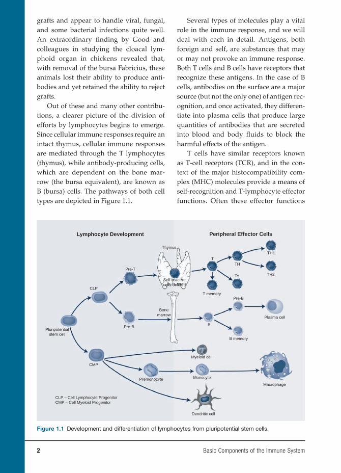
Out of these and many other contribu-tions, a clearer picture of the division of efforts by lymphocytes begins to emerge. Since cellular immune responses require an intact thymus, cellular immune responses are mediated through the T lymphocytes (thymus), while antibody-producing cells, which are dependent on the bone mar-row (the bursa equivalent), are known as B (bursa) cells. The pathways of both cell types are depicted in Figure 1.1.
Several types of molecules play a vital role in the immune response, and we will deal with each in detail. Antigens, both foreign and self, are substances that may or may not provoke an immune response. Both T cells and B cells have receptors that recognize these antigens. In the case of B cells, antibodies on the surface are a major source (but not the only one) of antigen rec-ognition, and once activated, they differen-tiate into plasma cells that produce large quantities of antibodies that are secreted into blood and body fl uids to block the harmful effects of the antigen.
T cells have similar receptors known as T-cell receptors (TCR), and in the con-text of the major histocompatibility com-plex (MHC) molecules provide a means of self-recognition and T-lymphocyte effector functions. Often these effector functions
2 Basic Components of the Immune System
Lymphocyte Development Peripheral Effector Cells
TH1
TH2
TH
Tc
T
B
Thymus
Self reactivecells deleted
T memory
B memory
Macrophage
Pre-B
Pre-BPluripotential
stem cell
Pre-T
CLP
CMP
Plasma cell
Myeloid cell
Dendritic cell
CLP – Cell Lymphocyte ProgenitorCMP – Cell Myeloid Progenitor
Bonemarrow
Premonocyte Monocyte
Figure 1.1 Development and differentiation of lymphocytes from pluripotential stem cells.
are carried out by messages transmitted between these cells. These soluble messen-gers are called interleukins or cytokines.
ANTIGENS
Antigens are any substances that are capable, under appropriate conditions, of inducing the formation of antibodies and reacting specifi cally with the antibodies so produced. They react with both T-cell recognition receptors and with antibodies. These antigenic molecules may have sev-eral antigenic determinants, called epitopes, and each epitope can bind with a specifi c antibody. Thus, a single antigen can bind to many different antibodies with different binding sites.
Some low-molecular-weight mol-ecules called haptens are unable to evoke an immune response but can react with existing antibodies. These molecules need to be coupled to a carrier molecule to be antigenic.
For some molecules such as drugs, the molecule needs to be conjugated to a car-rier. The carrier may be a host protein. The tertiary structure of the molecule as well as the amino acid sequence is important in determining antigenicity. Certain struc-tures such as lipids and DNA are generally poor antigens.
Most antigens are either thymus-dependent or thymus-independent anti-gens. Thymus-dependent antigens require T-cell participation: Most proteins and foreign red cells are examples of these molecules. Thymus-independent antigens do not require T-cell participation for anti-body production. Instead, they directly stimulate specifi c B lymphocytes by cross-linking antigen receptors on the surface of B cells. These molecules produce primarily
IgM and IgG2 antibodies and do not stimu-late long-lasting memory cells. Most bac-terial polysaccharides (found in bacterial cell walls) fall into this category. Certain polysaccharides, such as LPS (lipopoly-saccharide), not only induce specifi c B-cell activation but also can act as a polyclonal B-cell stimulant.
ANTIBODY
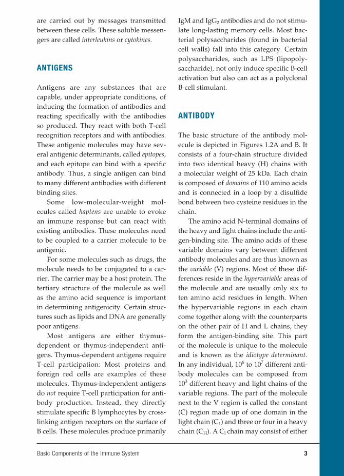
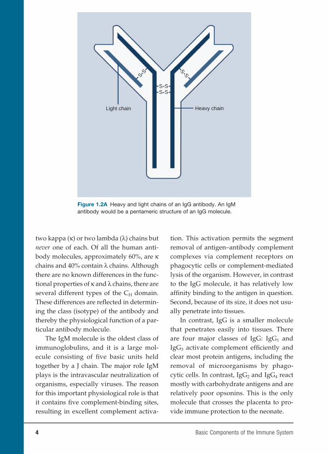
The basic structure of the antibody mol-ecule is depicted in Figures 1.2A and B. It consists of a four-chain structure divided into two identical heavy (H) chains with a molecular weight of 25 kDa. Each chain is composed of domains of 110 amino acids and is connected in a loop by a disulfi de bond between two cysteine residues in the chain.
The amino acid N-terminal domains of the heavy and light chains include the anti-gen-binding site. The amino acids of these variable domains vary between different antibody molecules and are thus known as the variable (V) regions. Most of these dif-ferences reside in the hypervariable areas of the molecule and are usually only six to ten amino acid residues in length. When the hypervariable regions in each chain come together along with the counterparts on the other pair of H and L chains, they form the antigen-binding site. This part of the molecule is unique to the molecule and is known as the idiotype determinant. In any individual, 106 to 107 different anti-body molecules can be composed from 103 different heavy and light chains of the variable regions. The part of the molecule next to the V region is called the constant (C) region made up of one domain in the light chain (C1) and three or four in a heavy chain (CH). A Cl chain may consist of either
Basic Components of the Immune System 3
4 Basic Components of the Immune System
two kappa (κ) or two lambda (λ) chains but never one of each. Of all the human anti-body molecules, approximately 60%, are κ chains and 40% contain λ chains. Although there are no known differences in the func-tional properties of κ and λ chains, there are several different types of the CH domain. These differences are refl ected in determin-ing the class (isotype) of the antibody and thereby the physiological function of a par-ticular antibody molecule.
The IgM molecule is the oldest class of immunoglobulins, and it is a large mol-ecule consisting of fi ve basic units held together by a J chain. The major role IgM plays is the intravascular neutralization of organisms, especially viruses. The reason for this important physiological role is that it contains fi ve complement-binding sites, resulting in excellent complement activa-
tion. This activation permits the segment removal of antigen–antibody complement complexes via complement receptors on phagocytic cells or complement-mediated lysis of the organism. However, in contrast to the IgG molecule, it has relatively low affi nity binding to the antigen in question. Second, because of its size, it does not usu-ally penetrate into tissues.
In contrast, IgG is a smaller molecule that penetrates easily into tissues. There are four major classes of IgG: IgG1 and IgG3 activate complement effi ciently and clear most protein antigens, including the removal of microorganisms by phago-cytic cells. In contrast, IgG2 and IgG4 react mostly with carbohydrate antigens and are relatively poor opsonins. This is the only molecule that crosses the placenta to pro-vide immune protection to the neonate.
Light chain Heavy chain
S S
SS
SS
S S
Figure 1.2A Heavy and light chains of an IgG antibody. An IgM antibody would be a pentameric structure of an IgG molecule.
Basic Components of the Immune System 5
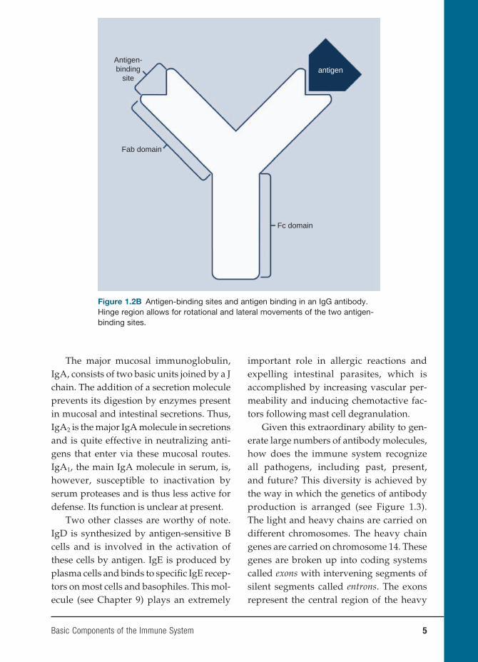
The major mucosal immunoglobulin, IgA, consists of two basic units joined by a J chain. The addition of a secretion molecule prevents its digestion by enzymes present in mucosal and intestinal secretions. Thus, IgA2 is the major IgA molecule in secretions and is quite effective in neutralizing anti-gens that enter via these mucosal routes. IgA1, the main IgA molecule in serum, is, however, susceptible to inactivation by serum proteases and is thus less active for defense. Its function is unclear at present.
Two other classes are worthy of note. IgD is synthesized by antigen-sensitive B cells and is involved in the activation of these cells by antigen. IgE is produced by plasma cells and binds to specifi c IgE recep-tors on most cells and basophiles. This mol-ecule (see Chapter 9) plays an extremely
important role in allergic reactions and expelling intestinal parasites, which is accomplished by increasing vascular per-meability and inducing chemotactive fac-tors following mast cell degranulation.
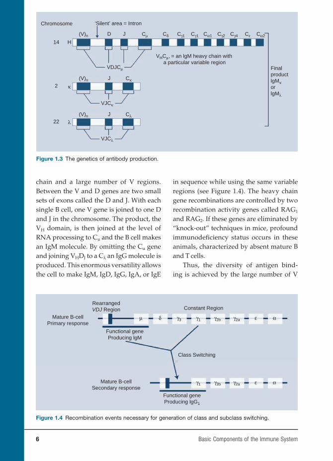
Given this extraordinary ability to gen-erate large numbers of antibody molecules, how does the immune system recognize all pathogens, including past, present, and future? This diversity is achieved by the way in which the genetics of antibody production is arranged (see Figure 1.3). The light and heavy chains are carried on different chromosomes. The heavy chain genes are carried on chromosome 14. These genes are broken up into coding systems called exons with intervening segments of silent segments called entrons. The exons represent the central region of the heavy
Antigen-binding
site
Fab domain
Fc domain
antigen
Figure 1.2B Antigen-binding sites and antigen binding in an IgG antibody. Hinge region allows for rotational and lateral movements of the two antigen-binding sites.
6 Basic Components of the Immune System
chain and a large number of V regions. Between the V and D genes are two small sets of exons called the D and J. With each single B cell, one V gene is joined to one D and J in the chromosome. The product, the VH domain, is then joined at the level of RNA processing to Cu and the B cell makes an IgM molecule. By omitting the Cu gene and joining VHDJ to a Cλ an IgG molecule is produced. This enormous versatility allows the cell to make IgM, IgD, IgG, IgA, or IgE
in sequence while using the same variable regions (see Figure 1.4). The heavy chain gene recombinations are controlled by two recombination activity genes called RAG1 and RAG2. If these genes are eliminated by “knock-out” techniques in mice, profound immunodefi ciency status occurs in these animals, characterized by absent mature B and T cells.
Thus, the diversity of antigen bind-ing is achieved by the large number of V
Chromosome
14
2
22
H
D
VDJCμ
VJCκ
VJCλ
VHCμ, = an lgM heavy chain with a particular variable region
Cμ Cδ CεCγ1 Cγ1 Cα1 Cα2Cγ2 Cγ4J(V)n
J Cκ
κ
λ
(V)n
J Cλ(V)n
‘Silent’ area = Intron
FinalproductlgMκorlgMλ
Figure 1.3 The genetics of antibody production.
RearrangedVDJ Region
Mature B-cellPrimary response
Constant Region
Mature B-cellSecondary response
Class Switching
Functional geneProducing lgM
Functional geneProducing lgG1
μ δ γ3 γ1 γ2b γ2a ε α
γ1 γ2b γ2a ε α
Figure 1.4 Recombination events necessary for generation of class and subclass switching.
Basic Components of the Immune System 7
genes available and their combination with different D and L genes to provide differ-ent antibodies. Furthermore, the inherited set of genes may be increased by somatic mutation during multiple divisions of lym-phoid cells, thereby increasing the number of antibody specifi cities to 1014, which far exceeds the number of B cells (1010) in the body.
Once a given B cell is preselected to pro-duce a particular VH and VL domain, all the ensuing progeny of that B cell will produce the same VH or VL domain. The sequence of events is as follows: initially, the B cell produces intracellular antigen-specific IgM, which becomes bound to the cell sur-face. The B cell is now antigen responsive with exposure to a given antigen. The com-mitted B cell begins producing a certain isotype or class of immunoglobulins and begins dividing, and all the progeny will produce the identical immunoglobulin mol-ecules. These B cells will later mature into either plasma cells or long-term memory B cells.
T CELLS AND THEIR RECEPTORS
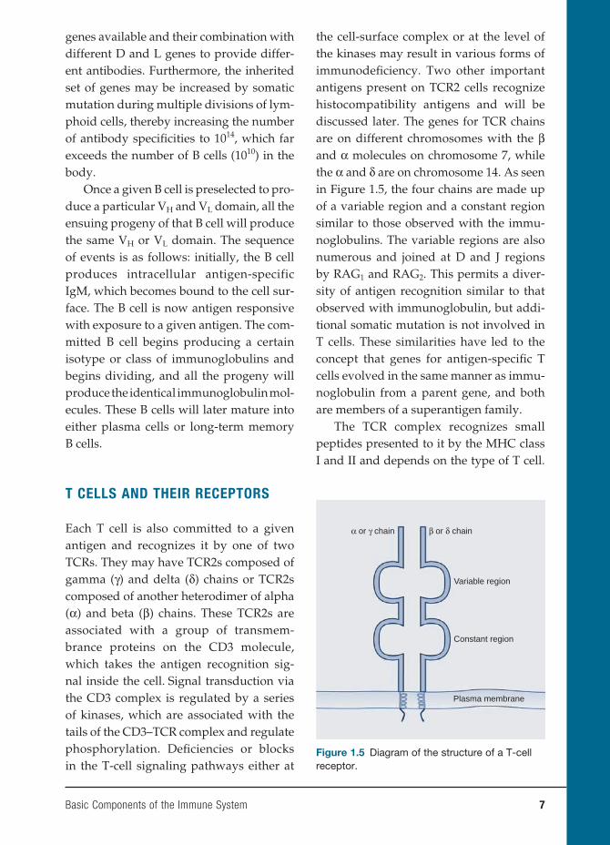
Each T cell is also committed to a given antigen and recognizes it by one of two TCRs. They may have TCR2s composed of gamma (γ) and delta (δ) chains or TCR2s composed of another heterodimer of alpha (α) and beta (β) chains. These TCR2s are associated with a group of transmem-brance proteins on the CD3 molecule, which takes the antigen recognition sig-nal inside the cell. Signal transduction via the CD3 complex is regulated by a series of kinases, which are associated with the tails of the CD3–TCR complex and regulate phosphorylation. Defi ciencies or blocks in the T-cell signaling pathways either at
the cell-surface complex or at the level of the kinases may result in various forms of immunodefi ciency. Two other important antigens present on TCR2 cells recognize histocompatibility antigens and will be discussed later. The genes for TCR chains are on different chromosomes with the β and α molecules on chromosome 7, while the α and δ are on chromosome 14. As seen in Figure 1.5, the four chains are made up of a variable region and a constant region similar to those observed with the immu-noglobulins. The variable regions are also numerous and joined at D and J regions by RAG1 and RAG2. This permits a diver-sity of antigen recognition similar to that observed with immunoglobulin, but addi-tional somatic mutation is not involved in T cells. These similarities have led to the concept that genes for antigen-specifi c T cells evolved in the same manner as immu-noglobulin from a parent gene, and both are members of a superantigen family.
The TCR complex recognizes small peptides presented to it by the MHC class I and II and depends on the type of T cell.
β or δ chain
Variable region
Constant region
Plasma membrane
α or γ chain
Figure 1.5 Diagram of the structure of a T-cell receptor.
8 Basic Components of the Immune System
Helper T cells (CD4) recognize class II anti-gens while suppressor cytotoxic T cells (CD8) recognize class I antigens. Because of the rather low affi nity of the reactions, recognition of processed antigen alone is not suffi cient to activate T cells. Soluble interleukins are needed to complete the picture and are generated during the anti-gen processing.
MAJOR HISTOCOMPATIBILITY
COMPLEX
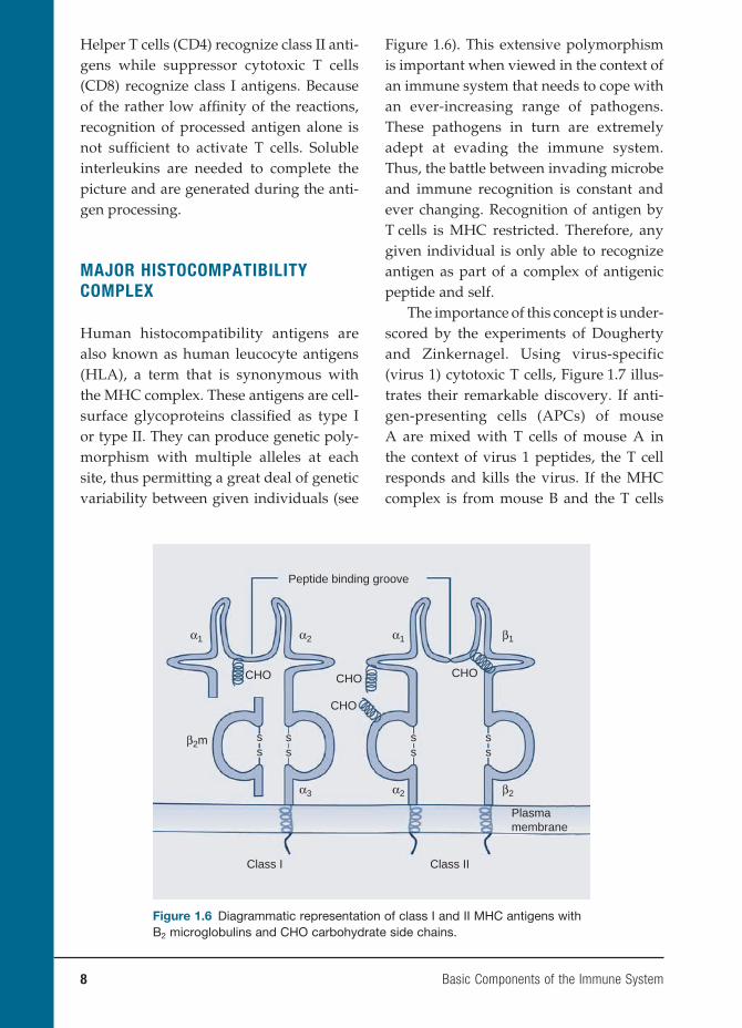
Human histocompatibility antigens are also known as human leucocyte antigens (HLA), a term that is synonymous with the MHC complex. These antigens are cell-surface glycoproteins classifi ed as type I or type II. They can produce genetic poly-morphism with multiple alleles at each site, thus permitting a great deal of genetic variability between given individuals (see
Figure 1.6). This extensive polymorphism is important when viewed in the context of an immune system that needs to cope with an ever-increasing range of pathogens. These pathogens in turn are extremely adept at evading the immune system. Thus, the battle between invading microbe and immune recognition is constant and ever changing. Recognition of antigen by T cells is MHC restricted. Therefore, any given individual is only able to recognize antigen as part of a complex of antigenic peptide and self.
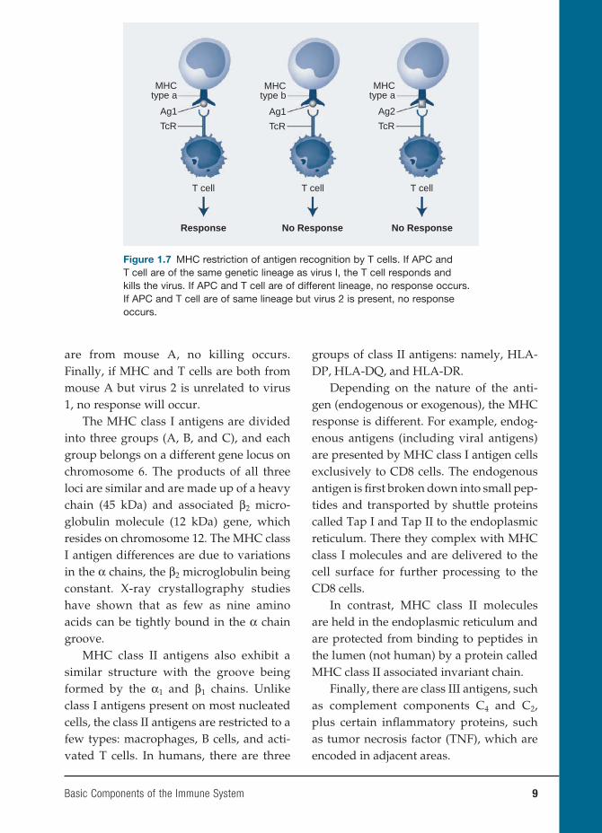
The importance of this concept is under-scored by the experiments of Dougherty and Zinkernagel. Using virus-specific (virus 1) cytotoxic T cells, Figure 1.7 illus-trates their remarkable discovery. If anti-gen-presenting cells (APCs) of mouse A are mixed with T cells of mouse A in the context of virus 1 peptides, the T cell responds and kills the virus. If the MHC complex is from mouse B and the T cells
α2α1 α1 β1
α3
β2m
α2 β2
Peptide binding groove
CHO
CHO
CHO CHO
Plasmamembrane
ss
ss
ss
ss
Class IIClass I
Figure 1.6 Diagrammatic representation of class I and II MHC antigens with B2 microglobulins and CHO carbohydrate side chains.
Basic Components of the Immune System 9
are from mouse A, no killing occurs. Finally, if MHC and T cells are both from mouse A but virus 2 is unrelated to virus 1, no response will occur.
The MHC class I antigens are divided into three groups (A, B, and C), and each group belongs on a different gene locus on chromosome 6. The products of all three loci are similar and are made up of a heavy chain (45 kDa) and associated β2 micro-globulin molecule (12 kDa) gene, which resides on chromosome 12. The MHC class I antigen differences are due to variations in the α chains, the β2 microglobulin being constant. X-ray crystallography studies have shown that as few as nine amino acids can be tightly bound in the α chain groove.
MHC class II antigens also exhibit a similar structure with the groove being formed by the α1 and β1 chains. Unlike class I antigens present on most nucleated cells, the class II antigens are restricted to a few types: macrophages, B cells, and acti-vated T cells. In humans, there are three
groups of class II antigens: namely, HLA-DP, HLA-DQ, and HLA-DR.
Depending on the nature of the anti-gen (endogenous or exogenous), the MHC response is different. For example, endog-enous antigens (including viral antigens) are presented by MHC class I antigen cells exclusively to CD8 cells. The endogenous antigen is fi rst broken down into small pep-tides and transported by shuttle proteins called Tap I and Tap II to the endoplasmic reticulum. There they complex with MHC class I molecules and are delivered to the cell surface for further processing to the CD8 cells.
In contrast, MHC class II molecules are held in the endoplasmic reticulum and are protected from binding to peptides in the lumen (not human) by a protein called MHC class II associated invariant chain.
Finally, there are class III antigens, such as complement components C4 and C2, plus certain infl ammatory proteins, such as tumor necrosis factor (TNF), which are encoded in adjacent areas.
MHCtype b
Ag1
TcR
MHCtype a
Ag1
TcR
Response No Response No Response
MHCtype a
Ag2
TcR
T cellT cellT cell
Figure 1.7 MHC restriction of antigen recognition by T cells. If APC and T cell are of the same genetic lineage as virus I, the T cell responds and kills the virus. If APC and T cell are of different lineage, no response occurs. If APC and T cell are of same lineage but virus 2 is present, no response occurs.
10 Basic Components of the Immune System
ADHESION MOLECULES
In spite of the known MHC complex consisting of binding of a TCR to the pro-cessed antigen, which in turn is bound to the class II molecule of APCs, this is not enough for T-cell activation. One must have additional stimuli that are provided by a series of adhesion molecules on the two cell surfaces.
These molecules are composed of a diverse set of cell-surface glycoproteins and play a pivotal role in mediating cell-to-cell adhesion. Adhesion molecules are divided into four major groups, (a) integrins, (b) selectins, (c) immunoglobulin superfamily, and (d) caherins.
a. Integrins are heterodimers: These are divided into α and β subunits. Depending on the substructure of the β unit, there are fi ve families, but for convenience β1 and β2 integrins are involved in leucocyte–endothelial inter-actions. β1 integrins, also known as very late activation proteins, are so named because they appear on lymphocytes several days after antigenic stimula-tion and are composed of a common β chain (CD29) paired with a different α chain. They mediate lymphocyte and monocyte binding to the endothelium receptors called vascular adhesion mol-ecule. β2 integrins also have a common β chain (CD18), which pairs with dif-ferent α chains (CD11 a, b, c) to form a number of separate molecules. These two sets of integrins mediate strong binding of leucocytes to the endothe-lial cell while β3–β5 are concerned with binding to extracellular matrix proteins such as fi bronectin and vitronectin.
b. Selectins: These molecules are com-posed of three glycoproteins and are
designated by three separate prefi xes: E (endothelial), P (platelet), and L (leu-cocyte). The letters denote the cells on which they were fi rst observed. These groups of selectins bind avidly to car-bohydrate molecules on leucocytes and endothelial cells.
c. Immunoglobulin superfamily: The molecules in this family are so called because they contain a common immunoglobulin-like structure. They strengthen the interaction between the T cells and APCs. They include some of the most powerful molecules in the immune system, such as the CD4, CD8, CD2, lymphocyte function antigen (LFA-3 or CD58), and the intercellular adhesion molecules such as ICAM-1 through 3.
d. Cadherins: These molecules are calcium-dependent adhesion mole-cules and are mainly important in establishing molecular connections between epithelial cells. Their particular importance is during embryonic devel-opment.
CYTOKINES
This group of soluble molecules plays an extremely important role in clinical immu-nology. They are secreted by macrophages and may act as stimulatory or inhibi-tory signals between cells. Cytokines that initiate chemotaxis of leucocytes are called chemokines.
Among the group of cytokines, there are a few of particular interest because of their stimulatory activity. Interleu-kins 1 (IL-1) and 2 (IL-2) are of particu-lar importance secondary to their role in amplifying the immune response. IL-1 acts on a wide range of cells including T
Basic Components of the Immune System 11
and B cells. In contrast, IL-2 primarily acts on lymphocytes, although it has similar trophic effects on IL-receptor B cells and natural killer (NK) cells. (See Table 1.1 and functions.)
INITIATION OF THE IMMUNE
RESPONSE
The effector cells are really divided into two types: B cells and T cells. B cells are primarily responsible for antibody produc-tion, whereas T cells act as effector cells and may function as both helpers and suppres-sors, depending on the stimulus provided by APCs.
The fi rst step in initiation of the immune response to an antigen must necessarily involve modifi cation of the antigen, and these specialized cells are called APCs. Without such processing, T cells cannot recognize antigen. Thus, it is the secretion of cytokines by APCs activated by antigen presentation that further activates antigen-
specifi c T cells. This interaction between APCs and T cells is strongly infl uenced by a group of molecules called co-stimulators. For example, it is CD80 (B7-1) and CD86 (B7-2) on the APC cells with receptors CD28 and CTLA-4 on the T cell that pro-vides this interaction. The absence of these co-stimulators leads to T-cell unrespon-siveness. The importance of this pathway is emphasized by the fact that antagonists to these co-stimulators do interrupt the immune response in both in vitro and in vivo experiments. For example, mice with a severe form of lupus exhibit a milder dis-ease following a CTLA-4 antagonist.
As stated before, processed antigen is presented to the T cells in the context of the MHC complex present on the surface of APCs. In this regard, the most effi cient APCs are the dendritic cells. These cells have high concentrations of MHC class I and II antigens, co-stimulatory molecules, and adhesion molecules on their surface.
These cells may be divided into two major groups. The dendritic cells of the
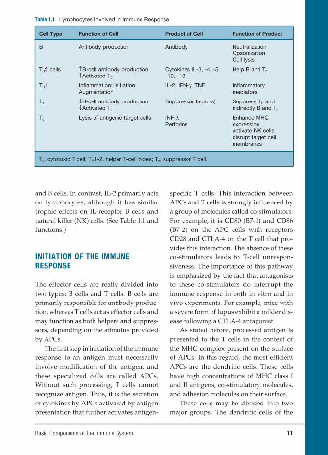
Table 1.1 Lymphocytes Involved in Immune Response
Cell Type Function of Cell Product of Cell Function of Product
B Antibody production Antibody Neutralization OpsonizationCell lysis
TH2 cells ↑B-cell antibody production↑Activated Tc
Cytokines IL-3, -4, -5, -10, -13
Help B and Tc
TH1 Infl ammation: InitiationAugmentation
IL-2, IFN-γ, TNF Infl ammatory mediators
Ts ↓B-cell antibody production↓Activated Tc
Suppressor factor(s) Suppress TH and indirectly B and Tc
Tc Lysis of antigenic target cells INF-λPerforins
Enhance MHC expression, activate NK cells, disrupt target cell membranes
Tc, cytotoxic T cell; TH1-2, helper T-cell types; Ts, suppressor T cell.
12 Basic Components of the Immune System
skin are called the Langerhans cells and play an important role in immune defenses since they are present in the largest protec-tive organ of the body. Because they are mobile, Langerhans cells can capture anti-gen in the periphery and migrate to sec-ondary lymph nodes where they become mature dendritic cells and interact with naïve T cells.
In contrast, the follicular dendritic cells reside in the follicular germinal cen-ter (B-cell area) of a lymph node. These cells have receptors for complement and immunoglobulins and their function is to trap immune complexes and feed them to B cells. This processed immune complex containing antigen is closely associated with MHC class II molecules on the APC surface and thus activates B cells.
ANTIBODY PRODUCTION
To achieve antibody production, at least four types of cells are required: APC, B cells, and two types of regulating cells.
B Cells
Antibodies are produced by naïve B cells and are called plasma cells. These cells express immunoglobulins on their sur-face. In the early stages, B cells fi rst show intracellular µ-chains and then surface IgM. Through the process described ear-lier, these cells can later express IgG, IgA, or IgE, a phenomenon known as isotype switching. The fi nal type of surface immu-noglobulin determines the class of anti-body secreted.
Isotype switching is mediated through two important protein interactions: CD40 on the B cell interacts with CD40L on acti-vated T cells (IL-4 induced) to stimulate B
cells to switch from IgM molecules to other isotypes. Defi ciencies in either molecule lead to severe immunodefi ciency states with only IgM produced but no IgG or IgA antibodies. This syndrome is called the hyper-IgM syndrome, and in this case of CD40L defi ciency, it is an X-linked immu-nodefi ciency.
As mentioned before, each B cell is committed to the production of antibody expressing a unique VH–VL combination, and the surface and excreted immuno-globulin are the same. These observations form the basis of Burnet’s clonal selection theory in that each B cell expresses a sur-face immunoglobulin that acts as its anti-gen-receptor site. Contact with the antigen and helper T-cell factors commit each B cell to divide and differentiate to pro-duce more of the same VH–VL antibody. A number of these B cells become memory cells so that a greater number of antigen-specifi c B cells are available on a second-ary contact with the same antigen. This phenomenon is known as clonal expan-sion and helps to account for the greater secondary response.
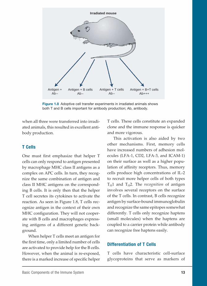
Perhaps more important is that the sec-ondary response of antibodies has a higher affi nity binding for these antigens. These latter antibodies will bind to antigen even when complexed to antibody and help clear the antigen more effectively from the circulation. It is important to remember, however, that B cells alone do not respond to antigen directly, even in the presence of APC cells. They must have a second signal, normally provided by the T cells. This point was elegantly shown in a series of transfer experiments using irradiated recipient ani-mals. As seen in Figure 1.8, antigen alone or antigen + B cells produced no antibody production in these animals. Similarly, T cells alone were ineffective. However,
Basic Components of the Immune System 13
when all three were transferred into irradi-ated animals, this resulted in excellent anti-body production.
T Cells
One must fi rst emphasize that helper T cells can only respond to antigen presented by macrophage MHC class II antigens as a complex on APC cells. In turn, they recog-nize the same combination of antigen and class II MHC antigens on the correspond-ing B cells. It is only then that the helper T cell secretes its cytokines to activate the reaction. As seen in Figure 1.8, T cells rec-ognize antigen in the context of their own MHC confi guration. They will not cooper-ate with B cells and macrophages express-ing antigens of a different genetic back-ground.
When helper T cells meet an antigen for the fi rst time, only a limited number of cells are activated to provide help for the B cells. However, when the animal is re-exposed, there is a marked increase of specifi c helper
T cells. These cells constitute an expanded clone and the immune response is quicker and more vigorous.
This activation is also aided by two other mechanisms. First, memory cells have increased numbers of adhesion mol-ecules (LFA-1, CD2, LFA-3, and ICAM-1) on their surface as well as a higher popu-lation of affi nity receptors. Thus, memory cells produce high concentrations of IL-2 to recruit more helper cells of both types TH1 and TH2. The recognition of antigen involves several receptors on the surface of the T cells. In contrast, B cells recognize antigen by surface-bound immunoglobulin and recognize the same epitopes somewhat differently. T cells only recognize haptens (small molecules) when the haptens are coupled to a carrier protein while antibody can recognize free haptens easily.
Differentiation of T Cells
T cells have characteristic cell-surface glycoproteins that serve as markers of
Irradiated mouse
Antigen +Ab–
Antigen + B cellsAb–
Antigen + T cellsAb–
Antigen + B+T cellsAb+++
Figure 1.8 Adoptive cell transfer experiments in irradiated animals shows both T and B cells important for antibody production; Ab, antibody.
14 Basic Components of the Immune System
“differentiation” of these cells. These markers are recognized by specifi c mono-clonal antibodies and divide them into two particular subsets.
TH1 cells secrete TNF and INF-α and mediate cellular immunity. Conversely, TH2 cells secrete IL-4, IL-5, IL-10, and IL-13 and are needed for stimulating antibody production by B cells. T cells secreting both cytokine profi les are designated THO.
What infl uences a naïve T cell to select which cytokine profi le to secrete is not known. However, experiments in which cells are exposed to IL-1 and IL-6 promote TH2 cells while IL-12 and IFN-α stimulate production of TH1 T cells.
In humans, a TH1 cytokine profi le is primarily directed toward protection against intracellular pathogens while a TH2 profi le interacts with diseases charac-terized by overproduction of antibodies including IgE.
An elegant example of these different pathways of the TH1 and TH2 response is seen in the disease leprosy. Patients that develop a TH1 response develop only limited disease (tuberculoid leprosy). In contrast, those patients mounting a TH2 response develop debilitating and spread-ing lepromatous leprosy since the antibody response will not protect against an intra-cellular pathogen.
Cellular Immunity
Cell-mediated responses are implemented by T lymphocytes. The major functions of T cells can be divided into two catego-ries: the fi rst (cytotoxicity) is to lyse cells expressing specifi c antigens; the second (delayed hypersensitivity) is to release cytokines, thereby triggering an infl am-matory response. These two types of cells are used to combat intracellular pathogens
such as viruses, certain bacteria, and para-sites inaccessible to antibodies.
Cytotoxic T cells lyse cells infected with viruses. This cytotoxicity is virus specifi c, and only cells expressing those proteins on the surface of the infected cell are killed. As stated before, this destruction occurs only in the presence of the same MHC class I molecules. This combination directly acti-vates CD8+ cells and is a potent killer of virally infected cells. The induction of the cytotoxic T cell requires precursor cells and IL-2 from helper cells and is subject to regulation by other T cells.
Cytotoxic T cells also play a role in graft rejection. This was shown years ago in a mixed lymphocyte reaction in which the lymphocytes from two genetically differ-ent individuals were placed in culture. In this case, helper cells responded to a for-eign MHC class II antigen, but cytotoxic T cells were able to lyse target cells carry-ing the MHC class I molecules of the stim-ulating (genetically different individual) cells. The in vivo reactions between indi-viduals undergoing transplantation will be discussed in more detail in a later chapter.
In contrast, delayed-type hypersensi-tivity reactions are mediated by specifi c T cells that produce TH1-type cytokines upon exposure to antigen. An example of this type of reaction is the PPD reaction, or tuberculin test. When the antigen is injected under the skin of an individual who was previously infected with Mycobacterium tuberculosis, a reaction in the skin evolves over 48 to 72 hours in which there is local swelling and induration >10 mm. If the site is biopsied, one fi nds a T-cell and macro-phage infi ltration. Injection of the same material in a noninfected individual pro-duces little or no induration, and the his-tology is essentially negative. Whereas the cells in this case do not kill the organism,
Basic Components of the Immune System 15
most individuals infected surround the organism in a caseous inflammatory lesion, which does not allow the organism to spread. The in vivo state of the lesions will be discussed in more detail in a later chapter.
Nonspecific Effector Molecules
There are a number of nonspecifi c mol-ecules that affect the immune response, especially antibody production. These major factors are as follows: phagocytic cells such as neutrophils and macrophages, which remove antigens and bacteria, and complement, which can either destroy the organism or facilitate its destruction. The role of many of these factors will be discussed in more detail in later chapters, but a brief outline of their functions is war-ranted here.
COMPLEMENTThe complement component system
consists of a series of heat-liable proteins, and they normally exist as inactive precur-sors. However, once activated each compo-nent may act as an enzyme and cleaves the next component in the sequence.
Each precursor is cleaved into two or more components, and the major frag-ment (designated “b”) has two biologically active sites. One is for binding to cell mem-branes and the other is for enzymatic cleav-age of the next component. The control of the sequence relies on either spontaneous decay or specifi c inactivation of these com-ponents. Minor fragments play a role in the fl uid phase, acting as chemotactins.
The major function of the complement system is to help in the opsonization of micro-organisms and immune complexes. These components plus antibody are more read-ily recognized by macrophages and more
readily bound and phagocytosed through IgG: Fc and C3b receptors. Immune com-plexes are handled in a similar fashion, activating the classical pathway comple-ments. Individuals who lack one of the classical pathway components are prone to immune complex disease.
The minor complement fragments con-tribute to the immune response by activating the infl ammatory response. For example, some increase vascular permeability (C3a); others are chemotactins for neutrophils and macrophages (C5a) and not only promote leucocytosis in the bone marrow but attract these cells to the site of infl ammation.
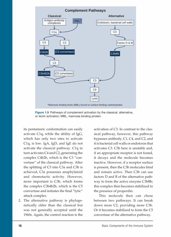
The critical step in complement activa-tion is the cleavage of the C3 component by complement-derived enzymes called C3 convertases. This results in the presence of C3b, which mediates a number of vital biological activities. The cleavage of C3b can be initiated by three routes (classical, alternative, and lectin), but each route is in response to different stimuli. Individu-als who are defi cient in C3 are obviously predisposed to bacterial infections and immune complex disease.
Each of these routes will be examined in more detail (see Figure 1.9).
1. Classical pathway: As its name implies, this is the usual pathway whereby anti-gen–antibody complexes in the presence of complement destroy the invading organism. The antibody (either IgM or IgG) causes a conformational change in the Fc portion of the antibody to reveal a binding site for the fi rst component of complement C1q. This component consists of six subunits and reacts with the Fc via its globular heads. The acti-vation of this component requires the binding of two globular heads for acti-vation. Thus, one molecule of IgM with
16 Basic Components of the Immune System
its pentameric conformation can easily activate C1q, while the ability of IgG, which has only two sites to activate C1q, is low. IgA, IgD, and IgE do not activate the classical pathway. C1q in turn activates C4 and C2, generating the complex C4b2b, which is the C3 “con-vertase” of the classical pathway. After the splitting of C3 into C3a and C3b is achieved, C3a possesses anaphylatoid and chemotactic activity. However, more important is C3b, which forms the complex C3b4b2b, which is the C5 convertase and initiates the fi nal “lytic” attack complex.
2. The alternative pathway is phyloge-netically older than the classical but was not generally accepted until the 1960s. Again, the control reaction is the
activation of C3. In contrast to the clas-sical pathway, however, this pathway bypasses antibody, C1, C4, and C2, and it is bacterial cell walls or endotoxin that activates C3. C3b here is unstable and, if an appropriate receptor is not found, it decays and the molecule becomes inactive. However, if a receptor surface is present, then the C3b molecules bind and remain active. Then C3b can use factors D and B of the alternative path-way to form the active enzyme C3bBb; this complex then becomes stabilized in the presence of properdin.
This molecule then can chose between two pathways. It can break down more C2, providing more C3b. Or it becomes stabilized to form the C5 convertase of the alternative pathway.
Complement PathwaysClassical Alternative
MBL*Antigen-antibodycomplexes
C1q
C4 C2
C5
C9
Lysis
*Mannose binding lectin (MBL) bound to surface-binding carbohydrates
C3
Endotoxin, bacterial cell walls
Factor D & B
C3
C3bB6C4b2b
C3b4B2b
(C3 convertase)
(C5 convertase)
Figure 1.9 Pathways of complement activation by the classical, alternative, or lectin activation; MBL, mannose binding protein.
Basic Components of the Immune System 17
3. Lectin pathway: The third pathway of complement activation is created by the mannose-binding lectin MBL, a circulating protein that binds to carbo-hydrate on the surface of many micro-organisms. MBL (structurally related to C1q) activates complement through a serine protease known as MBL-associated serine protease. Defi ciencies in circulating levels of MBL are asso-ciated with frequent infections in childhood.
Once these components are activated, that is, C3b, 4b2b or C3bBb, and proper-din, these molecules trigger sequentially C5, C6, C7, C8, and C9, which leads to the fi nal lytic pathway and lysis of the target cell. The target can be a red cell, a virally infected cell, or a bacterium. Electron microscopy has shown that this complex binds to the cell membrane and actually punches a hole in the cell. Salts and water pass through the hole and the water fi lls the cell, eventually leading to swelling and destruction of the cell.
The control of the complement activa-tion is important since many of its compo-nents induce infl ammation. This control is executed in the following ways. First, many of the activated components are unstable and will decay rapidly if the next sequence is not present. Second, there are specifi c inhibitors of each component, such as C1 esterase, which inhibits factors I and H. Finally, the cells themselves contain pro-teins that increase the rates of breakdown of these products.
In summary, all acute phase comple-ment components are acute phase proteins and the rate of increase occurs shortly after injury or infection. As will be seen later, there is considerable interaction between the complement system and other
pathways such as clotting, fi brinolytic, and kimin pathways.
FUNCTIONAL COMPONENTS OF THE
IMMUNE SYSTEM
Each of the cells in the immune response has a particular role to play. While many of these cells will be discussed in detail in subsequent chapters, a brief review of the functional capabilities is presented here.
Macrophages
These cells may be divided into two main groups: the dendritic cell and the mature macrophage. The dendritic cell’s major function is to present antigen to the lym-phocyte, and it is the earliest cell to recog-nize foreign antigen.
There are two forms of dendritic cells: immature and mature. The induction of an adaptive immune response begins when a pathogen is ingested by an immature den-dritic cell. These cells reside in most tissues and are relatively long-lived. As seen in Figure 1.1, they are derived from the same cell myeloid precursor as the macrophage. This immature cell carries receptors on its surface that recognize common features of many pathogens such as cell wall carbo-hydrates of bacteria. Once the bacterium is in contact with these receptors, the den-dritic cell is stimulated to engulf the patho-gen and degrade it intracellularly. These cells also continue engulfi ng extracellular material (both viruses and bacteria) by a receptor-independent mechanism of mac-ropinocytosis. Once accomplished, the main function of the “activated” dendritic cell is to carry pathogenic antigens to the periph-eral lymphoid organs to present them to T lymphocytes. Once arrived, the dendritic
18 Basic Components of the Immune System
cell matures into an APC, which now per-mits it to activate pathogen-specifi c lym-phocytes. Another function of activated dendritic cells is to secrete cytokines that infl uence both the innate and adaptive immune responses (see Chapter 4).
The mature macrophage also derives from primitive stem cells in the bone mar-row, but unlike the lymphocyte, it matures in the tissues. Thus monocytes, the pre-cursors of mature macrophages, circu-late for only a few hours before entering the tissues where they live for months as mature macrophages. There is great vari-ety in the tissue macrophages; they are heterogeneous in appearance and metab-olism. They include mobile alveolar and peritoneal macrophages. There are also fi xed cells in the liver called Kupffer cells and skin macrophages called Langerhans cells.
The primary function of these mono-nuclear cells is to phagocytose invading organisms, dead cells, immune complexes, and antigens. To do this, these cells are equipped with powerful lyososomal gran-ules containing acid hydrolases and other degrading enzymes. Macrophages need activation to carry out these functions. These include cytokines, which can bind to IgG: Fc receptors or most importantly (as we shall see later) receptors for bacterial polysaccharides. In addition, they can be activated by soluble infl ammatory prod-ucts such as C5a. In turn, the macrophages can release monokines, such as TNF or IL-1, which increase the infl ammation in infl amed tissues.
Neutrophils
These circulating cells also play an impor-tant role in the body’s defense against infection. These cells produce adhesin
molecule receptors, permitting them to adhere to and migrate from the blood vessels to the site of infection. They are attracted to the site by IL-8, C3a, and C3b, cytokines released by TH1 cells, and fi nally factors produced by mast cells. These cells are also phagocytic cells, and the process of phagocytosis is similar to that seen in macrophages. They are particularly effec-tive when the invading organism becomes coated with antigen-specifi c antibodies (often called opsonins) along with acti-vated complement components.
Other Functional Cells
NK cells also can kill target cells in the absence of either antigen or antibody stimulation. Their lineage is not known, but they are probably in some manner related to T cells. Unlike other cells, they can be nonspecifi cally activated by mito-gens, interferon, and IL-12. These cells are particularly useful in the early response to viral infection. As in other cells, they have receptors on their surface that recognize particular ligands. For example, NKR-PI is a lectin-like receptor that recognizes carbo-hydrate moieties on target cells, which ini-tiates killing. As in other cell systems, there is also an inhibiting receptor called KIR. This molecule binds to ligands on MHC class I ligands, and this prevents killing of the target cell.
NK cells are not immune cells, and they have a broad range of specifi city and no real memory. Studies of animals with NK defi ciencies indicate that they have a greater incidence of viral infections and malignancies. This suggests that they have broad “immunological surveillance” properties but the exact mechanisms whereby they exert those properties are not known.
Basic Components of the Immune System 19
The use of an antibody-coated target to destroy foreign target cells is called anti-body-dependent cell-mediated cytotoxic-ity, or ADCC. This killing is dependent on the recognition by cells bearing Fc receptors and includes monocytes, neutrophils, and NK cells. These cells do not need simulta-neous recognition by MHC molecules. The mechanisms of killing most likely involve the release of cytoplasmic components of the target cells and perforin, but additional factors are also probably involved.
TISSUE DAMAGE PATHWAYS
Although the major function of the com-ponents of the immune system is to neu-tralize or destroy the invading organisms or antigen, these reactions often cause “bystander” tissue damage as well. These are called hypersensitivity reactions, and Gell and Coombs conveniently divided them into fi ve types.
Hypersensitivity Reactions
TYPE I: IMMEDIATEThese reactions are those that involve
antigens that react with IgE bound to tissue mast cells or basophils. Activation of the mast cell results in the release of large amounts of pharmacologically active sub-stances. These reactions are rapid (hence immediate) and if injected into the skin a “wheel and fl are” reaction can be seen within fi ve to ten minutes. Most anti-gens stimulating IgE are either inhaled or ingested. A perfect example of the inhaled antigen is ragweed pollen. The IgE pro-duction requires helper T cells and T-cell-derived cytokines. IL-4 and IL-13 stimulate IgE production while IFN-γ is inhibiting. Many factors regulate the balance between
help and suppression, including route of administration, physical nature of the sub-stance, and the genetic background of either animals or humans. In the latter, there is a family tendency to these reactions but exact genetic factors are still ill defi ned.
TYPE II: CELL BOUNDThese reactions are initiated by anti-
body reacting with antigen on the cell mem-branes. IgM and IgG can be involved in these reactions. Clinical examples include organ-specifi c autoimmune diseases and immune hemolytic anemia. The role of autosensitized T cells in some diseases such as rheumatoid arthritis and multiple sclero-sis have been postulated, but the evidence for their involvement is far from clear. In Graves’ disease (hyperthyroidism), auto-antibodies have a primary pathogenic role but specifi c reactive T cells are also present. However, it is not clear whether the T cells exert a primary role in stimulating anti-body production or are really secondary to the tissue damage.
TYPE III: IMMUNE COMPLEXThese reactions result from the pres-
ence of either circulating immune com-plexes or immune complexes in the tissues. Deposition of immune complexes depends on their size, charge, local concentration of complement, and perhaps most impor-tant the nature of the antigen. An excel-lent example of this type of reaction is the arthritis reaction in which antigen is injected into the skin of an animal previously sensi-tized to the same antigen and has produced antibody to that antigen. The preformed antibody goes to the site of the injected antigen and forms a complex, thereby inducing complement activation and neu-trophil attraction. The result is intense local infl ammation, hemorrhage, and necrosis.
20 Basic Components of the Immune System
There are numerous examples of this type of hypersensitivity reaction, including serum sickness, glomerulonephritis, and systemic lupus erythematosus. Many of these conditions will be described in detail in later chapters.
TYPE IV: DELAYEDT cells drive this reaction when they
react with antigen and release TH1 cyto-kines. The cytokines in turn attract other cells, such as macrophages, which release their lysosomal enzymes. Histologically, the lesions consist of lymphocytes, mac-rophages, and occasionally eosinophilic polymorphonuclear leucocytes, leading to a chronic lesion of necrosis fi brosis and granulomatosus reaction. An excellent example of this reactivity is seen when PPD is injected into the skin of a person who has been previously infected with the tuberculosis organism.
BIBLIOGRAPHY
REVIEWSAbbas AK, Lichtman AH, Pober JS, eds.
Cellular and Molecular Immunology. 2nd ed. Philadelphia, PA: W. B. Saunders Co.; 1994.
Janeway CA, Travers P, Walport M, Schlomchik M, eds. Immunobiology: The Immune System in Health and Disease. New York: Garland Publishing; 2004.
Paul WE. Fundamental Immunology. 2nd ed. New York: Raven Press; 1994.
LANDMARK PAPERSChase MW. The cellular transfer of cutane-
ous hypersensitivity to tuberculin. Proc Soc Exp Biol Med. 1942;59:134–135.
Chase MW. Hypersensitivity to sim-ple chemicals. Harvey Lect. 1967;61: 169–203.
Del Prete G. The concept of type 1 and type 2 helper T cells and their cytokines in humans. Int Rev Immunol. 1998;16: 427–455.
Papermaster BW, Dalmasso AP, Martinez C, Good RA. Suppression of antibody forming capacity with thymectomy in mouse. Proc Soc Exp Biol Med. 1962;111:41.
Zinkernagel RM, Doherty PC. H-2 com-patibility requirement for T-cell medi-ated lysis of target cells infected with lymphocytic choriomeningitis virus: different cytotoxic T-cell specifi cities are associated with structures coded for in H-2K or H-2D. J Exp Med. 1975;141:1427–1436.
INTRODUCTION
This chapter is not designed to cover all the techniques and assays used in clinical immunology. Rather, it is an introduction to various techniques commonly used in diagnosing human disease or, rather, assays to evaluate the competence or incompe-tence of the immune system. Finally, it will serve as an introduction to the many new techniques emerging in the past several years that have widened our knowledge of the complex relationship of microbe–host interactions in human disease.
Laboratory tests vary widely in clinical immunology. Some are essential for diag-nosis while others are useful in subclassify-ing disorders. Finally, some are of research interest only but may add to our immuno-logical armamentarium in the future. In this regard, it is important to understand that these tests do vary in their sensitivity and specifi city.
The sensitivity of a test is defi ned as the number of diseased individuals that are positive for the test compared with those who are negative. The specifi city of a test is the proportion of individuals without a given disease that are negative. Thus, a positive test is really restricted to the dis-ease in question.
The various assays to be discussed later in this chapter can be conveniently divided into two main divisions. Some assays are quantitative in that they produce
precise results. Many of these assays are automated and can be related to interna-tional standards. Qualitative assays are less specifi c and will give answers such as normal–abnormal, or positive–negative results. The problem is that interpretation of results may be subjective and require special expertise in carrying out the test. Many research tests are in this category at fi rst, and many become quantitative assays when more fully developed.
ANTIBODY PRODUCTION
Antibodies for various tests can be pro-duced in a number of different ways, and we will discuss the prototype of each in turn.
a. Polyclonal antibodies: Many mammals have been used to produce antibodies, ranging from the horse, sheep, and goat down to mice and guinea pigs. Often an animal species is selected for antibody production because it will produce less-cross-reactive antibodies to a given tis-sue. Larger mammals, such as goats and sheep, are used to obtain larger volumes of serum to be used therapeutically in humans. A recent fear has been that ani-mals such as sheep or cows may have eaten animal foddage contaminated with prion disease. Thus, polyclonal antibody production for therapeutic
2. Immunological Techniques
John B. Zabriskie, M.D.
21
uses has often been limited to countries like Australia or New Zealand where there have been no recorded cases of prion disease in mammals.
b. Monoclonal antibodies: Over the past two decades, the revolutionary experi-ments of Kohler and Milstein have been a major advance in the production of antigen-specifi c antibodies. In brief, the key to this remarkable advance was the ability to obtain spleen cells from mice that had been immunized with a given antigen and fuse these cells to a non-secreting myeloma cell line, which then produces a single antibody clone when fused with a given B cell pres-ent in the spleen cells. Antibody clones are only produced when the mouse B cell fuses with the myeloma line. Non-fused B cells are eliminated by a spe-cial factor in the medium. The beauty of the hybridoma (fused) cell is that it produces only the antibody of a single mouse B cell and is therefore identical throughout its variable and constant regions, and the antibody reacts only with a single determinant on a given antigen. Finally, it is immortal and will produce the same specifi city of antibody for generations. Large-scale culture of these antibodies can provide large quantities of antibody that are precise in their reactivity.
However, a word of caution is war-ranted. These hybridoma clones can some-times partially lose their antibody produc-tion so that they no longer secrete as much antibody as before and may even stop production altogether. Finally, they may also lose their specifi city so that a given hybridoma line must be checked periodi-cally against the original antigen to deter-mine whether production or specifi city
remains the same as the original clone. As will be seen in many other chapters in this book, the use of monoclonal antibodies has expanded enormously in the past ten to fi f-teen years. They may be “humanized” by the introduction of human heavy and light chains so that they can be used as thera-peutic agents in many human diseases, ranging from rheumatoid arthritis to many forms of cancer.
IMMUNOLOGICAL ASSAYS
Measurements of Immunoglobulins
The introduction of automated machines to measure immunoglobulins and other proteins has proceeded rapidly in recent decades. Most clinical immunology labo-ratories rely almost exclusively on these machines, and research labs are also intro-ducing these automated techniques at a rapid pace. Precise measurement of serum immunoglobulins is an essential corner-stone in this area and is important for repeated and serious infections secondary to immunosuppressive agents, immunode-fi ciencies, in lymphoproliferate disorders, and for detection of autoantibodies.
The main principle behind this test is related to the formation of immune com-plexes between the antibody and a given antigen. If the concentration of antigen–antibody complex is low, then the immune complexes remain in suspension as fi ne particles, which can disperse a beam of light. As the complexes increase with concentration of antibody, the complexes will precipitate, and light scattering will decrease. This degree of dispersion can be measured on a nephelometer.
Using this method, a wide variety of proteins in serum, amniotic fl uid, cerebro-spinal fl uid, saliva, and gastrointestinal
22 Immunological Techniques
juices can be determined. The method includes a wide range of immune reac-tants, acute phase proteins, and tumor markers. Standard preparations are used and have been calibrated against interna-tional World Health Organization stan-dards. These tests primarily use polyclonal antibodies for each antigen since monoclo-nal antibodies do not form immune pre-cipitates because there are too few relevant epitopes.
Radioimmunoassay and Enzyme-
Linked Immunosorbent Assays
(ELISAs)
The use of these highly sensitive assays in human disease has virtually exploded in the past two decades. They can be used to detect the levels of a given antibody or hormone in human serum, and they are extremely sensitive methods of detecting low levels of autoantibodies.
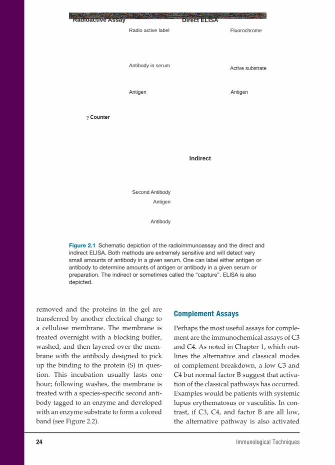
In the radioimmunoassay, one can radiolabel a particular antigen or antibody using either 125I or 14C tagged to the antigen or antibody. Once the serum or purifi ed antibody or antigen to be tested is placed in the well, a second radiolabeled antihuman IgG antibody is placed in the well. After appropriate binding and further washes, the degree of activity of the antibody to a given antigen can be determined in a γ counter (see Figure 2.1 top left).
The description of ELISAs; (Figure 2.1 top right) is similar to that described for the radioimmunoassay, but in place of the radioactively labeled antibody or anti-gen, various fl uorochromes have been substituted in place of the radioactive label. In the presence of an appropriate substrate, the fl uorochrome-labeled anti-body is activated to produce a given color, and the intensity of the color is read on a
spectrophotometer using a 450-nm fi lter. By keeping the known antigen constant and diluting the serum to be tested, one can produce a curve of decreasing optical density readings, thereby indicating the amount of antibody in a given serum when compared with a standard control.
For detection of small amounts of a given antigen or antibody in a test sample, the “capture” assay is used (Figure 2.1 bot-tom). In this case, an unlabeled antibody to a given molecule is laid down on the plate to “capture” the small amount of antigen or antibody present in the test sample. The second antibody to this antigen or anti-body is labeled with the appropriate fl uo-rochrome, and the rest of the tests proceed as in the direct assay described previously. While the radioimmunoassay remains the “gold” standard for many clinical labora-tories, more research and clinical laborato-ries are turning to the ELISA since it does not present the problem of radioactivity hazards or, perhaps more important, the removal of radioactive wastes (mainly a problem of disposal sites).
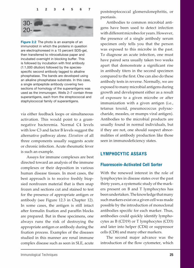
Immunoblots
This immunological technique has gained great favor with both basic immunolo-gists and clinical immunologists over the past decade. Its beauty is its simplicity and the fact that one can compare differ-ent proteins, toxins, and cellular products all at the same time and reach conclusions concerning their commonality or differ-ences or purity. The procedure is relatively simple. The proteins to be studied are run on a standard SDS gel, the percentage of which depends on the known or estimated size of the protein: larger proteins are run in 10 percent gels, while smaller proteins are run on 15 percent gels. The gel is then
Immunological Techniques 23
24 Immunological Techniques
removed and the proteins in the gel are transferred by another electrical charge to a cellulose membrane. The membrane is treated overnight with a blocking buffer, washed, and then layered over the mem-brane with the antibody designed to pick up the binding to the protein (S) in ques-tion. This incubation usually lasts one hour; following washes, the membrane is treated with a species-specifi c second anti-body tagged to an enzyme and developed with an enzyme substrate to form a colored band (see Figure 2.2).
Complement Assays
Perhaps the most useful assays for comple-ment are the immunochemical assays of C3 and C4. As noted in Chapter 1, which out-lines the alternative and classical modes of complement breakdown, a low C3 and C4 but normal factor B suggest that activa-tion of the classical pathways has occurred. Examples would be patients with systemic lupus erythematosus or vasculitis. In con-trast, if C3, C4, and factor B are all low, the alternative pathway is also activated
Radioactive Assay Direct ELISA
Radio active label Fluorochrome
Antibody in serum Active substrate
Second Antibody
Antigen
Antibody
Antigen
γ Counter
Indirect
Antigen
Figure 2.1 Schematic depiction of the radioimmunoassay and the direct and indirect ELISA. Both methods are extremely sensitive and will detect very small amounts of antibody in a given serum. One can label either antigen or antibody to determine amounts of antigen or antibody in a given serum or preparation. The indirect or sometimes called the “capture”. ELISA is also depicted.
Immunological Techniques 25
via either feedback loops or simultaneous activation. This would point to a gram-negative bacteremia. Normal C4 levels with low C3 and factor B levels suggest the alternative pathway alone. Elevation of all three components usually suggests acute or chronic infection. Acute rheumatic fever is such an example.
Assays for immune complexes are best directed toward an analysis of the immune complexes or their deposition in various human disease tissues. In most cases, the best approach is to receive freshly biop-sied nonfrozen material that is then snap frozen and sections cut and stained to test for the presence of appropriate antigen or antibody (see Figure 12.3 in Chapter 12). In some cases, the antigen is still intact after formalin fi xation and paraffi n blocks are prepared. But in these specimens, one always runs the risk of destroying the appropriate antigen or antibody during the fi xation process. Examples of the diseases studied in this manner are renal immune complex disease such as seen in SLE, acute
poststreptococcal glomerulonephritis, or psoriasis.
Antibodies to common microbial anti-gens have been used to detect infection with different microbes for years. However, the presence of a single antibody serum specimen only tells you that the person was exposed to this microbe in the past. To diagnose an acute infection, one must have paired sera usually taken two weeks apart that demonstrate a signifi cant rise in antibody titers in the second specimen compared to the fi rst. One can also do these antibody tests in reverse. Normally, we are exposed to many microbial antigens during growth and development either as a result of exposure to a given microbe or after immunization with a given antigen (i.e., tetanus toxoid, pneumococcus polysac-charide, measles, or mumps viral antigen). Antibodies to the microbial products are usually found in normal individuals, but if they are not, one should suspect abnor-malities of antibody production like those seen in immunodefi ciency states.
LYMPHOCYTIC ASSAYS
Fluorescein-Activated Cell Sorter
With the renewed interest in the role of lymphocytes in disease states over the past thirty years, a systematic study of the mark-ers present on B and T lymphocytes has been undertaken. The knowledge that many such markers exist on a given cell was made possible by the introduction of monoclonal antibodies specifi c for each marker. Thus, antibodies could quickly identify lympho-cytes as B (CD19) or T lymphocytes (CD3) and later into helper (CD4) or suppressor cells (CD8) and many other markers.
The second major advance was the introduction of the fl ow cytometer, which
1 2 3
30-20-
4 5 6 7
Figure 2.2 The photo is an example of an immunoblot in which the proteins in question are electrophoresed in a 15 percent SDS-gel, then transferred to nitrocellulose paper and incubated overnight in blocking buffer. This is followed by incubation with fi rst antibody (1:1,000 dilution) followed by the species-specifi c second antibody tagged to alkaline phosphatase. The bands are developed using an alkaline phosphatase substrate. In this case, a single antipeptide antibody covering two sections of homology of the superantigens was used as the immunogen. Wells 2–7 contain three superantigens, each from the streptococcal and staphylococcal family of superantigens.
26 Immunological Techniques
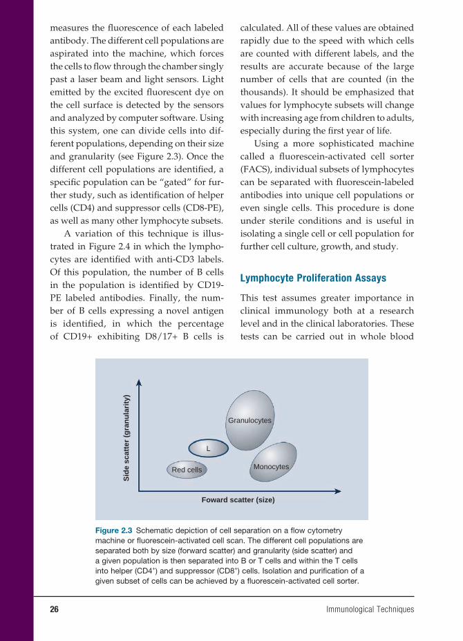
measures the fl uorescence of each labeled antibody. The different cell populations are aspirated into the machine, which forces the cells to fl ow through the chamber singly past a laser beam and light sensors. Light emitted by the excited fl uorescent dye on the cell surface is detected by the sensors and analyzed by computer software. Using this system, one can divide cells into dif-ferent populations, depending on their size and granularity (see Figure 2.3). Once the different cell populations are identifi ed, a specifi c population can be “gated” for fur-ther study, such as identifi cation of helper cells (CD4) and suppressor cells (CD8-PE), as well as many other lymphocyte subsets.
A variation of this technique is illus-trated in Figure 2.4 in which the lympho-cytes are identifi ed with anti-CD3 labels. Of this population, the number of B cells in the population is identifi ed by CD19-PE labeled antibodies. Finally, the num-ber of B cells expressing a novel antigen is identifi ed, in which the percentage of CD19+ exhibiting D8/17+ B cells is
calculated. All of these values are obtained rapidly due to the speed with which cells are counted with different labels, and the results are accurate because of the large number of cells that are counted (in the thousands). It should be emphasized that values for lymphocyte subsets will change with increasing age from children to adults, especially during the fi rst year of life.
Using a more sophisticated machine called a fl uorescein-activated cell sorter (FACS), individual subsets of lymphocytes can be separated with fl uorescein-labeled antibodies into unique cell populations or even single cells. This procedure is done under sterile conditions and is useful in isolating a single cell or cell population for further cell culture, growth, and study.
Lymphocyte Proliferation Assays
This test assumes greater importance in clinical immunology both at a research level and in the clinical laboratories. These tests can be carried out in whole blood
Red cells
L
Granulocytes
Monocytes
Foward scatter (size)
Sid
e sc
atte
r (g
ran
ula
rity
)
Figure 2.3 Schematic depiction of cell separation on a fl ow cytometry machine or fl uorescein-activated cell scan. The different cell populations are separated both by size (forward scatter) and granularity (side scatter) and a given population is then separated into B or T cells and within the T cells into helper (CD4+) and suppressor (CD8+) cells. Isolation and purifi cation of a given subset of cells can be achieved by a fl uorescein-activated cell sorter.
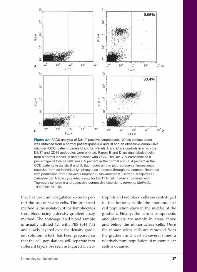
Immunological Techniques 27
that has been anticoagulated so as to per-mit the use of viable cells. The preferred method is the isolation of the lymphocytes from blood using a density gradient assay method. The anticoagulated blood sample is usually diluted 1:1 with PBS (pH 7.4) and slowly layered over the density gradi-ent solution, which has been prepared so that the cell populations will separate into different layers. As seen in Figure 2.5, neu-
trophils and red blood cells are centrifuged to the bottom, while the mononuclear cell population stays in the middle of the gradient. Finally, the serum components and platelets are mainly in areas above and below the mononuclear cells. Once the mononuclear cells are removed from the gradient and washed several times, a relatively pure population of mononuclear cells is obtained.
A
0.05%
33.4%
100
101
102
FL2
-H
103
104
B
101100 102
FL1-H
103 104
100
101
102
FL2
-H
103
104
101100 102
FL1-H
103 104
100
101
102
103
101100 102
FL1-H
103 104
100
101
102
FL2
-H
103
104
101100 102
FL1-H
103 104
C D
FL2
-H
104
Figure 2.4 FACS analysis of D8/17-positive lymphocytes. Whole venous blood was obtained from a normal patient (panels A and B) and an obsessive-compulsive disorder (OCD) patient (panels C and D). Panels A and C are controls in which the D8/17 and CD19 antibodies were omitted. Panels B and D are dual-labeled cells from a normal individual and a patient with OCD. The D8/17 fl uorescence as a percentage of total B cells was 0.5 percent in the normal and 33.4 percent in the OCD patients in panels B and D. Each point on this plot represents fl uorescence recorded from an individual lymphocyte as it passes through the counter. Reprinted with permission from Elsevier; Chapman F, Visvanathan K, Carreno-Manjarrez R, Zabriskie JB. A fl ow cytometric assay for D8/17 B cell marker in patients with Tourette’s syndrome and obsessive compulsive disorder. J Immunol Methods. 1998;219:181–186.
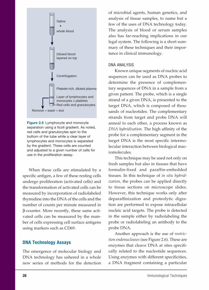
28 Immunological Techniques
When these cells are stimulated by a specifi c antigen, a few of these resting cells undergo proliferation (activated cells) and the transformation of activated cells can be measured by incorporation of radiolabeled thymidine into the DNA of the cells and the number of counts per minute measured in β-counter. More recently, these same acti-vated cells can be measured by the num-ber of cells expressing cell surface antigens using markers such as CD69.
DNA Technology Assays
The emergence of molecular biology and DNA technology has ushered in a whole new series of methods for the detection
of microbial agents, human genetics, and analysis of tissue samples, to name but a few of the uses of DNA technology today. The analysis of blood or serum samples also has far-reaching implications in our legal system. The following is a short sum-mary of these techniques and their impor-tance in clinical immunology.
DNA ANALYSISKnown unique segments of nucleic acid
sequences can be used as DNA probes to determine the presence of complemen-tary sequences of DNA in a sample from a given patient. The probe, which is a single strand of a given DNA, is presented to the target DNA, which is composed of thou-sands of nucleotides. The complementary strands from target and probe DNA will anneal to each other, a process known as DNA hybridization. The high affi nity of the probe for a complementary segment in the target DNA is the most specifi c intermo-lecular interaction between biological mac-romolecules.
This technique may be used not only on fresh samples but also in tissues that have formalin-fixed and paraffin-embedded tissues. In this technique of in situ hybrid-ization, the probes can be applied directly to tissue sections on microscope slides. However, this technique works only after deparaffi nization and proteolytic diges-tion are performed to expose intracellular nucleic acid targets. The probe is detected in the sample either by radiolabeling the probe or radiolabeling an antibody to the probe DNA.
Another approach is the use of restric-tion endonucleases (see Figure 2.6). These are enzymes that cleave DNA at sites specifi -cally related to the nucleotide sequences. Using enzymes with different specifi cities, a DNA fragment containing a particular
Saline
whole blood
Diluted bloodlayered on top
Centrifugation
Platetet-rich, diluted plasma
Layer of lymphocytes andmonocytes ± plateletsRed cells and granulocytes
Remove + wash + test
++
Figure 2.5 Lymphocyte and monocyte separation using a fi coll gradient. As noted, red cells and granulocytes spin to the bottom of the tube while a clear layer of lymphocytes and monocytes is separated by the gradient. These cells are counted and adjusted to a given number of cells for use in the proliferation assay.

Immunological Techniques 29
gene can be cut out from the DNA mole-cule. In the Southern blot, these fragments of DNA are electophoresed on agarose gels with the smaller fragments migrating fur-ther than the larger ones. Alkaline dena-turation of these fragments uncoils the fragment so that a single stranded DNA will hybridize with the complementary DNA after transfer to a special nitrocellu-lose fi lter. Blotting the gel with the nitrocel-lulose fi lter “fi xes” these DNA fragments after the electrophoresis. Finally, a radio-labeled probe containing DNA known to be complementary to the DNA of interest hybridizes to it, and the fragment can be identifi ed by autoradiography of the fi lter. The Northern blotting technique is very similar to the DNA experiment but uses RNA instead of DNA in the system.
POLYMERASE CHAIN REACTION (PCR) ASSAYAlthough the DNA analysis systems
described earlier are still used in research and clinical laboratories, a major revolu-
tion in detecting DNA material occurred with the introduction of the PCR assay. This method is particularly valuable since it can markedly amplify a small piece of DNA before cleavage with a restric-tion enzyme. Complementary oligonu-cleotide primers from either end of the target DNA are added to the denatured sample, along with a heat-resistant DNA polymerase. If the target sequence is pres-ent, the primers anneal to it and provide a starting point for the polymerase to begin the synthesis of second-strand DNA (see Figure 2.7). The newly synthesized double-stranded DNA is then denatured by heating and exposed again to the poly-merase enzyme at a lower temperature. In this way, newly synthesized molecules and original DNA can reassociate with the primer and act as templates for fur-ther rounds of DNA synthesis. After com-pleting about thirty cycles (usually two to three hours in an automated machine), the specifi c target sequence is amplifi ed
Restrictionendonuclease
Denaturation LabeledcDNA
Agarose gelelectrophoresis
gene ofinterest
dsDNA fragment containing gene of interest dsDNA fragment after denaturation
Nitrocelluloseblotting Hybridization
Identification oflabeled gene
fragment
Figure 2.6 The use of restriction endonucleases to carry out gene mapping using the Southern blotting technique. DNA hybridization is the most specifi c intermolecular interaction between biological macromolecules.
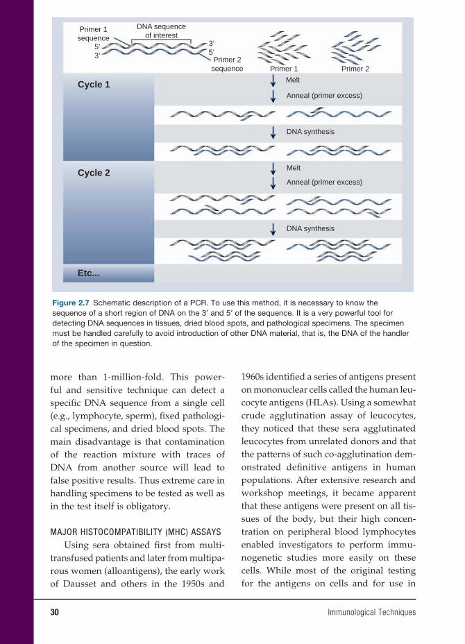
30 Immunological Techniques
more than 1-million-fold. This power-ful and sensitive technique can detect a specifi c DNA sequence from a single cell (e.g., lymphocyte, sperm), fi xed pathologi-cal specimens, and dried blood spots. The main disadvantage is that contamination of the reaction mixture with traces of DNA from another source will lead to false positive results. Thus extreme care in handling specimens to be tested as well as in the test itself is obligatory.
MAJOR HISTOCOMPATIBILITY (MHC) ASSAYSUsing sera obtained fi rst from multi-
transfused patients and later from multipa-rous women (alloantigens), the early work of Dausset and others in the 1950s and
1960s identifi ed a series of antigens present on mononuclear cells called the human leu-cocyte antigens (HLAs). Using a somewhat crude agglutination assay of leucocytes, they noticed that these sera agglutinated leucocytes from unrelated donors and that the patterns of such co-agglutination dem-onstrated defi nitive antigens in human populations. After extensive research and workshop meetings, it became apparent that these antigens were present on all tis-sues of the body, but their high concen-tration on peripheral blood lymphocytes enabled investigators to perform immu-nogenetic studies more easily on these cells. While most of the original testing for the antigens on cells and for use in
Primer 1sequence
Primer 2sequence Primer 1 Primer 2
Melt
Anneal (primer excess)
DNA synthesis
DNA synthesis
Melt
Anneal (primer excess)
DNA sequenceof interest
5’3’
Cycle 1
Cycle 2
Etc...
3’5’
Figure 2.7 Schematic description of a PCR. To use this method, it is necessary to know the sequence of a short region of DNA on the 3′ and 5′ of the sequence. It is a very powerful tool for detecting DNA sequences in tissues, dried blood spots, and pathological specimens. The specimen must be handled carefully to avoid introduction of other DNA material, that is, the DNA of the handler of the specimen in question.
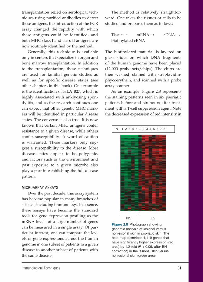
Immunological Techniques 31
transplantation relied on serological tech-niques using purifi ed antibodies to detect these antigens, the introduction of the PCR assay changed the rapidity with which these antigens could be identifi ed, and both MHC class I and class II antigens are now routinely identifi ed by the method.
Generally, this technique is available only in centers that specialize in organ and bone marrow transplantation. In addition to the transplantation, these techniques are used for familial genetic studies as well as for specifi c disease states (see other chapters in this book). One example is the identifi cation of HLA B27, which is highly associated with anklyosing spon-dylitis, and as the research continues one can expect that other genetic MHC mark-ers will be identifi ed in particular disease states. The converse is also true. It is now known that certain MHC antigens confer resistance to a given disease, while others confer susceptibility. A word of caution is warranted. These markers only sug-gest a susceptibility to the disease. Most disease states appear to be polygenic, and factors such as the environment and past exposure to a given microbe also play a part in establishing the full disease pattern.
MICROARRAY ASSAYSOver the past decade, this assay system
has become popular in many branches of science, including immunology. In essence, these assays have become the standard tools for gene expression profi ling as the mRNA levels of a large number of genes can be measured in a single assay. Of par-ticular interest, one can compare the lev-els of gene expressions across the human genome in one subset of patients in a given disease to another subset of patients with the same disease.
The method is relatively straightfor-ward. One takes the tissues or cells to be studied and prepares them as follows:
Tissue → mRNA → cDNA →Biotinylated cRNA
The biotinylated material is layered on glass slides on which DNA fragments of the human genome have been placed (12,000 probe sets/chips). The chips are then washed, stained with streptavidin-phycoerythrin, and scanned with a probe array scanner.
As an example, Figure 2.8 represents the staining patterns seen in six psoriatic patients before and six hours after treat-ment with a T-cell suppression agent. Note the decreased expression of red intensity in
NS LS
N 1 2 3 34 45 5 6 7 81 2
Figure 2.8 Photograph showing genomic analysis of lesional versus nonlesional skin in psoriatic skin. The heat map describes 1,119 genes that have signifi cantly higher expression (red area) by 1.2-fold (P < 0.05, after BH correction) in the lesional skin versus nonlesional skin (green area).
32 Immunological Techniques
the patterns following treatment, indicating a decrease in gene regulation.
FUTURE DIRECTIONS IN RESEARCH
The use of monoclonal antibodies, espe-cially “humanized” ones, will play an increasingly important role in both basic immunology and clinical immunology. Their use will be in both detection of small and early tumors and in the treatment of tumors. The success of monoclonal anti-bodies in metastatic prostate cancer is already well established. It is believed that other tumor therapies will follow in the future.
The use of microarray assays has grown in the past decade and will continue to grow. The use of these assays has a role in diseases associated with human genetics and provides information on which cyto-kines or lymphokines are important in dis-ease states.
BIBLIOGRAPHY
REVIEWSAbbas AK, Lichtman AH, Pober JS, eds.
Cellular and Molecular Immunology. 2nd ed. Philadelphia, PA: W. B. Saunders Co.; 1994.
Kohler G, Milstein C. Continuous cul-tures of fused cells secreting anti-body of predefi ned specifi city. Nature. 1975;256:495–497.
Rose NR, de Macario EC, Folds JD, Lane HC, Nakamura RM, eds. Manual of Clin-ical Laboratory Immunology. Washington, DC: American Society for Microbiol-ogy; 1997.
von Muhlen CA, Tan EM. Autoantibod-ies in the diagnosis of systemic rheu-matic diseases. Semin Arthritis Rheum. 1995;24:323–358.
SUGGESTED READINGAlvarez-Barrientos A, Arroyo J, Canton R,
Nombela C, Sanchez-Perez M. Appli-cations of fl ow cytometry to clini-cal microbiology. Clin Microbiol Rev. 2000;13:167–195.
Churchill GA. Fundamentals of experi-mental design for cDNA microarrays. Nat Genet Suppl. 2007;32:490–495.
Craig FE. Flow cytometric evaluation of B cell lymphoid neoplasms. Clin Lab Med. 2007;27:487–512.
Dausset J. The major histocompatibil-ity complex in man. Science. 1981;213: 1469–1474.
Davies MA, Marstone AJ, Viza DC, Colon-bani J, Dausset J. Human transplan-tation antigens: the HLA (Hu-1) sys-tem and its homology with the mouse H-2 system. Transplantation. 1968;4:571–586.
Dyer P, Middleton D. Histocompatibility Testing: A Practical Approach. Oxford: IRL Press; 1993.
3. Immune Regulation
Nina Bhardwaj, M.D., Ph.D., David O’Neill, M.D.,
and Thomas Waldmann, M.D.
33
INTRODUCTION
The immune system in general responds appropriately to the presence of foreign anti-gens. However, there are certain diseases that arise from either a defective or over-responsive immune system on the part of the host. Two major therapeutic approaches are possible: either immunosuppression or immunopotentiation of the immune system. The object of this chapter is to introduce the reader to the various approaches that have been used to either suppress or stimulate the immune response.
IMMUNOSUPPRESSION
Immunosuppressive Drugs
Several groups of drugs suppress the immune system (see Table 3.1). Among the oldest of these drugs are the corticoste-roids, which have long been known to alter immune responses. When corticosteroids are given, the result is a transient lympho-penia peaking at four hours and lasting up to twenty-four hours. Helper T cells are predominantly affected, and at higher doses of steroids inhibition of interleukin-2 (IL-2) production by helper T cells becomes increasing important. Another major effect in humans is on resting macrophages (activated macrophages are not sensitive).
In humans, steroids are used for two main purposes. One is the prevention
or reversal of graft rejection (see Chap-ter 18). The other is in the treatment of autoimmune and malignant diseases. Corticosteroids modulate infl ammation by suppressing cytokine- and chemokine-encoding genes, which inhibits the acti-vation and recruitment of infl ammatory cells.
The side effects of steroids are numer-ous and often depend on both the dose used and duration of treatment. These include an increased susceptibility to infection, osteoporosis, and growth disturbances in children, as well as gastric ulcers, hyperten-sion, acne, and hirsutism. By giving larger doses for shorter periods, many of these side effects are lessened.
The development of the thiopurines in the 1950s ushered in a new group of immunosuppressive agents, the most important of them being azathioprine. It is inactive until it is metabolized in the liver and takes three to four weeks to be effec-tive. The metabolites work by inhibiting DNA synthesis in dividing cells (such as activated lymphocytes). Like many other drugs, it has side effects, mainly in bone marrow toxicity, and long-term use even-tually results in granulocytopenia and thrombocytopenia.
Another group is the alkylating agents, of which cyclophosphamide is one of the best examples. This drug also requires activa-tion by the liver. It inhibits cell division and can suppress antibody production, and it
33
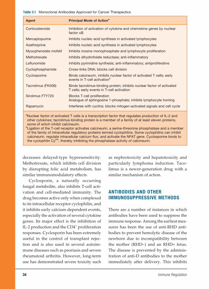
34 Immune Regulation
decreases delayed-type hypersensitivity. Methotrexate, which inhibits cell division by disrupting folic acid metabolism, has similar immunomodulatory effects.
Cyclosporin, a naturally occurring fungal metabolite, also inhibits T-cell acti-vation and cell-mediated immunity. The drug becomes active only when complexed to its intracellular receptor cyclophilin, and it inhibits early calcium-dependent events, especially the activation of several cytokine genes. Its major effect is the inhibition of IL-2 production and the CD4+ proliferation responses. Cyclosporin has been extremely useful in the control of transplant rejec-tion and is also used in several autoim-mune diseases such as psoriasis and severe rheumatoid arthritis. However, long-term use has demonstrated severe toxicity such
as nephrotoxicity and hepatotoxicity and particularly lym phoma induction. Taco-limus is a newer-generation drug with a similar mechanism of action.
ANTIBODIES AND OTHER
IMMUNOSUPPRESSIVE METHODS
There are a number of instances in which antibodies have been used to suppress the immune response. Among the earliest mea-sures has been the use of anti-RHD anti-bodies to prevent hemolytic disease of the newborn due to incompatibility between the mother (RHD−) and an RHD+ fetus. The disease is prevented by the adminis-tration of anti-D antibodies to the mother immediately after delivery. This inhibits
Table 3.1 Monoclonal Antibodies Approved for Cancer Therapeutics
Agent Principal Mode of Actiona
Corticosteroids Inhibition of activation of cytokine and chemokine genes by nuclear factor κB
Mercaptopurine Inhibits nucleic acid synthesis in activated lymphocytes
Azathioprine Inhibits nucleic acid synthesis in activated lymphocytes
Myocphenolate mofetil Inhibits inosine monophosphate and lymphocyte proliferation
Methotrexate Inhibits dihydrofolate reductase; anti-infl ammatory
Lefl unomide Inhibits pyrimidine synthesis; anti-infl ammatory; antiproliferative
Cyclophosphamide Cross-links DNA; blocks cell division
Cyclosporine Binds calcineurin, inhibits nuclear factor of activated T cells; early events in T-cell activationb
Tacrolimus (FK506) Binds tacrolimus-binding protein; inhibits nuclear factor of activated T cells; early events in T-cell activation
Sirolimus FTY720 Blocks T-cell proliferationAnalogue of sphingosine 1-phosphate; inhibits lymphocyte homing
Rapamycin Interferes with cyclins; blocks mitogen-activated signals and cell cycle
a Nuclear factor of activated T cells is a transcription factor that regulates production of IL-2 and other cytokines; tacrolimus-binding protein is a member of a family of at least eleven proteins, some of which inhibit calcineurin.
b Ligation of the T-cell receptor activates calcineurin, a serine-threonine phosphatase and a member of the family of intracellular regulatory proteins termed cyclophilins. Some cyclophilins can inhibit calcineurin, regulate intracellular calcium fl ux, and activate the NFAT gene. Cyclosporine binds to the cyclophilin CyPA, thereby inhibiting the phosphatase activity of calcineurin.
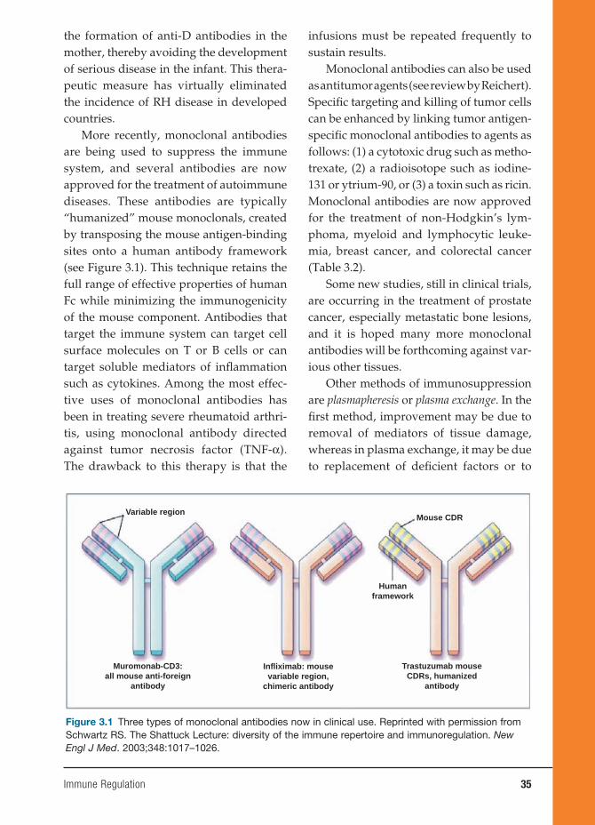
Immune Regulation 35
the formation of anti-D antibodies in the mother, thereby avoiding the development of serious disease in the infant. This thera-peutic measure has virtually eliminated the incidence of RH disease in developed countries.
More recently, monoclonal antibodies are being used to suppress the immune system, and several antibodies are now approved for the treatment of autoimmune diseases. These antibodies are typically “humanized” mouse monoclonals, created by transposing the mouse antigen-binding sites onto a human antibody framework (see Figure 3.1). This technique retains the full range of effective properties of human Fc while minimizing the immunogenicity of the mouse component. Antibodies that target the immune system can target cell surface molecules on T or B cells or can target soluble mediators of infl ammation such as cytokines. Among the most effec-tive uses of monoclonal antibodies has been in treating severe rheumatoid arthri-tis, using monoclonal antibody directed against tumor necrosis factor (TNF-α). The drawback to this therapy is that the
infusions must be repeated frequently to sustain results.
Monoclonal antibodies can also be used as antitumor agents (see review by Reichert). Specifi c targeting and killing of tumor cells can be enhanced by linking tumor antigen-specifi c monoclonal antibodies to agents as follows: (1) a cytotoxic drug such as metho-trexate, (2) a radioisotope such as iodine-131 or ytrium-90, or (3) a toxin such as ricin. Monoclonal antibodies are now approved for the treatment of non-Hodgkin’s lym-phoma, myeloid and lymphocytic leuke-mia, breast cancer, and colorectal cancer (Table 3.2).
Some new studies, still in clinical trials, are occurring in the treatment of prostate cancer, especially metastatic bone lesions, and it is hoped many more monoclonal antibodies will be forthcoming against var-ious other tissues.
Other methods of immunosuppression are plasmapheresis or plasma exchange. In the fi rst method, improvement may be due to removal of mediators of tissue damage, whereas in plasma exchange, it may be due to replacement of defi cient factors or to
Variable region
Muromonab-CD3:all mouse anti-foreign
antibody
Infliximab: mousevariable region,
chimeric antibody
Trastuzumab mouseCDRs, humanized
antibody
Humanframework
Mouse CDR
Figure 3.1 Three types of monoclonal antibodies now in clinical use. Reprinted with permission from Schwartz RS. The Shattuck Lecture: diversity of the immune repertoire and immunoregulation. New Engl J Med. 2003;348:1017–1026.
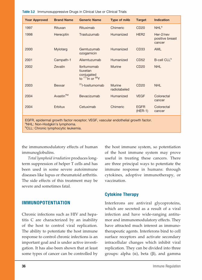
36 Immune Regulation
the immunomodulatory effects of human immunoglobulins.
Total lymphoid irradiation produces long-term suppression of helper T cells and has been used in some severe autoimmune diseases like lupus or rheumatoid arthritis. The side effects of this treatment may be severe and sometimes fatal.
IMMUNOPOTENTIATION
Chronic infections such as HIV and hepa-titis C are characterized by an inability of the host to control viral replication. The ability to potentiate the host immune response to control chronic infections is an important goal and is under active investi-gation. It has also been shown that at least some types of cancer can be controlled by
the host immune system, so potentiation of the host immune system may prove useful in treating these cancers. There are three principal ways to potentiate the immune response in humans: through cytokines, adoptive immunotherapy, or vaccination.
Cytokine Therapy
Interferons are antiviral glycoproteins, which are secreted as a result of a viral infection and have wide-ranging antitu-mor and immunomodulatory effects. They have attracted much interest as immuno-therapeutic agents. Interferons bind to cell surface receptors and activate secondary intracellular changes which inhibit viral replication. They can be divided into three groups: alpha (α), beta (β), and gamma
Table 3.2 Immunosuppressive Drugs in Clinical Use or Clinical Trials
Year Approved Brand Name Generic Name Type of mAb Target Indication
1997 Rituxan Rituximab Chimeric CD20 NHLa
1998 Herecptin Trastuzumab Humanized HER2 Her-2/nev positive breast cancer
2000 Mylotarg Gemtuzumabozogamicin
Humanized CD33 AML
2001 Campath-1 Aliemtuzumab Humanized CD52 B-cell CLLb
2002 Zevalin Ibritumomabtiuxetan conjugatedto 111In or 90Y
Murine CD20 NHL
2003 Bexxar 31I-tositumomab Murineradiolabeled
CD20 NHL
2004 AvastinTM Bevacizumab Humanized VEGF Colorectalcancer
2004 Erbitux Cetuximab Chimeric EGFR(HER-1)
Colorectalcancer
EGFR, epidermal growth factor receptor; VEGF, vascular endothelial growth factor.aNHL: Non-Hodgkin’s lymphoma.bCLL: Chronic lymphocytic leukemia.
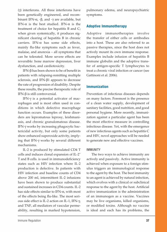
Immune Regulation 37
(γ) interferons. All three interferons have been genetically engineered, and recom-binant IFN-α, -β, and -γ are available, but IFN-α is the best studied. IFN-α is the treatment of choice for hepatitis B and C; when given systemically, it produces sig-nifi cant clearing of hepatitis B in chronic carriers. IFN-α has some side effects, mainly fl u-like symptoms such as fever, malaise, and anorexia – all symptoms that can be tolerated. More severe effects are reversible: bone marrow depression, liver dysfunction, and cardiotoxicity.
IFN-β has been shown to be of benefi t in patients with relapsing-remitting multiple sclerosis, and IFN-β1 appears to decrease the rate of progression of disability. Despite these results, the precise therapeutic role of IFN-β is still controversial.
IFN-γ is a potential activator of mac-rophages and is most often used in con-ditions in which defective macrophage function occurs. Examples of these disor-ders are lepromatous leprosy, leishmani-asis, and chronic granulomatous disease. IFN-γ works by increasing phagocytic bac-tericidal activity, but only some patients show enhanced superoxide activity, imply-ing that IFN-γ works by several different mechanisms.
IL-2 is produced by stimulated CD4 T cells and induces clonal expansion of IL-2+ T and B cells. is used in immunodefi ciency states such as HIV infection where IL-2 production is defective. In patients with HIV infection and baseline counts of CD4 above 200 ml, intermittent IL-2 infusions have been shown to produce substantial and sustained increases in CD4 counts. IL-2 has side effects similar to IFN-α, with most of the effects being fl u-like. The most seri-ous side effect is IL-2 action on IL-1, IFN-γ, and TNF, all mediators of vascular perme-ability, resulting in marked hypotension,
pulmonary edema, and neuropsychiatric symptoms.
Adoptive Immunotherapy
Adoptive immunotherapies involve the transfer of either cells or antibodies into a host. These are also referred to as passive therapies, since the host does not actively mount its own immune response. Examples include infusion of hepatitis B immune globulin and the adoptive trans-fer of antigen-specifi c T lymphocytes to treat a chronic viral infection or cancer (see Gattinoni et al. 2006).
Immunization
Prevention of infectious diseases depends on many factors. Foremost is the presence of a clean water supply, development of sanitary facilities, good nutrition, and good personal hygiene. More recently, immuni-zation against a particular agent has been the most effective measure in controlling infectious disease. Yet, with the emergence of new infectious agents such as hepatitis C and HIV, novel approaches will be needed to generate new and effective vaccines.
IMMUNITYThe two ways to achieve immunity are
actively and passively. Active immunity is achieved when exposure to a foreign stim-ulus triggers an immunological response to the agent by the host. The best immunity to an agent is achieved by natural infection, which evolves with a clinical or subclinical response to the agent by the host. Artifi cial active immunization is the administration of an immunogen as a vaccine. Vaccines may be live organisms, killed organisms, or modifi ed toxins. Although no vaccine is ideal and each has its problems, the
38 Immune Regulation
problems of live vaccines are generally related to their safety, while the problems of killed vaccines are related mainly to their effectiveness.
Live attenuated vaccines are useful because they infect, replicate, and immu-nize in a manner similar to natural infection but with milder clinical symptoms. Exam-ples include many of the childhood infec-tions such as measles, mumps, and rubella (MMR vaccine), chicken pox (varicella) and Bacille Calmette-Guérin (BCG) for tuberculosis. Although millions of doses have been administered with no complica-tions, if given to an immunocompromised host (such as primary immunodefi ciency or secondary to HIV infection), these live vaccines may cause serious disease.
Killed vaccines consist of suspen-sions of killed organisms such as typhoid, cholera, and pertussis (although there is now an acellular vaccine) or one of the products or fractions of the organism. These include toxoids of diphtheria and tetanus and subunits of viruses such as surface hepatitis B antigen. Among the most successful of these types of vaccines has been the use of polysaccharides in the pneumococcal, meningococcal, and Haemophilus infl uenza vaccines. In general, the killed vaccines are not as effective as the live viruses because they do not give long-lasting immunity as a live infection does. For example, although the tetanus toxoid vaccine is effective, it requires a booster dose every ten years.
The immunological response to the killed organism or product thereof has been enhanced by the use of adjuvants. Although the most common adjuvant for animal studies has been the complete Freund’s adjuvant, it cannot be used in humans because it causes liver, skin, and spleen dys-function. The most common adjuvant for
humans is aluminum compounds, which are generally safe for human use. Others include muramyl dipeptide, biodegradable polymers, and a glycoside adjuvant called Quil A from the bark of an Amazon oak tree. However, many others are being developed or will probably be given U.S. Food and Drug Administration (FDA) acceptance in the future. The key feature will be their immunogenic enhancement and their strength of safety for use in humans.
Although we have mainly been dis-cussing various forms of vaccination to protect against the invading organism, one of the most interesting new vaccines has not been developed to eliminate the infectious agent but rather to prevent the development of another far more serious disease – a complication of the initial infec-tion. This is the Gardisal vaccine manufac-tured by Merck to protect against human papilloma virus (HPV). HPV infection is usually sexually acquired, and it is esti-mated that currently 20 million people are infected in the United States. The infection has no real signs or symptoms, and HPV may lead to cervical cancer. It is estimated that ten of the thirty different serotypes of the virus can induce cervical cancer, so the vaccine has been directed at eliminat-ing those serotypes. Thus, if 10,000 women are infected with one of the high-risk viral serotypes, approximately 3,900 of them will die of cervical cancer. The new vac-cine, if given before active sexual activity in women, can prevent the viral infection and thereby markedly diminish the risk of cervical cancer. Because of the possible suc-cess of this vaccine, it may be worthwhile to look at how to prevent the Epstein-Barr virus in at-risk children to prevent or diminish the risk of Burkett’s lymphoma in children infected with the virus.
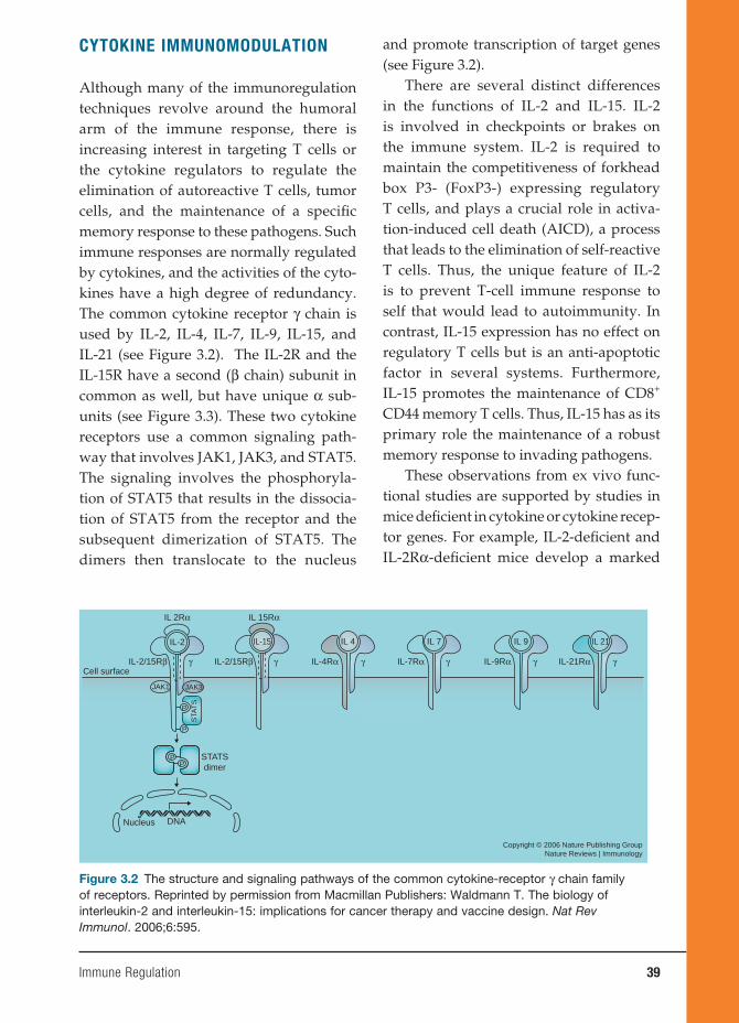
Immune Regulation 39
CYTOKINE IMMUNOMODULATION
Although many of the immunoregulation techniques revolve around the humoral arm of the immune response, there is increasing interest in targeting T cells or the cytokine regulators to regulate the elimination of autoreactive T cells, tumor cells, and the maintenance of a specifi c memory response to these pathogens. Such immune responses are normally regulated by cytokines, and the activities of the cyto-kines have a high degree of redundancy. The common cytokine receptor γ chain is used by IL-2, IL-4, IL-7, IL-9, IL-15, and IL-21 (see Figure 3.2). The IL-2R and the IL-15R have a second (β chain) subunit in common as well, but have unique α sub-units (see Figure 3.3). These two cytokine receptors use a common signaling path-way that involves JAK1, JAK3, and STAT5. The signaling involves the phosphoryla-tion of STAT5 that results in the dissocia-tion of STAT5 from the receptor and the subsequent dimerization of STAT5. The dimers then translocate to the nucleus
and promote transcription of target genes (see Figure 3.2).
There are several distinct differences in the functions of IL-2 and IL-15. IL-2 is involved in checkpoints or brakes on the immune system. IL-2 is required to maintain the competitiveness of forkhead box P3- (FoxP3-) expressing regulatory T cells, and plays a crucial role in activa-tion-induced cell death (AICD), a process that leads to the elimination of self-reactive T cells. Thus, the unique feature of IL-2 is to prevent T-cell immune response to self that would lead to autoimmunity. In contrast, IL-15 expression has no effect on regulatory T cells but is an anti-apoptotic factor in several systems. Furthermore, IL-15 promotes the maintenance of CD8+ CD44 memory T cells. Thus, IL-15 has as its primary role the maintenance of a robust memory response to invading pathogens.
These observations from ex vivo func-tional studies are supported by studies in mice defi cient in cytokine or cytokine recep-tor genes. For example, IL-2-defi cient and IL-2Rα-defi cient mice develop a marked
Figure 3.2 The structure and signaling pathways of the common cytokine-receptor γ chain family of receptors. Reprinted by permission from Macmillan Publishers: Waldmann T. The biology of interleukin-2 and interleukin-15: implications for cancer therapy and vaccine design. Nat Rev Immunol. 2006;6:595.
IL 2Rα IL 15Rα
γ γ γ γ γ γIL-2/15Rβ IL-2/15Rβ IL-4Rα IL-7Rα IL-9Rα IL-21Rα
IL-2 IL-15 IL 4 IL 7 IL 9 IL 21
Cell surface
Copyright © 2006 Nature Publishing GroupNature Reviews | Immunology
Nucleus DNA
STATSdimer
JAK1
P
PP
JAK3
STA
TS
P
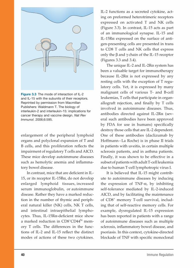
40 Immune Regulation
enlargement of the peripheral lymphoid organs and polyclonal expansion of T and B cells, and this proliferation refl ects the impairment of regulatory T cells and AICD. These mice develop autoimmune diseases such as hemolytic anemia and infl amma-tory bowel disease.
In contrast, mice that are defi cient in IL-15, or its receptor IL-15Rα, do not develop enlarged lymphoid tissues, in creased serum immunoglobulin, or au toimmune disease. Rather they have a marked reduc-tion in the number of thymic and periph-eral natural killer (NK) cells, NK T cells, and intestinal intraepithelial lympho-cytes. Thus, IL-15Rα-defi cient mice show a marked reduction in CD8+CD44hi mem-ory T cells. The differences in the func-tions of IL-2 and IL-15 refl ect the distinct modes of actions of these two cytokines.
IL-2 functions as a secreted cytokine, act-ing on preformed heterotrimeric receptors expressed on activated T and NK cells (Figure 3.3). In contrast, IL-15 acts as part of an immunological synapse. IL-15 and IL-15Rα expressed on the surface of anti-gen-presenting cells are presented in trans to CD8 T cells and NK cells that express only the β and γ chain of the IL-15 receptor (Figures 3.3 and 3.4).
The unique IL-2 and IL-2Rα system has been a valuable target for immunotherapy because IL-2Rα is not expressed by any resting cells with the exception of T regu-latory cells. Yet, it is expressed by many malignant cells of various T- and B-cell leukemias, T cells that participate in organ-allograft rejection, and fi nally by T cells involved in autoimmune diseases. Thus, antibodies directed against IL-2Rα (sev-eral such antibodies have been approved by FDA for use in humans) specifi cally destroy those cells that are IL-2 dependent. One of these antibodies (daclizumab by Hoffmann – La Roche) is in phase II trials in patients with uveitis, in certain multiple sclerosis patients, and in asthma patients. Finally, it was shown to be effective in a subset of patients with adult T-cell leukemia due to human T-cell lymphotropic virus I.
It is believed that IL-15 might contrib-ute to autoimmune diseases by inducing the expression of TNF-α, by inhibiting self-tolerance mediated by IL-2-induced AICD, and by facilitating the maintenance of CD8+ memory T-cell survival, includ-ing that of self-reactive memory cells. For example, dysregulated IL-15 expression has been reported in patients with a range of autoimmune diseases such as multiple sclerosis, infl ammatory bowel disease, and psoriasis. In this context, cytokine-directed blockade of TNF with specifi c monoclonal
Figure 3.3 The mode of interaction of IL-2 and IL-15 with the subunits of their receptors. Reprinted by permission from Macmillan Publishers: Waldmann T. The biology of interleukin-2 and interleukin-15: implications for cancer therapy and vaccine design. Nat Rev Immunol. 2006;6:595.
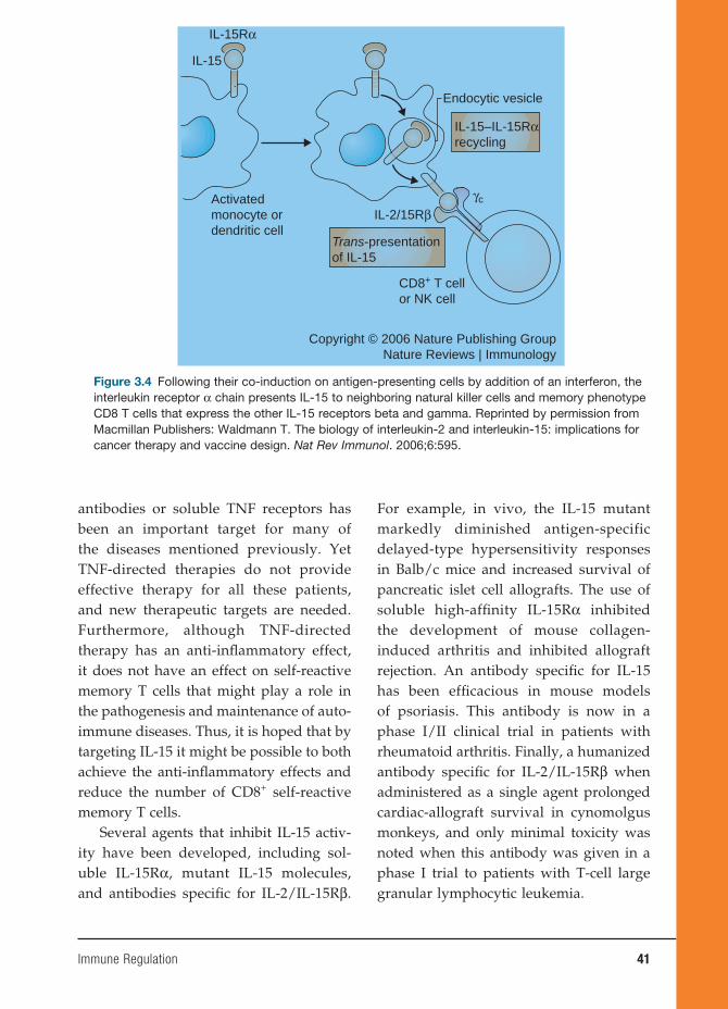
Immune Regulation 41
antibodies or soluble TNF receptors has been an important target for many of the diseases mentioned previously. Yet TNF-directed therapies do not provide effective therapy for all these patients, and new therapeutic targets are needed. Furthermore, although TNF-directed therapy has an anti-infl ammatory effect, it does not have an effect on self-reactive memory T cells that might play a role in the pathogenesis and maintenance of auto-immune diseases. Thus, it is hoped that by targeting IL-15 it might be possible to both achieve the anti-infl ammatory effects and reduce the number of CD8+ self-reactive memory T cells.
Several agents that inhibit IL-15 activ-ity have been developed, including sol-uble IL-15Rα, mutant IL-15 molecules, and antibodies specifi c for IL-2/IL-15Rβ.
For example, in vivo, the IL-15 mutant markedly diminished antigen-specific delayed-type hypersensitivity responses in Balb/c mice and increased survival of pancreatic islet cell allografts. The use of soluble high-affi nity IL-15Rα inhibited the development of mouse collagen-induced arthritis and inhibited allograft rejection. An antibody specifi c for IL-15 has been effi cacious in mouse models of psoriasis. This antibody is now in a phase I/II clinical trial in patients with rheumatoid arthritis. Finally, a humanized antibody specifi c for IL-2/IL-15Rβ when administered as a single agent prolonged cardiac-allograft survival in cynomolgus monkeys, and only minimal toxicity was noted when this antibody was given in a phase I trial to patients with T-cell large granular lymphocytic leukemia.
Figure 3.4 Following their co-induction on antigen-presenting cells by addition of an interferon, the interleukin receptor α chain presents IL-15 to neighboring natural killer cells and memory phenotype CD8 T cells that express the other IL-15 receptors beta and gamma. Reprinted by permission from Macmillan Publishers: Waldmann T. The biology of interleukin-2 and interleukin-15: implications for cancer therapy and vaccine design. Nat Rev Immunol. 2006;6:595.
IL-15Rα
γc
IL-2/15Rβ
Endocytic vesicle
IL-15
Activatedmonocyte ordendritic cell
Copyright © 2006 Nature Publishing GroupNature Reviews | Immunology
CD8+ T cellor NK cell
IL-15–IL-15Rαrecycling
Trans-presentationof IL-15
42 Immune Regulation
CELLULAR VACCINES AND
MODULATIONS THEREOF
Dendritic Cell Vaccines
Recently, much attention has focused on the area of dendritic cell (DC) vaccines in the treatment of cancers. The immunological basis of current approaches to therapeu-tic cancer vaccination (often called “vacci-treatment”) has been established over the past decade or longer. These new devel-opments are mainly based on the lessons learned from the clinical testing of these approaches. In particular, three lessons are worthy of note: First, recent randomized phase III trials suggest that vacci-treatment with autologous DCs expressing pros-tatic acid phosphatase or with autologous tumor-derived heat shock protein (HSP gp96) peptide complexes are showing progress in cancer patient survivals. Sec-ond, immunological monitoring of many clinical trials has failed to identify a sur-rogate marker for clinical outcomes. Third, many articles and reviews suggest that protective immunity to human cancer is elicited by the mutated antigenic repertoire unique to each cancer.
Focusing more closely on the type of DC needed to achieve its vaccine poten-tial, the subsets of DCs could prove criti-cal. Much of the current research is being carried out with monocyte-derived DCs, which are potent and homogeneous stimu-lators of immunity. Monocyte-derived DCs can be readily generated within a few days in large numbers (300 million–500 million mature DCs per apheresis) from precursors in the blood without the need for pretreat-ing patients with various cytokines such GM-CSF or FLT 3-L. Rather, one obtains populations of immature DCs by exposing monocytes to GM-CSF and IL-4, and then they are differentiated into mature DCs
by various stimuli such as toll-like recep-tor (TLR) ligands (LPS or poly I:C), infl am-matory cytokines (IL-1β, TNF-α, IL-6, and PGE2), or CD40L. The use of DCs that have received a maturation stimulus is likely to be important to induce strong immunity. It has become clear that antigen delivered on immature or incompletely matured DCs can even induce tolerance. However, the type and the duration of the maturation stimulus remain to be determined and may infl uence effi cacy. At this time, monocyte-derived DCs are the most accessible and homogeneous populations of DCs.
Monocyte-derived DCs were the fi rst to be used for treating melanoma patients, and several pilot studies have been published. Most used defi ned antigens in the form of peptides, but in some studies, tumor lysates or autologous DC tumor hybrids were also employed. The fi rst trial, published in 1998 by Nestle and colleagues, aroused great interest given an overall response rate of 30 percent in stage IV patients (i.e., distant metastases), including complete responses. An important point in the fi rst study by Nestle and colleagues was that they use fetal calf serum (FCS) during DC genera-tion, and this might have contributed to the observed effects by providing nonspe-cifi c helper epitopes and by promoting the maturation of DCs.
Jonuleit and colleagues directly com-pared within each of eight patients the immunogenicity of immature DCs (gen-erated according to Nestle and colleagues in FCS-containing media using GM-CSF and IL-4) to mature DCs generated in the absence of FCS and matured by a cocktail consisting of TNF-α, IL-1β, IL-6, and PGE2
mimicking the composition of monocyte-conditioned medium. These two differ-ent DC populations were administered intranodally into opposite inguinal lymph

Immune Regulation 43
nodes. FCS-free mature DCs induced stron-ger T-cell responses, both to the two recall antigens used (tetanus toxoid and PPD/tuberculin) and to tumor peptides. Interest-ingly, however, both immature and mature DCs showed an expansion of peptide-spe-cifi c T cells by tetramer staining; yet, only mature DCs induced IFN-γ-producing and lytic CD8+ T cells. These fi ndings suggest the interesting possibility that the imma-ture DCs might have induced regulatory T cells rather than effector T cells, an obser-vation previously noted in studies with normal volunteers.
Cancer patients and healthy subjects often harbor a repertoire of self-reactive T cells and antibodies. This led to the idea that if one could break immunological tolerance to these self-antigens in a con-trollable manner one would fi nd a “thera-peutic window” in which an autoimmune response might damage cancers more than normal tissues. This approach has worked reasonably well with chemotherapies, which, although not cancer specifi c, can confer clinical benefi t with acceptable mor-bidities.
The efforts to create cancer vaccines using allogeneic cell lines, differentiation antigens (such as gp100 and MART1), cancer testes (CT) antigens (such as MAGE, NY-ESO-1), or other common molecules (such as carcinoembryonic anti-gen, mucins, prostate-specifi c antigen, and prostatic acid phosphatase) represent this approach. Within this approach lie several subthemes. Thus, CT antigens, which are not expressed on normal somatic tissues but only on cancers or gonads, might be a better target for breaking tolerance than are differentiation antigens expressed on somatic tissues. Another subtheme is the idea that artifi cially mutated differentiation or CT antigens as vaccines might be better
at breaking tolerance than their wild-type counterparts. Finally, there is a multiplic-ity of choices of delivery agents for these antigens – whole proteins, peptides, RNA, DNA, viral vectors, DCs, and so on.
The importance of innate immunity and the emergence of the toll-like receptors have led to the recognition that immune modifi ers must be an essential compo-nent of any cancer vaccine. The inclusion of QS21, ISCOMS, Montanide, heat shock proteins, CpG, BCG, and granulocyte-macrophage colony-stimulating factor with cancer vaccines refl ects this. Of inter-est in this respect are the anecdotal success of Coley’s toxin at the turn of the nineteenth century (a heat-killed mixture of strep-tococcal cells and probably superantigen broths), which may now be reinterpreted in the light of the newly identifi ed immune response modifi ers.
Finally, a better understanding of the controls that act on T cells to stimulate or inhibit them has led to the use of reagents to enhance antitumor T-cell activity. For exam-ple, blocking antibodies to the inhibitory T-lymphocyte antigen CTLA-4, manipula-tion of regulatory T cells, antibodies to PDL or its ligand PDLI, and enhancement of co-stimulating molecules like B7 on antigen-presenting cells are a few examples of reagents that are being used in this manner in conjunction with vacci-treatments.
BIBLIOGRAPHY
REVIEWSBarnes P. Molecular mechanisms and
cellular effects of glucosteroids. Immu-nol Allergy Clin North Am. 2005;25: 451–468.
Berzofsky JA, Ahlers JD, Belyakov IM. Strategies for designing and optimizing
44 Immune Regulation
new generation vaccines. Nat Rev Immu-nol. 2001;1(3):209–219.
Fehniger TA, Caligiuri MA. Interleukin 15: biology and relevance to human dis-ease. Blood. 2001;97:14–32.
Gattinoni L, Powell DJ Jr, Rosenberg SA, Restifo NP. Adoptive immunotherapy for cancer: building on success. Nat Rev Immunol. 2006;6(5):383–393.
Schwartz RS. The Shattuck Lecture: diver-sity of the immune repertoire and immunoregulation. New Engl J Med. 2003;348:1017–1026.
Strand V, Kimberly R, Isaacs JD. Biologic therapies in rheumatology: lessons learned, future directions. Nat Rev Drug Discov. 2007;6:74–92.
SUGGESTED READINGBlachére NE, Darnell RB, Albert ML. Apop-
totic cells deliver processed antigen to dendritic cells for cross-presentation. PloS Biol. 2005;3:e185.
Dubois S, Mariner J, Waldmann TA, Tagaya Y. IL-15R alpha recycles and presents IL-15 in trans to neighboring cells. Immunity. 2002;17:537 – 547.
Jonuleit H, Giesecke-Tuettenberg A, Tüting T, et al. A comparison of two types of den-dritic cell as adjuvants for the induction of melanoma-specifi c T-cell responses in humans following intranodal injection. Int J Cancer. 2001(2):243–251.
Nestle FO, Alijagic S, Gilliet M, et al. Vacci-nation of melanoma patients with pep-tide or tumor lysate-pulsed dendritic cells. Nat Med. 1998;4:328–332.
Steinman RM, Palucka K, Bancherceau J, Schuler-Thurner B, Schuler G. Active immunization with dendritic cells bear-ing melanoma antigens. In: Levine MM, Kaper JB, Rappuoli R, Liu MA, Good MF, eds. New Generation Vaccines. 3rd ed. New York: Marcel Dekker; 2004:987–995.
Vincenti F, Kirkman R, Light S, et al. Interleukin-2-receptor blockade with daclizumab to prevent acute rejection in renal transplantation. N Engl J Med. 1998;338:161–165.
Waldmann TA. The biology of interleukin-2 and interleukin-15: implications for cancer therapy and vaccine design. Nat Rev Immunol. 2006;6:595–601.
4. Immunological Aspects of Infection
Kumar Visvanathan, M.D., Ph.D., Christopher M. MacIsaac, M.D., Ph.D.,
William W. Hall, M.D., Ph.D., and Vincent A. Fischetti, Ph.D.
45
INTRODUCTION
From the moment of birth, the host is constantly exposed to a wide variety of bacteria and viruses. In general, the host manages to either eliminate or ward off these invading organisms, and a symbiosis is achieved between microbes and the host. How does this occur? There are two major pathways to achieve this resistance: nonspecifi c and adaptive.
NONSPECIFIC RESISTANCE
Nonspecifi c or natural resistance refers to barriers, secretions, and normal fl ora that make up our external defenses. Phago-cytes and complement are also involved. Mechanical barriers are highly effective, and the skin (our largest organ) is highly suited to this protection (see Figure 4.1); loss of a major part of the skin (second-ary to burns, acids, etc.) immediately exposes the host to marked susceptibility to infection. The mucosal lining of mouth and respiratory tract is another excellent defense mechanism. Yet, a defect in the mucosal lining of the respiratory tract, which occurs in cystic fi brosis, results in a heightened susceptibility to many infec-tions. These are examples of a defect in the epithelium or epithelial lining. In gen-eral, however, it is the mobilization of the phagocytic cells such as monocytes/mac-
rophages and polymorphonuclear leuko-cytes that ingest invading microorganisms and kill them.
The polymorpholeukocytes are a large pool of phagocytic cells that are both cir-culatory and in the bone marrow. Invad-ing organisms trigger an infl ammatory cascade, which stimulates these cells to adhere to vascular epithelium and actively migrate toward the infection. Phagocytosis is promoted by opsonins (usually IgG anti-body) and complement.
The macrophages reside in the sub-epithelial tissues of the skin and intestine and line the alveoli of the lungs. Microbes that penetrate an epithelial surface will encounter local tissue macrophages called histocytes. If the organism enters via blood or lymph, then defense is provided by fi xed macrophages called Kupffer cells, which line the sinusoids of the liver. Simi-larly fi xed macrophages called Langerhans cells are also present in the epidermis of the skin. Once engaged with the organ-ism, these macrophages release a number of macrophage-derived cytokines, which nonspecifically amplify the immuno-logical and infl ammatory reactions to the invading microbe.
Most pathogenic microorganisms have evolved methods of resisting phagocy-tosis. For example, group A streptococci have cell surface structures called M pro-teins of which there are now more than 120 antigenically distinct molecules that
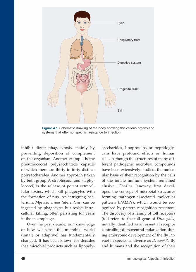
46 Immunological Aspects of Infection
inhibit direct phagocytosis, mainly by preventing deposition of complement on the organism. Another example is the pneumococcal polysaccharide capsule of which there are thirty to forty distinct polysaccharides. Another approach (taken by both group A streptococci and staphy-lococci) is the release of potent extracel-lular toxins, which kill phagocytes with the formation of pus. An intriguing bac-terium, Mycobacterium tuberculosis, can be ingested by phagocytes but resists intra-cellular killing, often persisting for years in the macrophage.
Over the past decade, our knowledge of how we sense the microbial world (innate or adaptive) has fundamentally changed. It has been known for decades that microbial products such as lipopoly-
saccharides, lipoproteins or peptidogly-cans have profound effects on human cells. Although the structures of many dif-ferent pathogenic microbial compounds have been extensively studied, the molec-ular basis of their recognition by the cells of the innate immune system remained elusive. Charles Janeway first devel-oped the concept of microbial structures forming pathogen-associated molecular patterns (PAMPs), which would be rec-ognized by pattern recognition receptors. The discovery of a family of toll receptors (toll refers to the toll gene of Drosophila, initially identifi ed as an essential receptor controlling dorsoventral polarization dur-ing embryonic development of the fl y lar-vae) in species as diverse as Drosophila fl y and humans and the recognition of their
Figure 4.1 Schematic drawing of the body showing the various organs and systems that offer nonspecifi c resistance to infection.
Eyes
Respiratory tract
Digestive system
Urogenital tract
Skin
Immunological Aspects of Infection 47
role in distinguishing molecular patterns that are common to microorganisms led to a renewed appreciation of the innate immune system.
From these humble beginnings, the fi eld of mammalian toll-like receptors (TLR) quickly evolved as a crucial sys-tem for alerting the host to the presence of numerous infectious agents. Infectious microbes display certain molecular pat-terns that are necessary for microbial virulence (see Table 4.1). Many of these molecular patterns such as lipopolysaccha-ride in the outer membranes of gram-nega-tive bacteria seem to be particularly potent activators of mammalian cells. The mam-malian receptors responsible for recogni-tion of PAMPs are called pattern recognition receptors. The failure of the immune system to recognize a pathogen’s PAMP could lead to a delay or blunting of the immune response, resulting in unchecked invasion by the microbe.
Perhaps the best example of an incom-plete recognition by the TLR system is a gram-negative bacterial infection in the C3H/HeJ mouse. As few as two colony-forming units of Salmonella typhimurium can kill this mouse. Further exploration of this extraordinary virulence revealed that the mouse harbored a point mutation (P712H) in the TLR4, which results in defective sig-nal transduction in response to LPS and a heightened susceptibility to gram-negative infections.
The family of TLRs is a highly special-ized system that can identify a number of microbial and endogenous ligands and acti-vate the immune system to respond. The body must be able to respond differently to various challenges. Thus, the specifi city in the immune response via TLRs is becoming increasingly complex. Currently, probes of the TLR family have been described, and it
is expected that many more receptors will be discovered in the future.
ADAPTIVE IMMUNITY
A specifi c immune response to invad-ing microbes is conveniently divided into humoral and cellular immunity. The importance of each arm of the specifi c response varies from infection to infection. Experimental animal models and naturally occurring immunodefi ciency states clearly demonstrate that certain components of the immune response are crucial for con-trolling a particular infection.
For example, individuals with anti-body defi ciencies are particularly prone to repeated infections with pyrogenic bacte-ria. Yet, replacement therapy with immu-noglobulin greatly reduces the number of infections. Interestingly, these individu-als can mount a normal response to most viruses (varicella, measles, mumps, etc.). Yet the absence of mucosal antibody does make them susceptible to some enterovi-ruses.
Although it is clear that innate immu-nity is the fi rst line of defense against invading organisms, the TLRs are also playing a role in adaptive immunity, and the dendritic cell (DC) appears to be play-ing a key role in linking the innate and adaptive immune responses. As immature cells, they are present in the peripheral tissues. However, with the appearance of invading organisms, DCs recognize these pathogens through their TLRs. Fortunately, they express the full repertoire of TLRs. After activation of the TLRs, the DCs are transformed into more mature cells with a high expression of major histocompatibil-ity complex (MHC) and the co-stimulatory molecules CD80 and CD86. The DCs then
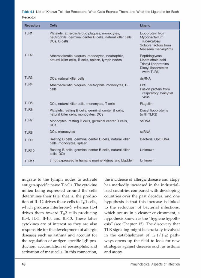
48 Immunological Aspects of Infection
migrate to the lymph nodes to activate antigen-specifi c naïve T cells. The cytokine milieu being expressed around the cells determines their fate; that is, the produc-tion of IL-12 drives these cells to TH1 cells, which produce interferon-δ, whereas IL-4 drives them toward TH2 cells producing IL-4, IL-5, Il-10, and IL-13. These latter cytokines are of interest as they are also responsible for the development of allergic diseases such as asthma and account for the regulation of antigen-specifi c IgE pro-duction, accumulation of eosinophils, and activation of mast cells. In this connection,
the incidence of allergic disease and atopy has markedly increased in the industrial-ized countries compared with developing countries over the past decades, and one hypothesis is that this increase is linked to the reduction of bacterial infections, which occurs in a cleaner environment, a hypothesis known as the “hygiene hypoth-esis” (see Chapter 15). The discovery that TLR signaling might be crucially involved in the establishment of TH1/TH2 path-ways opens up the fi eld to look for new strategies against diseases such as asthma and atopy.
Table 4.1 List of Known Toll-like Receptors, What Cells Express Them, and What the Ligand Is for Each
Receptor
Receptors Cells Ligand
TLR1 Platelets, atherosclerotic plaques, monocytes, neutrophils, germinal center B cells, natural killer cells, DCs, B cells
Lipoprotein fromMycobacterium
tuberculosisSoluble factors from Neisseria meningitidis
TLR2 Atherosclerotic plaques, monocytes, neutrophils, natural killer cells, B cells, spleen, lymph nodes
PeptidoglycanLipoteichoic acidTriacyl lipoproteinsDiacyl lipoproteins
(with TLR6)
TLR3 DCs, natural killer cells dsRNA
TLR4 Atherosclerotic plaques, neutrophils, monocytes, B cells
LPSFusion protein from
respiratory syncytial virus
TLR5 DCs, natural killer cells, monocytes, T cells Flagellin
TLR6 Platelets, resting B cells, germinal center B cells, natural killer cells, monocytes, DCs
Diacyl lipoproteins (with TLR2)
TLR7 Monocytes, resting B cells, germinal center B cells, DCs
ssRNA
TLR8 DCs, monocytes ssRNA
TLR9 Resting B cells, germinal center B cells, natural killer cells, monocytes, spleen
Bacterial CpG DNA
TLR10 Resting B cells, germinal center B cells, natural killer cells, DCs
Unknown
TLR11 ? not expressed in humans murine kidney and bladder Unknown
Immunological Aspects of Infection 49
In summary, the fi elds of both innate and adaptive immunity have experienced a new and rapid growth in interest in the past eight to ten years. Concomitant with this growth, the number of genomes that have been sequenced is expanding, thus enabling researchers to identify and to characterize the receptors and adaptors involved in the recognition.
Although much of the work over the past years has focused on defi ning ligands for the different TLRs, the molecular basis for this recognition is not known for a sin-gle ligand. Furthermore, the characteriza-tion of TLR-dependent signaling for the instruction of adaptive immune responses has just started to be explored. Because TLRs play such a crucial role in innate and adaptive immune responses to distinct vir-ulence factors, the development of selec-tive inhibitors/activators may be a worth-while endeavor to help manage a number of infectious and immunologic diseases.
BACTERIAL INFECTION
The immune system responds to bacterial infections in two major ways. First, it may respond to soluble products of the cell such as toxins or released structural antigens like LPS of a given gram-negative bacterial cell. Most bacterial antigens are T-cell depen-dent and require helper T cells for initia-tion of the immune response. Yet certain cell antigens, such as the pneumococcal polysaccharides, are T-cell independent. They are large-molecular-weight mol-ecules, and in children, antibody response to these antigens may take four to six years. Thus, younger children are susceptible to these infections.
An interesting sidelight to protection against these infections can be seen in
breast-fed infants who are less susceptible to infection than non-breast-fed babies. It now appears that it is not polysaccharides or antibodies that are responsible for this protection but rather a multimeric form of lactoalbumin (present in high concentra-tions in human breast milk (see “Suggested Reading”). A broader approach to protec-tion has been the production of pneumo-coccal polysaccharide vaccines specially designed to induce antibody induction in the young child.
In the following discussion, strepto-cocci, particularly S. pyogenes, are used as the example of a bacterial infection, but many other organisms produce a similar response. Streptococcal antigens include specifi c toxins such as streptolysins O and S that lyse blood and tissue cells and pyro-genic exotoxins, which act as superanti-gens to overstimulate the host responses. There are also specifi c enzymes such as hyaluronidase and streptokinase, which help promote the spread of infecting strep-tococcus. Perhaps most important is the M protein (Figure 4.2), a cell surface antigen of the group A streptococcus that allows the bacteria to evade immune defenses (especially neutrophils and complement). One way in which M protein functions is to bind host factor H, which prevents comple-ment C3 from depositing on the streptococ-cal surface. Since effi cient phagocytosis by neutrophils requires interaction with its C3 receptor, factor H prevents this interaction.
Antibodies to streptococcal antigens other than M protein are slow to appear, and most likely do not play a role in lim-iting the infection. However, antibodies to streptolysin O and deoxyribonuclease B have become important clinical tools to determine whether a given individual has had a recent streptococcal infection. This is partially true if a blood sample drawn
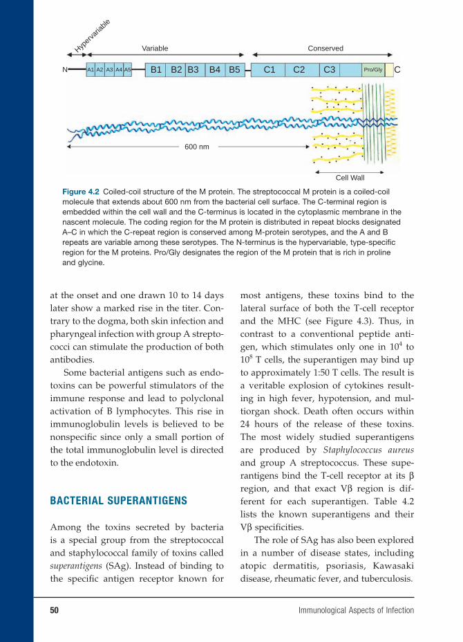
50 Immunological Aspects of Infection
at the onset and one drawn 10 to 14 days later show a marked rise in the titer. Con-trary to the dogma, both skin infection and pharyngeal infection with group A strepto-cocci can stimulate the production of both antibodies.
Some bacterial antigens such as endo-toxins can be powerful stimulators of the immune response and lead to polyclonal activation of B lymphocytes. This rise in immunoglobulin levels is believed to be nonspecifi c since only a small portion of the total immunoglobulin level is directed to the endotoxin.
BACTERIAL SUPERANTIGENS
Among the toxins secreted by bacteria is a special group from the streptococcal and staphylococcal family of toxins called superantigens (SAg). Instead of binding to the specifi c antigen receptor known for
most antigens, these toxins bind to the lateral surface of both the T-cell receptor and the MHC (see Figure 4.3). Thus, in contrast to a conventional peptide anti-gen, which stimulates only one in 104 to 108 T cells, the superantigen may bind up to approximately 1:50 T cells. The result is a veritable explosion of cytokines result-ing in high fever, hypotension, and mul-tiorgan shock. Death often occurs within 24 hours of the release of these toxins. The most widely studied superantigens are produced by Staphylococcus aureus and group A streptococcus. These supe-rantigens bind the T-cell receptor at its β region, and that exact Vβ region is dif-ferent for each superantigen. Table 4.2 lists the known superantigens and their Vβ specifi cities.
The role of SAg has also been explored in a number of disease states, including atopic dermatitis, psoriasis, Kawasaki disease, rheumatic fever, and tuberculosis.
Variable
600 nm
Cell Wall
N A1 A2 A3 A4 A5 B1 B2 B3 B4 B5 C1 C2 C3 CPro/Gly
ConservedHyper
varia
ble
Figure 4.2 Coiled-coil structure of the M protein. The streptococcal M protein is a coiled-coil molecule that extends about 600 nm from the bacterial cell surface. The C-terminal region is embedded within the cell wall and the C-terminus is located in the cytoplasmic membrane in the nascent molecule. The coding region for the M protein is distributed in repeat blocks designated A–C in which the C-repeat region is conserved among M-protein serotypes, and the A and B repeats are variable among these serotypes. The N-terminus is the hypervariable, type-specifi c region for the M proteins. Pro/Gly designates the region of the M protein that is rich in proline and glycine.

Immunological Aspects of Infection 51
Peptide
CβCα
Vα Vβ
TLymphocyte
MHC Class IIMHC Class II
AntigenPresenting
Cell
AntigenPresenting
Cell
CβCα
Vα Vβ
TLymphocyte
Superantigen
Figure 4.3 Binding of conventional (left) and superantigen (right) antigens to the T-cell receptor and antigen-presenting cell. Note that the conventional antigen binding is associated with the T-cell receptor and the major histocompatibility complex (MHC) directly and only a small proportion of T cells are activated. In contrast, the superantigen binds to the lateral sides of the T-cell receptor and MHC molecule, thereby triggering a much larger group of activated T cells (up to 25 percent of the cells).
BACTERIAL EVASION OF IMMUNE
DEFENSE
There are several ways in which bacteria can survive in the host by evasion of the immune defenses. These mechanisms will be briefl y summarized here, but the list of evasive mechanisms used by each bacterial species may be much longer in nature.
Capsules play an important role for long-term survival of pathogens. For example, group A streptococci have a hyaluronic acid capsule that is identical to hyaluronic acid in humans. While antibod-ies can be obtained following immuniza-tion with the streptococcal hyaluronic acid, they are nonprecipitating antibodies and are not effective in eliminating the organ-ism. Polysaccharide antigens of both the pneumococcus and the meningococcus capsules can inhibit phagocytosis of the
organism and mucoid secretions of these polysaccharides can block the activation of the alternate pathway of complement.
Antigenic variation or drift is another mechanism whereby bacteria evade the immune system. The M protein (the most important virulence factor of the group A streptococcus) has been shown to exhibit antigen variation in the environment, and new M protein molecules appear regularly in human isolates of group A streptococci.
Another example is the relapsing fever by Borrelia. During the fi rst episode, anti-bodies kill the bacteria and the fever sub-sides. However, some antigenic variants of the bacteria persist, and after fi ve to seven days these new variants can cause a relapse in the patient with fever reap-pearing. Other examples of evasion of the immune response are bacteria such as N. gonorrhea, N. meningitidis, Haemophilus
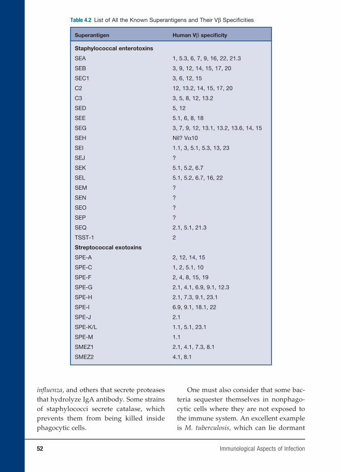
52 Immunological Aspects of Infection
Table 4.2 List of All the Known Superantigens and Their Vβ Specifi cities
Superantigen Human Vβ specifi city
Staphylococcal enterotoxins
SEA 1, 5.3, 6, 7, 9, 16, 22, 21.3
SEB 3, 9, 12, 14, 15, 17, 20
SEC1 3, 6, 12, 15
C2 12, 13.2, 14, 15, 17, 20
C3 3, 5, 8, 12, 13.2
SED 5, 12
SEE 5.1, 6, 8, 18
SEG 3, 7, 9, 12, 13.1, 13.2, 13.6, 14, 15
SEH Nil? Vα10
SEI 1.1, 3, 5.1, 5.3, 13, 23
SEJ ?
SEK 5.1, 5.2, 6.7
SEL 5.1, 5.2, 6.7, 16, 22
SEM ?
SEN ?
SEO ?
SEP ?
SEQ 2.1, 5.1, 21.3
TSST-1 2
Streptococcal exotoxins
SPE-A 2, 12, 14, 15
SPE-C 1, 2, 5.1, 10
SPE-F 2, 4, 8, 15, 19
SPE-G 2.1, 4.1, 6.9, 9.1, 12.3
SPE-H 2.1, 7.3, 9.1, 23.1
SPE-I 6.9, 9.1, 18.1, 22
SPE-J 2.1
SPE-K/L 1.1, 5.1, 23.1
SPE-M 1.1
SMEZ1 2.1, 4.1, 7.3, 8.1
SMEZ2 4.1, 8.1
infl uenza, and others that secrete proteases that hydrolyze IgA antibody. Some strains of staphylococci secrete catalase, which prevents them from being killed inside phagocytic cells.
One must also consider that some bac-teria sequester themselves in nonphago-cytic cells where they are not exposed to the immune system. An excellent example is M. tuberculosis, which can lie dormant
Immunological Aspects of Infection 53
for years inside a granulomatous, case-ous lesion called the Ghon complex. Once ground up, live M. tuberculosis organism can be extracted from this complex.
Finally, a number of organisms display antigens on their surface that are cross-reactive with human antigens. This could result in an enhanced immune response to host tissue antigens or a diminished response secondary to similarities between bacterial and self-antigens.
VIRAL INFECTIONS
The clinical spectrum of viral disease is wide, and because there is such variation, we will use the template of herpes viruses as an example. In general, viral infections are self-limited and usually produce long-term immunity, and secondary attacks by the same virus are uncommon. Of the large number of herpes viruses, only eight infect humans, and each will be discussed in some detail. However, two general features of pathogens of these viruses are important. First, there must be close contact between infected and noninfected individuals for transmission to occur and no intermediate host is involved. Second, after the primary infection, herpes virus will persist in the host for life.
To eliminate virions from entering noninfected cells and to eliminate virus-infected cells, two major pathways of the immune response are initiated. The humoral response is primarily directed against virions, while the T-cell response is primarily directed against infected cells. The humoral response may directly neutralize the virus, but complement-dependent enhancement of viral phago-cytosis or complement lysis of virus also may occur.
Epstein-Barr Infection
Infectious mononucleosis is caused by the Epstein-Barr virus (EBV), and in devel-oping countries, 99 percent of children demonstrate antibodies to the virus by age 3. In developed countries, the age is much later and clinical infection usually occurs in the age group of 15 to 25 years. The virus is excreted in the oropharyngeal fl uids for some months after the infection and is responsible for person-to-person transmission.
Hoagland observed that a female col-lege student traveling from New York to Boston met several different male students on the train and exchanged kisses with all of them. All of these contacts came down with infectious mononucleosis, which is often referred to as the “kissing disease.”
The pattern of antibody response to different EBV antigens helps distinguish acute or subclinical infections from postin-fections with the virus. IgM antibodies to viral capsid antigen (VCA) appear early in the course of the infection, but at the time of clinical symptoms, IgG levels to VCA are quite high. Thus, paired sera at the time of clinical infection do not help diagnose the disease. In contrast, antibod-ies to EB nuclear antigen appear months after the infection. Antibodies to early anti-gen appear in about 70 percent of primary infections and are usually indicative of an acute infection.
The clinical signs of fever, such as enlarged cervical lymph nodes, and red-dened pharynx with whitish exudates (also seen with other viruses) are often mistaken for group A streptococcal pharyngitis. Thus, culture and antibody tests are used to distinguish between the two.
EBV has one unique feature compared with other viruses in that it produces
54 Immunological Aspects of Infection
disease by infecting and transforming B lymphocytes via the CD21 molecule on the surface of B cells. These infected cells multiply like tumor cells, and up to half of the lymphoid cells from the tonsils of EBV-infected patients may be transformed.
The infection is stopped by a T-cell response that kills virus-infected cells and a humoral response that neutralizes free virions. The characteristic “atypical lym-phocytes” seen in this disease are CD8+ cytotoxic T cells that kill EBV-infected B cells.
Most individuals handle EBV infection quite well. However, rare individuals have a specifi c defect that fails to handle EBV infections and usually die of the disease. More important is the failure to contain EBV infections secondary to immunosup-pressive therapy. For example, the x-linked lymphoproliferative syndrome that affects males from age 6 months to 20 years is in this category. These patients die of lymphomas, aplastic anemia, or immunodefi ciency.
Immunosuppressive therapy may result in EBV reactivation, and about 1 to 10 percent of certain transplants may be com-plicated by EBV-induced lymphoprolifera-tive disease. Similar results are also seen in patients infected with the HIV virus. One of the most interesting effects of EBV infec-tion and transformation has been the occur-rence of Burkitt’s lymphoma. It is endemic in certain African countries (especially in regions of mosquito-borne disease), where it represents 90 percent of childhood can-cers in contrast to 3 percent in developed countries. The link between EBV and Burkitt’s lymphoma was established when it was found that EBV antigens and genome were present in the tumor cells. Since the incidence of Burkitt’s lymphoma follows the same belt as the chronic malaria belt of the region, it has been suggested that the
chronic malaria of these children induces EBV-infected lymphoproliferation, leading to chromosomal translocation in the long arms of chromosomes 8 and 14 and even-tually to the activation and translocation of the c-myc oncogene to the active region of the cellular genome (see Figure 4.4).
Viruses have evolved many mecha-nisms to evade the immune response, but one of the key elements in these evasive mechanisms has been their ability to induce a latent status within the cells of the host. Thus, all herpes viruses can remain latent, and no viral antigen is expressed. When there is a change in the host-virus equilib-rium secondary to certain other infections, metabolic disturbances, immunosuppres-sion, or aging, only then does the virus reactivate and cause disease. Examples are herpes simplex in the trigeminal gan-glia, which causes recurrent “cold sores,” or herpes zoster in a dorsal root ganglion, which causes “shingles.”
A second example is antigenic variation or drift in certain viruses. The best example is infl uenza A, an RNA virus surrounded by a lipid envelope into which two impor-tant proteins (hemagglutinin and neuro-minidase) are inserted. The virus evades the neutralizing antibodies to this virus by constantly modifying the structure of these two proteins by either antigenic drift or antigenic shift. In the former, the change is minor and accounts for minor epidemics of fl u during each winter. Antigenic shift is a major change in the structure of those pro-teins, which can cause a major pandemic of devastating proportions. Viewed in this light, the infl uenza vaccine currently in use is generally protective against common fl u epidemics in the world but would not be protective against a major antigenic shift.
A third mechanism is viral persistence in which the virus is not cleared and a chronic
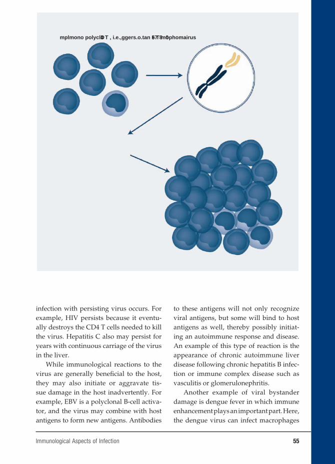
56 Immunological Aspects of Infection
via Fc receptors, and its capacity to enter the target cell is enhanced if it is bound to IgG antibodies. Thus, the fi rst infection with a specifi c dengue virus type elicits antibodies to that specifi c virus. How-ever, cross immunity to the other dengue serotypes is fl eeting and lasts only a few weeks. When the individual is exposed to a second dengue virus serotype, the pre-existing antibody to the fi rst type binds to it but does not neutralize the virus. The antibody-enhanced second virus enters the macrophage, setting off a veritable explo-sion of proteases, lymphokines comple-ment activation, coagulation cascade, and vascular permeability factors. The result is a much more severe disease called dengue hemorrhagic fever, whose mortality rate in areas with poor medical facilities and treat-ment may be as high as 50 percent.
FUNGAL INFECTION
Fungi cause many diseases, most of which are adequately handled by the immune system of the normal host. However, the increased use of immunosuppressive drugs – the immunosuppression secondary to HIV infection – has raised our awareness of these infections. For example, Candida albicans can cause superfi cial infections in the normal host and is found throughout the intestinal tract and commonly in the vagina. The organism is usually kept under control by the bacterial fl ora, but changes in these fl ora secondary to overuse of anti-biotics or changes in hormone balance will favor a chronic superfi cial infection. All of this is exacerbated in the immunocompro-mised host.
Another organism that is receiving wide attention is the fungus Pneumocystis carinii (now called Pneumocystis jiroveci),
which has a commensal relationship with the lung in the normal host. However, the sudden onset of pneumonia with this organism secondary to HIV infection in a child or adult is often the fi rst sign of an underlying suppressed immune system caused by HIV.
In contrast to the superfi cial infections of fungi, systemic infections in the immuno-compromised host have a higher mortality rate. This category includes Histoplasma cap-sulatum (pulmonary infection), Coccidiodes immitis (acute pneumonitis), and Crypto-coccus neoformas (meningitis, lung lesions). Such systemic infections may occur in normal individuals, and the cell-mediated immune response is the more important fac-tor in control or prevention of them. How-ever, all of these fungal infections (superfi -cial and systemic) are more likely to occur in immunosuppressed individuals, especially those in which the cell-mediated immune response is compromised.
There are several plausible outcomes to fungal infections. As stated before, an active cellular immune response plus topi-cal antifungal drugs usually handles most superfi cial infections. Systemic infections, especially in the immunocompromised host, carry a high mortality rate, and even the use of newer prophylactic and thera-peutic agents only partially decreases the mortality.
As with viruses, there is a third possible outcome. If the fungal infection is not elim-inated or causes persistent infection, then the host response may trigger a hypersen-sitivity reaction. As an example, Aspergil-lus fumigatus infections can persist as an aspergilloma in preexisting lung cavities secondary to tuberculosis or bronchiecta-sis following childhood pertussis (whoop-ing cough). Allergic bronchopulmonary aspergillosis may occur and is due to
Immunological Aspects of Infection 57
IgE-mediated hypersensitivity to the asper-gillus antigens. Clinically, the condition presents as recurrent episodes of increased wheezing and coughing, fever, and pleu-ritic pain similar to that seen in asthmatics. Another example is farmer’s lung in which antigen-antibody complexes of Micropoly-spora faeni cause a hypersensitivity reaction on the part of the host. The name farmer’s lung indicates that this particular fungus is found in moldy hay.
PARASITIC INFECTION
Protozoa are a diverse group of parasites, but malaria, leishmaniasis, and trypanoso-miasis globally account for most of the prob-lems encountered in parasitic diseases. The balance between host and parasite is two-fold. The parasite may be too virulent for the host or may evade the immune surveil-lance and thus kills the host. Conversely, the immune response may be vigorous and kill the parasite, thereby jeopardizing its survival. Thus, the survival of any parasite depends on a balance between induction of immunity and escape from surveillance.
Malaria
The worldwide incidence of malaria is esti-mated at 300 million–500 million people, and at least 1 million die each year of the disease, mostly of cerebral malaria and usually young children. Cerebral malaria is usually associated with infection with Plasmodium falciparum and not Plasmodium vivax. Patients react to protozoal infec-tion with activation of macrophages and monocytes with the release of cytokines TNF, IL-1, and IL-6. Clinically, they pro-duce fever, leukocytosis, and acute phase reactants.
Although most protozoa stimulate the production of IgG and IgM antibodies, these antibodies are probably not protec-tive, and thus vaccines have not yet been successful in the control or prevention of malaria. Also in the case of malaria, pro-tozoa invade erythrocytes and hepatocytes and thus are hidden from the immune response. Clinically, many of the signs and symptoms of these patients are related to the destruction of red blood cells and hepa-tocytes; therefore, anemia, jaundice, sple-nomegaly, hypostasis, hypotension, tender hepatomegaly, and biphasic fever are hall-marks of the disease.
Interestingly, there have been several mutations in the host that help provide resistance to malarial infection. Most strik-ing has been the appearance of the het-erozygous sickle cell trait (Hbas), which confers a survival advantage in endemic disease. Second, the absence of the red cell Duffy antigen (receptor for plasmodium vivax) is quite protective. Finally, the pres-ence of HLA-B53 in individuals is associ-ated with resistance to the disease.
Evasion mechanisms of protozoa fall into three major categories. The fi rst is entrance of the organism into the host cell, where it avoids immune surveillance. One example is malaria, as noted previously. Others include toxoplasma, leishmania, and Trypanosoma cruzi, which enter and can grow inside macrophages. For example, leishmania binds C3 avidly and thus serves as a ligand for the CR3 receptor on macro-phages. If one uses monoclonal antibodies to the CR3 receptor, this inhibits the uptake of the parasite into macrophage. Another approach is used by toxoplasma in which they prevent fusion of phagocytic vacuoles containing the parasite with lysosomes and thus are not destroyed. Finally, try-panosomes need activated macrophages for
58 Immunological Aspects of Infection
killing; thus they are resistant to intracellu-lar killing in nonactivated macrophages.
A second mechanism for evasion is anti-genic variation. Trypanosoma brucei is an excellent example of this. In this scenario, the trypanosomes are initially destroyed by host antibody. However, the organism resurfaces in the body with a different set of antigens or glycoproteins. The process continues, and the parasite possesses a number of genes that code for these anti-gens and can vary the genes used. Even-tually, the parasite succeeds and avoids host elimination. This type of variation is known as phenotypic variation and dif-fers from the genotypic variation seen in infl uenza epidemics.
As in bacteria, protozoa can also sup-press the immune response. Malaria and leishmania organisms release soluble anti-gens that nonspecifi cally suppress the im-mune response by acting on lymphocytes or the reticuloendothelial system. Several parasites undergo development stages that are resistant to complement-mediated lysis. Finally, leishmania can down-regu-late expression of MHC class II expression of parasitized macrophages, which reduces the effectiveness of CD8+ T cells.
In summary, protozoa have developed a wide variety of techniques to evade the immune system. This makes it extremely diffi cult both to eliminate these protozoa and to produce vaccines that are effective against them. Thus, the fi eld is wide open to new and innovative approaches to elimi-nate this class of organisms.
BIBLIOGRAPHY
REVIEWSAlcami A, Koszinowski UH. Viral mecha-
nisms of immune evasion. Immunol Today. 2000;21(9):447–455.
Fischetti VA. Streptococcal M protein. Sci Am. 1991;264(6):58–65.
Heine H, Lien E. Toll-like receptors and their function in innate and adaptive immunity. Int Arch Allergy Immunol. 2003;130:180–192.
Lavoie PM, Thibodeau J, Erard F, Sekaly RP. Understanding the mechanism of action of bacterial superantigens from a decade of research. Immunol Rev. 1999;168:257–269.
Wetzler LM. 2003. The role of Toll-like recep-tor 2 in microbial disease and immunity. Vaccine. 2003;21 (suppl 2):55–60.
SUGGESTED READINGBunikowski R, Mielke ME, Skarabis H, et
al. Evidence for a disease-promoting effect of Staphylococcus aureus-derived exotoxins in atopic dermatitis. J Allergy Clin Immunol. 2000;105(4):814–819.
Davison SC, Allen MH, Mallon E, Barker JN. Contrasting patterns of streptococcal superantigen-induced T-cell prolifera-tion in guttate vs. chronic plaque psoria-sis. Br J Dermatol. 2001;145(2): 245–251.
Hoagland RS. The transmission of infec-tious mononucleosis. Am J Med Sci. 1955;229:262–266.
Hoshino K, Takeuchi O, Kawai T, et al. Cutting edge: toll-like receptor 4 (TLR4)-defi cient mice are hyporesponsive to lipopolysaccharide: evidence for TLR4 as the Lps gene product. J Immunol. 1999;162:3749–3752.
Janeway CA Jr. Approaching the asymp-tote? Evolution and revolution in immunology. Cold Spring Harb Symp Quant Biol. 1989;54:1–13.
Kaisho T, Akira S. 2000. Critical roles of Toll-like receptors in host defense. Crit Rev Immunol. 2000;20:393–405.
Meissner HC, Leung DY. Superantigens, conventional antigens and the etiology

Immunological Aspects of Infection 59
of Kawasaki syndrome. Pediatr Infect Dis J. 2000;19(2):91–94.
Smoot LM, McCormick JK, Smoot JC, et al. Characterization of two novel pyro-genic toxin superantigens made by an acute rheumatic fever clone of Strep-tococcus pyogenes associated with mul-tiple disease outbreaks. Infect Immun. 2002;70(12):7095–7104.
Ohmen JD, Barnes PF, Grisso CL, Bloom BR, Modlin RL. Evidence for a
superantigen in human tuberculosis. Immunity. 1994;1(1):35–43.
Poltorak A, He X, Smirnova I, et al. Defec-tive LPS signaling in C3H/HeJ and C57BL/10ScCr mice: mutations in Tlr4 gene. Science. 1988;282:2085–2088.
Visvanathan K, Charles A, Bannan J, Pugach P, Kashfi K, Zabriskie JB. Inhi-bition of bacterial superantigens by peptides and antibodies. Infect Immun. 2001;69:875–884.

INTRODUCTION
The principal function of the immune system is to prevent microbial infection. Therefore, disorders resulting in impaired function of the immune system (immuno-defi ciency) result in increased susceptibil-ity to infection. Immunodefi ciency can arise from an intrinsic defect of a component of the immune system (primary immunode-fi ciency, or PID). Alternatively, immuno-defi ciency may be secondary to another pathological condition, which adversely affects immune function (Table 5.1). Both primary and secondary immunodefi cien-cies result in increased susceptibility to infection. The precise pattern of infection depends on the specifi c component of the immune system that is affected. Most PIDs are caused by defects in single genes and are hence heritable. Others may represent the consequence of an interaction between the genetic phenotype and an environmen-tal infl uence, like viral infections. Primary immunodefi ciencies are rare and based on information from national registers; these diseases are estimated to occur between 1 in 2,000 to 1 in 10,000 live births. In con-trast, secondary immunodefi ciencies are more commonly seen in clinical practice. (See Table 5.1 for examples of secondary immunodefi ciency.)
From a clinical perspective, immuno-defi ciencies can be classifi ed into eight categories (Table 5.2). Each category has a
characteristic pattern of clinical presenta-tion (Table 5.3), which will be elaborated on later.
DEFECTS IN ANATOMICAL
OR PHYSIOLOGICAL BARRIERS
TO INFECTION
One of the commonest predisposing causes of infection is a defect in an anatomical or physiological barrier to infection. Intact epithelial membranes, especially strati-fi ed squamous epithelial surfaces such as the skin, constitute an extremely effective barrier to infection. Thus, integumentary damage caused by burns, eczema, and trauma (including surgery), predisposes to infection. Skull fractures, particularly damage of the cribriform plate, may result in recurrent episodes of pyogenic menin-gitis. The existence of sinus tracts between deeper tissues and the skin surface or alternatively, the presence of foreign bod-ies or avascular areas (e.g., within bone) predisposes to infection. Obstruction to the drainage of hollow tubes and viscera also predisposes to infection, for example, obstruction of the biliary tract, urinary tract, or bronchi. Impaired vascular perfusion of the tissues due to edema and angiopathy (including microvascular changes follow-ing diabetes mellitus) also predisposes to infection. Alteration of the normal com-mensal fl ora by broad-spectrum antibiotic
5. Immunological Aspects of Immunodeficiency Diseases
Dinakantha S. Kumararatne, M.D.
61
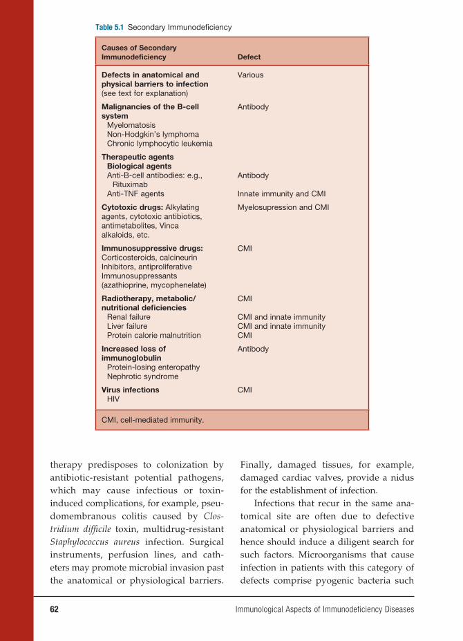
62 Immunological Aspects of Immunodeficiency Diseases
therapy predisposes to colonization by antibiotic-resistant potential pathogens, which may cause infectious or toxin-induced complications, for example, pseu-domembranous colitis caused by Clos-tridium diffi cile toxin, multidrug-resistant Staphylococcus aureus infection. Surgical instruments, perfusion lines, and cath-eters may promote microbial invasion past the anatomical or physiological barriers.
Finally, damaged tissues, for example, damaged cardiac valves, provide a nidus for the establishment of infection.
Infections that recur in the same ana-tomical site are often due to defective anatomical or physiological barriers and hence should induce a diligent search for such factors. Microorganisms that cause infection in patients with this category of defects comprise pyogenic bacteria such
Table 5.1 Secondary Immunodefi ciency
Causes of Secondary Immunodefi ciency Defect
Defects in anatomical and physical barriers to infection (see text for explanation)
Various
Malignancies of the B-cell system
MyelomatosisNon-Hodgkin’s lymphomaChronic lymphocytic leukemia
Antibody
Therapeutic agentsBiological agentsAnti-B-cell antibodies: e.g.,
RituximabAnti-TNF agents
Antibody
Innate immunity and CMI
Cytotoxic drugs: Alkylating agents, cytotoxic antibiotics, antimetabolites, Vinca alkaloids, etc.
Myelosupression and CMI
Immunosuppressive drugs:Corticosteroids, calcineurin Inhibitors, antiproliferative Immunosuppressants (azathioprine, mycophenelate)
CMI
Radiotherapy, metabolic/nutritional defi ciencies
Renal failureLiver failureProtein calorie malnutrition
CMI
CMI and innate immunityCMI and innate immunityCMI
Increased loss of immunoglobulin
Protein-losing enteropathyNephrotic syndrome
Antibody
Virus infectionsHIV
CMI
CMI, cell-mediated immunity.
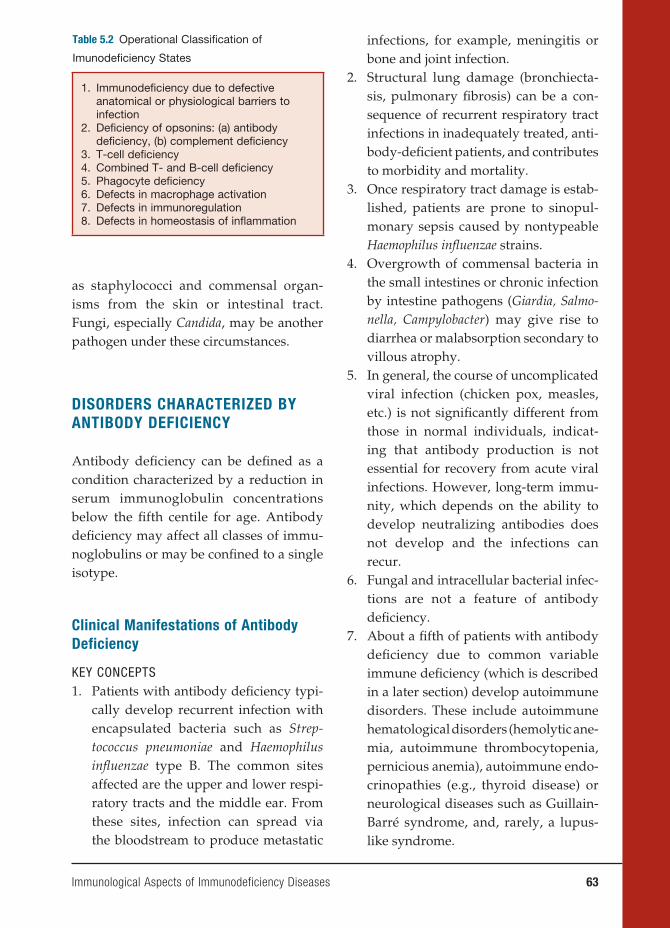
as staphylococci and commensal organ-isms from the skin or intestinal tract. Fungi, especially Candida, may be another pathogen under these circumstances.
DISORDERS CHARACTERIZED BY
ANTIBODY DEFICIENCY
Antibody defi ciency can be defi ned as a condition characterized by a reduction in serum immunoglobulin concentrations below the fi fth centile for age. Antibody defi ciency may affect all classes of immu-noglobulins or may be confi ned to a single isotype.
Clinical Manifestations of Antibody
Deficiency
KEY CONCEPTS1. Patients with antibody defi ciency typi-
cally develop recurrent infection with encapsulated bacteria such as Strep-tococcus pneumoniae and Haemophilus infl uenzae type B. The common sites affected are the upper and lower respi-ratory tracts and the middle ear. From these sites, infection can spread via the bloodstream to produce metastatic
infections, for example, meningitis or bone and joint infection.
2. Structural lung damage (bronchiecta-sis, pulmonary fi brosis) can be a con-sequence of recurrent respiratory tract infections in inadequately treated, anti-body-defi cient patients, and contributes to morbidity and mortality.
3. Once respiratory tract damage is estab-lished, patients are prone to sinopul-monary sepsis caused by nontypeable Haemophilus infl uenzae strains.
4. Overgrowth of commensal bacteria in the small intestines or chronic infection by intestine pathogens (Giardia, Salmo-nella, Campylobacter) may give rise to diarrhea or malabsorption secondary to villous atrophy.
5. In general, the course of uncomplicated viral infection (chicken pox, measles, etc.) is not signifi cantly different from those in normal individuals, indicat-ing that antibody production is not essential for recovery from acute viral infections. However, long-term immu-nity, which depends on the ability to develop neutralizing antibodies does not develop and the infections can recur.
6. Fungal and intracellular bacterial infec-tions are not a feature of antibody defi ciency.
7. About a fi fth of patients with antibody defi ciency due to common variable immune defi ciency (which is described in a later section) develop autoimmune disorders. These include autoimmune hematological disorders (hemolytic ane-mia, autoimmune thrombocytopenia, pernicious anemia), autoimmune endo-crinopathies (e.g., thyroid disease) or neurological diseases such as Guillain-Barré syndrome, and, rarely, a lupus-like syndrome.
Immunological Aspects of Immunodeficiency Diseases 63
Table 5.2 Operational Classifi cation of
Imunodefi ciency States
1. Immunodefi ciency due to defective anatomical or physiological barriers to infection
2. Defi ciency of opsonins: (a) antibody defi ciency, (b) complement defi ciency
3. T-cell defi ciency4. Combined T- and B-cell defi ciency5. Phagocyte defi ciency6. Defects in macrophage activation7. Defects in immunoregulation8. Defects in homeostasis of infl ammation
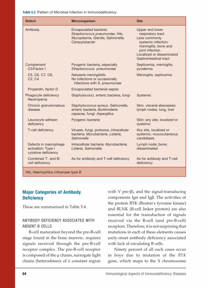
64 Immunological Aspects of Immunodeficiency Diseases
Table 5.3 Pattern of Microbial Infection in Immunodefi ciency
Defect Microorganism Site
Antibody Encapsulated bacteria: Streptococcus pneumoniae, Hib, Mycoplasma, Giardia, Salmonella, Campylobacter
Upper and lower respiratory tract
Less commonly systemic infection: meningitis, bone and joint infection
Localized or disseminated Gastrointestinal tract
ComplementC3/Factor I
Pyogenic bacteria, especially Streptococcus pneumoniae
Septicemia, meningitis, pyoderma
C5, C6, C7, C8,C2, C4
Neisseria meningitidisNo infections or occasionally
infections with S. pneumoniae
Meningitis, septicemia
Properdin, factor D Encapsulated bacterial sepsis
Phagocyte defi ciencyNeutropenia
Staphylococci, enteric bacteria, fungi Systemic
Chronic granulomatous disease
Staphylococcus aureus, Salmonella, enteric bacteria, Burkholderia cepacea, fungi: Aspergillus
Skin, visceral abscesses: lymph nodes, lung, liver
Leucocyte adhesin defi ciency
Pyogenic bacteria Skin; any site, localized or systemic
T-cell defi ciency Viruses, fungi, protozoa, intracellular bacteria: Mycobacteria, Listeria, Salmonella
Any site, localized or systemic; mucocutaneous candidiasis
Defects in macrophage activation: Type I cytokine defi ciency
Intracellular bacteria: Mycobacteria, Listeria, Salmonella
Lymph node; bone; disseminated
Combined T- and B-cell defi ciency
As for antibody and T-cell defi ciency As for antibody and T-cell defi ciency
Hib, Haemophilus infl uenzae type B
Major Categories of Antibody
Deficiency
These are summarized in Table 5.4.
ANTIBODY DEFICIENCY ASSOCIATED WITH ABSENT B CELLS
B-cell maturation beyond the pre-B-cell stage found in the bone marrow, requires signals received through the pre-B-cell receptor complex. The pre-B-cell receptor is composed of the µ chains, surrogate light chains (heterodimers of λ constant region
with V pre-β), and the signal-transducing components Igα and Igβ. The activities of the protein BTK (Bruton’s tyrosine kinase) and BLNK (B-cell linker protein) are also essential for the transduction of signals received via the B-cell (and pre-B-cell) receptors. Therefore, it is not surprising that mutations in each of these elements causes early-onset antibody defi ciency associated with lack of circulating B cells.
Ninety percent of all such cases occur in boys due to mutation of the BTK gene, which maps to the X chromosome
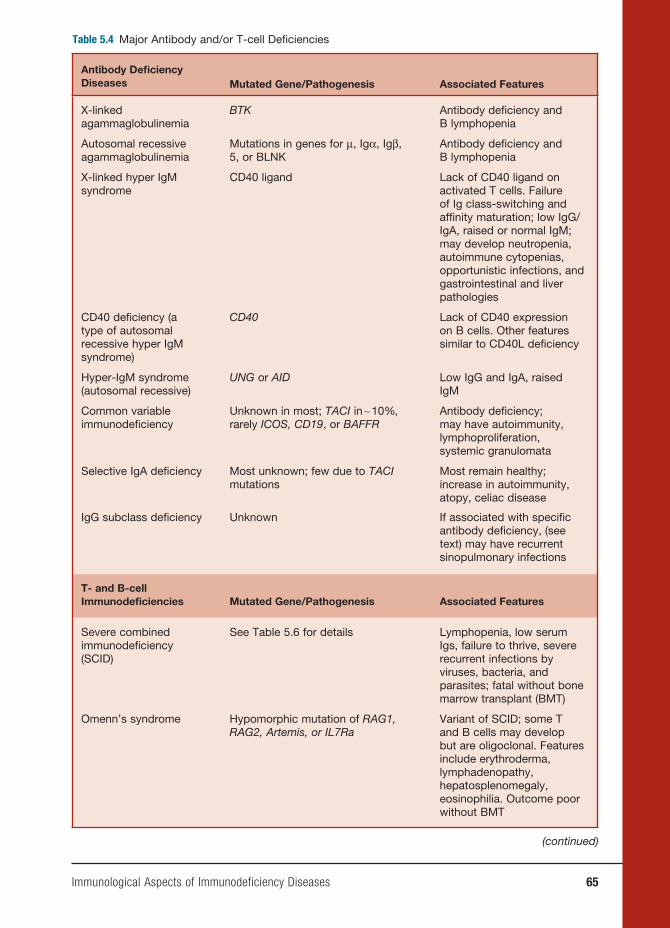
Immunological Aspects of Immunodeficiency Diseases 65
Table 5.4 Major Antibody and/or T-cell Defi ciencies
Antibody Defi ciency Diseases Mutated Gene/Pathogenesis Associated Features
X-linked agammaglobulinemia
BTK Antibody defi ciency andB lymphopenia
Autosomal recessive agammaglobulinemia
Mutations in genes for μ, Igα, Igβ, 5, or BLNK
Antibody defi ciency and B lymphopenia
X-linked hyper IgM syndrome
CD40 ligand Lack of CD40 ligand on activated T cells. Failure of Ig class-switching and affi nity maturation; low IgG/IgA, raised or normal IgM; may develop neutropenia, autoimmune cytopenias, opportunistic infections, and gastrointestinal and liver pathologies
CD40 defi ciency (a type of autosomal recessive hyper IgM syndrome)
CD40 Lack of CD40 expression on B cells. Other features similar to CD40L defi ciency
Hyper-IgM syndrome (autosomal recessive)
UNG or AID Low IgG and IgA, raised IgM
Common variable immunodefi ciency
Unknown in most; TACI in ∼ 10%, rarely ICOS, CD19, or BAFFR
Antibody defi ciency; may have autoimmunity, lymphoproliferation, systemic granulomata
Selective IgA defi ciency Most unknown; few due to TACI mutations
Most remain healthy; increase in autoimmunity, atopy, celiac disease
IgG subclass defi ciency Unknown If associated with specifi c antibody defi ciency, (see text) may have recurrent sinopulmonary infections
T- and B-cell Immunodefi ciencies Mutated Gene/Pathogenesis Associated Features
Severe combined immunodefi ciency (SCID)
See Table 5.6 for details Lymphopenia, low serum Igs, failure to thrive, severe recurrent infections by viruses, bacteria, and parasites; fatal without bone marrow transplant (BMT)
Omenn’s syndrome Hypomorphic mutation of RAG1, RAG2, Artemis, or IL7Ra
Variant of SCID; some T and B cells may develop but are oligoclonal. Features include erythroderma, lymphadenopathy, hepatosplenomegaly, eosinophilia. Outcome poor without BMT
(continued)
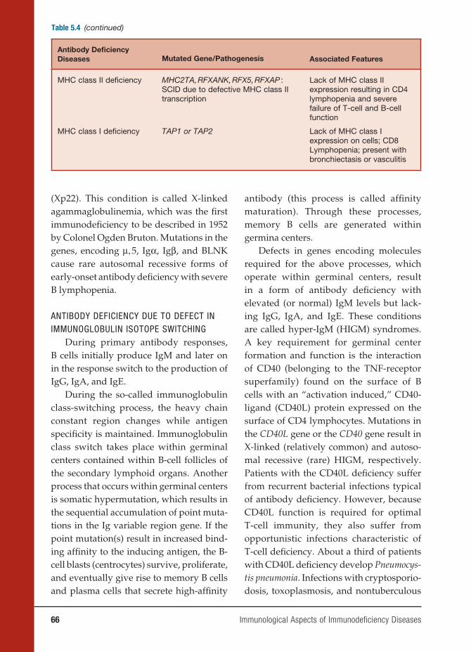
66 Immunological Aspects of Immunodeficiency Diseases
(Xp22). This condition is called X-linked agammaglobulinemia, which was the fi rst immunodefi ciency to be described in 1952 by Colonel Ogden Bruton. Mutations in the genes, encoding μ, 5, Igα, Igβ, and BLNK cause rare autosomal recessive forms of early-onset antibody defi ciency with severe B lymphopenia.
ANTIBODY DEFICIENCY DUE TO DEFECT IN IMMUNOGLOBULIN ISOTOPE SWITCHING
During primary antibody responses, B cells initially produce IgM and later on in the response switch to the production of IgG, IgA, and IgE.
During the so-called immunoglobulin class-switching process, the heavy chain constant region changes while antigen specifi city is maintained. Immunoglobulin class switch takes place within germinal centers contained within B-cell follicles of the secondary lymphoid organs. Another process that occurs within germinal centers is somatic hypermutation, which results in the sequential accumulation of point muta-tions in the Ig variable region gene. If the point mutation(s) result in increased bind-ing affi nity to the inducing antigen, the B-cell blasts (centrocytes) survive, proliferate, and eventually give rise to memory B cells and plasma cells that secrete high-affi nity
antibody (this process is called affi nity maturation). Through these processes, memory B cells are generated within germina centers.
Defects in genes encoding molecules required for the above processes, which operate within germinal centers, result in a form of antibody defi ciency with elevated (or normal) IgM levels but lack-ing IgG, IgA, and IgE. These conditions are called hyper-IgM (HIGM) syndromes. A key requirement for germinal center formation and function is the interaction of CD40 (belonging to the TNF-receptor superfamily) found on the surface of B cells with an “activation induced,” CD40-ligand (CD40L) protein expressed on the surface of CD4 lymphocytes. Mutations in the CD40L gene or the CD40 gene result in X-linked (relatively common) and autoso-mal recessive (rare) HIGM, respectively. Patients with the CD40L defi ciency suffer from recurrent bacterial infections typical of antibody defi ciency. However, because CD40L function is required for optimal T-cell immunity, they also suffer from opportunistic infections characteristic of T-cell defi ciency. About a third of patients with CD40L defi ciency develop Pneumocys-tis pneumonia. Infections with cryptosporio-dosis, toxoplasmosis, and nontuberculous
Table 5.4 (continued)
Antibody Defi ciency Diseases Mutated Gene/Pathogenesis Associated Features
MHC class II defi ciency MHC2TA, RFXANK, RFX5, RFXAP : SCID due to defective MHC class II transcription
Lack of MHC class II expression resulting in CD4 lymphopenia and severe failure of T-cell and B-cell function
MHC class I defi ciency TAP1 or TAP2 Lack of MHC class I expression on cells; CD8 Lymphopenia; present with bronchiectasis or vasculitis
Immunological Aspects of Immunodeficiency Diseases 67
mycobacteria also occur in this condition. These opportunistic infections can be ex-plained on the basis that the interaction of CD40L on activated T cells with CD40 ex-pressed on the surface of macrophages and dendritic cells, which in turn undergo matu-ration and activation, is required for the opti-mum expression of antimicrobial immunity.
A high proportion of CD40L-defi cient patients develop progressive liver dam-age (sclerosing cholangitis), probably the result of cryptosporidial infection of the bile ducts.
Defects in the RNA-editing enzymes, activation-induced cytidine deaminase (AID), and uracil-DNA glycosylase (UNG) result in two further types of HIGM syn-dromes, which are defective class switch-ing and affi nity maturation.
Signaling through CD40, which belongs to the TNF-receptor superfamily, depends on the activation of the inhibitor of κ kinase (IKK) complex, resulting in the induction of NFκB. Hypomorphic mutations of the gamma subunit of the IKK complex, which is called NEMO (NFκB essential modu-lator), impairs NFκB activation. Patients with mutations in NEMO develop a com-plex immunodefi ciency, which includes features of the HIGM syndrome.
COMMON VARIABLE IMMUNE DEFICIENCY Most patients with primary antibody
defi ciency are collected under the heading “common variable immune defi ciency,” which is a condition characterized by low serum IgG and IgA and a variable decrease in IgM and the impaired production of specific antibodies following natural microbial exposure or immunization. The estimated prevalence of CVID is between 1 in 10,000 and 1 in 50,000. Clinically, CVID is dominated by the effects of antibody defi ciency. About 20 percent of patients
with CVID develop autoimmune disor-ders (arthritis, cytopenias, endocrinopa-thies). Some patients with CVID develop noncaseating sarcoidlike granulomata infi ltrating various organs (lungs, liver, spleen, skin). The mechanisms underlying autoimmunity and granulomatous disease in CVID are unknown.
The immunological phenotype of CVID is heterogeneous with documented defects in B-cell survival, generation of B memory cells, and in vitro B- and T-cell activation. About 10 percent of cases of CVID are familial, with a predominance of autosomal dominant or autosomal reces-sive inheritance. CVID or selective IgA defi ciency can affect different members of the same kindred. Recently, a number of gene defects have been identifi ed in CVID patients, accounting for about 10–15 per-cent of the total patient pool.
The commonest defect (in approxi-mately 10% of CVID patients) is a mutation of the gene TACI (transmembrane activator and calcium modulator and cyclophilin ligand interacter). TACI, which belongs to the TNF-receptor superfamily, is a ligand for the cytokines, BAFF (B-cell-activat-ing factor of the TNF family) and APRIL (a proliferation-induced ligand), which induce immunoglobulin class-switch recombination.
Mice defi cient in BAFF or its recep-tor have impaired B-cell development and antibody defi ciency. TACI−/− mice have reduced serum IgA and IgM levels, reduced antibody responses to T-cell-independent antigens, and tend to develop autoimmunity and B lymphoprolifera-tion. Mutations in TACI have been found in CVID patients and their relatives, with selective IgA defi ciency, indicating variable penetrance of this gene defect. In the majority of currently documented
68 Immunological Aspects of Immunodeficiency Diseases
patients, TACI mutations affect only one allele, indicating a dominant negative effect of the mutated gene. A possible explana-tion is that TACI, like other members of the TNF-receptor family, undergoes ligand-independent preassociation and function as multimeric units. Thus, incorporation of a mutated TACI chain in this multimeric complex may disrupt ligand binding or signal-transducing capacity. Recent stud-ies have found that family members of CVID patients possessing heterozygous TACI mutations may have completely nor-mal serum immunoglobulin levels and “in vitro” B-cell function. This leads to the con-clusion that the development of antibody defi ciency in those carrying heterozygous TACI mutations may depend on modifi er genes or environmental factors.
ICOS (inducible co-stimulating recep-tor) is a co-stimulatory T-cell molecule that induces cytokines required for sup-porting class-switch recombination, Ig production and terminal B-cell differentia-tion. ICOS−/− and ICOS ligand−/− mice exhibit defective germinal center formation and antibody production. Mutations in the ICOS gene account for about 1 percent of CVID patients.
A single CVID patient was identifi ed to have a mutation on the gene encoding the BAFF receptor, a fi nding that could be pre-dicted from observations in BAFF−/− mice.
Finally, CD19 deficiency has been described in two families with CVID. CD19 is a B-cell accessory molecule that is required for B-cell activation, proliferation, and hence B-cell development. CD19−/− mice exhibit hypogammaglobulinemia and poor antibody responses to protein antigens.
In the case of the large majority of patients with CVID, the underlying molec-ular defect is unknown.
IgA DEFICIENCYIgA deficiency is characterized by
reduced (<0.07 g/L) or absent serum IgA levels. IgA defi ciency is the commonest form of primary antibody defi ciency and affects about 1 in 700 Caucasians. IgA defi ciency is rare in some racial groups (Japanese, Africans). The majority of IgA-defi cient individuals remain free of infec-tion due to the ability of IgG and IgM to compensate for the lack of IgA. Long-term studies have shown that a small propor-tion of IgA-deficient patients develop recurrent sinopulmonary or gastroin-testinal infections. Most infection-prone IgA-defi cient patients have concomitant IgG2 subclass defi ciency and a selective inability to produce antibodies against bacterial capsular polysaccharides. IgA defi ciency is associated with an increased incidence of atopy, celiac disease, and a range of autoimmune diseases, including arthritis, “lupus-like” syndrome, autoim-mune endocrinopathies, and autoimmune cytopenias.
IgA-defi cient patients with serum lev-els below 0.07 g/L are at the risk of devel-oping anti-IgA antibodies after receiving blood products. Such individuals are at risk of developing anaphylaxis on receiv-ing blood products.
IgA defi ciency and CVID can differ-entially affect members of the same kin-dred. Rarely, IgA defi ciency can precede the development of CVID. Therefore, in some instances, the molecular mechanisms underlying CVID and IgA defi ciency may be identical. TACI defi ciency can cause IgA defi ciency in some family members while others develop CVID. However, the molec-ular basis of IgA defi ciency in most cases is largely unknown.
Genetic analysis of kindred with CVID and IgA defi ciency have highlighted the
Immunological Aspects of Immunodeficiency Diseases 69
existence of susceptibility loci within the MHC region of chromosome 6. The stron-gest linkage lies within the DR/DQ within the MHC class II region.
IgG SUBCLASS DEFICIENCYSerum IgG comprises four subclasses
called IgG1, IgG2, IgG3, and IgG4, respec-tively refl ecting the relative abundance of these isotypes in the serum. IgG sub-class defi ciency is diagnosed on fi nding a reduction in the serum concentration of IgG subclasses, of more than two standard deviations below the mean value for age, despite the total IgG level being normal. In practice, IgG subclass assays are diffi cult to standardize due to the lack of an inter-nationally accepted reference preparation. Furthermore, genetic variations that infl u-ence serum IgG subclass levels exist among different ethnic groups. Hence, age- and population-related normal ranges are not always available.
Some individuals with IgG subclass deficiencies are asymptomatic. Others with IgG subclass defi ciencies are prone to recurrent sinopulmonary infections. Such infection-prone patients exhibit reduced antibody responses to bacterial capsular polysaccharides. Defective anti-polysac-charide antibody responses are most often seen in individuals with IgG2 subclass defi ciency with or without concomitant IgA defi ciency. The molecular mechanisms underlying IgG subclass defi ciencies are unknown.
TREATMENT OF ANTIBODY DEFICIENCYProspective clinical studies have
shown that optimal IgG replacement ther-apy reduces the incidence of sepsis and complications like structural lung dam-age. If replacement therapy is introduced early before organ damage is established,
antibody-defi cient individuals are likely to have a normal life span. Replacement immunoglobulin is prepared from plasma pooled from large cohorts of normal donors. Current practice of prescreening donors for infection and multiple antivi-ral steps employed during the manufac-ture of plasma have eliminated the risk of HIV and hepatitis B and C transmission. Different immunoglobulin products are licensed for administration via the intra-venous or subcutaneous route. Detailed management of a patient’s antibody defi -ciency is discussed in the references on the diagnosis and management of anti-body defi ciency (see list of references at end of chapter).
DEFECTS IN CELL-MEDIATED
IMMUNITY (T-CELL DEPENDENT
IMMUNITY)
These may be inherited (primary) condi-tions, which are rare or secondary to other pathologies (Table 5.5). HIV infection, which is an important cause of secondarily impaired T-cell immunity is discussed in Chapter 8.
Manifestations of T-Cell Deficiency
KEY CONCEPTSPatients with impaired T-cell function
may develop the following:
1. They show increased susceptibility to infections with intracellular microbial pathogens (viruses, intracellular bacte-ria, and protozoa).
2. Viral infections: Infections with exan-thematous viruses (measles, chicken pox) can be fatal in children with
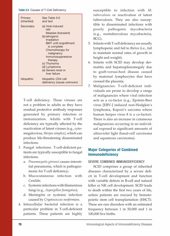
70 Immunological Aspects of Immunodeficiency Diseases
susceptible to infection with M. tuberculosis or reactivation of latent tuberculosis. They are also suscep-tible to disseminated infections with poorly pathogenic mycobacteria (e.g., nontuberculous mycobacteria, BCG).
5. Infants with T-cell defi ciency are usually lymphopenic and fail to thrive (i.e., fail to maintain normal rates of growth in height and weight).
6. Infants with SCID may develop der-matitis and hepatosplenomegaly due to graft-versus-host disease caused by maternal lymphocytes that have crossed the placenta.
7. Malignancies: T-cell-defi cient indi-viduals are prone to develop a range of malignancies where viral infection acts as a co-factor (e.g., Epstein-Barr virus [EBV-] induced non-Hodgkin’s lymphoma, Kaposi’s sarcoma where human herpes virus 8 is a co-factor). There is also an in crease in cutaneous malignancies occurring in an individ-ual exposed to signifi cant amounts of ultraviolet light (basal-cell carcinoma and squamous carcinoma).
Major Categories of Combined
Immunodeficiency
SEVERE COMBINED IMMUNODEFICIENCY SCID comprises a group of inherited
diseases characterized by a severe defi -cit in T-cell development and function with variable defects in B-cell and natural killer or NK cell development. SCID leads to death within the fi rst two years of life, unless patients are rescued by hemato-poietic stem cell transplantation (HSCT). These are rare disorders with an estimated frequency between 1 in 50,000 and 1 in 100,000 live births.
Table 5.5 Causes of T-Cell Defi ciency
Primary (inherited)
See Table 5.6 and text
Secondary (a) Viral-induced HIVMeasles (transient)
(b) IatrogenicIrradiationBMT until engraftment
is completeChemotherapy for
malignancyImmunosuppressive
therapy(c) Thymoma(d) Lymphoma(e) Severe renal or
liver failure
Idiopathic Idiopathic CD4 cell defi ciency (cause unknown)
T-cell defi ciency. These viruses are not a problem in adults as they have residual protective antibody responses generated by primary infection or immunization. Adults with T-cell defi ciency are typically affected by the reactivation of latent viruses (e.g., cyto-megalovirus, Herpes simplex), which can produce life-threatening disseminated infections.
3. Fungal infections: T-cell-defi cient pa-tients are typically susceptible to fungal infections.a. Pneumocystis giroveci causes intersti-
tial pneumonia, which is pathogno-monic for T-cell defi ciency;
b. Mucocutaneous infection with Candida;
c. Systemic infections with fi lamentous fungi (e.g., Aspergillus fumigatus);
d. Meningitis or systemic infection caused by Cryptococcus neoformans.
4. Intracellular bacterial infection is a particular problem in T-cell-defi cient patients. These patients are highly
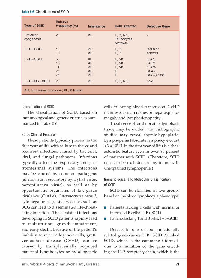
Immunological Aspects of Immunodeficiency Diseases 71
Classifi cation of SCIDThe classifi cation of SCID, based on
immunological and genetic criteria, is sum-marized in Table 5.6.
SCID: Clinical FeaturesThese patients typically present in the
fi rst year of life with failure to thrive and recurrent infections caused by bacterial, viral, and fungal pathogens. Infections typically affect the respiratory and gas-trointestinal systems. The infections may be caused by common pathogens (adenovirus, respiratory syncytial virus, parainfluenza virus), as well as by opportunistic organisms of low-grade virulence (Candida, Pneumocystis carinii, cytomegalovirus). Live vaccines such as BCG can lead to disseminated life-threat-ening infections. The persistent infections developing in SCID patients rapidly lead to malnutrition, growth impairment, and early death. Because of the patient’s inability to reject allogeneic cells, graft-versus-host disease (GvHD) can be caused by transplacentally acquired maternal lymphocytes or by allogeneic
cells following blood transfusion. GvHD manifests as skin rashes or hepatospleno-megaly and lymphadenopathy.
The absence of tonsils or other lymphatic tissue may be evident and radiographic studies may reveal thymic-hypoplasia. Lymphopenia (absolute lymphocyte count < 3 × 10⁹/L in the fi rst year of life) is a char-acteristic feature seen in over 80 percent of patients with SCID. (Therefore, SCID needs to be excluded in any infant with unexplained lymphopenia.)
Immunological and Molecular Classifi cation of SCID
SCID can be classifi ed in two groups based on the blood lymphocyte phenotype.
■ Patients lacking T cells with normal or increased B cells: T−B+ SCID
■ Patients lacking T and B cells: T−B−SCID
Defects in one of four functionally related genes causes T−B + SCID. X-linked SCID, which is the commonest form, is due to a mutation of the gene encod-ing the IL-2 receptor γ chain, which is the
Table 5.6 Classifi cation of SCID
Type of SCIDRelative Frequency (%) Inheritance Cells Affected Defective Gene
Reticular dysgenesis
<1 AR T, B, NK, Leucocytes, platelets
?
T − B − SCID 1010
ARAR
T, BT, B
RAG1/2Artemis
T − B + SCID 5010 1<1<1
XLARARARAR
T, NKT, NKT, NKTT
IL2R6JAK3IL7RACD45CD36,CD3E
T − B − NK − SCID 20 AR T, B, NK ADA
AR, antosomal recessive; XL, X-linked
72 Immunological Aspects of Immunodeficiency Diseases
signal-transducing chain common to the receptors for six cytokines (IL-2, IL-4, IL-7, IL-9, IL-15, and IL-21). The absence of responses to these cytokines causes defects in a broad range of T- and B-cell func-tions. IL-7 is required for early stages of T-cell development. Lack of response to this cytokine results in T lymphopenia. IL-15 is required for NK-cell development and its lack results in the failure of NK-cell development. Signal transduction through the aforementioned cytokine receptors involves the interaction of the common γ chain with the tyrosine kinase JAK3. This explains why mutations of the JAK3 gene result in an autosomal recessive form of SCID, with a phenotype similar to X-linked SCID. Mutations of the α chain of IL-2 or IL-7 receptors result in two rare forms of SCID.
T- and B-cell receptors consist of invari-ant signal-transducing elements combined with elements that make up the variable regions, which contribute to the antigen-binding portion of the receptor. The gene recombination required for generating these receptors requires the function of the product of recombination activating genes 1 and 2 and a number of proteins that are required for DNA repair (DNA-PKcs, KU70, KU80, XRLC4, and DNA-IV). In mice, mutations in any one of these genes produces analogues of SCID.
In humans, T−B−SCID is most common (50 percent of total) caused by mutations of the recombinase-activating genes, RAG1 or RAG2. RAG1 and RAG2 are enzymes responsible for introducing double-stranded DNA breaks, which initiate V(D)J gene rearrangements, required for gener-ating T- and B-cell receptors for antigen. Without normal RAG1 and RAG2 func-tion, T- and B-cell development is arrested early in ontogeny, producing T−B−SCID.
Hypomorphic mutations of RAG1 or RAG2 result in a leaky form of SCID called Omenn’s syndrome. In Omenn’s syn-drome, a few T- and B-cell clones may be generated but the full T- and B-cell reper-toire fails to develop. The few T- and B-cell clones that leak through may undergo secondary expansion. As a result, patients with Omenn’s syndrome may not be mark-edly lymphopenic but the lymphocyte rep-ertoire is oligoclonal and severe immuno-defi ciency is the outcome.
T- and B-cell antigen receptors are assembled from the recombination of variable region V(D)J and constant region genes. A protein called ARTEMIS is required for DNA repair, including the repair of DNA breaks generated during V(D)J recombination. Mutation of the gene encoding ARTEMIS results in a rare form of T−B−SCID. These patients also exhibit increased sensitivity to ionizing radiation.
About 15 percent of SCID cases are caused by defi ciency of adenosine deami-nase (ADA), an enzyme required for the sal-vage of nucleotides within lymphoid cells. The lack of ADA causes the accumulation of toxic metabolites of adenosine (deoxy-adenosine and deoxy-ATP) within lym-phoid cells, resulting in their demise. ADA defi ciency results in profound lymphopenia affecting T cells, B cells, and NK cells. Rarely, mutations of ADA causing milder forms of enzyme defi ciency lead to a milder form of combined immunodefi ciency presenting at a later stage in life. Purine nucleoside phos-phorylase (PNP) is an enzyme required for purine salvage within lymphocytes, and PNP defi ciency causes a milder phenotype of SCID than seen in ADA defi ciency. SCID due to PNP defi ciency not treated with HSCT is fatal in childhood.
Mutations in proteins required for nor-mal functioning and signal transduction
Immunological Aspects of Immunodeficiency Diseases 73
through the T-cell receptor (TCR) cause rare forms of SCID. Mutations of the tyro-sine phosphatase, CD45, which helps to initiate signaling by the TCR, results in T−B+ SCID in humans. Mutation of com-ponents of CD3-complex (CD3 γ, ε, and δ) result in a SCID phenotype. During signal transduction via TCRs, the protein tyrosine kinases Lck and ZAP70 are required for phosphorylation of ITAMs on the intracy-toplasmic segment of the TCR. Defi ciency of either of these kinases results in rare forms of SCID.
TCRs of CD8 cells recognize antigenic peptides that are complexed to MHC class I antigens, and TCR of CD4 cells recog-nize antigen bound to MHC class II on the surface of antigen-presenting cells. Cell-surface expression of MHC class I mol-ecules fails if either of the two transport-ers of antigenic peptides (TAP1 or TAP2) is lacking. TAP1 and TAP2 help to transfer peptides from the cytosol into the endo-plasmic reticulum, for subsequent loading onto newly synthesized MHC class I mol-ecules. In the absence of peptide loading, MHC class I molecules are degraded before reaching the cell surface. In the absence of MHC class I antigen expression, CD8 cell function is defi cient, and these cells are not generated within the thymus. The result-ing immunodefi ciency is milder than SCID and often presents in later life. Paradoxi-cally, viral infections are not a problem in these patients. Some MHC class I defi cient patients develop progressive bronchiecta-sis, while others develop vasculitis affect-ing the face and upper respiratory tract. It has been postulated that vasculitis seen in these patients may be due to self-destruc-tion of vascular endothelial cells by the unrestrained cytotoxicity of NK cells.
In contrast, MHC class II defi ciency results in a profound failure of CD4 cell
functions. Lack of thymic CD4+ CD8− cell selection for survival results in peripheral CD4 lymphopenia. Because CD4 func-tion is required for normal cell-mediated immunity, as well as antibody production, MHC class II defi ciency results in a severe form of SCID with a fatal outcome. MHC class II defi ciency is due to a mutation in one of four transcription factors (RFXAP, CIITA, RFX5, RFXANK), which regulate MHC class II expression.
TREATMENT OF SCIDHematopoietic Stem Cell Transplantation
Untreated SCID is invariably fatal in early infancy. Once a diagnosis of SCID is confi rmed, irrespective of the molecular diagnosis, HSCT from a human leucocyte antigen (HLA-) identical or haplo-identical family donor is the treatment of choice. Treatment of SCID with HSCT before 3.5 months of age results in good immune reconstitution and 95 percent survive long term. Delay in treatment, or the occurrence of infection, impairs outcome. Infection and GvHD are the main complications follow-ing HSCT. North American and European data indicate that long-term survival after transplants from HLA-matched unrelated donors was close to 60 percent. Review of European data between 1968 and 1999 indicates the progressive improvement of outcome, which is mainly because of better prevention of GvHD and the treatment of infection. Analysis of the outcome of European and U.S. bone marrow transplant programs for the treatment of SCID is ongoing and will be regularly reported.
Gene Therapy for SCIDLong-term immune reconstitution
has been achieved in patients with SCID caused by the common γ-chain defi ciency or ADA defi ciency, using gene therapy.
74 Immunological Aspects of Immunodeficiency Diseases
This was achieved by ex vivo gene transfer to hematopoietic stem cells isolated from the patient’s bone marrow. These gene-reconstituted stem cells were retransfused into the patient. To date, gene therapy has been restricted to patients without an HLA-matched family donor.
Several cases of leukemia have occurred among γ-chain-deficient patients who received gene therapy. In these cases, the retroviral vector had integrated close to the LMO2 proto-oncogene in the leukemia clone, leading to aberrant transcription and expression of LMO2. Because of this setback, clinical trials of gene therapy for SCID are being carefully evaluated, and more experience is required before the defi nitive role of gene therapy in SCID is established.
DiGeorge’s Syndrome (Thymic Aplasia)
DiGeorge’s syndrome (DGS) is second-ary to a hemizygous deletion of the short arm of chromosome 22 (DEL 22q. 11.2). This chromosomal defect causes a com-plex inherited syndrome characterized by cardiac malformations, thymic hypopla-sia, palatopharyngeal abnormalities with associated velopharyngeal dysfunction, hypoparathyroidism, and facial dysmor-phism. The 22q deletion has an incidence of approximately 1 in 2,500 live births. The associated clinical phenotype is highly variable. About 20 percent of individu-als with 22q deletion have thymic aplasia, resulting in T lymphopenia and impaired CMI. In most such cases, the degree of T lymphopenia is modest (partial DGS) and almost complete restitution of the T-cell repertoire and function occurs by two years of age. Therefore, infections characteristic of T-cell defi ciency are rare in these indi-viduals. A minority of infected individuals
(<1 percent) exhibit profound T lympho-penia, associated with opportunistic infec-tions and a poor outlook unless rescued with fetal thymic transplant.
The cardiac, velopharyngeal, thymic, and parathyroid abnormalities are due to the defective development of third and fourth pharyngeal arches during ontogeny.
The 22q. 11.2 region contains the TBX1 gene, which belongs to the T-BOX fam-ily of genes that incorporate proteins that regulate embryonic development. Patients with mutations in the TBX1 genes also develop the clinical features seen in 22q. 11.2 deletion syndrome, suggesting that haplo-insuffi ciency of the TBX1 gene may be responsible for the clinical features seen in those with a deletion of the 22q. 11.2 region.
PHAGOCYTE DEFICIENCIES
Neutrophils are the principal circulating phagocyte. During infl ammation, neutro-phils become activated and migrate into the tissues where they ingest, kill, and digest invading bacteria and fungi. Neu-trophil function can be defi cient because of a reduction in the number of circulating neutrophils (neutropenia) or due to inher-ited defects in neutrophil function, which are rare disorders.
Neutropenia
Neutropenia is defi ned as a decrease in the blood neutrophil count below 1.5 × 10⁹/L. In some racial groups (e.g., Africans), the normal neutrophil count is somewhat lower (up to 1.2 × 10⁹/L). While mild neu-tropnia may be asymptomatic, severe neu-tropenia (counts < 0.5 × 10⁹/L) is invariably associated with the risk of life-threatening
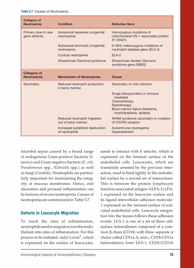
Immunological Aspects of Immunodeficiency Diseases 75
microbal sepsis caused by a broad range of endogenous Gram-positive bacteria (S. aureus) and Gram-negative bacteria (E. coli, Pseudomonas spp., Klebsiella spp.) as well as fungi (Candida). Neutrophils are particu-larly important for maintaining the integ-rity of mucous membranes. Hence, oral ulceration and perianal infl ammation can be features of severe neutropenia. Causes of neutropenia are summarized in Table 5.7.
Defects in Leucocyte Migration
To reach the sites of inflammation, neutrophils need to migrate across the endo-thelium into sites of infl ammation. For this process to be initiated, sialyl LewisX, which is expressed on the surface of leucocytes,
needs to interact with E selectin, which is expressed on the luminal surface of the endothelial cells. Leucocytes, which are transiently arrested by the previous inter-action, need to bind tightly to the endothe-lial surface by a second set of interactions. This is between the protein lymphocyte function-associated antigen-1(LFA-1) LFA-1 expressed on the leucocyte surface and its ligand intercellular adhesion molecule-1 expressed on the luminal surface of acti-vated endothelial cells. Leucocyte emigra-tion into the tissues follows these adhesion events. LFA-1 is one of a set of three cell-surface heterodimers composed of a com-mon β chain (CD18) with three separate α chains called CD11a, b, and c. CD18-CD11a heterodimers form LFA-1, CD18/CD11b
Table 5.7 Causes of Neutropenia
Category of Neutropenia Condition Defective Gene
Primary (due to rare gene defects)
Autosomal recessive congenital neutropenia
Homozygous mutations of mitochondrial HS-1 associated protein X1 (HAX1)
Autosomal dominant congenital neutropenia
In 60% heterozygous mutations of neutrophil elastase gene (ELA-2)
Cyclical neutropenia ELA-2
Shwachman Diamond syndrome Shwachman Bodian Diamond syndrome gene (SBDS)
Category of Neutropenia Mechanism of Neutropenia Cause
Secondary Reduced neutrophil production in bone marrow
Secondary to viral infection
Drugs (idiosyncratic) or immune mediated
ChemotherapyRadiotherapyBone marrow failure (leukemia,
myelodysplasia, aplasia)
Reduced neutrophil migration out of bone marrow
WHIM syndrome secondary to mutation of CXCR4 receptor
Increased peripheral destruction of neutrophils
Autoimmune neutropeniaHypersplenism
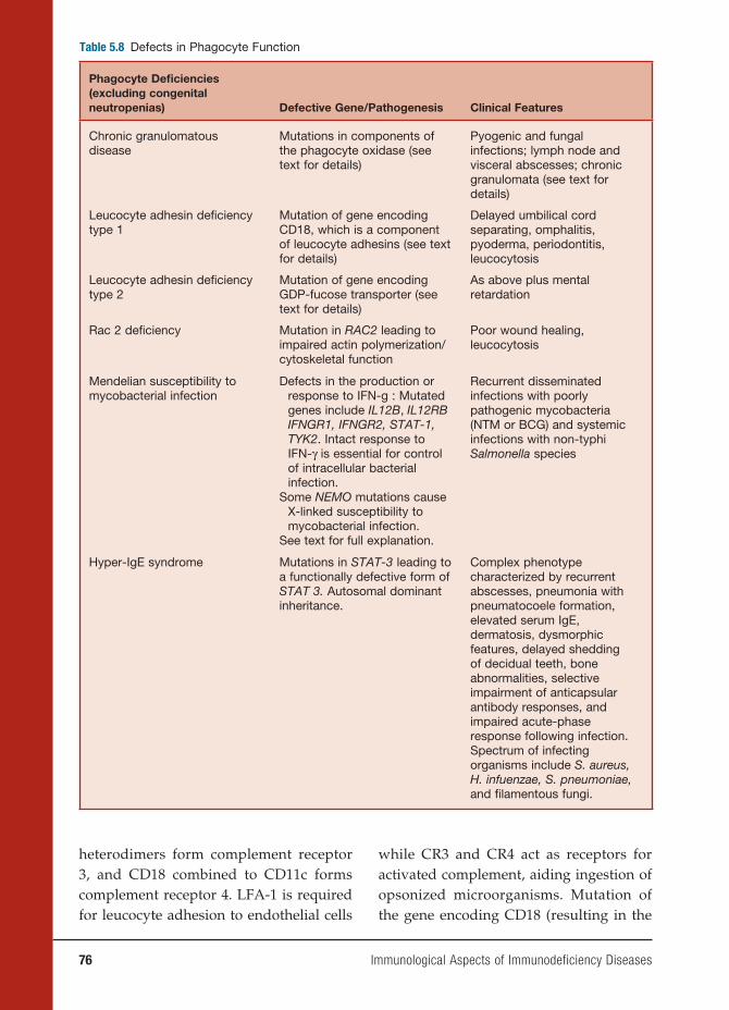
76 Immunological Aspects of Immunodeficiency Diseases
Table 5.8 Defects in Phagocyte Function
Phagocyte Defi ciencies (excluding congenital neutropenias) Defective Gene/Pathogenesis Clinical Features
Chronic granulomatous disease
Mutations in components of the phagocyte oxidase (see text for details)
Pyogenic and fungal infections; lymph node and visceral abscesses; chronic granulomata (see text for details)
Leucocyte adhesin defi ciency type 1
Mutation of gene encoding CD18, which is a component of leucocyte adhesins (see text for details)
Delayed umbilical cord separating, omphalitis, pyoderma, periodontitis, leucocytosis
Leucocyte adhesin defi ciency type 2
Mutation of gene encoding GDP-fucose transporter (see text for details)
As above plus mental retardation
Rac 2 defi ciency Mutation in RAC2 leading to impaired actin polymerization/cytoskeletal function
Poor wound healing, leucocytosis
Mendelian susceptibility to mycobacterial infection
Defects in the production or response to IFN-g : Mutated genes include IL12B, IL12RB IFNGR1, IFNGR2, STAT-1, TYK2. Intact response to IFN-γ is essential for control of intracellular bacterial infection.
Some NEMO mutations cause X-linked susceptibility to mycobacterial infection.
See text for full explanation.
Recurrent disseminated infections with poorly pathogenic mycobacteria (NTM or BCG) and systemic infections with non-typhi Salmonella species
Hyper-IgE syndrome Mutations in STAT-3 leading to a functionally defective form of STAT 3. Autosomal dominant inheritance.
Complex phenotype characterized by recurrent abscesses, pneumonia with pneumatocoele formation, elevated serum IgE, dermatosis, dysmorphic features, delayed shedding of decidual teeth, bone abnormalities, selective impairment of anticapsular antibody responses, and impaired acute-phase response following infection. Spectrum of infecting organisms include S. aureus, H. infuenzae, S. pneumoniae, and fi lamentous fungi.
heterodimers form complement receptor 3, and CD18 combined to CD11c forms complement receptor 4. LFA-1 is required for leucocyte adhesion to endothelial cells
while CR3 and CR4 act as receptors for activated complement, aiding ingestion of opsonized microorganisms. Mutation of the gene encoding CD18 (resulting in the
Immunological Aspects of Immunodeficiency Diseases 77
lack of expression of LFA-1, CR3, and CR4) results in an inherited primary immunode-fi ciency called leucocyte adhesin defi ciency type 1(LAD1). Mutations of the enzyme GDP-fucosyl transferase prevents post-transcriptional fucosylation of proteins. Such individuals cannot synthesize sialyl LewisX. This condition is called leucocyte adhesion defi ciency type 2. Leucocytes of the patient with LAD1 and 2 exhibit impaired ability to adhere to endothelial walls and therefore cannot migrate into infected sites.
These patients typically present in early childhood with recurrent pyogenic infection of skin, respiratory, and gastro-intestinal tracts as well as mucous mem-branes. Poor wound healing and delayed umbilical cord separation are typical. Because of impaired neutrophil migration, these patients develop a leucocytosis and pus fails to form at sites of infection. These inherited disorders are typically associated with severe gingivitis and periodontal disease, again indicating the particular importance of normal neutrophil function for maintenance of the health of the den-tal crevice. Outcome in both conditions is poor, with early death. BMT is curative in LAD1, and oral fucose supplementation is benefi cial in LAD2.
Defects in Bacterial Killing
The best example of an immunodefi ciency characterized by failure of phagocyte-mediated bacterial killing is chronic granulomatous disease (CGD). In normal neutrophils (and monocytes), bacterial phagocytosis results in the activation of the nicotinamide adenine dinucleotide phos-phate (NADPH) oxidase complex, which produces superoxide and generates a mi-lieu within the phagosome that activate
the proteolytic enzymes, cathepsin, and elastase. The activity of the proteolytic enzymes is bactericidal. The NADPH oxi-dase complex comprises two membrane-associated proteins, p91phox and p22phox (also called the α and β units of cytochrome B558, respectively), complexed with three cytosolic cofactors, p47phox, p40phox, and p67phox. Mutations have been identifi ed in four out of fi ve components (p91phox, p22phox, p47phox, p67phox), resulting in defective NADPH oxidase activity, leading to impairment of bacterial kill-ing by phagocytic cells. The result is a clinical syndrome, called CGD, character-ized by failure of bacterial degradation in vivo, resulting in the persistence of tissue infl ammation with granuloma formation in a variety of organs. CGD due to p91phox defi ciency is X-linked, while the other vari-ants of CGD are inherited in an autosomal recessive manner.
In CGD, the bacterial killing mecha-nisms that depend on the phagocyte oxidase system are inoperative, but the nonoxidative killing mechanisms are still intact. Therefore, patients with CGD are not troubled by the broad range of microbes that a neutropenic patient would be susceptible to. Instead, CGD patients get infections with a restricted range of micro-organisms, which are only susceptible to the bactericidal mechanisms initiated by NADPH oxidase activation. The spectrum of microorganisms that causes infections in CGD include S. aureus, Gram-negative bacteria (Burkholderia cepacea, Salmonella, Serratia), and fungi (Aspergillus spp.). Characteristic sites of infection include subcutaneous tissue, lymph nodes, lungs, and liver. Oral and perioral ulceration are common.
The formation of chronic granulomata in various tissues is a typical feature of
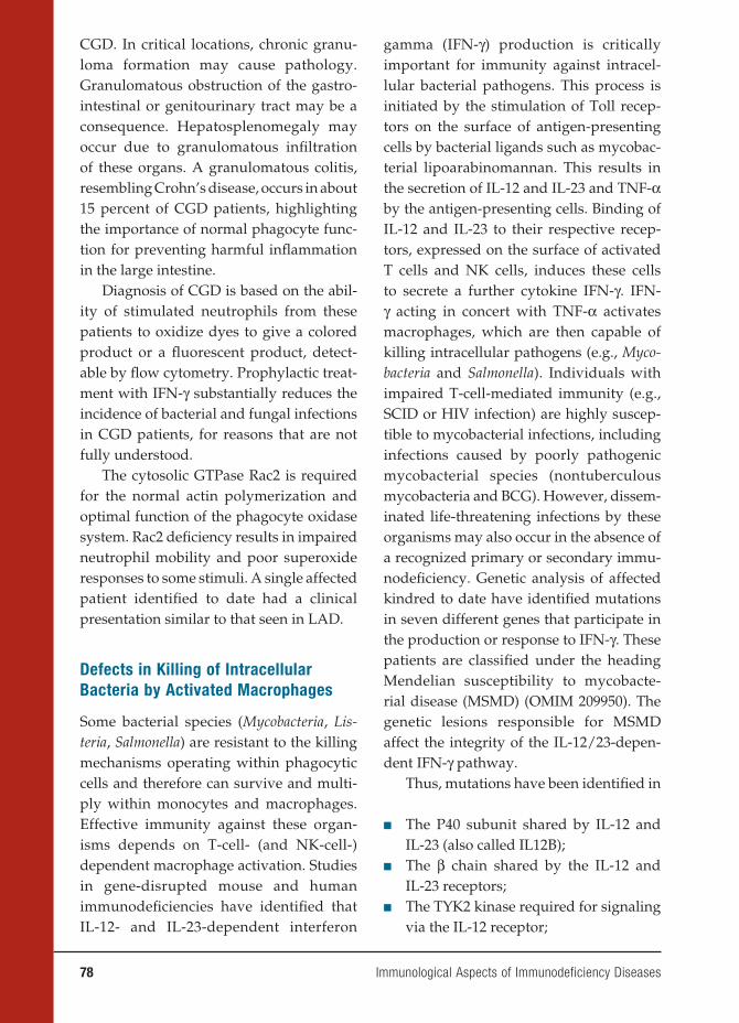
78 Immunological Aspects of Immunodeficiency Diseases
CGD. In critical locations, chronic granu-loma formation may cause pathology. Granulomatous obstruction of the gastro-intestinal or genitourinary tract may be a consequence. Hepatosplenomegaly may occur due to granulomatous infi ltration of these organs. A granulomatous colitis, resembling Crohn’s disease, occurs in about 15 percent of CGD patients, highlighting the importance of normal phagocyte func-tion for preventing harmful infl ammation in the large intestine.
Diagnosis of CGD is based on the abil-ity of stimulated neutrophils from these patients to oxidize dyes to give a colored product or a fl uorescent product, detect-able by fl ow cytometry. Prophylactic treat-ment with IFN-γ substantially reduces the incidence of bacterial and fungal infections in CGD patients, for reasons that are not fully understood.
The cytosolic GTPase Rac2 is required for the normal actin polymerization and optimal function of the phagocyte oxidase system. Rac2 defi ciency results in impaired neutrophil mobility and poor superoxide responses to some stimuli. A single affected patient identifi ed to date had a clinical presentation similar to that seen in LAD.
Defects in Killing of Intracellular
Bacteria by Activated Macrophages
Some bacterial species (Mycobacteria, Lis-teria, Salmonella) are resistant to the killing mechanisms operating within phagocytic cells and therefore can survive and multi-ply within monocytes and macrophages. Effective immunity against these organ-isms depends on T-cell- (and NK-cell-) dependent macrophage activation. Studies in gene-disrupted mouse and human immunodefi ciencies have identifi ed that IL-12- and IL-23-dependent interferon
gamma (IFN-γ) production is critically important for immunity against intracel-lular bacterial pathogens. This process is initiated by the stimulation of Toll recep-tors on the surface of antigen-presenting cells by bacterial ligands such as mycobac-terial lipoarabinomannan. This results in the secretion of IL-12 and IL-23 and TNF-α by the antigen-presenting cells. Binding of IL-12 and IL-23 to their respective recep-tors, expressed on the surface of activated T cells and NK cells, induces these cells to secrete a further cytokine IFN-γ. IFN-γ acting in concert with TNF-α activates macrophages, which are then capable of killing intracellular pathogens (e.g., Myco-bacteria and Salmonella). Individuals with impaired T-cell-mediated immunity (e.g., SCID or HIV infection) are highly suscep-tible to mycobacterial infections, including infections caused by poorly pathogenic mycobacterial species (nontuberculous mycobacteria and BCG). However, dissem-inated life-threatening infections by these organisms may also occur in the absence of a recognized primary or secondary immu-nodefi ciency. Genetic analysis of affected kindred to date have identifi ed mutations in seven different genes that participate in the production or response to IFN-γ. These patients are classifi ed under the heading Mendelian susceptibility to mycobacte-rial disease (MSMD) (OMIM 209950). The genetic lesions responsible for MSMD affect the integrity of the IL-12/23-depen-dent IFN-γ pathway.
Thus, mutations have been identifi ed in
■ The P40 subunit shared by IL-12 and IL-23 (also called IL12B);
■ The β chain shared by the IL-12 and IL-23 receptors;
■ The TYK2 kinase required for signaling via the IL-12 receptor;
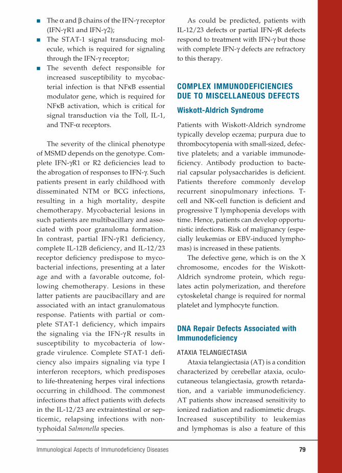
Immunological Aspects of Immunodeficiency Diseases 79
■ The α and β chains of the IFN-γ receptor (IFN-γ R1 and IFN-γ 2);
■ The STAT-1 signal transducing mol-ecule, which is required for signaling through the IFN-γ receptor;
■ The seventh defect responsible for increased susceptibility to mycobac-terial infection is that NFκB essential modulator gene, which is required for NFκB activation, which is critical for signal transduction via the Toll, IL-1, and TNF-α receptors.
The severity of the clinical phenotype of MSMD depends on the genotype. Com-plete IFN-γR1 or R2 defi ciencies lead to the abrogation of responses to IFN-γ. Such patients present in early childhood with disseminated NTM or BCG infections, resulting in a high mortality, despite chemotherapy. Mycobacterial lesions in such patients are multibacillary and asso-ciated with poor granuloma formation. In contrast, partial IFN-γ R1 defi ciency, complete IL-12B defi ciency, and IL-12/23 receptor defi ciency predispose to myco-bacterial infections, presenting at a later age and with a favorable outcome, fol-lowing chemotherapy. Lesions in these latter patients are paucibacillary and are associated with an intact granulomatous response. Patients with partial or com-plete STAT-1 defi ciency, which impairs the signaling via the IFN-γ R results in susceptibility to mycobacteria of low-grade virulence. Complete STAT-1 defi -ciency also impairs signaling via type I interferon receptors, which predisposes to life-threatening herpes viral infections occurring in childhood. The commonest infections that affect patients with defects in the IL-12/23 are extraintestinal or sep-ticemic, relapsing infections with non-typhoidal Salmonella species.
As could be predicted, patients with IL-12/23 defects or partial IFN-γR defects respond to treatment with IFN-γ but those with complete IFN-γ defects are refractory to this therapy.
COMPLEX IMMUNODEFICIENCIES
DUE TO MISCELLANEOUS DEFECTS
Wiskott-Aldrich Syndrome
Patients with Wiskott-Aldrich syndrome typically develop eczema; purpura due to thrombocytopenia with small-sized, defec-tive platelets; and a variable immunode-fi ciency. Antibody production to bacte-rial capsular polysaccharides is defi cient. Patients therefore commonly develop recurrent sinopulmonary infections. T-cell and NK-cell function is defi cient and progressive T lymphopenia develops with time. Hence, patients can develop opportu-nistic infections. Risk of malignancy (espe-cially leukemias or EBV-induced lympho-mas) is increased in these patients.
The defective gene, which is on the X chromosome, encodes for the Wiskott-Aldrich syndrome protein, which regu-lates actin polymerization, and therefore cytoskeletal change is required for normal platelet and lymphocyte function.
DNA Repair Defects Associated with
Immunodeficiency
ATAXIA TELANGIECTASIAAtaxia telangiectasia (AT) is a condition
characterized by cerebellar ataxia, oculo-cutaneous telangiectasia, growth retarda-tion, and a variable immunodefi ciency.AT patients show increased sensitivity to ionized radiation and radiomimetic drugs. Increased susceptibility to leukemias and lymphomas is also a feature of this
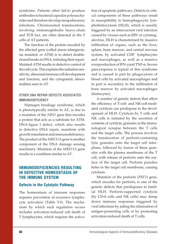
80 Immunological Aspects of Immunodeficiency Diseases
syndrome. Patients often fail to produce antibodies to bacterial capsular polysaccha-rides and therefore develop sinopulmonary infections. Chromosomal translocations, involving immunoglobulin heavy-chain and TCR loci, are often detected in the T cells of AT patients.
The function of the protein encoded by the affected gene (called ataxia telangiecta-sia mutated, or ATM) is to detect double-strand breaks in DNA, initiating their repair. Mutated ATM results in defective control of the cell cycle. This explains the radiation sen-sitivity, abnormal immune cell development and function, and the cytogenetic abnor-malities seen in AT.
OTHER DNA REPAIR DEFECTS ASSOCIATED IMMUNODEFICIENCY
Nijmegen breakage syndrome, which is phenotypically similar to AT, is due to a mutation of the NBS1 gene that encodes a protein that acts as a substrate for ATM. DNA-ligase I defect, which also results in defective DNA repair, manifests with growth retardation and immunodefi ciency. The product of the MRE11A gene is another component of the DNA damage sensing machinery. Mutation of the MRE11A gene results in a condition similar to AT.
IMMUNODEFICIENCIES RESULTING
IN DEFECTIVE HOMEOSTASIS OF
THE IMMUNE SYSTEM
Defects in the Cytolytic Pathway
The homeostasis of immune responses requires prevention of excessive lympho-cyte activation (Table 5.9). The mecha-nism by which such regulation occurs includes activation-induced cell death of T lymphocytes, which requires the activa-
tion of apoptotic pathways. Defects in criti-cal components of these pathways result in susceptibility to hemophagocytic lym-phohistiocytosis (HLH), which is usually triggered by an intercurrent viral infection caused by viruses such as EBV or cytomeg-alovirus. HLH is characterized by massive infi ltration of organs, such as the liver, spleen, bone marrow, and central nervous system, by activated CD8+ lymphocytes and macrophages, as well as a massive overproduction of IFN-γ and TNF-α. Severe pancytopenia is typical of this syndrome and is caused in part by phagocytosis of blood cells by activated macrophages and in part is secondary to the infi ltration of bone marrow by activated macrophages (histiocytes).
A number of genetic defects that affect the effi ciency of T-cell- and NK-cell-medi-ated cytolysis can predispose to the devel-opment of HLH. Cytolysis by T cells and NK cells is initiated by the secretion of contents of cytolytic granules at the immu-nological synapse between the T cells and the target cells. The process involves the translocation of perforin-containing lytic granules onto the target cell inter-phase, followed by fusion of these gran-ules with the plasma membrane of the T cell, with release of perforin onto the sur-face of the target cell. Perforin punches holes in the target cell membrane, causing cytolosis.
Mutation of the perforin (PRF1) gene, which encodes for perforin, is one of the genetic defects that predisposes to famil-ial HLH. Perforin-supported cytolysis (by CD-8 cells and NK cells) may damp down immune responses triggered by viral infections by aiding the elimination of antigen-presenting cells or by promoting activation-induced death of T cells.
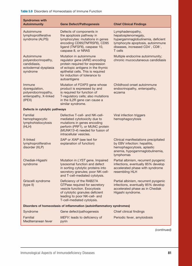
Immunological Aspects of Immunodeficiency Diseases 81
Table 5.9 Disorders of Homeostasis of Immune Function
Syndromes with Autoimmunity Gene Defect/Pathogenesis Chief Clinical Findings
Autoimmune lymphoproliferative syndrome (ALPS)
Defects of components in the apoptosis pathway in lymphocytes: mutations in genes encoding CD95(TNFRSF6), CD95 ligand (TNFSF6), caspase 10, caspase 8, or NRAS
Lymphadenopathy, hepatosplenomegaly, hypergammaglobulinemia, defi cient lymphocyte apoptosis, autoimmune diseases, increased CD4−, CD8−, T cells
Autoimmune polyendocrinopathy, candidiasis, ectodermal dysplasia syndrome
Mutation in autoimmune regulator gene (AIRE) encoding protein required for expression of ectopic antigens in the thymic epithelial cells. This is required for induction of tolerance to autoantigens
Multiple endocrine autoimmunity; chronic mucocutaneous candidiasis
Immune dysregulation, polyendocrinopathy, enteropathy, X-linked (IPEX)
Mutation of FOXP3 gene whose product is expressed by and is required for function of T-regulatory cells; also mutations in the IL2R gene can cause a similar syndrome.
Childhood onset autoimmune endocrinopathy, enteropathy, eczema
Defects in cytolytic pathways
Familial hemophagocytic lymphohistiocytosis (HLH)
Defective T-cell- and NK-cell-mediated cytotoxicity due to mutations in genes encoding perforin (PRF1), or MUNC protein (MUNK13-4) needed for fusion of intracellular vesicles.
Viral infection triggers hemophagocytosis
X-linked lymphoproliferative disorder (XLP)
SAP or XIAP (see text for explanation of function)
Clinical manifestations precipitated by EBV infection: hepatitis, hemophagocytosis, aplastic anemia, hypogammaglobulinemia, lymphomas
Chediak-Higashi syndrome
Mutation in LYST gene. Impaired lysosomal function and defect in sorting cytolytic proteins into secretory granules; poor NK-cell- and T-cell-mediated cytolysis.
Partial albinism, recurrent pyogenic infections, eventually 85% develop accelerated phase with syndrome resembling HLH
Griscelli syndrome (type II)
Defi ciency of the RAB27A GTPase required for secretory vesicle function. Exocytosis of cytolytic granules defi cient leading to poor NK-cell- and T-cell-mediated cytolysis.
Partial albinism, recurrent pyogenic infections, eventually 85% develop accelerated phase as in Chediak Higashi syndrome.
Disorders of homeostasis of infl ammation (autoinfl ammatory syndromes)
Syndrome Gene defect/pathogenesis Chief clinical fi ndings
Familial Mediterranean fever
MEFV: leads to defi ciency of pyrin
Periodic fever, amyloidosis
(continued)
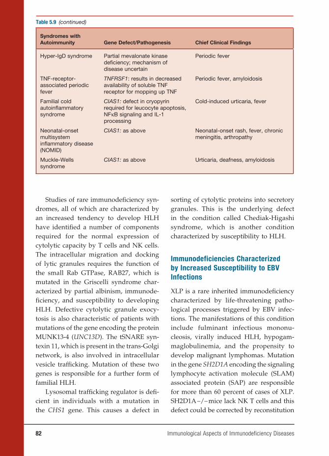
82 Immunological Aspects of Immunodeficiency Diseases
Table 5.9 (continued)
Syndromes with Autoimmunity Gene Defect/Pathogenesis Chief Clinical Findings
Hyper-IgD syndrome Partial mevalonate kinase defi ciency; mechanism of disease uncertain
Periodic fever
TNF-receptor-associated periodic fever
TNFRSF1: results in decreased availability of soluble TNF receptor for mopping up TNF
Periodic fever, amyloidosis
Familial cold autoinfl ammatory syndrome
CIAS1: defect in cryopyrin required for leucocyte apoptosis, NFκB signaling and IL-1 processing
Cold-induced urticaria, fever
Neonatal-onset multisystem infl ammatory disease (NOMID)
CIAS1: as above Neonatal-onset rash, fever, chronic meningitis, arthropathy
Muckle-Wells syndrome
CIAS1: as above Urticaria, deafness, amyloidosis
Studies of rare immunodefi ciency syn-dromes, all of which are characterized by an increased tendency to develop HLH have identifi ed a number of components required for the normal expression of cytolytic capacity by T cells and NK cells. The intracellular migration and docking of lytic granules requires the function of the small Rab GTPase, RAB27, which is mutated in the Griscelli syndrome char-acterized by partial albinism, immunode-fi ciency, and susceptibility to developing HLH. Defective cytolytic granule exocy-tosis is also characteristic of patients with mutations of the gene encoding the protein MUNK13-4 (UNC13D). The tSNARE syn-texin 11, which is present in the trans-Golgi network, is also involved in intracellular vesicle traffi cking. Mutation of these two genes is responsible for a further form of familial HLH.
Lysosomal traffi cking regulator is defi -cient in individuals with a mutation in the CHS1 gene. This causes a defect in
sorting of cytolytic proteins into secretory granules. This is the underlying defect in the condition called Chediak-Higashi syndrome, which is another condition characterized by susceptibility to HLH.
Immunodeficiencies Characterized
by Increased Susceptibility to EBV
Infections
XLP is a rare inherited immunodefi ciency characterized by life-threatening patho-logical processes triggered by EBV infec-tions. The manifestations of this condition include fulminant infectious mononu-cleosis, virally induced HLH, hypogam-maglobulinemia, and the propensity to develop malignant lymphomas. Mutation in the gene SH2D1A encoding the signaling lymphocyte activation molecule (SLAM) associated protein (SAP) are responsible for more than 60 percent of cases of XLP. SH2D1A –/– mice lack NK T cells and this defect could be corrected by reconstitution
Immunological Aspects of Immunodeficiency Diseases 83
of SH2D1A expression within bone mar-row cells derived from SH2D1A −/− mice. Patients with XLP also lack NK T cells. Therefore, it is possible that NK T-cell function is essential for controlling EBV infection as well as homeostasis of T-cell responses to this virus. Further support for this theory comes from the recent observation that XLP can also be caused by a mutation in the gene (BIRC4) that encodes for the protein X-linked inhibitor of apoptosis (XIAP). XIAP−/−patients also have reduced numbers of NK T cells.
The pathological manifestations of the XLP cannot be completely attributed to the reduced NK- and T-cell function seen in this condition. The SAP, which acts as a signaling adaptor molecule within lymphoid cells, and XIAP, which regulates apoptosis, help in the homeo-stasis of immune responses in complex ways. Therefore, mechanisms underlying the pathogenesis of XLP are likely to be complicated.
Immunodeficiencies Characterized
by Increased Liability to Develop
Autoimmunity
Immune responses to self-antigens are pre-vented by:
1. The elimination of self-reactive B and T cells in the thymus and bone marrow (central tolerance).
2. Inhibition of self-reactive T and B cells that escape central tolerance induc-tion, by the action of regulatory T cells found in the periphery. These T-regulatory cells, which are CD25+ and CD4+, are required for peripheral tol-erance and the active suppression of autoimmunity.
IMMUNE DYSREGULATION POLYENDOCRINOPATHY AND ENTEROPATHY, X-LINKED SYNDROME
The product of the FOXP3 gene (belong-ing to the forkhead family) is required for the generation and the function of CD4+ CD25+ T-regulatory cells. Mice that are FOXP3 −/− lack these cells and develop a syndrome characterized by lymphoprolif-eration and autoimmunity. Expression of ectopic FOXP3 confers suppressive func-tion on CD4+ and CD25+ T cells. Human males with mutations of the FOXP3 gene (which is located on the X chromo-some) develop a syndrome characterized by neonatal endocrinopathies (including type I diabetes) enteropathy, eczema, immune thrombocytopenia, and cachexia. In these patients, pancreatic islet cell and intestinal mucosal damage are secondary to infi ltration of the tissues with mononu-clear cells and plasma cells, signifying an autoimmune pathogenesis. This condition is called the immune dysregulation poly-endocrinopathy and enteropathy, X-linked (IPEX) syndrome. This condition provides evidence supporting current concepts on the role of T-regulatory cells in preventing autoimmunity.
Recently, mutations in the IL2R gene encoding for the α chain of the IL-2 receptor have been described in patients with the clinical phenotype of the IPEX syndrome. CD4 T cells of these patients fail to produce the immunosuppressive cytokine IL-10.
AUTOIMMUNE LYMPHOPROLIFERATIVE SYNDROME
During lymphocyte development, autoreactive cells are culled by undergo-ing apoptotic death. During the life span of mature lymphocytes, activation and effector function is followed by activation-
84 Immunological Aspects of Immunodeficiency Diseases
induced cell death by apoptosis. Apop-tosis thus maintains homeostasis in the immune system by minimizing autoim-mune reactions to self-antigens, as well as limiting the total size of the peripheral lymphocyte pool. Antigen-mediated acti-vation of T cells induces them to express a surface receptor FAS (CD95) as well as its ligand, the FAS-ligand. FAS/FAS-ligand interaction activates a biochemi-cal pathway culminating in cell death by apoptosis. Mice that are FAS −/− (lpr) or FAS-ligand −/− (gld) develop autoimmu-nity and develop expansion of their their lymphocyte pool.
ALPS is a rare disease caused by defective cellular apoptosis. Failure of lymphocytes to undergo apoptosis results in impaired homeostasis of the lymphoid population. Patients develop lymphocytosis, hyperplasia of lymphoid organs (spleen, lymph nodes), hyper-gammaglobulinemia, and autoimmunity (autoimmune cytopenias, Guillain-Barré syndrome). Peripheral blood and periph-eral lymphoid tissues characteristically contain an increased population of TCR̃ α̃ β positive CD4− and CD8− cells (dou-ble negative T cells). Mutations in the FAS-mediated pathway of apoptosis are responsible for most cases. These include mutations of FAS, FAS-ligand, and cas-pase 10. Caspase 8 defi ciency causes impaired CD95-mediated apoptosis as well as a defect in T-, B-, and NK-cell function, manifesting as an immunode-fi ciency. Recently, an activating muta-tion in the NRAS gene encoding a GTP-binding protein with a broad spectrum of signaling functions has been found in other patients with ALPS. Defective, IL-2 withdrawal induced, apoptosis of lym-phocytes is a characteristic abnormality in these patients.
Autoinflammatory Syndromes
The responses of acute infl ammation and fever are protective responses triggered by infection or tissue damage, acting through the innate immune system. The study of a collection of disorders termed familial periodic fever syndromes has contributed to our understanding of the physiological processes underlying the homeostasis of infl ammation and fever. These inherited disorders are characterized by recurrent episodes of fever and systemic infl amma-tion. These conditions are not associated with autoantibody production or self-reactive T-cell responses. They are col-lectively called autoinfl ammatory syndromes. Unraveling the pathophysiology of these disorders has given rise to the concept that these conditions are due to an innate immune system that is either oversensitive and prone to activation by minor stimuli or is poorly regulated.
The cleavage of pro-IL-1b to its active product IL-1 by the action of caspase-1 (interleukin converting-enzyme) is a key event in the generation of an acute infl am-matory response. Activated caspase-1, intern, is generated from pro-caspase-1 by the action of activated forms of two cyto-solic proteins called pyrin and cryopyrin, acting via adaptor proteins. The best-char-acterized adaptor protein is called ASC (apoptosis-associated speclike protein with a caspase recruitment domain). As summarized in Table 5.9, mutations of components of this system (also called an infl ammasome) give rise to most auto-infl ammatory syndromes characterized to date. The net effect of all these muta-tions is to impair homoeostasis of the pro-infl ammatory cytokine IL-1, NFκB acti-vation, and cellular apoptosis. Impaired cellular apoptosis leads to the persistence
Immunological Aspects of Immunodeficiency Diseases 85
of activated leucocytes that would other-wise undergo apoptosis, leading to the resolution of infl ammation. Collectively, these abnormalities permit the inappro-priate amplifi cation and persistence of infl ammatory responses leading to the clinical features of the autoinfl ammatory syndromes.
INHERITED DEFICIENCY OF THE
COMPLEMENT SYSTEM
Activation of the complement system underlies one of the main effector pathways contributing to antibody-mediated immu-nity. The normal function of the comple-ment system includes defending the body against pyogenic bacterial infection and helping to eliminate immune complexes and damaged cells (Table 5.10).
The key event in complement activa-tion is the proteolytic cleavage of C3 to C3a and C3b. Three pathways can lead to C3 cleavage, namely, classical, alternative, and mannose-binding lectin (MBL) pathways. C3 cleavage leads on to the activation of the terminal complement pathway, caus-ing the generation of the membrane attack complex (MAC), which assembles a lipo-philic complex capable of lysing plasma membranes of susceptible cells.
The inability to generate sufficient CD3b results in increased susceptibility to pyogenic sepsis, especially infections caused by encapsulated bacteria. C3 defi -ciency may be due to complement utiliza-tion (e.g., in systemic lupus erythematosus [SLE] or the presence of a C3 nephritic fac-tor) or rarely autosomal recessive C3 defi -ciency. Factor I defi ciency also leads to pro-found CD3 defi ciency due to uncontrolled progression of C3 cleavage. Hereditary C2 defi ciency and less commonly inherited C4
defi ciency may also be associated with the risk of pneumococcal sepsis.
Protection from Neisserial infection requires the ability to generate the MAC, which lyses these bacteria. Patients with inherited homozygous defi ciency of C5, C6, C7, C8, and C9 are susceptible to recur-rent meningococcal infections. In Japanese populations, an inherited terminal comple-ment component defi ciency leads to about a 5,000-fold increase in the risk of Neisse-rial sepsis. Primary or secondary CD3 defi -ciency, which in turn reduces the ability to generate MAC, also results in increased susceptibility to meningococcal infection. Complement defi ciency is not a risk factor in sporadic cases of meningococcal disease seen in the population at large.
MBL is a serum collectin that can bind to mannose residues on microbial cell walls. Upon binding of MBL to pathogens, two serine proteases found in serum (MBL-lectin associated serine proteases 1 and 2) become activated. These serine proteases in turn activate the classical pathway at C2 and C4. MBL defi ciency may arise because of one of three point mutations in the gene encoding for this protein. Polymorphisms in the promoter region of the gene also infl uence serum levels of MBL. Low serum levels of MBL may result in an increased incidence of pyogenic infections in young children below two years of age. MBL defi -ciency is also overrepresented in patients with pneumococcal or meningococcal sepsis. However, the clinical signifi cance of this fi nding has been disputed. This is because large, long-term prospective stud-ies of adults in Scandinavia have failed to identify increased morbidity due to infec-tious diseases in MBL-defi cient individuals. Therefore, except in young children who have not yet acquired a wide repertoire of protective antibodies, MBL defi ciency on
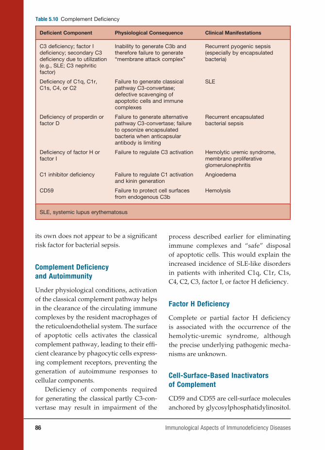
86 Immunological Aspects of Immunodeficiency Diseases
Table 5.10 Complement Defi ciency
Defi cient Component Physiological Consequence Clinical Manifestations
C3 defi ciency; factor I defi ciency; secondary C3 defi ciency due to utilization (e.g., SLE; C3 nephritic factor)
Inability to generate C3b and therefore failure to generate “membrane attack complex”
Recurrent pyogenic sepsis (especially by encapsulated bacteria)
Defi ciency of C1q, C1r, C1s, C4, or C2
Failure to generate classical pathway C3-convertase; defective scavenging of apoptotic cells and immune complexes
SLE
Defi ciency of properdin or factor D
Failure to generate alternative pathway C3-convertase; failure to opsonize encapsulated bacteria when anticapsular antibody is limiting
Recurrent encapsulated bacterial sepsis
Defi ciency of factor H or factor I
Failure to regulate C3 activation Hemolytic uremic syndrome, membrano proliferative glomerulonephritis
C1 inhibitor defi ciency Failure to regulate C1 activation and kinin generation
Angioedema
CD59 Failure to protect cell surfaces from endogenous C3b
Hemolysis
SLE, systemic lupus erythematosus
its own does not appear to be a signifi cant risk factor for bacterial sepsis.
Complement Deficiency
and Autoimmunity
Under physiological conditions, activation of the classical complement pathway helps in the clearance of the circulating immune complexes by the resident macrophages of the reticuloendothelial system. The surface of apoptotic cells activates the classical complement pathway, leading to their effi -cient clearance by phagocytic cells express-ing complement receptors, preventing the generation of autoimmune responses to cellular components.
Deficiency of components required for generating the classical partly C3-con-vertase may result in impairment of the
process described earlier for eliminating immune complexes and “safe” disposal of apoptotic cells. This would explain the increased incidence of SLE-like disorders in patients with inherited C1q, C1r, C1s, C4, C2, C3, factor I, or factor H defi ciency.
Factor H Deficiency
Complete or partial factor H defi ciency is associated with the occurrence of the hemolytic-uremic syndrome, although the precise underlying pathogenic mecha-nisms are unknown.
Cell-Surface-Based Inactivators
of Complement
CD59 and CD55 are cell-surface molecules anchored by glycosylphosphatidylinositol.
Immunological Aspects of Immunodeficiency Diseases 87
These two proteins inactivate any C3 con-vertase molecules deposited on cell sur-faces. Somatic mutation of the enzyme (PIGA: phosphatidylinositol glycan class A), needed to generate phosphatidyl-inositol anchors for cell-surface proteins, including CD55 and CD59, in erythroid precursors, results in a condition called paroxysmal nocturnal hemoglobinuria, which is due to the increased susceptibil-ity of red cells to complement-mediated hemolysis. Isolated CD59 defi ciency also results in hemolytic anemia.
C1 Inhibitor Deficiency
C1 inhibitor is a serine-protease inhibitor that inactivates the serine esterases gener-ated by complement activation (C1r and C1s), kallikrein of the kinin system, and activated factors XI and XII of the clotting cascade. In the absence of C1 inhibitor, C1 activation results in the depletion of the serum C4 level. (This is useful from a diagnostic point of view.) C1-inhibitor defi ciency also results in the inability to inactivate bradykinin, resulting from the unregulated activity of kallikrein. Produc-tion of bradykinin in the tissues results in increased vascular permeability, manifest-ing as attacks of angioedema. Angioedema of the respiratory tract can lead to death from asphyxia. Angioedema of the intes-tinal tissues results in recurrent episodes of severe abdominal pain due to partial intestinal obstruction, which can mimic an acute abdominal emergency.
C1-inhibitor defi ciency arises from a het-erozygous mutation of C1INH gene, which acts in an autosomal dominant manner. The single normal gene cannot maintain the synthesis of physiologically suffi cient quantities of C1 inhibitor. In 85 percent of C1-inhibitor-defi cient individuals, the
mutation prevents transcription of the defective gene. In 15 percent of affected individuals, the gene mutation abolishes the activity of the secreted protein. Rarely, autoantibodies to C1 inhibitor can lead to acquired C1-inhibitor defi ciency.
DEFECTS IN INNATE IMMUNITY
Defects in the complement pathway resulting in immunodefi ciency are sum-marized in the previous section. Recent work has identifi ed defects in pathways involved in the recognition and response to pathogen-associated molecular patterns. Some of these defects are outlined next.
Defective NFκB activation caused by X-linked hypomorphic mutations of the essential modulator gene (NEMO) compro-mises signaling mechanisms downstream of Toll, IL-1, and TNF-α receptors. These pa-tients are susceptible to infections caused by a range of microorganisms, including mycobacteria, Gram-positive and Gram-negative bacteria, fungi, and viruses.
UNC93B is a protein of the endoplas-mic reticulum involved in Toll-receptor activation. Mutations in UNC93B impair the production of IFN-α and IFN-β in response to HSV and other viruses. Affected patients present with Herpes sim-plex viral encephalitis. Recently, heterozy-gous dominant-negative mutations in the gene encoding toll receptor 3 (TLR3) have been identifi ed in patients with Herpes sim-plex encephalitis. TLR3 is expressed in the central nervous system where it helps to initiate IFN-α and IFN-β responses to viral duble-stranded DNA.
Interleukin-receptor-associated kinase-4 mediates signaling downstream of Toll receptors and members of the IL-1 receptor superfamily. IRAK-4-defi cient individuals
88 Immunological Aspects of Immunodeficiency Diseases
present during childhood with recurrent, severe, pyogenic sepsis. They are particu-larly susceptible to recurrent pneumococ-cal infections.
The signal-transducing molecule STAT-1 is required for signaling via recep-tors to IFN-γ as well as IFN-α and IFN-β. IFN-γ-receptor-mediated signaling involves the dimerization of phosphory-lated STAT-1 molecules. Signaling via IFN-α and IFN-β receptors involves the formation of a complex between STAT-1, STAT-2, and a third protein called inter-feron-stimulated-gene factor 3-γ. Com-plete (homozygous) defects of the signal-transducing molecules STAT-1 results in defective responses to IFN-γ, IFN-α, and IFN-β eater leading to the susceptibility to disseminated mycobacterial infections as well as fatal Herpes simplex viral infection. Partial STAT-1 defi ciency, which interferes with STAT-1 dimerization required for sig-nal transduction via IFN-γ receptors, leads to increased susceptibility to mycobacte-rial infections. In these patients, the cellu-lar responses to IFN-α and INF-β is intact, thus preserving antiviral immunity.
The WHIM syndrome is a condition characterized by severe warts, hypogam-maglobulinemia, and neutropenia. This is the fi rst example of an immunodefi ciency caused by aberrant chemokine-recep-tor function. WHIM syndrome is caused by a mutation in the gene encoding the CXCR 4 chemokine receptor. The mutant form of this receptor shows enhanced responsiveness to its ligand.
The hyper-IgE syndrome (HIES) is a complex clinical entity characterized by recurrent bacterial (S. aureus, Gram-nega-tive bacteria) and fungal infections of skin, lymph nodes, lungs, bones, and joints. These patients have elevated serum IgE levels, eosinophilia, dermatitis, facial dysmorphic
features, delayed shedding of primary dentition, osteopenia, and impaired acute-phase responses during infections. Most patients have an autosomal dominant inheritance while others are sporadic cases. Patients with classical HIES have heterozy-gous mutations in the gene encoding the signal-tranducing protein STAT-3. These mutations mainly involve the DNA-bind-ing domain or the SRC homology 2 domain of the protein and are permissive of protein expression. These mutant proteins severely impair the DNA binding of the phosphory-lated STAT-3 dimer in response to IFN-α and responses to the cytokines IL-10 and IL-6. Reduced response to IL-6 would explain the defective acute-phase response, and the defective response to IL-10 explains the overproduction of IgE. STAT-3 is essen-tial for the generation of TH17 cells and for IL-12 signaling, which are required for the secretion of the bactericidal peptides called β-defensins by epithelial cells of the skin and lungs. This may in part explain the increased incidence of severe sepsis.
CONCLUSIONS
Studies in genetically manipulated animals have helped in the development of mecha-nistic models of antimicrobial immunity. Gene knockout mice have been invaluable for the elucidation of the phenotypic conse-quences of single-gene defects. Collectively, these animal studies have highlighted can-didate genes that should be investigated in patients with increased susceptibility to microbial infection. Comparison of the phenotype of a mouse carrying a defi ned genetic defect with the clinical and immu-nological phenotype of patients has been helpful in identifying candidate genes that might be affected in the human patient.
Immunological Aspects of Immunodeficiency Diseases 89
Studies in humans with PID have been complementary. For example, human stud-ies have highlighted that (1) defects in more than one gene can give rise to a similar pri-mary immunodefi ciency syndrome; (2) the same genetic mutation can give rise to a variable phenotype, depending on the mod-ulating effect of genetic and environmental infl uences; and (3) defects in components of innate and adaptive immunity that are required for homeostasis of the immune response may result in autoimmunity or autoinfl ammatory syndromes rather than increased susceptibility to infection, thus extending the clinical spectrum of “primary immunodefi ciency diseases.”
In the past decade, we have realized that defects in some components of the innate and adaptive immunity may result in susceptibility to a narrow range of micro-bial pathogens. This indicates that some immune mechanisms have evolved to deal with specifi c pathogens but are redundant for immunity against other microorgan-isms. Such knowledge may lead to the development of novel treatment strategies targeted against specifi c microbes.
In this review, we have highlighted how defects in individual components of the immune system can lead to the sus-ceptibility to different categories of patho-gens. Such analyses help us to understand which components of immunity are non-redundant for protection against different microorganisms. This knowledge also helps us to be guided by the pattern of micro-bial infections in an individual patient in devising a rational approach to the inves-tigation of patients with suspected immu-nodefi ciency.
The past two decades have seen the development of novel treatment modali-ties for PIDs. These include the use of Ig replacement therapy (for antibody
defi ciency), bone marrow transplantation (for SCID), and gene therapy (for the treat-ment of X-linked SCID and SCID due to ADA defi ciency). In X-linked SCID and in ADA-defi ciency gene, corrected bone marrow cells have a selective advantage over unmodifi ed stem cells, contributing to successful engraftment by gene-recon-stituted cells. The correction of genetic defects in conditions where the expression of the normal molecule does not provide a selective survival advantage will be more diffi cult and will require the development of more effective genetic vectors.The elu-cidation of molecular defects underlying PIDs helps in the development of better methods of diagnosing these disorders and genetic counseling of affected families. The contribution of new genetic techniques for elucidating molecular defects underly-ing PIDs have been described elsewhere (see references list at end of chapter).
BIBLIOGRAPHY
COMPREHENSIVE REVIEWS (PRIMARY IMMUNODEFICIENCY DISEASES)Primary immunodeficiency diseases.
Report of an IUIS Scientifi c Committee. International Union of Immunological Societies. Clin Exp Immunol. 1999;118 (suppl 1):1–28.
Fischer A. Human primary immunode-fi ciency diseases: a perspective. Nat Immunol. 2004;5(1):23–30.
Geha, RS, Notarangelo, LD, et al. Pri-mary immunodefi ciency diseases: an update from the International Union of Immunological Societies Primary Immunodefi ciency Diseases Classifi ca-tion Committee. J Allergy Clin Immunol. 2007;120(4):776–794.
Ochs, HD, Smith, CIE, et al. Primary Immu-nodefi ciency Diseases: A Molecular and
90 Immunological Aspects of Immunodeficiency Diseases
Genetic Approach. New York: Oxford University Press; 2007.
SELECTED REVIEWS (MOLECULAR MECHANISMS UNDERLYING PRIMARY IMMUNODEFICIENCIES)Brydges S, Kastner DL. The systemic auto-
infl ammatory diseases: inborn errors of the innate immune system. Curr Top Microbiol Immunol. 2006;305:127–160.
Doffi nger R, Patel SY, Kumararatne, DS. Host genetic factors and mycobacte-rial infections: lessons from single gene disorders affecting innate and adaptive immunity. Microbes Infect. 2006;8(4):1141–1150.
Durandy A, Taubenheim N, Peron S, Fischer A. Pathophysiology of B-cell intrinsic immunoglobulin class switch recom-bination deficiencies. Adv Immunol. 2007;94:275–306.
Fischer A, Le Deist F, Hacein-Bey-Abina S, et al. Severe combined immunode-fi ciency. A model disease for molecu-lar immunology and therapy. Immunol Rev. 2005;203:98–109.
Gennery AR. Primary immunodefi ciency syndromes associated with defective DNA double-strand break repair. Br Med Bull. 2006.
Jouanguy E, Zhang SY, et al. Human pri-mary immunodefi ciencies of type I interferons. Biochimie. 2007; 89(6–7): 878–883.
Ku CL, Yang K, et al. Inherited disorders of human Toll-like receptor signaling: immunological implications. Immunol Rev. 2005;203:10–20.
Marodi L, Notarangelo, LD. Immunologi-cal and genetic bases of new primary immunodefi ciencies. Nat Rev Immunol. 2007;7(11):851–861.
Segal AW. How superoxide production by neutrophil leukocytes kills microbes.
Novartis Found Symp. 2006;279:92–98; discussion 98–100, 216–219.
Smith TF, Johnston, RB Jr. Functions of the spleen in host defense against infection. Am J Pediatr Hematol Oncol. 1979;1(4):355–362.
REFERENCES (DIAGNOSIS AND MANAGEMENT OF IMMUNODEFICIENCIES)Bonilla FA, Bernstein IL, et al. Practice
param eter for the diagnosis and man-agement of primary immunodefi ciency. Ann Allergy Asthma Immunol. 2005;94(5 Suppl 1):S1–63.
Cavazzana-Calvo M, Fischer, A. Gene therapy for severe combined immu-nodefi ciency: are we there yet? J Clin Invest. 2007;117(6):1456–1465.
de Vries E. Patient-centred screen-ing for primary immunodefi ciency: a multi-stage diagnostic protocol designed for non-immunologists. Clin Exp Immunol. 2006;145(2):204–214.
Ochs HD, Smith CIE, et al. Primary immu-nodefi ciency diseases : a molecular and genetic approach. New York: Oxford University Press; 2007.
Wood P, Stanworth S, et al. Recognition, clinical diagnosis and management of patients with primary antibody deficiencies: a systematic review. Clin Exp Immunol. 2007;149(3):410–423.
REVIEWS (SCIENTIFIC APPROACH TO UNRAVELING THE PATHOGENESIS OF PRIMARY IMMUNODEFICIENCY DISEASES)Casanova JL, Abel L. Primary immunode-
fi ciencies: a fi eld in its infancy. Science. 2007;317(5838):617–619.
Fischer A. Primary immunodeficiency diseases: an experimental model for molecular medicine. Lancet. 2001;357(9271): 1863–1869.
6. Autoimmunity
Haoyang Zhuang, Ph.D., Matthew Kosboth, M.D., Jennifer A. Sipos, M.D., Minoru
Satoh, M.D., Ph.D., Lijun Yang, M.D., and Westley H. Reeves, M.D.
91
DEFINITIONS AND TYPES
OF AUTOIMMUNITY
Autoimmunity versus Autoimmune
Disease
The classic studies of Paul Ehrlich in the early twentieth century laid the founda-tion for our current notions of the concept of autoimmunity. Ehrlich used the term autoimmunity to signify an immune re-sponse against self and introduced the phrase horror autotoxicus, suggesting that there are mechanisms to protect against autoimmunity. Over the years, autoim-munity has been recognized as not uncom-mon and not necessarily detrimental. Thus, an important distinction must be drawn between autoimmunity, which may be asymptomatic, and autoimmune disease, which occurs when autoimmunity leads to an infl ammatory response, resulting in tis-sue injury. An autoimmune response does not necessarily imply the existence of auto-immune disease.
T-Cell versus B-Cell-Mediated
Autoimmune Disease
Autoimmune disease may be mediated primarily by T cells, as in multiple sclero-sis or the animal model experimental auto-immune encephalomyelitis (EAE). In that case, disease can be transmitted from one animal to another by transferring antigen-
specific T lymphocytes. Alternatively, autoimmune disease may be caused by B cells that produce autoantibodies, as in the case of systemic lupus erythematosus (SLE). Autoantibodies bind to self-anti-gens (proteins, nucleic acids, or other mol-ecules from one’s own body, also known as autoantigens) and can damage cells either by binding directly to a cell surface or extracellular matrix antigen or through the formation of immune complexes (see the section “Mechanisms of Auto-immune Tissue Injury and Examples”). Autoantibody-mediated auto immune diseases sometimes can be transmitted transplacentally, as in the case of neona-tal Graves’ disease or congenital com-plete heart block and neonatal lupus. IgG antibodies/autoantibodies can cross the placenta, whereas IgM cannot. Thus, neo-natal autoimmune diseases are invariably caused by IgG, not IgM, autoantibodies. In view of the half-life of IgG (twenty-one to twenty-eight days), nearly all maternal IgG disappears from the circula-tion of the baby by six to twelve months postpartum. Thus, in most cases, neona-tal autoimmune disease is transient. One exception is congenital complete heart block, which is thought to be mediated by the transplacental passage of anti-Ro or anti-La autoantibodies that cross-react with cardiac antigens, causing permanent infl ammation-mediated damage to the cardiac conduction system.

92 Autoimmunity
Systemic versus Organ-Specific
Autoimmune Disease
Autoimmune disease also can be classi-fi ed as systemic or organ specifi c. Systemic autoimmune diseases, such as SLE, involve multiple organs or tissues, whereas organ-specifi c autoimmune diseases involve a single organ or tissue, such as the thy-roid gland in autoimmune thyroiditis or the islets of Langerhans in type I diabetes (TID). Some of the more common systemic and organ-specifi c autoimmune diseases are listed in Table 6.1.
MECHANISMS OF AUTOIMMUNE
TISSUE INJURY AND EXAMPLES
Tissue damage in autoimmune diseases can occur through several mechanisms, which are analogous to three of the classical types of hypersensitivity reactions: type II (caused by autoantibodies reactive with cell surface or matrix antigens), type III (caused by immune complexes), and type IV (delayed-type hypersensitivity, mediated by T cells).
Type II Autoimmune Reactions
Type II hypersensitivity reactions are caused by antibodies against altered self-proteins, such as penicillin–protein con-jugates. In the case of autoimmunity, antibodies generated against cell surface antigens/extracellular matrix proteins may be cytotoxic (type IIA) or they may have agonistic/antagonistic properties (type IIB). Autoantibodies to cell surface anti-gens may initiate cell destruction by com-plement-mediated lysis (cell destruction), phagocytosis, or antibody-dependent cell-mediated cytotoxicity (ADCC).
Examples include autoimmune hemolytic anemia (AIHA), and autoimmune throm-bocytopenia (Table 6.1). Some autoanti-bodies bind to surface receptors, either activating (e.g., anti-TSH receptor auto-antibodies in Graves’ disease) or inhibit-ing (e.g., anti-acetylcholine antibodies in myasthenia gravis) their function.
Type IIA Autoimmune Reaction:
Autoimmune Hemolytic Anemia
AIHA is an example of type IIA autoim-munity. In this disorder, a self-antigen on the surface of erythrocytes elicits an autoantibody response, resulting in the binding of autoantibody to the erythro-cyte surface followed by destruction of the antibody-coated erythrocytes by the reticuloendothelial system of the spleen and liver. The mechanism of hemolysis depends on the type of autoantibodies. Autoimmune hemolysis is classifi ed into two groups on the basis of thermal reactiv-ity of the autoantibodies. Warm autoanti-bodies react optimally at temperatures of 35°C–40°C, whereas cold agglutinins and other cold-reactive autoantibodies react maximally at 4°C. Warm autoantibod-ies are typically polyclonal IgG but may also be IgM or IgA. Most are IgG1 sub-class antibodies reactive with Rh antigens. These antibodies are detected by the direct antiglobulin (Coombs) test (Figure 6.1A). Erythrophagocytosis mediated by Fc receptors on Kupffer cells in the liver and macrophages in the splenic marginal zone is generally the major mechanism of eryth-rocyte destruction in patients with warm autoantibodies.
In contrast, AIHA induced by cold agglutinins is complement mediated. These autoantibodies are of the IgM class and cannot interact with Fc receptors
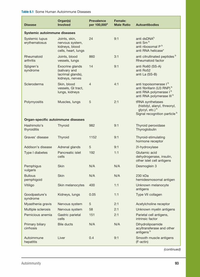
Autoimmunity 93
Table 6.1 Some Human Autoimmune Diseases
DiseaseOrgan(s) Involved
Prevalence per 100,000a
Female:Male Ratio Autoantibodies
Systemic autoimmune diseases
Systemic lupus erythematosus
Joints, skin, nervous system, kidneys, blood cells, heart, lungs
24 9:1 anti dsDNAb
anti Sm b
anti ribosomal P b
anti RNA helicase*
Rheumatoid arthritis
Joints, blood vessels, lungs
860 3:1 anti citrullinated peptides b
Rheumatoid factor
Sjögren’s syndrome
Exocrine glands (salivary and lacrimal glands), kidneys, nerves
14 9:1 anti Ro60 (SS-A)anti Ro52anti La (SS-B)
Scleroderma Skin, blood vessels, GI tract, lungs, kidneys
4 4:1 anti topoisomerase I b
anti fi brillarin (U3 RNP) b
anti RNA polymerase I b
anti RNA polymerase III b
Polymyositis Muscles, lungs 5 2:1 tRNA synthetases (histidyl, alanyl, threonyl, glycyl, etc.) b
Signal recognition particle b
Organ-specifi c autoimmune diseases
Hashimoto’s thyroiditis
Thyroid 982 9:1 Thyroid peroxidaseThyroglobulin
Graves’ disease Thyroid 1152 9:1 Thyroid-stimulating hormone receptor
Addison’s disease Adrenal glands 5 9:1 2I-hydroxylase
Type I diabetes Pancreatic islet cells
192 1:1 Glutamic acid dehydrogenase, insulin, other islet cell antigens
Pemphigus vulgaris
Skin N/A N/A Desmoglein 3
Bullous pemphigoid
Skin N/A N/A 230 kDa hemidesmosomal antigen
Vitiligo Skin melanocytes 400 1:1 Unknown melanocyte antigens
Goodpasture’s syndrome
Kidneys, lungs 0.05 1:1 Type VII collagen
Myasthenia gravis Nervous system 5 2:1 Acetylcholine receptor
Multiple sclerosis Nervous system 58 2:1 Unknown myelin antigens
Pernicious anemia Gastric parietal cells
151 2:1 Parietal cell antigens, intrinsic factor
Primary biliary cirrhosis
Bile ducts N/A N/A Dihydrolipoamide acyltransferase and other antigens b
Autoimmune hepatitis
Liver 0.4 9:1 Smooth muscle antigens (F-actin)
(continued)
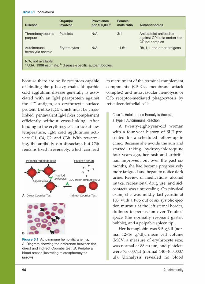
94 Autoimmunity
Table 6.1 (continued)
DiseaseOrgan(s) Involved
Prevalence per 100,000a
Female:male ratio Autoantibodies
Thrombocytopenic purpura
Platelets N/A 3:1 Antiplatelet antibodies against GPIIbIIIa and/or the GPIbα complex
Autoimmune hemolytic anemia
Erythrocytes N/A ~1.5:1 Rh, I, i, and other antigens
N/A, not available.a USA, 1996 estimate; b disease-specifi c autoantibodies.
to recruitment of the terminal complement components (C5–C9, membrane attack complex) and intravascular hemolysis or C3b receptor-mediated phagocytosis by reticuloendothelial cells.
Case 1. Autoimmune Hemolytic Anemia, a Type II Autoimmune Reaction
A twenty-eight-year-old woman with a four-year history of SLE pre-sented for a scheduled follow-up in clinic. Because she avoids the sun and started taking hydroxychloroquine four years ago, her rash and arthritis had improved, but over the past six months, she had become progressively more fatigued and began to notice dark urine. Review of medications, alcohol intake, recreational drug use, and sick contacts was unrevealing. On physical exam, she was mildly tachycardic at 105, with a two out of six systolic ejec-tion murmur at the left sternal border, dullness to percussion over Traubes’ space (the normally resonant gastric bubble), and a palpable spleen tip.
Her hemoglobin was 9.5 g/dl (nor-mal 12–16 g/dl), mean cell volume (MCV, a measure of erythrocyte size) was normal at 88 cu µm, and platelets were 75,000/µl (normal 140–400,000/µl). Urinalysis revealed no blood
A
B
Patient’s red blood cells
Direct Coombs Test Indirect Coombs Test
Patient’s serum
ABO and Rh compatible RBCs
YY
YY
Y+
Agglutination
Anti-lgGantibodies
Figure 6.1 Autoimmune hemolytic anemia. A, Diagram showing the difference between the direct and indirect Coombs test. B, Peripheral blood smear illustrating microspherocytes (arrows).
because there are no Fc receptors capable of binding the µ heavy chain. Idiopathic cold agglutinin disease generally is asso-ciated with an IgM paraprotein against the “I” antigen, an erythrocyte surface protein. Unlike IgG, which must be cross-linked, pentavalent IgM fi xes complement effi ciently without cross-linking. After binding to the erythrocyte’s surface at low temperature, IgM cold agglutinins acti-vate C1, C4, C2, and C3b. With rewarm-ing, the antibody can dissociate, but C3b remains fi xed irreversibly, which can lead
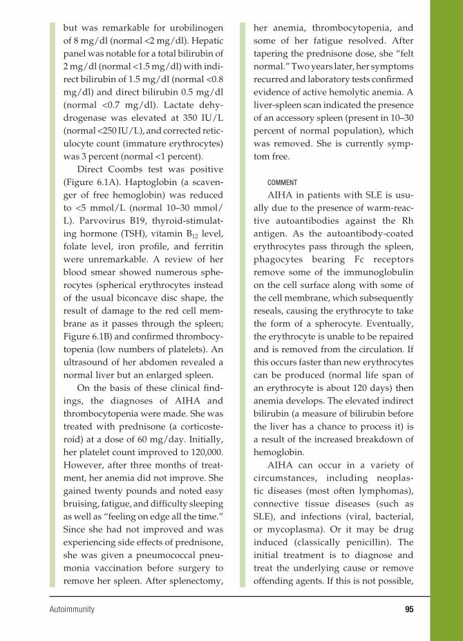
Autoimmunity 95
but was remarkable for urobilinogen of 8 mg/dl (normal <2 mg/dl). Hepatic panel was notable for a total bilirubin of 2 mg/dl (normal <1.5 mg/dl) with indi-rect bilirubin of 1.5 mg/dl (normal <0.8 mg/dl) and direct bilirubin 0.5 mg/dl (normal <0.7 mg/dl). Lactate dehy-drogenase was elevated at 350 IU/L (normal <250 IU/L), and corrected retic-ulocyte count (immature erythrocytes) was 3 percent (normal <1 percent).
Direct Coombs test was positive (Figure 6.1A). Haptoglobin (a scaven-ger of free hemoglobin) was reduced to <5 mmol/L (normal 10–30 mmol/L). Parvovirus B19, thyroid-stimulat-ing hormone (TSH), vitamin B12 level, folate level, iron profi le, and ferritin were unremarkable. A review of her blood smear showed numerous sphe-rocytes (spherical erythrocytes instead of the usual biconcave disc shape, the result of damage to the red cell mem-brane as it passes through the spleen; Figure 6.1B) and confi rmed thrombocy-topenia (low numbers of platelets). An ultrasound of her abdomen revealed a normal liver but an enlarged spleen.
On the basis of these clinical fi nd-ings, the diagnoses of AIHA and thrombocytopenia were made. She was treated with prednisone (a corticoste-roid) at a dose of 60 mg/day. Initially, her platelet count improved to 120,000. However, after three months of treat-ment, her anemia did not improve. She gained twenty pounds and noted easy bruising, fatigue, and diffi culty sleeping as well as “feeling on edge all the time.” Since she had not improved and was experiencing side effects of prednisone, she was given a pneumococcal pneu-monia vaccination before surgery to remove her spleen. After splenectomy,
her anemia, thrombocytopenia, and some of her fatigue resolved. After tapering the prednisone dose, she “felt normal.” Two years later, her symptoms recurred and laboratory tests confi rmed evidence of active hemolytic anemia. A liver-spleen scan indicated the presence of an accessory spleen (present in 10–30 percent of normal population), which was removed. She is currently symp-tom free.
COMMENT
AIHA in patients with SLE is usu-ally due to the presence of warm-reac-tive autoantibodies against the Rh antigen. As the autoantibody-coated erythrocytes pass through the spleen, phagocytes bearing Fc receptors remove some of the immunoglobulin on the cell surface along with some of the cell membrane, which subsequently reseals, causing the erythrocyte to take the form of a spherocyte. Eventually, the erythrocyte is unable to be repaired and is removed from the circulation. If this occurs faster than new erythrocytes can be produced (normal life span of an erythrocyte is about 120 days) then anemia develops. The elevated indirect bilirubin (a measure of bilirubin before the liver has a chance to process it) is a result of the increased breakdown of hemoglobin.
AIHA can occur in a variety of circumstances, including neoplas-tic diseases (most often lymphomas), connective tissue diseases (such as SLE), and infections (viral, bacterial, or mycoplasma). Or it may be drug induced (classically penicillin). The initial treatment is to diagnose and treat the underlying cause or remove offending agents. If this is not possible,
96 Autoimmunity
corticosteroids such as prednisone are often used. If patients do not respond, then consideration is given to the use of cytotoxic drugs (e.g., azathioprine or vincristine) or splenectomy.
Type IIB Hypersensitivity: Graves’
Disease
Graves’ disease is an organ-specifi c auto-immune disease of the thyroid mediated by stimulatory (agonistic) autoantibod-ies. Autoantibodies to the thyroid-stimu-lating hormone receptor (TSHR) cause hyperthyroidism in patients with Graves’ disease. The pathogenicity of anti-TSHR autoantibodies is demonstrated by the occurrence of neonatal Graves’ disease after passive transplacental transfer of IgG thyroid-stimulating autoantibodies from a mother with Graves’ disease to the fetus. The anti-TSHR autoantibodies in Graves’ disease inhibit binding of TSH to its recep-tor by binding to a conformational epitope (the part of the antigen recognized by an antibody) of the extracellular domain of the TSHR. Although the autoantibodies appear to interact with TSHR somewhat differently than the natural ligand, they nevertheless stimulate TSHR signaling, causing increased production of thyroid hormone.
Type IIB Hypersensitivity: Myasthenia
Gravis
Myasthenia gravis is an autoimmune disease caused by inhibitory (antagonis-tic) autoantibodies that bind and block the acetylcholine receptor (AChR), caus-ing muscular weakness and fatigue. The AChR is found at postsynaptic membranes of neuromuscular junctions and binds
acetylcholine released from a nerve end-ing, transiently opening a calcium channel. The signal is terminated by acetylcholine esterase, an enzyme located in the basal lamina between the nerve ending and the postsynaptic membrane. As in mothers with Graves’ disease, transplacental pas-sage of IgG autoantibodies from mothers with myasthenia gravis can cause transient neonatal myasthenia gravis. Anti-AChR autoantibodies cause disease by down-regulating expression of the receptor and by complement-mediated lysis of the cells bearing the AChR. Intermolecular cross-linking of AChR by the autoantibodies may lead to antigenic modulation.
Type III Autoimmune Reactions
(Immune Complex Disease)
Autoantibodies also cause disease by form-ing networks of autoantibodies bound to their antigens (immune complexes). The antigen-antibody complexes can deposit in tissues, causing infl ammatory lesions. Studies of serum sickness led to the fi rst description of an immune complex dis-ease. Serum sickness is manifested by fever, glomerulonephritis, vasculitis, urti-caria, and arthritis, appearing seven to twenty-one days after primary immuniza-tion or two to four days after secondary immunization with a foreign protein. Two consequences of immune complex forma-tion are complement fi xation and binding to Fc or complement receptors on phago-cytes. Clearance is facilitated by the bind-ing of immune complexes to C3b recep-tors (CR1) on erythrocytes, which retain the complexes in the circulation until their removal by the reticuloendothelial cells of the spleen or liver.
Immune complex formation is a nor-mal process that removes foreign antigens
Autoimmunity 97
from the circulation. Removal of immune complexes by phagocytes bearing Fc or complement receptors prevents their deposition at other sites. The effi ciency of uptake of immune complexes by either Fc receptors or CR1 is proportional to the number of IgG molecules associated with the complex.
Immune complexes can activate either the classical or the alternative complement pathway. The classical pathway plays a major role in maintaining immune com-plexes in a soluble form, preventing their deposition in tissues. C3b bound to the sol-ubilized immune complexes promotes their clearance by the erythrocyte complement receptor CR1. If the rate of immune com-plex formation exceeds the ability to clear these complexes via Fc receptors and CR1, the immune complexes can deposit within tissues, leading to inflammation. This effi cient immune complex transport and removal by Fc and complement receptors can be overwhelmed, however, leading to tissue deposition and immune complex disease. This situation may result from overproduction of immune complexes, blockade of phagocytosis by the reticulo-endothelial system, or complement deple-tion resulting in ineffi cient solubilization of immune complexes.
SLE is the prototype of human immune complex disease. Tissue damage in lupus is mainly caused by immune complexes containing autoantibodies to soluble anti-gens. These autoantibodies include anti-bodies against RNA-protein complexes (e.g., anti-Sm, RNP, Ro/SS-A, and La/SS-B antibodies) and DNA-protein com-plexes (e.g., anti-double-stranded DNA, antihistone, antichromatin antibodies). The target antigens are found mainly in the cell nucleus, although in some cases (e.g., antiribosomal antibodies), they may
be cytoplasmic. Immune complexes con-taining these autoantibodies, especially anti-dsDNA antibodies, are selectively enriched in the renal glomeruli (capillary tufts that produce urine as an ultrafi ltrate of blood) of patients with lupus nephritis and are thought to play a critical role in establishing the infl ammatory response. Immune complex deposition in the kidney leads to proliferative glomerulonephritis and effacement of the normal glomerular architecture (Figure 6.2). As is the case in serum sickness, active lupus nephritis is frequently associated with hypocomple-mentemia (Figure 6.2). In addition to the kidneys (glomeruli), immunoglobulin and complement deposits are found in the blood vessels (vasculitis), skin (rashes), nervous system, and other locations. Pre-formed immune complexes may become trapped in the glomerular fi lter, or immune complexes may develop in situ because of the interaction of cationic antigens (e.g., histones) with heparan sulfate glycos-aminoglycan in the glomerular basement membrane. The association of lupus with defi ciencies of the early classical comple-ment components, especially C2 and C4, is consistent with the role of complement pathways in solubilizing immune com-plexes (see the section “Pathogenesis of Autoimmune Disease”).
Case 2. Systemic Lupus Erythematosus, a Type III Autoimmune Reaction
A fi fteen-year-old girl developed myalgias (muscle pain), painful and swollen joints, and low-grade fevers and was found to have a positive anti-nuclear antibodies (ANA) test. Kidney function was normal. She was given a diagnosis of SLE and treated with hydroxychloroquine (an antimalarial), azathioprine (a nucleoside analog), and
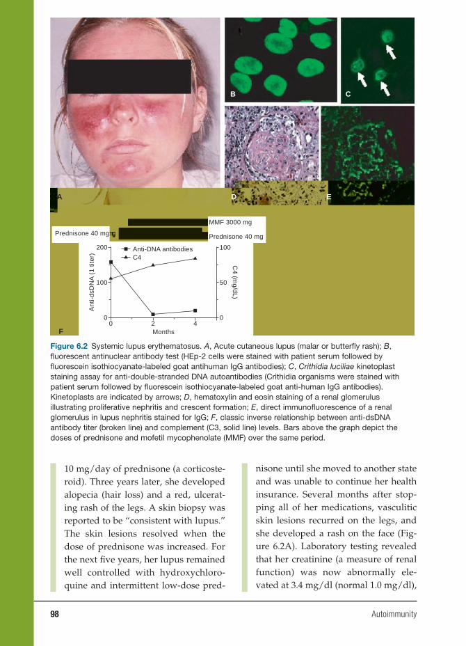
98 Autoimmunity
10 mg/day of prednisone (a corticoste-roid). Three years later, she developed alopecia (hair loss) and a red, ulcerat-ing rash of the legs. A skin biopsy was reported to be “consistent with lupus.” The skin lesions resolved when the dose of prednisone was increased. For the next fi ve years, her lupus remained well controlled with hydroxychloro-quine and intermittent low-dose pred-
nisone until she moved to another state and was unable to continue her health insurance. Several months after stop-ping all of her medications, vasculitic skin lesions recurred on the legs, and she developed a rash on the face (Fig-ure 6.2A). Laboratory testing revealed that her creatinine (a measure of renal function) was now abnormally ele-vated at 3.4 mg/dl (normal 1.0 mg/dl),
Figure 6.2 Systemic lupus erythematosus. A, Acute cutaneous lupus (malar or butterfl y rash); B, fl uorescent antinuclear antibody test (HEp-2 cells were stained with patient serum followed by fl uorescein isothiocyanate-labeled goat antihuman IgG antibodies); C, Crithidia luciliae kinetoplast staining assay for anti-double-stranded DNA autoantibodies (Crithidia organisms were stained with patient serum followed by fl uorescein isothiocyanate-labeled goat anti-human IgG antibodies). Kinetoplasts are indicated by arrows; D, hematoxylin and eosin staining of a renal glomerulus illustrating proliferative nephritis and crescent formation; E, direct immunofl uorescence of a renal glomerulus in lupus nephritis stained for IgG; F, classic inverse relationship between anti-dsDNA antibody titer (broken line) and complement (C3, solid line) levels. Bars above the graph depict the doses of prednisone and mofetil mycophenolate (MMF) over the same period.
A
F
D E
B C
00
100
200
Prednisone 40 mg
2 40
50
100
MMF 3000 mg
Prednisone 40 mg
Months
C4 (m
g/dL)
Ant
i-ds
DN
A (
1 tit
er)
C4Anti-DNA antibodies
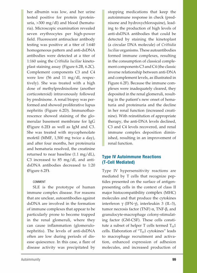
Autoimmunity 99
her albumin was low, and her urine tested positive for protein (protein-uria, >300 mg/dl) and blood (hematu-ria). Microscopic examination revealed seven erythrocytes per high-power fi eld. Fluorescent antinuclear antibody testing was positive at a titer of 1:640 homogeneous pattern and anti-dsDNA antibodies were detected at a titer of 1:160 using the Crithidia luciliae kineto-plast staining assay (Figure 6.2B, 6.2C). Complement components C3 and C4 were low (56 and 11 mg/dl, respec-tively). She was treated with a high dose of methylprednisolone (another corticosteroid) intravenously followed by prednisone. A renal biopsy was per-formed and showed proliferative lupus nephritis (Figure 6.2D). Immunofl uo-rescence showed staining of the glo-merular basement membrane for IgG (Figure 6.2E) as well as IgM and C3. She was treated with mycophenolate mofetil (MMF, 1,500 mg twice a day), and after four months, her proteinuria and hematuria resolved, the creatinine returned to near baseline (1.1 mg/dL), C3 increased to 85 mg/dl, and anti-dsDNA antibodies decreased to 1:20 (Figure 6.2F).
COMMENT
SLE is the prototype of human immune complex disease. For reasons that are unclear, autoantibodies against dsDNA are involved in the formation of immune complexes that appear to be particularly prone to become trapped in the renal glomeruli, where they can cause infl ammation (glomerulo-nephritis). The levels of anti-dsDNA often are low during periods of dis-ease quiescence. In this case, a fl are of disease activity was precipitated by
stopping medications that keep the autoimmune response in check (pred-nisone and hydroxychloroquine), lead-ing to the production of high levels of anti-dsDNA antibodies that could be detected by staining the kinetoplast (a circular DNA molecule) of Crithidia luciliae organisms. These autoantibodies formed immune complexes, resulting in the consumption of classical comple-ment components C3 and C4 (the classic inverse relationship between anti-DNA and complement levels, as illustrated in Figure 6.2F). Because the immune com-plexes were inadequately cleared, they deposited in the renal glomeruli, result-ing in the patient’s new onset of hema-turia and proteinuria and the decline in her renal function (increased creati-nine). With reinstitution of appropriate therapy, the anti-DNA levels declined, C3 and C4 levels recovered, and renal immune complex deposition dimin-ished, resulting in an improvement of renal function.
Type IV Autoimmune Reactions
(T-Cell Mediated)
Type IV hypersensitivity reactions are mediated by T cells that recognize pep-tides presented on the surface of antigen-presenting cells in the context of class II major histocompatibility complex (MHC) molecules and that produce the cytokines interferon γ (IFN-γ), interleukin 3 (IL-3), tumor necrosis factor (TNF) α, TNF-β, and granulocyte-macrophage colony-stimulat-ing factor (GM-CSF). These cells consti-tute a subset of helper T cells termed TH1 cells. Elaboration of “TH1 cytokines” leads to macrophage recruitment and activa-tion, enhanced expression of adhesion molecules, and increased production of
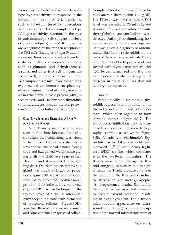
100 Autoimmunity
monocytes by the bone marrow. Delayed-type hypersensitivity in response to the intradermal injection of certain antigens, such as tuberculin (used for tuberculosis skin testing), is a classic example of a type IV hypersensitivity reaction. In the case of autoimmunity, self-antigens (instead of foreign antigens) plus MHC molecules are recognized by the antigen receptors of the TH1 cells. Examples of type IV autoim-mune reactions include insulin-dependent diabetes mellitus (pancreatic antigens, such as glutamic acid dehydrogenase, insulin, and other islet cell antigens are recognized), multiple sclerosis (unidenti-fi ed components of myelin are recognized), experimental antoimmune encephalomy-elitis (an animal model of multiple sclero-sis in which myelin basic protein (MBP) is recognized), and Hashimoto’s thyroiditis (thyroid antigens such as thyroid peroxi-dase and thyroglobulin are recognized).
Case 3. Hashimoto’s Thyroiditis: A Type IV Autoimmune Disease
A thirty-one-year-old woman was seen in the clinic because she had a sensation that something was stuck in her throat. Her older sister had a similar problem. She also noted feeling tired and had gained weight since giv-ing birth to a child fi ve years earlier. Her hair and skin seemed to be get-ting drier. On examination, her thyroid gland was mildly enlarged on palpa-tion (Figure 6.3A, 6.3B) and ultrasound revealed multiple small nodules and a pseudonodule indicated by the arrow (Figure 6.3C). A needle biopsy of the thyroid revealed a diffuse interstitial lymphocytic infi ltrate with formation of lymphoid follicles (Figure 6.3D). Residual thyroid follicles were small, and some contained inspissated colloid.
Complete blood count was notable for mild anemia (hemoglobin 11.3 g/dl). Her T4 level was low (1.9 µg/dl), TSH level was elevated at 25 mIU/L, and serum antithyroid peroxidase and anti-thyroglobulin autoantibodies were detected. Antithyroid-stimulating hor-mone receptor antibody was negative. She was given a diagnosis of autoim-mune (Hashimoto’s) thyroiditis on the basis of the low T4 level, elevated TSH, and the autoantibody profi le and was treated with thyroid replacement. Her TSH levels normalized and the ane-mia resolved and she noted a gradual decrease in her fatigue. Her skin and hair dryness improved.
COMMENT
Pathologically Hashimoto’s thy-roiditis represents an infi ltration of the thyroid gland with T and B lympho-cytes, which often organize to form germinal centers (Figure 6.3D). The lymphocytic infi ltration may be visu-alized on positron emission tomog-raphy scanning as shown in Figure 6.3E. Patients with Hashimoto’s thy-roiditis may exhibit a focal or diffusely increased 2-[18F]fl uoro-2-deoxy-d-glu-cose (FDG) uptake, which correlates with the T-/B-cell infi ltration. The B cells make antibodies against thy-roid antigens, as seen in this patient, whereas the T cells produce cytokines that stimulate the B cells and induce the thyroid cells to undergo apopto-sis (programmed death). Eventually, the thyroid is destroyed and is unable to secrete thyroid hormone, result-ing in hypothyroidism. The diffusely micronodular appearance on ultra-sound (Figure 6.3C) is due to disrup-tion of the normal microarchitecture of
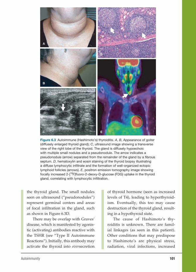
Autoimmunity 101
the thyroid gland. The small nodules seen on ultrasound (“pseudonodules”) represent germinal centers and areas of focal infi ltration in the gland, such as shown in Figure 6.3D.
There may be overlap with Graves’ disease, which is manifested by agonis-tic (activating) antibodies reactive with the TSHR (see “Type II Autoimmune Reactions”). Initially, this antibody may activate the thyroid into oversecretion
of thyroid hormone (seen as increased levels of T4), leading to hyperthyroid-ism. Eventually, this too may cause destruction of the thyroid gland, result-ing in a hypothyroid state.
The cause of Hashimoto’s thy-roiditis is unknown. There are famil-ial linkages (as seen in this patient). Other conditions that may predispose to Hashimoto’s are physical stress, radiation, viral infections, increased
A
D
E
B
C
Figure 6.3 Autoimmune (Hashimoto’s) thyroiditis. A, B, Appearance of goiter (diffusely enlarged thyroid gland); C, ultrasound image showing a transverse view of the right lobe of the thyroid. The gland is diffusely hypoechoic with multiple small nodules and a pseudonodule. The arrow indicates a pseudonodule (arrow) separated from the remainder of the gland by a fi brous septum. D, hematoxylin and eosin staining of the thyroid biopsy illustrating a diffuse lymphocytic infi ltrate and the formation of well-organized ectopic lymphoid follicles (arrows). E, positron emission tomography image showing focally increased 2-[18F]fl uoro-2-deoxy-D-glucose (FDG) uptake in the thyroid gland, correlating with lymphocytic infi ltration.
102 Autoimmunity
iodine, medications (most notably amiodarone, lithium, and interferon-α), other autoimmune diseases (most notably Sjögren’s syndrome), female gender, and pregnancy.
Epidemiology of Autoimmune Disease
There are nearly 100 different forms of auto-immune disease, making these disorders a major cause of chronic illness, affect-ing up to 3 percent of the general popula-tion. Nearly any organ can be affected by either systemic or organ-specifi c autoim-mune disease (Table 6.1). Women make up nearly 75 percent of all individuals affl icted by autoimmune disease, making these disorders one of the ten leading causes of death in women less than sixty-fi ve years old. However, the female-to-male ratio var-ies widely among different diseases, being as high as 9:1 in SLE, Sjögren’s syndrome, and autoimmune thyroiditis and as low as 1:1 in TID, Goodpasture’s syndrome, and vitiligo (Table 6.1). The mean age of onset also varies widely, with some disorders typically occurring early in childhood (e.g., TID, juvenile rheumatoid arthritis), oth-ers in the childbearing years (ages 15–45, e.g., SLE), and still others in later life (e.g., Sjögren’s syndrome). There may be strik-ing ethnic/racial predispositions to auto-immune disease. For example, SLE is about three times more prevalent in individuals of African, Asian, or Latin ancestry than in individuals of European ancestry, whereas Sjögren’s syndrome and multiple sclerosis are more prevalent in those of European ancestry. The racial/ethnic differences are likely to refl ect differences in the frequen-cies of disease susceptibility genes. The costs of these disorders to society are enor-mous. Rheumatoid arthritis (RA) affects
2.1 million Americans (1.5 million women and 600,000 men) at an annual cost of about $6,000 per patient (direct medical costs and indirect costs such as absence from work). Lupus affects 500,000 Americans at an esti-mated annual cost of $13,000 per patient, a total $6.5 billion per year.
ANIMAL MODELS OF AUTOIMMUNE
DISEASE
The diffi culty in carrying out randomized, well-controlled research in patients com-plicates studies of the pathogenesis and treatment of human autoimmune disease. Often the simplest course is to fi rst study the disease in an appropriate animal model. However, because animal models of dis-ease rarely are identical to the human dis-order, the suitability of a particular model in any given situation must be considered carefully before undertaking animal stud-ies. The list of animal models of autoim-mune disease is extensive, and only some of the more commonly studied models can be reviewed here (see also Table 6.2).
SLE (NZB X NZW F1, MRL, BXSB,
TMPD)
Numerous mouse strains have been stud-ied over the years as animal models of SLE. Some strains develop lupus spontane-ously, such as NZB X NZW (F1) (NZB/W) hybrid mice, MRL mice, and BXSB male mice. Other models, such as tetramethyl-pentadecane (TMPD, pristane) induced lupus, are inducible with chemicals. The spontaneous models afford hope that if the genetic defect(s) responsible for lupus-like disease in these mice can be identifi ed, similar defects will be found in human lupus. However, the inducible TMPD
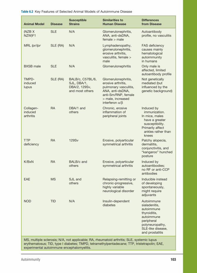
Autoimmunity 103
Table 6.2 Key Features of Selected Animal Models of Autoimmune Disease
Animal Model DiseaseSusceptible Strains
Similarities to Human Disease
Differences from Disease
(NZB X NZW)F1
SLE N/A Glomerulonephritis, ANA, anti-dsDNA, female > male
Autoantibody profi le, no vasculitis
MRL lpr/lpr SLE (RA) N/A Lymphadenopathy, glomerulonephritis, erosive arthritis, vasculitis, female > male
FAS defi ciency causes mainly hematological autoimmunity in humans
BXSB male SLE N/A Glomerulonephritis Only male is affected, limited autoantibody profi le
TMPD-induced lupus
SLE (RA) BALB/c, C57BL/6, SJL, DBA/1, DBA/2, 129Sv, and most others
Glomerulonephritis, erosive arthritis, pulmonary vasculitis, ANA, anti-dsDNA, anti-Sm/RNP, female > male, increased interferon α/β
Not genetically mediated (but infl uenced by the genetic background)
Collagen-induced arthritis
RA DBA/1 and others
Chronic, erosive infl ammation of peripheral joints
Induced by immunization.
In mice, males have a greater susceptibility.
Primarily affect ankles rather than knees
T TP defi ciency
RA 129Sv Erosive, polyarticular symmetrical arthritis
Patchy alopecia, dermatitis, conjunctivitis, and “kangaroo” hunched posture
K/BxN RA BALB/c and others
Erosive, polyarticular symmetrical arthritis
Induced by autoantibodies; no RF or anti-CCP antibodies
EAE MS SJL and others
Relapsing-remitting or chronic-progressive, highly variable neurological disorder
Inducible instead of developing spontaneously, might require adjuvants
NOD TID N/A Insulin-dependent diabetes
Autoimmune sialadenitis, autoimmune thyroiditis, autoimmune peripheral polyneuropathy, SLE-like disease, and prostatitis
MS, multiple sclerosis; N/A, not applicable; RA, rheumatoid arthritis; SLE, systemic lupus erythematosus; TID, type I diabetes; TMPD, tetramethylpentadecane; TTP, tristetrapolin; EAE, experimental autoimmune encephalomyelitis.
104 Autoimmunity
model more closely mimics the abnormali-ties in interferon (IFN) α and β production seen in most lupus patients.
NZB/W F1 MODELThe NZB/W model was the first
murine model of lupus nephritis. New Zealand Black (NZB) mice develop AIHA and the female New Zealand White (NZW) mice develop mesangial glomerulonephri-tis late in life. In contrast, the F1 hybrid (NZB/W) develops early-onset severe (proliferative, immune complex-medi-ated) glomerulonephritis along with ANA, antichromatin, and anti-dsDNA antibod-ies. However, these mice lack other classic clinical and serological manifestations of SLE, such as arthritis, infl ammatory skin rashes, serositis, and anti-Sm autoanti-bodies. Extensive genetic analysis of this strain has revealed three major susceptibil-ity intervals on chromosomes 1, 4, and 7. Each of these intervals appears to contain multiple disease-susceptibility genes and several candidate genes have been identi-fi ed. NZB/W mice have been used widely for preclinical studies of various therapeu-tic interventions for lupus nephritis.
MRL MODEL MRL mice, an inbred strain derived
from several other strains, develop ANAs and late-onset glomerulonephritis remi-niscent of SLE. A spontaneously occurring mutation led to impressive lymphoprolif-eration (lpr mutation), severe, early-onset nephritis closely resembling proliferative lupus nephritis, the development of ero-sive arthritis (more characteristic of RA than SLE), salivary gland infl ammation (reminiscent of Sjögren’s syndrome), vas-culitis, and skin disease resembling cuta-neous lupus. Both MRL and MRL lpr/lpr mice develop a host of autoantibodies
characteristic of SLE, including anti-Sm and anti-dsDNA as well as severe hyper-gammaglobulinemia. These autoantibodies develop earlier in the presence of the lpr mutation, which generally accelerates the onset of lupus-like disease in this strain. The abnormalities caused by the lpr mutation are due to an ETn retrotransposon inser-tion into the Fas gene, which encodes an important protein mediator of apoptosis. Defective apoptosis of lymphocytes leads to the accumulation of CD3+CD4−CD8− (“double negative”) T cells, accounting for the massive lymphoproliferation seen in MRL lpr/lpr mice.
BXSB MODELThis strain was created by crossing
male SB/Le and female C57BL/6J mice. Male, but not female, BXSB mice develop severe glomerulonephritis, lymphade-nopathy, splenomegaly, AIHA, and anti-dsDNA autoantibodies. Thus, the sex predilection is an important difference from human lupus and most other murine lupus models. A mutant gene located on the Y chromosome, designated Yaa (Y chromosome-linked autoimmune accel-eration), causes accelerated lupus-like dis-ease in male BXSB mice. A recent study showed that the Yaa mutation results from translocation of a 4-megabase portion of the X chromosome to the Y chromosome, leading to increased expression of several genes that are normally X linked, includ-ing TLR7.
TMPD-INDUCED LUPUSIntraperitoneal injection of pristane (2,
6, 10, 14 tetramethylpentadecane, TMPD) can induce a lupus-like syndrome in non-autoimmune-prone mice characterized by proliferative glomerulonephritis, erosive arthritis, pulmonary vasculitis,
Autoimmunity 105
and a variety of lupus autoantibodies, including anti-dsDNA and anti-Sm. All or nearly all immunocompetent mouse strains are susceptible to lupus induced by this hydrocarbon oil. This inducible model of lupus is, at least so far, unique in reproducing the increased levels of IFN-α and IFN-β seen in the majority of lupus patients. The disease is largely abrogated in type I interferon receptor-defi cient mice.
Rheumatoid Arthritis (Collagen-
Induced Arthritis, TTP Deficiency,
K/BxN Model)
RA is a systemic autoimmune disease char-acterized by prominent joint involvement. Arthritis is typically associated with ero-sion of cartilage and subchondral bone, formation of an infl ammatory tissue, con-sisting of activated macrophages, T cells, fi broblasts, and other immune cells (pan-nus). This can ultimately result in joint destruction and signifi cant joint deformi-ties. In addition to the joints, RA can cause vasculitis, splenomegaly, and leukopenia (Felty’s syndrome), interstitial lung dis-ease, and other abnormalities. Rheuma-toid factor (an autoantibody against the Fc portion of immunoglobulin) and anti-bodies against citrulline-modifi ed proteins or peptides (usually detected as antibod-ies against an artifi cially produced cyclic citrullinated peptide, or CCP) are typical serological fi ndings in RA, although not all patients exhibit these abnormalities. Several animal models of RA exist, but they do not precisely reproduce the clinical and laboratory abnormalities.
COLLAGEN-INDUCED ARTHRITIS (CIA)Immunization of susceptible rodent
strains with type II collagen (CII) leads to
the development of a severe polyarticular arthritis resembling human RA. Although induced by heterologous CII, immuniza-tion leads to a response against autologous CII. CIA can be induced in susceptible strains of mice, rats, and primates. Histo-logically, both RA and CIA are character-ized by an intense synovitis accompanied by erosion of cartilage and subchon-dral bone by a pannuslike tissue. Unlike human RA, CIA is monophasic. In addi-tion, there are important serological differ-ences. In general, rheumatoid factor is not produced in CIA and antibodies against CCP are absent.
TTP DEFICIENCYTristetraprolin (TTP) is a transcription
factor that can bind to and destabilize mRNAs encoding TNF-α and granu-locyte-macrophage colony-stimulating factor (GM-CSF). Mice defi cient in TTP develop a complex syndrome character-ized by cachexia, polyarticular arthritis, dermatitis, autoimmunity, and myeloid hyperplasia accompanied by extramedul-lary hematopoiesis (erythrocyte produc-tion outside of the bone marrow). TTP knockout mice exhibit exuberant infl am-matory pannus and bony erosions. These mice also produce high titers of anti-DNA, and ANAs; however, rheumatoid factors are absent.
K/BXN MODELAlthough RA has been considered pri-
marily a type IV autoimmune reaction for many years, the fi nding that autoantibod-ies against glucose-6-phosphate isomerase (GPI) can transfer RA-like joint disease to normal mice has rekindled interest in the possibility that antibody-mediated auto-immune mechanisms (type II or type III) could play a role in the pathogenesis of
106 Autoimmunity
RA. K/BxN T-cell-receptor transgenic mice express a transgenic T-cell receptor specifi c for a peptide of the ubiquitously expressed self-protein GPI. Arthritis in this model is initiated by antibodies against GPI. The resulting synovitis is chronic, erosive, and associated with pannus formation. Para-doxically, although the GPI antigen is ubiq-uitous, autoimmunity is focused on the joints. It appears that the GPI protein, not a cross-reactive synovial antigen, is the tar-get of the pathogenic antibodies. Although the histological appearance of the affected joints is reminiscent of RA, there is no evi-dence that RA in humans can be caused by antibodies against GPI. The classic sero-logical abnormalities, rheumatoid factor, and anti-CCP antibodies are not seen, and anti-TNF-α antibodies have little effect in this model.
Multiple Sclerosis (Experimental
Autoimmune Encephalomyelitis)
Multiple sclerosis (MS) is a chronic autoimmune disease affecting the central nervous system, including the brain and spinal cord. The disease affects about 350,000 Americans and about 1.1 mil-lion worldwide. Age of onset is typically twenty to forty years old, women are affected more frequently than men (2:1 ratio), and it is most prevalent in indi-viduals of northern European ancestry. It is thought to be mediated primarily by a T-cell-mediated attack on the myelin sheaths of certain nerve fi bers, resulting in infl ammation, demyelination, and glio-sis (scarring). In addition, autoantibod-ies against components of myelin such as myelin oligodendrocyte glycoprotein (MOG) may be seen and also may con-tribute to disease pathogenesis by fi xing complement. During the course of disease,
the lesions classically occur at different times and in different locations. Symp-toms include sensory loss, paresthesias (numbness, tingling), visual changes due to optic neuritis, tremor, ataxia, weakness, spasticity, and other neurological symp-toms. Patients with MS can exhibit either a relapsing-remitting or a progressive course.
EAE is a model of MS induced in susceptible animals by immunization by intact myelin or components of myelin, the sheath that surrounds certain neurons. Like CIA for RA, EAE can be induced in several species, including mice, rats, guinea pigs, rabbits, and primates. Although induced by heterologous antigen(s), the disease is autoimmune. Several proteins have been used to induce EAE, including MBP, pro-teolipid protein (PLP), and MOG. Different antigens cause somewhat different clini-cal manifestations. By administering the antigen with complete Freund’s adjuvant and pertussis toxin, the blood-brain bar-rier is disrupted, permitting access by immune cells. The resulting demyelinating disease closely resembles human MS and is thought to be mediated primarily by T cells because disease can be transferred to normal animals by T cells (type IV autoim-mune reaction). There is only limited evi-dence that an immune response to MBP, PLP, or MOG is involved in human disease, and it is hypothesized that other myelin antigens may be the targets of autoreactive T cells in MS.
Type I Diabetes (Nonobese Diabetic
Mouse Model)
TID is an autoimmune disease in which the insulin-producing β cells in the pan-creatic islets of Langerhans are gradually destroyed by autoreactive T cells over
Autoimmunity 107
a period of months to years. After about 80 percent of the islet cells are destroyed, insulin defi ciency and a severe form of insulin-dependent diabetes marked by ketoacidosis develops. The disease usually affects children and young adults but can occur at any age. Males and females are affected equally. The highest incidence is in Scandinavians (35 per 100,000 per year). Individuals with a genetic susceptibil-ity to the disease are thought to develop autoimmunity in response to an undefi ned environmental trigger. Most patients with TID produce anti-islet cell autoantibod-ies reactive with insulin, glutamic acid decarboxylase, ICA-512/IA-2, phogrin, or other antigens. These autoantibodies generally appear before the onset of clini-cal diabetes and have been used for early diagnosis of the condition.
The nonobese diabetic (NOD) mouse is the most useful model of autoimmune TID. NOD mice spontaneously develop marked infi ltration of T cells into the pancreatic islets. The infi ltrating T cells selectively destroy the pancreatic β cells. In addition to diabetes, NOD mice spon-taneously develop autoimmune responses involving other tissues, including sali-vary gland, lacrimal gland, thyroid gland, parathyroid gland, adrenal gland, testis, large bowel, and red blood cells. NOD mice also are susceptible to exogenously induced autoimmune diseases, such as experimental autoimmune thyroiditis, colitis-like wasting disease, encephalomy-elitis, and SLE. Defects related to several genes, including the MHC class II, CTLA-4, and IL-2, have been associated with the susceptibility to diabetes. T cells play an important role in the development and progression of disease, whereas B cells are not required at the effector stage of TID in NOD mice.
Autoimmune Thyroiditis (Experimental
Autoimmune Thyroiditis)
Experimental autoimmune thyroiditis is induced in mice by immunization with murine thyroglobulin plus complete Freund’s adjuvant. The mice develop autoantibodies against thyroglobulin and histological changes consistent with those seen in human autoimmune thyroiditis. It is a useful model for studying the patho-genesis of human chronic (Hashimoto’s) thyroiditis.
PATHOGENESIS OF AUTOIMMUNE
DISEASE
Genetic Predisposition
Genetic, environmental, and random (sto-chastic) factors all play a role in the patho-genesis of autoimmune diseases. Family members of affected individuals are at higher risk for developing autoimmune disease than the general population. The relative risk to siblings of affected individ-uals (probands) versus the risk in the gen-eral population (λs = disease prevalence in siblings of affected individuals ÷ disease prevalence in the general population) is a useful way to estimate the importance of genetic factors. The relative risk is between fi ve- and fi ftyfold higher in siblings of affected probands than in unrelated indi-viduals in most autoimmune diseases (Table 6.3). Part of this effect is accounted for by MHC-linked genes.
Twin studies illustrate the importance of these genetic factors. If the concordance rates in monozygotic and dizygotic twins are about the same, the genetic effect is small. For most autoimmune diseases, concordance rates are 15–30 percent for monozygotic twins versus 2–5 percent for

108 Autoimmunity
dizygotic twins, consistent with a sizeable genetic effect (Table 6.3). Identifi cation of the actual mutations or genetic polymor-phisms that confer susceptibility to auto-immune diseases has been complicated by the fact that most autoimmune disorders appear to involve multiple genes, each with only a small effect. Moreover, many autoimmune “diseases” are actually “syn-dromes” that may arise through a variety of different pathogenic mechanisms and genetic abnormalities. Even in the inbred lupus-prone mouse strain NZB/W, ten or more susceptibility loci are thought to contribute to disease severity in an addi-tive fashion (threshold liability model). Human SLE and other autoimmune dis-eases also are likely to be highly complex genetically. Interestingly, there may be some overlap genetically between different forms of autoimmune disease, such as SLE and type I diabetes. More than half of the linkages identifi ed in genomewide scan-
ning studies of a variety of systemic and organ-specifi c autoimmune diseases map nonrandomly into eighteen chromosomal clusters, possibly explaining the occur-rence of several autoimmune diseases in a given individual or family. For example, Hashimoto’s thyroiditis is associated with a variety of organ-specifi c (e.g., TID, per-nicious anemia, autoimmune hepatitis, and Addison’s disease) and systemic (e.g., lupus, RA, and Sjögren’s syndrome) auto-immune diseases. Pedigrees with more than one systemic autoimmune disorder are not unusual. Of course, shared envi-ronmental infl uences could also explain familial clustering.
Among candidate genes, the MHC class II molecule is the most comprehen-sively studied. MHC polymorphisms are associated with development of RA, SLE, MS, TID, and other autoimmune diseases (Table 6.3). RA is a striking example. A shared epitope, consisting of a 5-amino
Table 6.3 Twin Studies IIlustrating the Importance of Genetic Factors in Autoimmune Disease
Concordance Rates Sibling Risk/Population Risk (λs)
Disease Dizygotic Twins Monozygotic Twins Overall Attributable to MHC
Systemic autoimmunediseases
Rheumatoid arthritis
4% 12–15% 8 1.6
SLE 2% 24% 20 N/A
Organ-specifi c autoimmunediseases
Type 1 diabetes 5% 33% 15 2.4
Multiple sclerosis 3.5% 21–40% 20 2.4
Graves’ disease 0% 36% 15 N/A
Compiled from the following references: Vyse TJ, Todd JA. Genetic analysis of autoimmune disease. Cell. 1996;85:311–318; Jarvinen P, Aho K. Twin studies in rheumatic diseases. Semin Arthritis Rheum. 1994;24(1):19–28; Brix TH, Christensen K, Holm NV, Harvald B, Hegedus L. A population-based study of Graves’ disease in Danish twins. Clin Endocrinol. 1998;48(4):397–400; Redondo MJ, Fain PR, Eisenbarth GS. Genetics of type 1A diabetes. Recent Prog Horm Res. 2001;56:69–89; Kahana E. Epidemiologic studies of multiple sclerosis: a review. Biomed Pharmacother. 2000;54(2):100–102.
Autoimmunity 109
acid sequence motif in the third allelic hypervariable region of the HLA-DRβ1 chain (QKRAA in the *0401 allele, QRRAA in the *0404 and *0101 alleles), is carried by 90 percent of patients with RA and is associated with disease severity. In addi-tion to MHC-linked genes, mutations or genetic polymorphisms involving non-MHC genes also are strong candidates for autoimmune disease susceptibility genes (Table 6.4). These include genetic polymor-phisms or defi ciency of molecules involved in the response to or clearance of immune complexes (e.g., C1q, C4, FcγRIIa, FcγRIIb, FcγRIIIa, mannose-binding lectin), which are associated with SLE, as well as genes influencing T-cell activation (PTPN22, CTLA-4), cytokine responses (STAT-4, IRF-5, Tyk2), or programmed cell death (Fas, PDCD1; Table 6.4).
Many, if not most, systemic and organ-specifi c autoimmune diseases are thought to be multifactorial, involving multiple genetic defects consistent with the thresh-old liability model of multifactorial inheri-tance. This model supposes a continuously distributed genetically determined liability to the development of disease. Individuals who develop disease will bear multiple disease susceptibility genes. Because of the normal distribution of genes, fi rst-degree relatives will have much higher risk of developing disease than the general population, second-degree relatives will have a moderate risk, and third-degree relatives will have low risk.
Environmental Triggers of
Autoimmune Disease
Environmental factors can trigger autoim-mune disease in susceptible hosts, as illus-trated by the initiation of disease by sun exposure in a subset of lupus patients. The
wavelengths most likely to induce lupus fall in the UV range: UVC (200–290 nm), UVB (290–320 nm), and UVA (320–400 nm). UVB irradiation is mostly absorbed in the upper layers of the epidermis, whereas the longer wavelength UVA is able to reach the dermis. UV exposure can induce apop-tosis and release of immune mediators and activation of resident dendritic cells and T cells. Expression of certain self-antigens, such as Ro60/Ro52, on the surface of the apoptotic cells may lead to antibody-medi-ated infl ammatory responses that could play a role in the pathogenesis of skin rashes in lupus.
The importance of environmental fac-tors is further illustrated by the induc-tion of a murine lupus syndrome by the hydrocarbon pristane (see “Animal Mod-els of Autoimmune Disease”), which appears to act, in part, through the induc-tion of type I interferon (IFN-α and IFN-β) production. Many other chemicals and drugs have been implicated as triggers of autoimmunity or autoimmune disease. Procainamide, hydralazine, chlorproma-zine, methyldopa, quinidine, minocycline, and nitrofurantoin all have been associ-ated with the induction of ANAs and in some cases antineutrophil cytoplasmic antibodies as well as in the pathogenesis of “drug-induced lupus,” most frequently manifested by serositis (infl ammation of the pleura or pericardium) and arthri-tis. Silica is recognized as a precipitating factor for scleroderma, cigarette smoke may aggravate RA, and trichloroethyl-ene is thought to promote lupus in ani-mal models and possibly humans. Other chemical agents implicated in the patho-genesis of autoimmunity include heavy metals such as mercury, gold, and cad-mium, pesticides, herbicides, hydrazine, and certain dyes.
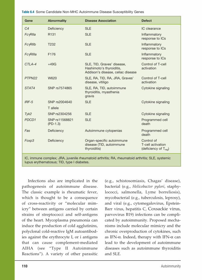
110 Autoimmunity
Infections also are implicated in the pathogenesis of autoimmune disease. The classic example is rheumatic fever, which is thought to be a consequence of cross-reactivity or “molecular mim-icry” between antigens carried by certain strains of streptococci and self-antigens of the heart. Mycoplasma pneumonia can induce the production of cold agglutinins, polyclonal cold-reactive IgM autoantibod-ies against the erythrocyte I, or i antigens that can cause complement-mediated AIHA (see “Type II Autoimmune Reactions”). A variety of other parasitic
(e.g., schistosomiasis, Chagas’ disease), bacterial (e.g., Helicobacter pylori, staphy-lococci, salmonella, Lyme borreliosis), mycobacterial (e.g., tuberculosis, leprosy), and viral (e.g., cytomegalovirus, Epstein-Barr virus, hepatitis C, Coxsackie virus, parvovirus B19) infections can be compli-cated by autoimmunity. Proposed mecha-nisms include molecular mimicry and the chronic overproduction of cytokines, such as IFN-α. Indeed, therapy with IFN-α can lead to the development of autoimmune diseases such as autoimmune thyroiditis and SLE.
Table 6.4 Some Candidate Non-MHC Autoimmune Disease Susceptibility Genes
Gene Abnormality Disease Association Defect
C4 Defi ciency SLE IC clearance
FcγRIIa R131 SLE Infl ammatory response to ICs
FcγRIIb T232 SLE Infl ammatory response to ICs
FcγRIIIa F176 SLE Infl ammatory response to ICs
CTLA-4 +49G SLE, TID, Graves’ disease, Hashimoto’s thyroiditis, Addison’s disease, celiac disease
Control of T-cell activation
PTPN22 W620 SLE, RA, TID, RA, JRA, Graves’ disease, vitiligo
Control of T-cell activation
STAT4 SNP rs7574865 SLE, RA, TID, autoimmune thyroiditis, myasthenia gravis
Cytokine signaling
IRF-5 SNP rs2004640
T allele
SLE Cytokine signaling
Tyk2 SNP rs2304256 SLE Cytokine signaling
PDCD1 SNP rs11568821 (PD-1.3)
SLE Programmed cell death
Fas Defi ciency Autoimmune cytopenias Programmed cell death
Foxp3 Defi ciency Organ-specifi c autoimmune disease (TID, autoimmune thyroiditis)
Control of T-cell activation (defi ciency of Treg)
IC, immune complex; JRA, juvenile rheumatoid arthritis; RA, rheumatoid arthritis; SLE, systemic lupus erythematosus; TID, type I diabetes.
Autoimmunity 111
Maintenance of Self-Tolerance
Environmental triggers, such as sunlight, drugs/chemicals, and infectious agents, act on a genetic background that regulates tolerance to self. The immune system has evolved a remarkable ability to distin-guish self from nonself. Immune tolerance is achieved by multiple mechanisms, operating both centrally and peripher-ally. Central tolerance occurs during the development of T and B lymphocytes in the thymus and bone marrow, respec-tively. This mostly involves the deletion of autoreactive cells before they exit the primary lymphoid organs. In general, lymphocytes exhibiting strong reactiv-ity for ubiquitously expressed self-anti-gens are deleted in this manner, whereas autoreactive cells of lower affi nity for self may escape central tolerance. These cells are held in check by peripheral tolerance mechanisms. Peripheral tolerance is medi-ated by deletion, anergy, and suppression as well as by “neglect” or “ignorance,” acting on autoreactive lymphocytes after they exit the primary organs. In general, lymphocyte activation requires two sig-nals, one delivered by the antigen receptor (T-cell antigen receptor or surface immu-noglobulin) and second, a co-stimulatory signal. For T-cell activation, this co-stimu-latory signal is delivered by the interac-tion of molecules expressed on the surface of professional antigen-presenting cells or B cells, such as CD80 and CD86, which interact with CD28 (or other receptors) on the T-cell surface. In the case of B cells, the co-stimulatory signal is delivered by CD40 ligand, a surface protein expressed by activated helper T cells that interacts with CD40 on the surface of B lympho-cytes. In the absence of a co-stimulatory signal, engagement of the T- or B-cell anti-
gen receptor leads to a state of anergy (the inability of the lymphocyte to respond to its specifi c antigen).
Many self-antigens are expressed at a very low level that is insuffi cient to induce T-cell activation. In the case of T cells, which recognize short peptides asso-ciated with MHC molecules, the induc-tion of self-tolerance requires the genera-tion of a suffi cient amount of self-peptide in antigen-presenting cells to stimulate T-cell deletion or anergy. Self-peptides that are generated ineffi ciently by the anti-gen-presenting cells can neither stimulate immunity nor induce tolerance; that is, the immune system remains “ignorant” of them. If these minor self-peptides are pro-duced in larger amounts and exposed to the immune system in the presence of an inducer of co-stimulatory molecules (e.g., adjuvants), they have the capacity to stim-ulate an immune response. This has been shown experimentally with peptides gen-erated in vitro using proteolytic enzymes or using synthetic self-peptides.
Regulatory T cells (Treg) also play an important role in maintaining peripheral tolerance. Several different subsets of Treg have been reported, but one of the most intensely studied is the CD4+CD25+Foxp3+ subset, which represents about 10 percent of total CD4+ cells. These cells regulate T-cell activation by a cell–cell contact-dependent mechanism and through the secretion of inhibitory cytokines such as IL-10 and transforming growth factor-beta (TGFβ). They suppress both naïve and memory T-cell responses and down-regu-late the expression of pro-infl ammatory cytokines and co-stimulatory molecules on the antigen-presenting cells. These cells are induced in an antigen-specifi c man-ner, but the subsequent suppressive effects are not antigen specifi c. Genetic defects in
112 Autoimmunity
Foxp3, a transcription factor that is the key controller of Treg function, lead to organ-specifi c autoimmune or autoinfl amma-tory diseases. The scurfy mouse has an X-linked defect of the Foxp3 gene that is lethal in males, which exhibit hyperactiva-tion of CD4+ T cells and overproduction of infl ammatory cytokines. Foxp3 muta-tions in humans are the cause of the IPEX (immune dysregulation, polyendocrinopa-thy, enteropathy, X-linked syndrome) syn-drome. These individuals develop organ-specifi c autoimmune diseases, such as TID, autoimmune thyroiditis, and infl ammatory bowel disease. Interestingly, however, the development of systemic autoimmune dis-ease is not part of the syndrome in either mice or humans.
Finally, antigen-presenting cells play an important role in the induction of tol-erance. Dendritic cells can either initiate T-cell activation and proliferation or promote peripheral tolerance through the deletion of autoreactive T cells, depending on their maturation state. Tolerance is induced when antigens are presented by immature dendritic cells and these cells also play a role in the generation and maintenance of Treg.
TREATMENT OF AUTOIMMUNE
DISEASE
Treatment for autoimmune disease is diverse, and in recent years, the options have increased rapidly. Organ-specifi c autoimmune diseases of endocrine func-tion, such as TID and autoimmune thy-roiditis, may be treated with hormone replacement. In contrast, other forms of organ-specifi c autoimmune disease such as autoimmune thrombocytopenia, AIHA, and multiple sclerosis are treated with
immunosuppressive medications, as are the majority of systemic autoimmune dis-eases. Immunosuppressive medications can be categorized by mode of action.
Anti-inflammatory Agents
Nonsteroidal anti-inflammatory drugs have been used since the 1800s when sali-cin was extracted from willow bark (1828) and sodium salicylate (1875) and aspirin (1899) were synthesized. A large number of these drugs, which either selectively or nonselectively inhibit the enzyme cyclo-oxygenase (a synthetic enzyme for pros-taglandins), are currently in use to treat infl ammatory disease. Although most of their anti-infl ammatory properties derive from the inhibition of prostaglandin syn-thesis, at high doses, there is inhibition of the transcription factor nuclear factor κB (NFκB), a key mediator of infl ammatory cytokine production. Corticosteroids have a more potent effect on NFκB and con-sequently a greater anti-inflammatory effect.
Philip S. Hench discovered the anti-infl ammatory properties of cortisone in 1949. Corticosteroids are a mainstay of therapy for many systemic autoimmune diseases, including SLE, RA, and infl am-matory myopathies such as polymyositis. Corticosteroid therapy also is used for the treatment of some of the more seri-ous organ-specifi c autoimmune diseases, such as AIHA, autoimmune thrombocy-topenia, multiple sclerosis, and Goodpas-ture’s syndrome. Corticosteroids reduce infl ammation by multiple mechanisms of action. One major action is enhanced tran-scription of an inhibitor of NFκB called IκB. IκB dimerizes with NFκB, inhibit-ing the production of infl ammatory cyto-kines mediated by this transcriptional
Autoimmunity 113
pathway. In addition, corticosteroids pro-mote the differentiation of a subset of anti-infl ammatory macrophages that produce the cytokine IL-10.
Antimalarial Drugs
Antimalarial drugs have been used for the treatment of SLE and RA since the early 1900s. The precise mechanism of action remains uncertain, but they have been shown to inhibit cytokine (IL-1 and IL-6) production in vitro. The antimalari-als pass freely through cell membranes at neutral pH, but in acidic environments, such as endosomes, they become proton-ated and can no longer diffuse freely. This leads to concentration of the drug within endosomes and the collapse of endosomal pH gradients. It has been proposed that the inhibition of endosomal acidifica-tion interferes with antigen processing or, alternatively, that there is an effect on the interaction of microbial substances such as unmethylated CpG DNA or uridine-rich RNA with endosomal toll-like recep-tors (TLR9 and TLR7/TLR8, respectively). In addition to SLE and RA, antimalarials are used in the treatment of juvenile rheu-matoid arthritis, Sjögren’s syndrome, and infl ammatory myopathies.
Anticytokine Agents
The development of TNF-α inhibitors in the 1990s ushered in a new era of therapy of autoimmune disease using “biologi-cals” capable of interfering with the interactions between cytokines and their receptors. The initial clinical use of TNF inhibitors such as etanercept (a soluble recombinant TNF receptor II linked to the Fc portion of human IgG1), infl iximab (a chimeric human-mouse anti-TNF-α
monoclonal antibody), and adalimumab (a fully humanized monoclonal antibody against TNF-α) in RA demonstrated that although multiple cytokines may be involved in disease pathogenesis (in RA, IL-1, and IL-6 in addition to TNF-α), inhibitors of a single cytokine pathway may show therapeutic effi cacy. In addi-tion to RA, TNF-α inhibitors are used for treating infl ammatory bowel disease, pso-riasis, and psoriatic arthritis and are being tested in sarcoidosis, Wegener’s granulo-matosis, pyoderma gangrenosum, SLE, and Behcet’s syndrome.
Anti-TNF therapy is only the tip of the “biological iceberg.” Recombinant IL-1 receptor antagonist (anakinra) has been approved for the treatment of RA, and numerous other cytokine antagonists are currently in clinical trials or under develop-ment.
Methotrexate
Methotrexate is a folic acid analog used extensively for the treatment of RA. It appears that its ability to inhibit dihydrofo-late reductase is not responsible for its effi -cacy in RA, however. Instead, activity may be related to effects on aminoimidazole-carboxamide ribotide transformylase, leading to the release of adenosine, a potent anti-infl ammatory molecule that inhibits neutophil adherence to fi bro-blasts and endothelial cells. Methotrexate inhibits IL-1 and increases the expression of TH2 cytokines (e.g., IL-4), leading to decreased production of TH1 cytokines (e.g., IFN-γ).
Anti-T-Lymphocyte Therapy
T cells play a key role in the pathogen-esis of type IV autoimmune reactions
114 Autoimmunity
and also are critical for generating the T-cell-dependent autoantibodies mediat-ing type II and type III autoimmune dis-eases. Consequently, considerable effort has gone into the development of thera-peutic agents that selectively or nonselec-tively target T lymphocytes. Drugs that target primarily T cells include cyclo-phosphamide, azathioprine, cyclosporin A, tacrolimus, and the biological CTLA4-Ig. Cyclophosphamide is an alkylating agent that substitutes alkyl radicals into DNA and RNA. The drug is inactive by itself but is converted to an active metabo-lite responsible for its immunosuppres-sive effects. It is used for the treatment of lupus nephritis and other life-threatening complications of SLE and other systemic autoimmune diseases.
Azathioprine is a purine analog that inhibits the synthesis of adenosine and guanine. Like cyclophosphamide, it is converted to an active metabolite (6-mer-captopurine), which inhibits the division of activated B and T cells. Azathioprine is used in the treatment of RA, SLE, auto-immune hepatitis, infl ammatory myopa-thy, vasculitis, and other autoimmune disorders.
Unlike cyclophosphamide and aza-thioprine, cyclosporine and tacrolimus (FK506) have immunosuppressive prop-erties that are highly selective for T cells. Both agents interfere with the phosphatase calcineurin, ultimately leading to an inhi-bition of the activation of the transcription factor NFAT (nuclear factor of activated T cells). Cyclosporine binds to the intracel-lular protein cyclophilin and tacrolimus to a protein called FK binding protein. The cyclosporine-cyclophilin and tacroli-mus-FK binding protein complexes bind to calcineurin, preventing its activation by intracellular calcium, and the activation of
NFAT. Although used most frequently to prevent transplant rejection, these agents have been shown to have activity in the treatment of RA, SLE, and certain forms of vasculitis.
The CTLA4 (CD152) molecule is an inhibitory receptor expressed by acti-vated T cells that block the co-stimulatory interaction between CD80 or CD86 on the surface of antigen-presenting cells and CD28 on T cells. It acts by binding CD80/CD86 with greater affi nity than CD28. CTLA4 is expressed late in T-cell activation and serves to turn off the activated state. CTLA4-Ig (abatacept) is a recombinant chimera of CTLA4 and the Fc fragment of IgG1. CTLA4-Ig/abatacept is used for the treatment of RA and is active in mouse models of lupus. Clinical trials in SLE patients are in progress.
Anti-B-Lymphocyte Therapy
Rituximab is a cytotoxic chimeric human-mouse monoclonal antibody with a high affi nity for CD20, a pan-B-cell surface anti-gen. It was developed originally for the treatment of B-cell lymphomas. The kill-ing of B cells by rituximab is thought to depend on both the specifi c recognition of B cells by this monoclonal antibody and natural killer (NK) cell-mediated antibody-dependent cellular cytotoxicity (ADCC) of those cells. There is considerable evi-dence that the interaction of B-cell-bound monoclonal antibodies with NK cell CD16 (FcγRIIIA) is a critical event leading to ADCC following treatment with rituximab. Rituximab appears to have activity in a variety of autoimmune diseases associated with autoantibody production, including RA, SLE, polymyositis/dermatomyositis, Sjögren’s syndrome, and cryoglobulinemic vasculitis.
Autoimmunity 115
Intravenous Immunoglobulin
Intravenous immunoglobulin (IVIG) is a preparation of human immunoglobulin pooled from thousands of healthy indi-viduals. It was originally developed for replacement therapy in humoral immu-nodefi ciency syndromes but has more recently become an important therapeutic modality in severe autoimmune disor-ders, such as thrombocytopenic purpura, AIHA, neuroimmunological diseases such as Guillain-Barré syndrome, SLE, certain forms of vasculitis, and polymyositis/dermatomyositis. The mechanism of action remains unclear, but IVIG may block the function of Fc receptors expressed by phagocytes of the reticuloendothelial sys-tem and also induces FcγRIIB (inhibitory Fc receptor) expression on infi ltrating mac-rophages in the K/BxN model of RA. An additional mode of action may involve the presence of anti-idiotypic antibodies that block the antigen combining sites of patho-genic antibodies. The duration of action is limited by the metabolism of serum immu-noglobulin, and generally, IVIG is regarded as a temporary measure that is followed by more defi nitive therapy.
Autologous Hematopoietic Stem Cell
Transplantation (HSCT)
The ability to adoptively transfer autoim-mune diseases with bone marrow trans-plantation in a variety of animal models provides strong evidence that these dis-orders are mediated by cells derived from hematopoietic cells. There is compelling evidence that autoimmune disease results from a loss of B- or T-cell tolerance to cer-tain self-antigens. Hematopoietic stem cells are the earliest progenitor cells of the immune system and give rise to B and
T lymphocytes as well as antigen-present-ing cells (monocytes, macrophages, and dendritic cells). The rationale for HSCT as a therapy for autoimmune disease is based on the concept that the peripheral expan-sion of autoreactive T- and B-cell clones is central to the pathogenesis of autoim-munity. If these autoantigen-specifi c cells can be deleted and the immune system regenerated with “normal” hematopoietic stem cells, there is the potential to effect a “cure” of autoimmune disease. Therapy is based on the mobilization of hematopoi-etic stem cells using C-CSF or G-CSF plus cyclophosphamide. There is the risk dur-ing mobilization of fl ares caused by G-CSF treatment. The stem cells are depleted of lymphocytes and enriched for CD34+ cells followed by expansion and reinfusion into the same donor after “conditioning.” The conditioning regimen involves cyclophos-phamide treatment or other immunosup-pressive treatments aimed at depleting mature lymphocytes. Phase III clinical tri-als of the effi cacy of autologous HSCT in MS, SLE, RA, and scleroderma are ongoing or planned. Promising preliminary results have been obtained with all of these condi-tions, but further study is needed.
THE FUTURE
Therapy Directed at Inflammatory
Pathways
The pace of changes in the fi eld of auto-immunity is rapid. Recent advances in understanding the importance of key infl ammatory pathways involved in spe-cifi c diseases, such as the TNF-α path-way in RA, infl ammatory bowel disease, and other disorders, have been quickly followed by new biological therapies designed to interfere with these pathways.
116 Autoimmunity
With recent data increasingly underscor-ing the importance of type I interferon pathways in the pathogenesis of lupus, it seems reasonable to expect that thera-pies directed at preventing the excessive production of IFN-α/-β will be tested in the near future. As always, a major challenge in immune therapy will be to treat the key immunological defects selec-tively, leaving the remainder of the immune system intact to deal with infections. As the important immunological pathways become better defi ned, it may be feasible to selectively target one part of the pathway while leaving others intact. For instance, type I interferon is produced through sev-eral interrelated pathways. If only one of them is found to be abnormal in SLE, it may be feasible to selectively blockade that pathway, leaving the others intact to deal with viral and other types of infections.
Gene Therapy
Although considerable progress has been made in defi ning the genetics of autoim-mune disease, nearly all of the major sys-temic autoimmune diseases are highly complex, multigene disorders, even in animal models. In comparison with single-gene diseases, such as cystic fi brosis or alpha 1-antitrypsin deficiency, it may prove considerably more diffi cult to correct genetically complex autoimmune disorders using standard gene therapy approaches. However, there may be reason for cautious optimism because inhibition of a single cytokine (TNF-α) can have a signifi cant benefi cial effect in the treatment of a multi-genic autoimmune disorder (RA). At least in mice, retroviral transduction of Foxp3 has been shown to convert naïve T cells into cells that phenotypically and function-ally resemble Treg. Whether this approach
will be applicable to the therapy of autoim-mune disease remains to be determined.
Cell Therapy
Another promising possibility involves manipulating tolerance through the use of suppressor T cells or immature den-dritic cells. Decreasing the numbers of CD25+CD4+Foxp3+ cells in mice can induce a variety of organ-specifi c autoimmune conditions. Conversely, expansion of this subset can be used to induce immune tol-erance in transplantation models. There is considerable interest in the potential use of Treg expansion either in vivo or in vitro in the treatment for autoimmune disease. Treg are highly proliferative in vivo, and certain drugs, such as rapamycin, may increase the ratio of Treg to T effector cells. Alternatively, Treg can be expanded in vitro in the presence of high doses of IL-2 and self-antigen followed by reinfusion, an approach that has been used successfully in the treatment of TID in NOD mice.
Whereas mature dendritic cells are highly potent stimulatory antigen-presenting cells, immature dendritic cells are tolerogenic. Dendritic cell therapy is being explored as a means of both promot-ing immunity (mature DCs) and inducing tolerance (immature DCs). Immune silenc-ing may be made feasible by loading DCs ex vivo with self-antigens followed by treatments that render them tolerogenic (TGF-β, retinoic acid, or rapamycin, for instance). Reinfusion of these cells may be useful for inducing tolerance, though numerous obstacles remain.
Stem Cell Therapy
Although the bulk of evidence suggests that autoimmune diseases arises primarily
Autoimmunity 117
due to defects in the immune system, ther-apy directed at repairing the target organs also may be equally important. Thus, there is increasing interest in the possibility of repairing damage at the level of the target organs with stem cell therapy.
MESENCHYMAL STEM CELLSFound in bone marrow, cord blood,
spleen, adipose tissue, and other tissues, mesenchymal stem cells (MSCs) are a well-characterized population of adult stem cells that can give rise to three main cell types, including adiopocytes, chondrocytes, and osteoblasts. However, these cells may be induced experimentally to undergo dif-ferentiation into other cell types as well, such as neural cells and myogenic cells. Isolation, amplifi cation, and large-scale in vitro culturing of MSCs has been mastered to a degree appropriate for clinical appli-cations, making MSCs good candidates for use in tissue repair. These cells can be maintained and propagated in culture for long periods, without losing their capac-ity to form the cell types discussed earlier. Another advantage is that MSCs can take up and retain introduced genes, a prop-erty that can be exploited for the delivery of clinically benefi cial proteins to targeted locations. MSCs are also amenable to cryo-preservation, allowing their future use in “off-the-shelf” therapies. Animal studies seeking to reconstitute or repair damaged cartilage, bone, muscle, heart muscle, and tendon using MSCs have shown great promise, raising the possibility that they might one day be used for repairing tis-sues damaged by autoimmune attack. Indeed, cell therapy using MSCs can prevent damage to the joints in collagen-induced arthritis and also can ameliorate end-organ damage in EAE and murine lupus.
Interestingly, allogeneic bone marrow–derived MSCs or stromal cells suppress in vitro T- and B-cell proliferation in a non-MHC-dependent manner. The immu-nosuppressive activity of MSCs has been attributed to effects on the expansion of the CD25+CD4+Foxp3+ Treg population.
Current tissue engineering strate-gies tend to rely on the use of autologous sources of adult stem cells. However, data demonstrating that the transplantation of allogeneic adult mesenchymal stem cells is feasible have the potential to revolutionize this fi eld. With routine access to adult stem cells at the point of care, physicians may be able to incorporate tissue-engineering approaches into the management of auto-immune disease.
HUMAN EMBRYONIC STEM CELLSVery recent advances in stem cell
technology enable the generation of plu-ripotent human stem cells from human somatic cells using a process known as reprogramming or dedifferentiation. The procedure exploits viral vector-mediated expression of only four genes (namely, c-myc, oct3/4, sox2, and klf4) to reprogram mouse and human somatic cells (specifi -cally, skin fi broblasts) into embryonic-like “induced pluripotent stem cells” (iPS cells). These cells appear to be just as plastic as embryonic stem cells, but one drawback to their clinical application in humans is the use of lentiviral or other retroviral vectors to introduce the genes. Although still at an early stage, the use of iPS cells could greatly advance the practicality of regenerative medicine as a therapeutic option. By creating patient-specifi c, pluripotent human stem cells, cell replacement therapies that avoid human embryo destruction may now be within reach.
118 Autoimmunity
ACKNOWLEDGMENTS
We thank Drs. Richard Lottenberg and Neil Harris (University of Florida Division of Hematology-Oncology) for providing the micrograph of spherocytes.
BIBLIOGRAPHY
REVIEWSAtkinson MA, Eisenbarth GS. Type 1 dia-
betes: new perspectives on disease pathogenesis and treatment. Lancet. 2001;358:221–229.
Drayton DL, Liao S, Mounzer RH, Ruddle NH. Lymphoid organ development: from ontogeny to neogenesis. Nat Immunol. 2006;7:344–353.
Feldmann M, Brennan FM, Maini RN. Role of cytokines in rheumatoid arthritis. Annu Rev Immunol. 1996;14:397–440.
Goodnow CC, Cyster JG, Hartley SB, et al. Self-tolerance checkpoints in B lym-phocyte development. Adv Immunol. 1995;59:279–368.
Miyara M, Sakaguchi S. Natural regulatory T cells: mechanisms of suppression. Trends Mol Med. 2007;13:108–116.
SUGGESTED READINGAnolik JH, Barnard J, Cappione A, et al.
Rituximab improves peripheral B cell abnormalities in human systemic lupus erythematosus. Arthritis Rheum. 2004;50:3580–3590.
Boackle SA, Holers VM, Chen X, et al. Cr2, a candidate gene in the murine Sle1c lupus susceptibility locus, encodes a dysfunctional protein. Immunity. 2001;15:775–785.
Bruhns P, Samuelsson A, Pollard JW, Ravetch JV. Colony-stimulating factor-1-dependent macrophages are responsible for IVIG protection in anti-body-induced autoimmune disease. Immunity. 2003;18:573–581.
Clynes R, Dumitru C, Ravetch JV. Uncou-pling of immune complex formation and kidney damage in autoimmune glomerulonephritis. Science. 1998;279: 1052–1054.
Ehrchen J, Steinmuller L, Barczyk K, et al. Glucocorticoids induce differentia-tion of a specifi cally activated, anti-inflammatory subtype of human monocytes. Blood. 2007;109:1265–1274.
Jacobson DL, Gange SJ, Rose NR, et al. Epi-demiology and estimated population burden of selected autoimmune dis-eases in the United States. Clin Immunol Immunopathol. 1997;84:223–243.
Kouskoff V, Korganow AS, Duchatelle V, Degott C, Benoist C, Mathis D. Organ-specifi c disease provoked by systemic autoimmunity. Cell. 1996;87:811–822.
Kremer JM, Westhovens R, Leon M, et al. 2003. Treatment of rheumatoid arthritis by selective inhibition of T-cell activa-tion with fusion protein CTLA4Ig. N Engl J Med. 349:1907–1915.
Nacionales DC, Kelly-Scumpia KM, Lee PY, et al. Defi ciency of the type I inter-feron receptor protects mice from experimental lupus. Arthritis Rheum. 2007;56:3770–3783.
Traynor AE, Schroeder J, Rosa RM, et al. Treatment of severe systemic lupus erythematosus with high-dose chemo-therapy and haemopoietic stem-cell transplantation: a phase I study. Lancet. 2000;356:701–707.
7. Chronic Lymphocytic Leukemia
Nicholas Chiorazzi, M.D., and Manlio Ferrarini, M.D.
119 119
INTRODUCTION
Any cell of the immune system (see Chapter 1, Figure 1.1) can undergo malig-nant transformation giving rise to leukemia, lymphoma, or myeloma. In this chapter, we focus on B-cell-type chronic lymphocytic leukemia (B-CLL), the most common leu-kemia in the Western Hemisphere. This B-cell lymphoproliferative disorder arises among the aging population, increasing in incidence in a linear fashion after age 50. Therefore, its incidence is likely to increase as the baby boomer generation enters the sixth decade. Because patients with the dis-ease in general have an extended clinical course (three to twenty-fi ve years), B-CLL is categorized as one of a group of indolent leukemias/lymphomas. However, despite progress in therapeutic strategies, B-CLL remains an incurable disease.
An abundance of new information has become available within the past decade, such that B-CLL is now divided into two related conditions, both originating from antigen-selected B lymphocytes but dif-fering in clinical course. Some patients live several decades, often without treat-ment, and others succumb to the disease in a few years. Here we shall discuss the possible mechanisms causing expansion and conversion of normal B lymphocytes to leukemic clones and determining the differences between the two major B-CLL subtypes. First, we will review normal
B-cell development and the aberrant trans-formation to leukemia.
B-CELL ACTIVATION AND
MATURATION
The enormous diversity of the normal B-cell-antibody repertoire initiates in the bone marrow where B lymphocytes rear-range their immunoglobulin (Ig) variable (V) region gene segments coding for the B cell’s receptor for antigen (BCR) (Figures 7.1 and 7.2). The diversity in the repertoire continues to grow after binding antigen, when the B cell enters a lymphoid follicle and, with the help of other cells and cyto-kines, creates a structure called the germinal center (GC) where the cell proliferates and accumulates somatic mutations in its BCR-encoding genes (Figure 7.3). These muta-tions may produce amino acid changes in the binding site of the BCR, which can improve or create new antigen-binding specifi city. Enhanced affi nity B cells survive, whereas those having BCRs that either do not bind antigen or bind self-antigens die. The GC reaction usually occurs in secondary lym-phoid follicles with the help of T lympho-cytes. This mutation and selection process can take place in response to bacteria with-out T-cell help in the marginal zones out-side of lymphoid follicles, although in this case, antigenic stimulation does not always induce mutation of IgVH genes.
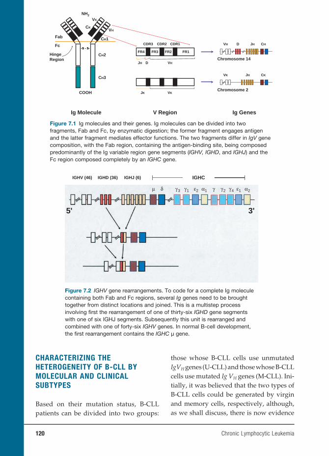
120 Chronic Lymphocytic Leukemia
CHARACTERIZING THE
HETEROGENEITY OF B-CLL BY
MOLECULAR AND CLINICAL
SUBTYPES
Based on their mutation status, B-CLL patients can be divided into two groups:
those whose B-CLL cells use unmutated IgVH genes (U-CLL) and those whose B-CLL cells use mutated Ig VH genes (M-CLL). Ini-tially, it was believed that the two types of B-CLL cells could be generated by virgin and memory cells, respectively, although, as we shall discuss, there is now evidence
VH D JH CH
Chromosome 14
Chromosome 2
VK JH CK
Ig GenesIg Molecule V Region
CK
COOH
NH2
--S S -- SS--
Fab
Fc
VK
CH1
VH
CH2
CH3
VKJK
VHDJH
CDR3 CDR1CDR2
FR4 FR3 FR2 FR1HingeRegion
Figure 7.1 Ig molecules and their genes. Ig molecules can be divided into two fragments, Fab and Fc, by enzymatic digestion; the former fragment engages antigen and the latter fragment mediates effector functions. The two fragments differ in IgV gene composition, with the Fab region, containing the antigen-binding site, being composed predominantly of the Ig variable region gene segments (IGHV, IGHD, and IGHJ) and the Fc region composed completely by an IGHC gene.
IGHV (46)
5' 3'
IGHD (36) IGHJ (6)
μ δ γ3 γ1 γ2 ε1γ4ε2 α1 α2γ
IGHC
Figure 7.2 IGHV gene rearrangements. To code for a complete Ig molecule containing both Fab and Fc regions, several Ig genes need to be brought together from distinct locations and joined. This is a multistep process involving fi rst the rearrangement of one of thirty-six IGHD gene segments with one of six IGHJ segments. Subsequently this unit is rearranged and combined with one of forty-six IGHV genes. In normal B-cell development, the fi rst rearrangement contains the IGHC µ gene.
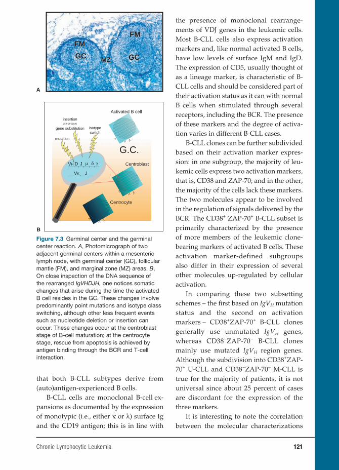
Chronic Lymphocytic Leukemia 121
the presence of monoclonal rearrange-ments of VDJ genes in the leukemic cells. Most B-CLL cells also express activation markers and, like normal activated B cells, have low levels of surface IgM and IgD. The expression of CD5, usually thought of as a lineage marker, is characteristic of B-CLL cells and should be considered part of their activation status as it can with normal B cells when stimulated through several receptors, including the BCR. The presence of these markers and the degree of activa-tion varies in different B-CLL cases.
B-CLL clones can be further subdivided based on their activation marker expres-sion: in one subgroup, the majority of leu-kemic cells express two activation markers, that is, CD38 and ZAP-70; and in the other, the majority of the cells lack these markers. The two molecules appear to be involved in the regulation of signals delivered by the BCR. The CD38+ ZAP-70+ B-CLL subset is primarily characterized by the presence of more members of the leukemic clone-bearing markers of activated B cells. These activation marker-defined subgroups also differ in their expression of several other molecules up-regulated by cellular activation.
In comparing these two subsetting schemes – the fi rst based on IgVH mutation status and the second on activation markers – CD38+ZAP-70+ B-CLL clones generally use unmutated IgVH genes, whereas CD38−ZAP-70− B-CLL clones mainly use mutated IgVH region genes. Although the subdivision into CD38+ZAP-70+ U-CLL and CD38−ZAP-70− M-CLL is true for the majority of patients, it is not universal since about 25 percent of cases are discordant for the expression of the three markers.
It is interesting to note the correlation between the molecular characterizations
GCGC GCGC
FMFMFMFM
MZMZ
A
G.C.
Activated B cell
Centroblast
Centrocyte
mutation
insertiondeletion
gene substitution
VH D J μ δ γ
VK J
isotypeswitch
B
Figure 7.3 Germinal center and the germinal center reaction. A, Photomicrograph of two adjacent germinal centers within a mesenteric lymph node, with germinal center (GC), follicular mantle (FM), and marginal zone (MZ) areas. B, On close inspection of the DNA sequence of the rearranged IgVHDJH, one notices somatic changes that arise during the time the activated B cell resides in the GC. These changes involve predominantly point mutations and isotype class switching, although other less frequent events such as nucleotide deletion or insertion can occur. These changes occur at the centroblast stage of B-cell maturation; at the centrocyte stage, rescue from apoptosis is achieved by antigen binding through the BCR and T-cell interaction.
that both B-CLL subtypes derive from (auto)antigen-experienced B cells.
B-CLL cells are monoclonal B-cell ex-pansions as documented by the expression of monotypic (i.e., either κ or λ) surface Ig and the CD19 antigen; this is in line with
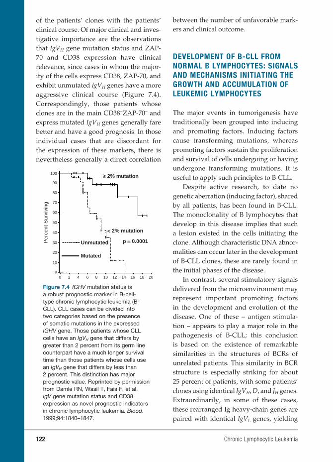
122 Chronic Lymphocytic Leukemia
of the patients’ clones with the patients’ clinical course. Of major clinical and inves-tigative importance are the observations that IgVH gene mutation status and ZAP-70 and CD38 expression have clinical relevance, since cases in whom the major-ity of the cells express CD38, ZAP-70, and exhibit unmutated IgVH genes have a more aggressive clinical course (Figure 7.4). Correspondingly, those patients whose clones are in the main CD38−ZAP-70− and express mutated IgVH genes generally fare better and have a good prognosis. In those individual cases that are discordant for the expression of these markers, there is nevertheless generally a direct correlation
between the number of unfavorable mark-ers and clinical outcome.
DEVELOPMENT OF B-CLL FROM
NORMAL B LYMPHOCYTES: SIGNALS
AND MECHANISMS INITIATING THE
GROWTH AND ACCUMULATION OF
LEUKEMIC LYMPHOCYTES
The major events in tumorigenesis have traditionally been grouped into inducing and promoting factors. Inducing factors cause transforming mutations, whereas promoting factors sustain the proliferation and survival of cells undergoing or having undergone transforming mutations. It is useful to apply such principles to B-CLL.
Despite active research, to date no genetic aberration (inducing factor), shared by all patients, has been found in B-CLL. The monoclonality of B lymphocytes that develop in this disease implies that such a lesion existed in the cells initiating the clone. Although characteristic DNA abnor-malities can occur later in the development of B-CLL clones, these are rarely found in the initial phases of the disease.
In contrast, several stimulatory signals delivered from the microenvironment may represent important promoting factors in the development and evolution of the disease. One of these – antigen stimula-tion – appears to play a major role in the pathogenesis of B-CLL; this conclusion is based on the existence of remarkable similarities in the structures of BCRs of unrelated patients. This similarity in BCR structure is especially striking for about 25 percent of patients, with some patients’ clones using identical IgVH, D, and JH genes. Extraordinarily, in some of these cases, these rearranged Ig heavy-chain genes are paired with identical IgVL genes, yielding
≥≥ 2% mutation
<< 2% mutation
p == 0.0001Unmutated
100
90
80
70
60
50
40
30Per
cent
Sur
vivi
ng
20
10
00 2 4 6 8 10 12 14 16 18 20
Mutated
Figure 7.4 IGHV mutation status is a robust prognostic marker in B-cell-type chronic lymphocytic leukemia (B-CLL). CLL cases can be divided into two categories based on the presence of somatic mutations in the expressed IGHV gene. Those patients whose CLL cells have an IgVH gene that differs by greater than 2 percent from its germ line counterpart have a much longer survival time than those patients whose cells use an IgVH gene that differs by less than 2 percent. This distinction has major prognostic value. Reprinted by permission from Damle RN, Wasil T, Fais F, et al. IgV gene mutation status and CD38 expression as novel prognostic indicators in chronic lymphocytic leukemia. Blood. 1999;94:1840–1847.
Chronic Lymphocytic Leukemia 123
antigen-binding sites that are virtually identical at the amino acid level. Given the enormous number of possible com-binations of IgV gene segments encoding antibody-binding domains, one would not expect to fi nd B-CLL patients having such structurally similar “stereotypic” BCRs by chance until well over 1 million cases have been screened. Hence, their occurrence is not likely random, making a plausible argument for the importance of antigen stimulation and drive in this disease.
Foreign antigens or autoantigens could prompt normal B lymphocytes to become B-CLL cells by selecting B-cell clones with restricted stereotypic BCRs. The nature of the antigens that select for these highly restricted BCRs (possibly unique to B-CLL) is largely unknown, although currently under investigation. It may be they result from infection due to a specifi c microbe common among patients, which has been found for gastric lymphomas. Alternative possibilities are that both environmental and autologous antigens may be involved. Indeed, it appears that intermittent and interchangeable encounters with microbial antigens and autoantigens, especially those generated during cell death and oxidative and other forms of stress, are key.
How would the transition from nor-mal B cells to leukemic cells via antigen stimulation occur? Normal B lymphocytes using unmutated IgV genes produce anti-bodies that are frequently polyreactive, binding carbohydrates, nucleic acids, and phospholipids. Such antibodies provide the fi rst line of defense against microor-ganisms and promote the clearance of autoantigens and their fragments. B-CLL cells frequently display polyreactive BCRs, thereby making it possible that they derive from normal polyreactive B lymphocytes that have been repeatedly stimulated in
vivo by a combination of nonprotein self- and microbial antigens.
Similarly, such a mechanism could also promote the origin of M-CLL. As already mentioned, IgVH gene mutations can occur without T-cell help in marginal zones outside of GCs. Because mutations can sometimes favor autoreactivity, such auto-reactive B cells would become expanded. However, expansion would stop if IgV gene mutations alter BCR structure in such a way that antigen binding is no longer suf-fi cient to induce B-cell signaling.
With continued expansion leading (or not) to accumulation of IgVH gene muta-tions as explained by the T-cell-dependent versus T-cell-independent models men-tioned earlier, it becomes increasingly likely that a cell develops a genetic abnormality as in an initial inducing lesion that would lead to relatively unrestrained expansion. Such a cell is primed for leukemic transfor-mation.
A hypothesis such as this implies that it might be possible to detect clonal expan-sions in healthy subjects. In fact, small numbers of apparently clonal B cells with B-CLL cell characteristics do exist in the blood of about 3.5 percent of disease-free individuals. An even higher proportion of such clones have been found in the blood of fi rst-degree relatives of patients with B-CLL (as often as 12 percent). Although such studies of the BCRs of B lymphocyte expansions in normal disease-free individ-uals are limited, they further support this hypothesis in that these expansions are not only monoclonal but also use some of the same genes commonly encoding the BCRs of B-CLL clones.
In addition to antigen stimulation, B-CLL cells also receive receptor-medi-ated signals as well as soluble factors, such as cytokines and chemokines, from other
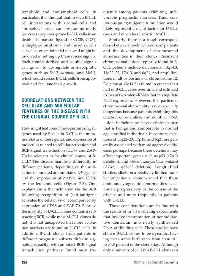
124 Chronic Lymphocytic Leukemia
lymphoid and nonlymphoid cells. In particular, it is thought that in vivo B-CLL cell interactions with stromal cells and “nurselike” cells can rescue normally (ex vivo) apoptosis-prone B-CLL cells from death. The natural ligand of CD38, CD31, is displayed on stromal and nurselike cells as well as on endothelial cells and might be involved in setting up these rescue signals. Such contact-derived and soluble signals can go on to up-regulate anti-apoptotic genes, such as Bcl-2, survivin, and Mcl-1, which could rescue B-CLL cells from apop-tosis and facilitate their growth.
CORRELATIONS BETWEEN THE
CELLULAR AND MOLECULAR
FEATURES OF THE DISEASE WITH
THE CLINICAL COURSE OF B-CLL
How might features of the repertoire of IgVH genes used by B cells in B-CLL, the muta-tion status of these genes, and expression of molecules related to cellular activation and BCR signal transduction (CD38 and ZAP-70) be relevant to the clinical course of B-CLL? The disease manifests differently in different patients, depending on the utili-zation of mutated or unmutated IgVH genes and the expression of ZAP-70 and CD38 by the leukemic cells (Figure 7.5). One explanation is that activation via the BCR following recognition of (self-)antigens activates the cells in vivo, accompanied by expression of CD38 and ZAP-70. Because the majority of U-CLL clones contain a self-reacting BCR, while most M-CLL clones do not, it is not unexpected that more activa-tion markers are found on U-CLL cells. In addition, B-CLL clones from patients in different prognostic subsets differ in sig-naling capacity, with an intact BCR signal transduction pathway found most fre-
quently among patients exhibiting unfa-vorable prognostic markers. Thus, con-tinuous (auto)antigenic stimulation would likely represent a major factor for U-CLL cases and much less likely for M-CLL.
Similarly, there is a rough correspon-dence between the clinical course of patients and the development of chromosomal abnormalities in their clones. Recurrent chromosomal lesions typically found in B-CLL patients include deletions at 13q14.3, 11q22–23, 17p13, and 6q21, and amplifi ca-tions of all or portions of chromosome 12. Deletion at 13q14.3 is found in greater than half of B-CLL cases over time and is linked to loss of two micro-RNAs that can regulate Bcl-2 expression. However, this particular chromosomal abnormality is not especially dangerous because patients exhibiting this deletion on one allele and no other DNA lesions in their clones have a clinical course that is benign and comparable to normal age-stratifi ed individuals. In contrast, dele-tions at 11q22–23, 17p13, and 6q21 are gen-erally associated with more aggressive dis-ease, perhaps because these deletions may affect important genes such as p53 (17p13 deletion), and ataxia telangiectasia mutated (ATM; 11q22–23 deletion). Longitudinal studies, albeit on a relatively limited num-ber of patients, demonstrated that these ominous cytogenetic abnormalities accu-mulate progressively in the course of the disease and more frequently in patients with U-CLL.
These considerations are in line with the results of in vivo labeling experiments that involve incorporation of nonradioac-tive deuterium into newly synthesized DNA of dividing cells. These studies have shown B-CLL clones to be dynamic, hav-ing measurable birth rates from about 0.1 to >1.0 percent of the clone/day. Although only a minority of cells in a B-CLL clone can
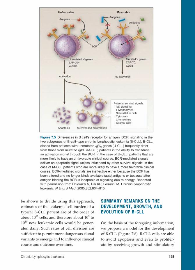
Chronic Lymphocytic Leukemia 125
be shown to divide using this approach, estimates of the leukemic cell burden of a typical B-CLL patient are of the order of about 1012 cells, and therefore about 109 to 1010 new leukemic cells would be gener-ated daily. Such rates of cell division are suffi cient to permit more dangerous clonal variants to emerge and to infl uence clinical course and outcome over time.
SUMMARY REMARKS ON THE
DEVELOPMENT, GROWTH, AND
EVOLUTION OF B-CLL
On the basis of the foregoing information, we propose a model for the development of B-CLL (Figure 7.6). B-CLL cells are able to avoid apoptosis and even to prolifer-ate by receiving growth and stimulatory
Mutated V genesZAP-70.CD38-
Unmutated V genesZAP-70+CD38+
No activationActivation
Apoptosis Survival and proliferation
Potential survival signals: IgD signaling T lymphocytes Natural killer cells Cytokines Chemokines Stromal cells
BCR
AntigensAntigens
Antigens
Unfavorable Favorable
Figure 7.5 Differences in B cell’s receptor for antigen (BCR) signaling in the two subgroups of B-cell–type chronic lymphocytic leukemia (B-CLL). B-CLL clones from patients with unmutated IgVH genes (U-CLL) frequently differ from those from mutated IgVH (M-CLL) patients in the ability to transduce an activation signal through the BCR. In the case of U-CLL, patients that are more likely to have an unfavorable clinical course, BCR-mediated signals deliver an apoptotic signal unless infl uenced by other survival signals. In the case of M-CLL patients who are more likely to have a more favorable clinical course, BCR-mediated signals are ineffective either because the BCR has been altered and no longer binds available (auto)antigens or because after antigen binding the BCR is incapable of signaling due to anergy. Reprinted with permission from Chiorazzi N, Rai KR, Ferrarini M. Chronic lymphocytic leukemia. N Engl J Med. 2005;352:804–815.
126 Chronic Lymphocytic Leukemia
A Initial inducing mutation C Repetitive antigen stimu- lation of polyreactive receptor
D Conversion to leukemia by new mutations
E Development of subclones
Subclone B
Subclone C
Subclone E
Clone A
Clone A
Clone D
Clone D
Apoptosis
Clone F Clone F
Antigens?
Antigens?Antigens
Antigens?
AntigensAntigens
Unm
utat
ed C
LLM
utat
ed C
LL Antigens Antigens
Antigens
Activated B cells
Nonactivated B cells
V-g
ene
mut
atio
nsN
o V
-gen
e m
utat
ions
Anergic B cells
B Response to self-antigen or foreign antigen
Figure 7.6 Model to explain the development and evolution of B-CLL. See text for description. Reprinted with permission from Chiorazzi N, Rai KR, Ferrarini M. Chronic lymphocytic leukemia. N Engl J Med. 2005;352:804–815.
signals from the environment delivered through their BCR or other receptors; these signals likely involve autologous and for-eign antigens, cytokines, and chemokines, as well as yet-to-be-defi ned ligands on accessory and stromal cells. The cell’s BCR mediates major growth effects in cases for which the receptor is polyreactive, binding autoantigens and foreign antigens, while maintaining its capacity to transmit stimu-latory signals to the cell nucleus. Both self-reactivity and intact BCR signal-transduc-ing capacity are more frequently found in U-CLL; hence, the more active clonal expansion and clinical aggressiveness of patients with such clones.
Because in vitro observations demon-strate the absence of lesions in the major
apoptotic pathways, our model posits the absence of an intrinsic cell death defect in the majority of the leukemic clones. We do not rule out the possibility that developing genetic alterations in the evolving clone can tip the balance between pro- and anti-apoptotic molecules in such a way as to favor B-CLL cell survival. However, the infl uence of external signals appears to dominate based on current knowledge. This is in line with the in vivo labeling studies indicating the dynamic nature of CLL clones.
ANIMAL MODELS OF B-CLL
While an enormous amount of new infor-mation has been gleaned by directly
Chronic Lymphocytic Leukemia 127
studying human B-CLL cells, animal mod-els are now contributing to our understand-ing of the human disease. For example, New Zealand Black (NZB) mice spontane-ously develop, with age, an expansion of IgM+CD5+ B cells that resembles B-CLL. However, because frank leukemia occurs randomly in only a minor subset of animals, this model has been used sparingly.
In recent years, however, a variety of transgenic mouse models have been devel-oped that lead to diseased phenotypes resembling human B-CLL more closely and reproducibly. We focus on three models that have been especially helpful.
Transgenic mice expressing the TCL1 gene in murine B cells develop a poly-clonal expansion of B lymphocytes early in life that becomes progressively more restricted until a monoclonal population emerges after about one year in most ani-mals. The genetic and phenotypic features of this murine leukemia resemble those of the aggressive, treatment-resistant cases of human U-CLL. Although it is of interest that TCL1 is an activator of the PI3K-Akt oncogenic signaling pathway, a pathway not infrequently active in human CLL, the extent to which overexpression of this gene leads to human B-CLL remain to be elucidated, as an overexpression of TCL1 in human B-CLL patients is not uniform.
Another mouse model that develops features resembling human B-CLL in-volves the overexpression of two genes: BCL-2 and TRAF2 (TNF-receptor-associated factor 2). This double transgenic animal is especially intriguing because of the already mentioned recent work showing that the deletion at 13q14, often seen in human B-CLL, involves the loss of micro-RNAs 15a and 16-1, which affects expression of BCL2. As with the TCL1 transgenic mice, these animals develop CD5+ B-cell clones,
eventually with massive splenomegaly and leukemia.
In the third transgenic model, overex-pression of APRIL ( a proliferation inducing ligand ) in murine T cells leads indirectly to B-cell proliferation and survival because of signaling through its receptors BCMA and TACI. Unlike the previous two transgenic animals, however, expansions of CD5+ B cells occur in only 40 percent of animals, with these B cells locating predominantly in the spleen and rarely passing into the blood. Nevertheless, as APRIL’s action involves the TRAFs and leads to NFκB activation, this model may prove helpful in linking signals from soluble ligands and surface receptors to the NF,κB pathway, a pathway known to be constitutively active in some B-CLL clones.
Finally, transfer of human B-CLL cells into immune-defi cient mice may bypass the ex vivo apoptosis tendency of B-CLL cells, enabling their survival and amplifi -cation in vivo. Such an approach may also make it possible to defi ne and test new therapeutics to treat this currently incur-able disorder.
CLINICAL IMPLICATIONS AND
THE DEVELOPMENT OF NOVEL
THERAPEUTICS
Currently used clinical staging systems defined by Rai and by Binet help clini-cians to monitor disease progression and decide when to treat patients. Because of difficulties distinguishing patients with poor prognosis at the onset of dis-ease using these staging systems, the generally used practice is to wait to start therapy until the patient’s clini-cal course becomes evident (“wait and watch” mode). However, the molecular
128 Chronic Lymphocytic Leukemia
and cellular markers that reflect intrin-sic properties of leukemic cells present at the disease onset can help to distin-guish patients that will follow worse clinical courses, regardless of their Rai and Binet risk categories at diagnosis. Although determination of IgVH gene mutation status is not yet routinely avail-able, measurement of CD38 and ZAP-70 has become more widely available. Thus, although the wait-and-watch mode is still being followed in current clinical practice, it may be substituted for by new more aggressive strategies in the future. The new prognostic markers indicate that 30–50 percent of the patients have features portending a poor outcome, and therefore an early start of therapy may be justified in such poor-prognosis groups. This strategy is plausible, considering that continued proliferation and expansion of the neoplastic clone facilitates accumula-tion of ominous cytogenetic abnormali-ties. However, before any recommended guidelines can be proposed, clinical trials must test the use of early intervention in patients in poor-prognosis groups.
New knowledge about the biology of B-CLL can provide clues for novel therapeu-tic targets. For example, since B-CLL cells must interact with the stroma in bone mar-row or other peripheral lymphoid tissues to survive, furthering our knowledge of these interactions may generate new objec-tives for innovative therapies. Another compelling set of options may derive from specifi c inhibition of the BCR or CD38 sig-naling pathways or other pathways in which ZAP-70 is crucial. Likewise, the pos-sibility of using cell-cycle-specifi c drugs is worth being explored in clinical trials, given the documented active turnover of B-CLL cells. Finally, because up to 20 per-cent of patients with the worst prognostic
markers have stereotypic antigen re-ceptors, these common structures may be feasible as vulnerable points of attack. As the antigens that engage these receptors become more precisely defi ned, it may be possible to use these to develop an arsenal of specifi c therapies.
BIBLIOGRAPHY
REVIEWSChiorazzi N, Ferrarini M. B cell chronic
lymphocytic leukemia: lessons learned from studies of the B cell antigen recep-tor. Annu Rev Immunol. 2003;21:841–894.
Chiorazzi N, Rai KR, Ferrarini M. Chronic lymphocyrtic leukemia. N Engl J Med. 2005;352:804–815.
Ferrarini M, Chiorazzi N. Recent advances in the molecular biology and immuno-biology of chronic lymphocytic leuke-mia. Semin Hematol. 2004;41:207–223.
Stevenson F, Caligaris-Cappio F. Chronic lymphocytic leukemia: revelations from the B-cell receptor. Blood. 2004; 103(12):4389–4395.
SUGGESTED READINGBichi R, Shinton SA, Martin ES, et al.
Human chronic lymphocytic leukemia modeled in mouse by targeted TCL1 expression. Proc Natl Acad Sci USA. 2002;99:6955–6960.
Binet JL, Auquier A, Dighiero G, et al. A new prognostic classifi cation of chronic lymphocytic leukemia derived from a multivariate survival analysis. Cancer. 1981;48:198–206.
Burger JA, Kipps TJ. Chemokine recep-tors and stromal cells in the homing and homeostasis of chronic lympho-cytic leukemia B cells. Leuk Lymphoma. 2002;43:461–466.
Chronic Lymphocytic Leukemia 129
Calin GA, Dumitru CD, Shimizu M, et al. Frequent deletions and down-regula-tion of micro-RNA genes miR15 and miR16 at 13q14 in chronic lympho-cytic leukemia. Proc Natl Acad Sci USA. 2002;99:15524–15529.
Dameshek W. Chronic lymphocytic leukemia – an accumulative disease of immunologically incompetent lympho-cytes. Blood. 1967;29(suppl):566–584.
Damle RN, Wasil T, Fais F, et al. Ig V gene mutation status and CD38 expres-sion as novel prognostic indicators in chronic lymphocytic leukemia. Blood. 1999;94:1840–1847.
Deaglio S, Capobianco A, Bergui L, et al. CD38 is a signaling molecule in B-cell chronic lymphocytic leukemia cells. Blood. 2003;102:2146–2155.
Dohner H, Stilgenbauer S, Benner A, et al. Genomic aberrations and survival in chronic lymphocytic leukemia. N Engl J Med. 2000;343:1910–1916.
Fais F, Ghiotto F, Hashimoto S, et al. Chronic lymphocytic leukemia B cells express restricted sets of mutated and unmutated antigen receptors. J Clin Invest. 1998;102:1515–1525.
Ghia P, Caligaris-Cappio F. The indis-pensable role of microenvironment in the natural history of low-grade B-cell neoplasms. Adv Cancer Res. 2000;79: 157–173.
Ghia P, Prato G, Scielzo C, et al. Mono-clonal CD5+ and CD5- B-lymphocyte expansions are frequent in the periph-eral blood of the elderly. Blood. 2004;103:2337–2342.
Hamblin TJ, Davis Z, Gardiner A, Oscier DG, Stevenson FK. Unmutated Ig
VH genes are associated with a more aggressive form of chronic lymphocytic leukemia. Blood. 1999;94:1848–1854.
Messmer BT, Messmer D, Allen SL, et al. In vivo measurements document the dynamic cellular kinetics of chronic lymphocytic leukemia B cells. J Clin Invest. 2005;115:755–764.
Pekarsky Y, Zanesi N, Aqelian RI, Croce CM. Animal models of chronic lympho-cytic leukemia. J Cell Biochem. 2006;100: 1109–1118.
Rai KR, Sawitsky A, Cronkite EP, Chanana AD, Levy RN, Pasternack BS. Clinical staging of chronic lymphocytic leuke-mia. Blood. 1975;46:219–234.
Rassenti LZ, Hunynh L, Toy TL, et al. ZAP-70 compared with immunoglobulin heavy-chain gene mutation status as a predictor of disease progression in chronic lymphocytic leukemia. N Engl J Med. 2004;351:893–901.
Rawstron AC, Yuille MR, Fuller J, et al. Inherited predisposition to CLL is detectable as subclinical monoclo-nal B-lymphocyte expansion. Blood. 2002;100:2289–2890.
Wiestner A, Rosenwald A, Barry TS, et al. ZAP-70 expression identifi es a chronic lymphocytic leukemia subtype with unmutated immunoglobulin genes, inferior clinical outcome, and dis-tinct gene expression profi le. Blood. 2003;101:4944–4951.
Zupo S, Isnardi L, Megna M, et al. CD38 expression distinguishes two groups of B-cell chronic lymphocytic leukemias with different responses to anti-IgM antibodies and propensity to apoptosis. Blood. 1996;88:1365–1374.

INTRODUCTION
An estimated 33 million people globally are infected with the AIDS virus (HIV-1). Most of the infected individuals are poor, live in developing countries, and have little access to health care. Although initiatives are under way to bring proper medications to these individuals via a large infusion of money, this will benefi t only approxi-mately 2.5 million individuals and pre-vent 12 million new cases, as pointed out in a 2007 New York Times editorial. The remainder of these infected individuals will continue to increase the number of newly infected individuals. In this context, the medications decrease the viral load in treated individuals, but medications are expensive and would not reach many of the infected individuals living in develop-ing countries.
Thus, many researchers are focusing on studies of the immunological concepts involved in the disease, not only to understand in greater detail why some individuals remain uninfected (they are not immune since some “resistant” women later acquire human immunodefi ciency virus, or HIV), despite repeated exposure to infected individuals, but also to explore various vaccine candidates and concepts that might prevent the disease before exposure to the virus – candidates that would be cheap to make and inexpensive to deliver.
IMMUNOLOGICAL AND BIOLOGICAL
PARAMETERS OF DISEASE
The pattern of disease progression has now been well documented. Following infec-tion with the virus, the virus hones to and infects cells with CD4 receptors. During the early phase, individuals may experience a fl u-like illness with mild fever, cough, and occasional chills. The symptoms subside, and the individual may be asymptomatic for many years. In reality, the disease is progressing, and it is a long battle between the immune response with production of new CD4+ cells and the dying (apoptotic) HIV-infected CD4 cells. Eventually, the host immune system deteriorates, and the individual succumbs to the complications secondary to loss of the cellular immune system (see Figures 8.1 and 8.2). The pat-tern and complications are quite similar to those seen in primary immunodefi ciency diseases (see Chapter 5). However, it has been a well-known observation that some individuals, especially sex workers with repeated exposure to the virus, are rela-tively resistant to HIV acquisition.
One of the earliest markers in the progression of HIV-1 infection was the presence of NEF in the viral strain. NEF is a 27–34 D myrisolated protein unique to primate lente viruses. A functional NEF protein is important for the development of high viremia and AIDS in simian immu-nodefi ciency virus (SIV) in infected rhesus
8. Immunology of HIV Infections
Anders G. Vahlne, M.D., Ph.D.
131
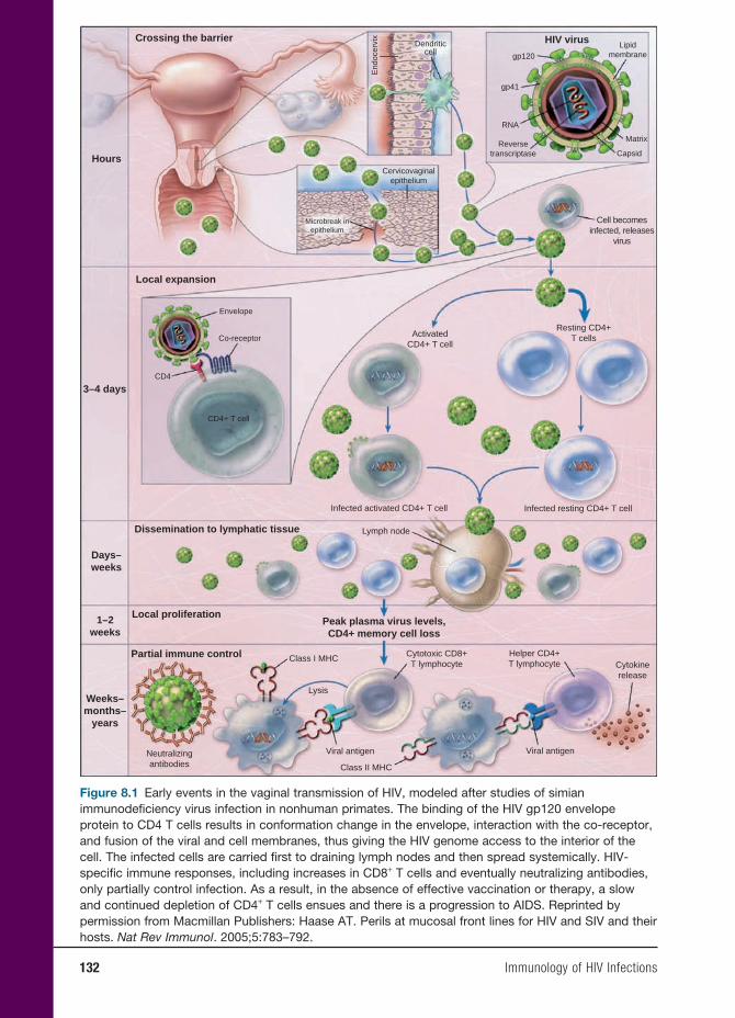
132 Immunology of HIV Infections
Crossing the barrier
Hours
Local expansion
ActivatedCD4+ T cell
Resting CD4+T cells
Microbreak inepithelium
Cervicovaginalepithelium
Reversetranscriptase
RNA
gp41
gp120
Dendriticcell
End
ocer
vix
Lipidmembrane
Matrix
Capsid
HIV virus
Cell becomesinfected, releases
virus
Infected resting CD4+ T cellInfected activated CD4+ T cell
Lymph node
Class I MHC
Lysis
Cytotoxic CD8+T lymphocyte
Helper CD4+T lymphocyte Cytokine
release
Viral antigenViral antigenNeutralizingantibodies Class II MHC
Dissemination to lymphatic tissue
Days–weeks
1–2weeks
Weeks–months–
years
Local proliferationPeak plasma virus levels,CD4+ memory cell loss
Partial immune control
3–4 days
Envelope
Co-receptor
CD4
CD4+ T cell
Figure 8.1 Early events in the vaginal transmission of HIV, modeled after studies of simian immunodefi ciency virus infection in nonhuman primates. The binding of the HIV gp120 envelope protein to CD4 T cells results in conformation change in the envelope, interaction with the co-receptor, and fusion of the viral and cell membranes, thus giving the HIV genome access to the interior of the cell. The infected cells are carried fi rst to draining lymph nodes and then spread systemically. HIV-specifi c immune responses, including increases in CD8+ T cells and eventually neutralizing antibodies, only partially control infection. As a result, in the absence of effective vaccination or therapy, a slow and continued depletion of CD4+ T cells ensues and there is a progression to AIDS. Reprinted by permission from Macmillan Publishers: Haase AT. Perils at mucosal front lines for HIV and SIV and their hosts. Nat Rev Immunol. 2005;5:783–792.
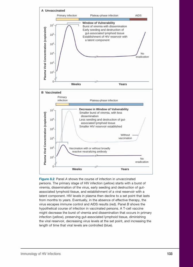
Immunology of HIV Infections 133
A Unvaccinated
B Vaccinated
Weeks Years
Primary infection
Primaryinfection
Pla
sma
Vir
al C
on
cen
trat
ion
(co
pie
s/m
l)
107
106
105
104
103
102
0
Weeks Years
Pla
sma
Vir
al C
on
cen
trat
ion
(co
pie
s/m
l)
107
106
105
104
103
102
0
Plateau-phase infection
Plateau-phase infection
Burst of viremia with disseminationEarly seeding and destruction of gut-associated lymphoid tissueEstablishment of HIV reservoir with a latent component
Window of Vulnerability
Smaller burst of viremia, with less disseminationLess seeding and destruction of gut- associated lymphoid tissueSmaller HIV reservoir established
Decrease in Window of Vulnerability
AIDS
Noeradication
Noeradication
Withoutvaccination
Vaccination with or without broadlyreactive neutralizing antibody
Figure 8.2 Panel A shows the course of infection in unvaccinated persons. The primary stage of HIV infection (yellow) starts with a burst of viremia, dissemination of the virus, early seeding and destruction of gut-associated lymphoid tissue, and establishment of a viral reservoir with a latent component. HIV levels in plasma then decline to a set point that lasts from months to years. Eventually, in the absence of effective therapy, the virus escapes immune control and AIDS results (red). Panel B shows the hypothetical course of infection in vaccinated persons. A T-cell vaccine might decrease the burst of viremia and dissemination that occurs in primary infection (yellow), preserving gut-associated lymphoid tissue, diminishing the viral reservoir, decreasing virus levels at the set point, and increasing the length of time that viral levels are controlled (blue).
134 Immunology of HIV Infections
macaques. An important observation fi rst noted in the macaques and later in humans was that animals infected with NEF-deleted virus were resistant to subsequent challenge with pathogenic wild-type viruses. This was followed by the observation in humans that some individuals with long-term non-progressive HIV-1 infection (persons who showed no clinical or immunological signs of immunodefi ciency despite being HIV seropositive for over a decade) turned out to be infected with viruses carrying deletion in their NEF gene.
More recently, a large body of evidence has appeared that shows that NEF has many important effects on many cells of the immune system. Both exogenous and endogenous NEF down-regulates HLA-ABC molecules critical for the initiation of cytotoxic T lymphocyte (CTL) responses, thus impairing antigen presentation to HIV-specifi c CD8+ lymphocytes. NEF and gp120 also are able to down-regulate major histocompatibility complex class I (MHC-I) in dendritic cells (DCs) and introduction of exogenous NEF leads to up-regulation of MHC-II molecules, thereby favoring CD4+ T-cell activation. This step increases the “pool” of lymphocytes permissive to infection. However, endogenous NEF does not modulate MHC-II surface expression; rather it induces a loss of co-stimulation. These results underscore the pleiotrophic action of NEF. On one hand, exogenous NEF triggers ABC-mediated bystander T-cell activation, ensuring viral spread, while endogenous NEF induces a loss of co-stimulation, favoring immune evasion.
Another area of interest has been the observation that NEF-pulsed DC produces a wide variety of cytokines and chemokines typical of mature DC. This up-regulation of cytokine production might promote bystander stimulation of T cells that cluster
around DC and could enhance HIV-1 replication in CD4+ T cells.
In summary, the selective effects of the NEF protein may markedly enhance viral pathogenesis and progression to disease. It does this by hijacking DC functional activity and favoring HIV replication via bystander activation of CD4+ cells and the escape of HIV-1 from immune surveil-lance by blocking CD8+ cells’ functional competence. Thus, efforts directed toward blocking NEF infl uence in viral replication may have long-term benefi cial effects on therapeutic management of the disease.
Approximately 5–10 percent of sex workers in Kenya remained HIV unin-fected despite engaging in unprotected sex with many male clients and the frequent acquisition of other sexually transmitted infections. Because of the pioneering work of Plummer and associates, the scientifi c reasons for this extraordinary resistance to the virus in these women are beginning to unfold.
Turning fi rst to examination of CD4+ T-cell immune responses in seronegative sex workers compared with HIV-positive women, CD4+ T cells that produced inter-feron gamma (IFN-γ) in response to HIV p24 were detected in exposed seronega-tive sex worker (ESN) women, albeit at a much lower level than in HIV-positive women. However, ESN women had a 4.5-fold stronger CD4+ proliferation response to the p24 peptide compared with the HIV-positive group. These data suggest that CD4+ T cells in ESN women recognize HIV and have an enhanced ability to proliferate to the p24 protein.
Perhaps more important has been the examination of the CD8+ cells in these ESN individuals. Because IFN-γ production cor-relates with cytotoxic functions, the CD8+ T lymphocyte IFN-γ response to HIV p24
Immunology of HIV Infections 135
peptide in ESN women was compared with HIV-positive individuals. Approximately 40 percent of ESN women had a CD8+ IFN-γ-positive response, and this was fi ve times lower in magnitude than that of HIV-posi-tive women. The breadth of the response was very narrow and focused primarily on one peptide that is similar to KK10 protec-tive peptide when compared with the HIV-positive group. In HIV-positive women, lower CD4+ counts infl uenced the num-ber of CD8+ cells producing IFN-γ, which may undermine the ability to control HIV. These results indicate ESP women have an HIV-1 p24 peptide-specifi c CD8+ IFN-γ response, providing evidence to the speci-fi city needed for an effective HIV vaccine.
Further studies of these women cen-tered around the known polymorphisms of the IL-4 gene and resistance to infection. Using microsatellite genotyping methods coupled with the genomic sequencing, three polymorphisms in the interferon reg-ulatory factor (IRF-1) located at 619a, 179, and 6516 of the gene, showed association with resistance to HIV infection. Periph-eral blood mononuclear cells from these patients showed signifi cantly lower-based IRF-1 expression and reduced respon-siveness to IFN-γ stimulation. This study added IRF-1, a transcriptional immunoreg-ulatory gene, to the list of genetic correlates of altered susceptibility to HIV-1.
One of the key cells in the immune sur-veillance system is the native DC, which is well equipped for activation of both the innate and adaptive immune response. It has been demonstrated that some DC-tropic viruses such as infl uenza virus leave DC function intact while other viruses such as HIV and cytomegalovirus have evolved strategies to impair DC functions, thereby enhancing the virus’s ability to persist and escape immune surveillance.
DC interaction with HIV is relevant to the pathogenesis of AIDS because they are present in the mucosa and skin of humans and are believed to be the fi rst HIV-1 tar-gets following sexual transmission of the virus. Both myeloid DC and plasmacytoid DC possess the receptors for HIV entry; that is, CD4, CXR4, and CCR5 can be infected but with a lower effi cacy than CD4+ cells or macrophages. The long-term result of this infection is that DC remains a reser-voir for the production and persistence of the HIV-1 virus, and the virus induces sev-eral functional impairments and variations in the DC population. Thus, the number of both myeloid DC (MDC) and peripheral DC (PDC) are signifi cantly decreased in HIV-positive progressors, while remain-ing unaltered in HIV-positive long-term nonprogression. The destruction of these cells may be a consequence of direct lytic infection or as targets for specifi c CTL or through a block in DC development from peripheral CD34+ stem cells. More recent evidence supports the notion that both midi and pad show impaired functional capacity in HIV-positive patients. Since both DC subsets participate in the initiation of innate and adaptive immune responses, infection, depletion, and dysfunction of DCs may contribute to the immunosup-pression seen in HIV disease. Therefore, DCs play a dual role in HIV infection: they trigger both innate and adaptic immune responses to control the infection, but they also represent a viral reservoir for infection of permissive CD4+ T cells.
Although the story of the African sex workers who seem to be immune to HIV infection has captured the imagina-tion of many researchers, there is another model of natural immunity that needs examination in more detail. Most babies born to HIV-infected mothers also escape
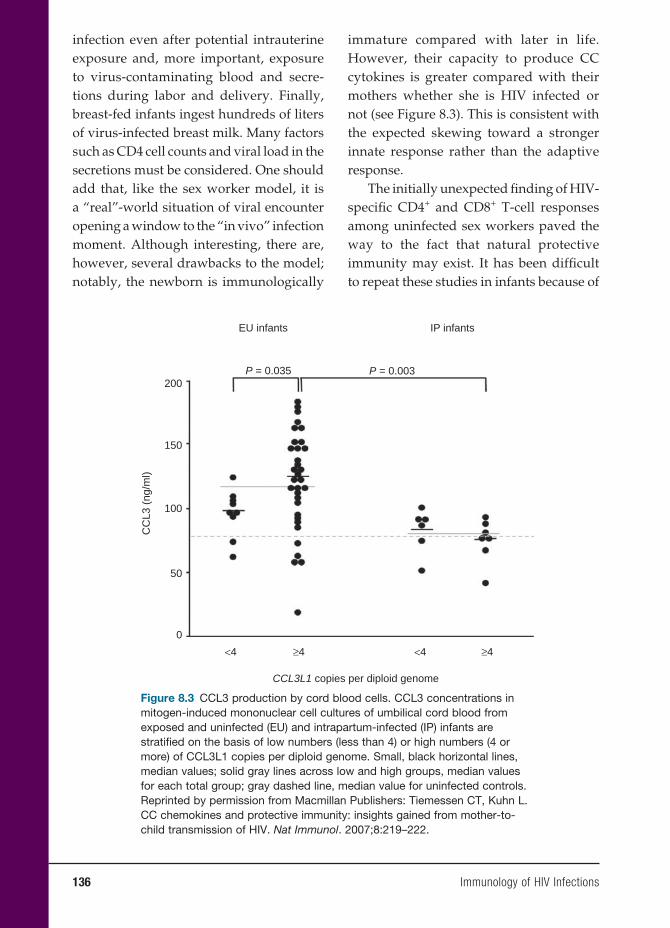
136 Immunology of HIV Infections
infection even after potential intrauterine exposure and, more important, exposure to virus-contaminating blood and secre-tions during labor and delivery. Finally, breast-fed infants ingest hundreds of liters of virus-infected breast milk. Many factors such as CD4 cell counts and viral load in the secretions must be considered. One should add that, like the sex worker model, it is a “real”-world situation of viral encounter opening a window to the “in vivo” infection moment. Although interesting, there are, however, several drawbacks to the model; notably, the newborn is immunologically
immature compared with later in life. However, their capacity to produce CC cytokines is greater compared with their mothers whether she is HIV infected or not (see Figure 8.3). This is consistent with the expected skewing toward a stronger innate response rather than the adaptive response.
The initially unexpected fi nding of HIV-specifi c CD4+ and CD8+ T-cell responses among uninfected sex workers paved the way to the fact that natural protective immunity may exist. It has been diffi cult to repeat these studies in infants because of
200
150
CCL3L1 copies per diploid genome
100
CC
L3 (
ng/m
l)
50
0
<4 ≥4 ≥4<4
IP infantsEU infants
P = 0.035 P = 0.003
Figure 8.3 CCL3 production by cord blood cells. CCL3 concentrations in mitogen-induced mononuclear cell cultures of umbilical cord blood from exposed and uninfected (EU) and intrapartum-infected (IP) infants are stratifi ed on the basis of low numbers (less than 4) or high numbers (4 or more) of CCL3L1 copies per diploid genome. Small, black horizontal lines, median values; solid gray lines across low and high groups, median values for each total group; gray dashed line, median value for uninfected controls. Reprinted by permission from Macmillan Publishers: Tiemessen CT, Kuhn L. CC chemokines and protective immunity: insights gained from mother-to-child transmission of HIV. Nat Immunol. 2007;8:219–222.
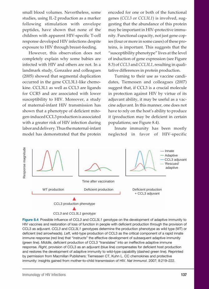
Immunology of HIV Infections 137
small blood volumes. Nevertheless, some studies, using IL-2 production as a marker following stimulation with envelope peptides, have shown that none of the children with apparent HIV-specifi c T-cell response developed HIV infections despite exposure to HIV through breast-feeding.
However, this observation does not completely explain why some babies are infected with HIV and others are not. In a landmark study, Gonzalez and colleagues (2005) showed that segmental duplication occurred in the gene CCL3L1-like chemo-kine. CCL3L1 as well as CCL3 are ligands for CCR5 and are associated with lower susceptibility to HIV. Moreover, a study of maternal-infant HIV transmission has shown that a phenotype of defi cient mito-gen-induced CCL3 production is associated with a greater risk of HIV infection during labor and delivery. Thus the maternal-infant model has demonstrated that the protein
encoded for one or both of the functional genes (CCL3 or CCL3L1) is involved, sug-gesting that the abundance of this protein may be important in HIV-protective immu-nity. Functional capacity, not just gene cop-ies (four or more in some cases) of these pro-teins, is important. This suggests that the “susceptibility phenotype” lives at the level of induction of gene expression (see Figure 8.3) of CCL3 and CCL3L1, resulting in quali-tative differences in protein production.
Turning to their use as vaccine candi-dates, Tiemessen and colleagues (2007) suggest that, if CCL3 is a crucial molecule in protection against HIV by virtue of its adjuvant ability, it may be useful as a vac-cine adjuvant. In this manner, one does not have to rely on the host’s ability to produce it (production may be defi cient in certain populations; see Figure 8.4).
Innate immunity has been mostly neglected in favor of HIV-specific
Res
pons
e m
agni
tude
Time after vaccination
WT production
CCL3 production phenotype
CCL3 and CCL3L1 genotype
Deficient production Deficient production+ CCL3 adjuvant
InnateAdaptiveCCL3 adjuvant‘Rescued’ adaptive
Figure 8.4 Possible infl uence of CCL3 and CCL3L1 genotype on the development of adaptive immunity to HIV vaccines and restoration of loss of function in people with defi cient production through the provision of CCL3 as adjuvant. CCL3 and CCL3L1 genotypes determine the production phenotype as wild type (WT) or defi cient (red arrowheads). Left, wild-type production of CCL3 as the critical component of a rapid innate immune response (red line) that “instructs” the effective development of subsequent adaptive immunity (green line). Middle, defi cient production of CCL3 “translates” into an ineffective adaptive immune response. Right, provision of CCL3 as an adjuvant (blue line) compensates for defi cient host production and restores the development of adaptive immunity to wild-type capability (dashed green line). Reprinted by permission from Macmillan Publishers: Tiemessen CT, Kuhn L. CC chemokines and protective immunity: insights gained from mother-to-child transmission of HIV. Nat Immunol. 2007; 8:219–222.
138 Immunology of HIV Infections
immunity in HIV vaccine studies. CCL3 production should be considered a cor-relate of protective immunity. A desired response would be suffi cient production of CCL3 at the site of HIV and vaccine encounter. It is imperative to delineate the precise functions of CCL3 in the immune response to HIV, aside from its noncyto-lytic inhibitory effect on HIV, as the abil-ity of CCL3 to drive the development of adaptive immunity might be the crucial factor in overall protection. Many impor-tant questions arise, given the fi ndings outlined here. How should HIV vaccines be designed? How can people who would be poor responders to vaccines (defi cient CCL3 producers) be identifi ed? How can the genetically encoded loss of function be overcome, and how can molecules be identifi ed that can compensate for this? Innate immunity must become a more integral component of studies of HIV vac-cines, as understanding the interaction between innate and adaptive immunity may hold the key to understanding what constitutes protective immunity to HIV. Studies of uninfected infants born to HIV-infected mothers offer a unique human experimental model for extending the understanding of such phenomena.
ANIMAL MODELS OF HIV INFECTION
Although many questions concerned with HIV biology were fi rst learned in cul-tured cell lines infected with HIV or SIV, it became obvious that these cultured lines did not truly mimic the wild-type infec-tions seen in animal models. A number of small rodent models, such as the SCID mouse, were developed and were helpful but somewhat artifi cial and did not really mimic human disease.
In fact, a signifi cant obstacle in HIV vaccine research has been the diffi culty in developing an appropriate animal model with many of the features of human HIV infection. The only animals susceptible to experimental infections with HIV are chimpanzees, Pantroglodytes, and pigtail macaques, Macaca nemestrina. These ani-mals maintain low levels of persistent virus but do not develop clinical manifesta-tions of AIDS. In contrast, Asian monkeys, especially rhesus monkeys from India, are highly susceptible to SIVmac infec-tion and progressively develop an immu-nodefi ciency syndrome that is similar to human AIDS. Plasma viral levels during acute and chronic SIVmac251 infection in these animals parallel those observed in humans, with some animals containing virus spontaneously and progressing to disease slowly as in HIV-1 infected human long-term nonprogressors. In con-trast, others maintain high viral loads and behave like human rapid progresses. As in HIV-1 infected humans, the cellular immune response to SIVmac during acute and chronic disease differs signifi cantly, and evidence of immune escape has been well documented. This is particularly true of the mucosal immune system, especially the many CD4+ CCR-5+ T cells in the gut-associated lymphoid tissue, the major site of viral replication and of CD4+ T-cell depletion in SIV-infected macaques. This also mimics the human disease and thus the SIV macaque model is currently con-sidered the most appropriate animal model for studies on potential protective immune responses against HIV.
A second animal model of HIV vaccine research is based on the use of SIV/HIV hybrid viruses that were engineered to carry the env gene from HIV into the con-tent of the SIV genome. These viruses do
Immunology of HIV Infections 139
replicate in rhesus macaques and after serial macaque passages do lead to the emergence of highly pathogenic SHIV variants that can wipe out circulating CD4+ T cells within a few weeks and lead to a lethal immunodefi ciency syndrome within a year of infection.
However, the problem has been whether these variants, like SHIV 89.6P, are relevant to HIV infections. For exam-ple, they exhibit an “X4” cellular tropism and are therefore easier to contain by vac-cine-induced immune response than “R5” viruses such as SIV. If we could obtain SHIV strains with R5 tropism, this might be a better model to study.
The real problem with the monkey models is that experiments to evaluate vac-cine effi cacy requires challenging the vac-cinated animals with high doses of virus to get 100 percent infection, that is, 103 – 105 TCID50 equivalent to 5 × 10⁷ SIV RNA cop-ies/ml. This is in marked contrast to natu-ral exposure to HIV in humans wherein the doses of 103 HIV RNA copies/ml of semi-nal plasma have been reported. However, these results are quite different in mucosal challenge, wherein reported low doses (10–30 TCID50) with SIV results in the same viral and immunological kinetics of infection as high-dose challenges. Thus, this new type of mucosal challenge might change the results of preclinical vaccine effi cacy studies in the future.
VACCINES TO PREVENT HIV
INFECTION
Despite the many efforts to develop a safe and effective vaccine against HIV infection, the results to date have been negative or controversial at best. Among the reasons for this state of affairs have been the enormous
genetic diversity of the virus and the unique features of the HIV envelope protein. One thing we have learned from studies of HIV pathogenesis, however, has been that a vac-cine that induces a strong and specifi c T-cell immune response in the absence of broadly neutralizing antibodies may blunt initial viremia, even if the infection is not com-pletely prevented. Moreover, they may pre-vent the mass destruction of CD4+ T cells, thereby helping to control the infection and prolong disease-free survival.
Before we go to the actual vaccine can-didates, some knowledge of the structure of the HIV is warranted. HIV, together with the simian, feline, and bovine immu-nodefi ciency viruses (SIV, FIV, and BIV) all belong to the genus lentevirus in the family of Retroviridae. These viruses char-acteristically produce slowly progressive infections, and the replication depends on an active reverse transcriptase enzyme to transform the viral RNA genome into a proviral DNA copy that integrates into the host cell chromosome. This step provides the enormous genetic diversity of HIV iso-lates in the infected cell, since the reverse transcriptase step is often fraught with errors in replication.
Two types of HIV have been described: HIV-1 and HIV-2, the latter being less virulent, less transmissible, and confi ned mostly to West Africa. HIV-1 is phylo-genetically close to SIVcpz commensural virus in chimpanzees, which probably arose as a specifi c transmission event from chimps to humans. However, SIVmac is the etiological agent of simian AIDS and has many of the features of HIV AIDS. HIV-1 is further divided into three major groups: M (major), O (outlier), and N (non M or O). Most of the strains responsible for the AIDS epidemic belong to the M group, and this group has been divided
140 Immunology of HIV Infections
into ten different subgroups known as clades (A–K).
The genome of HIV is a single-stranded positive small RNA molecule, approximately 9.5 kbs in length and encodes the typical retrovirus proteins Gag further divided into M (matrix), C (capsid), and N (nucleocapsid); Pol, cleaved into protease, reverse transcriptase, and integrase; and ENV 160 kD glycoprotein, divided into an external gp120 and a trans-membrane gp41 subunit that together form trimeric spikes on the surface of the virion. Furthermore, the HIV genome encodes a variety of nonstructural proteins such as transactivation protein (Tat) splice regulator protein (Rev) and accessory pro-teins such as Nef, Vpr, and Vpn.
VACCINE CANDIDATES
There have been many attempts to con-struct a vaccine that is both protective and has a low cost of production; yet, there is still not a promising candidate. Part of the problem is that to be effective the vaccine has to be given early or before exposure to the virus. Ideally a vaccine could prevent the disease entirely or at least shut down the viremie phase (see panel B of Figure 8.4). For the sake of convenience, we have listed the vaccine approaches separately with their promises and caveats.
Live Attenuated Vaccines
The observation that NEF strains could offer protection against challenge with pathogenic SIV infections in rhesus macaques served as the model for the use of this type of attenuated vaccine. However, this mutant’s drawback is that it produces a lifelong persistent viral infection in the
host. The positive side is that although the attenuated vaccine does not prevent infection with a wild-type SIV infection, it does prevent that infection from going on to produce the disease AIDS. Thus, this approach does not appear feasible now. However, what is being explored is the nature of the protective immune response in these monkeys after infection with the attenuated virus.
Several reports raise the question whether these attenuated vaccines will give the broad protection so eagerly sought. As Anne Piantadosi and colleagues (2007) have pointed out, HIV-1 superinfection (reinfec-tion) does not always protect against other strains. In their studies, they screened a cohort of high-risk Kenyan women by com-paring partial gag and envelope sequences over a fi ve-year period, beginning with the primary infection. Of thirty-seven women screened, seven were found to have superinfections, including cases in which both viruses were from the same HIV-1 subtype A. In fi ve cases, the superinfect-ing strain was detected in only one of the two genomes examined, suggesting that recombination frequently occurs following HIV-1 superinfection. They conclude that superinfection commonly occurs after the immune response against the initial infec-tion has had time to develop and mature. Supporting this claim was an earlier report by Altfeld et al. in Nature in which this superinfection occurred despite broad CD8+ T-cell responses (twenty-fi ve distinct epitopes) to many HIV viral proteins. They conclude that superinfection can occur in the setting of a strong and broadly directed virus-specifi c CD8+ T-cell response.
Yet, other reports by Chakraborty and colleagues indicate that superinfection may not be so high. They studied fourteen HIV seroconcordant couples (i.e., partners were
Immunology of HIV Infections 141
independently infected with different HIV-1 strains) with high risk of re-exposure to the virus. Phylogenetic analyses based on pol and env global sequences obtained from more than a 100 longitudinal plasma samples over one to four years failed to detect HIV-1 superinfection in this cohort of patients. They conclude that chronic HIV infection seems to confer protection against superinfection with a second HIV-1 strain. Obviously, more work needs to be done to determine what factors are responsible for superinfection in some individuals and protection in others, both at high risk of reinfection.
Inactivated Viral Vaccines
Although the initial results with inacti-vated vaccine were negative with high doses of formalin treatment (loss of anti-genicity of the viral envelope proteins), it was quickly discovered that by using low doses of formalin, the antigenicity of the envelope proteins was preserved. This preparation was capable of inducing viral neutralizing antibodies in both mice and nonhuman primates. This area is presently being actively pursued, but the problem will be overcoming the rapidly changing antigens of the wild-type virus.
A second approach has been to inacti-vate the domains of two nucleocapsid pro-tein zinc fi ngers. This has been achieved by treating these complexes with mild oxidation or alkylation procedures, which completely inactivates both HIV-1 and SIV but keeps the envelope glycoprotein spikes intact and functional. Studies in the SIV macaque model revealed that monkeys vaccinated with the inactivated virus were not protected against infection with the wild-type virus, but the levels of SIV vire-mia were low and there was no depletion of CD4+ T cells.
Subunit Vaccines
Most of the research efforts in HIV vaccines have gone into the subunit vaccines involv-ing the gp120 envelope proteins, which also includes the gp41 domain. Both of these proteins do elicit neutralizing antibodies to the homologous vaccine strain but not to heterologous primary isolates in the animal model. Despite these caveats, two large-scale phase III clinical trials of this type of vaccine have been carried out involving 7,500 high-risk individuals. Neither of these trials showed a signifi cant reduction in HIV infection in the vaccinated individuals, even with continuous booster shots dur-ing the three-year trial. A number of other trials involving trimeric gp140, a DNA vac-cine encoding a V2-deleted gp140 and other combinations, are in various phases, but the results are not known at present.
Cellular Vaccines
A strong and specific T-cell immune response in the absence of broadly neu-tralizing antibodies may blunt the ini-tial viremia, even if the infection is not completely prevented. Thus, more recent vaccine efforts have been directed toward stimulating the cellular immune response. Particular attention has been paid to those vaccines that induce an HIV-specific CD8+ CTL response whose role in the con-trol of virus load and evolution of disease has been well documented in the macaque model. Although the T-cell vaccines do not prevent the HIV infection, they do help vaccinees who get infected to control viral replication and reduce viral loads, thus resulting in less risk of transmission of the disease to seronegative partners.
Vaccines have been based on this strat-egy with particular emphasis on the use of
142 Immunology of HIV Infections
naked DNA vaccines and live recombinant vectors, for example, naked DNA vaccines expressing the HIV-1 gag gene and either IL-12 or Il-15, which was developed by Wyeth and is presently in several phase I trials in the United States, Brazil, and Thailand. DNA vaccines have been found to be most useful as priming vaccines in prime-boost strategies, using live recombi-nant vaccines for booster immunization.
While the original recombinant HIV vaccines used vaccinia virus and were well tolerated, safety considerations in immu-nodefi cient hosts has led investigators to substitute the canarypox virus for the vac-cinia virus. However, the immunogenicity of the pox-virus-based vaccines in humans has been relatively modest with less than 35 percent of the vaccinees scoring positive for T-cell responses. In contrast, replication-defective adenovirus type 5 (ad5) appears to be one of the most promising live virus vectors for HIV vaccines. Merck has used this complex and showed that 50 percent of the volunteers had signifi cantly long-lasting CD8+ T-cell responses to HIV-1 peptides. A trivalent recombinant ad5-gag/pol/nef complex has been engineered and retested in human volunteers. A phase II trial of this candidate is being investigated in 1,200 men and 400 women at high risk of exposure to HIV infection who will be followed for three years. The results of this trial will appear in 2008.
FUTURE DIRECTIONS FOR
RESEARCH
The use of antiretroviral combination regimes has made signifi cant advances in the treatment of HIV infections in indus-trialized countries. However, these treat-ments are expensive and would only reach
a limited number of infected individuals, especially in developing countries where most of the epidemic is occurring.
Thus, the need for a safe, effective, inex-pensive vaccine to prevent further spread of the infection becomes an increasingly important goal. Unfortunately, most of the vaccine trials to date have not been success-ful in this regard, and many investigators have turned their attention to vaccines that, if given early in the infection, may pro-tect individuals from developing AIDS. A number of these vaccines show prom-ise and are in various stages of clinical or nonhuman primate trials. As stated in the 2007 New England Journal of Medicine article by Johnson and Fauci, “There is optimism that even a less than perfect vaccine could benefi t individual recipients and the at risk community…. However such a vaccine will have to be delivered as part of a comprehen-sive multifaceted prevention program.”
These are, however, to be considered as fi rst-generation vaccines. The ultimate goal will be to produce an effective preventive vaccine. This will require new thinking about vaccine approaches, innovative solutions, and persistence, but the total prevention of this devastating disease is a worthwhile goal.
BIBLIOGRAPHY
REVIEWSDuerr A, Wasserheit JN, Corey L. HIV vac-
cines: new frontiers in vaccine develop-ment. Clin Infect Dis. 2006;43:500–511.
Girard MP, Osmanor SK, Kieny MP. A review of vaccine research and devel-opment: the human immunodefi ciency virus (HIV). Vaccine. 2006;24:4062–4081.
Johnson MJ, Fauci AS. Current concepts: an HIV vaccine – evolving concepts. New Engl J Med. 2007;356:2073–2081.
Immunology of HIV Infections 143
SUGGESTED READINGAltfeld M, Allen TM, Yu XG, et al. HIV-1
superinfection despite broad CD8+ T cell responses containing replication of the primary virus. Nature. 2002;420:434–439.
Chakraborty B, Valer L, De Mendoza C, Soriano V, Quiñones-Mateu ME. Fail-ure to detect human immunodefi-ciency virus type 1 superinfection in 28 HIV-seroconcordant individuals with high risk of reexposure to the virus. AIDS Res Hum Retroviruses. 2004;20:1026–1031.
Devito C, Broliden K, Kaul R, et al. Mucosal and plasma IgA from HIV-1-exposed uninfected individuals inhibit HIV-1 transcytosis across human epithelial cells. J Immunol. 2000;165:5170–5176.
Devito C, Hinkula J, Kaul R, et al. Muco-sal and plasma IgA from HIV-exposed seronegative individuals neutral-ize a primary HIV-1 isolate. AIDS. 2000;14:1917–1920.
Editorial. New York Times. June 1, 2007.Ellenberger D, Otten R, Li B, et al. Evidence
of protection after reported mucosal SHIV challenges to HIV-1 vaccinated rhesus types. AIDS Vaccine Meeting, Montreal Quebec, Canada. Abs. 110; 2005.
Gonzalez E, Kulkarni H, Mangamo A, et al. The infl uence of CCL3L1 gene-containing segmented duplications M HIV-1/AIDS susceptibility. Science. 2005;307:1434–1440.
Heeney JL. Primate models for AIDS vaccine development. AIDS. 1996;10(suppl A):S115–S122.
Kaul R, Dong T, Plummer FA, et al. CD8+ lymphocytes respond to different HIV epitopes in seronegative and infected subjects. J Clin Invest. 2001;107(10):1303–1310.
Kaul R, Plummer FA, Kimani J, et al. HIV-1-specific mucosal CD8+ lym-phocyte responses in the cervix of HIV-1-resistant prostitutes in Nairobi. J Immunol. 2000;164:1602–1611.
Kaul R, Rowland-Jones SL, Kimani J, et al. Late seroconversion in HIV-resistant Nairobi prostitutes despite pre-existing HIV-specifi c CD8+ responses. J Clin Invest. 2001;107:341–349.
Piantadosi A, Chohan B, Chohan V, et al. Chronic HIV-1 infection frequently fails to protect against superinfection. PLoS Pathog. 2007;3(11):e177.
Quaranta MC, Mattiol B, Giordoni L, Viora M. The immunoregulatory effects of HIV-1 Nef on dendritic cells and the pathogenesis of AIDS. FASEB J. 2006;20:2198–2208.
Rowland-Jones S, Pinheiro S, Kaul R. New insights into host factors in HIV-1 pathogenesis. Cell. 2001;104:473–476.
Tiemessen CT, Kuhn L. CC chemokines and protective immunity: insights gained from mother-to-child transmission of HIV. Nat Immunol. 2007;8(3):219–222.

9. Immunological Aspects of Allergy and Anaphylaxis
Paul M. Ehrlich, M.D., and Jonathan D. Field, M.D., F.A.A.A.I.
145 145
INTRODUCTION
Adverse responses to otherwise innocuous substances we are exposed to constitute the crux of allergic reactions. Reaction to exposure to these substances may vary from a slight rash, easily treated with an antihistamine, leukotriene modifi er, or corticosteroid cream, to a multisystemic reaction, with catastrophic consequences or anaphylaxis. All that will be discussed in this chapter have, as a common factor, various aspects of the immune system with infl ammatory responses involving these seemingly innocuous substances.
Allergic reactions may be found in up to 20 percent of the general U.S. popula-tion, but by the age 6, 40 percent of the children in the United States have some sort of allergic problems. Although most of these children have respiratory prob-lems, such as allergic rhinitis or bronchial asthma, many of those with allergies may also have atopic reactions to foods or medi-cations. As antigens are slowly introduced into an infant’s environment or diet, the child’s propensity to deal with these new substances may not be developed. In addi-tion, children’s airways are small, their gastrointestinal tracts are not developed, and their immune systems are not ready to meet the challenges of these newly introduced proteins called allergens. Most responses are Gell and Coombs type I or
immediate hypersensitivity reaction. This reaction will be described in the next section.
ALLERGIC HYPERSENSITIVITY
In 1964, Gell and Coombs classifi ed four types of immunologically mediated hyper-sensitivity states. The majority of disease states encountered in the clinical practice of allergy are related to type I, or immediate-type hypersensitivity. In this model, an allergen interacts with preformed IgE on the surface of a mast cell or basophil. This interaction causes cross-linking of the FceRI receptor and release of multiple mediators, including histamine, leukotri-enes, and various interleukins. Depending on the relative localization of release, clini-cal states such as allergic asthma, allergic rhinitis, or systemic anaphylaxis occur. Of the remaining clinical allergy hypersensi-tivity states, type IV, or delayed-type hyper-sensitivity, is most common. In this type, T-cell antigen receptors on TH1 or TH2 lymphocytes bind to tissue antigens, caus-ing clonal expansion of lymphocytes and release of pro-infl ammatory lymphokines. Distinct clinical entities such as contact der-matitis (e.g., poison ivy) or tuberculin skin test sensitivity in pulmonary tuberculosis may occur relative to the site of the tissue antigen.
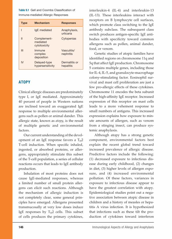
146 Immunological Aspects of Allergy and Anaphylaxis
ATOPY
Clinical allergic diseases are predominately type I, or IgE mediated. Approximately 40 percent of people in Western nations are inclined toward an exaggerated IgE response to multiple environmental aller-gens such as pollen or animal dander. This allergic state, known as atopy, is the result of multiple genetic and environmental factors.
Our current understanding of the devel-opment of an IgE response favors a TH2 T-cell induction. When specifi c inhaled, ingested, or absorbed proteins, or aller-gens, appropriately stimulate this subset of the T-cell population, a series of cellular reactions occurs that leads to IgE antibody production.
Inhalation of most proteins does not cause IgE-mediated responses, whereas a limited number of small protein aller-gens can elicit such reactions. Although the mechanism of allergic induction is not completely clear, some general prin-ciples have emerged. Allergens presented transmucosally at very low doses induce IgE responses by TH2 cells. This subset of cells produces the primary cytokines,
interleukin-4 (IL-4) and interleukin-13 (IL-13). These interleukins interact with receptors on B lymphocyte cell surfaces, which promote class switching to the IgE antibody subclass. The subsequent class switch produces antigen-specifi c IgE anti-bodies with specifi city toward common allergens such as pollen, animal dander, food, or venom.
Genetic studies of atopic families have identifi ed regions on chromosome 11q and 5q that affect IgE production. Chromosome 5 contains multiple genes, including those for IL-4, IL-5, and granulocyte-macrophage colony-stimulating factor. Eosinophil sur-vival and mast cell proliferation are just a few pro-allergic effects of these cytokines. Chromosome 11 encodes the beta subunit of the high-affi nity IgE receptor. Increased expression of this receptor on mast cells leads to a more vehement response to small numbers of antigens. This increased expression explains how exposure to min-ute amounts of allergen, such as venom from a stinging insect, can produce sys-temic anaphylaxis.
Although atopy has a strong genetic component, environmental factors best explain the recent global trend toward increased prevalence of allergic disease. Predictive factors include the following: (1) decreased exposure to infectious dis-ease during early childhood, (2) changes in diet, (3) higher levels of allergen expo-sure, and (4) increased environmental pollution. Of these factors, variances in exposure to infectious disease appear to have the greatest correlation with atopy. Epidemiological studies point out a nega-tive association between atopic disease in children and a history of measles or hepa-titis A virus infection. It is hypothesized that infections such as these tilt the pro-duction of cytokines toward interferon
Table 9.1 Gell and Coombs Classifi cation of
Immune-mediated Allergic Responses
Type Mechanism Responses
I IgE mediated Anaphylaxis, urticaria
II Complement-mediated cytotoxicity
Cytopenias
III Immune complex deposition
Vasculitis/nephritis
IV Delayed-type hypersensitivity
Dermatitis or hepatitis
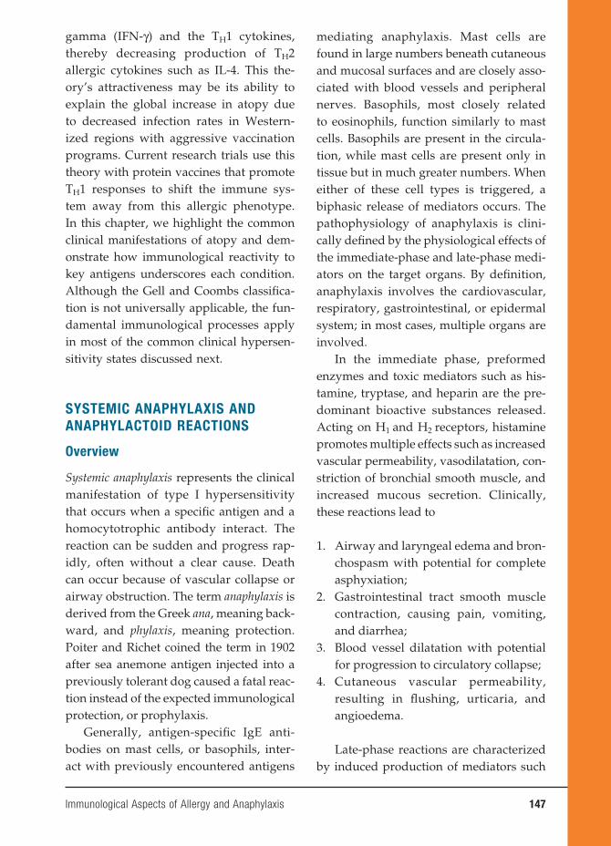
Immunological Aspects of Allergy and Anaphylaxis 147
mediating anaphylaxis. Mast cells are found in large numbers beneath cutaneous and mucosal surfaces and are closely asso-ciated with blood vessels and peripheral nerves. Basophils, most closely related to eosinophils, function similarly to mast cells. Basophils are present in the circula-tion, while mast cells are present only in tissue but in much greater numbers. When either of these cell types is triggered, a biphasic release of mediators occurs. The pathophysiology of anaphylaxis is clini-cally defi ned by the physiological effects of the immediate-phase and late-phase medi-ators on the target organs. By defi nition, anaphylaxis involves the cardiovascular, respiratory, gastrointestinal, or epidermal system; in most cases, multiple organs are involved.
In the immediate phase, preformed enzymes and toxic mediators such as his-tamine, tryptase, and heparin are the pre-dominant bioactive substances released. Acting on H1 and H2 receptors, histamine promotes multiple effects such as increased vascular permeability, vasodilatation, con-striction of bronchial smooth muscle, and increased mucous secretion. Clinically, these reactions lead to
1. Airway and laryngeal edema and bron-chospasm with potential for complete asphyxiation;
2. Gastrointestinal tract smooth muscle contraction, causing pain, vomiting, and diarrhea;
3. Blood vessel dilatation with potential for progression to circulatory collapse;
4. Cutaneous vascular permeability, resulting in fl ushing, urticaria, and angioedema.
Late-phase reactions are characterized by induced production of mediators such
gamma (IFN-γ) and the TH1 cytokines, thereby decreasing production of TH2 allergic cytokines such as IL-4. This the-ory’s attractiveness may be its ability to explain the global increase in atopy due to decreased infection rates in Western-ized regions with aggressive vaccination programs. Current research trials use this theory with protein vaccines that promote TH1 responses to shift the immune sys-tem away from this allergic phenotype. In this chapter, we highlight the common clinical manifestations of atopy and dem-onstrate how immunological reactivity to key antigens underscores each condition. Although the Gell and Coombs classifi ca-tion is not universally applicable, the fun-damental immunological processes apply in most of the common clinical hypersen-sitivity states discussed next.
SYSTEMIC ANAPHYLAXIS AND
ANAPHYLACTOID REACTIONS
Overview
Systemic anaphylaxis represents the clinical manifestation of type I hypersensitivity that occurs when a specifi c antigen and a homocytotrophic antibody interact. The reaction can be sudden and progress rap-idly, often without a clear cause. Death can occur because of vascular collapse or airway obstruction. The term anaphylaxis is derived from the Greek ana, meaning back-ward, and phylaxis, meaning protection. Poiter and Richet coined the term in 1902 after sea anemone antigen injected into a previously tolerant dog caused a fatal reac-tion instead of the expected immunological protection, or prophylaxis.
Generally, antigen-specifi c IgE anti-bodies on mast cells, or basophils, inter-act with previously encountered antigens
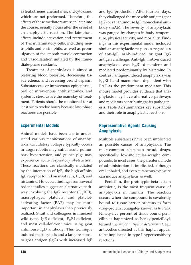
148 Immunological Aspects of Allergy and Anaphylaxis
as leukotrienes, chemokines, and cytokines, which are not preformed. Therefore, the effects of these mediators are seen later into the course, usually hours after the onset of an anaphylactic reaction. The late-phase effects include activation and recruitment of TH2 infl ammatory cells, including neu-trophils and eosinophils, as well as prom-ulgation of the smooth muscle contraction and vasodilatation initiated by the imme-diate-phase reactants.
Treatment of anaphylaxis is aimed at restoring blood pressure, decreasing tis-sue edema, and reversing bronchospasm. Subcutaneous or intravenous epinephrine, oral or intravenous antihistamines, and systemic steroids are the mainstay of treat-ment. Patients should be monitored for at least six to twelve hours because late-phase reactions are possible.
Experimental Models
Animal models have been use to under-stand various manifestations of anaphy-laxis. Circulatory collapse typically occurs in dogs; rabbits may suffer acute pulmo-nary hypertension; and guinea pigs may experience acute respiratory obstruction. These reactions are classically mediated by the interaction of IgE; the high-affi nity IgE receptor found on mast cells, FcεRI; and histamine. However, fi ndings from several rodent studies suggest an alternative path-way involving the IgG receptor (Fc γ RIII), macrophages, platelets, and platelet-activating factor (PAF) may be more important in anaphylaxis than previously realized. Strait and colleagues immunized wild-type, IgE-defi cient, FcεRI-defi cient, and mast cell–defi cient mice using goat antimouse IgD antibody. This technique induced mastocytosis and a large response to goat antigen (IgG) with increased IgE
and IgG production. After fourteen days, they challenged the mice with antigen (goat IgG) or rat antimouse IgE monoclonal anti-body (mAb). The severity of anaphylaxis was gauged by changes in body tempera-ture, physical activity, and mortality. Find-ings in this experimental model included similar anaphylactic responses regardless of anti-IgE, mAb-induced, or goat IgG antigen challenge. Anti-IgE, mAb-induced anaphylaxis was FcεRI dependent and mediated predominantly by histamine. In contrast, antigen-induced anaphylaxis was Fc γ RIII and macrophase dependent with PAF as the predominant mediator. This mouse model provides evidence that ana-phylaxis may have alternative pathways and mediators contributing to its pathogen-esis. Table 9.2 summarizes key substances and their role in anaphylactic reactions.
Representative Agents Causing
Anaphylaxis
Multiple substances have been implicated as possible causes of anaphylaxis. The most common substances include drugs, specifi cally low-molecular-weight com-pounds. In most cases, the parenteral mode of administration is implicated, although oral, inhaled, and even cutaneous exposure can induce anaphylaxis as well.
Penicillin, the prototypic beta-lactam antibiotic, is the most frequent cause of anaphylaxis in humans. The reaction occurs when the compound is covalently bound to tissue carrier proteins to form drug-protein conjugates known as haptens. Ninety-fi ve percent of tissue-bound peni-cillin is haptenized as benzylpenicilloyl, termed the major antigenic determinant. IgE antibodies directed at this hapten appear to be implicated in type I hypersensitivity reactions.
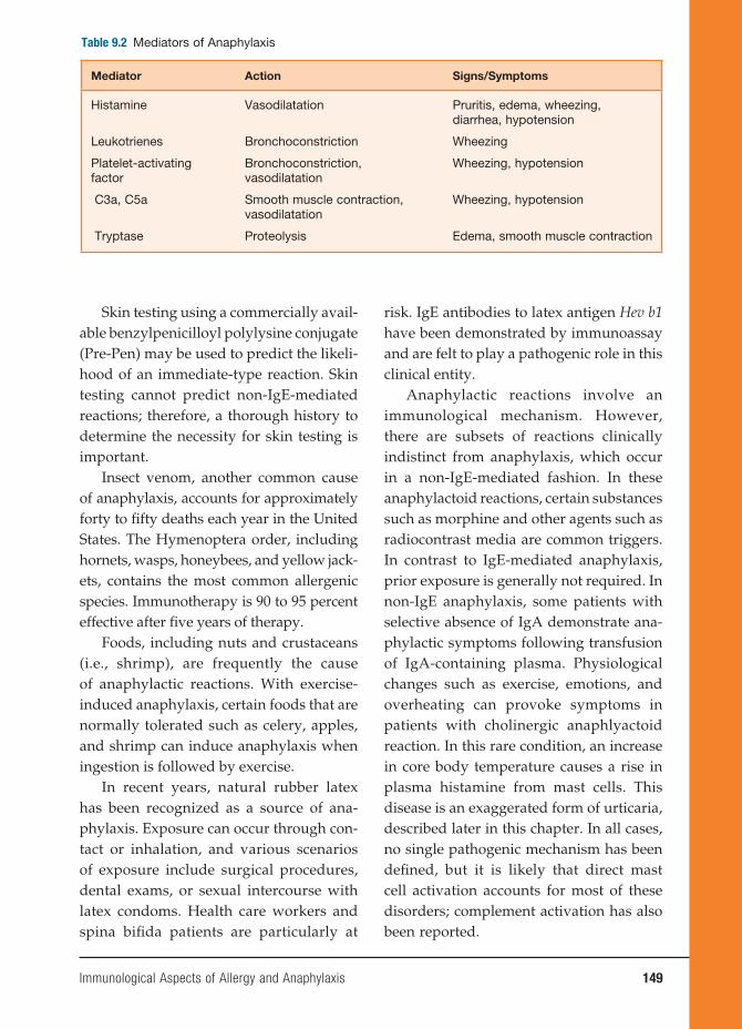
Immunological Aspects of Allergy and Anaphylaxis 149
Skin testing using a commercially avail-able benzylpenicilloyl polylysine conjugate (Pre-Pen) may be used to predict the likeli-hood of an immediate-type reaction. Skin testing cannot predict non-IgE-mediated reactions; therefore, a thorough history to determine the necessity for skin testing is important.
Insect venom, another common cause of anaphylaxis, accounts for approximately forty to fi fty deaths each year in the United States. The Hymenoptera order, including hornets, wasps, honeybees, and yellow jack-ets, contains the most common allergenic species. Immunotherapy is 90 to 95 percent effective after fi ve years of therapy.
Foods, including nuts and crustaceans (i.e., shrimp), are frequently the cause of anaphylactic reactions. With exercise-induced anaphylaxis, certain foods that are normally tolerated such as celery, apples, and shrimp can induce anaphylaxis when ingestion is followed by exercise.
In recent years, natural rubber latex has been recognized as a source of ana-phylaxis. Exposure can occur through con-tact or inhalation, and various scenarios of exposure include surgical procedures, dental exams, or sexual intercourse with latex condoms. Health care workers and spina bifi da patients are particularly at
risk. IgE antibodies to latex antigen Hev b1 have been demonstrated by immunoassay and are felt to play a pathogenic role in this clinical entity.
Anaphylactic reactions involve an immunological mechanism. However, there are subsets of reactions clinically indistinct from anaphylaxis, which occur in a non-IgE-mediated fashion. In these anaphylactoid reactions, certain substances such as morphine and other agents such as radiocontrast media are common triggers. In contrast to IgE-mediated anaphylaxis, prior exposure is generally not required. In non-IgE anaphylaxis, some patients with selective absence of IgA demonstrate ana-phylactic symptoms following transfusion of IgA-containing plasma. Physiological changes such as exercise, emotions, and overheating can provoke symptoms in patients with cholinergic anaphlyactoid reaction. In this rare condition, an increase in core body temperature causes a rise in plasma histamine from mast cells. This disease is an exaggerated form of urticaria, described later in this chapter. In all cases, no single pathogenic mechanism has been defi ned, but it is likely that direct mast cell activation accounts for most of these disorders; complement activation has also been reported.
Table 9.2 Mediators of Anaphylaxis
Mediator Action Signs/Symptoms
Histamine Vasodilatation Pruritis, edema, wheezing, diarrhea, hypotension
Leukotrienes Bronchoconstriction Wheezing
Platelet-activating factor
Bronchoconstriction, vasodilatation
Wheezing, hypotension
C3a, C5a Smooth muscle contraction, vasodilatation
Wheezing, hypotension
Tryptase Proteolysis Edema, smooth muscle contraction

Immunological Aspects of Allergy and Anaphylaxis 151
milieu, it appears that vernal conjunctivi-tis is a combined immediate- and delayed-type hypersensitivity reaction.
Giant papillary conjunctivitis is a chronic infl ammatory process that leads to the production of giant mucosal ducts, or papillae, on the conjunctival lining of the upper eyelids. The immunopathologic mechanism is complex and is theorized to be a mechanical trauma culminating in a mast cell–mediated delayed-type hyper-sensitivity.
The major signs and symptoms include itching and a clear or white stringy dis-charge. A “cobblestone” appearance is typical. It is commonly associated with contact lens use. Foreign bodies and ocu-lar sutures or prosthetics may be causal as well. Treatment typically involves avoid-ance of the inciting process or use of ocular anti-infl ammatory agents such as cromo-lyn sodium or topical corticosteroids.
Atopic keratoconjunctivitis (AK) is observed in approximately 15 to 40 percent of patients with atopic dermatitis. Whereas allergic conjunctivitis is usually a self-lim-iting process, AK is chronic and can poten-tially cause loss of vision. Immunological and immunohistochemical studies reveal mast cells, IgE antibody, eosinophils, and other infl ammatory cells in similar quanti-ties to those found in allergic conjunctivi-tis. The fi nding of lymphocyte involvement explains the chronic nature of the disease as well as the threat to sight. Additional fi ndings include antibodies to ICAM-1 and HLA-DR throughout the ocular epithe-lium, suggesting increased antigen presen-tation. Also, increased level of RANTES, an eosinophil homeostasis chemokine, are observed in immunohistochemical studies of the epithelium. Fibroblast numbers are increased in the connective tissue with an increased level of collagen compared with
normal tissue. This infi ltration is likely critical to the sight-threatening nature of the disease. Table 9.3 summarizes the his-topathological and other laboratory mani-festations of ocular disease.
Treatment is aimed at reducing the local infl ammation. Ocular mast cell stabilizer drops such as olopatadine are the mainstay of treatment. In addition, treatment of nasal symptoms with nasal anti-infl ammatory sprays promotes patency of the nasolac-rimal duct. This combination treatment allows the eye to drain excess allergens, therefore diminishing allergic responses in the eye.
Experimental Models
Animal models of ocular allergies have been developed in the past years to estab-lish new therapeutic approaches and assess immunological mechanisms. The current animal models are based on sensitization to and subsequent challenge with small numbers of allergens such as ovalbumin (Ova), ragweed pollen, or cat epithelium. The murine model of allergic conjunctivitis represents the presently preferred species for investigating the immunological basis of the disease.
Magone and colleagues applied such a model to evaluate the role of various cyto-kines, including IL-4, IFN-γ, and IL-12, in the early and late phases of ocular allergy using knockout (KO) mice and neutral-izing antibodies. In this model, multiple mice experimental groups, including (1) ragweed-sensitized wild type, (2) IL-4KO type, (3) IL-12KO type, (4) IFN-γ KO type, (5) anti-IL-12/mAb-treated, and (6) anti-IFN-γ mAb-treated mice were chal-lenged with ragweed allergen ten days after immunization. An additional group received recombinant murine IL-12. They
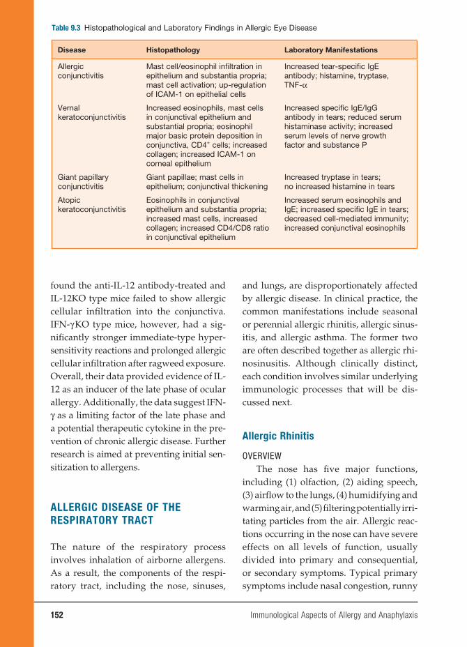
152 Immunological Aspects of Allergy and Anaphylaxis
found the anti-IL-12 antibody-treated and IL-12KO type mice failed to show allergic cellular infi ltration into the conjunctiva. IFN-γ KO type mice, however, had a sig-nifi cantly stronger immediate-type hyper-sensitivity reactions and prolonged allergic cellular infi ltration after ragweed exposure. Overall, their data provided evidence of IL-12 as an inducer of the late phase of ocular allergy. Additionally, the data suggest IFN-γ as a limiting factor of the late phase and a potential therapeutic cytokine in the pre-vention of chronic allergic disease. Further research is aimed at preventing initial sen-sitization to allergens.
ALLERGIC DISEASE OF THE
RESPIRATORY TRACT
The nature of the respiratory process involves inhalation of airborne allergens. As a result, the components of the respi-ratory tract, including the nose, sinuses,
and lungs, are disproportionately affected by allergic disease. In clinical practice, the common manifestations include seasonal or perennial allergic rhinitis, allergic sinus-itis, and allergic asthma. The former two are often described together as allergic rhi-nosinusitis. Although clinically distinct, each condition involves similar underlying immunologic processes that will be dis-cussed next.
Allergic Rhinitis
OVERVIEWThe nose has fi ve major functions,
including (1) olfaction, (2) aiding speech, (3) airfl ow to the lungs, (4) humidifying and warming air, and (5) fi ltering potentially irri-tating particles from the air. Allergic reac-tions occurring in the nose can have severe effects on all levels of function, usually divided into primary and consequential, or secondary symptoms. Typical primary symptoms include nasal congestion, runny
Table 9.3 Histopathological and Laboratory Findings in Allergic Eye Disease
Disease Histopathology Laboratory Manifestations
Allergic conjunctivitis
Mast cell/eosinophil infi ltration in epithelium and substantia propria; mast cell activation; up-regulation of ICAM-1 on epithelial cells
Increased tear-specifi c IgE antibody; histamine, tryptase, TNF-α
Vernal keratoconjunctivitis
Increased eosinophils, mast cells in conjunctival epithelium and substantial propria; eosinophil major basic protein deposition in conjunctiva, CD4+ cells; increased collagen; increased ICAM-1 on corneal epithelium
Increased specifi c IgE/IgG antibody in tears; reduced serum histaminase activity; increased serum levels of nerve growth factor and substance P
Giant papillary conjunctivitis
Giant papillae; mast cells in epithelium; conjunctival thickening
Increased tryptase in tears;no increased histamine in tears
Atopic keratoconjunctivitis
Eosinophils in conjunctival epithelium and substantia propria; increased mast cells, increased collagen; increased CD4/CD8 ratio in conjunctival epithelium
Increased serum eosinophils and IgE; increased specifi c IgE in tears; decreased cell-mediated immunity; increased conjunctival eosinophils
Immunological Aspects of Allergy and Anaphylaxis 153
nose or rhinorrhea, as well as itchy palate and ears. Secondary symptoms include involvement of the middle ears, eustachian tubes, and sinuses, inducing symptoms such as headache, ear pain, and decreased hearing. Multiple central nervous system complaints may occur, including fatigue, irritability, anxiety, and even depression. As in other atopic diseases, the pathophys-iology of allergic rhinitis involves specifi c IgE production after exposure to airborne allergens. The clinical manifestations and underlying immunological mechanisms of the disorder will be discussed later.
CLINICAL DISEASE STATESAllergic rhinitis is the most common
allergic disease, affecting 25 percent of the population. It consists of two forms, sea-sonal and perennial. Seasonal allergic rhi-nitis is commonly referred to as “hay fever” or “rose fever” and is triggered by pollens with a typically well-defi ned season of ger-mination. Perennial allergic rhinitis has similar symptoms, yet involved substances are present year-round, including animal dander, dust mites, and mold. In both forms, allergens interact with mast cells or basal cells in the nasal mucosa. They are then presented by antigen-presenting cells such as dendritic cells and macrophages. CD4+ lymphocytes are stimulated by this presentation to release interleukins includ-ing IL-3, IL-4, IL-5, IL-13, and other cyto-kines that promote local and systemic IgE production by plasma cells. In addition, these cytokines lead to enhanced chemo-taxis, infl ammatory cell recruitment, prolif-eration, activation, and prolonged immune cell survival in the airway mucosa.
Within minutes of allergen inhalation in sensitized individuals, IgE antibodies fi xed to mast cells and basophils trigger release of acute preformed mediators such
as histamine and tryptase in the early-phase response. Shortly afterward, de novo generation of mediators, including cysteinyl-leukotrienes (LTC4, LTD5, and LTE4) as well as prostaglandin D2 (PGD2) occurs. These substances cause a pro-nounced infl ammatory response leading to the typical symptoms of paroxysmal sneezing, nasal pruritis and congestion, clear rhinorrhea, and palatal itching.
Over the next four to eight hours, the mediators released during the initial response set off a sequence of events, with enhanced infl ammatory responses known as the late-phase response. In this phase, cytokines and various mediators released earlier promote the infl ux of other immune cells by enhancing expression of vascular cell adhesion molecules (VCAMs) that help traffi c circulating eosinophils, neutrophils, and lymphocytes to the nasal endothelium. Although each of these cell types plays a role in the late response, eosinophils appear to be the main effector cell in allergic rhi-nitis. Nasal obstruction and secretions can lead to secondary effects, including ear and sinus infections, sleep apnea, and asthma exacerbations. Furthermore, the infl amma-tory cytokines may circulate to the central nervous system, eliciting malaise, irritabil-ity, and impaired concentration.
Treatment of allergic rhinitis involves environmental control for indoor aller-gens, reduction of swelling and congestion by nasal corticosteroids or oral leukotriene receptor antagonists, and relief of rhinor-rhea and nasal pruritis by oral or nasal antihistamines. Recalcitrant cases may require desensitization by immunotherapy (allergy shots).
EXPERIMENTAL MODELSRecent research into the underlying
immunological mechanisms of rhinitis
154 Immunological Aspects of Allergy and Anaphylaxis
has borne new insights into the patho-physiology of allergic rhinitis. Prior under-standing favored initial IgE production in regional lymph nodes or bone marrow. Failure of prior experiments to co-localize IgE protein within B cells to the local tis-sue environment weighed heavily against localized tissue IgE production. In the past ten years, however, a growing body of research has challenged the former dogma that IgE production occurred remotely from the allergen–tissue interface. Studies of local messenger RNA (mRNA) sup-port the hypothesis of local protein syn-thesis. Durham and colleagues, using a combination of in situ hybridization and immunohistochemistry, showed that cells expressing epsilon–heavy chain mRNA (Cε) were present in the nasal mucosa of allergic individuals, and marked increases of these cells occurred on exposure to aller-gen. Furthermore, the increases seen in IgE and Cε mRNA after allergen challenge were inhibited by topical corticosteroids, favoring a localized process. In addition, Kleinjan and colleagues used similar tech-niques to demonstrate fi nally IgE-produc-ing B cells in the nasal mucosa. Biopsies were obtained from normal subjects and from seasonal and perennial subjects dur-ing pollen season and during house dust exposure. The study found no differences in B-cell numbers, either CD19+ (B cells) or CD138+ (immunoglobulin-secreting plasma cells) in allergic and normal patients. Allergic patients, however, exhib-ited signifi cantly greater numbers of IgE-positive B cells than normal patients, and allergen-positive cells were only found in allergics with almost all such cells staining positive for IgE or CD138. The combination of these factors suggests local IgE produc-tion may take place in the mucosa during natural allergen exposure.
Other models focus on the eosino-philic inflammation that characterizes allergic rhinitis as well as modulation of the allergic response. Hussain and col-leagues sensitized BALB/c mice using Ova intraperitoneally. Subsequent challenge with aerosolized Ova took place. Vari-ous outcomes, including nasal symptoms, nasal submucosal eosinophilia, and bone marrow eosinophilia, were measured. Afterward, a subset of mice received CpG oligodeoxynucleotides (ODNs), potent inducers of a TH1 nonallergenic response. Using enzyme-linked immunosorbent assay, cytokine levels were measured. Findings included elevated IL-4 and IL-5 and suppressed IFN-γ in Ova-sensitized mice compared with ODN-treated mice. In addition, the administration of ODNs abrogated nasal symptoms and upper air-way eosinophilia compared with controls. Collectively, these models elucidate the complex immunological basis of allergic rhinitis and demonstrate how manipula-tion of the immune response can be used clinically as a potential treatment.
Allergic Asthma
OVERVIEW The human lung provides the funda-
mental function of gas exchange. As the terminal level of the respiratory tract after the nasal cavity and pharynx, it is constantly exposed to airborne particulate matter. Allergic asthma is the manifestation of a pul-mo nary immune response to various in haled substances. Clinically, the cardinal symptoms include (1) generalized but reversible air-way obstruction, (2) wheeze, (3) dyspnea, and (4) cough. Symptoms can range from mild to life threatening. Typical allergens include house dust mite, pollen, cockroach epithelium, animal dander, and fungi.
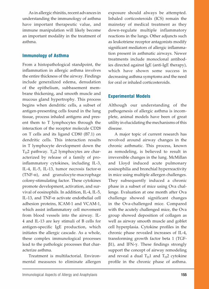
Immunological Aspects of Allergy and Anaphylaxis 155
As in allergic rhinitis, recent advances in understanding the immunology of asthma have important therapeutic value, and immune manipulation will likely become an important modality in the treatment of asthma.
Immunology of Asthma
From a histopathological standpoint, the infl ammation in allergic asthma involves the entire thickness of the airway. Findings include generalized edema, denudation of the epithelium, subbasement mem-brane thickening, and smooth muscle and mucous gland hypertrophy. This process begins when dendritic cells, a subset of antigen-presenting cells found in the lung tissue, process inhaled antigens and pres-ent them to T lymphocytes through the interaction of the receptor molecule CD28 on T cells and its ligand CD80 (B7.1) on dendritic cells. This interaction results in T lymphocyte development down the TH2 pathway. TH2 lymphocytes are char-acterized by release of a family of pro-infl ammatory cytokines, including IL-3, IL-4, IL-5, IL-13, tumor necrosis factor-α (TNF-α), and granulocyte-macrophage colony-stimulating factor. These cytokines promote development, activation, and sur-vival of eosinophils. In addition, IL-4, IL-5, IL-13, and TNF-α activate endothelial cell adhesion proteins, ICAM-1 and VCAM-1, which assist infl ammatory cell movement from blood vessels into the airway. IL-4 and IL-13 are key stimuli of B cells for antigen-specifi c IgE production, which initiates the allergic cascade. As a whole, these complex immunological processes lead to the pathologic processes that char-acterize asthma.
Treatment is multifactorial. Environ-mental measures to eliminate allergen
exposure should always be attempted. Inhaled corticosteroids (ICS) remain the mainstay of medical treatment as they down-regulate multiple inflammatory reactions in the lungs. Other adjuncts such as leukotriene receptor antagonists modify signifi cant mediators of allergic infl amma-tion present in asthmatic airways. Newer treatments include monoclonal antibod-ies directed against IgE (anti-IgE therapy), which have shown some success in decreasing asthma symptoms and the need for oral or inhaled corticosteroids.
Experimental Models
Although our understanding of the pathogenesis of allergic asthma is incom-plete, animal models have been of great utility in elucidating the mechanisms of this disease.
A major topic of current research has revolved around airway changes in the chronic asthmatic. This process, known as remodeling, is believed to result in irreversible changes in the lung. McMillan and Lloyd induced acute pulmonary eosinophilia and bronchial hyperreactivity in mice using multiple allergen challenges. They subsequently induced a chronic phase in a subset of mice using Ova chal-lenge. Evaluation at one month after Ova challenge showed significant changes in the Ova-challenged mice. Compared with the acutely challenged mice, the Ova group showed deposition of collagen as well as airway smooth muscle and goblet cell hyperplasia. Cytokine profi les in the chronic phase revealed increases of IL-4, transforming growth factor beta 1 (TGF-β1), and IFN-γ . These fi ndings strongly support the concept of airway remodeling and reveal a dual TH1 and TH2 cytokine profi le in the chronic phase of asthma.
156 Immunological Aspects of Allergy and Anaphylaxis
Another important focus of asthma research centers on so-called inner-city asthma, an increasing epidemic in devel-oped countries. Many epidemiological studies have shown disproportional rates of asthma in urban, lower socioeconomi-cally stratifi ed patients. Although many socioeconomic factors are thought to play a role, distinct allergic triggers specifi c to these environments appear to be impor-tant. Using recombinant proteins, Sarpong, Zhang, and Kleeberger (2004) evaluated two such allergens, cockroach (Bla g 2) and dust mite (Der f 1), in inbred mouse strain (A/J). Mice were immunized with Bla g 2 and Der f 1 or a combination on days 0 and 7 and were inhalant challenged on day 14. Airway hyperreactivity and airway cellular content were subsequently studied. Findings included dose-related statistically signifi cant increases in airway reactivity and infl ammatory and epithe-lial cell measurements. Compared with individual antigens, however, enhanced infl ammatory cell levels and epithelial cell numbers, but not airway reactivity, were noted in the combined group. This model, which has been subsequently validated, has practical implications for preventing asthma.
FOOD ALLERGY AND INTOLERANCE
Perhaps of all allergic reactions, the pub-lic, clinicians, and medical personnel mis-understand food allergy most. As little as 1 percent of the U.S. population has true food allergies compared with 20 percent who are perceived to have them. True food allergy is a typical, and sometimes catastrophic, type I, IgE-mediated reaction, which preferably must be proven by in vitro or in vivo testing.
Diagnosis of Food Allergy and
Intolerance
The gold standard of food allergy test-ing is the double-blinded, placebo-con-trolled food challenge, which will establish whether the patient is truly allergic to a specifi c food. Because this method of test-ing for food allergies is time consuming, expensive, and potentially dangerous, in vivo testing is usually performed in food allergy centers. Occasionally, straight food challenges may be tried when the chances of reactions are minimal and the serum IgE (radioallergosorbent test, or RAST) specifi c for the allergen is low or negative.
Approximately 20 percent of the U.S. population may perceive food as causative reaction in food allergies, whereas the true prevalence of food allergies is approxi-mately 1 percent. Food allergies are usually found in individuals with a strong per-sonal and family history of allergies. Atopic responses are associated with many foods. In children under the age 2, 90 percent of the incriminating foods are eggs, milk, legumes (such as peanuts, which are not nuts), and soy. In adults, fi sh, shellfi sh, fruits, and tree nuts might be added to the list.
Case 9.1 Peanut AllergyN.G. is a two-year-old female who
was well until the age of seventeen months when she developed a watery, runny nose that persisted throughout the spring but was gone by the sum-mer. She responded well to conventional antihistamines and did well during the summer. In the middle of August, she broke out in hives and wheezing soon after eating a peanut butter sandwich, but the symptoms were reversed with the antihistamine. She had never had peanut butter before that event, but her
Immunological Aspects of Allergy and Anaphylaxis 157
mother was a self-described “peanut but-ter addict” and consumed large amounts during her pregnancy with and breast-feeding of N.G. N.G.’s subsequent skin prick test with peanut revealed a 20 mm by 30 mm wheal and fl air response with a markedly elevated RAST to the peanut protein. She has remained peanut free since that time. She is able to eat tree nuts (e.g., almonds and cashews) without a problem because peanuts are not nuts.
Food Intolerance
True allergic reactions (IgE mediated) to food must be differentiated from food intol-erance, which is rarely multisystemic, not necessarily found among atopic patients, and with no positive skin prick tests or in vitro responses (RAST). A nonallergic reac-tion that typifi es food intolerance would be an acute gastrointestinal response in a lactose-intolerant individual. This patient does not have the enzyme lactase and, therefore, cannot break down sugar lactose in milk and other foods into glucose and galactose. Profound diarrhea or vomiting usually results without any other system involved (respiratory or skin, for example). Food avoidance is the treatment of choice, as it is with food allergy, and over time, food intolerance may resolve. Lactaid is a brand of milk in which the enzyme lactase is added so that the milk may be consumed.
SKIN DISEASES AND ALLERGY
Case 9.2 Skin ReactionsA.A. is a fi fty-six-year-old female
who was well until she began to develop acute, episodic, or transient swelling of her skin (urticaria), with intense pru-ritis (itching) and erythema (redness)
that she described as “maddening.” She assumed she had food allergies, so she tried to eliminate various substances from her diet, including dairy products, fi sh, and nuts without any positive response. Eventually, she took antihis-tamines, with marginal improvement and sought medical attention after a three-month ordeal. An extensive his-tory was taken and allergy tests (all negative) and a variety of serum stud-ies were performed. She was diagnosed with a thyroid abnormality. She was referred to an endocrinologist and did well on therapy, which had nothing to do with an allergic diathesis.
Urticaria and Angioedema
Angioedema and urticaria are two of the more perplexing problems evaluated by physicians, and cases of chronic urticaria (hives of more than six weeks duration) may yield diagnoses in about 20 percent of those evaluated. Perhaps most important in the evaluation is the medical history, fol-lowed by understanding whether physical issues such as pressure, cold, and scratch-ing affect the skin. These issues must be carefully explored in the initial questioning of the patient.
Certain types of urticaria may be brought on by physical disruption of mast cells by either cold, scratching, or pressure. There is no IgE mediation of these reac-tions, unlike those from foods, but hista-mine release from the cells is common to all. Swelling of the skin due to damage to small blood vessels causes burning and pain of the areas involved and may last for several days. The burning and pain are unrelated to allergies or physical mast cell disruption. These might typically be related to a systemic disorder, and precipitating
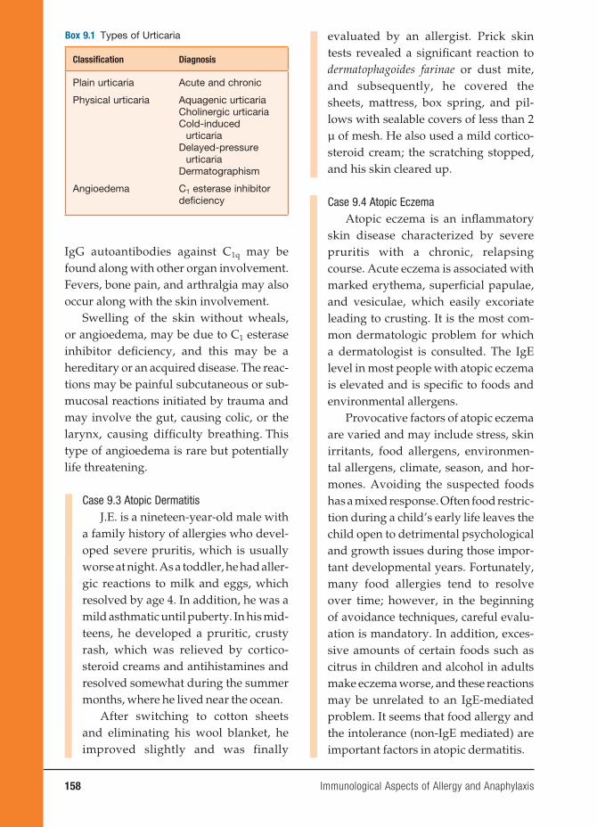
158 Immunological Aspects of Allergy and Anaphylaxis
IgG autoantibodies against C1q may be found along with other organ involvement. Fevers, bone pain, and arthralgia may also occur along with the skin involvement.
Swelling of the skin without wheals, or angioedema, may be due to C1 esterase inhibitor defi ciency, and this may be a hereditary or an acquired disease. The reac-tions may be painful subcutaneous or sub-mucosal reactions initiated by trauma and may involve the gut, causing colic, or the larynx, causing diffi culty breathing. This type of angioedema is rare but potentially life threatening.
Case 9.3 Atopic DermatitisJ.E. is a nineteen-year-old male with
a family history of allergies who devel-oped severe pruritis, which is usually worse at night. As a toddler, he had aller-gic reactions to milk and eggs, which resolved by age 4. In addition, he was a mild asthmatic until puberty. In his mid-teens, he developed a pruritic, crusty rash, which was relieved by cortico-steroid creams and antihistamines and resolved somewhat during the summer months, where he lived near the ocean.
After switching to cotton sheets and eliminating his wool blanket, he improved slightly and was finally
evaluated by an allergist. Prick skin tests revealed a signifi cant reaction to dermatophagoides farinae or dust mite, and subsequently, he covered the sheets, mattress, box spring, and pil-lows with sealable covers of less than 2 µ of mesh. He also used a mild cortico-steroid cream; the scratching stopped, and his skin cleared up.
Case 9.4 Atopic EczemaAtopic eczema is an infl ammatory
skin disease characterized by severe pruritis with a chronic, relapsing course. Acute eczema is associated with marked erythema, superfi cial papulae, and vesiculae, which easily excoriate leading to crusting. It is the most com-mon dermatologic problem for which a dermatologist is consulted. The IgE level in most people with atopic eczema is elevated and is specifi c to foods and environmental allergens.
Provocative factors of atopic eczema are varied and may include stress, skin irritants, food allergens, environmen-tal allergens, climate, season, and hor-mones. Avoiding the suspected foods has a mixed response. Often food restric-tion during a child’s early life leaves the child open to detrimental psychological and growth issues during those impor-tant developmental years. Fortunately, many food allergies tend to resolve over time; however, in the beginning of avoidance techniques, careful evalu-ation is mandatory. In addition, exces-sive amounts of certain foods such as citrus in children and alcohol in adults make eczema worse, and these reactions may be unrelated to an IgE-mediated problem. It seems that food allergy and the intolerance (non-IgE mediated) are important factors in atopic dermatitis.
Box 9.1 Types of Urticaria
Classifi cation Diagnosis
Plain urticaria Acute and chronic
Physical urticaria Aquagenic urticariaCholinergic urticariaCold-induced
urticariaDelayed-pressure
urticariaDermatographism
Angioedema C1 esterase inhibitor defi ciency
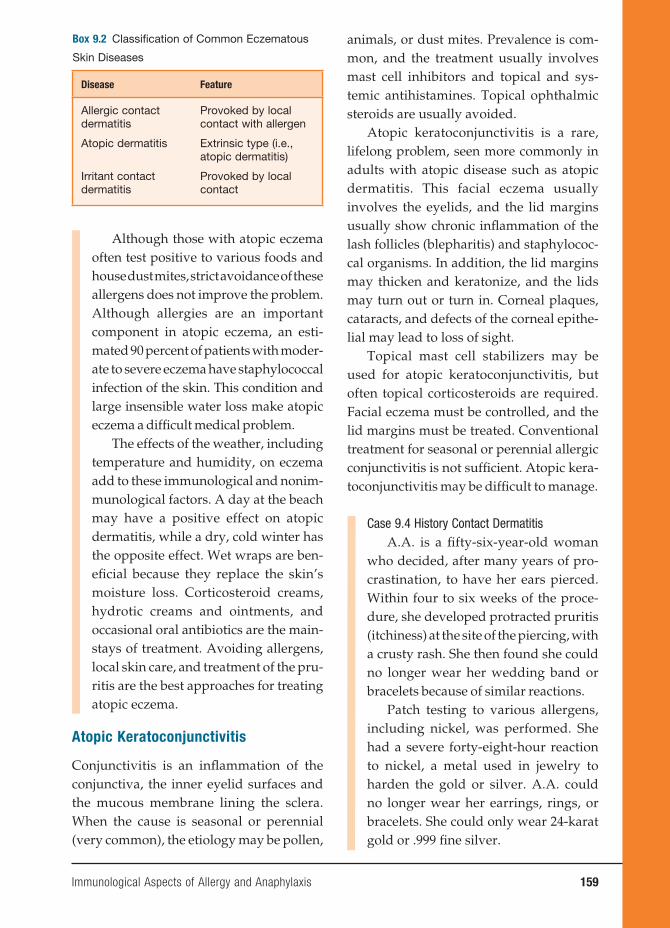
Immunological Aspects of Allergy and Anaphylaxis 159
Although those with atopic eczema often test positive to various foods and house dust mites, strict avoidance of these allergens does not improve the problem. Although allergies are an important component in atopic eczema, an esti-mated 90 percent of patients with moder-ate to severe eczema have staphylococcal infection of the skin. This condition and large insensible water loss make atopic eczema a diffi cult medical problem.
The effects of the weather, including temperature and humidity, on eczema add to these immunological and nonim-munological factors. A day at the beach may have a positive effect on atopic dermatitis, while a dry, cold winter has the opposite effect. Wet wraps are ben-efi cial because they replace the skin’s moisture loss. Corticosteroid creams, hydrotic creams and ointments, and occasional oral antibiotics are the main-stays of treatment. Avoiding allergens, local skin care, and treatment of the pru-ritis are the best approaches for treating atopic eczema.
Atopic Keratoconjunctivitis
Conjunctivitis is an infl ammation of the conjunctiva, the inner eyelid surfaces and the mucous membrane lining the sclera. When the cause is seasonal or perennial (very common), the etiology may be pollen,
animals, or dust mites. Prevalence is com-mon, and the treatment usually involves mast cell inhibitors and topical and sys-temic antihistamines. Topical ophthalmic steroids are usually avoided.
Atopic keratoconjunctivitis is a rare, lifelong problem, seen more commonly in adults with atopic disease such as atopic dermatitis. This facial eczema usually involves the eyelids, and the lid margins usually show chronic infl ammation of the lash follicles (blepharitis) and staphylococ-cal organisms. In addition, the lid margins may thicken and keratonize, and the lids may turn out or turn in. Corneal plaques, cataracts, and defects of the corneal epithe-lial may lead to loss of sight.
Topical mast cell stabilizers may be used for atopic keratoconjunctivitis, but often topical corticosteroids are required. Facial eczema must be controlled, and the lid margins must be treated. Conventional treatment for seasonal or perennial allergic conjunctivitis is not suffi cient. Atopic kera-toconjunctivitis may be diffi cult to manage.
Case 9.4 History Contact DermatitisA.A. is a fi fty-six-year-old woman
who decided, after many years of pro-crastination, to have her ears pierced. Within four to six weeks of the proce-dure, she developed protracted pruritis (itchiness) at the site of the piercing, with a crusty rash. She then found she could no longer wear her wedding band or bracelets because of similar reactions.
Patch testing to various allergens, including nickel, was performed. She had a severe forty-eight-hour reaction to nickel, a metal used in jewelry to harden the gold or silver. A.A. could no longer wear her earrings, rings, or bracelets. She could only wear 24-karat gold or .999 fi ne silver.
Box 9.2 Classifi cation of Common Eczematous
Skin Diseases
Disease Feature
Allergic contact dermatitis
Provoked by local contact with allergen
Atopic dermatitis Extrinsic type (i.e., atopic dermatitis)
Irritant contact dermatitis
Provoked by local contact

160 Immunological Aspects of Allergy and Anaphylaxis
Contact Dermatitis
Classical contact dermatitis is a Gell and Coombs type IV reaction, mediated by previously sensitized lymphocytes, which is exhibited by raised, very pruritic rash at the sight of the contact. Unlike allergic reactions of a type I, IgE-mediated contact dermatitis, as a type IV reaction, involving low-molecular-weight allergens (less than 1 kDa). These contact allergens are haptens and need to link with proteins in the skin to become allergenic. These haptens may be readily absorbed into the skin, a reac-tion that renders them antigenic. If the skin is exposed to humidity or warmth, the penetration of the hapten is greater, and the chance of developing contact dermatitis is greater. As these haptens make their way into extravascular spaces, they combine with serum proteins or cell membranes of antigen-presenting cells. The processed antigens is presented by Langerhans cells to T cells leading to a cascade of events that result in an infl ux of mononuclear cells into the dermis and epidermis, hence dermatitis.
Although most people exposed to these presenting allergens do not develop contact dermatitis, certain substances such as dinitrochlorobezene may sensitize as
many as 90 percent of normal individuals, which is why the patient’s history with suspected contact dermatitis is impor-tant. Among those situations patients need to elucidate are occupation, cosmet-ics, topical or systemic drugs, recreational activities, effects of holidays, and time course.
Testing for contact dermatitis involves placing a patch of the suspected substance or substances on the back. The area must be kept clean and dry for forty-eight hours, after which the various patches are removed, and the individual areas are evaluated for infl ammatory responses. A positive response then characterizes a type IV reaction, and avoidance of the offending allergen is the treatment of choice. Thirty milligrams per day of systemic steroids may be required when large areas of the skin are involved (>25 percent of body surface). Otherwise, corticosteroid creams may be employed; however, avoidance of the contactant is most crucial.
BIBLIOGRAPHY
GENERALAdkinson NF Jr, Yunginger JW, Busse
WW, et al., eds. Middleton’s Allergy:
Box 9.3 Where Contact Agents are Found
Contact Agents Where Contactant is Found
Chloroisothiazolinone Preservatives in creams
Chromate Leathers and bleaches
Formaldehyde Cosmetics, newsprint, fabric softeners, wrinkle-resistant clothing
Mercaptobenzothiazole Rubber products like boots and gloves
Thiuram Rubber products, clothing dye
Vegetation Rhus (poison ivy), tulips
Immunological Aspects of Allergy and Anaphylaxis 161
Principles and Practice. 6th ed. Philadel-phia, PA: Mosby; 2003.
Janeway CA Jr, Travers P, Walport M, Sholmchik M. Immunobiology: The Immune System in Health and Disease. 6th ed. New York: Garland Science; 2004.
Patterson R, et al. eds. Allergic Diseases: Diagnosis and Management. 6th ed. Philadelphia, PA: Lippincott-Raven Publishers; 2002.
ANIMAL MODELSGronenberg DA, Bielory L, Fischer A,
Bonini S, Wahn U. Animal models of allergic and infl ammatory conjunctivitis. Allergy. 2003;58(11):1101–1113.
Hussain I, Jain VV, Kitagaki K, Businga TR, O’Shaughnessy P. Modulation of murine allergic rhinosinusitis by CpG
oligodeoxynucleoides. Laryngoscope. 2002;112(10):1819–1826.
Magone MT, Whitcup SM, Fukushima A, Chan CC, Silver PB, Rizzo LV. The role of IL-12 in the induction of late-phase cellular infiltration in a murine model of allergic conjunctivitis. J Clin Immunol. 2000;105:299–308.
McMillan SJ, Lloyd CM. Prolonged aller-gen challenge in mice leads to persistent airway remodeling. Clin Exp Allergy. 2004;34(3):497–507.
Sarpong SB, Zhang LY, Kleeberger SR. A novel mouse model of experimen-tal asthma. Int Arch Allergy Immunol. 2004;132(4):346–354.
Strait RT, Morris SC, Yang M, Qu X-W, Finkelman FD. Pathways of anaphy-laxis in the mouse. J Clin Immunol. 2002;109(4):658–668.

INTRODUCTION
The skin is the largest human organ, and its surface (measuring about 2 square meters) protects the body from invading organisms, toxins, and viruses. Loss of the protective epidermal barrier (second-ary to burns or disease states) results in an increased risk of infection from a variety of organisms.
Normal skin may be divided into three major parts: epidermis, dermis, and hypodermis. The epidermis is a stratifi ed, squamous epithelia composed mainly of keratinocytes that differentiate to a physi-cal barrier (the stratum corneum). The dermis is composed largely of collagen and elastin fi bers synthesized by dermal fi broblasts. The dermis also contains a rich vascular supply, nerves, and various appendages such as hair follicles, eccrine glands, and apocrine glands. The hypoder-mis contains subcutaneous adipose tissue with associated vascular and neural ele-ments and contributes the largest bulk of the cutaneous organ.
The skin is also an important immuno-logical organ and is capable of mediating or initiating both innate and acquired immune responses. Keratinocytes synthesize a range of proteins such as defensins that directly kill bacteria. Activated keratinocytes can also rapidly recruit neutrophils and other innate immune cells through release of a large number of mediators such as S100
proteins, chemokines, cytokines, and lipid-derived molecules. Keratinocytes can also activate acquired immunity through syn-thesis of heat shock proteins that activate dendritic cells (DCs) in the skin.
Normal skin contains at least two resi-dent populations of antigen-presenting DCs Langerhans cells and dermal DCs Langerhans cells are randomly distrib-uted throughout the living cell layers of the epidermis and constitute about 1 per-cent of all epidermal cells. Although this cell type originates from precursors in the bone marrow, continued proliferation of Langerhans cells in the epidermis appears to sustain steady state levels. However, if large numbers of Langerhans cells are lost from the epidermis (e.g., induced migra-tion after antigen exposure or epidermal damage), bone marrow precursors can replete epidermal stores. Langerhans cells may be identifi ed visually by their char-acteristic tennis racket–shaped organelles known as Birbeck granules that are a sub-domain of the endosomal recycling com-partment. CD1a and langerin (CD207) are antigens used to identify Langerhans cells on a molecular level. CD1a is an MHC-like protein that mediates the presentation of nonpeptide antigens to T cells, and lan-gerin (CD207) is an endocytic receptor that recognizes bacterial mannose residues and transports them to the Birbeck granules. Langerhans cells are “immature” DCs that survey the epidermal environment for
10. Immunological Aspects of Skin Diseases
James G. Krueger, M.D., Ph.D., and Lisa Zaba, M.D., Ph.D.
163
foreign antigens. If these cells capture an antigen, or are triggered by cytokines or other danger signals, maturation ensues such that the cells up-regulate MHC and co-stimulatory molecules and migrate through dermal lymphatics to skin-drain-ing lymph nodes. Activated, or “mature,” Langerhans cells, in turn, activate naïve T cells, inducing T-cell proliferation and differentiation into effector T cells. These Langerhans cell-induced effector T cells home specifi cally to the skin because they express cutaneous lymphocyte-associated antigen (CLA) that binds to E-selectin on endothelial cells.
Presumably, dermal DCs (HLA-DR+ cells that possess co-stimulatory markers and the integrin CD11c, but lack Birbeck granules, CD1a, and CD207) have a similar potential to activate, mature, and migrate to lymph nodes; however, there is less experimental evidence for this outcome. Potentially, activation of CLA+ T cells is not limited to lymph nodes via Langerhans cell migration but may also occur directly in the skin with activated dermal DCs.
This chapter will explore in detail some skin diseases that have a major immuno-logical component in disease pathogen-esis. Two other immune-mediated skin diseases – atopic eczema and contact der-matitis – are dealt with in other chapters.
PSORIASIS
Psoriasis vulgaris is an infl ammatory dis-ease of the skin that affects 2–3 percent of people across North America and Europe. Until recently, this disease was thought to be an abnormality in skin cell differ-entiation, resulting in keratinocyte hyper-proliferation. However, work begun in the late 1970s showed evidence that immune
cells accumulating in the skin may play an active role in disease pathogenesis. Selective targeting of activated T cells and immune-related cytokines was then shown to reverse psoriasis in a large num-ber of cases. It is now believed that the epidermal hyperplasia is a consequence of the immune activation of the focal skin lesions. We consider that psoriasis and atopic dermatitis are the most prevalent T-cell-mediated infl ammatory diseases in humans.
To understand the cellular features of the psoriasis plaque, a clinical and patho-logical description of the lesion is war-ranted. Clinically, the plaque is a red, raised, and scaling (fl aking) region of the skin ≥1 cm in size. Usually, many indi-vidual lesions cover the skin separated by normal skin. In extreme cases, virtually all of the skin surface can be affected. Histo-logical features include (1) marked thicken-ing of the epidermis with rete elongation, keratinocyte hyperplasia, and incomplete terminal differentiation of keratinocytes (parakeratosis); (2) infi ltration of skin lesions by many types of leukocytes; and (3) increased vascular growth (angiogen-esis) and dilation of blood vessels (Figure 10.1). Leukocyte alterations in psoriatic skin are extensive and include marked numbers of type I T cells (TH1) and DCs, both activated and nonactivated subsets of each cell type, and neutrophils are present in the stratum corneum in most cases. T cells are mainly skin-homing CLA+ mem-ory cells, either CD4+ TH1 or CD8+ TC1 cells. A subset of CD8+CD103+ T cells is further specialized for epithelial homing through surface expression of the E-cad-herin binding integrin αeβ7. Few “allergic” type II T cells (TH2) cells exist in psoriasis skin lesions. Instead, strong TH1 differen-tiation is suggested from genetic profi ling
164 Immunological Aspects of Skin Diseases
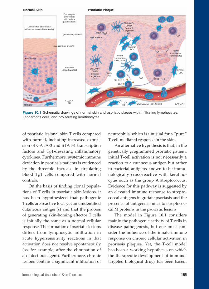
of psoriatic lesional skin T cells compared with normal, including increased expres-sion of GATA-3 and STAT-1 transcription factors and TH1-deviating infl ammatory cytokines. Furthermore, systemic immune deviation in psoriasis patients is evidenced by the threefold increase in circulating blood TH1 cells compared with normal controls.
On the basis of fi nding clonal popula-tions of T cells in psoriatic skin lesions, it has been hypothesized that pathogenic T cells are reactive to as yet an unidentifi ed cutaneous antigen(s) and that the process of generating skin-homing effector T cells is initially the same as a normal cellular response. The formation of psoriatic lesions differs from lymphocytic infi ltration in acute hypersensitivity reactions in that activation does not resolve spontaneously (as, for example, after the elimination of an infectious agent). Furthermore, chronic lesions contain a signifi cant infi ltration of
neutrophils, which is unusual for a “pure” T-cell-mediated response in the skin.
An alternative hypothesis is that, in the genetically programmed psoriatic patient, initial T-cell activation is not necessarily a reaction to a cutaneous antigen but rather to bacterial antigens known to be immu-nologically cross-reactive with keratino-cytes such as the group A streptococcus. Evidence for this pathway is suggested by an elevated immune response to strepto-coccal antigens in guttate psoriasis and the presence of antigens similar to streptococ-cal M proteins in the psoriatic lesions.
The model in Figure 10.1 considers mainly the pathogenic activity of T cells in disease pathogenesis, but one must con-sider the infl uence of the innate immune response on chronic cellular activation in psoriasis plaques. Yet, the T-cell model has been a working hypothesis on which the therapeutic development of immune-targeted biological drugs has been based.
Immunological Aspects of Skin Diseases 165
Normal Skin Psoriatic PlaqueCorneocytesdifferentiatewith nucleus
(parakeratosis)
Corneocytes differentiatewithout nucleus (orthokeratosis)
granular layer absent
granular layer present
EPIDERMIS
DERMIS DERMISplasmacytoid (CD123+)DC
CD11C+DC
CLA
CLA
Flow
PMN
EPIDERMIS
DC-LAMP+CD83+
(Mature)Langerhans
cell
DC-LAMP+CD83+
(Mature)Langerhans
cell
MatureLangerhanscell(CD83+)
(CDB+)TC1TC1
(CDB+)TC1TH1
(CD4+)TH1
immatureLangerhans Cell
CLA+T-cell
CD11C+DCs
CD11C+DDC
CD11C+DC
CD68+cell
HighlyProliferative
Keratinocytes
Figure 10.1 Schematic drawings of normal skin and psoriatic plaque with infi ltrating lymphocytes, Langerhans cells, and proliferating keratinocytes.
166 Immunological Aspects of Skin Diseases
The use of various animal models, but most especially transplanted human skin on immunodefi cient mice, has also been helpful in our understanding of the patho-genic mechanisms involved in the disease.
Animal Models
At the onset, there is no spontaneous model of psoriasis in animals. However, the use of genetically engineered mice or the implan-tation of psoriasis plaque xenografts in immunodefi cient mice has aided greatly in understanding some of the basic mecha-nisms of skin infl ammation as they apply to psoriasis and other infl ammatory diseases.
Several types of transcription factors, including STAT-1 and interferon (IFN) reg-ulatory factors (IRFs), convert IFN-induced genes. IRF-1 is a transcriptional activator, whereas IRF-2 suppresses IRF-1 activity. The IRF-2 null mouse chronically overex-presses IRF-1. These mice spontaneously develop infl ammatory skin lesions similar to psoriasis, including CD8+ T-cell infi ltra-tion in the epidermis, CD4+ T cells in the der-mis, and marked epidermal hyperplasia.
IL-12 and IL-23 (related cytokines that share a p40 subunit) stimulate T cells to differentiate and produce IFN-γ. Mice were genetically engineered to express constitu-tively p40 in the skin by joining the kera-tin-14 promoter with the p40 gene. These animals developed inflammatory skin lesions with marked epidermal hyperpla-sia and increased levels of DCs and T-cell-derived cytokines similar to those found in psoriasis lesions. Thus, IL-23 appears to be an important “upstream” infl ammatory product leading to possible IFN-γ produc-tion and synthesis of “downstream” genes controlled by IFN-γ, STAT-1, and IRFs.
Another approach that has yielded useful information has been to transplant
unaffected (nonlesional) skin or lesional skin from a psoriasis patient on severe combined immunodefi ciency (SCID) mice. Injection of cytokines into the uninvolved tissue grafts induces some mild hyperpla-sia, whereas injection of superantigen-acti-vated mononuclear leucocytes obtained from peripheral blood of the same patient induces a full psoriasis phenotype (but without neutrophil infi ltration), suggest-ing that the psoriasis phenotype can be induced in genetically predisposed skin by bacterial antigen-primed leucocytes. When psoriasis lesional skin is grafted, long-term grafts continue to show viable T cells and other infi ltrating mononuclear leucocytes. Thus, it appears that viable T cells can con-tinue to expand in situ in skin lesions at a rate that matches the rate of programmed cell death, and one does not need new T cells from the peripheral circulation to perpetuate the lesion.
Perhaps of greater importance has been the observation that when nonlesional “normal” skin from psoriasis patients is grafted onto highly immunodefi cient AGR129 mice, the bystander T cells in the graft expand in situ and cause a full-blown psoriatic lesion. Unlike SCID mice, AGR129 mice lack both natural killer (NK) cells and IFN receptors, which may leave them unable to reject graft T cells or create a cytokine environment more conducive to T-cell activation. Either a tumor necrosis factor (TNF) antagonist or anti-CD3 anti-bodies can block the onset of lesions.
Global View of Psoriasis Through
Genomics
Another informative approach to under-standing the underlying mechanisms of pathogenesis of the psoriatic plaque has been the study of differences in gene
Immunological Aspects of Skin Diseases 167
expression detected through transcrip-tional profi ling on 63,000-element gene arrays. Using this technique, it appears that 1,338 genes have altered expression in psoriasis. Having a global view of these differences in gene expression between psoriatic plaques and normal skin back-ground is important because it provides an unbiased means to assess activation pathways in psoriasis. Through genomic analysis, new infl ammatory or regulatory cytokine and chemokine products have been identifi ed as overexpressed in psori-atic lesions. Unexpectedly, there is strong expression of numerous lymphoid-orga-nizing chemokines, for example, CCL19, CCL21, and SDF-1 in psoriasis, as expres-sion is normally confi ned to lymph nodes or formal lymphoid tissues. Most likely, these chemokines orchestrate a striking accumulation of immature dendritic cells (iDCs) and mature dendritic cells (mDCs) in skin lesions; T-cell activation in situ may be regulated through interaction with these DCs in the skin or through release of acti-vating cytokines (such as IL-23) produced by these infi ltrating DCs. If one considers that the vascular changes in psoriasis bear a striking resemblance to those in lymph nodes, then the picture of dense perivas-cular DC/T-cell aggregates and increased expression of lymphoid chemokines sug-gests that the dermal infi ltrates are actually a type of secondary lymphoid tissue.
Genomic profi ling permits us to iden-tify which of the many cytokines detected in psoriasis plaques are transcriptional activators. Thus, expression of more than sixty-fi ve genes with increased expres-sion in psoriasis lesions can be linked to IFN-γ and subsequent activation of STAT-1 signaling by this cytokine. For example, the chemokines CXCL9/MIG, CXCL10/IP-10, and CXCL11/I-Tac are STAT-1-regulated
products and are synthesized in large part by keratinocytes in plaques. These che-mokines direct CXCR3+ T cells to migrate to the epidermis, where they may trigger epidermal hyperplasia as a result of physi-cal damage (disruption of basement mem-brane and desmosomes) done to epider-mal structure through T-cell traffi cking or through secreted products.
Interleukin-8 (also induced by IFN-γ) is a key regulatory chemokine for neutrophil traffi cking into lesions. Inducible nitric oxide synthase (iNOS) transcription is also regulated by IFN-γ and is highly overex-pressed in psoriasis lesions, so that its prod-uct (NO, or nitric oxide) may be responsible for either cell damage or vascular dilation in lesions. Hence, IFN-γ plays a key role in leukocyte migration, as well as in epider-mal and vascular alterations. The T cells in skin lesions do not become activated to release TNF, IFN-γ, and other cytokines unless triggered by antigen recognition, specifi c cytokines, or both. Hence, both the clonal nature of these T-cell infi ltrates in psoriasis and the ability of CTLA4-Ig to reverse disease activity argue for an ongo-ing activation of T cells via classical TCR engagement and co-stimulation. Two cyto-kines produced by activated DCs, IL-12 and IL-23, have been detected in elevated levels in psoriasis lesions. Both of these factors augment IFN-γ production from T cells and may stimulate excessive expan-sion of TH1-biased clones. Therefore, in this model, the presence of activated DCs in skin lesions may be as important in sus-taining disease activity as are T-cell infi l-trates. Long-term disease persistence in the skin (individual plaques can last for years without suppression by therapy) has been hypothesized to be a consequence of orga-nized lymphoid tissue that forms in these infl ammatory skin lesions.
168 Immunological Aspects of Skin Diseases
Insights into Psoriasis Pathogenesis
from Treatments
In 2003, on the basis of many observa-tions that T cells play an important role in the pathogenesis of psoriasis, two T-cell-targeting biologics, alefacept and efali-zumab, were initially tested for activity in psoriasis and are now approved by the U.S. Food and Drug Administration (FDA). Ale-facept is a fusion protein that contains the extracellular domain of LFA-3 fused with immunoglobulin constant region domains. This agent binds to CD2, which is expressed at high levels on memory T cells. Although this binding interaction with CD2 could inhibit normal LFA-3/CD2 signaling, suc-cessful therapeutic outcomes with this agent are most closely associated with depletion of T cells from psoriasis skin lesions and, to a lesser extent, depletion of memory T cells (CD8 > CD4) from the circulation.
Efalizumab is a humanized monoclonal antibody that binds to the alpha subunit (CD11a) of the integrin lymphocyte func-tional antigen (LFA-1). LFA-1 is expressed at high levels on T lymphocytes, while other integrins are specifi c for monocytes, macro-phages, neutrophils, and DCs. Thus, LFA-1 blockade with efalizumab, specifi cally targets T cells, and alters T-cell function in several ways: (1) prevents fi rm adhesion of T cells to infl amed (ICAM+) endothelium and thus blocks entry of cells from the cir-culation; (2) prevents binding of T cells to ICAM+ keratinocytes, thus reducing traf-fi cking of cells into the epidermis; and (3) reduces T-cell activation, with attendant cytokine release, either directly or by block-ing DC/T-cell immune synapse formation.
Although alefacept and efalizumab are potent immune therapies, only 25–30 percent of treated patients experience maximal disease improvement after
a twelve-week treatment course. For unknown reasons, responses to these tar-geted agents are more variable than to more general immunosuppressive treat-ments. Possible explanations include vari-able expression of redundant T-cell activa-tion pathways, restrictions in the access of large molecules to relevant T-cell pools, or more complex interactions between T cells and other types of leukocytes that contrib-ute to disease pathogenesis in different patients. Genetic/genomic heterogeneity of humans seems likely to underlie the vari-able response to molecule-specifi c antago-nists, but more consistent improvements in disease are seen with less selective T-cell antagonists, for example, cyclosporine, suggesting that psoriasis is fundamentally caused by cellular immune system dis-regulation. In general, suppression of T-cell-mediated infl ammation – functionally defi ned as genomic suppression of IFN-γ, STAT-1, and downstream genes regulated by STAT-1 in psoriasis skin lesions – leads to objective improvement or clearing of disease, whereas failure to suppress these infl ammatory genes leads to continuing disease activity. A review of therapeutic trials with many different agents indicates that there are no documented cases in which T cells and associated infl ammatory genes were reduced without correspond-ing improvements in disease-defi ning epi-dermal hyperplasia. Hence, no data argues against the fundamental hypothesis that activation of the cellular immune system triggers psoriasis.
Another therapeutic approach in pso-riasis has been to antagonize infl amma-tory cytokines. Although an antibody to IL-8 was administered to psoriasis patients with moderate improvement in disease activity, the fi rst major success of the anti-cytokine approach was antagonizing TNF
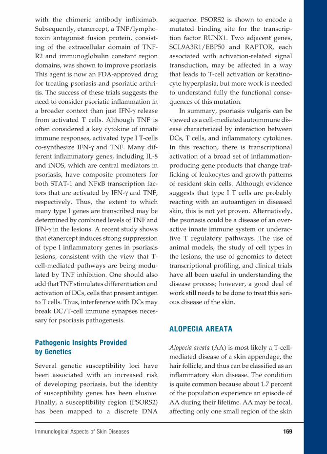
Immunological Aspects of Skin Diseases 169
with the chimeric antibody infl iximab. Subsequently, etanercept, a TNF/lympho-toxin antagonist fusion protein, consist-ing of the extracellular domain of TNF-R2 and immunoglobulin constant region domains, was shown to improve psoriasis. This agent is now an FDA-approved drug for treating psoriasis and psoriatic arthri-tis. The success of these trials suggests the need to consider psoriatic infl ammation in a broader context than just IFN-γ release from activated T cells. Although TNF is often considered a key cytokine of innate immune responses, activated type I T-cells co-synthesize IFN-γ and TNF. Many dif-ferent infl ammatory genes, including IL-8 and iNOS, which are central mediators in psoriasis, have composite promoters for both STAT-1 and NFκB transcription fac-tors that are activated by IFN-γ and TNF, respectively. Thus, the extent to which many type I genes are transcribed may be determined by combined levels of TNF and IFN-γ in the lesions. A recent study shows that etanercept induces strong suppression of type I infl ammatory genes in psoriasis lesions, consistent with the view that T-cell-mediated pathways are being modu-lated by TNF inhibition. One should also add that TNF stimulates differentiation and activation of DCs, cells that present antigen to T cells. Thus, interference with DCs may break DC/T-cell immune synapses neces-sary for psoriasis pathogenesis.
Pathogenic Insights Provided
by Genetics
Several genetic susceptibility loci have been associated with an increased risk of developing psoriasis, but the identity of susceptibility genes has been elusive. Finally, a susceptibility region (PSORS2) has been mapped to a discrete DNA
sequence. PSORS2 is shown to encode a mutated binding site for the transcrip-tion factor RUNX1. Two adjacent genes, SCL9A3R1/EBP50 and RAPTOR, each associated with activation-related signal transduction, may be affected in a way that leads to T-cell activation or keratino-cyte hyperplasia, but more work is needed to understand fully the functional conse-quences of this mutation.
In summary, psoriasis vulgaris can be viewed as a cell-mediated autoimmune dis-ease characterized by interaction between DCs, T cells, and infl ammatory cytokines. In this reaction, there is transcriptional activation of a broad set of infl ammation-producing gene products that change traf-fi cking of leukocytes and growth patterns of resident skin cells. Although evidence suggests that type I T cells are probably reacting with an autoantigen in diseased skin, this is not yet proven. Alternatively, the psoriasis could be a disease of an over-active innate immune system or underac-tive T regulatory pathways. The use of animal models, the study of cell types in the lesions, the use of genomics to detect transcriptional profi ling, and clinical trials have all been useful in understanding the disease process; however, a good deal of work still needs to be done to treat this seri-ous disease of the skin.
ALOPECIA AREATA
Alopecia areata (AA) is most likely a T-cell-mediated disease of a skin appendage, the hair follicle, and thus can be classifi ed as an infl ammatory skin disease. The condition is quite common because about 1.7 percent of the population experience an episode of AA during their lifetime. AA may be focal, affecting only one small region of the skin
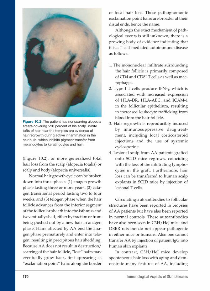
170 Immunological Aspects of Skin Diseases
(Figure 10.2), or more generalized total hair loss from the scalp (alopecia totalis) or scalp and body (alopecia universalis).
Normal hair growth cycle can be broken down into three phases (1) anagen growth phase lasting three or more years, (2) cata-gen transitional period lasting two to four weeks, and (3) telogen phase when the hair follicle advances from the inferior segment of the follicular sheath into the isthmus and is eventually shed, either by traction or from being pushed out by a new hair in anagen phase. Hairs affected by AA end the ana-gen phase prematurely and enter into telo-gen, resulting in precipitous hair shedding. Because AA does not result in destruction/scarring of the hair follicle, “lost” hairs may eventually grow back, fi rst appearing as “exclamation point” hairs along the border
of focal hair loss. These pathognomonic exclamation point hairs are broader at their distal ends, hence the name.
Although the exact mechanism of path-ological events is still unknown, there is a growing body of evidence indicating that it is a T-cell-mediated autoimmune disease as follows:
1. The mononuclear infi ltrate surrounding the hair follicle is primarily composed of CD4 and CD8+ T cells as well as mac-rophages.
2. Type I T cells produce IFN-γ, which is associated with increased expression of HLA-DR, HLA-ABC, and ICAM-1 in the follicular epithelium, resulting in increased leukocyte traffi cking from blood into the hair follicle.
3. Hair regrowth is reproducibly induced by immunosuppressive drug treat-ment, including local corticosteroid injections and the use of systemic cyclosporine.
4. Lesional scalp from AA patients grafted onto SCID mice regrows, coinciding with the loss of the infi ltrating lympho-cytes in the graft. Furthermore, hair loss can be transferred to human scalp explants in SCID mice by injection of lesional T cells.
Circulating autoantibodies to follicular structures have been reported in biopsies of AA patients but have also been reported in normal controls. These autoantibodies have also been seen in C3H/HeJ mice and DEBR rats but do not appear pathogenic in either mice or humans. Also one cannot transfer AA by injection of patient IgG into human skin explants.
In contrast, C3H/HeJ mice develop spontaneous hair loss with aging and dem-onstrate many features of AA, including
Figure 10.2 The patient has nonscarring alopecia areata covering >90 percent of his scalp. White tufts of hair near the temples are evidence of hair regrowth during active infl ammation in the hair bulb, which inhibits pigment transfer from melanocytes to keratinocytes and hair.
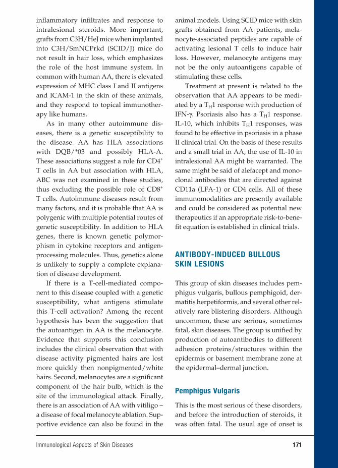
Immunological Aspects of Skin Diseases 171
infl ammatory infi ltrates and response to intralesional steroids. More important, grafts from C3H/HeJ mice when implanted into C3H/SmNCPrkd (SCID/J) mice do not result in hair loss, which emphasizes the role of the host immune system. In common with human AA, there is elevated expression of MHC class I and II antigens and ICAM-1 in the skin of these animals, and they respond to topical immunother-apy like humans.
As in many other autoimmune dis-eases, there is a genetic susceptibility to the disease. AA has HLA associations with DQB/*03 and possibly HLA-A. These associations suggest a role for CD4+ T cells in AA but association with HLA, ABC was not examined in these studies, thus excluding the possible role of CD8+ T cells. Autoimmune diseases result from many factors, and it is probable that AA is polygenic with multiple potential routes of genetic susceptibility. In addition to HLA genes, there is known genetic polymor-phism in cytokine receptors and antigen-processing molecules. Thus, genetics alone is unlikely to supply a complete explana-tion of disease development.
If there is a T-cell-mediated compo-nent to this disease coupled with a genetic susceptibility, what antigens stimulate this T-cell activation? Among the recent hypothesis has been the suggestion that the autoantigen in AA is the melanocyte. Evidence that supports this conclusion includes the clinical observation that with disease activity pigmented hairs are lost more quickly then nonpigmented/white hairs. Second, melanocytes are a signifi cant component of the hair bulb, which is the site of the immunological attack. Finally, there is an association of AA with vitiligo – a disease of focal melanocyte ablation. Sup-portive evidence can also be found in the
animal models. Using SCID mice with skin grafts obtained from AA patients, mela-nocyte-associated peptides are capable of activating lesional T cells to induce hair loss. However, melanocyte antigens may not be the only autoantigens capable of stimulating these cells.
Treatment at present is related to the observation that AA appears to be medi-ated by a TH1 response with production of IFN-γ. Psoriasis also has a TH1 response. IL-10, which inhibits TH1 responses, was found to be effective in psoriasis in a phase II clinical trial. On the basis of these results and a small trial in AA, the use of IL-10 in intralesional AA might be warranted. The same might be said of alefacept and mono-clonal antibodies that are directed against CD11a (LFA-1) or CD4 cells. All of these immunomodalities are presently available and could be considered as potential new therapeutics if an appropriate risk-to-bene-fi t equation is established in clinical trials.
ANTIBODY-INDUCED BULLOUS
SKIN LESIONS
This group of skin diseases includes pem-phigus vulgaris, bullous pemphigoid, der-matitis herpetiformis, and several other rel-atively rare blistering disorders. Although uncommon, these are serious, sometimes fatal, skin diseases. The group is unifi ed by production of autoantibodies to different adhesion proteins/structures within the epidermis or basement membrane zone at the epidermal–dermal junction.
Pemphigus Vulgaris
This is the most serious of these disorders, and before the introduction of steroids, it was often fatal. The usual age of onset is
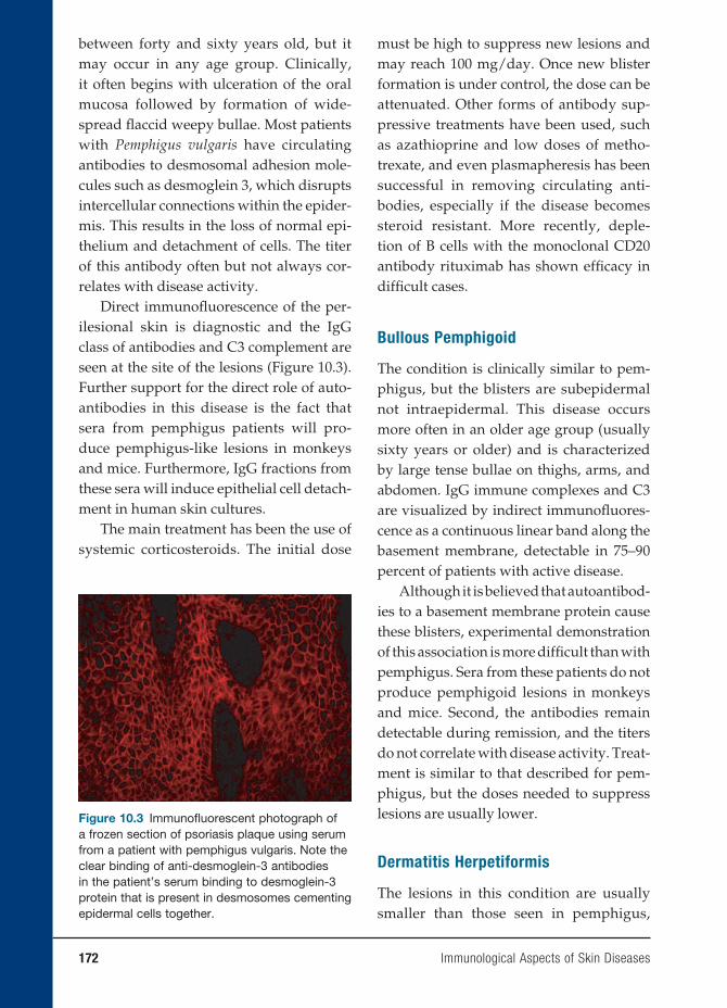
172 Immunological Aspects of Skin Diseases
between forty and sixty years old, but it may occur in any age group. Clinically, it often begins with ulceration of the oral mucosa followed by formation of wide-spread fl accid weepy bullae. Most patients with Pemphigus vulgaris have circulating antibodies to desmosomal adhesion mole-cules such as desmoglein 3, which disrupts intercellular connections within the epider-mis. This results in the loss of normal epi-thelium and detachment of cells. The titer of this antibody often but not always cor-relates with disease activity.
Direct immunofl uorescence of the per-ilesional skin is diagnostic and the IgG class of antibodies and C3 complement are seen at the site of the lesions (Figure 10.3). Further support for the direct role of auto-antibodies in this disease is the fact that sera from pemphigus patients will pro-duce pemphigus-like lesions in monkeys and mice. Furthermore, IgG fractions from these sera will induce epithelial cell detach-ment in human skin cultures.
The main treatment has been the use of systemic corticosteroids. The initial dose
must be high to suppress new lesions and may reach 100 mg/day. Once new blister formation is under control, the dose can be attenuated. Other forms of antibody sup-pressive treatments have been used, such as azathioprine and low doses of metho-trexate, and even plasmapheresis has been successful in removing circulating anti-bodies, especially if the disease becomes steroid resistant. More recently, deple-tion of B cells with the monoclonal CD20 antibody rituximab has shown effi cacy in diffi cult cases.
Bullous Pemphigoid
The condition is clinically similar to pem-phigus, but the blisters are subepidermal not intraepidermal. This disease occurs more often in an older age group (usually sixty years or older) and is characterized by large tense bullae on thighs, arms, and abdomen. IgG immune complexes and C3 are visualized by indirect immunofl uores-cence as a continuous linear band along the basement membrane, detectable in 75–90 percent of patients with active disease.
Although it is believed that autoantibod-ies to a basement membrane protein cause these blisters, experimental demonstration of this association is more diffi cult than with pemphigus. Sera from these patients do not produce pemphigoid lesions in monkeys and mice. Second, the antibodies remain detectable during remission, and the titers do not correlate with disease activity. Treat-ment is similar to that described for pem-phigus, but the doses needed to suppress lesions are usually lower.
Dermatitis Herpetiformis
The lesions in this condition are usually smaller than those seen in pemphigus,
Figure 10.3 Immunofl uorescent photograph of a frozen section of psoriasis plaque using serum from a patient with pemphigus vulgaris. Note the clear binding of anti-desmoglein-3 antibodies in the patient’s serum binding to desmoglein-3 protein that is present in desmosomes cementing epidermal cells together.
Immunological Aspects of Skin Diseases 173
are extremely itchy, and are seen on exte-rior surfaces of elbows, knees, buttocks, necks, and shoulders. Visually, the lesions resemble herpetic papules; however, the herpes virus is not involved in this condi-tion. The age group is quite different from that of pemphigus (twenty-fi ve to forty years old) but it may occur at any age. IgA is the major class of antibody detected, deposited in a granular fashion in the tips of the dermal papillae, forming subepi-thelial bullae. However, unlike pemphi-gus, no circulating autoantibodies to skin are seen in the sera of these patients. An interesting and as yet unexplained obser-vation is that many of the DH patients have a concomitant enteropathy similar to celiac disease. Clinically, they do not have the symptoms of celiac disease and the associated condition is usually only demonstrable by jejunal biopsy. About 30 percent of these patients have antibodies to gliadin and endomysium in their sera. They also have an increased risk of lym-phoma (like celiac disease patients) and a genetic component exhibited by a mark-edly increased inheritance of HLA-B8, DR3-DQ2 haplotypes.
Dermatitis herpetiformis responds to a strict gluten-free diet, which may need to be in force for several years before the lesions heal. Reintroduction of gluten results in the reappearance of the lesions, indicating the important role of gluten in the condition. Yet the presence of IgA deposits in the skin and their immunologi-cal basis remain unknown.
FUTURE DIRECTIONS
FOR RESEARCH
Infl ammatory diseases of the skin are an exciting and evolving fi eld of research in
which basic research frequently translates into clinical applications. Accessibility of human skin biopsies and intersection with biopharma production of immune-modulating therapies increases research velocity and immediacy. Current “hot” top-ics for researchers studying psoriasis, for example, include the newly defi ned TH17 T cell, and immunomodulating effects of innate antimicrobial peptides contribut-ing to disease initiation and maintenance. Since the advent of effective immune-modulating therapies targeting T cells, and the discovery of a large IFN-γ signature in psoriatic lesions, psoriasis has been known as a TH1-mediated disease. TH17 cells pro-ducing IL-17 and IL-22, fi rst discovered in animal models of autoimmunity, have now been found to promote psoriasis disease pathogenesis, both through IL-17-depen-dent leukocyte migration, and through IL-22-dependent keratinocyte hyperprolifera-tion. Current questions in the fi eld include defi ning the relationship between TH1 and TH17 cells in humans: Are they synergistic or antagonistic? Are they both involved in initiation and maintenance and is block-ing one or both subsets most effective for patient therapy? Another current topic of research is the role of innate antimicrobials, including β-defensins and cathelicidins, as upstream versus downstream infl am-matory mediators. LL-37 cathelicidin was recently found to bind ribonucleic acids and initiate plasmacytoid DC activation through TLR9. Activated plasmacytoid DCs produce large quantities of IFN-α, which leads to downstream myeloid DC activation and subsequent T-cell activation. However, both IL-17 and IL-22 are potent inducers of keratinocyte-elaborated anti-microbial peptides, forming a potential for positive feedback and continued infl am-mation. Unanswered questions include
174 Immunological Aspects of Skin Diseases
the temporal relationship between innate antimicrobial peptides and T-cell adaptive immunity, as well as the genetic basis for this breakdown in tolerance and immune regulation.
In summary, infl ammatory diseases of the skin are an important and exciting area of research. Working with other research-ers in both academia and industry rap-idly moves the fi eld from the bench to the bedside. Researching infl ammatory skin diseases also allows for collaboration with other related fi elds where the immune system is deviated – as is the case for skin cancer.
BIBLIOGRAPHY
SUGGESTED READINGChamian F, Krueger JG. Psoriasis vulgaris:
an interplay of T lymphocytes, den-dritic cells, and infl ammatory cytokines in pathogenesis. Curr Opin Rheumatol. 2004;16:331–337.
Kalish RS, Gilhar A. Alopecia areata: autoimmunity – the evidence is com-pelling. J Investig Dermatol Symp Proc. 2003;8:164–167.
Krueger JG. Treating psoriasis with biolog-ical agents. Sci Med. 2002;8:149–161.
Lew W, Bowcock AM, Krueger JG. Psoria-sis vulgaris: cutaneous lymphoid tissue supports T-cell activation and “type 1”
infl ammatory gene expression. Trends Immunol. 2004;25:295–305.
LANDMARK PAPERSAsadullah K, Sterry W, Stephanek K,
et al. IL-10 is a key cytokine in psoriasis. Proof of principle by IL-10 therapy: A new therapeutic approach. J Clin Invest. 1998;101(4):783–794.
Cicardi M, Agostoni A. Hereditary angio-edema. N Engl J Med. 1996;334(25): 1666–1667.
Gilhar A, Ullmann Y, Berkutzki T, Assy B, Kalish RS. Autoimmune hair loss (alo-pecia areata) transferred by T lympho-cytes to human scalp explants on SCID mice. J Clin Invest. 1998;101:62–67.
Gottlieb SL, Gilleaudeau P, Johnson R, et al. Response of psoriasis to a lymphocyte-selective toxin (DAB389IL-2) suggests a primary immune, but not keratino-cyte, pathogenic basis. Nat Med. 1995;1: 442–447.
Kalish RS, Johnson KL, Hordinsky MK. Alopecia areata: autoreactive T cells are variably enriched in scalp lesions rela-tive to peripheral blood. Arch Dermatol. 1992;128:1072–1077.
McElvee KJ, Boggess D, King LE Jr, Sund-berg JP. Experimental induction of alo-pecia areata-like hair loss in C3H/HeJ mice using full-thickness skin grafts. J Investig Dermatol. 1998;11:797–803.
11. Experimental Approaches to the Study of Autoimmune
Rheumatic Diseases
Dalit Ashany, M.D., and Mary K. Crow, M.D.
175
INTRODUCTION
The ultimate objective of biomedical research is to gain new understanding of the mechanisms of disease such that advances in treatment, and ultimately pre-vention and cure, are applied to that dis-ease. However, progress is complicated by the inherent heterogeneity of human popu-lations, even among those diagnosed with a particular disease. Even when appropriate study populations can be defi ned, access to the most relevant tissues and respect for patient privacy and autonomy present challenges to unfettered human-subject research. Although recent large-scale genomewide association studies in patients with systemic autoimmune diseases have identifi ed candidate disease susceptibility genes, in vitro systems and experimental models are required to characterize the immunopathogenic signifi cance of those genes and their products. Once targets for potential new therapies are identifi ed, ini-tial testing for toxicities of those therapeu-tics in humans is rarely appropriate. These limitations are among those that have led investigators to identify and to develop useful animal models that closely mimic human disease. Such models have been a particular focus of research relevant to the autoimmune rheumatic diseases. Although the use of animal models will always be limited by the likelihood of genetic and molecular distinctions between humans
and lower mammals, current data from the analysis of the genomes of mice and man, including study of disease susceptibility loci, indicate that important hints regard-ing disease mechanisms can be gleaned from study of animal models. This chapter will review the major animal models inves-tigators are using to explore the mecha-nisms of autoimmune rheumatic diseases, emphasizing the degree of fi delity of those models to the relevant human disease. The most frequently studied mammal, the mouse, will be the focus of discussion.
TYPES OF ANIMAL MODELS
Spontaneous
Mouse strains that exhibit spontaneous development of syndromes that closely resemble human autoimmune rheumatic diseases have been identifi ed by experi-enced investigators carefully observing mouse colonies. In addition, some efforts to induce mutation using chemical muta-gens have generated mice that express immune system defects or pathology that provide clues to human diseases with simi-lar problems. Advantages of spontaneous models of disease are that the phenotype is generally quite reproducible from ani-mal to animal (although, in some cases, the disease is only partially penetrant) and development of the disease does not require intervention by the investigator.
A disadvantage of spontaneous models is that the disease often takes months to develop, slowing the pace of investigation.
Inducible
Some of the most useful animal models are those that are induced by the investigator by administration of a drug, an antigen, an adjuvant, an antibody, or through surgi-cal manipulation of the immune system. For example, demethylating drugs alter the structure of chromatin, resulting in increased accessibility of positive or nega-tive regulatory elements in gene promoters or enhancers and subsequent alteration of the gene expression profi le of the animal, sometimes resulting in disease. Removal of the thymus at day 3 after birth has been demonstrated to promote development of several organ-targeted autoimmune diseases, most likely based on removal of an important regulatory T-cell population. Induction of immune system activation, as in the pristane model of lupus, can lead to altered patterns of cytokine production and autoimmunity. In some cases, autoimmune disease can be simply transferred from one animal to another by administration of an autoantibody or autoreactive T-cell popu-lation. Under these circumstances, the cells and molecules of the immune system that are required for disease expression can be identifi ed and the investigation narrowed to one or at least a more narrow range of immune system components. The contribu-tion of background genetic factors to devel-opment of a disease phenotype can also be studied in inducible animal models.
Transgenic and Knockout
Many investigators have used the tech-nology that generates animals that either
overexpress or are defi cient in a single gene to investigate the relevance of that gene product to development of a dis-ease phenotype. This approach has been particularly fruitful in the study of lupus, with transgenic and knockout mice dem-onstrating that any number of genetic modifi cations that alter self-antigen acces-sibility or threshold for immune system activation can lead to production of the classic lupus autoantibody, anti-DNA antibody, and deposition of those autoan-tibodies in the kidney, another character-istic of human lupus. Defi ciencies in the complement system, members of which help to clear apoptotic debris and solu-bilize immune complexes; increased or decreased expression of cell surface mol-ecules, kinases, phosphatases, or adaptor molecules relevant to intracellular signal-ing and that modulate the threshold for lymphocyte activation; and altered expres-sion of death pathway molecules, such as Fas, can result in increased targeting of the immune response toward self compo-nents and manifestations of autoimmu-nity. An advantage of these sophisticated animal systems is that the modifi cation is often restricted to one gene, or at least a small genomic region adjacent to the gene of interest, allowing the study of the impact of that particular molecular prod-uct and its relevant pathway on disease expression. However, there are inherent dangers in relying solely on transgenic and knockout models. That the gene of interest is absent during embryonic and fetal development means that other gene products can take over some of the func-tions of the modifi ed gene, obfuscating the predicted phenotype. The role of a gene in development of a disease can also be over-estimated if background genomic factors in the neighborhood of the modifi ed gene
176 Experimental Approaches to the Study of Autoimmune Rheumatic Diseases
also contribute to disease. Finally, the fact that overexpression or deletion of a given gene results in a disease phenotype does not necessarily indicate that the same human gene is altered in the correspond-ing human disease. In fact, few of the genes that result in disease when modifi ed in murine systems have been confi rmed as disease genes in humans. Nonetheless, the transgenic and knockout approaches help to identify important pathways for further study and that may be eventually targeted therapeutically.
Congenic
Like the transgenic and knockout approaches, congenic mice have proved very useful in studying the specifi c con-tributions of narrow genomic regions to aspects of disease pathology. The util-ity of congenic mouse strains, in which a segment of a chromosome is bred into a desired genetic background, is nicely illus-trated by the NZM 2410 strain. That mouse model has allowed identifi cation of at least three chromosomal loci that confer distinct immune system alterations, which when present together result in severe lupus-like disease. As in the case of the transgenic and knockout models, gene sequences in the chromosomal segment of interest can interact with background genes to compli-cate interpretation of the role of gene prod-ucts in the congenic region.
RHEUMATOID ARTHRITIS
Introduction and Epidemiology
Rheumatoid arthritis (RA) is the most com-mon infl ammatory arthritis. Its incidence worldwide is estimated to be between 0.5 and 1 percent of the general population.
Certain populations, such as the Pima Indians in North America, have a much higher incidence of RA (about 5 percent) than the general population. RA is one of many chronic infl ammatory diseases that predominate in women. The ratio of female to male patients is estimated to be from 2:1 to 4:1.
Etiology and Pathogenesis
The etiology of RA has not been elucidated although a variety of studies suggest that a combination of environmental and genetic factors is responsible; a contribution of either one is necessary but not suffi cient for full expression of the disease. A genetic component is demonstrated by studies of monozygotic twins, in whom the concor-dance rate is perhaps 30 to 50 percent when one twin is affected, compared with 1 per-cent for the general population. The immu-nogenetics is under intense study although there is a clear risk factor in the class II MHC haplotype of an individual. Addi-tional genes associated with RA include PTPN22 and STAT4.
The major pathology of RA is local-ized to the joints. The synovial membrane surrounding the joints becomes highly vascular and infi ltrated with virtually all cellular components of the immune sys-tem. T and B lymphocytes are highly rep-resented and form lymphoid aggregates where B cells differentiate to antibody-forming cells. Poorly controlled produc-tion of pro-infl ammatory cytokines, such as tumor necrosis factor (TNF) and inter-leukin-1 (IL-1), promote recruitment of additional infl ammatory cells, and pro-duction of metalloproteinases contributes to tissue damage. The presence of immune complexes in joint fl uid and direct deposi-tion of those complexes on the surface of
Experimental Approaches to the Study of Autoimmune Rheumatic Diseases 177
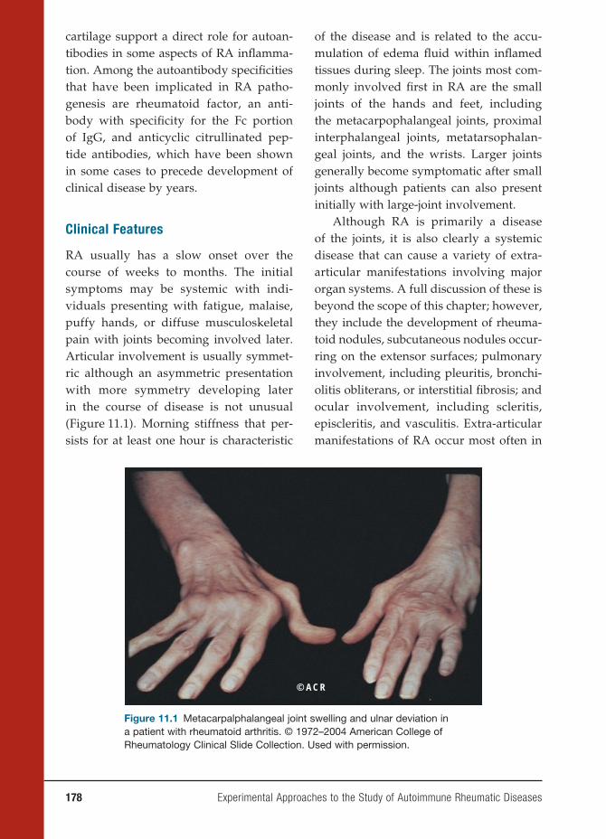
178 Experimental Approaches to the Study of Autoimmune Rheumatic Diseases
cartilage support a direct role for autoan-tibodies in some aspects of RA infl amma-tion. Among the autoantibody specifi cities that have been implicated in RA patho-genesis are rheumatoid factor, an anti-body with specifi city for the Fc portion of IgG, and anticyclic citrullinated pep-tide antibodies, which have been shown in some cases to precede development of clinical disease by years.
Clinical Features
RA usually has a slow onset over the course of weeks to months. The initial symptoms may be systemic with indi-viduals presenting with fatigue, malaise, puffy hands, or diffuse musculoskeletal pain with joints becoming involved later. Articular involvement is usually symmet-ric although an asymmetric presentation with more symmetry developing later in the course of disease is not unusual (Figure 11.1). Morning stiffness that per-sists for at least one hour is characteristic
of the disease and is related to the accu-mulation of edema fl uid within infl amed tissues during sleep. The joints most com-monly involved fi rst in RA are the small joints of the hands and feet, including the metacarpophalangeal joints, proximal interphalangeal joints, metatarsophalan-geal joints, and the wrists. Larger joints generally become symptomatic after small joints although patients can also present initially with large-joint involvement.
Although RA is primarily a disease of the joints, it is also clearly a systemic disease that can cause a variety of extra-articular manifestations involving major organ systems. A full discussion of these is beyond the scope of this chapter; however, they include the development of rheuma-toid nodules, subcutaneous nodules occur-ring on the extensor surfaces; pulmonary involvement, including pleuritis, bronchi-olitis obliterans, or interstitial fi brosis; and ocular involvement, including scleritis, episcleritis, and vasculitis. Extra-articular manifestations of RA occur most often in
© ACR
Figure 11.1 Metacarpalphalangeal joint swelling and ulnar deviation in a patient with rheumatoid arthritis. © 1972–2004 American College of Rheumatology Clinical Slide Collection. Used with permission.
Experimental Approaches to the Study of Autoimmune Rheumatic Diseases 179
seropositive patients with more severe joint disease.
Treatment of RA
Patients with RA have variable courses, ranging from those who progress rapidly to complete destruction of joints if left untreated, to those who, with few phar-macologic interventions, smolder along with minimal disease that leaves cartilage functional and activities of daily living unchanged from the premorbid state. The challenge for physicians is to determine into which category a patient falls. Treat-ment choices include nonsteroidal anti-infl ammatory drugs (NSAIDs), which are useful to help control some of the infl am-matory signs of RA but do not prevent progression of the disease. A large class of drugs known as DMARDs (disease-modifying antirheumatic drugs) includes hydroxycholoroquine, an agent that has been shown to have anti-infl ammatory properties in the disease but does not prevent erosive arthritis. Other DMARDs include those that have been shown to prevent joint destruction, which include sulfasalazine, methotrexate, lefl unomide, and azathioprine. In recent years, the development of biologics targeting spe-cifi c cytokines, immune cells, and immune receptors have provided additional potent therapeutics with which to treat RA. In this category are three different drugs that target the TNF cytokine (etancercept, adalimumb, and infl iximab), an IL-1 recep-tor antagonist (anakinra), an anti-CD20 antibody that selectively depletes B cells (rituximab), and a selective co-stimulation modulator that inhibits T-cell activation by binding to CD80 and CD86, thereby blocking CD28 interaction (abatacept).
ANIMAL MODELS OF RHEUMATOID
ARTHRITIS
No animal model is regarded as a high-fi delity replicate of human RA. However, several models that variably use T cells or antibodies that target the joint, or that induce monocyte activation and infl am-mation, have been used to study distinct aspects of the disease phenotype.
Animal models of arthritis can be used to understand elements of the arthritic process in patients. Recent therapeutic approaches in patients with RA, includ-ing the use of biological agents, are based on initial fi ndings in murine models of experimental arthritis, although final proof of concept must come from clini-cal studies. Animal models are powerful tools for studying pathological changes in articular cartilage and bone in great detail and can be used to evaluate mecha-nisms of erosive processes. Although, in general, more infl ammation drives more destruction, the uncoupling of infl amma-tion and erosion can be seen as well, and different mediators are involved in these processes.
COLLAGEN-INDUCED ARTHRITISCollagen-induced arthritis (CIA) is an
example of an inducible animal model for human RA. In this model, the disease is induced in susceptible strains of animals by immunization with cartilage type II collagen. The resulting disease is a T-cell-dependent, antibody-mediated autoim-mune disease directed against the type II collagen immunogen. Similar to RA, CIA is characterized by massive infi ltration of synovial joints by infl ammatory cells and hyperplasia of the synovial mem-brane (pannus). In addition, the disease is
180 Experimental Approaches to the Study of Autoimmune Rheumatic Diseases
characterized by both cartilage and bone destruction as seen in RA.
Both collagen-specifi c T and B lym-phocytes are involved in the induction of CIA. The effector mechanisms that lead to joint tissue destruction are less well under-stood, and a number of infl ammatory cell types have been implicated. These include fibroblast-like synoviocytes, bone mar-row–derived macrophages, granulocytes, and dendritic cells as well as lymphocytes. Pro-infl ammatory cytokines (IL-1β, TNF) and various chemokines are involved in the pathogenesis of the immune-mediated joint damage observed in both CIA and RA. Another similarity between RA and CIA is that the development of synovial infl am-mation is associated with pronounced angiogenesis with growth of high endothe-lial venules.
In contrast to RA, where large num-bers of T lymphocytes accumulate in the infl amed synovium, few T cells are found in the synovium of CIA. Another differ-ence between collagen arthritis and RA is that the former is more highly destructive than RA and in contrast to RA has a marked sensitivity to suppression of infl ammation and joint destruction by NSAIDs such as indomethacin. In contrast to the female preponderance in RA, males are more sus-ceptible than females in murine CIA.
ADJUVANT ARTHRITISAdjuvant arthritis is the oldest stud-
ied model of polyarthritis. It is induced by intradermal injection of complete Freund’s adjuvant, containing heat-killed myco-bacteria, in susceptible rat strains, mostly Lewis rats. Arthritis develops within two weeks. The active component in bacteria is the cell wall peptidoglycan.
Histopathological features are primar-ily a periarthritis, with marked periostitis
rather than synovitis, and massive infl am-mation in the bone marrow. Immune-complex deposition in the cartilage is not a characteristic feature, and cartilage destruction is limited in early disease.
Adjuvant arthritis has a pure T-cell-driven pathogenesis, and passive trans-fer of T cells from diseased animals can induce arthritis. The joint infl ammation is thought to refl ect the generation of a T-cell reaction to bacterial epitopes cross-react-ing with endogenous bacterial fragments continuously present in synovial tissues or with cartilaginous antigens. Similar to RA, TNF and IL-1 have been shown to play a role in pathogenesis. Treatment with anti-TNF or anti-IL-1 therapy blocks infl amma-tion and tissue destruction in the model, with the most optimal blockade occurring with combination therapy. As seen in CIA, NSAIDs are effective inhibitors of cartilage and bone destruction in this model and this represents a signifi cant difference from human RA.
Adjuvant-induced arthritis is severe but self-limited, and the rat recovers within a few months. This is a general shortcoming of most animal models when compared with the chronic process of human RA. An additional feature of adjuvant-induced arthritis is that once the rats undergo spontaneous remission the animal can no long er be reinduced to develop disease. This resistance has made the model suit-able for studies in regulation of T-cell tolerance.
ANTIGEN-INDUCED ARTHRITISAntigen-induced arthritis is a model
of RA that is induced by immunizing animals with a foreign antigen, usually bovine serum albumin, and subsequently injecting the same antigen into a joint. As a result, a pronounced T-cell-dependent
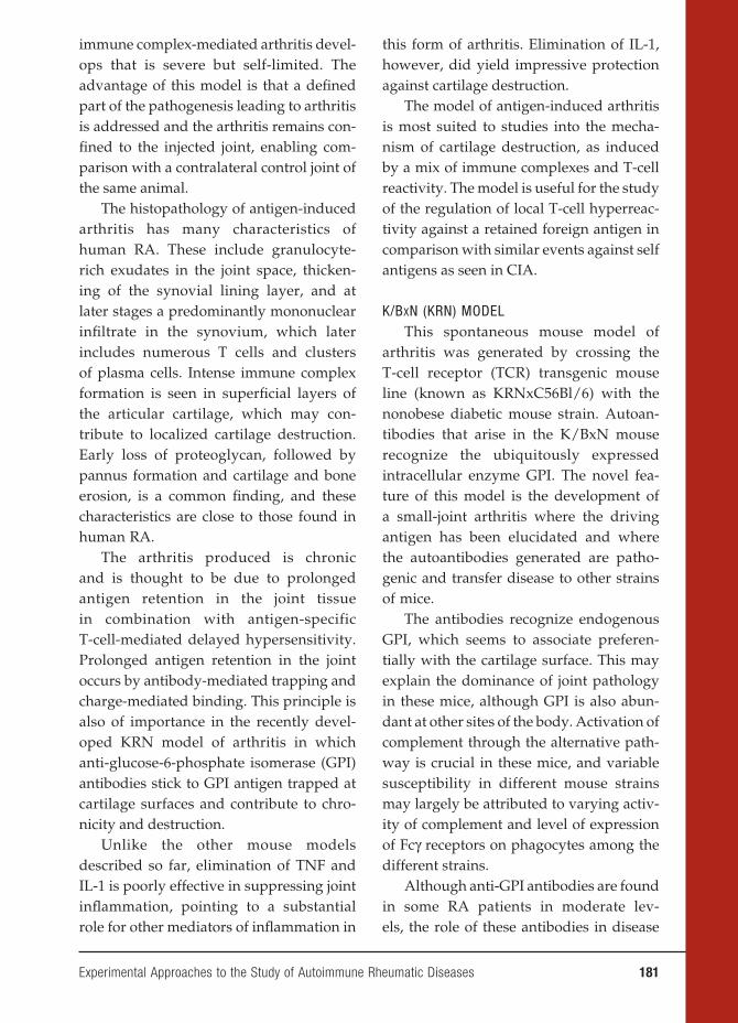
Experimental Approaches to the Study of Autoimmune Rheumatic Diseases 181
immune complex-mediated arthritis devel-ops that is severe but self-limited. The advantage of this model is that a defi ned part of the pathogenesis leading to arthritis is addressed and the arthritis remains con-fi ned to the injected joint, enabling com-parison with a contralateral control joint of the same animal.
The histopathology of antigen-induced arthritis has many characteristics of human RA. These include granulocyte-rich exudates in the joint space, thicken-ing of the synovial lining layer, and at later stages a predominantly mononuclear infi ltrate in the synovium, which later includes numerous T cells and clusters of plasma cells. Intense immune complex formation is seen in superfi cial layers of the articular cartilage, which may con-tribute to localized cartilage destruction. Early loss of proteoglycan, followed by pannus formation and cartilage and bone erosion, is a common fi nding, and these characteristics are close to those found in human RA.
The arthritis produced is chronic and is thought to be due to prolonged antigen retention in the joint tissue in combination with antigen-specific T-cell-mediated delayed hypersensitivity. Prolonged antigen retention in the joint occurs by antibody-mediated trapping and charge-mediated binding. This principle is also of importance in the recently devel-oped KRN model of arthritis in which anti-glucose-6-phosphate isomerase (GPI) antibodies stick to GPI antigen trapped at cartilage surfaces and contribute to chro-nicity and destruction.
Unlike the other mouse models described so far, elimination of TNF and IL-1 is poorly effective in suppressing joint infl ammation, pointing to a substantial role for other mediators of infl ammation in
this form of arthritis. Elimination of IL-1, however, did yield impressive protection against cartilage destruction.
The model of antigen-induced arthritis is most suited to studies into the mecha-nism of cartilage destruction, as induced by a mix of immune complexes and T-cell reactivity. The model is useful for the study of the regulation of local T-cell hyperreac-tivity against a retained foreign antigen in comparison with similar events against self antigens as seen in CIA.
K/BXN (KRN) MODELThis spontaneous mouse model of
arthritis was generated by crossing the T-cell receptor (TCR) transgenic mouse line (known as KRNxC56Bl/6) with the nonobese diabetic mouse strain. Autoan-tibodies that arise in the K/BxN mouse recognize the ubiquitously expressed intracellular enzyme GPI. The novel fea-ture of this model is the development of a small-joint arthritis where the driving antigen has been elucidated and where the autoantibodies generated are patho-genic and transfer disease to other strains of mice.
The antibodies recognize endogenous GPI, which seems to associate preferen-tially with the cartilage surface. This may explain the dominance of joint pathology in these mice, although GPI is also abun-dant at other sites of the body. Activation of complement through the alternative path-way is crucial in these mice, and variable susceptibility in different mouse strains may largely be attributed to varying activ-ity of complement and level of expression of Fcγ receptors on phagocytes among the different strains.
Although anti-GPI antibodies are found in some RA patients in moderate lev-els, the role of these antibodies in disease
182 Experimental Approaches to the Study of Autoimmune Rheumatic Diseases
pathogenesis remains to be defi ned. The role of IL-1 and TNF in the pathogenesis of KRN arthritis is similar to other murine models. IL-1-defi cient mice do not develop arthritis and TNF-defi cient mice have a more variable course.
SYSTEMIC LUPUS ERYTHEMATOSUS
Introduction and Epidemiology
Systemic lupus erythematosus (SLE) is an autoimmune disease in which the immune system targets intracellular particles that contain both nucleic acids and nucleic acid-binding proteins. Virtually all components of the immune system have been impli-cated in SLE, with B-cell hyperactivity, antigen-presenting cell activation, T-cell dysfunction, and altered cytokine pro-fi les documented. The consequence of this broad scope of autoimmune manifestations is a multisystem disease with the potential for affecting many organs, including kid-ney, brain, heart, skin, and joint. Studies exploring the prevalence of SLE in the con-tinental United States have reported rates of disease ranging between 14.6 and 50.8 per 100,000. The disease occurs predomi-nantly in women of childbearing age, with a female to male ratio of approximately 10:1, suggesting a role for hormonal fac-tors in the pathogenesis of the disease. The incidence of disease also appears to vary among different races. In the United States, for example, African American women have an increased relative risk of disease compared with Caucasian women.
Etiology and Pathogenesis
Multiple factors are associated with the development of SLE, including genetic, racial, hormonal, and environmental
factors. Recent genomewide association studies of single-nucleotide polymor-phisms have identifi ed gene variants that show a signifi cant association with SLE in patients of European descent. These include HLA-DRB1, ITGAM, IRF5, PXK, PTPN22, FCGR2A, STAT4, BLK, and BANK1. While the functional correlates of these gene variants have yet to be charac-terized, most components of the innate and adaptive immune responses contribute to lupus pathogenesis. Current understand-ing of the disease implicates self-reactive B cells and helper T cells in the production of autoantibodies, with cytokines, particu-larly IFN-α, promoting augmented anti-gen-presenting cell function and immune system activation.
One proposed mechanism for the development of autoantibodies involves a defect in apoptosis or clearance of apoptotic cells, leading to a disturbance in immune tolerance. The redistribution of cellular antigens during apoptosis leads to a dis-play of cytoplasmic and nuclear antigens on the cell surface, enhancing immune reactivity to antigens, which are normally protected intracellularly. Activation of antigen-presenting cells by IFN-α might promote presentation of autoantigens to self-reactive T cells. Immune complexes form in the microvasculature, leading to complement activation and infl ammation. Antibody–antigen complexes deposit on the basement membranes of skin and kid-neys. In active SLE, this process has been confi rmed based on the presence of com-plexes of nuclear antigens such as DNA, immunoglobulins, and complement pro-teins at these sites. Immune complexes con-taining nucleic acids also play an important role in lupus pathogenesis based on their capacity to stimulate toll-like receptors and amplify production of IFN-α. Gene
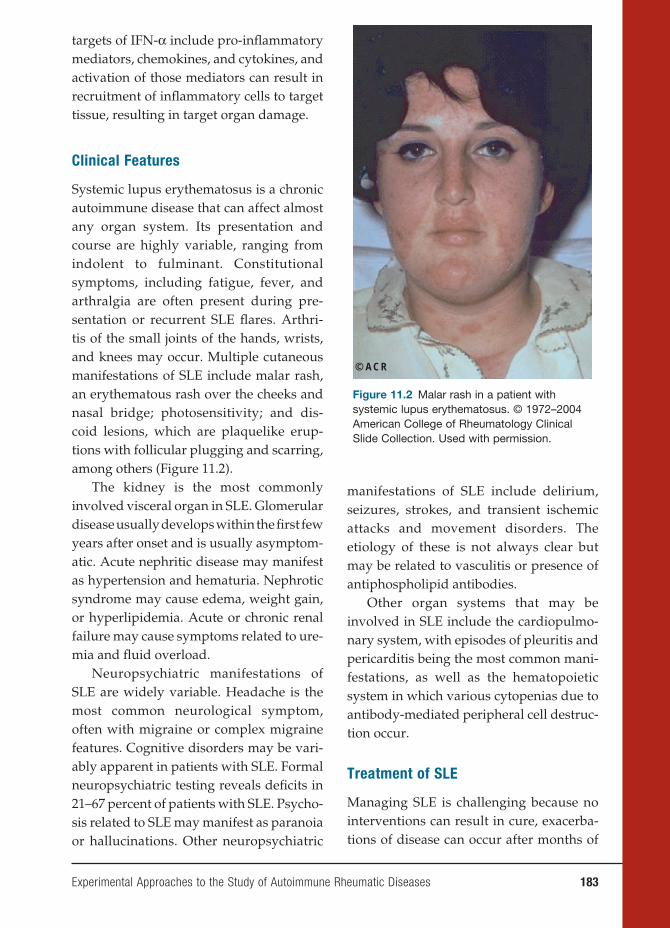
Experimental Approaches to the Study of Autoimmune Rheumatic Diseases 183
targets of IFN-α include pro-infl ammatory mediators, chemokines, and cytokines, and activation of those mediators can result in recruitment of infl ammatory cells to target tissue, resulting in target organ damage.
Clinical Features
Systemic lupus erythematosus is a chronic autoimmune disease that can affect almost any organ system. Its presentation and course are highly variable, ranging from indolent to fulminant. Constitutional symptoms, including fatigue, fever, and arthralgia are often present during pre-sentation or recurrent SLE fl ares. Arthri-tis of the small joints of the hands, wrists, and knees may occur. Multiple cutaneous manifestations of SLE include malar rash, an erythematous rash over the cheeks and nasal bridge; photosensitivity; and dis-coid lesions, which are plaquelike erup-tions with follicular plugging and scarring, among others (Figure 11.2).
The kidney is the most commonly involved visceral organ in SLE. Glomerular disease usually develops within the fi rst few years after onset and is usually asymptom-atic. Acute nephritic disease may manifest as hypertension and hematuria. Nephrotic syndrome may cause edema, weight gain, or hyperlipidemia. Acute or chronic renal failure may cause symptoms related to ure-mia and fl uid overload.
Neuropsychiatric manifestations of SLE are widely variable. Headache is the most common neurological symptom, often with migraine or complex migraine features. Cognitive disorders may be vari-ably apparent in patients with SLE. Formal neuropsychiatric testing reveals defi cits in 21–67 percent of patients with SLE. Psycho-sis related to SLE may manifest as paranoia or hallucinations. Other neuropsychiatric
manifestations of SLE include delirium, seizures, strokes, and transient ischemic attacks and movement disorders. The etiology of these is not always clear but may be related to vasculitis or presence of antiphospholipid antibodies.
Other organ systems that may be involved in SLE include the cardiopulmo-nary system, with episodes of pleuritis and pericarditis being the most common mani-festations, as well as the hematopoietic system in which various cytopenias due to antibody-mediated peripheral cell destruc-tion occur.
Treatment of SLE
Managing SLE is challenging because no interventions can result in cure, exacerba-tions of disease can occur after months of
© ACR
Figure 11.2 Malar rash in a patient with systemic lupus erythematosus. © 1972–2004 American College of Rheumatology Clinical Slide Collection. Used with permission.
184 Experimental Approaches to the Study of Autoimmune Rheumatic Diseases
stable maintenance treatment, and unde-sirable side effects of the therapies can be signifi cant. Careful and frequent monitor-ing of patients is important in selecting management plans, monitoring effi cacy, and changing treatments. The physician must fi rst decide whether a patient needs treatment and, if so, whether conserva-tive management is suffi cient or aggres-sive immunosuppression is necessary. For manifestations that are not life or organ threatening such as rash, musculoskel-etal disease, or mild serositis, conserva-tive management with NSAIDs, topical or low-dose glucocorticoids, or hydroxy-chloroquine are mainstays of treatment. For severe manifestations of disease such as renal or central nervous system disease, immunosuppression with high-dose ste-roids and cytotoxic agents such as cyclo-phosphamide, mycophenolate mofetil, or azathioprine is a therapeutic alternative. Most recently, studies investigating the role of the anti-CD20 monoclonal antibody rituximab, co-stimulatory blockade with abatacept and blockade of the cytokines IFN-α, IL-6, and B-lymphocyte stimulator in controlling various manifestations SLE are being actively pursued in clinical trials.
Animal Models of SLE
Several murine strains have served as clas-sic models of lupus-like disease, with each having many features that mimic immuno-logical and pathological aspects of human lupus. Although several spontaneous mouse models of lupus have striking simi-larities to human SLE, none of the animal models reproduces human SLE perfectly. What makes these animal models particu-larly interesting to study is that the dif-ferences may refl ect different forms of the human disease.
NZB X NZW F1 (B/W F1) LUPUS MODELThe F1 hybrid is the earliest recognized
spontaneous model of lupus. The mouse was developed from a cross between NZB mice, which develop a spontaneous auto-immune disease resembling hemolytic anemia, and NZW mice, which are phe-notypically normal. The B/W F1 strain is one of the best-studied models of human SLE and is considered by many to be the murine model most closely resembling human SLE. The disease is characterized by autoantibodies, including antinuclear antibodies (ANAs), IgG autoantibodies to double-stranded DNA (dsDNA), as well as lupus erythematosus (LE) cells, repre-senting phagocytic cells that have engulfed antibody-opsonized cell debris. Death results from a severe glomerulonephri-tis (GN) due to the deposition of immune complexes, including those containing anti-DNA antibodies. The disease is more severe and occurs earlier in female mice than in their male counterparts.
The B/W F1 is a good representative of human SLE because female B/W F1 mice manifest similar autoantibody profi les, and the disease develops in the absence of any known disease-accelerating gene, unlike other mouse models. Production of anti-dsDNA antibodies in this mouse, as in
human SLE, is thought to be T-cell depen-dent as suggested by the IgG isotype, by somatic mutations in the antibody genes, and by experiments in which depletion of T cells suppresses antibody production and ameliorates disease. As is true for humans with lupus, the correlation between nephri-tis and anti-DNA antibody levels is not universal. In addition to anti-dsDNA anti-bodies, the mice develop other autoanti-bodies found in human disease, including those that bind single-stranded (ssDNA), transfer RNA (tRNA), polynucleotides,
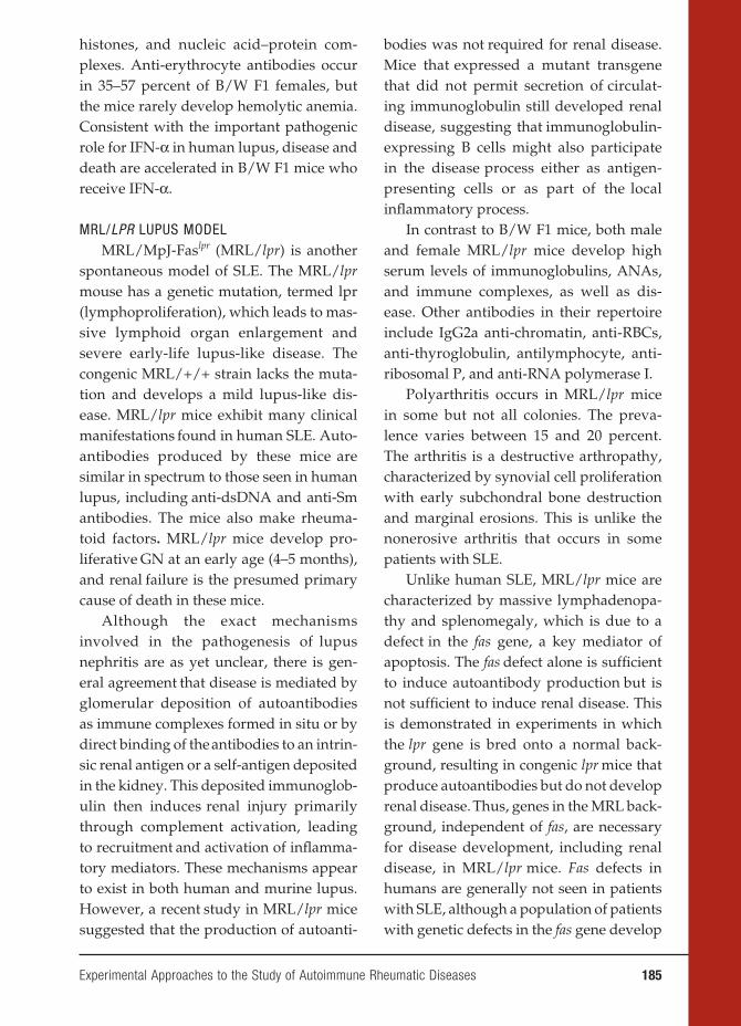
Experimental Approaches to the Study of Autoimmune Rheumatic Diseases 185
histones, and nucleic acid–protein com-plexes. Anti-erythrocyte antibodies occur in 35–57 percent of B/W F1 females, but the mice rarely develop hemolytic anemia. Consistent with the important pathogenic role for IFN-α in human lupus, disease and death are accelerated in B/W F1 mice who receive IFN-α.
MRL/LPR LUPUS MODEL MRL/MpJ-Faslpr (MRL/lpr) is another
spontaneous model of SLE. The MRL/lpr mouse has a genetic mutation, termed lpr (lymphoproliferation), which leads to mas-sive lymphoid organ enlargement and severe early-life lupus-like disease. The congenic MRL/+/+ strain lacks the muta-tion and develops a mild lupus-like dis-ease. MRL/lpr mice exhibit many clinical manifestations found in human SLE. Auto-antibodies produced by these mice are similar in spectrum to those seen in human lupus, including anti-dsDNA and anti-Sm antibodies. The mice also make rheuma-toid factors . MRL/lpr mice develop pro-liferative GN at an early age (4–5 months), and renal failure is the presumed primary cause of death in these mice.
Although the exact mechanisms involved in the pathogenesis of lupus nephritis are as yet unclear, there is gen-eral agreement that disease is mediated by glomerular deposition of autoantibodies
as immune complexes formed in situ or by direct binding of the antibodies to an intrin-sic renal antigen or a self-antigen deposited
in the kidney. This deposited immunoglob-ulin then induces renal injury primarily through complement activation, leading to recruitment and activation of infl amma-tory mediators. These mechanisms appear to exist in both human and murine lupus. However, a recent study in MRL/lpr mice suggested that the production of autoanti-
bodies was not required for renal disease. Mice that expressed a mutant transgene that did not permit secretion of circulat-ing immunoglobulin still developed renal disease, suggesting that immunoglobulin-expressing B cells might also participate in the disease process either as antigen-presenting cells or as part of the local infl ammatory process.
In contrast to B/W F1 mice, both male and female MRL/lpr mice develop high serum levels of immunoglobulins, ANAs, and immune complexes, as well as dis-ease. Other antibodies in their repertoire include IgG2a anti-chromatin, anti-RBCs, anti-thyroglobulin, antilymphocyte, anti-ribosomal P, and anti-RNA polymerase I.
Polyarthritis occurs in MRL/lpr mice in some but not all colonies. The preva-lence varies between 15 and 20 percent. The arthritis is a destructive arthropathy, characterized by synovial cell proliferation with early subchondral bone destruction and marginal erosions. This is unlike the nonerosive arthritis that occurs in some patients with SLE.
Unlike human SLE, MRL/lpr mice are characterized by massive lymphadenopa-thy and splenomegaly, which is due to a defect in the fas gene, a key mediator of apoptosis. The fas defect alone is suffi cient to induce autoantibody production but is not suffi cient to induce renal disease. This is demonstrated in experiments in which the lpr gene is bred onto a normal back-ground, resulting in congenic lpr mice that produce autoantibodies but do not develop renal disease. Thus, genes in the MRL back-ground, independent of fas, are necessary
for disease development, including renal disease, in MRL/lpr mice. Fas defects in humans are generally not seen in patients with SLE, although a population of patients with genetic defects in the fas gene develop
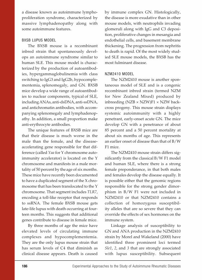
186 Experimental Approaches to the Study of Autoimmune Rheumatic Diseases
a disease known as autoimmune lympho-proliferation syndrome, characterized by massive lymphadenopathy along with some autoimmune features.
BXSB LUPUS MODELThe BXSB mouse is a recombinant
inbred strain that spontaneously devel-ops an autoimmune syndrome similar to human SLE. This mouse model is charac-terized by the production of autoantibod-ies, hypergammaglobulinemia with class switching to IgG3 and IgG2b, hypocomple-mentemia, splenomegaly, and GN. BXSB mice develop a wide range of autoantibod-ies to nuclear components, typical of SLE, including ANAs, anti-dsDNA, anti-ssDNA,
and antichromatin antibodies, with accom-panying splenomegaly and lymphadenop-athy. In addition, a small proportion make anti-erythrocyte antibodies.
The unique features of BXSB mice are that their disease is much worse in the male than the female, and the disease-accelerating gene responsible for that dif-ference (called Yaa for Y chromosome auto-immunity accelerator) is located on the Y chromosome and manifests in a male mor-tality of 50 percent by the age of six months. These mice have recently been documented to have a duplicated segment of the X chro-mosome that has been translocated to the Y chromosome. That segment includes TLR7, encoding a toll-like receptor that responds to ssRNA. The female BXSB mouse gets late-life lupus with death occurring at four-teen months. This suggests that additional genes contribute to disease in female mice.
By three months of age the mice have elevated levels of circulating immune complexes and hypocomplementemia. They are the only lupus mouse strain that has serum levels of C4 that diminish as clinical disease appears. Death is caused
by immune complex GN. Histologically, the disease is more exudative than in other mouse models, with neutrophils invading glomeruli along with IgG and C3 deposi-tion, proliferative changes in mesangia and endothelial cells, and basement membrane thickening. The progression from nephritis to death is rapid. Of the most widely stud-ied SLE mouse models, the BXSB has the most fulminant disease.
NZM2410 MODELThe NZM2410 mouse is another spon-
taneous model of SLE and is a congenic recombinant inbred strain (termed NZM for New Zealand Mixed) produced by inbreeding (NZB × NZW)F1 × NZW back-cross progeny. This mouse strain displays systemic autoimmunity with a highly penetrant, early-onset acute GN. The mice develop GN with a penetrance of about 85 percent and a 50 percent mortality at about six months of age. This represents an earlier onset of disease than that of B/W F1 mice.
The NZM2410 mouse strain differs sig-nifi cantly from the classical B/W F1 model and human SLE, where there is a strong female preponderance, in that both males and females develop the disease equally. It is possible either that the genomic regions responsible for the strong gender dimor-phism in B/W F1 were not included in NZM2410 or that NZM2410 contains a collection of homozygous susceptibil-ity alleles that are so severe that they can override the effects of sex hormones on the immune system.
Linkage analysis of susceptibility to GN and ANA production in the NZM2410 strain by Morel and Wakeland (2000) have identifi ed three prominent loci termed Sle1, 2, and 3 that are strongly associated with lupus susceptibility. Subsequent
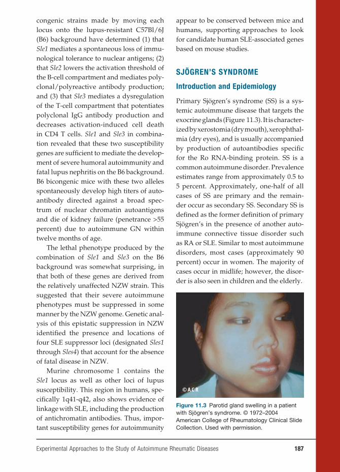
Experimental Approaches to the Study of Autoimmune Rheumatic Diseases 187
congenic strains made by moving each locus onto the lupus-resistant C57Bl/6J (B6) background have determined (1) that Sle1 mediates a spontaneous loss of immu-nological tolerance to nuclear antigens; (2) that Sle2 lowers the activation threshold of the B-cell compartment and mediates poly-clonal/polyreactive antibody production; and (3) that Sle3 mediates a dysregulation of the T-cell compartment that potentiates polyclonal IgG antibody production and decreases activation-induced cell death in CD4 T cells. Sle1 and Sle3 in combina-tion revealed that these two susceptibility genes are suffi cient to mediate the develop-ment of severe humoral autoimmunity and fatal lupus nephritis on the B6 background. B6 bicongenic mice with these two alleles spontaneously develop high titers of auto-antibody directed against a broad spec-trum of nuclear chromatin autoantigens and die of kidney failure (penetrance >55 percent) due to autoimmune GN within twelve months of age.
The lethal phenotype produced by the combination of Sle1 and Sle3 on the B6 background was somewhat surprising, in that both of these genes are derived from the relatively unaffected NZW strain. This suggested that their severe autoimmune phenotypes must be suppressed in some manner by the NZW genome. Genetic anal-ysis of this epistatic suppression in NZW identifi ed the presence and locations of four SLE suppressor loci (designated Sles1 through Sles4) that account for the absence of fatal disease in NZW.
Murine chromosome 1 contains the Sle1 locus as well as other loci of lupus susceptibility. This region in humans, spe-cifi cally 1q41-q42, also shows evidence of linkage with SLE, including the production of antichromatin antibodies. Thus, impor-tant susceptibility genes for autoimmunity
appear to be conserved between mice and humans, supporting approaches to look for candidate human SLE-associated genes
based on mouse studies.
SJÖGREN’S SYNDROME
Introduction and Epidemiology
Primary Sjögren’s syndrome (SS) is a sys-temic autoimmune disease that targets the exocrine glands (Figure 11.3). It is character-ized by xerostomia (dry mouth), xerophthal-mia (dry eyes), and is usually accompanied by production of autoantibodies specifi c for the Ro RNA-binding protein. SS is a common autoimmune disorder. Prevalence estimates range from approximately 0.5 to 5 percent. Approximately, one-half of all cases of SS are primary and the remain-der occur as secondary SS. Secondary SS is defi ned as the former defi nition of primary Sjögren’s in the presence of another auto-immune connective tissue disorder such as RA or SLE. Similar to most autoimmune disorders, most cases (approximately 90 percent) occur in women. The majority of cases occur in midlife; however, the disor-der is also seen in children and the elderly.
© ACR
Figure 11.3 Parotid gland swelling in a patient with Sjögren’s syndrome. © 1972–2004 American College of Rheumatology Clinical Slide Collection. Used with permission.
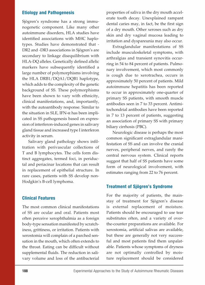
188 Experimental Approaches to the Study of Autoimmune Rheumatic Diseases
Etiology and Pathogenesis
Sjögren’s syndrome has a strong immu-nogenetic component. Like many other autoimmune disorders, HLA studies have identifi ed associations with MHC haplo-types. Studies have demonstrated that -DR2 and -DR3 associations in Sjögren’s are secondary to linkage disequilibrium with HLA-DQ alleles. Genetically defi ned allelic markers have subsequently identifi ed a large number of polymorphisms involving the HLA DRB1/DQA1/DQB1 haplotype, which adds to the complexity of the genetic background of SS. These polymorphisms have been shown to vary with ethnicity, clinical manifestations, and, importantly, with the autoantibody response. Similar to the situation in SLE, IFN-α has been impli-cated in SS pathogenesis based on expres-sion of interferon-induced genes in salivary gland tissue and increased type I interferon activity in serum.
Salivary gland pathology shows infi l-tration with perivascular collections of T and B lymphocytes. The cells form dis-tinct aggregates, termed foci, in periduc-tal and periacinar locations that can result in replacement of epithelial structure. In rare cases, patients with SS develop non-Hodgkin’s B-cell lymphoma.
Clinical Features
The most common clinical manifestations of SS are ocular and oral. Patients most often perceive xerophthalmia as a foreign body-type sensation manifested by scratch-iness, grittiness, or irritation. Patients with xerostomia will complain of a parched sen-sation in the mouth, which often extends to the throat. Eating can be diffi cult without supplemental fl uids. The reduction in sali-vary volume and loss of the antibacterial
properties of saliva in the dry mouth accel-erate tooth decay. Unexplained rampant dental caries may, in fact, be the fi rst sign of a dry mouth. Other xeroses such as dry skin and dry vaginal mucosa leading to irritation and dyspareunia may also occur.
Extraglandular manifestations of SS include musculoskeletal symptoms, with arthralgias and transient synovitis occur-ring in 54 to 84 percent of patients. Pulmo-nary involvement, which most commonly is cough due to xerotrachea, occurs in approximately 50 percent of patients. Mild autoimmune hepatitis has been reported to occur in approximately one-quarter of primary SS patients, with smooth muscle antibodies seen in 7 to 33 percent. Antimi-tochondrial antibodies have been reported in 7 to 13 percent of patients, suggesting an association of primary SS with primary biliary cirrhosis (PBC).
Neurologic disease is perhaps the most common signifi cant extraglandular mani-festation of SS and can involve the cranial nerves, peripheral nerves, and rarely the central nervous system. Clinical reports suggest that half of SS patients have some form of neurological involvement, with estimates ranging from 22 to 76 percent.
Treatment of Sjögren’s Syndrome
For the majority of patients, the main-stay of treatment for Sjögren’s disease is external replacement of moisture. Patients should be encouraged to use tear substitutes often, and a variety of over-the-counter preparations are available. For xerostomia, artifi cial salivas are available, but these are generally not very success-ful and most patients fi nd them unpalat-able. Patients whose symptoms of dryness are not optimally controlled by mois-ture replacement should be considered
Experimental Approaches to the Study of Autoimmune Rheumatic Diseases 189
for treatment with secretory stimulants. There are two approved agents available for use as secretagogues in SS: pilocar-pine (Salagen) and cevemiline (Evoxac). Both agents have been shown, in con-trolled clinical trials, to increase salivary fl ow rate signifi cantly in SS. There are no disease-modifying drugs that have been demonstrated to have specifi c effi cacy for SS. Minor musculoskeletal symptoms usu-ally respond to NSAID therapy. Because erosive joint disease is rare, therapy with DMARDs is usually unnecessary; how-ever, hydroxychloroquine has been used to treat fatigue, arthralgia, and myalgia in primary SS. Cranial and peripheral neu-ropathy may be treated with low-dose tricyclic antidepressants or gabapentin. Symptomatic cases resistant to the previ-ously mentioned therapies may be treated with intravenous gammaglobulin. Central nervous system manifestations, which are felt to be due to Sjögren’s, should be treated aggressively with high-dose oral or pulse intravenous corticosteroid and daily or monthly intravenous pulse cyclophosphamide.
Animal Models of Sjögren’s Syndrome
Nonobese diabetic mice have been used predominantly as a model of autoimmune diabetes mellitus, but they also develop an autoimmune exocrinopathy characterized by hyposecretion of saliva and acinar cell atrophy.
MRL+/+ and MRL/lpr mice spontane-ously develop lacrimal and salivary gland infl ammation and have been used as mod-els for SS.
IQI/Jic (IQI) mice are a model of pri-mary SS, unlike the animal models above that are autoimmune-prone mice that develop SS-like pathology associated with
other autoimmune conditions, such as lupus, infl ammatory arthritis, or insulitis. IQI mice spontaneously develop autoim-mune infi ltration of lymphocytes into the lacrimal and salivary glands, leading to dacryoadenitis and sialoadenitis. The inci-dence of the disease is higher in females than in males. Sialoadenitis in female mice can be detected from two months of age onward and signifi cant progression of the lesions is observed after nine months of age. Infl ammatory lesions occurring at young ages mainly consist of CD4+ T cells with lesser abundance of CD8+ T cells, B cells, and macrophages, and the pro-portions of B cells and plasma cells are elevated in accordance with increasing
magnifi cation of the lesions. Production of ANAs, one of the prominent pathophysi-ological features in patients with SS, is also observed in old IQI mice. Moreover, IQI mice develop infl ammatory lesions in the lung, pancreas, and kidney in addi-tion to the lacrimal and salivary glands that are similar to those in patients with primary SS. Thus, this murine model may be the most suitable for the investigation of the pathogenesis of SS, with progression from oral and ocular disease to a systemic
disorder.
PROGRESSIVE SYSTEMIC
SCLEROSIS
Introduction and Epidemiology
Progressive systemic sclerosis (PSS) is a disorder of connective tissue clinically characterized by thickening and fi bro-sis of the skin (scleroderma) and by dis-tinctive forms of involvement of internal organs, including the heart, lungs, kid-neys, and gastrointestinal tract. The hall-marks of the disease are autoimmunity
190 Experimental Approaches to the Study of Autoimmune Rheumatic Diseases
and inflammation, widespread vascu-lopathy (blood vessel damage), affecting multiple vascular beds, and progressive interstitial and perivascular fi brosis. The etiology is unknown. The incidence of sys-temic sclerosis is estimated to be between eighteen and twenty individuals per one million persons per year. All age groups may be affected, but the onset of disease is highest between the ages of thirty and fi fty years old. Systemic sclerosis is three to four times more common in women than in men, with women of childbearing age at peak risk.
Etiology and Pathogenesis
The pathogenesis of systemic sclerosis involves an interplay between oblitera-tive vasculopathy in multiple vascular beds, infl ammation, and autoimmunity,
and progressive fi brosis. Vascular injury and activation are the earliest and possibly primary events in the pathogenesis of PSS. This is suggested by histopathological evi-dence of vascular damage that is present before fi brosis, and clinical manifestations, such as Raynaud’s phenomenon, an epi-sodic and reversible cold-induced vaso-spasm of the fi ngers and toes, that precede other disease manifestations. Additional manifestations of PSS-associated vascu-lopathy include cutaneous telangiectasia, nail-fold capillary alterations, pulmo-nary arterial hypertension, gastric antral vascular ectasia, and scleroderma renal crisis with malignant hypertension. In
late-stage PSS, there is a striking paucity of small blood vessels in lesional skin and other organs. Endothelial cell injury might
be triggered by granzymes, endothe-lial cell-specifi c autoantibodies, vasculo-tropic viruses, infl ammatory cytokines, or
reactive oxygen radicals generated during ischemia/reperfusion.
Clinical Features
Patients with systemic sclerosis have a wide array of symptoms and diffi culties, ranging from complaints related to spe-cifi c internal organ involvement as well as the symptoms of a chronic catabolic illness. Raynaud’s phenomenon is the initial complaint in approximately 70 per-cent of patients with systemic sclerosis. Peripheral vasoconstriction in response to cold is physiologic, and individu-als with Ra ynaud’s, no matter the cause, have undue intolerance to environmental cold. The digital arteries of patients with systemic sclerosis exhibit marked intimal hyperplasia as well as adventitial fi bro-sis. Severe narrowing (more than 75 per-cent) of the arterial lumen results, which may be suffi ciently severe to account for Raynaud’s phenomenon. The skin thick-ening of systemic sclerosis begins on the fi ngers and hands in nearly all cases. The skin initially appears shiny and taut and may be erythematous at early stages (Figure 11.4). The skin of the face and neck is usually involved next and is associated with an immobile and pinched facies. The skin change may stay restricted to fi ngers, hands, and face and may remain rela-tively mild. Extension to the forearms is often followed by rapid centripetal spread to the upper arms, shoulders, anterior chest, back, abdomen, and legs (diffuse scleroderma).
Involvement of the gastrointestinal tract is the third most common manifestation of systemic sclerosis. Impaired function of the lower esophageal sphincter is associated with symptoms of intermittent heartburn,
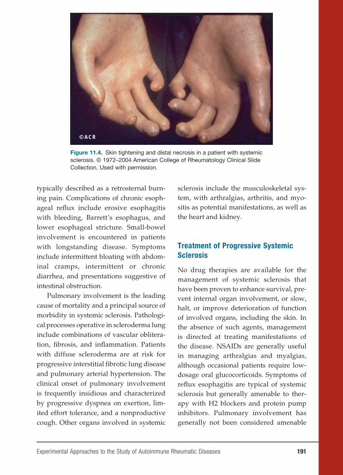
Experimental Approaches to the Study of Autoimmune Rheumatic Diseases 191
typically described as a retrosternal burn-ing pain. Complications of chronic esoph-ageal refl ux include erosive esophagitis with bleeding, Barrett’s esophagus, and lower esophageal stricture. Small-bowel involvement is encountered in patients with longstanding disease. Symptoms include intermittent bloating with abdom-inal cramps, intermittent or chronic diarrhea, and presentations suggestive of intestinal obstruction.
Pulmonary involvement is the leading cause of mortality and a principal source of morbidity in systemic sclerosis. Pathologi-cal processes operative in scleroderma lung include combinations of vascular oblitera-tion, fi brosis, and infl ammation. Patients with diffuse scleroderma are at risk for progressive interstitial fi brotic lung disease and pulmonary arterial hypertension. The clinical onset of pulmonary involvement is frequently insidious and characterized by progressive dyspnea on exertion, lim-ited effort tolerance, and a nonproductive cough. Other organs involved in systemic
sclerosis include the musculoskeletal sys-tem, with arthralgias, arthritis, and myo-sitis as potential manifestations, as well as the heart and kidney.
Treatment of Progressive Systemic
Sclerosis
No drug therapies are available for the management of systemic sclerosis that have been proven to enhance survival, pre-vent internal organ involvement, or slow, halt, or improve deterioration of function of involved organs, including the skin. In the absence of such agents, management is directed at treating manifestations of the disease. NSAIDs are generally useful in managing arthralgias and myalgias, although occasional patients require low-dosage oral glucocorticoids. Symptoms of refl ux esophagitis are typical of systemic sclerosis but generally amenable to ther-apy with H2 blockers and protein pump inhibitors. Pulmonary involvement has generally not been considered amenable
© ACR
Figure 11.4. Skin tightening and distal necrosis in a patient with systemic sclerosis. © 1972–2004 American College of Rheumatology Clinical Slide Collection. Used with permission.
192 Experimental Approaches to the Study of Autoimmune Rheumatic Diseases
to therapy. A patient in whom there is evi-dence of pulmonary interstitial infl amma-tion might be treated with glucocorticoids and immunosuppressive agents. A recent double-blind, randomized, placebo-con-trolled trial to determine the effects of oral cyclophosphamide on lung function and health-related symptoms in patients with evidence of active alveolitis and sclero-derma-related interstitial lung disease reported a signifi cant but modest ben-efi cial effect on lung function, dyspnea, thickening of the skin, and health-related quality of life. Pulmonary hyperten-sion has emerged as a principal cause of morbidity and mortality in late systemic sclerosis. Centrally infused prostacyclin (epoprostenol) improves both short- and long-term hemodynamics, as well as the quality of life and survival. Studies have demonstrated that treatment with the oral endothelin receptor anatagonist bosen-tan is well tolerated in PSS patients with interstitial lung disease and is effective for treatment of severe pulmonary hyperten-sion in study patients. Scleroderma renal crisis demands prompt recognition of the diagnosis and aggressive treatment of the accompanying accelerated hyper-tension. Angiotensin-converting enzyme inhibitors such as captopril and enala-pril are mechanistically ideal to treat the hyperreninemic hypertension of sclero-derma renal crisis and are the treatment of choice.
Animal Models of Progressive
Systemic Sclerosis
The pathogenesis of PSS involves a triad of small-vessel vasculopathy; infl amma-tion, and autoimmunity; and interstitial
and vascular fi brosis in the skin, lungs, and multiple other organs. Various ani-
mal models have been investigated as
spontaneous or inducible models for SSC. Although none of them reproduce all three pathogenetic components of the disease, some models do recapitulate selected phenotypic features. Two useful animal models of PSS include bleomy-cin-induced skin fi brosis and the chronic graft-versus-host disease system, which results in skin fi brosis. In addition, the tight skin (Tsk-1) mouse has been pro-posed as a model for PSS based on skin pathology that is similar to that of the human disease, with increased accumula-tion of collagen and glycosaminoglycans in the skin and production of serum auto-antibodies. The gene mutated in the Tsk-1 mouse is fi brillin-1, whose product can form a part of elastic fi bers.
POLYMYOSITIS
Introduction and Epidemiology
Polymyositis (PM) is an infl ammatory muscle disease characterized by injury and death of muscle cells and resulting in severe weakness. Polymyositis is considered to be an autoimmune disease based on the pres-ence of autoantibodies with characteristic specifi cities, including those reactive with tRNA synthetases. In addition, infi ltration of T cells, predominantly CD8+ cytotoxic T cells, may contribute directly to muscle damage. PM is a rare disease and only a few epidemiological studies have been published. The reported incidence ranges between two and eight new cases per mil-lion people per year.
Etiology and Pathogenesis
The idiopathic inflammatory myopa-thies, including PM, are believed to be
Experimental Approaches to the Study of Autoimmune Rheumatic Diseases 193
immune-mediated processes possibly triggered by environmental factors in genetically susceptible individuals. This hypothesis is based on the recognized association with other autoimmune and collagen vascular diseases, the preva-lence and type of circulating autoanti-bodies, animal models, immunogenetic observations, specifi cs of the infl amma-tory changes in muscle, and the treat-ment response to immunosuppressive agents. Autoantibodies associated with PM include the anti-tRNA synthetase antibodies (anti-Jo-1 and others) as well as antibodies directed against the sig-nal recognition particle. In PM, CD8+ cytotoxic T cells invade muscle fi bers that express MHC class I antigens, lead-ing to fi ber necrosis via the perforin pathway.
Clinical Features
The main symptoms of PM are weakness affecting primarily the proximal muscles. The onset is typically subacute, occurring over several weeks, with patients com-plaining of increasing fatigue in rising from a chair, lifting their arms, climbing steps, or combing their hair. The neck extensor muscles may be involved caus-ing diffi culty in holding up the head. In severe advanced cases, the respiratory and oropharyngeal muscles are involved causing dysphagia and respiratory dif-fi culties. Myalgias are less frequent than muscular weakness, and when present, accompany the weakness. The most com-mon clinical signs are decreased strength in the proximal muscles, contractures, and late in the course of the disease, muscular atrophy, which occurs in up to 40 percent of cases. Dysphagia occurs in one-third of cases and is due to involvement of the
oropharyngeal muscles or involvement of the striated muscle fi bers of the upper esophagus. Pulmonary symptoms are due to weakness of the thoracic muscles or interstitial lung disease. Fibrosis is radiologically demonstrated in 5–10 per-cent of cases and is often associated with anti-tRNA synthetase antibodies such as anti-Jo-1.
Treatment of Polymyositis
Glucocorticoids are the standard fi rst-line medication for patients with idiopathic infl ammatory myopathy. Initially, predni-sone is given in a single dose of 1 to 2 mg/kg per day. Regular evaluations of muscle strength and serum enzymes should be performed during treatment. Ideally, the initial steroid dosage is maintained until strength and creatine kinase values have returned to normal and have remained normal for four to eight weeks and then tapered. If a patient fails to respond to glucocorticoid therapy or if a patient has had some improvement but the level of strength has reached a plateau, additional therapy with either the immunosuppres-sive agent azathioprine or methotrexate is added. Another therapeutic option is intra-venous immune globulin, which is being used increasingly to treat infl ammatory myopathies. This therapy is associated with little toxicity but is extremely expensive. Controlled trials involving larger numbers of patients are needed to prove a benefi t for this and other therapies in PM.
Animal Models of Polymyositis
Several animal models have been devel-oped that may prove useful for understand-ing the pathogenesis of PM. Experimental autoimmune myositis (EAM) can be

194 Experimental Approaches to the Study of Autoimmune Rheumatic Diseases
induced by immunization of rodents with skeletal muscle homogenate and adju-vant. Similarly, Lewis rats immunized with purified skeletal muscle myosin develop EAM with the same pattern and severity as EAM induced by whole-rabbit skeletal-muscle homogenate. Multiple infl ammatory lesions are detected histo-pathologically in various muscle groups in animals immunized with either prepa-ration. Dendritic cells presenting pyruvate kinase M1/M2 peptide 464-472 to T cells have also been effective in inducing EAM in BALB/c mice, with >40 percent of the mice developing pathological changes in skeletal muscle similar to those seen in human PM.
PM can also be induced in mice by viral infection. Intraperitoneal inoculation of Coxsackie B1 virus, Tucson strain, into young mice can result in proximal hind-quarter weakness that persists for more than ten weeks. The myositis persists long after the virus is cleared and in the pres-ence of neutralizing antibody, suggesting that the immunological response is con-tributing to pathology.
A possible murine model of infl amma-tory myositis has resulted from a genetic defi ciency in a member of the synapto-tagmin family, comprising Ca2+ sensors involved in cell membrane fusion. Syt VII is a ubiquitously expressed synaptotagmin previously implicated in plasma membrane repair and Trypanosoma cruzi invasion, events mediated by the Ca2+-regulated exocytosis of lysosomes. Mice that are Syt VII defi cient have an infl ammatory myopathy, with muscle-fi ber invasion by leukocytes and endomysial collagen depo-sition. This pathological picture is associ-ated with elevated creatine kinase release and progressive muscle weakness as well as a strong ANA response.
SERONEGATIVE
SPONDYLOARTHROPATHY
Introduction and Epidemiology
Ankylosing spondylitis (AS) and related spondyloarthropathies are characterized by sacroiliitis and infl ammation of the intervertebral discs in the lumbar spine, as well as an enthesitis at sites of ligamentous insertions into bone. Patients with AS develop calcifi cation of the ligamentous insertions, back stiffness, and pain. The immunologic basis of the specifi c loca-tions of infl ammation in this disorder is not understood, but a strong association of the disease with particular HLA alleles, particularly HLA-B27, suggests a possible role for antigen presentation to T cells in the immunopathogenesis. The prevalence of AS closely parallels the frequency of HLA-B27. Among whites, the estimated prevalence rate of AS is 197 per 100,000 in the United States. AS, in general, is diag-nosed more frequently in males. Females, however, may have milder or subclinical disease. AS is more common in males with a male to female ratio of 3:1. The age of onset of AS is usually from the late teens to age 40.
Etiology and Pathogenesis
The exact etiology of AS is unclear. The strong association with most subtypes of HLA-B27 supports the hypothesis that the disease is due to a genetically determined immune response to environmental fac-tors in susceptible individuals. HLA-B27 is present in 80 to 98 percent of white patients with AS, in contrast to only about 8 percent of the general population. The arthritogenic peptide hypothesis postulates that AS results when external antigenic challenge activates autoreactive T cells that recognize
Experimental Approaches to the Study of Autoimmune Rheumatic Diseases 195
endogenous peptides presented by HLA-B27. In normal situations, the HLA-B27 molecule on the surface of the antigen-presenting cells presents endogenously derived peptides to CD8+ T cells. These peptides, which are usually nine amino acids long and have arginine in position 2, are mostly self-derived, but they may also be of viral or bacterial origin. Recent studies also suggest that the intracellu-lar handling of molecular complexes that include HLA-B27 molecules may activate an infl ammatory stress response and con-tribute to disease.
Clinical Features
Back pain is the most common symptom and the fi rst manifestation in approxi-mately 75 percent of patients with AS. The infl ammatory back pain of AS has particular features that differentiate it from mechanical back pain. These include insidious onset of the pain occurring over months or years, generally with at least three months of symptoms before presenta-tion. Symptoms include morning stiffness lasting at least thirty minutes, improve-ment of symptoms with moderate physical activity, and diffuse nonspecifi c radiation of pain into both buttocks. Patients often experience stiffness and pain that awakens them in the early morning hours, a dis-tinctive symptom not generally found in patients with mechanical back pain. Ten-derness at tendon insertion points due to enthesitis, an infl ammatory reaction, is a common complaint. Typical tender sites include the costosternal junctions, spinous processes, iliac crests, greater trochan-ters, ischial tuberosities, tibial tubercles, or heels (Achilles tendinitis or plantar fasciitis). AS is also characterized by a number of extra-articular manifestations
including uveitis, occurring in 25–35 per-cent of patients during the course of the disease. Manifestations of cardiac involve-ment include ascending aortitis, aortic valve incompetence, conduction abnor-malities, cardiomegaly, and pericarditis. In rare situations, aortitis may precede other features of AS. Aortic incompetence was noted in 3.5 percent of patients who had the disease for fi fteen years and in 10 percent after thirty years. Cardiac conduc-tion disturbances are seen with increasing frequency with the passage of time, occur-ring in 2.7 percent of those with disease of fi fteen years’ duration and in 8.5 percent after thirty years.
Treatment of Ankylosing Spondylitis
AS is a relatively mild disease for most patients with a good functional prognosis. The majority of patients do not experience signifi cant extraskeletal manifestations, except for acute anterior uveitis. Usu-ally, the eye disease can be well managed with eye drops containing corticosteroids to reduce infl ammation and with pupil-dilating, atropine-like agents to prevent or diminish synechiae. The objectives for treatment of AS are to relieve pain, stiff-ness, and fatigue and to maintain good pos-ture and good physical and psychosocial functioning. Physiotherapy provided as exercises is effective in improving thoraco-lumbar mobility and fi tness, at least in the short term (up to one year). Until recently, no drug had been shown to signifi cantly infl uence the course of spinal disease and retard the process of ossifi cation in AS. Similarly, there was no evidence to suggest that any of the conventional DMARDs, including sulfasalazine and methotrexate, altered or inhibited the infl ammation seen in the spine and entheses in AS. Recently,
196 Experimental Approaches to the Study of Autoimmune Rheumatic Diseases
the introduction of anti-TNF agents in the treatment of AS can be regarded as a defi -nite advantage in the therapy of this dis-ease. Both infl iximab and etancercept have been shown to improve signs and symp-toms of disease. Continuous treatment is necessary in most patients. More than two-thirds of the patients stay on therapy after one year. Guidelines for the use of anti-TNF therapies have recently been devel-oped. The new therapeutic modalities identify important clinical questions to be answered by further research. Clearly, inf-liximab and etanercept have disease-modi-fying properties, but their long-term safety and disease-controlling effects in terms of improvement or maintenance of func-tion, as well as the prevention of structural damage, still have to be demonstrated.
Animal Models of Seronegative
Spondyloarthropathy
HLA-B27 transgenic rats and strains of HLA-B27 transgenic β2-microglobulin-deficient mice develop a multisystem infl ammatory disease affecting the joints, skin, and bowel with strong similarity to human spondyloarthritis. This model supports a direct mechanistic role for that MHC product in the disease, although the details of that mechanism have not been elucidated.
As is the case for other autoimmune rheumatic diseases, antigen-induced murine models are proving useful in study of AS. Aggrecan and versican are large pro-teoglycan molecules that are present in the intervertebral disc and hyaline cartilages of the sacroiliac joint, as well as in entheses. Versican is generally absent from cartilage tissue except in the sacroiliac joint but is concentrated in ligaments and the annulus of the intervertebral disc. Immunization
of BALB/c mice with versican results in an AS-like pathology, including sacroi-liitis, enthesitis, and discitis. Those mice develop an ankylosis of the spine similar to that seen in AS. It has also been noted that immunity to versican can induce uveitis, a clinical feature of the human disease.
Future Directions of Research
Signifi cant progress in characterizing the underlying immunopathogenic mecha-nisms of rheumatic diseases is being stim-ulated by accelerated success in defi ning disease susceptibility genes using high-throughput single-nucleotide polymor-phism analysis. In addition, increased focus on the activation of the innate immune response, resulting in activation of molecu-lar pathways involved in either production of type I interferon or TNF, is leading to new understanding of the effect of those cytokines on altered immunoregulation and infl ammation and has substantially advanced development of new targeted therapies.
Future advances in rheumatic disease research are likely to derive from func-tional studies related to disease-associated genes identifi ed in the large-scale studies. The regulation of innate immune system activation, alterations in function of cell surface molecules, and signaling pathways that control the threshold for lymphocyte activation and regulation of the comple-ment system are areas for further study that may identify functional correlates of the genes characterized in recent collab-orative projects. The regulation of cytokine production, with a particular emphasis on the type I interferon pathway, will be an important topic that can link genetics and translational research that is based on care-ful clinical characterization in association
Experimental Approaches to the Study of Autoimmune Rheumatic Diseases 197
with laboratory investigations. The use of the experimental murine models described in this chapter will represent an important approach to elucidating the mechanisms through which gene variants and altered immune function result in tissue damage and disease consistent with the pathol-ogy that occurs in humans. Future studies should be aimed at identifying molecular targets that can be practically modulated using new therapeutics, with animal stud-ies serving to speed translation to humans.
BIBLIOGRAPHY
SUGGESTED READINGBriani C, Doria A, Sarzi-Puttini P,
Dalakas MC. Update on idiopathic infl ammatory myopathies. Autoimmu-nity. 2006;39(3):161–170.
Christner PJ, Jimenez SA. Animal models of systemic sclerosis: insights into sys-temic sclerosis pathogenesis and poten-tial therapeutic approaches. Curr Opin Rheumatol. 2004;16:746–752.
Crow MK. Collaboration, genetic associa-tions, and lupus erythematosus. N Engl J Med. 2008;358(9):956–961.
Hahn BH. Animal models of systemic lupus erythematosus. In: Wallace DJ, Hahn BH, eds. Dubois’ Lupus Erythema-tosus. 6th ed. Philadelphia, PA: Lippin-cott Williams & Wilkins; 2002:339.
Holmdahl R, Andersson M, Goldschmidt TJ, Gustafsson K, Jansson L, Mo JA. Type II collagen autoimmunity in ani-mals and provocations leading to arthri-tis. Immunol Rev. 1990;118:193–232.
Holmdahl R. Experimental models for rheumatoid arthritis. In: Firestein
GS, Panayi GS, Wollheim FA, eds. Rheumatoid Arthritis New Frontiers in Pathogenesis and Treatment. New York, NY: Oxford University Press; 2000:39.
Jorgensen TN, Gubbels MR, Kotzin BL. New insights into disease pathogenesis from mouse lupus genetics. Curr Opin Immunol. 2004;16:787–793.
Kono DH, Theofi lopoulos AN. Genetics of systemic autoimmunity in mouse models of lupus. Int Rev Immunol. 2000;19:367–387.
Lauwerys BR, Wakeland EK. Genetics of lupus nephritis. Lupus. 2005;14:2–12.
Morel L, Wakeland EK. Lessons from the NZM2410 model and related strains. Int Rev Immunol. 2000;19:423–446.
Nagaraju K, Plotz PH. Animal models of myositis. Rheum Dis Clin North Am. 2002;28:917–933.
Theofi lopoulos AN, Dixon FJ. Murine models of systemic lupus erythemato-sus. Adv Immunol. 1985;37:269–390.
Van den Berg WB. 2004. Animal models. In: St. Clair EW, Pisetsky DS, Haynes BF, eds. Rheumatoid Arthritis. Philadelphia, PA: Lippincott Williams & Wilkins; 2004.
Varga J, Abraham D. Systemic sclero-sis: a prototypic multisystem fi brotic disorder. J Clin Invest. 2007;117(3): 557–567.
Wakeland EK, Liu K, Graham RR, Behrens TW. Delineating the genetic basis of systemic lupus erythematosus. Immu-nity. 2001;15:397–408.
Zhang Y. Animal models of infl amma-tory spinal and sacroiliac joint diseases. Rheum Dis Clin North Am. 2003;29:631–645.

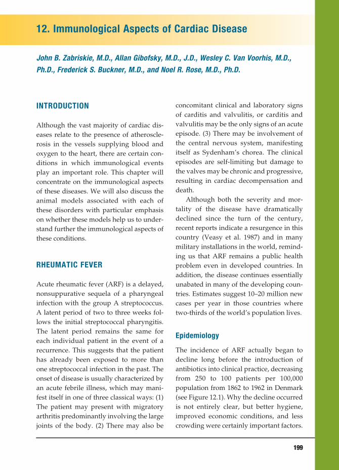
12. Immunological Aspects of Cardiac Disease
John B. Zabriskie, M.D., Allan Gibofsky, M.D., J.D., Wesley C. Van Voorhis, M.D.,
Ph.D., Frederick S. Buckner, M.D., and Noel R. Rose, M.D., Ph.D.
199
INTRODUCTION
Although the vast majority of cardiac dis-eases relate to the presence of atheroscle-rosis in the vessels supplying blood and oxygen to the heart, there are certain con-ditions in which immunological events play an important role. This chapter will concentrate on the immunological aspects of these diseases. We will also discuss the animal models associated with each of these disorders with particular emphasis on whether these models help us to under-stand further the immunological aspects of these conditions.
RHEUMATIC FEVER
Acute rheumatic fever (ARF) is a delayed, nonsuppurative sequela of a pharyngeal infection with the group A streptococcus. A latent period of two to three weeks fol-lows the initial streptococcal pharyngitis. The latent period remains the same for each individual patient in the event of a recurrence. This suggests that the patient has already been exposed to more than one streptococcal infection in the past. The onset of disease is usually characterized by an acute febrile illness, which may mani-fest itself in one of three classical ways: (1) The patient may present with migratory arthritis predominantly involving the large joints of the body. (2) There may also be
concomitant clinical and laboratory signs of carditis and valvulitis, or carditis and valvulitis may be the only signs of an acute episode. (3) There may be involvement of the central nervous system, manifesting itself as Sydenham’s chorea. The clinical episodes are self-limiting but damage to the valves may be chronic and progressive, resulting in cardiac decompensation and death.
Although both the severity and mor-tality of the disease have dramatically declined since the turn of the century, recent reports indicate a resurgence in this country (Veasy et al. 1987) and in many military installations in the world, remind-ing us that ARF remains a public health problem even in developed countries. In addition, the disease continues essentially unabated in many of the developing coun-tries. Estimates suggest 10–20 million new cases per year in those countries where two-thirds of the world’s population lives.
Epidemiology
The incidence of ARF actually began to decline long before the introduction of antibiotics into clinical practice, decreasing from 250 to 100 patients per 100,000 population from 1862 to 1962 in Denmark (see Figure 12.1). Why the decline occurred is not entirely clear, but better hygiene, improved economic conditions, and less crowding were certainly important factors.
199
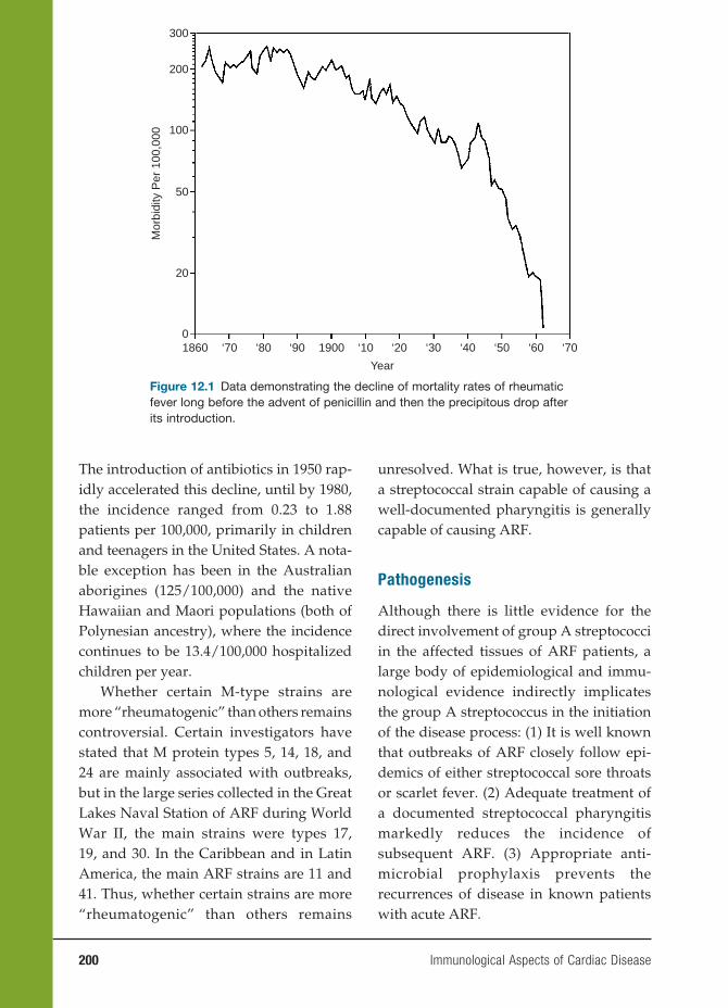
200 Immunological Aspects of Cardiac Disease
The introduction of antibiotics in 1950 rap-idly accelerated this decline, until by 1980, the incidence ranged from 0.23 to 1.88 patients per 100,000, primarily in children and teenagers in the United States. A nota-ble exception has been in the Australian aborigines (125/100,000) and the native Hawaiian and Maori populations (both of Polynesian ancestry), where the incidence continues to be 13.4/100,000 hospitalized children per year.
Whether certain M-type strains are more “rheumatogenic” than others remains controversial. Certain investigators have stated that M protein types 5, 14, 18, and 24 are mainly associated with outbreaks, but in the large series collected in the Great Lakes Naval Station of ARF during World War II, the main strains were types 17, 19, and 30. In the Caribbean and in Latin America, the main ARF strains are 11 and 41. Thus, whether certain strains are more “rheumatogenic” than others remains
unresolved. What is true, however, is that a streptococcal strain capable of causing a well-documented pharyngitis is generally capable of causing ARF.
Pathogenesis
Although there is little evidence for the direct involvement of group A streptococci in the affected tissues of ARF patients, a large body of epidemiological and immu-nological evidence indirectly implicates the group A streptococcus in the initiation of the disease process: (1) It is well known that outbreaks of ARF closely follow epi-demics of either streptococcal sore throats or scarlet fever. (2) Adequate treatment of a documented streptococcal pharyngitis markedly reduces the incidence of subsequent ARF. (3) Appropriate anti-microbial prophylaxis prevents the recurrences of disease in known patients with acute ARF.
300
200
100
50
Mor
bidi
ty P
er 1
00,0
00
20
1860 ‘70 ‘80 ‘90 1900 ‘10 ‘20
Year
‘30 ‘40 ‘50 ‘60 ‘700
Figure 12.1 Data demonstrating the decline of mortality rates of rheumatic fever long before the advent of penicillin and then the precipitous drop after its introduction.
Immunological Aspects of Cardiac Disease 201
A note of caution is necessary concerning documentation (either clinically or micro-biologically) of an antecedent streptococ-cal infection. The frequency of isolation of group A streptococci from the oropharynx is extremely low during the acute stage of rheumatic fever even in populations with limited access to antibiotics. Further, there appears to be an age-related discrepancy in the clinical documentation of an ante-cedent sore throat. In older children and young adults, the recollection of a strepto-coccal sore throat approaches 70 percent; in younger children, this rate approaches only 20 percent. Thus, it is important to have a high index of suspicion of ARF in children or young adults presenting with signs of arthritis or carditis even in the absence of a clinically and microbiologically docu-mented sore throat.
Another intriguing, yet unexplained observation has been the invariable asso-ciation of ARF only with streptococcal pharyngitis. Although there have been many outbreaks of streptococcal skin infec-tions (impetigo), ARF almost never occurs following infection with these strains. Furthermore, in Trinidad, where both impetigo and ARF are concomitant infec-tions, the strains colonizing the skin are dif-ferent from those associated with ARF and did not infl uence the incidence of ARF.
Group A Streptococcus
Figure 12.2 is a schematic cross section of the group A streptococci. Many of the anti-gens present in the group A streptococcus cross-react with antigens present in mam-malian tissues: a concept we call molecular mimicry. Thus, antibodies present in the sera of ARF patients bind to cardiac tissue or cells in the caudate nucleus or to human kidney tissues. The controversial question
is whether any or some of these antibodies play a role in initiating the disease process. This will be discussed in more detail in the etiology section. For a more detailed dis-cussion of these cross-reactions, the reader is referred to several excellent reviews of this subject matter.
Although, numerous attempts to pro-duce antibodies to this capsule have been unsuccessful, more recently reports have emerged demonstrating high antibody titers to hyaluronic acid using techniques designed to detect nonprecipitating anti-bodies in the sera of immunized animals. Similar antibodies have been noted in humans. The data establishing the impor-tance of this capsule in human infections have been almost nonexistent, although Stollerman has commented on the pres-ence of a large mucoid capsule as being one of the more important characteristics of certain “rheumatogenic” strains.
Genetics
That ARF might be the result of a host genetic predisposition has intrigued inves-tigators for over a century. It has been variously suggested that the disease gene is transmitted in an autosomal dominant fashion, in an autosomal recessive fashion with limited penetrance, or that it is pos-sibly related to the genes conferring blood-group secretor status.
Renewed interest in the genetics of ARF occurred with the recognition that gene products of the human MHC (major his-tocompatibility complex) were associated with certain clinical disease states. Using an alloserum from a multiparous donor, an increased frequency of a B-cell alloantigen was reported in several genetically distinct and ethnically diverse populations of ARF individuals and was not MHC related.
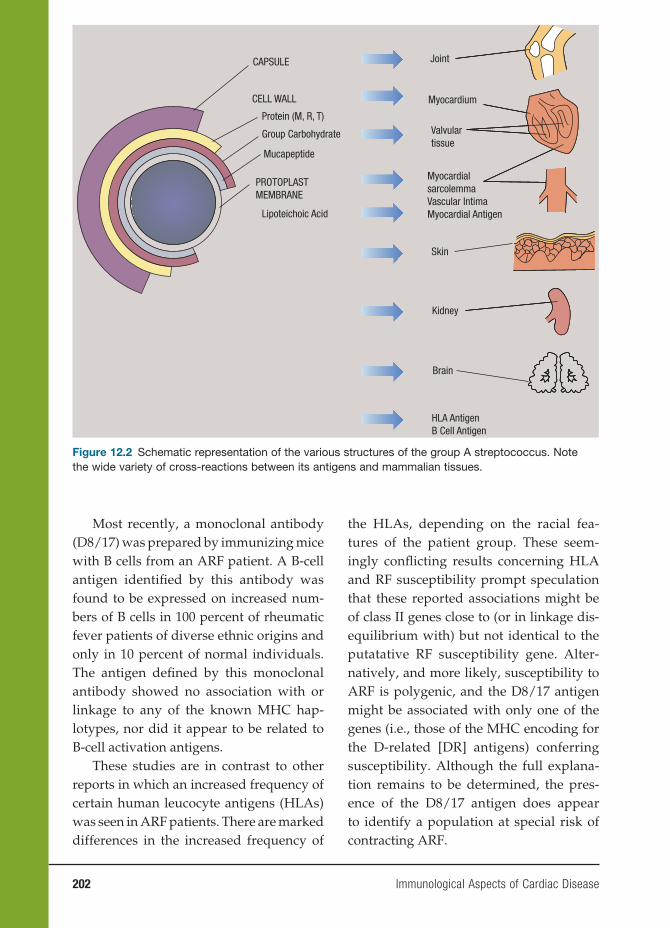
202 Immunological Aspects of Cardiac Disease
Most recently, a monoclonal antibody (D8/17) was prepared by immunizing mice with B cells from an ARF patient. A B-cell antigen identifi ed by this antibody was found to be expressed on increased num-bers of B cells in 100 percent of rheumatic fever patients of diverse ethnic origins and only in 10 percent of normal individuals. The antigen defi ned by this monoclonal antibody showed no association with or linkage to any of the known MHC hap-lotypes, nor did it appear to be related to B-cell activation antigens.
These studies are in contrast to other reports in which an increased frequency of certain human leucocyte antigens (HLAs) was seen in ARF patients. There are marked differences in the increased frequency of
the HLAs, depending on the racial fea-tures of the patient group. These seem-ingly confl icting results concerning HLA and RF susceptibility prompt speculation that these reported associations might be of class II genes close to (or in linkage dis-equilibrium with) but not identical to the putatative RF susceptibility gene. Alter-natively, and more likely, susceptibility to ARF is polygenic, and the D8/17 antigen might be associated with only one of the genes (i.e., those of the MHC encoding for the D-related [DR] antigens) conferring susceptibility. Although the full explana-tion remains to be determined, the pres-ence of the D8/17 antigen does appear to identify a population at special risk of contracting ARF.
CAPSULE
CELL WALL
Protein (M, R, T)
Group Carbohydrate
Mucapeptide
PROTOPLASTMEMBRANE
Lipoteichoic Acid
Joint
Myocardium
Valvulartissue
MyocardialsarcolemmaVascular IntimaMyocardial Antigen
Skin
Kidney
Brain
HLA AntigenB Cell Antigen
Figure 12.2 Schematic representation of the various structures of the group A streptococcus. Note the wide variety of cross-reactions between its antigens and mammalian tissues.
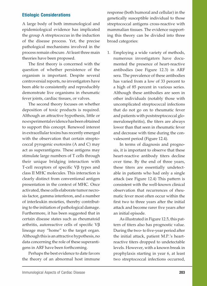
Immunological Aspects of Cardiac Disease 203
Etiologic Considerations
A large body of both immunological and epidemiological evidence has implicated the group A streptococcus in the induction of the disease process. Yet, the precise pathological mechanisms involved in the process remain obscure. At least three main theories have been proposed.
The fi rst theory is concerned with the question of whether persistence of the organism is important. Despite several controversial reports, no investigators have been able to consistently and reproducibly demonstrate live organisms in rheumatic fever joints, cardiac tissues, or valves.
The second theory focuses on whether deposition of toxic products is required. Although an attractive hypothesis, little or no experimental evidence has been obtained to support this concept. Renewed interest in extracellular toxins has recently emerged with the observation that certain strepto-coccal pyrogenic exotoxins (A and C) may act as superantigens. These antigens may stimulate large numbers of T cells through their unique bridging interaction with T-cell receptors of specifi c Vβ types and class II MHC molecules. This interaction is clearly distinct from conventional antigen presentation in the context of MHC. Once activated, these cells elaborate tumor necro-sis factor, gamma interferon, and a number of interleukin moieties, thereby contribut-ing to the initiation of pathological damage. Furthermore, it has been suggested that in certain disease states such as rheumatoid arthritis, autoreactive cells of specifi c Vβ lineage may “home” to the target organ. Although this is an attractive hypothesis, no data concerning the role of these superanti-gens in ARF have been forthcoming.
Perhaps the best evidence to date favors the theory of an abnormal host immune
response (both humoral and cellular) in the genetically susceptible individual to those streptococcal antigens cross-reactive with mammalian tissues. The evidence support-ing this theory can be divided into three broad categories:
1. Employing a wide variety of methods, numerous investigators have docu-mented the presence of heart-reactive antibodies (see Figure 12.3) in ARF sera. The prevalence of these antibodies has varied from a low of 33 percent to a high of 85 percent in various series. Although these antibodies are seen in other individuals (notably those with uncomplicated streptococcal infections that do not go on to rheumatic fever and patients with poststreptococcal glo-merulonephritis), the titers are always lower than that seen in rheumatic fever and decrease with time during the con-valescent period (Figure 12.4).
In terms of diagnosis and progno-sis, it is important to observe that these heart-reactive antibody titers decline over time. By the end of three years, these titers are essentially undetect-able in patients who had only a single attack (see Figure 12.4) This pattern is consistent with the well-known clinical observation that recurrences of rheu-matic fever most often occur within the fi rst two to three years after the initial attack and become rarer fi ve years after an initial episode.
As illustrated in Figure 12.5, this pat-tern of titers also has prognostic value. During the two- to fi ve-year period after the initial attack, patient M.P.’s heart-reactive titers dropped to undetectable levels. However, with a known break in prophylaxis starting in year 6, at least two streptococcal infections occurred,
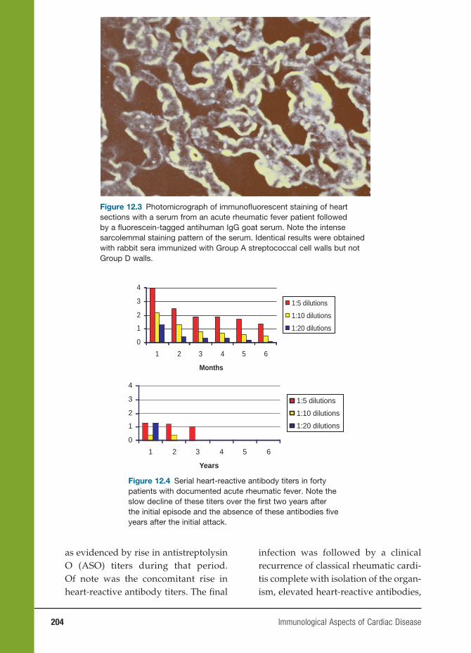
204 Immunological Aspects of Cardiac Disease
0
1
2
3
4
1 2 3 4 5 6
Months
1:5 dilutions
1:10 dilutions
1:20 dilutions
0
1
2
3
4
1 2 3 4 5 6
Years
1:5 dilutions
1:10 dilutions
1:20 dilutions
Figure 12.4 Serial heart-reactive antibody titers in forty patients with documented acute rheumatic fever. Note the slow decline of these titers over the fi rst two years after the initial episode and the absence of these antibodies fi ve years after the initial attack.
as evidenced by rise in antistreptolysin O (ASO) titers during that period. Of note was the concomitant rise in heart-reactive antibody titers. The fi nal
infection was followed by a clinical recurrence of classical rheumatic cardi-tis complete with isolation of the organ-ism, elevated heart-reactive antibodies,
Figure 12.3 Photomicrograph of immunofl uorescent staining of heart sections with a serum from an acute rheumatic fever patient followed by a fl uorescein-tagged antihuman IgG goat serum. Note the intense sarcolemmal staining pattern of the serum. Identical results were obtained with rabbit sera immunized with Group A streptococcal cell walls but not Group D walls.
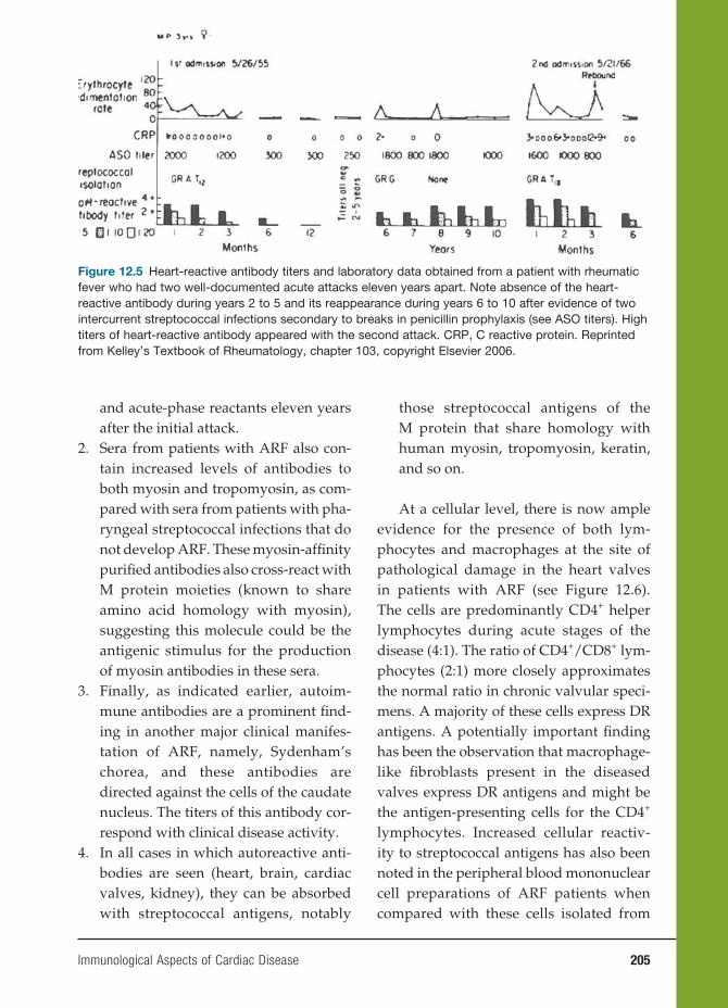
Immunological Aspects of Cardiac Disease 205
and acute-phase reactants eleven years after the initial attack.
2. Sera from patients with ARF also con-tain increased levels of antibodies to both myosin and tropomyosin, as com-pared with sera from patients with pha-ryngeal streptococcal infections that do not develop ARF. These myosin-affi nity purifi ed antibodies also cross-react with M protein moieties (known to share amino acid homology with myosin), suggesting this molecule could be the antigenic stimulus for the production of myosin antibodies in these sera.
3. Finally, as indicated earlier, autoim-mune antibodies are a prominent fi nd-ing in another major clinical manifes-tation of ARF, namely, Sydenham’s chorea, and these antibodies are directed against the cells of the caudate nucleus. The titers of this antibody cor-respond with clinical disease activity.
4. In all cases in which autoreactive anti-bodies are seen (heart, brain, cardiac valves, kidney), they can be absorbed with streptococcal antigens, notably
those streptococcal antigens of the M protein that share homology with human myosin, tropomyosin, keratin, and so on.
At a cellular level, there is now ample evidence for the presence of both lym-phocytes and macrophages at the site of pathological damage in the heart valves in patients with ARF (see Figure 12.6). The cells are predominantly CD4+ helper lymphocytes during acute stages of the disease (4:1). The ratio of CD4+/CD8+ lym-phocytes (2:1) more closely approximates the normal ratio in chronic valvular speci-mens. A majority of these cells express DR antigens. A potentially important fi nding has been the observation that macrophage-like fi broblasts present in the diseased valves express DR antigens and might be the antigen-presenting cells for the CD4+ lymphocytes. Increased cellular reactiv-ity to streptococcal antigens has also been noted in the peripheral blood mononuclear cell preparations of ARF patients when compared with these cells isolated from
Figure 12.5 Heart-reactive antibody titers and laboratory data obtained from a patient with rheumatic fever who had two well-documented acute attacks eleven years apart. Note absence of the heart-reactive antibody during years 2 to 5 and its reappearance during years 6 to 10 after evidence of two intercurrent streptococcal infections secondary to breaks in penicillin prophylaxis (see ASO titers). High titers of heart-reactive antibody appeared with the second attack. CRP, C reactive protein. Reprinted from Kelley’s Textbook of Rheumatology, chapter 103, copyright Elsevier 2006.
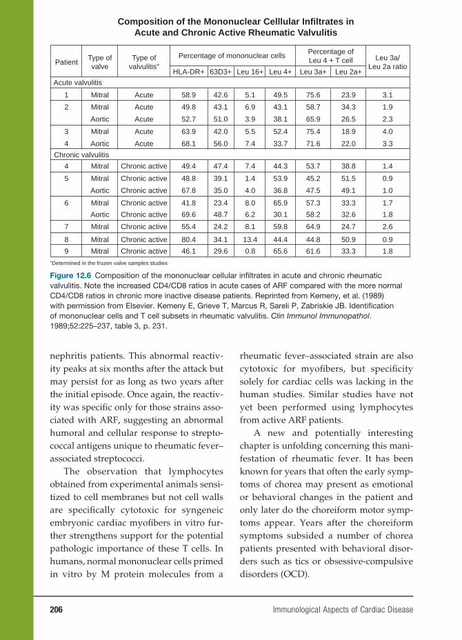
206 Immunological Aspects of Cardiac Disease
nephritis patients. This abnormal reactiv-ity peaks at six months after the attack but may persist for as long as two years after the initial episode. Once again, the reactiv-ity was specifi c only for those strains asso-ciated with ARF, suggesting an abnormal humoral and cellular response to strepto-coccal antigens unique to rheumatic fever–associated streptococci.
The observation that lymphocytes obtained from experimental animals sensi-tized to cell membranes but not cell walls are specifi cally cytotoxic for syngeneic embryonic cardiac myofi bers in vitro fur-ther strengthens support for the potential pathologic importance of these T cells. In humans, normal mononuclear cells primed in vitro by M protein molecules from a
rheumatic fever–associated strain are also cytotoxic for myofi bers, but specifi city solely for cardiac cells was lacking in the human studies. Similar studies have not yet been performed using lymphocytes from active ARF patients.
A new and potentially interesting chapter is unfolding concerning this mani-festation of rheumatic fever. It has been known for years that often the early symp-toms of chorea may present as emotional or behavioral changes in the patient and only later do the choreiform motor symp-toms appear. Years after the choreiform symptoms subsided a number of chorea patients presented with behavioral disor-ders such as tics or obsessive-compulsive disorders (OCD).
Composition of the Mononuclear Celllular Infiltrates inAcute and Chronic Active Rheumatic Valvulitis
PatientType ofvalve
Type ofvalvulitis"
Percentage of mononuclear cells Percentage ofLeu 4 + T cell Leu 3a/
Leu 2a ratioHLA-DR+
Acute valvulitis
Chronic valvulitis
63D3+ Leu 16+ Leu 4+ Leu 3a+ Leu 2a+
1
2
3
4
4
5
6
7
8
9
Acute
Mitral
Mitral
Mitral
Mitral
Mitral
Mitral
Mitral
Mitral
Mitral
Aortic
Aortic
Aortic
Aortic
Acute
Acute
Acute
Acute
Chronic active
Chronic active
Chronic active
Chronic active
Chronic active
Chronic active
Chronic active
Chronic active
58.9
49.8
52.7
63.9
68.1
42.6
43.1
51.0
42.0
56.0
5.1
6.9
3.9
5.5
7.4
49.5
43.1
38.1
52.4
33.7
75.6
58.7
65.9
75.4
71.6
23.9
34.3
26.5
18.9
22.0
3.1
1.9
2.3
4.0
3.3
49.4
48.8
67.8
41.8
69.6
55.4
80.4
46.1
47.4
39.1
35.0
23.4
48.7
24.2
34.1
29.6
7.4 44.3
1.4
4.0
8.0
6.2
8.1
13.4
0.8
53.9
36.8
65.9
30.1
59.8
44.4
65.6
53.7
45.2
47.5
57.3
58.2
64.9
44.8
61.6
38.8
51.5
49.1
33.3
32.6
24.7
50.9
33.3
1.4
0.9
1.0
1.7
1.8
2.6
0.9
1.8
"Determined in the frozen valve samples studies
Figure 12.6 Composition of the mononuclear cellular infi ltrates in acute and chronic rheumatic valvulitis. Note the increased CD4/CD8 ratios in acute cases of ARF compared with the more normal CD4/CD8 ratios in chronic more inactive disease patients. Reprinted from Kemeny, et al. (1989) with permission from Elsevier. Kemeny E, Grieve T, Marcus R, Sareli P, Zabriskie JB. Identifi cation of mononuclear cells and T cell subsets in rheumatic valvulitis. Clin Immunol Immunopathol. 1989;52:225–237, table 3, p. 231.
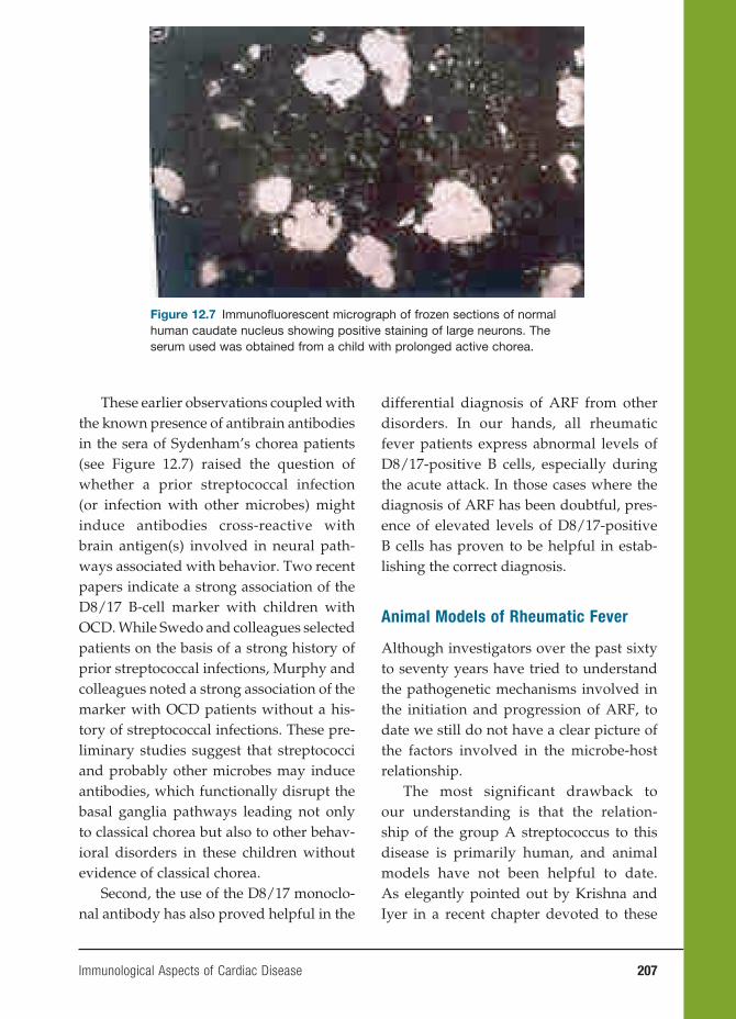
Immunological Aspects of Cardiac Disease 207
These earlier observations coupled with the known presence of antibrain antibodies in the sera of Sydenham’s chorea patients (see Figure 12.7) raised the question of whether a prior streptococcal infection (or infection with other microbes) might induce antibodies cross-reactive with brain antigen(s) involved in neural path-ways associated with behavior. Two recent papers indicate a strong association of the D8/17 B-cell marker with children with OCD. While Swedo and colleagues selected patients on the basis of a strong history of prior streptococcal infections, Murphy and colleagues noted a strong association of the marker with OCD patients without a his-tory of streptococcal infections. These pre-liminary studies suggest that streptococci and probably other microbes may induce antibodies, which functionally disrupt the basal ganglia pathways leading not only to classical chorea but also to other behav-ioral disorders in these children without evidence of classical chorea.
Second, the use of the D8/17 monoclo-nal antibody has also proved helpful in the
differential diagnosis of ARF from other disorders. In our hands, all rheumatic fever patients express abnormal levels of D8/17-positive B cells, especially during the acute attack. In those cases where the diagnosis of ARF has been doubtful, pres-ence of elevated levels of D8/17-positive B cells has proven to be helpful in estab-lishing the correct diagnosis.
Animal Models of Rheumatic Fever
Although investigators over the past sixty to seventy years have tried to understand the pathogenetic mechanisms involved in the initiation and progression of ARF, to date we still do not have a clear picture of the factors involved in the microbe-host relationship.
The most significant drawback to our understanding is that the relation-ship of the group A streptococcus to this disease is primarily human, and animal models have not been helpful to date. As elegantly pointed out by Krishna and Iyer in a recent chapter devoted to these
Figure 12.7 Immunofl uorescent micrograph of frozen sections of normal human caudate nucleus showing positive staining of large neurons. The serum used was obtained from a child with prolonged active chorea.
208 Immunological Aspects of Cardiac Disease
models, the design of the animal mod-els has closely paralleled the prevailing hypothesis at any given time. These have involved (1) whether persistent and sub-clinical infection by the organism was a factor, (2) whether direct injury to the myo-cardium or valves by streptococcal toxins was involved, or (3) whether streptococcal antibodies cross-reactive with human tis-sues can initiate immune-mediated injury. Unfortunately, none of these approaches have yielded any unequivocal evidence of the classic lesions of ARF. Further-more, none have clearly demonstrated the known clinical features of the human dis-ease such as a prior streptococcal pharyn-gitis followed by a latent period followed by clinical and pathological signs of an ARF episode.
However, since the 1970s, observation in ARF patients both in circulatory blood samples and serum as well as studies in tis-sues removed from ARF and RHD patients have strengthened the concept that an abnormal response to streptococcal anti-gens at a cellular and humoral level in the genetically susceptible host results in the clinical and laboratory signs of ARF. Fur-ther studies have revealed the following: (1) heart-reactive antibodies cross-reactive with streptococcal antigens are present in the sera of these patients; (2) the number of CD4+ cells are increased both in the plasma and tissues of ARF patients compared with CD8; (3) the total number of B cells is increased compared with age-matched controls; (4) the infl ammatory cytokines such as TNF-α, IL-2, and IL-1 are elevated in the acute phase and return to normal levels after the episode; and (5) group A streptococci secrete several exotoxins called SPEA, SPEB, and SPEC, which are called superantigens because they bind to the lateral side of TCR and the MHC stimu-
lating a large release of cytokines that could initiate the ARF process. Furthermore they are even capable of deleting certain TCR alleles, possibly inducing loss of self-tol-erance and the induction of self-reactive autoantibodies.
All of these observations coupled with the unique association of group A streptococcal infections in humans ver-sus animals suggest that introduction of human genes in mice or rats to create transgenic animals may be more helpful in understanding the human rheumatic fever process. These animals are now available for study mainly based on the long-term commitment to transgenic mice by Dr. Chella David. These mice contain the gene for human CD4+ cells and genes for several different MHC molecules. These mice are more susceptible to the lethal efforts of the superantigen toxins (similar to humans) when compared with nontransgenic littermates, but to date, no studies have been carried out to determine how these mice might react to oral infection with several different group A streptococcal M types, particularly with respect to myo-cardial and valvular damage. The gene for human B-cell production may also be needed to complete the pathological fi nd-ings seen in human disease.
Whether these human gene introduc-tions are suffi cient to mimic the human disease is not known. But the creation of these and other transgenic models may well be more fruitful since all natural mod-els tested so far have had little success.
Streptococcal Vaccine Candidates
As early as the 1930s, researchers were pursuing the study of streptococcal vac-cinations, with the injection of whole-killed group A streptococci and cell walls,
Immunological Aspects of Cardiac Disease 209
thereof culminating in injections of par-tially purifi ed M protein extracts in the 1970s. However, all interest, especially by the pharmaceutical companies, ceased at that point because the U.S. Food and Drug Administration (FDA) proclaimed that one could work on streptococcal vac-cines as long as no streptococcal compo-nent was used! The FDA was afraid that induction of antibodies cross-reactive with human tissues, especially cardiac tis-sues, could be detrimental to the vaccinee. However, it was soon apparent that many individuals had antibodies cross-reactive with a variety of human tissues and were perfectly normal. In the past decade, the restriction was removed on the condition that toxicity studies in animals did not reveal any deletion effects in the animals when injected with a streptococcal vaccine candidate.
This ushered in the search for an effec-tive, safe, and inexpensive group A strep-tococcal vaccine, and there are now at least four prominent candidates with more in the “pipeline.” This area has recently been reviewed in New Generation Vaccines (chapter 57) and will only briefl y be sum-marized here.
1. Perhaps the most advanced candidate is by Dale and colleagues (1996), which is synthetic peptide sequences of a vari-ety of M protein types taken from the variable region of the M protein and hooked together by linkers. This has induced protective immunity in ani-mals to a number of different M protein types and is safe for use in humans. Clinical trial of its effi cacy to prevent streptococcal infections is currently under way.
2. The use of the C-repeat constant region of the M protein advanced by Fischetti
(1989) produces protection against oral colonization of the throat by group A streptococci.
3. A surface protein called C5 peptidase, which is present on the surface of all group A and group B streptococci, produces antibodies that block both the colonization of group A and group B streptococci in oral colonization studies.
4. The streptococcal group A carbohydrate (CHO) as proposed by Zabriskie and colleagues has been purifi ed and used as an immunogen to protect against streptococcal infections. This vaccine candidate promotes phagocytosis of group A streptococci of several different M types, will protect against infection using passive and active immunization studies, and also protects against oral colonization. Table 12.1 summarizes all the studies on these four candidates and it can be seen that the variable region of the M protein and the CHO molecule offer the most convincing evidence for a protective vaccine against group A streptococcal infections.
Directions for Future Research
Obviously, the most active area for strep-tococcal research in the future will be the development of a good group A strepto-coccal vaccine. Several good candidates are present and more will be studied and evaluated in the future. There are also candidates such as the hyaluronic acid capsule (see Figure 12.2) and many other surface antigens, including the T antigen (similar in many cases but not identical to the M antigen) and others still unknown. It is an exciting area and the production of a safe and inexpensive effi cacious vaccine will be the desired goal, especially in
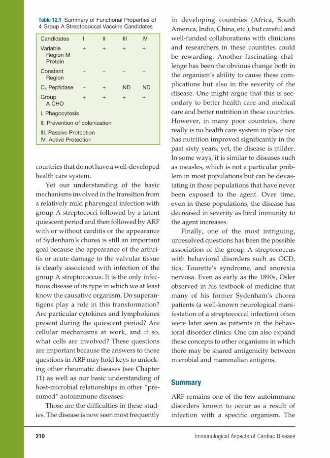
210 Immunological Aspects of Cardiac Disease
countries that do not have a well-developed health care system.
Yet our understanding of the basic mechanisms involved in the transition from a relatively mild pharyngeal infection with group A streptococci followed by a latent quiescent period and then followed by ARF with or without carditis or the appearance of Sydenham’s chorea is still an important goal because the appearance of the arthri-tis or acute damage to the valvular tissue is clearly associated with infection of the group A streptococcus. It is the only infec-tious disease of its type in which we at least know the causative organism. Do superan-tigens play a role in this transformation? Are particular cytokines and lymphokines present during the quiescent period? Are cellular mechanisms at work, and if so, what cells are involved? These questions are important because the answers to those questions in ARF may hold keys to unlock-ing other rheumatic diseases (see Chapter 11) as well as our basic understanding of host-microbial relationships in other “pre-sumed” autoimmune diseases.
Those are the diffi culties in these stud-ies. The disease is now seen most frequently
in developing countries (Africa, South America, India, China, etc.), but careful and well-funded collaborations with clinicians and researchers in these countries could be rewarding. Another fascinating chal-lenge has been the obvious change both in the organism’s ability to cause these com-plications but also in the severity of the disease. One might argue that this is sec-ondary to better health care and medical care and better nutrition in these countries. However, in many poor countries, there really is no health care system in place nor has nutrition improved signifi cantly in the past sixty years; yet, the disease is milder. In some ways, it is similar to diseases such as measles, which is not a particular prob-lem in most populations but can be devas-tating in those populations that have never been exposed to the agent. Over time, even in these populations, the disease has decreased in severity as herd immunity to the agent increases.
Finally, one of the most intriguing, unresolved questions has been the possible association of the group A streptococcus with behavioral disorders such as OCD, tics, Tourette’s syndrome, and anorexia nervosa. Even as early as the 1890s, Osler observed in his textbook of medicine that many of his former Sydenham’s chorea patients (a well-known neurological mani-festation of a streptococcal infection) often were later seen as patients in the behav-ioral disorder clinics. One can also expand these concepts to other organisms in which there may be shared antigenicity between microbial and mammalian antigens.
Summary
ARF remains one of the few autoimmune disorders known to occur as a result of infection with a specifi c organism. The
Table 12.1 Summary of Functional Properties of 4 Group A Streptococcal Vaccine Candidates
Candidates I II III IV
Variable Region M Protein
+ + + +
Constant Region
− − − −
C5 Peptidase − + ND ND
Group A CHO
+ + + +
I. Phagocytosis
II. Prevention of colonization
III. Passive ProtectionIV. Active Protection
Immunological Aspects of Cardiac Disease 211
confi rmed observation of an increased frequency of a B-cell alloantigen in several populations of rheumatics suggests that it might be possible to identify individuals susceptible to ARF at birth. If so, then from a public health standpoint, these individu-als would be prime candidates for immu-nization with any streptococcal vaccine that might be developed in the future. In addition, careful monitoring of streptococ-cal disease in the susceptible population could lead to early and effective antibiotic strategies, resulting in disease prevention. Finally, in individuals previously infected, who later present with subtle or nonspecifi c manifestations of the disease, the presence or absence of the marker could be useful in arriving at a diagnosis.
The continued study of ARF as a para-digm for microbial-host interactions also has important implications for the study of autoimmune diseases in general and rheumatic diseases in particular. Further insights into this intriguing host–parasite relationship may shed additional light into those diseases in which the infection is presumed but has not yet been identifi ed.
CHAGAS’ DISEASE
Chagas’ disease is named after the Brazilian physician Carlos Chagas who fi rst discovered the parasite responsible for the signs and symptoms of the disease. The causative agent is the hemofl agellate protozoan Trypanosoma cruzi. This proto-zoan parasite is transmitted to the blood-sucking reduviid bug vector when the reduviids take a blood meal from animals or humans who are infected with circulat-ing T. cruzi. When the infected bug takes a subsequent blood meal, it usually def-ecates on the skin of the next victim and
deposits T. cruzi as well. The individual then becomes infected either by the feces deposited at the site of the puncture or inadvertent contamination of a mucous membrane of the human secondary to hand contamination with organisms. The distribution of the arthropod vectors is limited to the Western Hemisphere and is widely distributed in the southern United States, Mexico, and Central America. The reduviid vectors are common in most South American countries, especially Brazil, Argentina, Bolivia, and Chile. Chagas’ is a disease primarily of the poor largely because the reduviid bugs favor living in adobe mud or enter poor housing through cracks or holes in the walls and thatched roofs.
Mode of Transmission and
Epidemiology
An estimated 20 million people are cur-rently infected with T. cruzi, and approxi-mately 100 million people are at risk of infection. The endemic area of Chagas’ disease is huge, stretching from Mexico to southern Argentina, but is limited to the Western Hemisphere. The incidence, prev-alence, and severity of Chagas’ disease and T. cruzi infection seem to be higher in South America than Central America; this may be due to differences in vector behavior or strain variation of T. cruzi or differences in disease recognition and reporting.
T. cruzi is transmitted by blood-feed-ing reduviid insect vectors, vertically from mother to child, or by blood transfusions. Vector transmission is responsible for most of T. cruzi infections, and most of this transmission is secondary to the deposit of infected feces and urine by the reduviid bug at the time of the blood meal. As an alternative, humans have been reported to

Immunological Aspects of Cardiac Disease 213
granulocyte-macrophage colony-stimulat-ing factor.
Serum taken from an uninfected human does not lyse trypomastigotes, but serum taken from infected patients does lyse trypomastigotes. Thus, the comple-ment system requires the participation of the acquired immune response to T. cruzi to become operational. Parasite lysis by immune serum occurs primarily via the alternative complement pathway. This means that the complement cascade is ini-tiated by direct association of complement components (C3) with the parasite surface, rather than by fi xation of complement by antibody Fc receptors. In fact, Fab and F(ab)2
′ prepared from chagasic patients’ sera (which are incapable of fi xing com-plement) were nearly as effi cient as intact immunoglobulin G (IgG) in complement lysis assays with trypomastigotes. This implied that the effect of immune anti-bodies on complement lysis of trypomas-tigotes was a function of the specifi city of the antigen-binding sites, rather than complement fi xation by bound antibod-ies. In other words, there was a surface component on the parasites that, when neutralized by immune sera, rendered the parasites susceptible to complement lysis.
Antibodies directed to a 160-kDa pro-tein correlated with the capacity of the serum for complement-mediated lysis of trypomastigotes. Subsequent work led to the [purifi cation and characterization of the specifi c parasite product, gp160. This glyco-protein bound the complement component C3b and inhibited C3 convertase formation, thus inhibiting activation of the alterna-tive complement pathway. It is membrane bound and shares genetic and functional similarities to the human complement reg-ulatory protein, decay accelerating factor. Further work indicated that this protein
also bound human C4b, a component of the classical pathway C3 convertase, and therefore may restrict classic complement activation. The T. cruzi complement regu-latory protein is stage specifi c in that it is only expressed by mammalian forms of the parasite. A recent study reported that stable transfection of T. cruzi epimastigotes (which are susceptible to complement lysis by normal serum) with the complementary DNA for the T. cruzi complement regula-tory protein conferred complement resis-tance.
TISSUE TROPISMWhy does T. cruzi preferentially para-
sitize muscle and nerve tissue? Arguing teleologically, the host immune response needs to control the parasite infection but not at the cost of destroying vital organs (i.e., heart and peripheral nerves). The down-regulatory immune responses may be more exuberant in these critical tissues where even minor injury can be fatal and regenerative capacity is limited (e.g., the cardiac conduction system). T. cruzi may have evolved to preferentially infect these tissues as relatively immune-privileged sites. Several other parasites that preferen-tially infect muscle tissue or nervous tissue include toxoplasmosis, trichinella, Taenia species (beef and pork tapeworms), and sar-cocystis. Each of these species depends on a carnivorous-defi nitive host ingesting the meat of an intermediate host to complete its life cycle. There is no evidence, how-ever, that consumption of T. cruzi–infected tissues plays a role in this parasite’s life cycle. Thus, the predilection for growth in muscle and nervous tissue is more likely to be related to a strategy for long-term parasite survival in the host, rather than a strategy related to direct transmission to secondary hosts.
214 Immunological Aspects of Cardiac Disease
IFN-γ induction of macrophage try-panocidal activity is associated with the production of hydrogen peroxide. How-ever, treatment of activated macrophages with catalase, superoxide dismutase, or sodium benzoate to scavenge respira-tory burst metabolites failed to inhibit trypanocidal activity in vitro, suggesting an oxygen-independent mechanism of T. cruzi destruction. Subsequent studies have revealed the importance of nitric oxide (NO) production in the killing mechanism in murine systems. Macrophages primed with IFN-γ produce NO, and the addition of an inhibitor to NO synthase blocks the trypanocidal activity. Levels of extracellu-lar l-arginine (the substrate for NO produc-tion) modulate the trypanocidal activity of macrophages. NO may be directly toxic to T. cruzi; however, it reacts with superoxide (O2
−) to yield peroxynitrite (ONDO−), which is highly toxic to T. cruzi. In vivo, inducible nitric oxide synthase (iNOS) is induced at the protein and messenger RNA levels, and NO is released during acute infection in coincidence with secretion of IFN-γ and TNF. The administration of inhibitors of NO production leads to greater suscepti-bility of T. cruzi–infected mice. Mice carry-ing disruption of the iNOS genes are highly susceptible to T. cruzi infection. The IFN-γ–induced, NO-dependent mechanism of macrophage killing of T. cruzi can be inhib-ited by the addition of interleukin-10 or transforming growth factor-β in vitro.
NK cells participate in the innate immune response to T. cruzi infection, and NK cells secrete IFN-γ after incubation with T. cruzi in vitro. This was shown by culturing splenocytes from athymic nude mice with T. cruzi and detecting the secre-tion of IFN-γ, then demonstrating that the pretreatment of the splenocytes with anti-NK1.1 monoclonal antibody blocked
the IFN-γ production. Since IFN-γ acti-vates macrophages to kill T. cruzi, the role of NK cells in T. cruzi infection would be expected to be protective. In fact, a rela-tively T. cruzi–resistant strain of mouse was rendered highly susceptible to infec-tion by pretreatment with anti-NK1.1 anti-bodies. Thus, NK cells appear to be an early source of IFN-γ that helps control parasite replication before the acquired immune response becomes predominant. NK cells may also be involved in the immune response later in infection, since they are present in infl ammatory lesions of muscle in experimental mice 270 days after infection.
ANIMAL MODELSTrypanosoma cruzi multiplies and dis-
seminates throughout the host before a specifi c antiparasitic immune response is mounted. In mice, parasites fi rst become apparent in blood approximately five to seven days after infection and rise in numbers until three or four weeks into infection when the mice either die or the infection is controlled by the immune response (Figure 12.1). RAG knockout mice, which are defi cient in both B- and T-cell function, have similar levels of par-asitemia compared with wild-type mice until day 13 of infection, at which point the parasitemia level becomes higher in the RAG knockouts. This indicates that the acquired immune response has little effect until about two weeks into the infec-tion. Although anti–T. cruzi antibodies are detectable at about day 7 in murine mod-els, protective antibodies are not present until several weeks later. This was shown in experiments in which antibodies taken from acutely infected mice were not protec-tive against T. cruzi infection in immune-naïve mice. However, experimental mice
Immunological Aspects of Cardiac Disease 215
or rats that received sera from animals that survived acute infection experience a sig-nifi cant decrease in parasitemia level and mortality following a challenge with viru-lent parasites.
The importance of IgG in the humoral immune response was fi rst demonstrated by Castelo Branco. The protective compo-nent of serum from chronically infected mice could be removed by staphylococcal protein A (which absorbs IgG). Further-more, the purifi ed IgG component from whole serum was capable of conferring protection. Both IgG subclasses 1 and 2 are capable of clearing T. cruzi. Antibodies mediate protection from T. cruzi by opsonization, complement activation, and antibody-dependent cellular cytotoxicity.
T cells have a protective role in the acquired immune response to T. cruzi. Experiments in mice showed that T-cell activation correlated with resistance to infection. Passive transfer of T cells from mice immunized against T. cruzi conferred resistance in mice challenged with T. cruzi. Defi cient T-cell function is associated with increased sensitivity to infection. This has been shown in nude mice; thymec-tomized mice; mice treated with cyclo-sporine A, anti-CD4, and anti-CD8; and mice genomically deleted of CD4, CD8, β2-microglobulin, TAP-1, or MHC mol-ecules. The increased susceptibility associ-ated with defi cient T-cell function is most apparent during acute infection. Mice that are depleted of T cells (CD4+ or CD8+) after they have survived the acute infection do not have altered parasitemia or longevity. The declining role of T cells and the impor-tance of humoral immunity in controlling infection after the acute stage are schemati-cally illustrated (Figure 12.1).
The presence of parasite-specifi c T cells has been demonstrated for both CD4+ and
CD8+ T cells. Nickell and colleagues iso-lated CD4+ T cells from spleens of infected mice that proliferate to T. cruzi antigen in a HLA-restricted fashion. The CD4+ T-cell line recognized an undefi ned trypomasti-gote antigen(s), did not cross-react with Leishmania spp. or Toxoplasma gondii, and was able to passively protect syngeneic recipients from lethal T. cruzi challenge infection. Kahn and Wleklinski isolated and cloned CD4+ T cells from T. cruzi–infected mice that proliferate and secrete cytokines in response to the surface protein, SA-85, a member of the sialidase superfamily. The MHC class II epitope in this protein was mapped to a 20-amino acid sequence. Nickell and associates were able to isolate CD8+ T cells from infected mice that lysed parasite-infected target cells in an MHC-restricted manner. The parasite antigens involved in the stimulation of CD8+ T cells were not characterized. However, Wizel and colleagues detected class I–restricted CD8+ T cells from spleens of infected mice that lyse target cells, presenting epitopes from the trans-sialidase family of proteins. These cytotoxic lymphocytes passively transferred protection against challenge infection.
The inflammatory infiltrates in T. cruzi–infected experimental animals have been analyzed for cellular surface mark-ers. Mirkin and colleagues studied tis-sues from C3H/HeN mice during acute, early chronic, and late chronic infection. The infl ammatory infi ltrates consisted mainly of lymphocytes (60–90 percent) and macrophages (10–40 percent). The lymphocytes primarily carried the T-cell marker, Thy1.2. Both CD4+ and CD8+ T cells were present in the infi ltrates of skeletal muscle, sciatic nerve, and spinal cord. The general trend was for a slight predominance of CD8+ cells over CD4+,
216 Immunological Aspects of Cardiac Disease
particularly during the early chronic and late chronic stages. Sun and Tarleton also found that Thy1.2+ cells were the major lymphocyte population in tissues (cardiac and skeletal muscle) during acute infec-tion. CD8+ T cells (47–59 percent) domi-nated over CD4+ T cells (9–19 percent). B cells and macrophages each repre-sented less than 1 percent of the cells in the infl ammatory infi ltrates. Tissues from humans with chronic chagasic cardiomy-opathy were similarly analyzed. As with mice, T cells were the primary cell type in the infl ammatory infi ltrates, with a greater proportion of CD8+ cells than CD4+. Many of the CD8+ T cells expressed granzyme A. The extent that the tissue lymphocytes are directed toward parasites and para-site antigen as opposed to self-targets (i.e., autoimmunity) has not been fully clari-fi ed (see “Pathogenesis and Modulation of Immune Function” section).
T cells in T. cruzi infection perform a variety of antiparasitic functions. They provide helper T-cell function by stimu-lating B cells to produce parasite-specifi c antibody. Nickell and colleagues showed that a T-cell line derived from the spleens of T. cruzi–infected mice was able to induce normal spleen cells to produce parasitic-directed antibodies when stimu-lated in vitro with T. cruzi antigen. The activation of helper T-cell function is sup-ported by the predominance of IgG2a and IgG2b antibodies in T. cruzi infection, which are typical of CD4+ T-cell-depen-dent responses. T cells produce cytokines in T. cruzi infection, which mediate impor-tant antiparasitic functions. CD8+ T cells lyse T. cruzi–infected host cells, which pre-sumably interrupts the parasite life cycle, thus limiting its replication. Interestingly, mice genetically defi cient for genes con-trolling perforin or granzyme B–mediated
cytolytic pathways had parasitemia and mortality rates similar to wild-type mice, suggesting that cytolytic function, in fact, may not be the protective effector mecha-nism of CD8+ T cells. Wizel and cowork-ers showed that T. cruzi–specifi c CD8+ T cells produce IFN-γ and TNF-α upon stimulation, thus raising the possibility that these cells mediate their effects by cytokine release.
Pathogenesis and Modulation of
Immune Function
PATHOGENESISThe pathogenesis of acute Chagas’ dis-
ease is not in dispute. On standard histo-pathological study, infl ammatory lesions are found to co-localize with abundant parasites, demonstrating that infl amma-tory damage is directed to the parasite. The pathophysiology of chronic Chagas’ disease is still in question, but the compet-ing (nonexclusive) views are that disease results from an autoimmune response directed at the affected organ systems or from damage resulting from infl ammation related to the persistence of the parasite. Supporting the autoimmune hypothesis are observations that live T. cruzi para-sites have been diffi cult to demonstrate in involved organs by conventional histologi-cal study and that autoimmune T cells and antibodies develop during infection with T. cruzi. Furthermore, autoimmune anti-bodies and autoimmune T cells have been associated with chronic Chagas’ disease or lesions. Some T. cruzi antigens molecularly mimic affected host tissues. It is hypothe-sized that the chronic infection leads to loss of tolerance and, combined with antigenic mimicry, results in specifi c autoimmune attack of cardiac, gut, and peripheral ner-vous tissues.
Immunological Aspects of Cardiac Disease 217
The other hypothesis is that progres-sive infl ammation directed at parasites that reside in target organs cause the pathologic damage of chronic infection. Studies that support this hypothesis have found that parasite antigens and DNA can be detected in many chronic infl ammatory infi ltrate and that parasitologic treatment of chronically infected animals or humans tends to lead to improvement and resolu-tion of disease. The evidence for the com-peting theories of chronic Chagas’ disease pathogenesis is discussed in detail in the next section. Since the autoimmune and parasite-directed pathogenesis theories are not mutually exclusive and since evi-dence exists to support each of them, it seems likely that both mechanisms could be pathogeneic in chronic Chagas’ disease.
AUTOIMMUNITY AND PATHOGENESIS OF CHAGAS’ DISEASE
The theory of autoimmunity leading to damage in chronic Chagas’ disease is sup-ported by a number of observations. First, few parasites can be demonstrated in the infl ammatory lesions by conventional his-tological study. This observation suggests the disease may not be driven solely by reac-tion to the parasite. Second, only 10 percent to 30 percent of chronically infected people develop Chagas’ disease, although most can be shown to have chronic parasitemia. This suggests that, in addition to chronic T. cruzi infection, some other factor(s) determines which individual develops dis-ease. Susceptibility to autoimmune disease could be the host factor that determines who develops disease. Third, chronic Cha-gas’ disease is very organ specifi c, gener-ally limited to the heart, nervous tissues, or innervation of the gut. Since the parasite can reside in almost any cell type, the organ specifi city of Chagas’ disease suggests that
additional specifi city of Chagas’ disease is imposed during infection, perhaps by the particular autoimmune response that is generated by chronic T. cruzi infection. Fourth, the long lag time from infection to disease could be necessary to generate dis-ease by autoimmunity. Fifth, the presence of autoimmune T cells and antibodies in infected individuals, especially when they are associated with disease, is evidence that autoimmunity may play a role in pathogenesis. It should also be noted that each of these properties of chronic Chagas’ disease could be due to parasite-directed pathogenesis, as discussed below.
Autoimmune antibodies are easily demonstrated in T. cruzi-infected indi-viduals. Antibodies to myocardium and nervous tissues are found in high levels in persons and mice infected with T. cruzi compared with those that are not. Since the myocardium and nervous tissues are key organs that are damaged in chronic Cha-gas’ disease, fi nding antibodies to these tissues could mean that these antibodies cause autoimmune damage to these tis-sues. As an alternative, antigens from myo-cardium and nervous tissues are probably exposed by the damage of infection of these organs, and antibodies may be generated to these tissues without being pathogenic.
Many of the autoantibodies found in the sera of T. cruzi–infected persons and mice are so-called natural autoantibodies, which can be found in low levels in the serum of normal humans and mice. The “natural autoantibodies” are directed against pro-teins that are highly conserved in evolu-tion and are not necessarily an indication of autoimmune disease. The high levels of natural autoantibodies after T. cruzi infec-tion may be the result of the polyclonal lym-phocytic proliferative response that occurs during acute infection. Levels of natural
218 Immunological Aspects of Cardiac Disease
autoantibodies do not correlate with dis-eases in individuals who are chronically infected with T. cruzi.
MOLECULAR MIMICRY IN CHAGAS’ DISEASEMolecular mimicry of host antigens by
parasite antigens has been found to gener-ate autoantibodies that are found in chronic T. cruzi infection (See table 12.2). Autoim-mune pathogenesis by molecular mimicry requires breakdown in tolerance of the immune system to self-antigens. A break-down in tolerance could occur in response to chronic T. cruzi infection or because of the polyclonal lymphocyte proliferative response that occurs early after infection. Although many molecular mimicry epit-opes have been described, only a few have been shown to correlate with disease in chronic T. cruzi infection.
For example, high levels of antibod-ies to rodent endocardium, blood vessels, and interstitium (EVI) have been found in the sera of T. cruzi–infected individu-als and the mammalian host and were absorbed by epimastigote antigens, thus demonstrating cross-reactivity. However, these EVI antigens shown to be directed against α-galactose epitopes were spe-cifi c for murine laminin and not human laminin. Furthermore, anti-EVI antibodies (α-galactose/laminin) or other autoanti-bodies have not been shown to transfer disease to noninfected animals.
Perhaps more interesting (in terms of the organs infected) has been a protein found on the surface of trypomastigotes in asso-ciation with the fl agellum, Fl-160. This pro-tein molecularly mimics myenteric plexus and peripheral neurons. These antibodies are found in 30 percent of individuals with chronic Chagas’ disease; yet, they do not appear to correlate with disease activity in
these patients. In addition, T-cell responses to this protein have not been seen in indi-viduals with Chagas’ disease, and passive transfer of these immune T-cells was not consistently effective.
Another peptide of interest has been the ribosomal Po protein (R13 peptide), which has been shown to cross-react with a functional protein on human B1-adenergic receptors. Antibodies to this receptor via immunization with the R13 peptide have led to electrocardiographic (ECG) changes in mice that are similar to those seen in chronic Chagas’ carditis but no changes in digestive symptoms have been noted.
Antibodies to an interesting T. cruzi protein called B13 is seen in the sera of all patients with chronic Chagas’ cardio-myopathy and this antibody cross-reacts with human cardiac myosin but in only 17 percent of asymptomatic individu-als. Furthermore, T cells directed to both B13 and myosin have been detected in biopsy specimens from persons with chronic Chagas’ cardiomyopathy. These results suggest that antibody and T cells directed to cardiac myosin may be patho-genic in chronic Chagas’ cardiomyopathy. However, in animal models transfer of anti-body and T cells directed to myosin have not been reported to lead to pathogenic changes. Perhaps the mouse model may not be correct and as in rheumatic fever the introduction of myosin in the Lewis rat may be a better animal model.
PARASITE-DIRECTED PATHOGENESISSince the studies reporting that autoim-
munity may be involved in the infl amma-tory infi ltrates of chronic Chagas’ disease are not conclusive, the alternative theory is that parasites residing in chronically infected host tissue may cause chronic
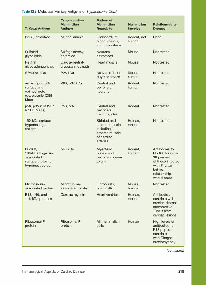
Immunological Aspects of Cardiac Disease 219
Table 12.2 Molecular Mimicry Antigens of Trypanosoma Cruzi
T. Cruzi Antigen
Cross-reactive Mammalian Antigen
Pattern of Mammalian Reactivity
Mammalian Species
Relationship to Disease
(α1-3)-galactose Murine laminin Endocardium, blood vessels, and interstitium
Rodent, not human
None
Sulfated glycolipids
Sulfagalactosyl-ceramide
Neurons, astrocytes
Mouse Not tested
Neutral glycosphingolipids
Cardia-neutral-glycosphingolipids
Heart muscle Mouse Not tested
GP50/55 kDa P28 kDa Activated T and B lymphocytes
Mouse, human
Not tested
Amastigote cell-surface and epimastigote cytoplasmic (CE5 Mab)
P60, p32 kDa Central and peripheral neurons
Rodent, human
Not tested
p58, p35 kDa (5H7 & 3H3 Mabs)
P58, p37 Central and peripheral neurons, glia
Rodent Not tested
150-kDa surface trypomastigote antigen
Striated and smooth muscle including smooth muscle of cardiac arteries
Human, mouse
Not tested
FL-160, 160-kDa fl agellar-associated surface protein of trypomastigotes
p48 kDa Myenteric plexus and peripheral nerve axons
Rodent, human
Antibodies to FL-160 found in 30 percent of those infected with T. cruzi but no relationship with disease
Microtubule-associated protein
Microtubule-associated protein
Fibroblasts, brain cells
Mouse, bovine
Not tested
B13, 140, and 116-kDa proteins
Cardiac myosin Heart ventricle Human, mouse
Antibodies correlate with cardiac disease, autoreactive T cells from cardiac lesions
Ribosomal P protein
Ribosomal P protein
All mammalian cells
Human High levels of antibodies to R13 peptide correlate with Chagas cardiomyophy
(continued)
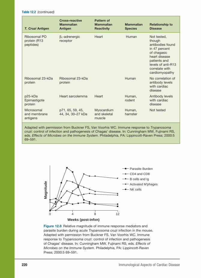
220 Immunological Aspects of Cardiac Disease
Table 12.2 (continued)
T. Cruzi Antigen
Cross-reactive Mammalian Antigen
Pattern of Mammalian Reactivity
Mammalian Species
Relationship to Disease
Ribosomal PO protein (R13 peptides)
β1-adrenergic receptor
Heart Human Not tested, though antibodies found in 47 percent of chagasic heart disease patients and levels of anti-R13 correlate with cardiomyopathy
Ribosomal 23-kDa protein
Ribosomal 23-kDa protein
Human No correlation of antibody levels with cardiac disease
p25-kDa Epimastigote protein
Heart sarcolemma Heart Human, rodent
Antibody levels with cardiac disease
Microsomal and membrane antigens
p71, 65, 59, 45, 44, 34, 30–27 kDa
Myocardium and skeletal muscle
Human, hamster
Not tested
Adapted with permission from Buckner FS, Van Voorhis WC. Immune response to Trypanosoma cruzi: control of infection and pathogenesis of Chagas’ disease. In: Cunningham MW, Fujinami RS, eds. Effects of Microbes on the Immune System. Philadelphia, PA: Lippincott-Raven Press; 2000:5 69–591.
0 4 8 12
Parasite Burden
CD4 and CD8
B cells and Ig
Activated M'phages
NK cells
Weeks (post-infxn)
Mag
nit
ud
e
Figure 12.8 Relative magnitude of immune response mediators and parasite burden during acute Trypanosoma cruzi infection in the mouse. Adapted with permission from Buckner FS, Van Voorhis WC. Immune response to Trypanosoma cruzi: control of infection and pathogenesis of Chagas’ disease. In: Cunningham MW, Fujinami RS, eds. Effects of Microbes on the Immune System. Philadelphia, PA: Lippincott-Raven Press; 2000:5 69–591.
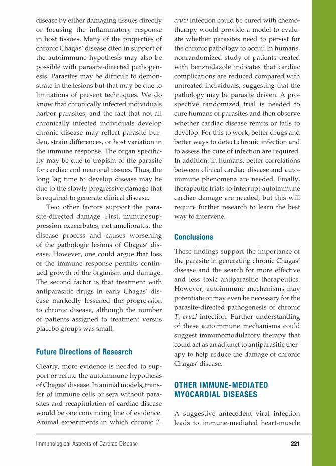
Immunological Aspects of Cardiac Disease 221
disease by either damaging tissues directly or focusing the infl ammatory response in host tissues. Many of the properties of chronic Chagas’ disease cited in support of the autoimmune hypothesis may also be possible with parasite-directed pathogen-esis. Parasites may be diffi cult to demon-strate in the lesions but that may be due to limitations of present techniques. We do know that chronically infected individuals harbor parasites, and the fact that not all chronically infected individuals develop chronic disease may refl ect parasite bur-den, strain differences, or host variation in the immune response. The organ specifi c-ity may be due to tropism of the parasite for cardiac and neuronal tissues. Thus, the long lag time to develop disease may be due to the slowly progressive damage that is required to generate clinical disease.
Two other factors support the para-site-directed damage. First, immunosup-pression exacerbates, not ameliorates, the disease process and causes worsening of the pathologic lesions of Chagas’ dis-ease. However, one could argue that loss of the immune response permits contin-ued growth of the organism and damage. The second factor is that treatment with antiparasitic drugs in early Chagas’ dis-ease markedly lessened the progression to chronic disease, although the number of patients assigned to treatment versus placebo groups was small.
Future Directions of Research
Clearly, more evidence is needed to sup-port or refute the autoimmune hypoth esis of Chagas’ disease. In animal models, trans-fer of immune cells or sera without para-sites and recapitulation of cardiac disease would be one convincing line of evidence. Animal experiments in which chronic T.
cruzi infection could be cured with chemo-therapy would provide a model to evalu-ate whether parasites need to persist for the chronic pathology to occur. In humans, nonrandomized study of patients treated with benznidazole indicates that cardiac complications are reduced compared with untreated individuals, suggesting that the pathology may be parasite driven. A pro-spective randomized trial is needed to cure humans of parasites and then observe whether cardiac disease remits or fails to develop. For this to work, better drugs and better ways to detect chronic infection and to assess the cure of infection are required. In addition, in humans, better correlations between clinical cardiac disease and auto-immune phenomena are needed. Finally, therapeutic trials to interrupt autoimmune cardiac damage are needed, but this will require further research to learn the best way to intervene.
Conclusions
These fi ndings support the importance of the parasite in generating chronic Chagas’ disease and the search for more effective and less toxic antiparasitic therapeutics. However, autoimmune mechanisms may potentiate or may even be necessary for the parasite-directed pathogenesis of chronic T. cruzi infection. Further understanding of these autoimmune mechanisms could suggest immunomodulatory therapy that could act as an adjunct to antiparasitic ther-apy to help reduce the damage of chronic Chagas’ disease.
OTHER IMMUNE-MEDIATED
MYOCARDIAL DISEASES
A suggestive antecedent viral infection leads to immune-mediated heart-muscle
222 Immunological Aspects of Cardiac Disease
disease. These diseases may be conve-niently divided into two main categories: myocarditis and dilated cardiomyopa-thy (DCM). We will not discuss drug- or allergy-mediated myocarditis or myo-cardial damage related to ischemic heart disease.
Myocarditis
The major features of myocarditis are dis-turbances in heart rhythms, congestive heart failure, or cardiogenic shock. ECG changes include nonspecifi c ST-Twave changes, while two-dimensional echocar-diograms reveal (1) normal ventricular size but decreased contractility early in the dis-ease and (2) later heart enlargement with thinning of the muscle in chronic cases. In addition, both ventricles may be affected in chronic cases. None of the clinical fea-tures described are diagnostic for myocar-ditis, and until the use of the endomyocar-dial biotome, the diagnosis could only be established with certainty by postmortem examination.
The incidence of the disease varies from 1 to 10/100,000. The reasons for this wide discrepancy may be related to exposure to the different types and strains of cardiotro-pic viruses as well as genetic differences in host populations. Many patients recover spontaneously inter supportive treatment but the fi ve-year survival of biopsy-proven giant cell myocarditis is only 56 percent, and in pediatric patients, the mortality rates may be even higher.
Dilated Cardiomyopathy
DCM is a chronic form of heart disease characterized by right ventricular dila-tion and impaired contraction. The clini-cal spectrum varies from asymptomatic
cardiomopathy to severe congestive heart failure. Patients may also display signs or symptoms of arrhythmias, systemic pulmonary vein congestion, triple gallup rhythms, and mitral or tricuspid regurgita-tion. Once the diagnosis is established, the outcome is poor, with a fi ve-year mortal-ity of 46 percent. In 1985 in Minnesota, the prevalence was 36.5/100,000, and African Americans and males were associated with increased risk. In Sweden, the incidence was 10/100,000.
It is uncertain how many cases rep-resent progression from myocarditis to DCM since in general the endomyocar-dial biopsy is not routinely performed in DCM patients. Some reports have indi-cated that there is a relationship between the two, but biopsies in both diseases are needed to prove or disprove this concept.
Etiologic Considerations
In both myocarditis and DCM, there appears to be an association with prior Coxsackie virus infections. In children and infants with DCM, signifi cantly increased titers of neutralizing antibody to the virus has been found. In addition, Coxsackie virus B–specifi c nucleic acid sequences have been found in the heart tissues of a small number of these patients. Furthermore, using poly-merase chain reaction (PCR) techniques, many children with suspected myocarditis were PCR positive for several viral agents in endomyocardial biopsies (twenty-six of thirty-eight samples from thirty-four patients), and the viral sequences were more often adenoviral than enteroviral.
Autoimmune Considerations
As in other immune-mediated diseases of the heart, many investigators have now
Immunological Aspects of Cardiac Disease 223
explored the role of autoantibodies to the heart as an explanation for the dis-ease. Using cardiac tissue from rats and humans (either frozen tissue or isolated myocytes), immunofluorescence stud-ies showed the antibody localized on the myocyte, giving a sarcolemmal, myolem-nal pattern or on the striations, producing a fi brillar pattern. Whether these two pat-terns represent different forms of the dis-ease is unclear because both patterns were seen in the sera of mice immunized with cardiac myosin. The specifi city of these patterns is unclear because many normal sera gave the same pattern in one series (91 percent of patients vs. 35 percent of controls). Another series was more spe-cifi c where 59 percent of acute myocarditis patients where positive versus 0 percent of healthy controls.
Perhaps more sensitive has been the use of the Western immunoblot technique. Here 97 of 103 samples exhibited positive reactivity but no single pattern was seen to be unique in either group. However, myocarditis sera reacted more to myosin heavy chain, while cardiomyopathy sera reacted more to muscle actin. Again, many normal sera also reacted to these antigens but in lower titers. Further techniques such as enzyme-linked immunosorbent assay (ELISA), using purifi ed cardiac myosin heavy chain material, may be needed for clinical evaluation of these antibodies.
Using a variety of well-defi ned cardiac cytoskeletal proteins (myosin, laminin, and β1-adrenergic receptors) as well as mitochondrial components such as ade-nosine nucleotide translocator (ANT) and branched-chain ketodehydrogenase (BCKD), ELISA studies have revealed that autoantibodies to ANT were elevated in 24/32 patients with DCM. Control sera were within normal limits.
These results show that many patients with myocarditis and with DCM develop autoantibodies to a number of cardiac constituents. Large-scale evaluation is necessary before one can conclude that the detection of any single antibody, or group of antibodies, is suffi ciently sensitive and specifi c to replace the endomyocardial biopsy as a primary diagnostic tool.
None of these antibodies to cardiac anti-gens is known to play a direct pathogenetic role in the disease. However, the presence of antibodies to β1-adrenergic receptors in DCM and Chagas’ disease is highly suggestive of a direct pathogenetic effect, since the antigen is accessible on the surface of the myocardiocyte. β1-adrenergic recep-tor antibodies can induce apoptosis in iso-lated adult cardiomyocytes and antibodies activating the receptors are associated with reduced cardiac function in chronic heart failure. Antibodies to the mitochondrial antigens, ANT and BCKD, may also have adverse consequences on cardiac function. It is not clear, however, whether these anti-bodies have access to their target antigens in vivo.
Genetic Features
Because of the possible autoimmune origin of myocarditis and DCM in humans, and the well-documented association of experimental myocarditis with MHC in mice (Rose et al. 1988) a number of stud-ies to determine the relationship with the human MHC (HLA) have been carried out. Several investigators have reported that DCM patients had an increased frequency of HLA-DR4 and a decreased frequency of HLA-DR6. The largest study to date recon-fi rmed these fi ndings and also found that DR4-Dqw4 haplotype conferred height-ened risk of disease.
224 Immunological Aspects of Cardiac Disease
A predominance of myocarditis in males has been reported in a number of studies. The proportion of male patients is about 60 percent. In this respect, myocardi-tis differs from most autoimmune diseases, which predominantly affect females.
Environmental Features
Myocarditis has both infectious and non-infectious causes. Acute myocarditis is associated with infections of many types, including bacterial, rickettsial, viral, mycotic, protozoan, and helminthic. As stated previously, several viruses have been implicated in this disease and, in some cases, multiple viruses may be detected in the heart. In Europe and North America, among the most common agents are the enteroviruses and the adenoviruses. It has been reported that Coxsackie virus group B infections were associated with at least half of the acute cases of myocarditits and by immunofl uorescence, the Coxsackie virus B antigen was found in the myocardium of 30.9 percent of routine autopsy specimens of myocarditis. Serotype B3 is identifi ed most frequently.
Animal Models
Since enteroviruses have been most often implicated in human myocardi-tis and DCM, animal models with these agents have been used to investigate the pathogenic mechanisms of these diseases. Although infections with Cox Sackie Virus B3 are relatively common in humans, the development of clinically signifi cant car-diac disease in humans is uncommon, sug-gesting that differences in host response may be crucial. These differences may be related to genetic factors or may relate to virus-specifi c receptors on host tissue.
Thus, mice with a wide variety of genetic backgrounds have been used to study the viral host relationships related to human disease.
All strains of mice tested developed acute myocarditis starting two or three days after CB3 infection. The disease reached its peak on day 7 and gradually resolved, so that by twenty-one days after infection the heart was histologically normal. In a few strains of mice, however, the myocar-ditis persisted. Further studies resolved CB3-induced myocarditis into two distinct phases. The fi rst phase occurred during the fi rst week after infection and was charac-terized by focal necrosis of myocytes and an accompanying focal acute infl amma-tory response with a mixed-cell infi ltrate, consisting of polymorphonuclear and mononuclear cells. The second phase of CB3-induced myocarditis becomes evident about nine days after infection and is fully manifested by fi fteen to twenty-one days after infection. Histologically, the infl am-matory process was diffuse rather than focal and consisted mainly of mononuclear interstitial infi ltrate. Little or no myocyte necrosis was evident at that time. Infectious virus could be cultured only during the fi rst phase of disease; no virus was isolated after day 9. Heart-reactive autoantibodies were present in all strains that developed the second phase of myocarditis. Only cer-tain inbred strains of mice developed this secondary autoimmune myocarditis. Sus-ceptibility was determined primarily by non-H-2-background genes, although H-2-encoded differences infl uenced the sever-ity of the autoimmune response initiated by molecular mimicry between the virus and heart antigens. Available evidence suggests that the autoimmune response depends on virus-induced damage to the heart since it was observed that transgenic
Immunological Aspects of Cardiac Disease 225
mice expressing IFN-γ in their pancreatic beta cells failed to develop CB3-induced myocarditis, even though the virus prolifer-ated in other sites. This work challenges the concept of molecular mimicry as the mech-anism initiating the autoimmune response and suggests that it is due to a “bystander effect.” The virus infections may serve as an adjuvant for the cardiac antigens liber-ated during the viral infection of the heart.
The presence of autoantibodies in the late phase of CB3-induced myocarditis pro-vided the opportunity to evaluate the patho-genic signifi cance of the immune response. The fi rst step was to characterize the car-diac antigen, and it was found that cardiac myosin heavy chain was the major antigen. Knowing this, Rose and colleagues demon-strated that purifi ed mouse cardiac myosin injected with complete Freund’s adjuvant (CFA) produced lesions that resembled the late phase of CB3-induced myocardi-tis. Furthermore, only strains of mice that were genetically susceptible to the disease produced these lesions, whereas resistant strains did not. One can also induce the disease with peptides derived from the car-diac myosin molecule.
Cellular immunity also played a role in that autoreactive T cells were present in the lesions. Two types of cytotoxic T cells were discovered. One was specifi c for CB3-infected myocytes, and one was specifi c for uninfected myocytes. The latter reactivity was probably a result of the appearance of novel determinants expressed on the myo-cyte following altered myocyte metabolism. With regard to the myosin-specifi c T cells, mice depleted of CD4+ or CD8+ T cells failed to develop myocarditis after myosin immunization. Both humoral and cellular responses to cardiac myosin participate in the lesion as antibody plus complement is deposited in the hearts of these mice.
Recent investigations have focused on the critical role of the initial, innate immune response in switching from harmless autoimmunity to a pathogenic autoimmune response. Administration of pro-infl ammatory cytokines, IL-1 or TNF-α, to genetically resistant B10A mice confers susceptibility to myocarditis after challenge with CB3 or murine myosin in CFA. However, antibody to TNF-α or IL-1 receptor antagonist prevents or delays the onset of myocarditis in genetically suscep-tible A/J mice, leading to the conclusion that these early infl ammatory mediators are required for autoimmune myocarditis.
Myocarditis can be induced in mice by other viruses, including mouse cytomega-lovirus (CMV), a herpes virus. Sublethal mouse CMV infections of BALB/c mice induce infl ammation of the heart similar to that seen in the CB3-induced disease in mice. Infectious virus was not detected in the heart after ten days, but antibodies to cardiac myosin were evident.
Treatment and Outcome
Until recently, the only treatments for myo-carditis and DCM have been supportive therapies, such as bed rest and treatment of heart failure, arrhythmias, and embolic events if present. In many centers, cardiac transplantation has become the eventual treatment of choice in patients with refrac-tory heart failure. The role of immunosup-pressive therapy in myocarditis remains controversial. Numerous reported studies on relatively small numbers of patients have generally found that, while some individuals respond well to immuno-suppression (prednisone and cyclospo-rine), others fail to respond or even have serious adverse reactions that preclude continued treatment. The major problem
226 Immunological Aspects of Cardiac Disease
at present is the diffi culty in distinguishing immune-mediated cardiac disease from infectious, genetic, or toxic forms of the disease. Obviously, until there are reliable biomarkers to distinguish autoimmune myocarditis/DCM, treatment cannot be rational or capable of evaluation.
Future Directions for Research
At this time, the treatment of both myocar-ditis and DCM is problematic. In the case of myocarditis, the distinction between viral disease and autoimmune myocarditis remains uncertain. The appropriate treat-ment of DCM presents an even greater challenge. The disease process ends essen-tially in an enlarged, fi brotic, nonfunction-ing heart. Although the idea of replacing the fi brous connective tissue with function-ing myocytes using stem cells is attractive, the obstacles remain well beyond our pres-ent capabilities. We are left, therefore, with the diffi cult alternatives of supportive treat-ment or cardiac transplantation. The latter course of treatment raises its own series of problems, especially because it entails life-long treatment with anti-infl ammatory and immune-suppressive agents.
In considering future treatments for myocarditis or DCM, the most feasible strategy available to us at this time is pre-vention. The initial goal is to identify patients with viral myocarditis who are likely to progress to an autoimmune dis-order. Although the task is daunting, some information from experimental studies gives hope that the goal is not out of reach. In the fi rst instance, the genetic constitution of the host determines the transition from viral to autoimmune myocarditis. In mice, the MHC plays a role, but additional immu-noregulatory genes are even more promi-nent. For example, in the BALB/c mice,
susceptibility genes have been identifi ed on chromosome 1 and chromosome 6. On the basis of preliminary evidence, both of these genes may act through modulation of apoptosis in T cells. Obviously, any genetic trait that impairs T-cell apoptosis following antigen stimulation increases the probabil-ity of a later autoimmune consequence. If similar genes operate in humans, their pres-ence in the form of specifi c alleles would provide important initial information on the risk of autoimmune progression. A second clue comes from detailed studies of the early innate immune response to the virus. Susceptible strains of mice produce somewhat higher levels of certain key cyto-kines such as IL-1β and TNF-α. Early eleva-tion in these or similar key cytokines may provide useful biomarkers for subsequent autoimmune disease.
The second goal is to identify patients with autoimmune myocarditis who are most likely to progress to irreversible DCM. Again, studies of the experimental disease in mice provide some clues. Early data suggest that two key cytokines, IFN-γ and IL-13, exert important protective effects during autoim-mune myocarditis. Consequently, mice defi -cient in either of these cytokine markers are likely to progress to a severe form of dilated cardiopathy. If these fi ndings can be repli-cated in humans, valuable information can be gleaned about the risk of DCM.
Prediction of oncoming disease is only half of the story since that information must be coupled to some useful interven-tions. Turning again to experimental mod-els, evidence shows that inhibiting some critical cytokines at the earliest stages of pathogenesis can avoid the autoimmune sequelae of viral infection. For example, administration of either the IL-1β recep-tor antagonist or monoclonal antibody to TNF-α prevents the development of
Immunological Aspects of Cardiac Disease 227
autoimmune myocarditis, even in the most susceptible strains of mice. The use of such anti-infl ammatory treatment has already proven to be benefi cial in a num-ber of other autoimmune disorders. A sec-ond, and even more targeted, approach is to focus on the responsible antigen. In the case of myocarditis, it seems that car-diac myosin is an important, if not the only, etiologic antigen. If that is the case in human disease, many strategies can be mobilized to induce antigen-specifi c tol-erance. Nasal administration of myosin prevents development of autoimmune myocarditis. Recently published evidence suggests that a conjugate vaccine prepared with the myocarditic peptide can prevent the development of autoimmune myocar-ditis and even reverse the process if given during the early stages of immunization. Of course, the possible prophylactic and therapeutic use of such altered peptide vaccines has proved to be a double-edged sword in the past because these prepara-tions can induce detrimental or even fatal immunological consequences. A careful study of the immunological events sur-rounding such peptide-based vaccines should provide the understanding neces-sary to avoid these unfortunate effects.
There are many reasons to believe that the future treatment of autoimmune disease in general depends on early predic-tion and appropriate prevention. The auto-immune diseases of the heart, myocarditis, and DCM are ideal candidates in which to develop feasible strategies to attain this long-range goal.
SUMMARY
The three major cardiac diseases described in this chapter exemplify the broad
range of cardiovascular disorders with which autoimmune responses have been implicated. Until recently, the role of autoimmunity in cardiovascular diseases had been rather neglected. Yet before the early 1960s, rheumatic fever was a major topic of investigation. Although the decline of rheumatic fever in the 1960s in developed (but not in developing) coun-tries accounted for a loss of interest in this topic, one must recognize that the stud-ies of rheumatic fever were the stimulus for many current concepts of autoim-munity and autoimmune disease. Efforts to understand the role of autoimmunity in Chagas’ disease and the viral myocar-diopathies spearheaded the renaissance in cardiovascular immunology. There is now a substantial challenge in develop-ing reliable and robust in vitro assays that defi ne autoimmune heart disorders with the same sensitivity and specifi city avail-able to autoimmune disorders affecting other major organs. These studies have also served as the impetus to delineate the contribution of autoimmunity to other enigmatic cardiovascular diseases such as atherosclerosis. Further, they have served as a general model for studies relating infection to the onset of autoimmune disease.
BIBLIOGRAPHY – RHEUMATIC FEVER
REVIEWSCunningham MW. Molecular mimicry
between Group A streptococci and myosin in the pathogenesis of rheu-matic fever. In: Narula J, Vermani R, Ruddy KS, Tandon, R, eds. Rheumatic Fever. Washington, DC: American Reg-istry of Pathology, Armed Forces Insti-tute of Pathology; 1999:Chapter 7.
228 Immunological Aspects of Cardiac Disease
Gibofsky A, Zabriskie JB. Rheumatic fever: new insights into an old disease. Bull Rheum Dis. 1994;42(7):5–7.
Gibofsky A, Zabriskie J. Acute rheumatic fever and post-streptococcal arthritis. In: Harris E Jr, Budd RC, Firestein GS, et al. Kelley’s Textbook of Rheumatology. Elsevier Sanders; 2005:1684–1696.
SUGGESTED ARTICLESDale JB, Simmons M, et al. Recombinant,
octavalent group A streptococcal M protein vaccine. Vaccine. 1996;14(10): 944–948.
Fischetti VA. Streptococcal M protein: molecular design and biological behav-ior. Clin Microbiol Rev. 1989;2:285–314.
Guilherme L, Kalil J. Rheumatic fever: the T cell response leading to autoimmune aggression in the heart. Autoimmun Rev. 2002;1:261–266.
Husby G, van de Rijn I, Zabriskie JB, Abdin ZH, Williams RC Jr. Antibodies reacting with cytoplasm of subthalmic and caudate nuclei neurons in chorea and acute rheumatic fever. J Exp Med 1976;144:1094–1110.
Kemeny E, Grieve T, Marcus R, Sareli P, Zabriskie JB. Identification of mononuclear cells and T cell subsets in rheumatic valvulitis. Clin Immunol Immunopathol. 1989;52:225–237.
Khanna AK, Buskirk DR, Williams RC Jr, Gibofsky A, Crow MK, Menon A. Pres-ence of a non HLA B cell antigen in rheumatic fever patients and their fam-ilies as defi ned by a monoclonal anti-body. J Clin Invest. 1989;83:1710–1716.
Krishna G, Iyer RR. Animal models of rheumatic fever. In: Narula J, Vermani R, Ruddy KS, Tandon R, eds. Rheumatic Fever. Washington, DC: American Reg-istry of Pathology, Armed Forces Insti-tute of Pathology; 1999:Chapter 10.
Li Y, Heuser JS, Kosanke SD, Hemric M, Cunningham MW. Cryptic epitope identifi ed in rat and human cardiac myosin S2 region induces myocarditis in the Lewis rat. J Immunol. 2004; 172(5):3225–3234.
Murphy TK Goodman WK, Fudge MW, et al. B lymphocyte antigen D8/17: a peripheral marker for childhood-onset obsessive compulsive disorder and Tourette’s syndrome? Am J Psychiatry. 1997;154:402–407.
Swedo SE, Leonard HL, Mittleman BB, et al. Children with PANDAS (Pediatric autoimmune neuropsychiatric disor-ders associated with strep. infections) are identifi ed by a marker associated with rheumatic fever. Am J Psychiatry. 1997;154:110–112.
Veasy LG, Wiedmeier SE, Orsmond GS, et al. Resurgence of acute rheumatic fever in the intermountain area of the United States. N Engl J Med. 1987;316:421–427.
Welcher BC, Carra JH, Dasilva L, Hanson J, David CS, Bavari S. Lethal shock induced by streptococcal pyrogenic exo-toxin A in mice transgenic for human leukocyte antigen-DQ8 and human CD4 receptors: implications for devel-opment of vaccines and therapeutics. J Infect Dis. 2002;186(4):501–510.
BIBLIOGRAPHY – CHAGAS’ DISEASE
REVIEWSDutra WO, Rocha MO, Teixeira MM.
The clinical immunology of human Chagas disease. Trends Parasitol. 2005;21(12):581–587.
Girones N, Cuervo H, Fresno M. Trypano-soma cruzi-induced molecular mimicry and Chagas’ disease. Curr Top Microbiol Immunol. 2005;296:89–123.
Immunological Aspects of Cardiac Disease 229
SUGGESTED ARTICLESChagas C. (Nova tripanosomiase humana:
estudos sobre a morfolojia e o ciclo evolutivo do Schizotrypanum cruzi n.g., n.s.p., ajente etiolojico de nova entidade morbida no homen. Mem Inst/Oswaldo Cruz. 1909;1:159–218.
Cunha-Neto E, Coelho V, Guileherme L, Fiorelli A, Stolf N, Kalil J. Autoimmu-nity in Chagas’ disease: identifi cation of cardiac myosin-B13 Trypanosoma cruzi protein cross-reactive T cell clones in heart lesions of a chronic Chagas’ disease cardiomyopathy patient. J Clin Invest. 1996;98:1709–1712.
dos Santos RR, Rossi MA, Laus JL, Silva JS, Savino W, Mengel J. Anti-CD4 abrogates rejection and reestablishes long-term tolerance to syngeneic new-born hearts grafted in mice chronically infected with Trypanosoma cruzi. J Exp Med. 1992;175:29–39.
Jones EM, Colley DG, Tostes S, Reis Lopes E, Vnencak-Jones CL, McCurley TL. Amplifi cation of a Trypanosoma cruzi DNA sequence from inflammatory le sions in human Chagasic cardiomy-opathy. Am J Trop Med Hyg. 1993;48: 348–357.
Krettli AU, Brenner Z. Protective effects of specifi c antibodies in Trypanosoma cruzi infection. J Immunol. 1976;116: 755–760.
Noguiera N, Cohn Z. Trypanosoma cruzi: mechanism of entry and intracellular fate in mammalian cells. J Exp Med. 1976;143:1402–1420.
Reed SG. 1980. Adoptive transfer of resis-tance to acute Trypanosoma cruzi infec-tion with T lymphocyte-enriched spleen cells. Infect Immun. 28:404–410.
Santos-Busch CA, Acosta AM, Zweerink HJ, et al. Primary muscle disease: defi ni-tion of a 25-kDa polypeptide myopathic
specifi c Chagas antigen. Clin Immunol Immunopathol. 1985;37:334–350.
Tarleton RL, Zhang L, Downs MO. “Auto-immune rejection” of neonatal heart transplants in experimental Chagas disease is a parasite-specifi c response to infected host tissue. Proc Natl Acad Sci USA. 1997; 94:3932–3937.
Van Voorhis, WC Eisen H. FL-160: a sur-face antigen of Trypanosoma cruzi that mimics mammalian nervous tissue. J Exp Med. 1989;169:641–652.
Viotti R, Vigliano C, Armenti H, Segura E. Treatment of chronic Chagas’ disease with benznidazole: clinical and sero-logical evolution of patients with long-term followup. Am Heart J. 1994;127: 151–162.
Viotta R, Vigliano C, Lococo B, et al. Long-term cardiac outcomes of treating chronic Chagas disease with benznidazole ver-sus no treatment: a non-randomized trial. Ann Int Med. 2006;144:722–734.
BIBLIOGRAPHY – IMMUNE-
MEDIATED MYOCARDIAL DISEASES
REVIEWSAfanasyeva M, Rose NR. Viral infec-
tion and heart disease: autoimmune mechanisms. In: Shoenfeld Y, Rose NR, eds. Infection and Autoimmunity. Amsterdam: Elsevier; 2004:299–318.
Rose NR, Baughman KL. Immune Mediated Cardiovascular Diseases in Autoimmune Diseases IV, Rose NR and Mckay IR, eds. London: Elsevier/Academic Press.
SUGGESTED ARTICLESFairweather DL, Rose NR. Infl ammatory
heart disease: a role for cytokines. Lupus. 2005;14:1–6.
Fairweather D, Kaya Z, Shellam GR, Law-son CM, Rose NR. From infection to
230 Immunological Aspects of Cardiac Disease
autoimmunity. J Autoimmun. 2001;16: 175–186.
Lane JR, Neumann DA, Lafond-Walker A, Herskowitz A, Rose NR. Interleukin 1 or tumor necrosis factor can promote Coxsackie B3-induced myocarditis in resistant B10.A mice. J Exp Med. 1992;175:1123–1129.
Neu N, Rose NR, Beisel KW, Herskowitz A, Gurri-Glass G, Craig SW. Cardiac myo-sin induces myocarditis in genetically predisposed mice. J Immunol. 1987;139: 3630–3636.
Rose NR, Beisel KW, Herskowitz A, et al. Cardiac myosin and autoimmune myo-carditis. In: Evered D, Whelan J, eds. Autoimmunity and Autoimmune Dis-ease. Ciba Foundation Symposium 129. Chichester, UK: John Wiley & Sons; 1987:3–24.
Rose NR, Neumann DA, Herskowitz A, Traystman MD, Beisel KW. Genet-ics of susceptibility to viral myocar-ditis in mice. Pathol Immunopathol Res. 1988;7:266–278.
13. Immunological Aspects of Chest Diseases:
The Case of Tuberculosis
Ernesto Muñoz-Elías, Ph.D., and Robert J. Wilkinson Ph.D., FRCP
231
INTRODUCTION
The respiratory tract is one of the fi rst portals of entry for many viral and bacte-rial microorganisms. The local defense systems are generally suffi cient to handle most invading microorganisms in healthy individuals but if the lung is damaged by bronchiectasis or fi brosis, the infecting microorganisms are able to establish an infection, cross the epithelial layer, and cause invasive disease.
The respiratory tract has two main com-partments. The fi rst is the airways, which extend from the nose to the terminal bron-chioli. The second area is the alveoli in the lung tissue. The airways’ defenses include many features such as ciliary movement, mucus, antimicrobial proteins, and rapid arrival of neutrophils whose combined action make it diffi cult for organisms to establish an infection. The access to the alve-oli is also generally limited to very small inhaled particles. In the alveoli, invading microorganisms encounter resident alveo-lar macrophages that play a major role in engulfi ng and killing invaders.
The lung has its own immune system, which is known as bronchus-associated lymphoid tissue. Antigen-specifi c immune responses are generated at these sites, which, similarly to the Peyer’s patches in the intestine, contain dendritic cells (DCs), which are the main antigen-presenting cells, as well as T and B cells, the latter
organized into B-cell follicles, as well as macrophages. Immune responses occur in response to infections or injury to the tract. The main antibodies found in respiratory tract secretions are IgA and IgG. However, B cells producing IgE and IgG are also found in the lung. Why IgE is present in this tissue is unclear but IgE may have a role in controlling parasites that reach the lung as it does in the intestines. IgE plays a major role in hypersensitivity reactions that occur in the respiratory tract, such as asthma and hay fever.
There are a variety of respiratory dis-eases including infectious diseases, inter-stitial lung disease, and allergic diseases. However, we will concentrate on tubercu-losis (TB), one of the most important global granulomatous diseases in the world today.
TB is arguably the single most success-ful pathogen humankind has ever known. The unparalleled penetrance of TB in the human population refl ects the extraordi-nary ability of Mycobacterium tuberculosis to cause a lifelong persistent infection, a peculiarity that is likely the result of a long dynamic interaction with its host. Although microbial infections, including those caused by highly pathogenic bac-teria, tend to run an acute course, with infectious cycles of a few weeks, mycobac-terial infections such as leprosy and TB are unusually protracted due to these patho-gens’ proclivity to persist in their hosts.
231
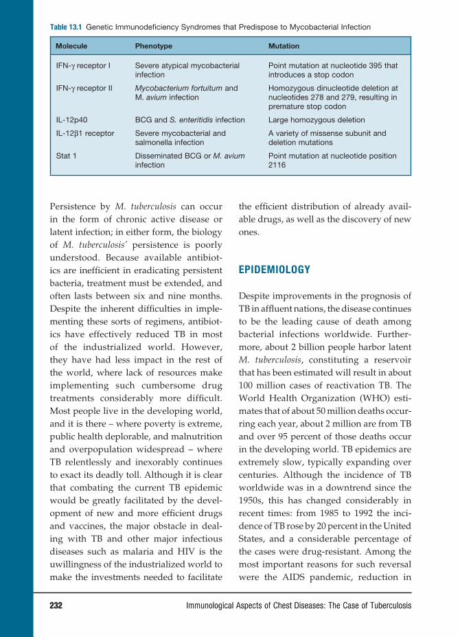
232 Immunological Aspects of Chest Diseases: The Case of Tuberculosis
Persistence by M. tuberculosis can occur in the form of chronic active disease or latent infection; in either form, the biology of M. tuberculosis’ persistence is poorly understood. Because available antibiot-ics are ineffi cient in eradicating persistent bacteria, treatment must be extended, and often lasts between six and nine months. Despite the inherent diffi culties in imple-menting these sorts of regimens, antibiot-ics have effectively reduced TB in most of the industrialized world. However, they have had less impact in the rest of the world, where lack of resources make implementing such cumbersome drug treatments considerably more diffi cult. Most people live in the developing world, and it is there – where poverty is extreme, public health deplorable, and malnutrition and overpopulation widespread – where TB relentlessly and inexorably continues to exact its deadly toll. Although it is clear that combating the current TB epidemic would be greatly facilitated by the devel-opment of new and more effi cient drugs and vaccines, the major obstacle in deal-ing with TB and other major infectious diseases such as malaria and HIV is the uwillingness of the industrialized world to make the investments needed to facilitate
the effi cient distribution of already avail-able drugs, as well as the discovery of new ones.
EPIDEMIOLOGY
Despite improvements in the prognosis of TB in affl uent nations, the disease continues to be the leading cause of death among bacterial infections worldwide. Further-more, about 2 billion people harbor latent M. tuberculosis, constituting a reservoir that has been estimated will result in about 100 million cases of reactivation TB. The World Health Organization (WHO) esti-mates that of about 50 million deaths occur-ring each year, about 2 million are from TB and over 95 percent of those deaths occur in the developing world. TB epidemics are extremely slow, typically expanding over centuries. Although the incidence of TB worldwide was in a downtrend since the 1950s, this has changed considerably in recent times: from 1985 to 1992 the inci-dence of TB rose by 20 percent in the United States, and a considerable percentage of the cases were drug-resistant. Among the most important reasons for such reversal were the AIDS pandemic, reduction in
232 Immunological Aspects of Chest Diseases: The Case of Tuberculosis
Table 13.1 Genetic Immunodefi ciency Syndromes that Predispose to Mycobacterial Infection
Molecule Phenotype Mutation
IFN-γ receptor I Severe atypical mycobacterial infection
Point mutation at nucleotide 395 that introduces a stop codon
IFN-γ receptor II Mycobacterium fortuitum and M. avium infection
Homozygous dinucleotide deletion at nucleotides 278 and 279, resulting in premature stop codon
IL-12p40 BCG and S. enteritidis infection Large homozygous deletion
IL-12β1 receptor Severe mycobacterial and salmonella infection
A variety of missense subunit and deletion mutations
Stat 1 Disseminated BCG or M. avium infection
Point mutation at nucleotide position 2116
Immunological Aspects of Chest Diseases: The Case of Tuberculosis 233
public health resources in inner cities, and continued immigration from areas of high TB prevalence.
TB and AIDS
AIDS in HIV-infected individuals (e.g., low CD4+ T cells) is one of the most important contributors to the develop-ment of TB in co-infected individuals in many parts of the world, including the United States. The low CD4+ T cell counts translate into suboptimal macrophage activation and an inability to mount a protective response. HIV-co-infection is the greatest single risk factor for progres-sion from M. tuberculosis infection to dis-ease. The risk of developing disease after infection with M. tuberculosis in an HIV-co-infected person is about 5 to 15 percent per year, compared with a lifetime risk of developing active TB after infection of about 10 percent in immunocompetent individuals. Furthermore, the inability to mount an effective adaptive immune response leads to a more progressive course of TB in HIV-infected individuals. Miliary TB, a highly disseminated form of TB that can affect multiple organs, is also more frequent in AIDS patients. Fur-thermore, some evidence suggests that the interaction might be reciprocal, since active TB might stimulate viral replication, thereby accelerating the onset of AIDS, perhaps through tumor necrosis factor-alpha (TNF-α,) which induces viral tran-scription, and/or activation of the CD4+ T cells that support viral replication. Cur-rent estimates indicate that there are about 11 million individuals co-infected with HIV and M. tuberculosis worldwide. A recent study found that of an estimated 1.8 million deaths from TB, 12 percent were attributable to HIV, and similarly, TB was
responsible for 11 percent of all deaths due to AIDS in adults.
The emergence of drug-resistant strains of M. tuberculosis is another important fac-tor modifying the current TB epidemic. A strain is considered multidrug resistant (MDR) if it is resistant to at least isonia-zid and rifampicin, the two most effective drugs for TB. Rapidly spreading MDR M. tuberculosis strains, that are resistant to all four fi rst-line TB drugs, including isonia-zid, rifampin, pyrazinamide, and etham-butol, have already emerged. According to WHO estimates, around 5 percent of all active TB cases worldwide are caused by MDR strains.
The Tubercle Bacillus
Mycobacterium tuberculosis belongs to the class Actinobacteria, order Actinomyce-tales, suborder Corynebacterineae, family Mycobacteriaceae. The suborder Coryne-bacterineae encompasses other high G+C gram-positive bacteria including members of the Nocardia, Rhodococcus, and Cory-nebacterium families. M. tuberculosis bacilli are nonmotile rod-shaped bacteria with general dimensions ranging from 1 to 4 μm in length and 0.3 to 0.6 μm in diameter. Though M. tuberculosis requires oxygen for growth, it can grow at low oxygen ten-sions and even survive complete oxygen deprivation.
Mycobacteria are notorious for being very slow growers. M. smegmatis, a fast- growing innocuous mycobacterial sap-rophyte used as a surrogate to study M. tuberculosis, has a division time of about 3 hours in axenic culture and takes 3–4 days to form a colony on agar. M. tubercu-losis has a doubling time of about 20 hours in culture and formation of a colony on agar requires 18–21 days. One of the most
Immunological Aspects of Chest Diseases: The Case of Tuberculosis 233
234 Immunological Aspects of Chest Diseases: The Case of Tuberculosis
striking features of mycobacterial cells is their tremendously complex lipid-rich envelope, which comprises half of the cell’s dry weight. This property causes mycobac-terial cells to aggregate into clumps in cul-ture, making their experimental manipula-tion diffi cult. Limited studies suggest that mycobacterial cells might be relatively less permeable to hydrophilic substrates due to the presence of mycolic acids and myco-sides; this impermeability could prevent transport of hydrophilic substrates into the cell. This lipid “shield,” which causes M. tuberculosis to retain carbol fuchsin dye despite acid treatment (“acid fastness”), plays an important protective function from physical and chemical stress, and accumulating evidence indicates that its components mediate complex interac-tions with the host immune response. These properties might also limit the abil-ity of certain drugs to effi ciently enter the cell.
In 1998, Cole and colleagues were the fi rst to sequence an M. tuberculosis genome – that of virulent laboratory strain H37Rv. A recent clinical isolate, termed CDC1551, was sequenced shortly after, and a third clinical isolate (strain 210) has also been completed. This wealth of information has uncovered unique aspects about mycobac-terial biology. The constant G+C content of around 65 percent throughout mycobacte-rial genomes suggests the absence of hori-zontally acquired pathogenicity islands, which are widespread in other pathogenic bacteria. The most salient characteristic of the M. tuberculosis and other mycobacterial genomes is the presence of lots of genes dedicated to lipid metabolism (more than 8 percent of the M. tuberculosis genome); considering that nearly 40 percent of the dry weight of the mycobacterial cell wall is made up of lipids, it is reasonable to
conclude that lipid metabolism plays a major role in the biology of mycobacteria.
CLINICAL TUBERCULOSIS
M. tuberculosis contagion occurs when a susceptible person inhales bacilli-containing infectious droplets that have been expelled from the lungs of a tubercu-lous individual by sneezing, coughing, or simply talking. Of those infected, only a minority (around 10 percent) will develop disease; about half will do so within a few months to a couple of years immediately following infection (primary TB), while the other half will do so only after an indeter-minately long period of latency (postpri-mary TB).
Primary TB
After reaching the alveoli, phagocytosis of the bacilli by resident macrophages is fol-lowed by unrestricted bacterial and expo-nential intracellular growth. Uptake of bacteria by DCs and migration of infected DCs to the draining lymph nodes for anti-gen presentation likely contribute to the lymphohematogenous dissemination of bacteria to extrapulmonary organs and uninfected parts of the lung, leading to the formation of secondary lesions. Expo-nential bacillary growth is thought to con-tinue in primary and secondary lesions until a specifi c T-cell response is gener-ated around each lesion, curtailing bacil-lary growth and likely killing some of the bacteria within macrophages as these bac-teria become mycobacteriostatic or myco-bacteriocidal after activation. The caseous centers of newly formed lesions are sur-rounded by activated macrophages. Pre-sumably, bacterial antigens being released
Immunological Aspects of Chest Diseases: The Case of Tuberculosis 235
by the bacteria or captured from them are constantly being processed and presented to surrounding T cells in the context of IL-12 and other cytokines that induce a type of immune response. Constant presence of antigen likely contributes to expansion of the T-cell populations and continued acti-vation of macrophages, which avidly ingest and inhibit or kill bacilli accumulated dur-ing the acute phase of the infection. In a lesion where the bacteria are effectively controlled by cell-mediated immunity, the caseous center becomes surrounded by a capsule, forming a protective granuloma. Over time, the number of viable bacilli in these granulomas likely diminishes. Old caseous foci of this sort contain little to no bacilli, and some of the old lesions can ossify and be reabsorbed as Opie and Aron-son demonstrated in 1927. The interactions just described result in control of the infec-tion in most infected individuals.
For reasons poorly understood, some individuals fail to generate the protective type of immune response just described. In these cases, bacteria continue to replicate and the granulomas become increasingly necrotic. Part of the tissue damage that occurs is the result of immunopathology, that is, an excessive host-mediated infl am-matory response to bacterial antigens. When the centers of granulomas undergo caseating necrosis, the sheaths of mono-cytes and T cells that normally suppress the intracellular growth of the bacilli can also become necrotic; as the bacteria con-tinue to replicate in nonactivated mac-rophages at the periphery of the lesion, progressive destruction of lung tissue ensues, ultimately leading to the death of the host. Massive infl ammation and tis-sue damage can also be caused directly by some components of the bacterial cell wall as well as by secreted lipids and proteins;
lipoarabinomannan, for instance, triggers TNF-α secretion by macrophages, while some secreted lipids lead to secretion of proinfl ammatory IL-6.
Although M. tuberculosis is not an obli-gate intracellular pathogen, whether it is capable of extracellular growth in the acel-lular necrotic centers of caseous lesions is unclear. In contrast, solid caseum that has undergone liquefaction is believed to be permissive for bacillary replication. The coughing up of this highly infectious material is in fact responsible for the trans-mission of the bacilli. These observations illustrate how pathology is a necessary step in the life cycle of M. tuberculosis infec-tion in humans.
Latent TB Infection and Reactivation
Little is known about the factors that deter-mine whether an individual that is exposed to M. tuberculosis will go on to develop pri-mary TB or become latently infected. That the immune status of the host is involved in both the establishment and maintenance of latency can be inferred from the obser-vation that HIV–M. tuberculosis co-infected individuals have a much higher risk of developing TB after infection. The only sign of infection in individuals with latent TB infection (LTBI) is the appearance (between two and six weeks postexposure) of a detectable delayed-type hypersensi-tivity (DTH) reaction to a mycobacterial purifi ed protein derivative (tuberculin). By taking necropsy specimens of lung lesions from immune-competent individuals who had died of causes other than TB and inoc-ulating necropsy speciments into guinea pigs to determine the presence of infectious bacilli, Opie and Aronson (1927) showed that some individuals can harbor viable tubercle bacilli for many years without
236 Immunological Aspects of Chest Diseases: The Case of Tuberculosis
showing any symptoms of disease. In this and other early studies, it was found that while specimens from encapsulated or cal-cifi ed lesions rarely were infectious, those isolated from caseous lesions in the lung’s apex generally were. Unexpectedly, appar-ently healthy tissues were also often infec-tious. Recently, Gomez and McKinney (2004) have reviewed several other studies from the pre-antibiotic era that indicated that lung lesions from asymptomatic indi-viduals can harbor viable bacilli to differ-ent degrees. Recent molecular evidence of potential LTBI in normal human lung tissue was provided by the fi nding that about 30 to 50 percent of samples from individuals from endemic regions (Mexico and Ethiopia) could harbor M. tuberculosis DNA.
Perhaps the most compelling evidence for the prevalence of LTBI has come from the observed increased rates of TB in AIDS patients, as well as the high reactivation TB associated with the intake of TNF-α inhibi-tors prescribed for rheumatoid arthritis. These observations indicate that many individuals with LTBI accomplish life-long suppression of the bacillus, but never actually achieve sterilization. Reactivation TB usually presents clinically as a slowly progressive, chronic condition; individuals with chronic subacute TB may infect scores of contacts without realizing that they have the disease. Patients with cavitary TB are particularly infectious because they shed enormous numbers of tubercle bacilli in their sputa. Cavitation is caused by the liq-uefaction of necrotic tuberculous tissue and its expulsion through the airways; liquefac-tion is linked to a strong DTH response and is an important example of immunopathol-ogy in TB. The molecular basis of cavity formation has not been determined, nor is it clear whether the immune mechanisms
responsible for pathogenesis and protec-tion are the same.
EXPERIMENTAL STUDIES OF
TUBERCULOSIS
The Mouse Model of Chronic
Tuberculosis
Animals infected with M. tuberculosis – mice, guinea pigs, or rabbits – typically develop a chronic, progressive form of the disease that varies in severity depending on the species, the guinea pig being the most susceptible, the rabbit the most resistant, and the mouse somewhere in between.
The mouse has become the animal of choice in the study of TB. In fact, Robert Koch himself employed mice in his pio-neering experiments over 100 years ago. A chronic infection in mice can be established by inoculation of bacilli by intravenous injection or by aerosol exposure. The bacilli multiply logarithmically during the fi rst two to four weeks postinfection and then stop increasing in numbers in the lung and start to slowly decline in numbers in the liver and spleen. Depending on the titer of the initial inoculum, the chronic infection can last from months to over a year. The model is useful in that it allows host–M. tuberculosis interactions to be studied in the context of a prolonged persistent infection. The maintenance of a relatively high bac-terial load, accompanied by accumulating pathology, might pertain to human active TB, while the apparent lack of bacterial replication might be relevant to both active TB and LTBI. Among the limitations of the mouse model are differences in the com-position and organization of the granulo-matous structures compared with those formed in human lungs, and in the location of the bacilli in the infected lungs – almost
Immunological Aspects of Chest Diseases: The Case of Tuberculosis 237
exclusively intracellular in mice, consider-ably more extracellular in humans.
Spontaneous M. tuberculosis latency in animals only occurs in nonhuman pri-mates. A drug-induced model of latency known as the Cornell model, in which M. tuberculosis–infected mice are sterilized by drug treatment, and then allowed to reac-tivate spontaneously or by immunesup-pression, was developed in the 1950s by McCune and Tompsett. This model allows host–pathogen interactions to be stud-ied in the context of an infection where very low numbers of bacteria are present and has been used to study host factors involved in establishing and maintaining this type of “latency.” However, the rel-evance of the model to true latency awaits validation.
Host–Pathogen Interactions in TB
Upon entering the respiratory tract, bacilli reaching the lung alveoli are phagocytosed by resident macrophages through man-nose, complement, and Fc receptors (in differentiated macrophages). As dem-onstrated more than three decades ago by Armstrong and Hart (1975), bacterial uptake through Fc receptors increases phagolysosomal fusion, triggers the oxida-tive burst, and leads to bacterial killing in human macrophages ex vivo. Regardless of the receptor involved, uptake apparently requires recruitment of cholesterol to the nascent phagosome. Uptake by resident DCs is believed to occur through DC-SIGN adhesion.
CELLULAR INTERACTIONS LEADING TO A PROTECTIVE IMMUNE RESPONSE
A protective immune reponse to M. tuberculosis involves mainly the T-cell-mediated branch of adaptive immunity.
As early as one week postinfection, bacte-ria-specifi c CD4+ and CD8+ T cells migrate to the lung, where they peak in numbers at around four weeks postinfection, Studies in immune-defi cient mice have established that IFN-γ, TNF-α, and IL-12 are the main cytokines involved in orchestrating a pro-tective immune response after M. tubercu-losis infection. The relevance of IFN-γ and IL-12 in antimycobacterial immunity is sup-ported by human genetic data; the recent association between administration of TNF-α inhibitors used to treat rheumatoid arthri-tis and the reactivation of LTBI has under-scored the protective role of this cytokine in control of M. tuberculosis in humans.
The initiation of the immune response occurs through presentation of M. tuber-culosis antigens, presumably by DCs, on MHC class I and II molecules to naïve T cells, which in the context of IL-12 and IL-23 produced by infected macrophages and DCs themselves, are instructed to dif-ferentiate into IFN-γ-secreting cells (helper T type 1, TH1) cells (see Figure 1). Both CD4+ and CD8+ T cells contribute to the activation of infected macrophages through IFN-γ secretion, while CD8+ T cells might additionally contribute to protection by causing apoptosis of infected macrophages through the Fas-FasL pathway, as well as through secretion of granulysin and other microbicidal molecules. However, the relative contribution of CD8+ T cells to the overall protective cell-mediated immunity might not be as signifi cant as that by CD4+ T cells because M. tuberculosis–infected mice defi cient in the former live almost as long as infected wild-type mice, while those defi cient in the latter are signifi cantly more susceptible.
Mice lacking CD4+ T cells exhibit reduced macrophage activation, which is associated with lower IFN-γ production,
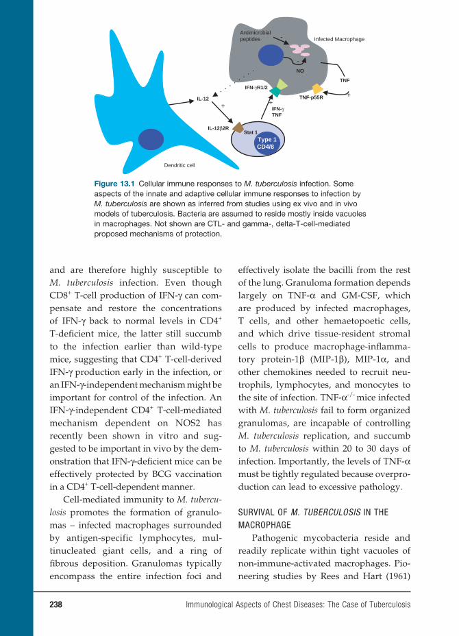
238 Immunological Aspects of Chest Diseases: The Case of Tuberculosis
and are therefore highly susceptible to M. tuberculosis infection. Even though CD8+ T-cell production of IFN-γ can com-pensate and restore the concentrations of IFN-γ back to normal levels in CD4+ T-defi cient mice, the latter still succumb to the infection earlier than wild-type mice, suggesting that CD4+ T-cell-derived IFN-γ production early in the infection, or an IFN-γ-independent mechanism might be important for control of the infection. An IFN-γ-independent CD4+ T-cell-mediated mechanism dependent on NOS2 has recently been shown in vitro and sug-gested to be important in vivo by the dem-onstration that IFN-γ-defi cient mice can be effectively protected by BCG vaccination in a CD4+ T-cell-dependent manner.
Cell-mediated immunity to M. tubercu-losis promotes the formation of granulo-mas – infected macrophages surrounded by antigen-specific lymphocytes, mul-tinucleated giant cells, and a ring of fi brous deposition. Granulomas typically encompass the entire infection foci and
effectively isolate the bacilli from the rest of the lung. Granuloma formation depends largely on TNF-α and GM-CSF, which are produced by infected macrophages, T cells, and other hemaetopoetic cells, and which drive tissue-resident stromal cells to produce macrophage-infl amma-tory protein-1β (MIP-1β), MIP-1α, and other chemokines needed to recruit neu-trophils, lymphocytes, and monocytes to the site of infection. TNF-α-/- mice infected with M. tuberculosis fail to form organized granulomas, are incapable of controlling M. tuberculosis replication, and succumb to M. tuberculosis within 20 to 30 days of infection. Importantly, the levels of TNF-α must be tightly regulated because overpro-duction can lead to excessive pathology.
SURVIVAL OF M. TUBERCULOSIS IN THE MACROPHAGE
Pathogenic mycobacteria reside and readily replicate within tight vacuoles of non-immune-activated macrophages. Pio-neering studies by Rees and Hart (1961)
Figure 13.1 Cellular immune responses to M. tuberculosis infection. Some aspects of the innate and adaptive cellular immune responses to infection by M. tuberculosis are shown as inferred from studies using ex vivo and in vivo models of tuberculosis. Bacteria are assumed to reside mostly inside vacuoles in macrophages. Not shown are CTL- and gamma-, delta-T-cell-mediated proposed mechanisms of protection.
Stat 1
IL-12 +
Infected Macrophage
IFN-γTNF
Type 1CD4/8
Dendritic cell
+
TNF
+
-
NO
IL-12β2R
IFN-γR1/2
TNF-p55R
Antimicrobial peptides
-
Immunological Aspects of Chest Diseases: The Case of Tuberculosis 239
showed that tubercle bacilli prevent the fusion of lysosomes to M. tuberculosis–containing vacuoles, thus preventing the vacuoles’ normal acidifi cation and facili-tating bacterial survival and replication. Vacuoles containing live virulent Mycobac-terium avium have a relatively high pH (6.3–6.5), which correlates with a lower density of vesicular H+ ATPase pumps on their membranes. Mycobacterial vacuoles reside within the recycling and sorting endosomal compartment – markers of early endo-somes (Rab5) are retained, markers of late endosomes (Rab7) and lysosomes (LAMP1-1 and LAMP-2) are largely excluded; communication with the cytoplasmic mem brane appears to be maintained, as suggested by the cycling of transferrin in and out of the vacuoles. Diversion of the mycobacterial phagosome from the normal maturation pathway correlates with reten-tion of the actin-binding coronin TACO on the phagosomal membrane. Active evasion of phagolysosomal fusion by M. tuberculosis involves bacterial effectors such mannose-capped lipoarabinomannan (ManLAM), which interferes with phagolysosomal fusion by blocking the acquisition of the lysosomal cargo and syntaxin 6 from the trans-Golgi network. LAM alone can spe-cifi cally block the cytosolic Ca2+ increase necessary for a signaling cascade involving phosphatidylinositol (PI) 3 kinase hVPS34, which is needed for production of PI 3 phosphate (PI3P) on phagosomes.
The importance of type 1 cytokines in human immunity to M. tuberculosis infec-tion has been confi rmed by studies of naturally occurring polymorphisms in the genes encoding type 1 cytokines and their receptors in humans. The principal role of type 1 cytokines in anti-TB immunity is to activate the antimicrobial functions of mac-rophages. IFN-γ activation and infection of
murine macrophages with M. tuberculo-sis results in the differential expression of some 40 percent of the macrophage genes, an indication of just how dynamic the host–M. tuberculosis interaction is. Many of these differentially expressed genes likely encode functions involved in the curtailment of growth and killing of the intracellular bacilli.
EFFECTOR MECHANISMS OF THE IMMUNE-ACTIVATED MACROPHAGE
Schaible and colleagues (1998) found that upon activation of M. avium–infected macrophages with IFN-γ and lipopolysac-charide (LPS), the mycobacterial vacuoles fuse to each other, become more acidic (pH 5.3), and as a result of fusion with lyso-somes accumulate hydrolases. These and other changes in the intracellular milieu lead to stasis and killing of the bacilli.
MacMicking and colleagues (2003) indicate that LRG47, a member of a family of IFN-γ–dependent GTPases that mediate fusion events in endosomal networks, is one of the facilitators of phagolysosomal fusion and acidifi cation downstream of IFN-γ and is essential for protection. These GTPases play different functions in response to infection by different pathogens as sug-gested by the fi ndings that IGTP–/– and IRG-47–/– mice, which are susceptible to infection by viral, bacterial, and eukaryotic pathogens are resistant to infection by M. tuberculosis. In contrast, LRG47-/- mice suc-cumb to infection by M. tuberculosis with kinetics similar to those of NOS2-/- mice, the other major pathway of antimycobacte-rial immunity.
Work from the laboratories of Carl Nathan and Ferric Fang over the years has demonstrated that the immune system relies on chemically reactive micromol-ecules to neutralize and eliminate invading
240 Immunological Aspects of Chest Diseases: The Case of Tuberculosis
pathogens and their products. Some of the most effi cient of these reactive molecules in killing and controlling M. tuberculosis include the reactive nitrogen intermediates derived from nitric oxide (NO) produced by NO synthase 2. M. tuberculosis is killed by murine peritoneal macrophages and cell lines activated with IFN-γ and either LPS or TNF-α through reactive nitrogen inter-mediate (RNI) production, and IFN-γ and TNF-α synergistically increase NO synthe-sis by macrophages. The demonstration that NOS2-/- mice are highly susceptible to M. tuberculosis infection provides clear functional genetic evidence that NOS2 is essential for the protective immune response against M. tuberculosis.
Even though macrophage activation and resulting RNI production result in bac-teriostasis, M. tuberculosis is nonetheless able to resist eradication in mouse lungs and in activated macrophages in tissue cul-ture, despite the fact that in vitro reagent NO is a potent mycobactericidal. Having evolved to infect the mammalian lung, M. tuberculosis appearently has ways of countering the nossive effects of NO and related RNI. One such mechanism involves the product of the mpa gene, which forms hexamers that have ATPase activity, and is believed to be associated with a myco-bacterial proteasome. Δmpa M. tuberculosis bacteria are attenuated in wild-type mice and show some reversion to virulence in NOS2-/-. It remains to be shown whether Δmpa M. tuberculosis bacteria have defects in protein degradation through this presumptive proteasome, and whether this proposed function is involved in protect-ing the bacteria from RNI. An additional strategy adopted by M. tuberculosis to deal with RNI appears to be simple avoidance of NOS2. Recent microscopy studies indi-cate that M. tuberculosis might be able to
prevent NOS2 from being recruited to bacteria-containing phagosomes.
The role of NO in protection against M. tuberculosis in humans is somewhat con-troversial. While some investigators have reported NOS2 protein induction, NO, which is measured by determining the concentration of its breakdown products nitrate (NO3) and nitrite (NO2), cannot be consistently detected in M. tuberculosis–infected cultures of human monocytes/macrophages. Furthermore, no mutations in nos2 in humans have been associated with susceptibility to M. tuberculosis. How-ever, patients with active TB apparently exhale high levels of NO, which correlate with high expression of NOS2 and spon-taneous production of nitrite by alveolar macrophages.
Another reactive micromolecule that is a potent antimicrobial effector of the immune response is the nicotinamide adenine dinucleotide phosphate (NADPH) oxidase (PHOX)-mediated production of reactive oxygen intermediates (ROIs) including reduction products of O2 such as superoxide, hydrogen peroxide, and hydroxyl radical. ROI generation is an effec-tive antimicrobial defense against many bacterial pathogens including Salmonella and Burkholderia cepacia. However, recent work from John McKinney’s laboratory indicates that the protective role of ROI in TB appears to be less prominent than dur-ing Salmonella infections. Chronic granu-lomatous disease (CGD) patients that lack phagocyte oxidase have not been reported to be more susceptible to M. tuberculosis infection. Furthermore, gp91(phox-/-) mutant mice, which lack a functional phago-cyte oxidase, are not more susceptible to M. tuberculosis than wild-type mice, while mice lacking the gp47 subunit of PHOX are less able to control M. tuberculosis
Immunological Aspects of Chest Diseases: The Case of Tuberculosis 241
replication only transiently. Furthermore, M. tuberculosis appears to be able to effec-tively counteract the damaging effects of ROIs. ROI detoxifi cation mediated by the KatG catalase is essential for bacterial per-sistence in mice following induction of adaptive immunity, and an M. tuberculosis mutant of katG is attenuated in wild-type mice, but reverts to wild-type levels of vir-ulence in mice lacking PHOX. The product of katG is also a peroxynitritetase, thus it may protect M. tuberculosis from both RNI and ROI. At least one of the superoxide dismutases of M. tuberculosis contributes to intracellular survival.
IMMUNITY DURING M. TUBERCULOSIS CHRONIC INFECTION
Several of the components that are important for generating a protective immune response to M. tuberculosis infec-tion are also important for maintaining the equilibrium between host and pathogen during the chronic phase of the infection. As expected, TH1 maintenance is important for controlling M. tuberculosis during the chronic phase of the infection, and sustain-ing this type of response requires IL-12. Neutralization of TNF-α by antibodies or by infection with an adenovirus express-ing a soluble TNF-α receptor resulted in marked increases in bacterial loads and exacerbation of disease in the mouse model of chronic TB and in a variation of the Cor-nell model of drug-induced latency. Work from the laboratories of John Chan and Joan Flynn using the Cornell model of TB, has demonstrated that IFN-γ is involved in preventing reactivation. Similarly, chemical inhibition of NOS2 in chronically infected mice resulted in rapid increases in lung bacterial loads. NOS2-inhibiton also reactivated M. tuberculosis in variations of the Cornell model of latency.
EXTRACELLULAR PERSISTENCE OF M. TUBERCULOSIS IN VIVO
In the human lung, M. tuberculosis is thought to reside both intracellularly and extracellularly, depending on the type of lesion. Pioneering work by Lurie (1964) and continued by Dannenberg using the rabbit model showed the existence of extracellular M. tuberculosis in caseous necrotic lesions. Because immunity to TB was thought to be mainly mediated by acti-vated phagocytes that kill intracellular bac-teria, it was suspected that the extracellular milieu represented a safe heaven for bacte-rial replication and persistence. In order to study this population of bacteria directly back in 1939, Lurie devised a very inge-nious method of cultivating bacteria extra-cellularly in vivo: Parlodion-fi lm–encased silk bags that were impervious to host cells, but not to plasma, were inoculated with bacilli and then implanted subcutane-ously in rabbits. These extracellular bacte-ria replicated unhindered during the fi rst two weeks of infection, but then leveled off at around two weeks postinfection. When immunized animals were studied, bacteria stopped growing earlier and were killed more effi ciently than in naïve counterparts, strongly implicating acquired immunity in controlling the growth of extracellular bac-teria. Because control of growth correlated with a more rapid mobilization of mono-nuclear cells toward the encased focus of infection, it was proposed that factors secreted by activated macrophages ren-dered the fl uids entering the infection foci inimical to bacterial replication.
Recently, work in William Bishai’s laboratory adapted Lurie’s strategy to the widely used model of murine TB: M. tubercu-losis was encapsulated in cell-impermeable hollow fi bers, and these were implanted subcutaneously into mice for in vivo
242 Immunological Aspects of Chest Diseases: The Case of Tuberculosis
cultivation. As expected, an initial period of bacterial replication was followed by cur-tailment of growth, followed by a consider-able decline in numbers, and, interestingly, an eventual stabilization of the counts. These stationary-state colony forming unit (CFU) counts, similarly to those in the equi-librium that characterized the chronic phase of infection in the mouse model of chronic TB as described by Rees and Hart in 1965, represented a somewhat static scenario: they found that about half of the encapsu-lated bacilli were alive by differential stain-ing; a highly dynamic equilibrium would have resulted in a large accumulation of dead bacteria and a decreasing proportion of live ones. Furthermore, indirect analysis – comparison of ATP utilization and CFU at corresponding time points – indicated that soon after implantation, bacteria decreased their metabolism. The rapid cessation of multiplication and rather swift reduction in metabolic activity suggest that innate defenses or perhaps immune-independent mechanisms prompt these adaptations. These adaptations were also suggested by Lurie’s studies using vaccinated animals, which implicated an unknown plasma fac-tor in bacterial growth inhibition. This fac-tor could be nitric oxide, which is known to block M. tuberculosis replication in vitro by inhibiting bacterial respiration. Care-ful analysis of the hollow fi ber’s contents in this extracellular model of persistence in vivo, including concentrations of NO, oxygen, essential elements, and nutrients, might be of value.
VACCINATION STRATEGIES
FOR TUBERCULOSIS
This section is not meant to be a com-plete description of the whole vaccination
program against TB but rather a brief summary of where the fi eld is headed. A review by Wilkinson and Young (2004) gives a much more comprehensive sum-mation of the fi eld. Within recent years the approach to controlling and/or preventing TB infections has undergone a remarkable transformation. This transformation is cer-tainly in part because we now know the entire genome sequence of M. tuberculosis. It is further aided by the completion of the Human Genome Project. Novel analyti-cal techniques now allow us to study the organism from varying sources under dif-fering conditions. It is also now possible to achieve genetic recombination in M. tuber-culosis. This has shifted the emphasis from purely microbiological characterization of recombinant strains to their cellular char-acterization in model systems. Indeed TB was one of the fi rst diseases to be shown to be potentially preventable by DNA vaccines.
With the advent of HIV coexisting with TB coupled with MDR strains emerg-ing, there is now a greater need for new approaches to improved or additional vac-cines against TB. We will now discuss a number of these potential candidates.
Bacille Calmette-Guérin (BCG)
For some time a safe, inexpensive TB vac-cine has been effective in humans and animals. It has been used in more than 100 million newborn children each year but the effi cacy of this vaccine has been under continuous debate.
The vaccine was originally made by Calmette and Guérin by continuous lab-oratory passage of a virulent strain of Mycobacterium bovis on defi cient media for thirteen years. The resulting isolate was tested in mice, guinea pigs, and humans
Immunological Aspects of Chest Diseases: The Case of Tuberculosis 243
and was found to be effective as an experi-mental vaccine in animals.
The situation in humans appears to be more controversial. Several studies such as the one in the United Kingdom by Hart and Sutherland (1977) have shown that the inci-dence of TB decreased by 77 percent when compared with a control group. In contrast, other studies, most notably in South India, indicated close to zero protection. The difference is mainly in the vaccine’s inabil-ity to protect adults from pulmonary TB because the vaccine has consistently been effective against the disseminated forms of TB that are a major cause of childhood mortality. In addition, a small amount of limited data indicates that the effi cacy of BCG may wane over time.
What are the possible reasons for this variation in vaccination studies? One view is that, with continuous passage over the years, the BCG organism has changed genetically and is not like the original strain. In fact, many of these trials were carried out with six different BCG strains. Thus, using the H37Rv genome of M. tuberculosis as a template, it is not surprising that identify-ing genetic regions that are missing in the various substrains of the original BCG vac-cine has been possible. Furthermore, fi ve regions encompassing thirty-eight open reading frames are also deleted in some or all BCG substrains. Calmette maintained that isolates of BCG that caused extreme scar formation and regional lymphade-nopathy were better vaccines.
Other factors that could infl uence the success of BCG are that strains may dif-fer in their immunogenicity. The route, administration, and the effi cacy of the immunizing technique must also be con-sidered. Finally, a favored hypothesis (see Black et al. 2001) is that saphophytic myco-bacteria seen more commonly in warmer
climates may infl uence the results of the trials. Thus, sensitization by environmental mycobacteria could reverse the immuno-logical benefi ts conferred by BCG vaccina-tion since all individuals had already been sensitized by saphophytic organisms.
Finally, the comorbidity of HIV infec-tions coupled with the recrudescence of latent TB or just HIV infection has raised the possibility that administration of BCG (attenuated strain) in an immunocomprised host may allow the BCG strain to multiply rapidly in the host and not prevent disease. These caveats, coupled with the emer-gence of MDR strains of the TB organism and present poor adherence in taking the medicines (long-lasting regimens), have only served to emphasize the urgency for an effective and safe vaccine to control the current TB pandemic. In the following sec-tion, we will discuss the merits and draw-backs of these various vaccine candidates. Before that, however, some understanding of the various immune responses to this organism needs to be discussed.
The Framework for Vaccine
Development and Evaluation
Although most vaccines are produced by large pharmaceutical companies, vaccines for the prevention of TB may not inter-est the pharmaceutical industry because such a vaccine would be primarily used in resource-poor countries. Thus, the major impetus for vaccine development may have to come from the public, not the private, sector.
Considerable momentum toward test-ing and developing novel vaccines has built up in the past few years and the National Institutes of Health after consul-tations with many worldwide organiza-tions has issued a blueprint for TB vaccine
244 Immunological Aspects of Chest Diseases: The Case of Tuberculosis
development. A specifi c commitment to evaluate novel vaccines in aerosol-infected mice and guinea pigs was made, and many candidates and combinations thereof have been tested. The European community has also recognized the need for a wide pre-clinical research approach and includes a primate model for testing novel vaccines. Another important area has been to iden-tify high-incidence areas of infection for future effi cacy trials and the establish-ment of long-term clinical and epidemio-logical support for such ventures. Even in high-incidence areas, phase III trials may involve thousands of individuals because the effi cacy of BCG is known to vary by geographical region and thus parallel trials may need to be established in differ-ent parts of the world.
Vaccine Candidates
Many new vaccine candidates have been produced, and there is no clear rationale for the design of the optimal TB vaccine. However, BCG provides a precedent that a live mycobacterium can confer protection. However, the coexistence of HIV infec-tion in many areas of the world favors a nonreplicating subunit formulation. This approach, however, does require identifi -cation of relevant antigens and the selec-tion of appropriate delivery systems.
LIVE VACCINESExperience with BCG does prove that a
live attenuated vaccine does confer protec-tion, and the improvement of the existing BCG vaccine represents one general approach to the question of new vaccine candidates. In view of the complete dissec-tion of the genome for the organism, new tools for the genetic manipulation of myco-
bacterial antigens allow us to refi ne the live vaccine strategy of Calmette and Guérin by either addition of genes to BCG or targeted removal of genes from an initially virulent M. tuberculosis strain.
1. Modifi ed BCG Strains. BCG has now been engineered to express recombinant mam-malian cytokines designed to augment immunogenicity or decrease immuno-pathology. The addition of the listero-lysin gene has been shown to enhance the ability of BCG antigens to traffi c into the cytoplasm, thereby inducing a CD8+ T-cell response. Another approach has been the overexpression of antigen 85B carried on a multicopy episomal plas-mid in recombinant BCG.
Another possible reason for the incomplete effi cacy of BCG is that it lacks “protective” antigens present in the original M. tuberculosis. For exam-ple, comparison of these two genomes showed that a 9.5-kb region called RD1 is missing from all strains of BCG. The RD1 encodes two antigens (ESAT-6 and CFP-10) that are highly immunogenic and prominent immune targets in M. tuberculosis infected humans. Obviously, reintroduction of the ESAT-6 and CFP-10 is an attractive concept but one must be sure this reintroduction does not increase virulence.
2. Attenuation of M. tuberculosis. A number of novel attenuated strains have been developed and three strategies have evolved: (1) The fi rst involves auxotro-phic mutations that render the bacte-ria dependent on exogenous growth factors that are in limited supply. For example, the derivation of attenuated strains dependent on leucine and tryp-tophan has been successful. (2) A second
Immunological Aspects of Chest Diseases: The Case of Tuberculosis 245
approach is to inactivate M. tuberculosis analogously to those involved in viru-lence in other bacterial pathogens. A recent example is the generation of a phoP attenuated mutant, based on the knowledge of the key role of this regu-lator in pathogenic salmonella. (3) The third strategy exploits the technique of signature-tagged transposon mutagen-esis. This involves screening of a library of transposon mutants to identify clones selectively lost during murine infec-tion. Using this technique has identifi ed mutants with defects in biosynthesis of cell wall lipids. Decreased persistence within tissues and altered patterns of tis-sue dissemination have been described for several attenuated variants of M. tuberculosis.
Whether any of these mutants could function as a vaccine candidate still needs further testing, and issues such as safety, the properties required for an improved live vaccine, interactions with antigen-presenting cells all need to be investigated further.
PROTEIN SUBUNIT VACCINESTwo criteria have been used to identify
M. tuberculosis antigens for incorporation into programs for subunit vaccine develop-ment. The fi rst is the ability to elicit a strong recall response in humans or experimental animals exposed to M. tuberculosis infection. The response is assessed primarily in terms of the release of IFN-γ from antigen-specifi c CD4+ T cells. Although this represents a realistic strategy to identify antigens avail-able for T-cell recognition, it will (like BCG) tend to reproduce rather than augment the natural immune response. Amplifi cation of naturally subdominant responses rep-resents a potentially interesting alternative
approach, particularly in the context of postexposure vaccination.
A second common criterion for anti-gen selection is its identifi cation in the supernatant of in vitro cultures. This idea is supported by experiments from sev-eral laboratories demonstrating successful vaccination with culture fi ltrate prepara-tions. However, live mycobacteria also require immunogenic proteins that are not secreted, such as the small heat-shock pro-tein Acr or 16-kDa antigen.
Several purifi ed protein antigens have been shown to induce protective immu-nity following immunization in appropri-ate adjuvants. The most extensively stud-ied are antigen 85B (Ag85B) and ESAT-6. Ag85B is a member of an antigen complex, a set of three closely related proteins that function as mycolyl transferases and are the most abundant protein components in culture fi ltrate preparations. Because any single protein may be insuffi ciently immu-nogenic, the use of fusion proteins could be important for future subunit vaccine development. Subunit vaccination with purifi ed proteins requires delivery of the protein in adjuvant to generate a strong immune response. Unfortunately, the adju-vants currently licensed for use in humans generally optimize the antibody rather than the T-cell responses, although novel preparations such as the SBAS2 adjuvant or microspheres may be useful in moving mycobacterial protein subunit candidates into clinical trials.
DNA VACCINESDNA vaccination against TB has
proved effective in small animal mod-els using some, but not all, antigens. Two examples from DNA vaccine studies highlight the potential importance of fi ne
246 Immunological Aspects of Chest Diseases: The Case of Tuberculosis
antigenic specificity in the protective response. First, Ag85A, but not Ag85B, is effective when administered as a DNA vac-cine to mice. Interestingly, when antigen 85B is delivered to guinea pigs either as a subunit or overexpressed in a recombinant BCG, it is an effective vaccine, and protec-tion has also been reported using an alter-native Ag85B DNA formulation. Thus, the form of vaccination and host species are also crucial for any given antigen. The second instance, indicating a potential contribution of fi ne specifi city to the immune response in TB, concerns the closely related Hsp60 molecules of Mycobacterium leprae and M. tuberculosis. DNA vaccination using Hsp60 of M. leprae is effective in mice both as a prophylactic and an immunothera-peutic vaccine. The vaccine effi cacy could not be reproduced using a construct of the closely related Hsp60 of M. tuberculosis.
COMBINATION VACCINESVaccinia virus has been used as a
delivery system for mycobacterial anti-gens. This live vector has the advantage of proven safety and effi cacy in the con-text of the smallpox eradication program. The combination of three vaccinations of plasmid DNA containing MPT63 and ESAT-6 plus one “boost” with a recom-binant Vaccinia construct expressing the same antigens induced protection against intraperitoneal infection of mice close to that conferred by BCG. However, there is at least one report of a theoretically ele-gant prime-boost strategy using ESAT-6 in which there was no enhancement above the protection provided by individual subunits. Perhaps of greatest interest is that sequential immunization with antigen AG85B-expressing DNA followed by BCG was more effective than BCG immunization alone. In this case, deple-
tion of the CD8+ T cells in the immunized mice impaired protection in their spleens, indicating that this improved efficacy was partially mediated by CD8+ T cells.
STRATEGIES FOR VACCINE EVALUATIONAlthough animal models have been
helpful in designing new vaccine strategies for TB, there is no guarantee that success or failure in animal models will not neces-sarily produce similar behavior in humans. With respect to animal models, mice and guinea pigs have been used most exten-sively. However, other larger models such as rabbits, cattle, and nonhuman primates are available for further characterization of a given vaccine candidate. A consensus exists that nonhuman primate studies are necessary before phase I studies of novel anti-TB vaccines in humans.
1. Mouse: This is the least costly model and has been a valuable model for investigating genetics and immunology of the host response to TB. Yet, they are relatively resistant to the disease, the skin test reactivity is not very good, and pathological examination of tissues does not show the caseation and fi bro-sis of the lungs so commonly seen in human TB.
2. Guinea pig: These animals are quite susceptible to low-dose infection with virulent M. tuberculosis and exhibit some of the necrotic pathology seen in humans. Their exquisite sensitivity to TB provides an experimental window of suffi cient width to allow the ranking of different vaccines according to their individual protective effi cacies.
3. Rabbit: Perhaps the most striking feature of this model is not rabbits’ response to M. tuberculosis but rather to M. bovis strains. Infection with these
Immunological Aspects of Chest Diseases: The Case of Tuberculosis 247
strains produces cavities in the lungs by caseation, and the extracellular mul-tiplication of bacilli are very similar to that of humans.
4. Cattle: TB in cattle and farmed deer is of considerable economic signifi cance, and efforts to develop a new vaccine against human TB are paralleled by attempts to develop diagnostic agents and vaccine control strategies for bovine TB. Given the relationship between the respective infectious agents, M. tuberculosis and M. bovis, it may be that the prevention of human and bovine TB can be achieved using similar vaccines. Therefore, vaccine trials in cattle can be considered a relevant animal model for human TB, given that the endpoint of this model will be the reduction of transmission in a naturally susceptible host system.
5. Nonhuman primates: Establishment of experimental TB in nonhuman primates provides an opportunity to study protec-tive and pathological mechanisms with close resemblance to those in humans. BC6 vaccination has been characterized in rhesus and cynomolgus monkeys, and studies have been initiated with new candidates. Nonhuman primate models are relatively poorly standard-ized, however, and ethical and fi nancial aspects of such research dictate that it should be reserved for the end stages of preclinical evaluation.
Summary
In each of these model systems, challenge with virulent mycobacteria can be by par-enteral infection or, in an attempt to mimic the natural route of infection, by aerosol infection. Measuring the reduction of bac-terial load in target tissues, particularly in
the lung and spleen, at a specifi c time after challenge is the most widely accepted sys-tem to assess protection in the above ani-mal models, although there is increasing recognition that the effect of vaccination on longer-term pathological changes is important in the comprehensive analysis of a new candidate. Typically, BCG vac-cination results in a reduction of approxi-mately tenfold in bacterial numbers in mice and up to 5-log reduction in guinea pigs. The slow growth rate of mycobacte-ria makes the standard determination of CFUs both labor intensive and time-con-suming. Recently, reporter gene technol-ogy has been applied to mycobacteria with luciferase-expressing mycobacteria allow-ing the measurement of luminescence in organ homogenates as a rapid endpoint for vaccine evaluation. Extension of the use of such bacteria into simple whole-blood-based assays in humans also has consider-able potential as a surrogate of protective immunity that makes no assumptions of the detailed nature of this response.
Clinical Trials in Humans
General logistic and ethical issues related to TB vaccine trials will determine the type of vaccine that can be tested and may there-fore have an important infl uence on more immediate research strategies. These issues should be addressed now, rather than wait-ing until vaccine candidates have already reached the stage of fi eld evaluation.
1. Preinfection trials: In designing new trials for novel TB vaccines designed to replace BCG, one runs into an imme-diate ethical problem – to use a novel untested vaccine candidate side by side with a proven BCG effect, namely, the prevention of childhood forms
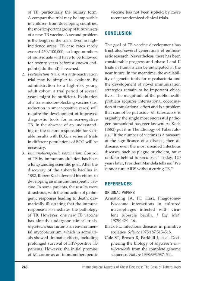
248 Immunological Aspects of Chest Diseases: The Case of Tuberculosis
of TB, particularly the miliary form. A comparative trial may be impossible in children from developing countries, the most important group of future users of a new TB vaccine. A second problem is the length of the trials. Even in high-incidence areas, TB case rates rarely exceed 250/100,000, so huge numbers of individuals will have to be followed for twenty years before a known end-point (adulthood) is reached.
2. Postinfection trials: An anti-reactivation trial may be simpler to evaluate. By administration to a high-risk young adult cohort, a trial period of several years might be suffi cient. Evaluation of a transmission-blocking vaccine (i.e., reduction in smear-positive cases) will require the development of improved diagnostic tools for smear-negative TB. In the absence of an understand-ing of the factors responsible for vari-able results with BCG, a series of trials in different populations of BCG will be necessary.
3. Immunotherapeutic vaccination: Control of TB by immunomodulation has been a longstanding scientifi c goal. After the discovery of the tubercle bacillus in 1882, Robert Koch devoted his efforts to developing an immunotherapeutic vac-cine. In some patients, the results were disastrous, with the induction of patho-genic responses leading to death, dra-matically illustrating that the immune response also mediates the pathology of TB. However, one new TB vaccine has already undergone clinical trials. Mycobacterium vaccae is an environmen-tal mycobacterium, which in some tri-als showed dramatic effects, including prolonged survival of HIV-positive TB patients. However, the initial promise of M. vaccae as an immunotherapeutic
vaccine has not been upheld by more recent randomized clinical trials.
CONCLUSION
The goal of TB vaccine development has frustrated several generations of enthusi-astic research. Nevertheless, there has been considerable progress and phase I and II trials in humans can be anticipated in the near future. In the meantime, the availabil-ity of genetic tools for mycobacteria and the development of novel immunization strategies remain to be important objec-tives. The magnitude of the public health problem requires international coordina-tion of translational effort and is a problem that cannot be put aside. M. tuberculosis is arguably the single most successful patho-gen humankind has ever known. As Koch (1882) put it in The Etiology of Tuberculo-sis: “If the number of victims is a measure of the signifi cance of a disease, then all disease, even the most dreaded infectious diseases, such as plague or cholera, must rank far behind tuberculosis.” Today, 120 years later, President Mandela tells us: “We cannot cure AIDS without curing TB.”
REFERENCES
ORIGINAL PAPERSArmstrong JA, PD Hart. Phagosome-
lysosome interactions in cultured macrophages infected with viru-lent tubercle bacilli. J Exp Med. 1975;142:1–16.
Black FL. Infectious diseases in primitive societies. Science 1975;187:515–518.
Cole ST, Brosch R, Parkhill J, et al. Deci-phering the biology of Mycobacterium tuberculosis from the complete genome sequence. Nature 1998;393:537–544.
Immunological Aspects of Chest Diseases: The Case of Tuberculosis 249
Dannenberg AM Jr, Rook GAW. Rabbit model of tuberculosis. In: Bloom BR, ed. Tuberculosis, Pathogenesis, Protection and Control. Washington, DC: ASM Press; 1994:149–156.
Hart PD, Sutherland I. BCG and vole bacillus vaccines in the prevention of tuberculosis in adolescence and early adult life. Br Med J. 1977;2:293–295.
Lurie MB. Studies on the mechanism of immunity in TB: the role of extracellular factors and local immunity in the fi xa-tion and inhibition of growth of tuber-cle bacilli. J Exp Med. 1939;69:555–578.
MacMicking JD, North RJ, LaCourse R, Mudgett JS, Shah SK, Nathan CF. Iden-tifi cation of nitric oxide synthase as a protective locus against TB. Proc Natl Acad Sci USA, 1997;94:5243–5248.
McCune RM, Feldmann FM, Lambert HP, McDermott W. Microbial persistence. The capacity of tubercle bacilli to sur-vive sterilization in mouse tissues. J Exp Med. 1966;123:445–468.
Opie EL, Aronson JD. Tubercle bacilli in latent tuberculous lesions and in lung tissue without tuberculous lesions. Arch of Pathol. 1927;4:1–21.
Raviglione MC, Snider DE Jr., Kochi A. Global epidemiology of tuberculosis. Morbidity and mortality of a worldwide epidemic. JAMA 1995;273:220–226.
Rees R, Arcy Hart, PD. Analysis of the host-parasite equilibrium in chronic murine tuberculosis by total and via-ble bacillary counts. Brit J Exp Pathol. 1961;42:83–88.
Sambandamurthy VK, Wang X, Chen B, et al. A pantothenate auxotroph of Myco-bacterium tuberculosis is highly attenu-ated and protects mice against tubercu-losis. Nat Med. 2002;8(10):1171–1174.
Schaible UE, Sturgill-Koszycki, S, Schlesinger PH, Russell DG. Cytokine
activation leads to acidifi cation and increases maturation of mycobacte-rium avium-containing phagosomes in murine macrophages. J Immunol. 1998;160:1290–1296.
Sturgill-Koszycki S, Schlesinger, PH, Chakraborty P, et al. Lack of acidifi -cation in Mycobacterium phagosomes produced by exclusion of the vesicular proton-ATPase. Science 1994;263:678–681; erratum. 1994;263:1359.
REVIEWSBloom BR, Murray CJ. Tuberculosis: com-
mentary on a reemergent killer. Science 1992;257:1055–1064.
Bloom BR, Fine PEM. The BCG experi-ence: implications for future vaccines against tuberculosis. In: Bloom BR, ed. Tuberculosis: Pathogenesis Protection and Control. Washington DC: ASM; 531–557.
Flynn, JL. Immunology of Tuberculosis and implications in vaccine develop-ment. Tuberculosis 2004;84:93–101.
Gomez JE, McKinney JD. M. tuberculosis persistence, latency, and drug toler-ance. Tuberculosis 2004;84:29–44.
Farmer P. Social inequalities and emerg-ing infectious diseases. Emerg Infect Dis 1996;2:259–269.
Gormus BJ, Meyers WM. Under-explored experimental topics related to inte-gral mycobacterial vaccines for lep-rosy. Expert Rev of Vaccines. 2003;2(6):791–804.
Lurie MB. Resistance to Tuberculosis: Experi-mental Studies in Native and Acquired Defensive Mechanisms. Cambridge, MA: Harvard University Press; 1964.
Nathan C. Antibiotics at the crossroads. Nature 2004;431:899–902.
Nicod LP. Immunology of tuberculosis Swiss Med Weekly. 2007;1137:357–362.

250 Immunological Aspects of Chest Diseases: The Case of Tuberculosis
North RJ, Jung YJ. Immunity to TB. Annu. Rev. Immunol. 2004;22:599–623.
Wayne LG, Sohaskey CD. Nonreplicat-ing persistence of Mycobacterium tuber-culosis. Annu Rev Microbiol 2001;55:139–163.
Wilkinson RJ, Young DB. Novel vaccines against tuberculosis. In: Levine MM,
Kaper JB, Rappuoli R, Liu MA, Good MF, eds. New Generation Vaccines. 3rd ed. New York: Marcel Dekker; 2004:519–535.
Young, DB, Duncan K. Prospects for new interventions in the treatment and pre-vention of mycobacterial disease. Annu Rev Microbiol. 1995;49:641–673.
14. Immunological Aspects of Gastrointestinal
and Liver Disease
Christine Moung, M.D., and Lloyd Mayer, M.D.
251
MUCOSAL IMMUNITY
The mucosal immune system is distinct from its systemic counterpart. The periph-eral immune system is characterized pri-marily by its ability to eradicate foreign antigens and functions in a relatively antigen-free environment. In contrast, the mucosal immune system is in con-stant juxtaposition with luminal fl ora and dietary proteins. Therefore, instead of mounting active immune responses, which would result in devastating consequences to the host, the general immune response of the gut is suppression. These responses are supported by several phenomena that have been observed in the gut, including oral tolerance and controlled or physiolog-ical infl ammation. Suppression appears to be a selective process, as it does not pre-clude the ability of the gut to mount appro-priate responses to pathogens (e.g., secre-tory IgA response). This emphasizes the dynamic ability of the mucosal immune system to adapt to environmental stimuli in a way that best suits the needs of the host. Aberrations in this balance result in infl ammatory diseases of the bowel and food allergy.
The alternative pathways of immune regulation observed in the mucosal immune system are most likely explained by the distinct organization of the lym-phoid structures and lymphocyte popu-lations that are present. At this point,
study of the anatomy and components of the mucosal immune system will facili-tate understanding of the mechanisms involved in both health and disease of the gastrointestinal (GI) tract.
Anatomy of the Gut-Associated
Lymphoid Tissue
Anatomically, the gut-associated lymphoid tissue (GALT) is composed of a single layer of epithelial cells separating the external environment from the underlying loose connective tissue, the lamina propria (LP) (Figure 14.1). Two general categories of defenses that serve to protect the mucosa are innate and adaptive (Figure 14.2). Innate defenses include both physical and chemical barriers. The fi rst line of defense includes the glycocalyx and mucous coat, which cover the epithelium. Bacteria and viral particles are trapped in the mucous, and these are subsequently expelled from the body through rectal, vaginal, and nasal secretions. The epithelium also prevents the entry of microorganisms into the LP. Breeches in the epithelium are rapidly restored by intestinal trefoil factors. Some pathogens are capable of invading the host through penetration of the epithelium, but the tight junctions that join the epithelial cells are virtually impermeable to foreign particles. The crypt epithelium in the small intestine (Paneth cells) produces defensins that inhibit bacterial growth. In addition,
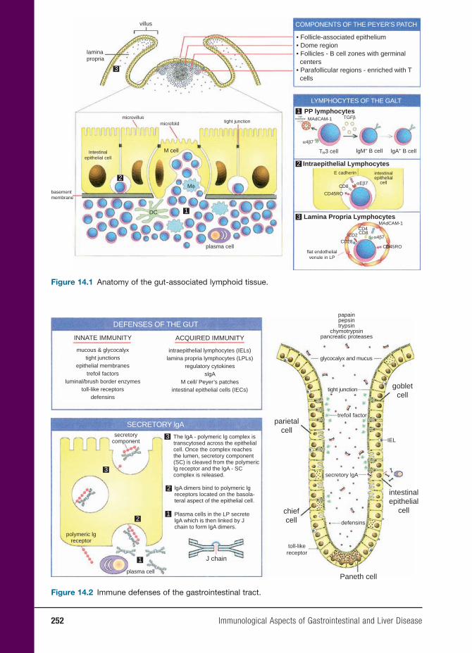
252 Immunological Aspects of Gastrointestinal and Liver Disease
• Follicle-associated epithelium• Dome region• Follicles - B cell zones with germinal centers• Parafollicular regions - enriched with T cells
COMPONENTS OF THE PEYER’S PATCH
LYMPHOCYTES OF THE GALT
PP lymphocytesTGFβ
E cadherin intestinalepithelial
cell
α4β7
α4β7
αEβ7CD8
CD8CD4
CD2CD28
flat endothelialvenule in LP
CD45RO
CD45ROplasma cell
DC
M cell
laminapropria
villus
microvillus
basementmembrane
Intestinalepithelial cell
microfold tight junction
Mφ
TH3 cell lgM+ B cell lgA+ B cell
MAdCAM-1
MAdCAM-1
1
Intraepithelial Lymphocytes2
Lamina Propria Lymphocytes31
2
3
Figure 14.1 Anatomy of the gut-associated lymphoid tissue.
INNATE IMMUNITY
mucous & glycocalyxtight junctions
epithelial membranestrefoil factors
luminal/brush border enzymestoll-like receptors
defensins
intraepithelial lymphocytes (IELs)lamina propria lymphocytes (LPLs)
regulatory cytokinesslgA
M cell/ Peyer’s patchesintestinal epithelial cells (IECs)
The lgA - polymeric lg complex istranscytosed across the epithelialcell. Once the complex reachesthe lumen, secretory component(SC) is cleaved from the polymericlg receptor and the lgA - SCcomplex is released.
lgA dimers bind to polymeric lgreceptors located on the basola-teral aspect of the epithelial cell.
Plasma cells in the LP secretelgA which is then linked by Jchain to form lgA dimers.
J chain
plasma cell
polymeric lgreceptor
secretorycomponent
papainpepsintrypsin
chymotrypsinpancreatic proteases
glycocalyx and mucus
tight junction
trefoil factor
secretory lgA
parietalcell
chiefcell
toll-likereceptor
Paneth cell
intestinalepithelial
cell
gobletcell
defensins
IEL
ACQUIRED IMMUNITY
DEFENSES OF THE GUT
SECRETORY lgA
3
3
2
21
1
Figure 14.2 Immune defenses of the gastrointestinal tract.
Immunological Aspects of Gastrointestinal and Liver Disease 253
enzymes such as papain, pepsin, trypsin, chymotrypsin, and pancreatic proteases allow for the destruction and degradation of antigens to nonimmunogenic di- and tri-peptides.
Within the GALT, lymphocytes are found at three sites in the mucosa (Figure 14.1). These lymphocytes, in addi-tion to secretory IgA (SIgA) make up the adaptive arm of mucosal immunity. Lym-phocytes are found in the Peyer’s patch (PP) – the main organized lymphoid tissue of the GALT, in the epithelial layer, and in the LP. Each group has demonstrated spe-cial characteristics, which will be discussed in detail later.
The GALT also contains cells that may be indirectly responsible for the initiation or down-regulation of immune responses through their role as antigen-sampling cells. One such cell is the M (microfold) cell or follicle-associated epithelium, which overlies the PP. These cells lack a glycocalyx or microvilli. They allow for the uptake and transport of particulate antigens to the underlying PP, which ultimately results in an active immune response. Intestinal epithelial cells (IECs), which make up the majority of the epi-thelial layer, are capable of sampling soluble antigens and have been observed to stimulate regulatory CD8+ T-cell popu-lations, suggesting their role in the path-ways involved in down-regulation of the immune response.
Lymphocyte Populations of the GALT
INTRAEPITHELIAL LYMPHOCYTESIntraepithelial lymphocytes (IELs)
are predominantly CD8+ T cells situated between epithelial cells of the mucosa, on the basement membrane. These cells express a variety of activation markers
including CD45RO+, which is characteris-tic of memory cells. The function of these cells is uncertain as they demonstrate lit-tle proliferation when activated through their T-cell receptor (TCR). Some studies suggest that they may be cytotoxic and involved in maintaining the health of the epithelium. They do not appear to move in and out of the epithelium as epithelial cells generally grow over them. IELs express the surface molecule, αEβ7, which recog-nizes the ligand E-cadherin on epithelial cells. Unlike α4β7 (a mucosal addressin discussed with lamina propria lympho-cytes TH3 cells), αEβ7 does not appear to be a homing molecule because the number of IELs is not signifi cantly decreased when this integrin is knocked out by targeted gene deletion. Transforming growth fac-tor β (TGF-β) induces the expression of this integrin.
LAMINA PROPRIA LYMPHOCYTESUnlike IELs, which are predominantly
T cells, lamina propria lymphocytes (LPLs) represent a heterogeneous mix of IgA plasma cells, T cells, B cells, macrophages, and dendritic cells. Similar to IELs, LPLs express an activated memory phenotype. They express the mucosal addressin α4β7 and up to 40 percent express αEβ7. Simi-lar to IELs, they do not respond to signals mediated through TCRs but rather can be activated through alternate pathways such as CD2 and CD28. This down-regulated response to TCR activation in both LPLs and IELs suggests this mechanism may be a means by which the mucosal immune system dampens the response to luminal fl ora and diet.
TH3 CELLSParticulate antigens taken up by the
M cells are ultimately presented to the
254 Immunological Aspects of Gastrointestinal and Liver Disease
lymphocyte populations in the PP through macrophages and dendritic cells. One such lymphocyte population, commonly referred to as TH3 cells, secretes a key sup-pressor cytokine, TGF-β. The signifi cance of TGF-β will become evident with the discussion of secretory IgA and oral tol-erance. These lymphocytes can migrate systemically from the PP to other mucosal sites, which comprise the common muco-sal immune system (described later). These activated cells express surface molecules, α4β7, which provide a homing signal for mucosal sites through the ligand MAd-CAM-1. Specifi cally in the gut, they can home to the LP where they are terminally differentiated (e.g., into plasma cells for B cells).
Common Mucosal Immune System
Evidence suggests that the various mucosa-associated lymphoid tissues (MALT) throughout the body are part of a common mucosal immune system. One study intro-duced labeled PP lymphocytes in mice by injection and observed the pathway they appeared to migrate from the PP to the mesenteric lymph nodes, the thoracic duct, the inferior vena cava, the systemic circu-lation, and fi nally to the LP. These labeled lymphocytes could also be found in the LP throughout the remainder of the GI tract, in the mammary gland, and the lung and genitourinary tract. More recent studies suggest there is compartmentalization in the MALT; in other words, immunizing at one site induces lymphocytes with a pref-erence for migration to certain MALT sites over others. For instance, GI tract immuni-zation provides effector cells throughout the gut and mammary gland as demon-strated by SIgA in breast milk, while nasal and intratracheal immunization results in
lymphocytes in the lung, genitourinary tract, and rectum. The important implica-tion is that immunization at one site can confer protection at another.
Secretory IgA
Although IgG is the most abundant immu-noglobulin in the serum, secretory IgA (SIgA) is the most abundant immunoglob-ulin in secretions. Serum IgA is a mono-mer, whereas SIgA exists as a dimer bound together by J (joining) chain (Figure 14.2). TGF-β initiates class switching of B cells within the PP from production of IgM to IgA. The basolateral surface of the epithe-lial cell expresses the polymeric immuno-globulin receptor (pIgR), which serves as a site of attachment for SIgA produced by LP plasma cells. Once bound to the pIgR, the SIgA–pIgR complex is endocytosed within vesicles and transported to the apical sur-face where proteolysis of the pIgR occurs. The proteolytic fragment of the pIgR is called the secretory component (SC), and it remains complexed to SIgA following secretion into the lumen of the gut. SC, a 55-kDa fragment, protects IgA against enzymatic (papain, pepsin) degradation by wrapping itself around the Fc portion of the dimeric antibody. The SIgA–SC com-plex remains situated in the mucous coat where it binds up bacterial and viral par-ticles, preventing their attachment to the epithelium.
Secretory IgA complexed with anti-gens in the lumen is recycled through the enterohepatic circulation. After the SIgA-antigen complex is absorbed in the distal small intestine, it travels to the liver sinusoids through the portal vein, where Kupffer cells take up the complex, destroy the microorganism, and release free SIgA. The pIgR seen on the gut epithelial
Immunological Aspects of Gastrointestinal and Liver Disease 255
cells is also expressed on the basal surface of bile duct epithelium. This epithelium takes up the free SIgA and transports it into the bile duct. Bile containing SIgA then fl ows to the duodenum to be reused in the gut lumen.
Antigen Sampling
M cells sample particulate antigens such as intact bacteria, viruses, and parasites. This process ultimately leads to an immune response to clear these potentially harmful and infectious agents. IECs, however, have been found to sample soluble antigens. Studies have demonstrated that IECs, a type of nonclassical antigen-presenting cell, express class I and class II major his-tocompatibility complex (MHC) molecules constitutively. However, they have been shown to activate CD8+ T cells through expression of the nonclassical MHC class I molecule CD1d. This restriction element interacts with gp180, a member of the carcinoembryonic antigen family, which binds to CD8.
Oral Tolerance
There are several phenomena that sup-port the idea that suppression is the gen-eral response of the gut. One of these is oral tolerance (OT), which is defi ned as the active nonresponse to a soluble antigen that is administered through the oral route. H. G. Wells carried out one of the fi rst doc-umented experiments demonstrating this phenomenon in guinea pigs. By feeding them ovalbumin (OVA), he was subsequently unable to elicit an allergic/anaphylactic response when OVA was introduced sys-temically. This tolerant state has been deter-mined to be active as it can be transferred to a naïve recipient by T cells.
Multiple mechanisms are involved in the induction of OT. Low-dose tolerance is mediated by the CD4+ cells secreting the cytokines TGF-β, IL-10, and IL-4. Higher doses of fed antigens can induce anergy or deletion. Antigen-specifi c CD8+ suppressor T cells may also play a role in this process. Loss of OT may be the mechanism respon-sible for the immune response to commen-sal fl ora and dietary antigens, leading to food allergies and possibly infl ammatory bowel disease (IBD).
Oral tolerance has recently gained much interest as it proposes a mecha-nism by which to generate suppression to specifi c antigens. Several groups have suggested its use in the treatment of autoimmune/inflammatory disorders. Autoantigen can be fed to hosts, which would then result in the stimulation of regulatory T cells to these same antigens. Although induction of OT is specifi c for a given antigen, the effector response is not. This induction is potentially due to the effects of suppressive cytokines such as TGF-β; that is, by feeding an antigen situated in close proximity to the auto-antigen of interest, you can suppress an immune response to both antigens. Thus, you do not necessarily need to feed the antigen to which the autoreactive response is targeted. This process has been termed bystander suppression.
IMMUNE-MEDIATED DISEASES OF
THE GASTROINTESTINAL TRACT
Pernicious Anemia
Pernicious anemia (PA) is an organ-spe-cifi c autoimmune disease characterized by chronic infl ammation of the stomach (gastritis) with subsequent loss of pari-etal cells. This loss of parietal cells results
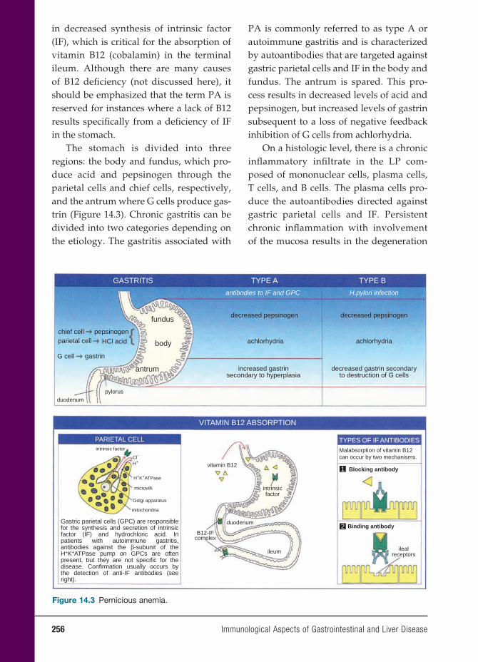
256 Immunological Aspects of Gastrointestinal and Liver Disease
in decreased synthesis of intrinsic factor (IF), which is critical for the absorption of vitamin B12 (cobalamin) in the terminal ileum. Although there are many causes of B12 defi ciency (not discussed here), it should be emphasized that the term PA is reserved for instances where a lack of B12 results specifi cally from a defi ciency of IF in the stomach.
The stomach is divided into three regions: the body and fundus, which pro-duce acid and pepsinogen through the parietal cells and chief cells, respectively, and the antrum where G cells produce gas-trin (Figure 14.3). Chronic gastritis can be divided into two categories depending on the etiology. The gastritis associated with
PA is commonly referred to as type A or autoimmune gastritis and is characterized by autoantibodies that are targeted against gastric parietal cells and IF in the body and fundus. The antrum is spared. This pro-cess results in decreased levels of acid and pepsinogen, but increased levels of gastrin subsequent to a loss of negative feedback inhibition of G cells from achlorhydria.
On a histologic level, there is a chronic infl ammatory infi ltrate in the LP com-posed of mononuclear cells, plasma cells, T cells, and B cells. The plasma cells pro-duce the autoantibodies directed against gastric parietal cells and IF. Persistent chronic infl ammation with involvement of the mucosa results in the degeneration
decreased pepsinogen
antibodies to IF and GPC
GASTRITIS TYPE A
VITAMIN B12 ABSORPTION
PARIETAL CELL TYPES OF IF ANTIBODIES
Malabsorption of vitamin B12can occur by two mechanisms.
Blocking antibody
Binding antibody
ilealreceptorsileum
duodenum
intrinsicfactor
vitamin B12
B12-IFcomplex
Cl-
H+
H+K+ATPase
intrinsic factor
microvilli
Golgi apparatus
mitochondria
TYPE B
H.pylori infection
chief cell pepsinogenparietal cell HCl acid
G cell gastrin
pylorusduodenum
fundus
body
antrum
achlorhydria
increased gastrinsecondary to hyperplasia
decreased pepsinogen
achlorhydria
decreased gastrin secondaryto destruction of G cells
1
2Gastric parietal cells (GPC) are responsiblefor the synthesis and secretion of intrinsicfactor (IF) and hydrochloric acid. Inpatients with autoimmune gastritis,antibodies against the β-subunit of theH+K+ATPase pump on GPCs are oftenpresent, but they are not specific for the disease. Confirmation usually occurs bythe detection of anti-IF antibodies (seeright).
Figure 14.3 Pernicious anemia.

258 Immunological Aspects of Gastrointestinal and Liver Disease
may be considered as an alternative. This is supported by studies that demonstrate B12 absorption may also occur by a pathway independent of IF. Treatment is impor-tant to prevent anemia and neurologic sequelae.
Gluten-Sensitive Enteropathy
Gluten-sensitive enteropathy (GSE), also known as celiac disease or celiac sprue, is characterized by infl ammatory injury to the mucosa of the small intestine after ingestion of gluten in genetically predis-posed individuals. The inciting agent is gluten, a storage protein commonly found in wheat. More specifi cally, GSE is caused by T-cell-mediated recognition of glia-din, an alcohol-soluble fraction of gluten. Removal of gluten from the diet has been found to result in signifi cant regression of the infl ammatory lesion and symptoms seen with the disease.
For some time, GSE was thought to be rare, as its often nondescript clinical pre-sentation masked its true prevalence. How-ever, the advent of new serologic tests has allowed for more accurate diagnosis in the general population. Currently, it is thought to affect as many as 1 in 100 to 250 individ-uals worldwide – predominantly affecting Caucasians and people mainly in North America, Europe, and South America. GSE has also been observed among populations in North Africa, Iran, and India, which attests to the widespread occurrence of the disease.
Genetics plays a major role in the patho-genesis of GSE. Individuals expressing the MHC haplotypes -DR3, -DR5, -DR7, and, more importantly, -DQ2 or -DQ8 are at increased risk of developing GSE. The dis-ease tends to cluster in families, and the monozygotic twin concordance rate of
75 percent further emphasizes the strong genetic component. Less is known about the involvement of non-HLA (human leu-cocyte antigen) genes, which can modify the clinical expression of the disease. GSE has been associated with a region on the long arm of chromosome 5 (5q31 – 33), which is where other candidate suscep-tibility genes that have been implicated in Crohn’s disease and asthma are also located. It has been speculated that there is some involvement of the gene that encodes the negative co-stimulatory mol-ecule, cytotoxic T-lymphocyte antigen-4 (CTLA-4).
The myriad of symptoms associated with GSE has made detection of disease especially diffi cult. It was once thought to occur only in childhood, but GSE can manifest at any age. Patients commonly present with malabsorption, steatorrhea, and weight loss relating to the fl attening of proximal small intestinal villi. Diarrhea is not usually present unless the distal small bowel, in addition to the proximal portion, is affected. Some patients do not present with any gastrointestinal symptoms, but with effects secondary to malabsorption of vitamins such as iron, folate, and calcium. These patients may only present with anemia and osteoporosis, respectively. In addition, there is a strong association between GSE and autoimmune disease. Some individuals with GSE also present with dermatitis herpetiformis (DH), a pru-ritic vesicular rash that commonly presents on the extensor surfaces and the scalp. Most DH patients have GSE, whereas all individuals with GSE do not have DH. These lesions improve with the removal of gluten from the diet. Dapsone can help expedite the healing process.
Immune responses to gliadin occur in two places: the LP and the epithelium. At
Immunological Aspects of Gastrointestinal and Liver Disease 259
a low pH, tissue transglutaminase (tTG, a calcium-dependent enzyme), deamidates glutamine on gliadin, rendering these pep-tides more capable of binding HLA-DQ2 and HLA-DQ8. After CD4+ LP T cells rec-ognize the gliadin epitope presented by DQ2 and DQ8, they are thought to secrete interferon-γ (IFN-γ), which then causes the destructive lesions seen in GSE. The expansion of IELs (predominantly CD8+) that occurs is more controversial and dif-fi cult to understand, but may relate to the reduction of IL-15 by IECs in the presence of gliadin. However, the development of lymphoma in individuals with refractory sprue has been shown to consist of a clonal expansion of CD8+ T cells, implicating the IELs as a possible source. However, as glia-din-specifi c IELs have not been identifi ed, this has led to the conclusion that expan-sion of CD8+ T cells is secondary to the CD4+ T-cell response to gliadin. But this same expansion of CD8+ T cells does not occur in other infl ammatory disorders of the GI tract such as IBD and autoimmune enteropathy, where CD4+ T cells are also activated.
A mechanism to explain this incon-sistency may involve the expression of nonclassical MHC class I molecules such as major histocompatibility class I chain related (MIC) and human leucocyte anti-gen E (HLA-E), which are induced by stress and IFN-γ. Natural killer receptors NKG2D and CD94 present on IELs are up-regulated by IL-15 and can recognize MIC and HLA-E on damaged cells. Up-regula-tion of IL-15 could cause the uncontrolled activation of IELs and villous atrophy. In this situation, IELs do not have to recognize gliadin peptides directly. These data have laid the groundwork for an alternate ther-apy for GSE, using antibodies against IL-15 in refractory sprue because IL-15 activates
IELs and promotes the development of lymphoma.
An ongoing controversy has been whether gliadin is acting as an immuno-logic trigger for an adaptive (CD4+ T-cell) response or whether gliadin fractions are directly toxic to the epithelium, initiating the disease process. Several groups have shown that gliadin peptide can cause the lysis of certain epithelial cell lines. More recently, newer studies have supported both sides of the argument. A major glia-din fragment possesses two activities: (1) a T-cell epitope in the N terminus and (2) a fragment that is rapidly taken up by epithe-lium and subsequently subepithelial cells, inducing the production of IL-15 (an innate immune response). The combination of both activities accounts for the CD4+ T-cell acti-vation, CD8+ IEL lymphocytosis, and IFN-γ secretion. The ability or inability to gener-ate the complete response may ac count for some of the disease heterogeneity.
The gold standard for the diagnosis of GSE is a small-bowel biopsy. Histologically, GSE is characterized by a triad of villous atrophy, hyperplastic crypts, and intraepi-thelial lymphocytosis. Empiric therapy with a trial of gluten-free diet should be avoided as this will alter the small-bowel biopsy fi ndings. In addition, serologic tests are useful for the diagnosis and manage-ment of GSE as they allow for a noninvasive means of monitoring adherence to diet and the identifi cation of individuals who are at high risk for the disease who otherwise might not have been detected. IgA anti-bodies to endomysium, tTG, and gliadin are typically identifi ed. Even though tTG is the autoantigen recognized by endomysial antibodies, there is not always concordance between antibodies against endomysium and tTG. Detection of GSE is optimized by looking for both antibodies. Interestingly,
260 Immunological Aspects of Gastrointestinal and Liver Disease
antibodies to tTG do not alter the function of this enzyme. The presence of antibodies against endomysium is 100 percent specifi c for the diagnosis of GSE. IgA defi ciency occurs in about 1.7 percent to 2.6 percent of patients with GSE, a rate that is ten- to sixteenfold higher than the general popula-tion. In these instances, it is acceptable to look at IgG antibodies to endomysium, tTG, and gliadin instead of IgA. The detection of both IgA and IgG antibodies requires indi-rect immunofl uorescence with either mon-key esophagus or human umbilical cord as the substrate or tTG by enzyme-linked immunosorbent assay.
The only disease-controlling action is the removal of all gluten from the diet for life. The major limiting factor in healing is noncompliance especially in adoles-cents with GSE. In cases in which there is poor clinical improvement, continued ingestion of gluten is the most likely cause. Poor control is thought to be a major factor in the development of IEL lymphomas.
Inflammatory Bowel Disease
Crohn’s disease (CD) and ulcerative colitis (UC) are chronic relapsing and remitting infl ammatory disorders of the gastroin-testinal tract, which together are referred to as IBD. Although clinical evidence sug-gests they are distinct entities, CD and UC are classifi ed under the common name of IBD as they tend to occur in members of the same family and are thought to share genetic factors. While the exact pathoge-netic mechanisms remain to be uncovered, the current hypothesis is that IBD results from an inappropriate mucosal immune response to commensal fl ora in genetically predisposed hosts.
Much of what is known about CD and UC is the result of extensive study of mouse models of intestinal infl amma-tion that resemble IBD. The four general categories of such models are as follows: (1) spontaneous colitis due to a naturally occurring genetic abnormality; (2) sponta-neous colitis in mice that have been geneti-cally manipulated – knockout and trans-genic mice; (3) colitis induced by exposure to a haptenating agent – an antigen that is immunogenic only upon being complexed with a carrier; and (4) adoptive trans-fer models that involve transfer of T-cell populations to a lymphopenic host such as mice with severe combined immunodefi -ciency (SCID).
Current research is focused on elu-cidating the complex interplay between genetic and environmental factors in the pathogenesis of IBD. Mouse models have allowed several broad conclusions to be made. The fi rst, as demonstrated by the numerous murine models of colitis, is that different genetic defects can result in the same features of intestinal infl ammation. The implication here is that there may be several mechanisms of IBD pathogenesis, which rely on a common pathway. Sec-ond, regardless of the genetic defect that is known to cause colitis, the host back-ground can largely determine whether infl ammation occurs. One example is that IL-10 knockout mice bred onto different backgrounds express varying clinical phe-notypes. Careful study of these mice could provide insights about why some people develop IBD while others do not. Third, the normal luminal fl ora is an essential part of the development of colitis. Studies in several mouse models have demonstrated that colitis does not occur when mice are bred and maintained in a germ-free
Immunological Aspects of Gastrointestinal and Liver Disease 261
environment. However, once these mice are colonized by normal commensal fl ora, colitis rapidly ensues.
It is thought that IBD is the result of loss of tolerance to commensal fl ora and hence much interest is focused on uncovering the mechanisms that govern OT induction and the regulatory T-cell populations that are involved. By understanding OT, it may be possible to determine what defects occur in IBD, which would subsequently result in novel targets for immunotherapy.
CROHN’S DISEASEIn the United States, approximately
380,000 to 400,000 individuals are thought to be affected with CD. Even though it can occur at any age, a bimodal peak in the third and fi fth decades of life has been observed. The incidence appears to be greater in whites than in blacks, and it has a predilection to affect the Ashkenazi-Jewish population.
There is a high concordance rate of 40–58 percent in monozygotic twins with CD. The use of microsatellite markers in genomewide screening of affected indi-viduals and family members has reaped the greatest benefi ts in terms of identify-ing susceptibility loci and genes that pre-dispose to the development of CD. There is a strong association with a single gene, NOD2, also referred to as CARD15 (caspase activation and recruitment domain) located on chromosome 16 and referred to as IBD1. NOD2 is an intracellular pattern recogni-tion receptor involved in the binding of muramyl dipeptide, the product of pep-tidoglycan derived from Gram-positive organisms. Of the three major mutations in NOD2 defi ned in CD, all result in the fail-ure of muramyl dipeptide binding and loss of NFκB activation.
CD can involve any part of the GI tract, although it commonly affects the terminal ileum (30 percent), cecum and ileum (40 percent), perianal area (10 percent) and colon (20–30 percent). Endoscopy will often reveal diseased areas interspersed with normal areas of the bowel, called skip lesions. The nature of the infl amma-tory response leads to what is seen as a “cobblestone” appearance of the mucosa. This response is the result of linear ulcers intersecting with sections of normal tis-sue. Histologically, infl ammation affects all the layers of the bowel wall (trans-mural). There is a dense infi ltration of lymphocytes and macrophages and the presence of granulomas. Clinical features include abdominal pain, narrowing of the gut lumen (fi brosis) leading to strictures and bowel obstruction, abscess formation, fi stulization to skin and internal organs, and if the colon is involved, diarrhea and bleeding (Figure 14.4).
Much evidence supports that CD is pri marily a TH1-mediated immune response. However, recent studies impli-cated TH1 T-cells in the disease process. In mouse models, the introduction of the haptenating agent, trinitrobenzene sul-fonic (TNBS) acid, results in increased secretion of the cytokines IL-12, IFN-γ, and TNF-α. Histologic fi ndings reveal a transmural infl ammation in these mice that resembles that seen in CD. Increased secretion of IL-12 both in vivo and ex vivo by macrophages and dendritic cells isolated from patients with CD has also been observed. In CD patients, STAT-4 (signal transducer and activator of tran-scription–4) is increased in the bowel. IL-12 signals through STAT-4 to up-regulate IFN-γ. Antibodies specifi c for the p40 chain of IL-12 have been shown to cause
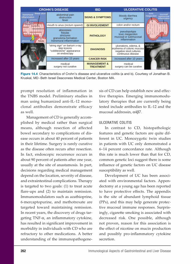
262 Immunological Aspects of Gastrointestinal and Liver Disease
prompt resolution of infl ammation in the TNBS model. Preliminary studies in man using humanized anti-IL-12 mono-clonal antibodies demonstrate effi cacy as well.
Management of CD is generally accom-plished by medical rather than surgical means, although resection of affected bowel secondary to complications of dis-ease occurs in about 40 percent of patients in their lifetime. Surgery is rarely curative as the disease often recurs after resection. In fact, endoscopic recurrence occurs in about 90 percent of patients after one year, usually at the site of anastamosis. In part, decisions regarding medical management depend on the location, severity of disease, and extraintestinal complications. Therapy is targeted to two goals: (1) to treat acute fl are-ups and (2) to maintain remission. Immunomodulators such as azathioprine, 6-mercaptopurine, and methotrexate are targeted toward maintaining remission. In recent years, the discovery of drugs tar-geting TNF-α, an infl ammatory cytokine, has resulted in signifi cant improvement in morbidity in individuals with CD who are refractory to other medications. A better understanding of the immunopathogene-
sis of CD can help establish new and effec-tive therapies. Emerging immunomodu-latory therapies that are currently being tested include antibodies to IL-12 and the mucosal addressin, α4β7.
ULCERATIVE COLITISIn contrast to CD, histopathologic
features and genetic factors are quite dif-ferent in UC. Monozygotic twin studies in patients with UC only demonstrated a 6–14 percent concordance rate. Although this rate is much lower than that for CD, common genetic loci suggest there is some infl uence of genetic factors on UC disease susceptibility as well.
Development of UC has been associ-ated with environmental factors. Appen-dectomy at a young age has been reported to have protective effects. The appendix is the site of abundant lymphoid tissue (PPs), and this may help generate protec-tive mucosal immune responses. Surpris-ingly, cigarette smoking is associated with decreased risk. One possible, although not proven, reason for this association is the effect of nicotine on mucin production and possibly pro-infl ammatory cytokine secretion.
Figure 14.4 Characteristics of Crohn’s disease and ulcerative colitis (a and b). Courtesy of Jonathan B. Kruskal, MD - Beth Israel Deaconess Medical Center, Boston MA.
CROHN’S DISEASEtransmural
inflammation
abscess
stringsign
a
fistula
abdominal painobstruction
feverSIGNS & SYMPTOMS bloody diarrhea
urgency
colon and/or rectum
pseudopolypstoxic megacolon
mucosal or submucosalinflammation
ulcerations, edema, &erythema of colonic mucosa
negative stool culturescontinuous disease
increased after 10 years
medicalsurgery can be curative
GI INVOLVEMENT
PATHOLOGY
DIAGNOSIS
CANCER RISK
MANAGEMENT &TREATMENT
abscessesfistulas
stricturesgranuloma formation
transmural inflammation
increased after 15 years
mouth to anus (rectum spared)
“string sign” on barium x-rayskip lesions
“cobblestone” appearanceon endoscopy
medicalsurgery not curative
linear ulcer toxicmegacolon
pseudopolyp
lead-pipecolon
b
mucosal/submucosalinflammation
granuloma
IBD ULCERATIVE COLITIS
Immunological Aspects of Gastrointestinal and Liver Disease 263
UC is thought to be mediated by a mixed infl ammatory immune response that is generally characterized by the secretion of cytokines IL-5, IL-13, and IFN-γ. Thus, it is neither TH1 nor TH2. Mouse models of colitis in which the haptenating agent, oxa-zolone, is introduced as well as the TCRα −/− mouse demonstrate a similar cytokine profi le.
UC only affects the colon and spreads proximally from the rectum in a con-tinuous fashion. In cases in which it only affects the rectum, it is called ulcerative proctitis. It affects the superfi cial layers of the bowel wall with infi ltration of lympho-cytes, granulocytes, and mast cells. There are often ulcerations and crypt abscesses (collections of polymorphonuclear cells in the crypt). Clinically, patients often pres-ent with severe diarrhea, blood loss, and, with time and poor control, progressive loss of peristaltic function secondary to rigid “lead-pipe” colon. In severe cases, one can see the complications of this toxic megacolon and perforation. Of the IBDs, UC is strongly associated with an increased risk for colon cancer (occurs with long-standing – greater than ten years – disease) although this risk is present in CD as well (Figure 14.4).
Given that UC is generally confi ned to the colon, surgery is curative (total colec-tomy), in contrast to CD. Medical thera-pies include those that are used to treat CD – 5-ASA formulations, steroids, and immunomodulators. Surgery is reserved for those patients who are refractory to medications, develop intractable toxic megacolon, or show early or overt signs of colonic malignancy. The standard pro-cedure up until fi fteen years ago was a total proctocolectomy and end ileostomy. However, more recently, a procedure that spares the rectal musculature and
strips the rectal muscosa has been used, known as an ileoanal pull-through (restor-ative proctocolectomy with ileal pouch anal anastamosis). A total colectomy is performed, leaving the rectal muscula-ture layer that is essentially stripped of its mucosa, as the disease is superfi cial, and an internal pouch is created from the dis-tal ileum with outfl ow through the rectal muscular tube. A complication of this pro-cedure is infl ammation of the pouch, or pouchitis. Interestingly, this complication occurs in UC patients in up to 40 percent of the cases but does not occur in individ-uals in which this procedure is performed for noninfl ammatory disease (e.g., famil-ial polyposis). These fi ndings support an intrinsic defect in mucosal immunomo-regulation in UC.
LIVER IMMUNOLOGY
The liver is essential in metabolism, the removal of pathogens and antigens from the blood, and the synthesis of proteins. As a result, it is exposed to numerous antigens and by-products of gastrointestinal metab-olism on a daily basis. It is critical that the liver is able to mount active responses to eliminate microorganisms while avoiding unnecessary immune responses toward innocuous antigens, which could also result in injury to hepatocytes. Similar to the gut, the liver has a natural tendency toward immune tolerance rather than the induction of immunity.
Anatomically, the liver is divided into functional units termed hepatic lobules (Figure 14.5). These lobules are hexagonally shaped and consist of a central venule or vein and portal triads at its corners. Within the lobule, the hepatic sinusoids – the walls of which comprise liver sinusoidal
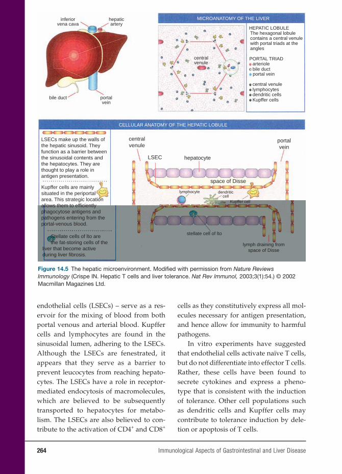
264 Immunological Aspects of Gastrointestinal and Liver Disease
endothelial cells (LSECs) – serve as a res-ervoir for the mixing of blood from both portal venous and arterial blood. Kupffer cells and lymphocytes are found in the sinusoidal lumen, adhering to the LSECs. Although the LSECs are fenestrated, it appears that they serve as a barrier to prevent leucocytes from reaching hepato-cytes. The LSECs have a role in receptor-mediated endocytosis of macromolecules, which are believed to be subsequently transported to hepatocytes for metabo-lism. The LSECs are also believed to con-tribute to the activation of CD4+ and CD8+
cells as they constitutively express all mol-ecules necessary for antigen presentation, and hence allow for immunity to harmful pathogens.
In vitro experiments have suggested that endothelial cells activate naïve T cells, but do not differentiate into effector T cells. Rather, these cells have been found to secrete cytokines and express a pheno-type that is consistent with the induction of tolerance. Other cell populations such as dendritic cells and Kupffer cells may contribute to tolerance induction by dele-tion or apoptosis of T cells.
inferiorvena cava
hepaticartery
centralvenule
LSECs make up the walls ofthe hepatic sinusoid. Theyfunction as a barrier betweenthe sinusoidal contents andthe hepatocytes. They arethought to play a role inantigen presentation.
Kupffer cells are mainlysituated in the periportalarea. This strategic locationallows them to efficientlyphagocytose antigens andpathogens entering from theportal-venous blood.
Stellate cells of lto are the fat-storing cells of theliver that become activeduring liver fibrosis.
LSEC hepatocyte
space of Disse
lymphocyte
stellate cell of lto
lymph draining fromspace of Disse
dendriticcell
Kupffer cell
portalvein
portalvein
MICROANATOMY OF THE LIVER
CELLULAR ANATOMY OF THE HEPATIC LOBULE
centralvenule
HEPATIC LOBULEThe hexagonal lobulecontains a central venulewith portal triads at theangles
PORTAL TRIADarteriolebile ductportal vein
central venulelymphocytesdendritic cellsKupffer cellsbile duct
Figure 14.5 The hepatic microenvironment. Modifi ed with permission from Nature Reviews Immunology (Crispe IN. Hepatic T cells and liver tolerance. Nat Rev lmmunol, 2003;3(1):54.) © 2002 Macmillan Magazines Ltd.
Immunological Aspects of Gastrointestinal and Liver Disease 265
DISEASES OF THE LIVER
Hepatitis B
Hepatitis B virus (HBV) is thought to affect more than 400 million people world-wide. About 1.25 million Americans have chronic HBV. Chronic HBV infection con-tributes to 5,000 deaths annually from cirrhosis and hepatocellular carcinoma (HCC) in the United States. The rate of progression to chronic HBV infection is higher in individuals who are infected at a younger age (25–30 percent before fi ve years of age; 3–5 percent after fi ve years of age or as adults). HBV is especially preva-lent in Southeast Asia, China, and Africa, where more than half the population is affected. It is passed on through vertical (mother to fetus) and horizontal (primar-ily child to child) transmission. In contrast, in the United States and Europe, infection occurs primarily by horizontal transmis-sion among young adults.
HBV belongs to the family of hepad-naviruses. The virus is present in serum in large quantities (108 to 1010 virions) and can be transmitted not only through blood but also through bodily fl uids such as semen, saliva, and cervical secretions. There is a 30 percent chance of transmis-sion through needlestick. Acute infection is asymptomatic in 70 percent of adults and 90 percent of children younger than fi ve years old. However, patients can present with nausea, anorexia, fatigue, fever, and right upper quadrant pain. Treatment for acute infection is generally supportive.
The HBV genome is composed of a partially double-stranded circular DNA that is enclosed within a nucleocapsid (or core antigen). This genome is surrounded by a spherical envelope or surface anti-gen. The genome encodes both core and
surface proteins and a DNA polymerase, which also acts as a reverse transcriptase. The variable expression of core and surface antigens and antibodies to these proteins serve as useful markers of past, current, or chronic infection.
Four distinct stages of HBV infec-tion have been described. The fi rst stage is characterized by high levels of HBV DNA replication and normal serum transaminase levels. HBeAg, a marker of active viral replication, is positive. The second stage refl ects the immune response in which infl ammation results in destruction of HBV-infected cells and a subsequent elevation in transaminase levels. The highest risk of progression to cirrhosis and HCC occurs with chronic HBV infection when the patient stays in this stage beyond 6 months. The third stage is thought to indicate the end of viral replication although low levels of HBV DNA may still be present. HBeAg becomes negative, HBe antibody appears, and transaminase levels normalize. The fi nal stage is defi ned by the clearance of hepatitis B surface antigen (HBsAg). Antibody to HBsAg confers protective immunity.
HBV replication is not directly cyto-toxic. This is supported by the fi nding that despite ongoing viral replication within hepatocytes, many HBV carriers are asymptomatic and exhibit minimal liver injury. Rather, the liver injury that occurs with HBV infection is thought to be an unfortunate consequence of the immune response, which is directed at viral clear-ance. Clinical studies in patients with acute and self-limited hepatitis B infection have demonstrated strong T-cell responses involving both MHC class II restricted CD4+ cells and MHC class I restricted CD8+ cells to HBV antigens in contrast to
266 Immunological Aspects of Gastrointestinal and Liver Disease
the attenuated responses seen in chronic carriers of HBV. More specifi cally, cyto-toxic T lymphocytes are targeted at epi-topes in the HBV core, polymerase, and envelope proteins, suggesting the impor-tance of cytotoxic T cells in the immune response.
To further characterize the role of cyto-toxic T cells, transgenic mice that express HBV antigens or viral genomes in the liver have proven useful. These mice are tolerant to HBV proteins and therefore liver damage does not develop. However, when antiviral cytotoxic T cells from syn-geneic mice are introduced to these mice, they develop acute liver injury similar to that seen in hepatitis B. Interestingly, the extent of hepatocyte injury cannot be entirely explained by the few interactions that occur between cytotoxic T cells and their targets. It is believed that the major-ity of the injury is the result of antigen nonspecifi c cytotoxic by-products such as TNF, free radicals, and proteases, which are released secondary to the effects of cytotoxic T lymphocytes.
Additional studies in mice suggest that the antiviral response does not nec-essarily involve killing the infected cells. When cytotoxic T cells are introduced in mice with active HBV replication, both DNA and RNA replication decreased even in hepatocytes that were uninjured and viable. Antibodies to TNF-α and IFN-γ inhibit this response, suggesting that these cytokines have antiviral effects.
Vaccination against HBV has been the cause of a tremendous decline of hepati-tis B infection in the United States, from 300,000 cases per year to about 79,000 cases. A three-injection series with a recombi-nant vaccine has been found to induce protective antibody levels in 95 percent of children and 90 percent of adults.
Chronic HBV infection impairs the abil-ity of the liver to respond to endogenous interferon. IFN-α-2b, a recombinant IFN, resembles naturally occurring antiviral cytokines. It is thought to up-regulate MHC class I molecules generally not expressed by hepatocytes, thereby increasing their susceptibility to recognition by CD8+ T lymphocytes. In the fi rst year after treat-ment, HBeAg seroconversion was seen in as many as 46 percent of patients treated with IFN-α-2b. The goal of immunomodu-latory therapy using IFN is to accelerate the transition from stage II to stage III by eliminating the hepatocytes that house the replicating virus.
Lamivudine, a nucleoside analog fi rst used to treat HIV infection, was approved for the treatment of HBV in 1999. It inhibits reverse transcriptase, thereby terminating viral DNA synthesis. Despite the advan-tages of lamivudine over IFN-α-2b in terms of its tolerability and safety in patients with decompensated cirrhosis, disadvantages include the uncertainty about the duration of therapy and the development of lami-vudine-resistant strains of HBV. Recently, a nucleotide analog, adefovir dipivoxil has been approved for the treatment of HBV infection. One benefi t of this drug over lamivudine is that no adefovir dipivoxil-resistant strains have been developed. It is speculated that because of its tolerability and oral route of administration, it may replace IFN-α-2b as fi rst-line therapy in most patients.
Hepatitis C
Hepatitis C virus (HCV) affects 170 mil-lion people worldwide. Around 1.8 per-cent of the U.S. population is positive for HCV antibodies. Given that about 75 per-cent of these individuals demonstrate viral
Immunological Aspects of Gastrointestinal and Liver Disease 267
replication (viremia) in addition to serocon-version, about 2.7 million Americans have active HCV infection. For some time, blood transfusion was the primary cause of HCV infection in developed countries. With the introduction of blood-screening measures in the early 1990s, transmission by blood transfusion has decreased considerably. However, new cases continue to emerge, primarily as a result of intravenous drug use and percutaneous or mucous mem-brane exposure. Nosocomial infection is also a cause of HCV as the rate of transmis-sion is estimated to be 3 percent in needle-stick injuries.
HCV belongs to the family of fl avivi-ruses. The structural components include the core and two envelope proteins. One of the envelope proteins, E2, contains the binding site for CD81, which is present on hepatocytes and B lymphocytes and is thought to function as a cellular recep-tor for the virus. The regulatory proteins include helicase, protease, and polymerase. HCV replicates by means of an RNA-dependent RNA polymerase that lacks a “proofreading” function. This process can result in the evolution of genomic varia-tions of the virus within an individual, making immune-mediated control of HCV diffi cult. In addition, six distinct genotypes and around 100 subtypes of HCV have been identifi ed. Genotypes 1a and 1b are the most prevalent in the United States and western Europe, followed by genotypes 2 and 3. Genotype helps to predict effective-ness of antiviral therapy, with genotypes 2 and 3 demonstrating the best responses.
Study of acute HCV infection has been limited because these individuals are often asymptomatic. It is currently thought that the pathology, which results from HCV infection, is the result of both direct cytopathic effects of the virus and the
immune response. The immune response to HCV, as with HBV, is still incompletely understood.
Due to the strict affi nity of HCV for human cells, in the past only humans and higher primates such as monkeys could be infected with HCV. However, rodents make for a more appropriate and useful biological model in that their gestational periods are short, they are small, and cost less to maintain. Some models, which are currently being developed, include an immunotolerized rat and a Trimera mouse model. In the immunotolerized rat model, human hepatoma cells (Huh7) are introduced to fetal rats in utero and transplanted with the same cell line after birth. The rat is subsequently infected with HCV using HCV-positive human serum. In the Trimera model, mice are irradi-ated and reconstituted using SCID mouse bone marrow. HCV-infected liver frag-ments from patients with HCV or ex vivo infected HCV liver fragments are trans-planted into the ear pinna or under the kidney capsule. More research is needed, but these particular models could be help-ful in studying the effects of therapeutic regimens against HCV. In addition, several transgenic mouse models have been stud-ied that have allowed for the analysis of the direct cytopathic effects of HCV protein and its correlation with the pathogenesis of chronic hepatitis C.
In the rare instances in which clinical acute hepatitis has been identifi ed, symp-toms have included jaundice, malaise, and nausea. Although infection becomes chronic in about 75–80 percent of affected individuals, it is often characterized by a prolonged asymptomatic period of any-where from twenty to thirty years. Patients may present with nonspecifi c symptoms of fatigue (90 percent). Severe complications
268 Immunological Aspects of Gastrointestinal and Liver Disease
and death tend to occur primarily in indi-viduals who progress to cirrhosis, which is around 15–20 percent of infected patients. Extrahepatic manifestations of HCV tend to be associated with autoimmune and lymphoproliferative states. These include cryoglobulinemia, vasculitis, and mem-branoproliferative glomerulonephritis. In addition, a correlation between HCV infec-tion and lichen planus, sicca syndrome, and porphyria cutanea tarda has been noted. Co-infection with other viruses such as HIV-1 and HBV also tend to accelerate the disease process.
Liver biopsy is the gold standard for determining the activity of HCV-related disease as histologic staging is the only reliable predictor of prognosis. A biopsy can also help to rule out other causes of dis-ease. Patients who demonstrate fi brosis or cirrhosis on liver biopsy, have genotypes 2 and 3, and present with symptoms such as fatigue and extrahepatic manifestations of disease should strongly be considered for medical therapy.
The mainstay of treatment for HCV includes the immunomodulatory drugs, pegylated (addition of polyethylene glycol prolongs the half-life and duration of activ-ity) IFN-α-2a, -2b, and an antiviral agent, ribavirin. The HCV RNA level and HCV genotype must be obtained before starting medical therapy. Serum HCV RNA testing is the gold standard to determine effec-tiveness of medical therapy. Treatment is aimed at a sustained response – HCV RNA should be undetectable twenty-four weeks after discontinuation of medical therapy in individuals with either genotype 2 or 3 and for forty-eight weeks in individuals with genotypes 1a or 1b.
Patients with decompensated HCV-related cirrhosis and some patients with early stages of HCC require liver transplan-
tation for survival. Complications include reinfection of the graft with HCV and recurrence of hepatitis and even cirrhosis. New therapies are necessary to improve long-term outcome of liver transplanta-tion, either to prevent infection of the liver transplant or to treat HCV infection more effectively. With better characterization of the replicative cycle of HCV, it may be pos-sible to develop virus-specifi c inhibitors. Potential targets include the HCV prote-ases, helicase, and polymerase as well as the cell surface receptor CD81.
Other Hepatitis Viruses
Hepatitis A virus (HAV) is a small RNA virus that belongs to the picornavirus fam-ily. Prevalence of the virus is generally correlated with areas of poor hygiene and sanitation. Even in countries such as the United States with relatively good sanita-tion, one-third of the population has evi-dence of previous infection from hepatitis A. Although infection is acute and resolves spontaneously in the majority of these individuals, a small number of cases result in fulminant hepatitis and death.
HAV replicates in the liver and is trans-ported by bile into the stool. Transmis-sion occurs through the fecal-oral route. Two formalin-inactivated hepatitis A vac-cines are currently available in the United States. These vaccines cause formation of antibodies (seroconversion) to HAV in 94 percent of vaccine recipients after the fi rst dose has been administered and in nearly all recipients after the second dose. In addition, immune globulin is available for individuals who require passive immuno-prophylaxis. It is generally given within two weeks after exposure to HAV or two weeks before travel to areas where HAV is endemic.
Immunological Aspects of Gastrointestinal and Liver Disease 269
Hepatitis D (delta hepatitis) is an RNA-containing “passenger” virus that requires co-infection with HBV. Hepatitis D needs the nucleocapsid assembly function of HBV in addition to the HBsAg-derived envelope. Without these, HDV replication cannot occur. The best prevention is vac-cination against hepatitis B.
Hepatitis E virus (HEV), an RNA virus belonging to the family of caliciviruses, causes endemic and epidemic hepatitis mostly in Asia, the Middle East, North Africa, Mexico, and South America. Clini-cal illness generally occurs between the ages of 15 and 34, with a peak incidence from the age of 20 to 29. Most epidemics result from fecal contamination of water sources. Person-to-person transmission is uncommon and exposure to blood prod-ucts and intravenous drug use do not seem to increase risk. The disease is acute and often resolves spontaneously. Fulminant hepatic failure is rare with mortality at 0.1 percent to 0.6 percent. However, infec-tion in pregnancy has a mortality of 15 to 25 percent. Repeated episodes of infection are not uncommon as anti-HEV antibodies decline over time.
Primary Biliary Cirrhosis
Primary biliary cirrhosis (PBC) is an auto-immune disease of the liver that results in chronic injury to the intrahepatic bile duct epithelium. The gradual destruction of the bile ducts causes cholestasis with the sub-sequent retention of toxins, inciting further hepatic injury and resulting in fi brosis, cirrhosis, and eventual liver failure. Early detection is optimal, before signifi cant liver injury has occurred.
PBC primarily affects middle-aged women. The ratio of affected women to men has been reported to be as high as 9:1.
The most common presenting symptoms include pruritis and fatigue. While pruritis can be severe, there is marked improve-ment by medications such as ursodiol. Fatigue on the other hand causes signifi -cant disability, as there are no medications that can provide relief. Other fi ndings may include hyperlipidemia and osteo-porosis. In addition, patients may have concurrent autoimmune diseases, which include but are not limited to Sjögren’s syndrome and scleroderma. Patients with more advanced disease may present with symptoms commonly seen with other chronic liver diseases such as ascites, por-tal hypertension, and esophageal varices. An increased incidence of HCC in patients with longstanding advanced disease has been described.
The diagnostic criteria for PBC include an elevation in liver enzymes (most nota-bly alkaline phosphatase) for a duration of six or more months, histologic fi ndings, and the presence of antimitochondrial antibodies in the serum. The presence of two criteria is highly suggestive of the disease while a defi nite diagnosis requires all three.
Involvement of the liver is heteroge-neous, so a biopsy may demonstrate dif-ferent stages of disease. In such instances, the disease is assigned the most advanced stage. Stage I is characterized by portal inflammation comprised of predomi-nantly lymphoplasmacytic infi ltrates. The pathognomonic lesion of PBC, the fl orid duct lesion, represents focal duct oblitera-tion by granuloma formation. In stage II there is extension of infl ammation to the periportal areas. There is formation of fi brous septa that link adjacent portal tri-ads and bile duct loss (ductopenia) in stage III. Finally, stage IV is defi ned by frank cirrhosis.
270 Immunological Aspects of Gastrointestinal and Liver Disease
The targets of the antimitochondrial antibodies belong to the family of 2-oxo-acid dehydrogenase enzyme complexes located in the inner mitochondrial mem-brane. The major autoantigen is the E2 subunit of the pyruvate dehydrogenase complex (PDC-E2). Given that 90 to 95 percent of patients with PBC have anti-mitochondrial antibodies, some of which are asymptomatic, serologic detection has allowed for earlier diagnosis of disease and improvement in prognosis.
Molecular mimicry is the most widely proposed explanation as to the induction of autoimmunity in PBC. Briefl y, a host is infected with a microorganism that con-tains antigens similar to antigens present in the host. These microbial antigens induce an immunologic response when presented to the immune system of the host. As a result, what began as a pathogen-specifi c response then cross-reacts with the host antigens and results in tissue injury and disease. This mechanism has also been associated with other autoimmune dis-eases of the liver such as primary scleros-ing cholangitis (PSC) and autoimmune hepatitis (AIH).
Bacteria such as Escherichia coli have been implicated due to the reported high incidence of urinary tract infec-tions in patients with PBC. Antibodies against human pyruvate dehydrogenase in PBC patients react well against the E. coli pyruvate dehydrogenase complex. More recently, a Gram-negative aerobe, Novosphingobium aromaticivorans has been demonstrated to have an even higher degree of homology with the immunodominant epitope on human PDC-E2 than E. coli. The titers of antibodies in PBC patients against pyruvate dehydrogenase in N. aromaticiv-orans are as much as 1,000 times higher than the titers of antibody against E. coli.
Other potential infectious triggers include Chlamydia pneumoniae and lactobacilli.
The exact mechanism of biliary destruc-tion is not entirely clear, as it appears that the autoantibodies do not have a direct cytotoxic effect. This is supported by a lack of correlation between antibody titer and extent of hepatic involvement, the lack of immediate recurrence despite the persis-tence of antibodies status post-liver trans-plantation, and the absence of antibodies in about 5–10 percent of patients with histo-logically confi rmed PBC.
The involvement of infl ammatory cells in the pathogenesis of PBC is supported by several fi ndings. The epitopes recognized by portal CD4+ helper and CD8+ cyto-toxic T cells overlap on the lipoyl domain of PDC-E2. In patients with PBC, studies have demonstrated that the frequency of autoreactive CD4+ T cells is higher in the hepatic lymph nodes than in the circula-tion. In addition, CD8+ T cells and B cells that are reactive with PDC-E2 are higher in the liver than in the circulation. A decrease in CD4+ CD25-high regulatory (Treg) cells may also contribute to an acceleration of autoimmunity in PBC. Bile duct destruc-tion secondary to the accumulation of bile acids is thought to play a role in dis-ease progression. Cholestasis increases the expression of HLA class I antigens in hepa-tocytes in PBC. The implication here is that there is increased presentation of antigens to cytotoxic cells.
Some puzzling observations are that the autoantibodies are specifi cally directed against the mitochondria in bile duct epi-thelial cells despite the fact that nucle-ated cells are ubiquitous in the body. One proposed theory is that the altered state of apoptotic biliary epithelial cells predis-poses them to having antibodies devel-oped against them. It is thought that the
Immunological Aspects of Gastrointestinal and Liver Disease 271
blockage of glutathione attachment to the lysine-lipoyl moiety of the E2 protein during biliary epithelial cell apoptosis ren-ders the PDC-E2 susceptible to recognition by autoantibodies. When compared with PSC, the injury is limited to the intrahepatic ducts in PBC. The extrahepatic ducts often remain intact even at the cirrhotic stage. This selectivity for cholangiocytes may be attributed to inherent genetic differences (observed in mice) present in intra- and extrahepatic cholangiocytes.
Recently, studies have identified three murine models that spontaneously develop PBC. These models include the NOD.c3c4 mouse, a mouse with the domi-nant negative form of tTGF-β receptor II (dnTGFβ RII), and the IL-2Rα knockout mouse. In all three models, the target of the antimitochondrial antibodies is PDC-E2 and the immunodominant epitope is the lipoyl domain. In addition, they all dem-onstrate lymphocyte infi ltration around the portal tracts accompanied by chol-angiocyte injury. These animal models could help elucidate many unanswered questions.
Medical therapy is geared toward both symptomatic improvement and treatment of underlying disease. Cholestyramine and rifampin are common agents used to treat the pruritis. Ursodiol (ursodeoxycho-lic acid) is the only medical treatment for PBC that has received approval by the U.S. Food and Drug Administration. Ursodiol has been found to cause a normalization of enzymes and improve histologic fi nd-ings in approximately 25 to 30 percent of patients with PBC. In addition, at least 20 percent of patients treated with ursodiol have no histologic progression over four years. Its effects are thought to be mul-tifactorial. It promotes both endogenous bile acid secretion and membrane stabili-
zation. This compound is also associated with reduced aberrant HLA type 1 expres-sion on hepatocytes and a fall in cytokine production. Colchicine and methotrexate are also commonly utilized if there is inad-equate response to ursodeoxycholic acid. PBC still remains one of the top fi ve indica-tions for liver transplantation. The survival rates are 92 percent and 85 percent at one and fi ve years, respectively. PBC recurs in 15 percent of patients at three years and 30 percent at ten years.
Primary Sclerosing Cholangitis
PSC is a chronic liver disease that is char-acterized by chronic infl ammation and obliterative fi brosis of the intra- and extra-hepatic bile ducts. Similar to PBC, the bile ducts are the initial target of injury with subsequent hepatic injury and progres-sion to liver failure. Unlike PBC, there is a great deal more uncertainty as to the opti-mal means of diagnosis, monitoring, and therapy. Although the exact cause of PSC is unknown, it is considered autoimmune due to the presence of autoantibodies. However, therapy is limited because PSC does not respond as well to conventional immunosuppressive therapies as do other autoimmune diseases.
PSC typically presents in the fourth to fi fth decades of life. Men are affected more often than women, which is in contrast to PBC. Patients often present with pruritis and fatigue. Essentially, the clinical course represents a progressive worsening of symptoms associated with bile stasis and eventual development of jaundice and end-stage liver disease. About a third of patients have bacterial cholangitis, especially fol-lowing biliary intervention, during which time the disease can progress. In addition, PSC is closely related to IBD (typically UC)
272 Immunological Aspects of Gastrointestinal and Liver Disease
and 10 percent of patients develop cholan-giocarcinoma, which can occur relatively early, before the onset of cirrhosis.
Similar to PBC, patients exhibit abnor-malities in liver enzymes, characteristic histologic fi ndings, and serum autoanti-bodies. However, the diagnosis of PSC requires demonstration of a “beaded” pattern by endoscopic retrograde cholan-giopancreatography (ERCP) or magnetic resonance cholangiopancreatography, which represents alternating areas of stric-ture and dilatation of the bile ducts.
The primary injury is against medium- and large-sized bile ducts. The smaller ducts gradually disappear as a result of obstruction. The classic, nearly pathogno-monic, lesion of PSC is concentric periduc-tal fi brosis (onion-skinning) that causes narrowing and obliteration of the small bile ducts, ultimately leaving a scar. Other nonspecifi c changes commonly seen in chronic cholestasis include pseudoxantho-matous changes, Mallory bodies, and peri-portal copper accumulation. The staging and grading of PSC has not been tested for reliability. But similar to PBC, four stages have been described.
The autoimmune nature of PSC is sup-ported by the presence of hypergamma-globulinemia, multiple autoantibodies, and specifi c MHC haplotypes commonly associated with autoimmune diseases. As mentioned earlier, it is also associated with other immune-mediated infl ammatory dis-orders such as IBD. While many autoanti-bodies may be present in PSC, it is most closely linked to perinuclear antineutro-phil cytoplasmic antibody (pANCA). Stud-ies suggest that the antigen targeted by pANCA is either a nuclear envelope pro-tein, myeloid-specifi c tubulin-beta isotype 5, or histone H1. These antibodies do not appear to be directly cytotoxic.
A recent study has suggested that the association between IBD and PSC is the result of inappropriate recruitment of mucosal lymphocytes to extraintesti-nal tissue. In PSC, the mucosal addressin, MAdCAM-1 and the gut-specifi c chemo-kine, CCL25, which are normally present in the intestine, are aberrantly expressed in the liver where they recruit mucosal lym-phocytes. As a result, in PSC, the liver is infi ltrated by activated mucosal T cells that secrete pro-infl ammatory cytokines. The activated T cells bind to biliary epithelial cells. Whether this process represents a pri-mary or a secondary change is not clear. In other studies, it has been suggested that toll-like receptors (TLRs), involved in the trig-gering of innate immune responses in IBD, may also play a role in the pathogenesis of PSC. TLRs are expressed in both gastroin-testinal epithelial cells and cholangiocytes. It is possible that activation of TLRs can manipulate transepithelial resistance and decrease epithelial barrier function.
Unfortunately, no animal models are able to fully demonstrate the spectrum of clinical and pathological features of PSC. Although various toxins can induce chol-angiocyte injury in rodents, the end result does not adequately mimic human dis-ease. One model that shows promise is that of the Mdr2 knockout mouse. Normal bile secretion depends on the presence of bile acid and lipid transporters. In humans, MDR3/ABCB4 (the murine analog is Mdr2) encodes a canalicular fl ipase that is respon-sible for transporting phospholipids into bile. Humans with MDR3 mutations have been found to develop progressive famil-ial intrahepatic cholestasis. Mice that have Mdr2 knocked out have spontaneous scle-rosing cholangitis with serum as well as histologic features resembling human PSC. These mice also develop autoantibodies to
Immunological Aspects of Gastrointestinal and Liver Disease 273
pANCA. These mice have been utilized as models for therapy in PSC. In this model, nor-ursodeoxycholic acid improved histo-logic fi ndings and liver enzymes.
The improvement in PSC using ursode-oxycholic acid is controversial. While there may be an improvement in liver enzymes, there is no actual improvement in liver histology or liver transplant-free survival. Between 10 and 15 percent of patients with PSC will experience high-grade obstruc-tion from a discrete area of narrowing within the extrahepatic tree. In the past, these were managed surgically. Since then, improvements in endoscopic techniques have allowed for management by ERCP with balloon or coaxial dilatation or stent-ing. Clinical response can be achieved in 80 percent of patients without cirrhosis. Liver transplant survival is excellent with 90 per-cent at one year and 84 percent at two years. Retransplantation rates are higher for PSC patients than those with other diagnoses (9.6 percent versus 4.6 percent within two years) potentially due to the persistence of underlying immunologic defects.
Autoimmune Hepatitis
AIH is a chronic hepatitis of unknown cause that affects children and adults of all ages. It tends to present with a wax-ing and waning course. As with PBC and PSC, diagnosis is based on an elevation of liver enzymes (transaminase levels tend to be affected the most), histology, and the presence of antibodies in serum. In addi-tion, variant, overlapping, or mixed forms of AIH have been described that share fea-tures with PBC and PSC. It is important to distinguish AIH from PBC and PSC, as a high percentage of patients with AIH respond to anti-infl ammatory and immu-nosuppressive therapy.
Although the exact mechanisms under-lying AIH are not known, environmental triggers such as infection with hepatitis, measles, cytomegalovirus, and Epstein-Barr virus are thought to be involved in molecular mimicry. Certain drugs such as oxyphenisatin, methyldopa, nitrofurantoin, diclofenac, IFN, pemoline, minocycline, and atorvastatin can also cause hepatocel-lular injury, which resembles AIH.
Histologically, AIH can be indistin-guishable from a chronic hepatitis. Virol-ogy panels and cholangiography are espe-cially helpful in narrowing the differential diagnosis. AIH is characterized by a mono-nuclear infi ltrate that permeates the limit-ing plate (the row of hepatocytes lining the portal triad), in what is referred to as piece-meal necrosis or interface hepatitis, which pro-gresses to hepatocyte infl ammation (lobu-lar hepatitis). Plasma cells and eosinophils are frequently present and fi brosis is often present in all but the mildest of cases.
Classifi cation of AIH is based on auto-antibody patterns. Type 1 AIH is character-ized by antinuclear (ANA) and anti-smooth muscle antibodies. Anti-actin is more spe-cifi c. Atypical pANCA and autoantibodies against soluble liver antigen and liver pan-creas antigen (SLA/LP) may also be pres-ent. Type 1 is also associated with HLA-DR3 and HLA-DR4. Type 2 AIH, which is rare, is characterized by antibodies to liver kidney microsome (LKM-1) and liver cyto-sol (ALC-1).
The autoantigens responsible for AIH are still being explored. One popular contender is the liver-specifi c membrane protein, asialoglycoprotein receptor. It is expressed in high levels in peripor-tal hepatocytes and shares some com-mon amino acid sequences with SLA/LP antigen. Even more compelling is the discovery that LKM-1 antibodies in type
274 Immunological Aspects of Gastrointestinal and Liver Disease
2 AIH react with epitopes on the 2D6 isoform of cytochrome P450 (CYP2D6). One study found that the sera of 38 percent of chronic hepatitis C patients reacted specifically with CYP2D6, whereas the sera of hepatitis B patients did not show CYP2D5 reactivity. This lends credence to the role of HCV in molecular mimicry.
CD4+ regulatory T cells (Treg) that express the IL-2 receptor α chain (CD25) are known to suppress the proliferation and effector function of autoreactive CD4+ and CD8+ cells. Absence of these T cells has been shown to result in spontaneous autoimmune disease such as autoimmune thyroiditis, gastritis, and insulin-depen-dent diabetes in animal models. A study in humans demonstrated that Treg cells were decreased in patients with AIH as com-pared with normal controls. In addition, the percentage of Treg cells was signifi -cantly decreased at diagnosis rather than remission and was inversely proportional to the titers of anti-LKM and soluble liver antigen antibodies. The suppressor activity of the Treg cells, however, was maintained. This suggests that therapeutic measures in the treatment of AIH may include increas-ing Treg cells.
Several animal models of AIH have been described. The major limitations to most models are that they require complicated induction protocols and the hepatitis observed is generally transient. One of the more recently proposed models is based on CYP2D6 and its involvement in molecular mimicry. In this model, mice express human CYP2D6 under their own promoter in the liver. To break tolerance, these CYP2D6 mice were infected with adenovirus-CYP2D6 vector, which resulted in long-lasting hepatic damage, infi ltration with B cells, CD4 and CD8 T cells, and a
transient elevation in serum aminotrans-ferases. These CYP2D6 mice also had high titers of anti-CYP2D6 antibodies, which were found to react with the same immu-nodominant epitope in AIH patients.
Early diagnosis is essential as medical treatment is successful at improving long-term outcomes for patients. Prednisolone alone or in combination with azathioprine is the mainstay of treatment. Most patients require lifelong immunosuppression as maintenance therapy. Liver transplantation is required in patients who are refractory to or intolerant of immunosuppressive ther-apy and in whom end-stage liver disease develops. The survival rate among patients and grafts fi ve years after liver transplanta-tion is approximately 80 to 90 percent, the ten-year survival rate is approximately 75 percent, and the recurrence rate has been reported to be as high as 42 percent.
REFERENCES
MUCOSAL IMMUNOLOGYMayer L. Mucosal immunity and gastroin-
testinal antigen processing. J Pediatr Gas-troenterol Nutr 2000;30(suppl):S4–S12.
Mayer L. Mucosal immunity. Pediatrics 2003;111(6):1595–1600.
PERNICIOUS ANEMIAAlderuccio F, Sentry JW, Marshall ACJ,
Biondo M, Toh BH. Animal models of human disease: experimental autoim-mune gastritis – a model for autoimmune gastritis and pernicious anemia. Clin Immunol 2002;102(1):48–58.
Oh R, Brown DL. Vitamin B12 defi ciency. Am Fam Physician 2003;67(5):979–986.
Toh BH, van Driel IR, Gleeson PA. Pernicious anemia. N Engl J Med 1997;337(20):1441–1448.
Immunological Aspects of Gastrointestinal and Liver Disease 275
Waters HM, Dawson DW, Howarth JE, Geary CG. High incidence of type II autoantibodies in pernicious anaemia. J Clin Pathol 1993;46:45–47.
GLUTEN-SENSITIVE ENTEROPATHYGreen PH, Jabri B. Coeliac disease. Lancet
2003;362(9381):383–391.Mention JJ, Ben Ahmed M, Begue B,
et al. Interleukin-15: a key to disrupted intraepithelial lymphocyte homeo-stasis and lymphomagenesis in celiac disease. Gastroenterology 2003;125(3):730–745.
Sollid LM. Coeliac disease: dissecting a complex infl ammatory disorder. Nat Rev Immunol 2002;2(9):647–655.
INFLAMMATORY BOWEL DISEASEBach SP, Mortensen NJ. Ileal pouch surgery
for ulcerative colitis. World J Gastroen-terol 2007;13(24):3288–3300.
Bouma G, Stober W. Immunological and genetic basis of infl ammatory bowel disease. Nat Rev Immunol 2003;3(7): 521–533.
Knutson D, Greenberg G, Cronau H. Management of Crohn’s disease – a practical approach. Am Fam Physician 2003;68(4):707–714.
Podolsky DK. Infl ammatory bowel disease. N Engl J Med 2002;347(6):417–429.
LIVER IMMUNOLOGYCrispe, IN. Hepatic T cells and liver toler-
ance. Nat Rev Immunol 2003;3(1):51–62.Knolle PA, Gerken G. Local control of the
immune response in the liver. Immunol Rev 2000;174(1):21–34.
VIRAL HEPATITISCraig AS, Shaffner W. Prevention of hepa-
titis A with the hepatitis A vaccine. N Engl J Med 2004;350(5):476–481.
Flamm SI. Chronic hepatitis C virus infec-tion. JAMA 2003;289(18):2413–2417.
Ganem D, Prince AM. Hepatitis B virus infection – natural history and clini-cal consequences. N Engl J Med 2004;350(11):1118–1129.
Kremsdorf S, Brezillon N. New animal models for hepatitis C viral infection and pathogenesis studies. World J Gas-troenterol 2007;13(17):2427–2435.
Lauer GM, Walker BD. Hepatitis C virus infection. N Engl J Med 2001;345(1): 41–52.
Lin KW, Kirchner JT. Hepatitis B. Am Fam Phys 2004;69(1):75–82.
Marsano LS. Hepatitis. Prim Care 2003;30(1):81–107.
PRIMARY BILIARY CIRRHOSISKaplan MM. Primary biliary cirrhosis. N
Engl J Med 2005;353(12):1261–1273.Talwalkar JA, Lindor KD. Primary
biliary cirrhosis. Lancet 2003;362(9377): 53–61.
Ueno Y, Moritoki Y, Shimosegawa T, Gersh-win ME. Primary biliary cirrhosis: what we know and what we want to know about human PBC and spontaneous PBC mouse models. J Gastroenterol 2007 42(3):189–195.
PRIMARY SCLEROSING CHOLANGITISLaRusso NF, Shneider BL, Black D,
et al. Primary sclerosing cholangitis: summary of a workshop. Hepatology 2006;44(3):746–764.
AUTOIMMUNE HEPATITISChristen U, Holdener M, Hintermann
E. Animal models for autoimmune hepatitis. Autoimmun Rev. 2007;6(5): 306–311.
Krawitt EL. Autoimmune hepatitis. N Engl J Med. 2006;354(1):54–66.

15. Immunological Aspects of Endocrine Disease
Jean-François Bach, M.D.
277
INTRODUCTION
Endocrine cells may be localized in a defi ned glandular organ such as the pancreas (islet cells) or in the adrenal gland. Others may be distributed throughout a nonendocrine gland such as the stomach. Functional dis-orders of the endocrine gland may result from overactivity of the glands or atrophy. The former results in overproduction of a given hormone, while the latter is a failure to produce the hormone.
Although a number of endocrine glands are involved in this process and other organs such as the thyroid and adrenal gland will be discussed in other chapters (see “Autoimmunity,” Chapter 6), we will concentrate our discussion on a single organ, the pancreas, and the dis-ease insulin-dependent diabetes mellitus (IDDM).
INSULIN-DEPENDENT DIABETES
MELLITUS
IDDM is a T-cell-mediated autoimmune disease. Its etiology is multifactorial, involving several predisposing genes and complex environmental factors. The analy-sis of disease pathogenesis and the search for new treatments have benefi ted enor-mously from the availability of two spon-taneous animal models of the disease: (1)
the nonobese diabetic (NOD) mouse and (2) the bio-breeding (BB) rat.
CLINICAL PRESENTATION
Most commonly, IDDM starts suddenly in a previously healthy individual, usually a child. The initial clinical symptoms include polyuria and polydipsia as a consequence of osmotic diuresis induced by glycosuria. If the disease is not diagnosed early, weight loss is observed, and in some cases keto-acidosis, leading to coma. The biological hallmarks of the disease are hyperglycemia and glycosuria. The treatment is based on the regular administration of exogenous insulin in doses and with a timing adapted to each individual patient. Once it has appeared, insulin dependency is defi ni-tive, as explained by the complete destruc-tion of β cells, the insulin-producing cells in the islets of Langerhans of the pancreas in patients with longstanding disease. In some patients, however, the disease may regress for a few months after initiation of intensive insulin therapy (the so-called honeymoon). This remission is linked to the relief of B cells from glucotoxicity.
An important and yet unsettled ques-tion is the percentage of β cells that are still alive at the time of diagnosis. This is a cru-cial question for immunotherapy strategies. Only limited data are available in human IDDM, since pancreas biopsies have only
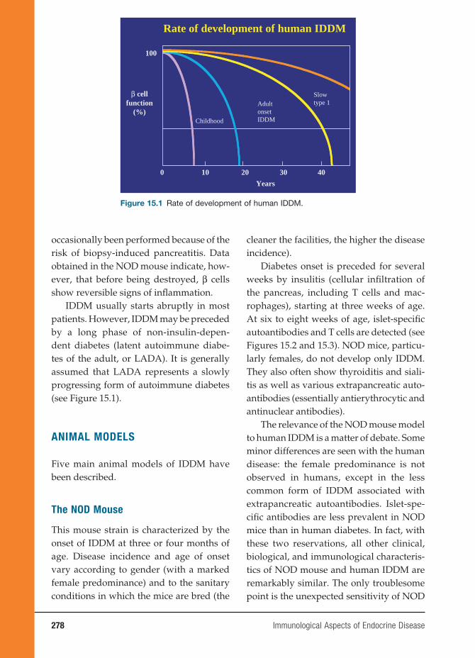
278 Immunological Aspects of Endocrine Disease
occasionally been performed because of the risk of biopsy-induced pancreatitis. Data obtained in the NOD mouse indicate, how-ever, that before being destroyed, β cells show reversible signs of infl ammation.
IDDM usually starts abruptly in most patients. However, IDDM may be preceded by a long phase of non-insulin-depen-dent diabetes (latent autoimmune diabe-tes of the adult, or LADA). It is generally assumed that LADA represents a slowly progressing form of autoimmune diabetes (see Figure 15.1).
ANIMAL MODELS
Five main animal models of IDDM have been described.
The NOD Mouse
This mouse strain is characterized by the onset of IDDM at three or four months of age. Disease incidence and age of onset vary according to gender (with a marked female predominance) and to the sanitary conditions in which the mice are bred (the
cleaner the facilities, the higher the disease incidence).
Diabetes onset is preceded for several weeks by insulitis (cellular infi ltration of the pancreas, including T cells and mac-rophages), starting at three weeks of age. At six to eight weeks of age, islet-specifi c autoantibodies and T cells are detected (see Figures 15.2 and 15.3). NOD mice, particu-larly females, do not develop only IDDM. They also often show thyroiditis and siali-tis as well as various extrapancreatic auto-antibodies (essentially antierythrocytic and antinuclear antibodies).
The relevance of the NOD mouse model to human IDDM is a matter of debate. Some minor differences are seen with the human disease: the female predominance is not observed in humans, except in the less common form of IDDM associated with extrapancreatic autoantibodies. Islet-spe-cifi c antibodies are less prevalent in NOD mice than in human diabetes. In fact, with these two reservations, all other clinical, biological, and immunological characteris-tics of NOD mouse and human IDDM are remarkably similar. The only troublesome point is the unexpected sensitivity of NOD
Rate of development of human IDDM
β cellfunction
(%)
Years
0 10 20 30 40
Childhood
AdultonsetIDDM
Slowtype 1
100
Figure 15.1 Rate of development of human IDDM.
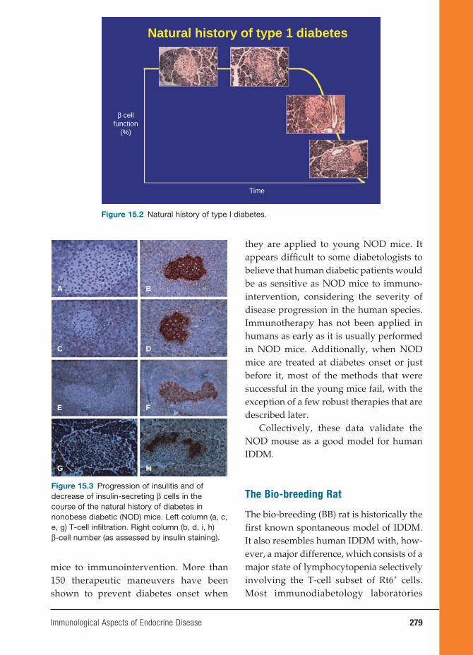
Immunological Aspects of Endocrine Disease 279
they are applied to young NOD mice. It appears diffi cult to some diabetologists to believe that human diabetic patients would be as sensitive as NOD mice to immuno-intervention, considering the severity of disease progression in the human species. Immunotherapy has not been applied in humans as early as it is usually performed in NOD mice. Additionally, when NOD mice are treated at diabetes onset or just before it, most of the methods that were successful in the young mice fail, with the exception of a few robust therapies that are described later.
Collectively, these data validate the NOD mouse as a good model for human IDDM.
The Bio-breeding Rat
The bio-breeding (BB) rat is historically the fi rst known spontaneous model of IDDM. It also resembles human IDDM with, how-ever, a major difference, which consists of a major state of lymphocytopenia selectively involving the T-cell subset of Rt6+ cells. Most immunodiabetology laboratories
Natural history of type 1 diabetes
β cellfunction
(%)
Time
Figure 15.2 Natural history of type I diabetes.
A B
C D
E F
G H
Figure 15.3 Progression of insulitis and of decrease of insulin-secreting β cells in the course of the natural history of diabetes in nonobese diabetic (NOD) mice. Left column (a, c, e, g) T-cell infi ltration. Right column (b, d, i, h) β-cell number (as assessed by insulin staining).
mice to immunointervention. More than 150 therapeutic maneuvers have been shown to prevent diabetes onset when
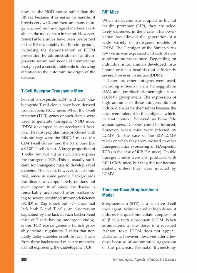
280 Immunological Aspects of Endocrine Disease
now use the NOD mouse rather than the BB rat because it is easier to handle, it breeds very well, and there are many more genetic and immunological markers avail-able in the mouse than in the rat. However, remarkable studies have been performed in the BB rat, notably the Rossini groups, including the demonstration of IDDM prevention by administration of antilym-phocyte serum and neonatal thymectomy that played a considerable role in drawing attention to the autoimmune origin of the disease.
T-Cell Receptor Transgenic Mice
Several islet-specifi c CD4+ and CD8+ dia-betogenic T-cell clones have been derived from diabetic NOD mice. When the T-cell receptor (TCR) genes of such clones were used to generate transgenic NOD mice, IDDM developed in an accelerated fash-ion. The most popular mice produced with this strategy were the BDC2.5 mouse (for CD4 T-cell clones) and the 8.1 mouse (for a CD8+ T-cell clone). A large proportion of T cells (but not all) in such mice express the transgenic TCR. This is usually suffi -cient for transgenic mice to develop rapid diabetes. This is not, however, an absolute rule, since in some genetic backgrounds the disease develops slowly or does not even appear. In all cases, the disease is remarkably accelerated after backcross-ing to severe combined immunodefi ciency (SCID) or Rag knock out −/− mice that lack both B and T cells, an observation explained by the lack in such backcrossed mice of T cells having undergone endog-enous TCR rearrangements (which prob-ably include regulatory T cells) that nor-mally delay diabetes onset. In fact, T cells from these backcrossed mice are monoclo-nal, all expressing the diabetogenic TCR.
RIP Mice
When transgenes are coupled to the rat insulin promoter (RIP), they are selec-tively expressed in the β cells. This obser-vation has allowed the generation of a wide variety of transgenic models of IDDM. The T antigen of the Simian virus (SV) virus was expressed in β cells of non-autoimmune-prone mice. Depending on individual mice, animals developed insu-linoma or major insulitis (not suffi ciently severe, however, to induce IDDM).
Later on, other antigens were used, including infl uenza virus hemagglutinin (HA) and lymphochoriomeningitis virus (LCMV) glycoprotein. The expression of high amounts of these antigens did not induce diabetes by themselves because the mice were tolerant to the antigens, which, in that context, behaved as bona fi de autoantigens. Diabetes could be induced, however, when mice were infected by LCMV (in the case of the RIP-LCMV mice) or when they were crossed to other transgenic mice expressing an HA-specifi c TCR (in the case of RIP-HA mice). Double transgenic mice were also produced with RIP-LCMV mice, but they did not become diabetic unless they were infected by LCMV.
The Low-Dose Streptozotocin
Model
Streptozotocin (STZ) is a selective β-cell toxic agent. Administered at high doses, it induces the quasi-immediate apoptosis of all β cells with subsequent IDDM. When administered at low doses in a repeated fashion, toxic IDDM does not appear. Diabetes is, however, observed after a few days because of autoimmune aggression of the pancreas. Neonatal thymectomy
Immunological Aspects of Endocrine Disease 281
and anti-T-cell antibody treatment prevent low-dose STZ-induced IDDM. This IDDM model has the great advantage of being rapidly induced and obtained in a large variety of mouse strains. Not all strains are equally sensitive to STZ in a geneti-cally controlled fashion. STZ doses must be selected with caution because only a small difference between toxic and immune-trig-gering doses is apparent. The mechanisms leading to STZ-induced diabetes are ill defi ned but probably involve T-cell trig-gering by β-cell autoantigens released by apoptotic β cells.
THE β-CELL LESION:
NATURE OF EFFECTOR CELLS
IDDM is characterized by the selective destruction of β cells (other islet cells, notably glucagon-producing cells remain intact). The β-cell destruction is due to apoptosis. It is preceded by a long phase of prediabetes that has been well charac-terized in animal models. Initial insulitis is essentially of peripheral nature (perinsuli-tis) without β-cell aggression. It becomes progressively more active, resulting in “malignant” insulitis. Even at a late stage (diabetes onset), much of this aggression is reversible because of T-cell-mediated infl ammation. One can thus explain that hyperglycemia in recently diagnosed dia-betic mice is corrected within twenty-four to forty-eight hours by treatment with an anti-T-cell antibody (anti-TCR or anti-CD3) and that islets derived from recently diagnosed diabetic NOD mice recover the capacity to produce insulin after in vitro culture in the absence of T cells or autoan-tibodies.
The nature of the cellular and molecular events at the origin of β-cell infl ammation
and destruction is still a matter of debate. The role of T cells is unquestionable. Dia-betes can be transferred to NOD-SCID mice (which do not spontaneously develop diabetes because they lack T cells) after infusion of purifi ed T cells or T-cell clones. Both CD4 and CD8 T cells are necessary to obtain diabetes transfer when using poly-clonal T cells, even though transfer can be obtained with CD4+ or CD8+ T-cell clones injected alone. Emphasis has recently been put on the role of CD8+ T cells, but there has not yet been a clear demonstration of islet-specifi c cytotoxic T lymphocytes capable of destroying β cells in a FAS or a perforin-dependent manner (probably because of the diffi culties met in using 51Cr release assay with β cells). Using major histocompatibility complex (MHC) class I tetramers with islet antigen mimetopes, it was shown that diabetes progression in the NOD mouse is associated with increasing numbers of high-avidity islet-specifi c CD8 T cells. CD4+ T cells are also important. They may have direct diabetogenic action through cytokine release. They may also intervene as helper T cells for CD8 T cells. A number of studies have attempted to dis-sect the molecular events leading to β-cell apoptosis. The role of free radicals and var-ious cytokines, notably IL-1, has been the matter of particular attention.
No role for islet-specifi c autoantibod-ies has been demonstrated so far. Transfer of sera from diabetic mice does not induce diabetes onset. Pure T-cell populations derived from diabetic mice injected into syngeneic recipients still induce diabetes when the recipients have previously been rendered agammaglobulinemic by post-natal anti-IgM antibody treatment. These observations do not preclude the possi-bility that B cells may play a role in the pathogenesis of IDDM since B-cell-less
282 Immunological Aspects of Endocrine Disease
NOD mice (Ig-/-) do not develop diabe-tes. One may assume that in this context, B cells act as antigen-presenting cells, rather than as producers of diabetogenic autoantibodies. The only evidence in favor of the pathogenic role of autoanti-bodies is the observation that newborns from diabetic autoantibody-positive NOD mouse mothers develop diabetes more rapidly than newborns from antibody-negative mothers. This intriguing obser-vation does not fi t with clinical studies showing the higher IDDM incidence in descendants of diabetic fathers than of diabetic mothers and with the absence of difference in the incidence of diabe-tes in children from antibody-positive or antibody-negative mothers.
THE RUPTURE OF TOLERANCE
TO β-CELL ANTIGENS
β-Cell-Specific T Cells Are Present in
Healthy Individuals but Do Not Attack
β Cells
It is possible to derive T-cell lines and to produce T-cell clones specifi c to vari-ous β-cell autoantigens (insulin, glutamic acid decarboxylase, or GAD) in healthy individuals. Diabetes does not develop, though such β-cell-specifi c T cells are likely to have access to β cells. Similarly, double-RIP LCMV transgenic mice (in which large amounts of LCMV glycopro-tein are expressed in β cells and a large proportion of CD8 T cells expressed an LCMV glycoprotein-specifi c TCR) do not become diabetic unless they are infected with LCMV. This state of ignorance implies that development of diabetes requires that autoreactive T cells be activated to become pathogenic.
Is IDDM Associated with a Defect in
Intrathymic Negative Selection?
T cells differentiate in the thymus where they undergo two successive stages of selection: the fi rst positive, giving rise to the emergence of the TCR repertoire, and the second negative, eliminating high-affi nity self-reactive TCR-carrying T cells. In any event, negative selection is not absolute, because autoreactive T cells are found in the periphery. They can then be stimulated by the corresponding autoantigen when it is adequately presented to them in the context of MHC molecules. The hypothesis has recently been put forward that IDDM could result, at least in part, from defective intrathymic negative selection due to either underexpression of intrathymic β-cell autoantigens (perhaps under the control of the AIRE gene) or to partial expression of these autoantigens in the thymus due to alternative splicing. Also, NOD mouse thymocytes show a defect in thymocyte apoptosis, which could reduce the effi cacy of negative selection.
Driving of the Islet-Specific Response
by β-Cell Antigens Search for IDDM
Autoantigen(s)
There is strong evidence that the diabe-togenic autoimmune response is driven by β-cell autoantigens. (Diabetogenic T cells are rapidly exhausted in the absence of β cells.) The nature of the IDDM tar-get autoantigens remains elusive. Several candidates have been proposed, namely, insulin (or proinsulin), GAD, IA.2, a tyro-sine phosphatase, and IGRP. All four molecules have an exclusive or a prefer-ential β-cell distribution. They induce the production of T cells or autoantibodies in NOD mice or diabetic patients. Insulin
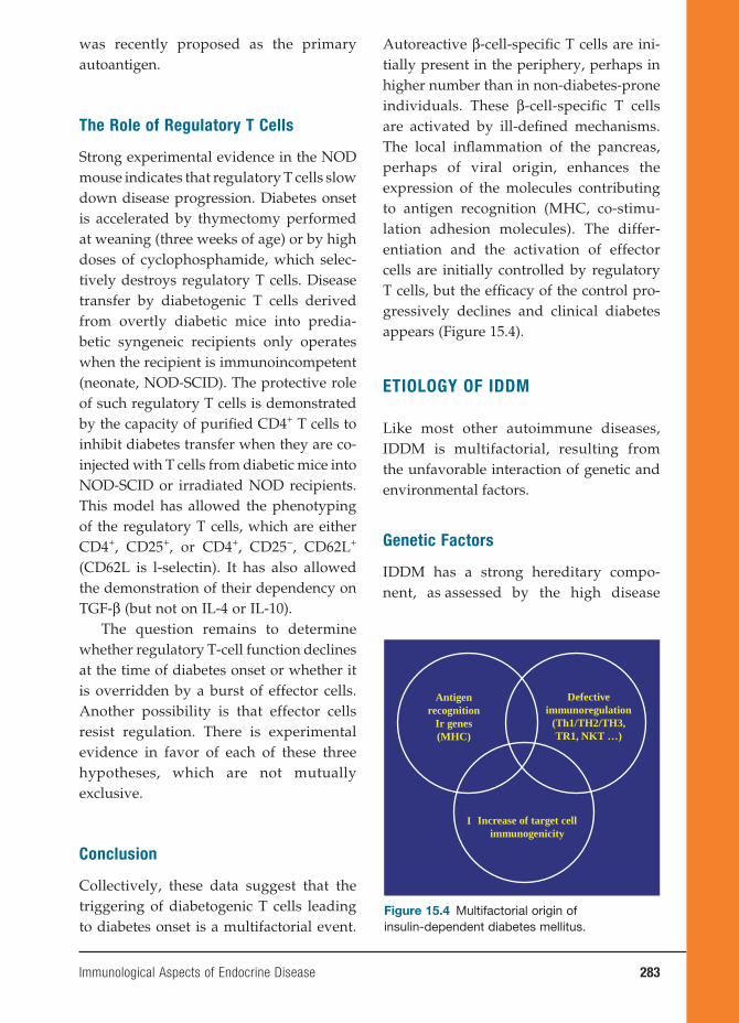
Immunological Aspects of Endocrine Disease 283
was recently proposed as the primary autoantigen.
The Role of Regulatory T Cells
Strong experimental evidence in the NOD mouse indicates that regulatory T cells slow down disease progression. Diabetes onset is accelerated by thymectomy performed at weaning (three weeks of age) or by high doses of cyclophosphamide, which selec-tively destroys regulatory T cells. Disease transfer by diabetogenic T cells derived from overtly diabetic mice into predia-betic syngeneic recipients only operates when the recipient is immunoincompetent (neonate, NOD-SCID). The protective role of such regulatory T cells is demonstrated by the capacity of purifi ed CD4+ T cells to inhibit diabetes transfer when they are co-injected with T cells from diabetic mice into NOD-SCID or irradiated NOD recipients. This model has allowed the phenotyping of the regulatory T cells, which are either CD4+, CD25+, or CD4+, CD25−, CD62L+ (CD62L is l-selectin). It has also allowed the demonstration of their dependency on TGF-β (but not on IL-4 or IL-10).
The question remains to determine whether regulatory T-cell function declines at the time of diabetes onset or whether it is overridden by a burst of effector cells. Another possibility is that effector cells resist regulation. There is experimental evidence in favor of each of these three hypotheses, which are not mutually exclusive.
Conclusion
Collectively, these data suggest that the triggering of diabetogenic T cells leading to diabetes onset is a multifactorial event.
Autoreactive β-cell-specifi c T cells are ini-tially present in the periphery, perhaps in higher number than in non-diabetes-prone individuals. These β-cell-specifi c T cells are activated by ill-defi ned mechanisms. The local infl ammation of the pancreas, perhaps of viral origin, enhances the expression of the molecules contributing to antigen recognition (MHC, co-stimu-lation adhesion molecules). The differ-entiation and the activation of effector cells are initially controlled by regulatory T cells, but the effi cacy of the control pro-gressively declines and clinical diabetes appears (Figure 15.4).
ETIOLOGY OF IDDM
Like most other autoimmune diseases, IDDM is multifactorial, resulting from the unfavorable interaction of genetic and environmental factors.
Genetic Factors
IDDM has a strong hereditary compo-nent, as as sessed by the high disease
Antigenrecognition
Ir genes(MHC)
Defectiveimmunoregulation
(Th1/TH2/TH3,TR1, NKT …)
I Increase of target cellimmunogenicity
Figure 15.4 Multifactorial origin of insulin-dependent diabetes mellitus.
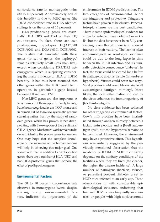
284 Immunological Aspects of Endocrine Disease
concordance rate in monozygotic twins (30 to 40 percent). Approximately half of this heredity is due to MHC genes (the IDDM concordance rate in HLA identical sib lings is on the order of 15 percent).
HLA-predisposing genes are essen-tially HLA DR3 and DR4 or their DQ counterparts. In fact, there are two predisposing haplotypes: DQA1*0501 DQB1*0201 and DQA1*0301 DQB1*0302. The relative risk associated with these genes (or set of genes, the haplotype) remains relatively small (less than fi ve), except when considering DR3/DR4 het-erozygotes, which is surprising consider-ing the major infl uence of HLA on IDDM heredity. It has thus been assumed that other genes within the MHC could be in operation, in particular a gene located between HLA-B and TNF.
Non-MHC genes are also important. A large number of them (approximately twenty) have been recognized in the NOD mouse and in human IDDM thanks to systematic genome scanning rather than by the study of candi-date genes, which has proven rather disap-pointing, with the exception of the insulin and CTLA-4 genes. Much more work remains to be done to identify the precise genes in question. One may hope that the complete knowl-edge of the sequence of the human genome will help in achieving this major goal. One should add that in addition to predisposition genes, there are a number of HLA (DR2) and non-HLA-protective genes that oppose the effect of predisposition genes.
Environmental Factors
The 60 to 70 percent discordance rate observed in monozygotic twins, despite sharing many environmental fac-tors, indicates the importance of the
environment in IDDM predisposition. The two categories of environmental factors are triggering and protective. Triggering factors have proven to be elusive. Pancrea-totropic viruses are the best candidates. There is some epidemiological evidence for a role for enteroviruses, notably Coxsackie B4, but the data have never been fully con-vincing, even though there is a renewed interest in their validity. The lack of clear epidemiological or serological evidence could be due to the long lapse in time between the initial infection and its clini-cally detectable consequence (diabetes). In fact, the virus could be cleared long before its pathogenic effect is visible (hit-and-run hypothesis). Viruses could act in two ways. Viral proteins could resemble certain β-cell autoantigens (antigen mimicry). More likely, the local infl ammation induced by the virus enhances the immunogenicity of β-cell autoantigens.
No clear evidence has been collected for other triggering environmental factors. Cow’s milk proteins have been incrimi-nated through antigen mimicry between a lactalbumin peptide and a β-cell autoan-tigen (p69) but the hypothesis remains to be confi rmed. However, the environment may have a protective effect. This hypoth-esis was initially suggested by the pre-viously mentioned observation that the incidence of IDDM in NOD mice tightly depends on the sanitary conditions of the facilities where they are bred (the cleaner, the higher the disease incidence). A large number of pathogens (bacteria, viruses, or parasites) prevent diabetes onset in NOD mice infected at an early age. These observations fi t with considerable epi-demiological evidence, indicating that human IDDM occurs frequently in coun-tries or people with high socioeconomic
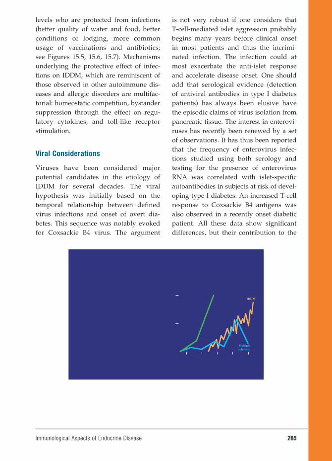
Immunological Aspects of Endocrine Disease 285
levels who are protected from infections (better quality of water and food, better conditions of lodging, more common usage of vaccinations and antibiotics; see Figures 15.5, 15.6, 15.7). Mechanisms underlying the protective effect of infec-tions on IDDM, which are reminiscent of those observed in other autoimmune dis-eases and allergic disorders are multifac-torial: homeostatic competition, bystander suppression through the effect on regu-latory cytokines, and toll-like receptor stimulation.
Viral Considerations
Viruses have been considered major potential candidates in the etiology of IDDM for several decades. The viral hypothesis was initially based on the temporal relationship between defi ned virus infections and onset of overt dia-betes. This sequence was notably evoked for Coxsackie B4 virus. The argument
is not very robust if one considers that T-cell-mediated islet aggression probably begins many years before clinical onset in most patients and thus the incrimi-nated infection. The infection could at most exacerbate the anti-islet response and accelerate disease onset. One should add that serological evidence (detection of antiviral antibodies in type I diabetes patients) has always been elusive have the episodic claims of virus isolation from pancreatic tissue. The interest in enterovi-ruses has recently been renewed by a set of observations. It has thus been reported that the frequency of enterovirus infec-tions studied using both serology and testing for the presence of enterovirus RNA was correlated with islet-specifi c autoantibodies in subjects at risk of devel-oping type I diabetes. An increased T-cell response to Coxsackie B4 antigens was also observed in a recently onset diabetic patient. All these data show signifi cant differences, but their contribution to the
IDDM
Multiplesclerosis
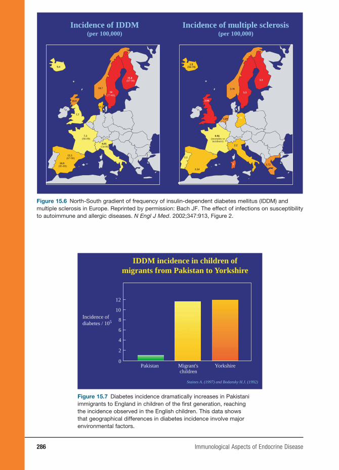
286 Immunological Aspects of Endocrine Disease
5.3
1.9 (56-74)
3.78
1.69 24
0.96 (mortality or incidence)
2.2
5
1.1
2.24
3.94
3.3
1.72
Incidence of multiple sclerosis(per 100,000)
24(77-85)
9.4
7.3 (66-89)
8.05 (54-86)
11.5 (67-90)
19.9
10.9 (85-88)
7.7
19.7
35.8 (87-99)
13.7
Incidence of IDDM(per 100,000)
Figure 15.6 North-South gradient of frequency of insulin-dependent diabetes mellitus (IDDM) and multiple sclerosis in Europe. Reprinted by permission: Bach JF. The effect of infections on susceptibility to autoimmune and allergic diseases. N Engl J Med. 2002;347:913, Figure 2.
IDDM incidence in children of
migrants from Pakistan to Yorkshire
Staines A. (1997) and Bodansky H.J. (1992)
0
2
4
6
8
10
12
Pakistan Migrant'schildren
Yorkshire
Incidence of
diabetes / 105
Figure 15.7 Diabetes incidence dramatically increases in Pakistani immigrants to England in children of the fi rst generation, reaching the incidence observed in the English children. This data shows that geographical differences in diabetes incidence involve major environmental factors.
Immunological Aspects of Endocrine Disease 287
etiologic role of enteroviruses remains indirect since positive results are also observed in a large percentage of the nor-mal population.
At the experimental level, there are rather limited data also. Encephalo-myocarditis virus has been reported to induce IDDM in mice, but IDDM is of the cytopathic type in that case without much immunological involvement. The Coxsackie B4 virus can induce diabetes in mice with some features of autoimmune type I diabetes, but it is not clear whether disease pathogenesis involves a direct cytopathic effect or an immune β-cell attack secondary to virus-induced infl am-mation. This interpretation is compat-ible with data showing that Coxsackie B4 infection of BDC 2.5 mice, which have a limited nontransgenic TCR repertoire, show accelerated diabetes.
Other interesting data have been de rived from the RIP-LCMV transgenic mice. These mice develop IDDM after LCMV infection, according to a hit-and-run mechanism. The virus infection stimulates the induction of LCMVgp-specifi c CD8 cytotoxic T lymphocytes, which cause the disease even though the virus is rapidly cleared through the action of such cyto-toxic T cells.
Collectively, these data are compat-ible with a viral etiology of type I diabetes even though the diabetogenic virus is still unidentifi ed. The etiological infection may take place many years before clini-cal onset, questioning the role of preclini-cal infections and the disease’s seasonal nature. This lag time would explain the diffi culty in identifying the diabetogenic viruses, which in addition might not be unique. Several pancreatotropic viruses could operate through nonspecifi c infl am-matory mechanisms. Antigen mimicry is
possible, but there is no good evidence for this hypothesis in humans (except a ques-tionable homology between a Coxsackie B4 virus protein and GAD) and very little evidence in the mouse. IDDM is accel-erated in BB rats infected with Kilham virus.
CLINICAL CONSIDERATIONS
Diagnosis
IMMUNOASSAYSSeveral assays have been proposed to
detect and quantitate islet-specifi c auto-immunity. Because IDDM is T-cell medi-ated, one would certainly prefer to use T-cell-based rather than humoral assays. Unfortunately, this has proven diffi cult. No fully reliable T-cell assay has so far been described that is suffi ciently sensi-tive (positive in a signifi cant proportion of patients) and disease-specifi c (negative in most controls). Some hope relies in more recent studies using peptides eluted from IDDM-predisposing HLA molecules, but the results obtained must be confi rmed and rendered applicable to their routine usage. One may also hope that other new T-cell targets will become available, such as IGRP, which stimulates CD8 T cells. The emergence of the tetramer technol-ogy could prove useful, particularly for CD8+ T cells.
At variance with T-cell-based assays, autoantibody detection has been extremely informative. Islet cell-specifi c autoanti-bodies (ICA) were fi rst detected by indi-rect immunofl uorescence using sections of human pancreas (ICA). These tests proved extremely useful for several decades. It is not fully quantitative and has now been replaced by assays using chemi-cally defi ned β-cell antigens. There is not
288 Immunological Aspects of Endocrine Disease
a single IDDM target autoantigen, but rather autoantibodies are found against a variety of islet autoantigens using highly reliable assays. Anti-insulin autoantibodies are measured using a radioimmunoassay rather than ELISA, whose interpretation is hampered by low-avidity autoantibodies. Anti-GAD and anti-IA-2 autoantibodies are measured by radioligand assays in which the autoantigen is synthesized de novo in vitro as a radiolabeled (35S) recombinant protein in cell-free transla-tion systems.
HELP FOR DIAGNOSISThe diagnosis of IDDM is essentially
made on a metabolic basis (hyperglycemia and glycosuria). There are certain settings, however, where confi rmation of the pres-ence of islet-specifi c autoimmunity may be useful.
At the onset of clinical diabetes, there may be some hesitation about the auto-immune origin of the disease even in relatively young subjects (young adults) particularly in non-HLA DR3 non-DR4 subjects. It may be important to confi rm the presence of islet-specifi c autoimmu-nity, which will become critical when more patients are enrolled in immuno-therapy trials in which nonautoimmune patients are excluded.
Another important setting in this context is that of LADA. These patients appear to be conventional type II diabetes patients (with the exception of an unusual absence of excess weight). Orientation toward LADA (slowly progressing IDDM) is done in the presence of β-cell-specifi c autoantibodies. Recognition of LADA is important clinically because the knowl-edge of a likely short- or medium-term evolution toward insulin dependency will prompt clinicians to accelerate the onset
of insulin therapy, protecting patients from exposure to the risk of degenerative complications associated with unsatisfac-tory metabolic control (usually observed when insulin dependency is progressively recognized).
PREDIABETESA large (and yet ill-defi ned) propor-
tion of β cells are destroyed at the clinical onset of IDDM, even though there is still a signifi cant number of living β cells that are rescuable by anti-CD3 antibodies. In any event, reserving immunotherapy for subjects with high β-cell mass would be highly desirable. This can be achieved by identifying subjects progressing toward IDDM but still capable of maintaining normal glycemia despite some reduction of the β-cell mass. These subjects can be recognized by the early presence of islet-specifi c autoantibodies using the reliable assays described earlier. These predia-betic subjects can be identifi ed with a high degree of precision. Subjects with the three autoantibodies (insulin, GAD, and IA-2) will become diabetic within fi ve years in more than 90 percent of cases. However, the situation is less clear for subjects with only one or two antibodies. Furthermore, diabetes prediction in the general popula-tion (in the absence of an IDDM heredity) is much less effi cacious than in relatives of diabetic patients in whom it is essen-tially performed today both because of the higher risk (7 percent versus 0.2 percent) and the higher prediction value of the autoantibody assays.
One may complement autoantibody detection by genetic testing, which is today essentially limited to HLA, DR3, and DR4 (or their DQ equivalents). This approach has been used for systematic DR3/DR4 screening of young children born of
Immunological Aspects of Endocrine Disease 289
diabetic parents, starting antibody moni-toring at birth.
IMMUNOTHERAPYInsulin is not a fully satisfactory treat-
ment for IDDM. It is associated with major constraints (monitoring) and hazards (hypoglycemia) and above all does not com-pletely prevent the onset of degenerative complications; hence, the interest given over the past two decades to the immuno-therapeutic approach.
On the basis of results obtained in ani-mal models, treatment was fi rst attempted with recently diagnosed IDDM patients with nonspecifi c immunosuppressants, namely, cyclosporin A and azathioprin. A signifi cant retardation of disease pro-gression was obtained with observation of complete disease remission in a sta-tistically signifi cant percentage of cases. Drug-specifi c side effects were observed and the disease relapsed when treatment was stopped. Inasmuch as chronic immu-nosuppression is hardly possible in young subjects like IDDM patients, it was con-sidered that the immunotherapy research should be oriented toward induction of long-term restoration of islet-specifi c tol-erance after a short course of induction treatment, considering the side effects of long-term immunosuppression. Two approaches were undertaken. The fi rst one was inspired by the remarkable abil-ity of some β-cell autoantigens to induce long-term tolerance and diabetes preven-tion in NOD mice when administered at an early age either parenterally or orally. It was rapidly realized that oral insulin did not slow IDDM progression in recently diagnosed subjects. Similar negative results were observed when administering parenteral insulin in prediabetic subjects, even though the trial was performed in a
large number of patients. More recently, a new approach was developed based on the demonstration that administration of nonmitogenic anti-CD3 antibodies could induce long-term remission of recently diagnosed diabetes, an effect associated with the parallel elimination of effector cells and resetting of immunoregulatory TGF β-dependent regulatory T cells. An open phase I/II trial provided promising results that have now been confi rmed in a phase II randomized placebo-controlled study. Importantly, in the latter trial, there was not only maintenance of β-cell func-tion but also real rescue in a large propor-tion of patients, confi rming that at the time of diabetes onset, there is a signifi cant role for reversible islet infl ammation in addi-tion to β-cell destruction.
Other trials are now in progress using β-cell-antigen peptides or altered peptides. It will be important to determine whether the antigen-specifi c approach is operational in advanced disease. Perhaps the solution will be the combined usage of tolerance-inducing regimens of the CD3 antibody type complemented in a second phase by antigen-specifi c therapy.
One should also follow with interest the prolifi c research of new immunothera-peutic methods tested in the NOD mouse, with the caveat of not overinterpreting data obtained in early treatments that may act in a nonspecifi c fashion or at least in conditions that do not correspond to the clinical setting.
GENERAL CONCLUSION
Insulin-dependent diabetes mellitus is an autoimmune disease essentially due to β-cell aggression (infl ammation and killing) by CD4+ and CD8+ T cells. The
290 Immunological Aspects of Endocrine Disease
autoimmune nature of the disease and the complex underlying mechanisms (triggering of β-cell-specifi c responses by local infl ammation overriding regula-tory T cells) should not mask the impor-tance of etiologic initial events, which probably involve an as-yet-unraveled pancreatotropic virus. The major present issue and logical corollary of the multi-tude of clinical and experimental studies performed over the past three decades is to establish a safe and effi cacious immu-notherapy active before disease onset, which means that major efforts should be developed to detect prediabetic sub-jects with high reliability. When this goal is achieved, one may envision, thanks to the use of tolerance-inducing regimens, the progressive eradication of the disease using vaccination strategies to achieve these goals.
BIBLIOGRAPHY
REVIEWSBach J-F. The effect of infections on
susceptibility to autoimmune and allergic diseases. N Engl J Med. 2002;347: 911–920.
Bach J-F. Insulin-dependent diabetes mel-litus as an autoimmune disease. Endocr Rev. 1994;15:516–542.
Bach J-F. Immunotherapy of insulin-depen-dent diabetes mellitus. Curr Opin Immu-nol. 2001;13:601–605.
Bach J-F, Chatenoud L. Tolerance to islet autoantigens in type 1 diabetes. Annu Rev Immunol. 2001;19:131–161.
SUGGESTED READINGChristen U, Juedes A, Homann D, Von Her-
rath MG. Virally induced infl ammation
and therapeutic avenues in type 1 diabetes. Endocrinol Metab Clin North Am. 2004;33:45–58.
Greenbaum CJ. Type 1 diabetes interven-tion trials: what have we learned? A critical review of selected interven-tion trials. Clin Immunol. 2002;104: 97–104.
Hanninen A, Hamilton-Williams E, Kurts C. Development of new strategies to prevent type 1 diabetes: the role of ani-mal models. Ann Med. 2003;35:546–563.
Hirschhorn JN. Genetic epidemiology of type 1 diabetes. Pediatr Diabetes. 2003;4:87–100.
Ide A, Eisenbarth GS. Genetic susceptibil-ity in type 1 diabetes and its associated autoimmune disorders. Rev Endocr Metab Disord. 2003;4:243–253.
Jun HS, Yoon JW. 2003. A new look at viruses in type 1 diabetes. Diabetes Metab Res Rev. 2003;19:8–31.
Karvonen M, Sekikawa A, La Porte R, Tuomilehto J, Tuomilehto-Wolf E. Type 1 diabetes: global epidemiology. In: Ekoe JM, Zimmet P, Williams R. eds. The Epidemiology of Diabetes Mellitus: An International Perspective. Chichester: Wiley; 2001:71–102.
Knip M. Natural course of preclinical type 1 diabetes. Horm Res. 2002;57(suppl 1):6–11.
Knip M. Environmental triggers and deter-minants of beta-cell autoimmunity and type 1 diabetes. Rev Endocr Metab Dis-ord. 2003;4:213–223.
McDevitt H. The role of MHC class II mol-ecules in the pathogenesis and preven-tion of type I diabetes. Adv Exp Med Biol. 2001;490:59–66.
Pociot F, McDermott MF. Genetics of type 1 diabetes mellitus. Genes Immun. 2002;3:235–249.

Immunological Aspects of Endocrine Disease 291
Roep BO. The role of T-cells in the patho-genesis of type 1 diabetes: from cause to cure. Diabetologia. 2003;46:305–321.
Varela Calvino R, Peakman M. Entero-viruses and type 1 diabetes. Diabetes Metab Res Rev. 2003;19:431–441.
Yang Y, Santamaria P. Dissecting autoimmune diabetes through genetic manipulation of non-obese diabetic mice. Diabetologia. 2003;46: 1447–1464.

16. Immune-Mediated Neurological Syndromes
Jacqueline Friedman, M.D.
293
This chapter will discuss the clinical phenomena and underlying theories of autoimmune diseases affecting the human nervous system. Examples will be drawn from diseases affecting (1) the central nervous system (CNS): brain and spinal cord, multiple sclerosis (MS); (2) a disease of the peripheral nerves, Guil-lain-Barré syndrome (GBS); and (3) an autoimmune disease affecting the endplate between nerve and muscle: myasthenia gravis (MG). In these diseases, the nervous system or motor endplate is the unique tar-get of autoimmunity. Systemic lupus ery-thematosus (SLE), an example of a systemic autoimmune disease with nervous system involvement, will also be discussed.
Animal models for the study of patho-genesis and treatment have played a large role in the study of immunologically medi-ated neurological diseases. The success has been variable and not always well corre-lated with the human condition. Animal models will be mentioned within the con-text of each disease addressed, and a gen-eral discussion of this topic is included at the end of the chapter.
MULTIPLE SCLEROSIS
MS is the most prevalenat infl ammatory disease of the CNS in humans, affecting 1 person per 1,000 adults. This disease affects women in a 2:1 ratio to men, and,
for the most part, respects the latitudi-nal geographic presentation; that is, it occurs mostly in the northern European population or those of northern European descent. The incidence of MS gradually decreases as one approaches the equator. MS increases in incidence as one moves south, away from the equator. Epidemio-logical studies have also determined that the risk rate is fi xed at the age of puberty; that is, if a person moves from an area of high risk to low risk before the age of fi f-teen that person takes on the risk rate of the new location. If a person moves after the age of 15, that person maintains the risk rate from the area he or she migrated from. The reverse is true in going from low-risk areas to high-risk areas. Epide-miologists have interpreted this to mean that the inciting event or exposure that will ultimately lead to the clinical picture of MS occurs decades before the dis-ease is manifested in the mid-twenties and thirties.
Some epidemiologists point to the geo-graphical parameters of the disease as evi-dence of a genetic predisposition. In fact, even within the northern latitudes, the Laplanders and Inuits are not susceptible to MS. Specifi c human leucocyte antigens (HLA) types, such as DR2, occur more fre-quently in MS patients than in the general population. Twin studies also support this hypothesis, with identical twins experienc-ing concordance for the disease at a rate
294 Immune-Mediated Neurological Syndromes
twenty to fi fty times higher than expected from the population as a whole.
Newer studies are focusing on the role of 25-hydroxy vitamin D, an immuno-modulating factor, in the pathogenesis of MS vis-à-vis the latitudinal gradient. High levels of vitamin 25-hydroxy vitamin D in the serum signifi cantly lower the odds ratio of MS risk in large population studies. Pos-sibly, the higher exposure to sunlight in the equatorial latitudes enhances natural absorption of vitamin D from the skin, resulting in higher serum levels, which may protect against MS.
By defi nition, the disease must affect two parts of the CNS at two discrete periods. Historically, the neurological examination and patient’s clinical history were used to fulfi ll these criteria. More recently, the MRI scanner has become a powerful adjunct to the history and examination in these patients. Multiple areas of the CNS can now be imaged to confi rm the presence of more than one lesion. Serial MRI scans can now confi rm multiple episodes in time, even when the patient is asymptomatic. A scan may be performed at three-month intervals from the fi rst symptom to deter-mine whether new lesions present them-selves. New evidence of infl ammation fi ts the criterion for separate episodes in time and thus the defi nition of MS. This is called the McDonald criterion for MS and has strengthened the practice of treating patients earlier in the disease course before signifi cant disability sets in.
Cerebrospinal fl uid (CSF) analysis has also proven helpful in diagnosing MS. The CSF is usually normal in terms of protein and cells but may have a slight protein elevation or increase in lymphocytes, rarely more than 50 cells/ml. Spinal fl uid analysis, which directs one to the diagnosis of MS, includes evidence of intrathecal synthesis
of immunoglobulins and the presence of oligoclonal bands. These antibody bands, primarily IgG, are found in approximately 90 percent of patients at some point in their disease course. These antibodies are detected on an agarose gel in the presence of an electric fi eld and isoelectric focusing. The bands present in the basic area of the gel were initially felt to show a spe-cifi c immune response to an autoantigen or infectious agent in MS patients. How-ever, years of investigation have detected no consistent antibody response in MS patients. In fact, the pattern differs from patient to patient. Newer interpretation for the presence of oligoclonal antibodies is that they represent polyclonal dysregu-lation, rather than B-cell populations with specifi c immune responses induced. None-theless, the oligoclonal bands are a use-ful adjunctive test in the diagnosis of MS. Other diseases, which can produce bands, can be readily ruled out by other means, such as syphilis with a rapid plasma reagin (RPR) test or Lyme disease with enzyme-linked immunosorbent assay (ELISA) and Western blotting against Borrelia antigens.
The symptoms of MS depend on their localization in the nervous system. If the optic nerves are affected, the patient can present with decreased vision, ranging from color blindness to total loss of vision, which is accompanied by pain due to eye move-ment. Lesions of the brain stem can present with double vision, trigeminal neuralgia, severe pain on the face, or speech or swal-lowing diffi culties. Lesions in the spinal cord can present with numbness, tingling, or weakness of the upper or lower extremities, as well as bladder/bowel and sexual dys-function. Symptoms are often accompanied by severe fatigue, possibly caused by release of cytokines in the CNS. The symptoms are manifest because nerve conduction is
Immune-Mediated Neurological Syndromes 295
signifi cantly slowed in de myelinated fi bers. Saltatory conduction between the nodes of Ranvier is disorganized, and the slowed conduction results in neurological symp-toms of nerve dysfunction.
In addition, local edema in the area of the plaque may interrupt function tempo-rarily. Some patients develop symptoms that last a few hours or less. It is believed that low-affi nity antibodies present in MS directly interact with antigens com-posing the sodium channels on neurons directly. The attachment and subsequent freeing of the antibodies from these chan-nels may be responsible for a rapid change in symptoms not involving an entire autoimmune/infl ammatory cascade.
This fi nding points to the premise that MS is not solely a disease of white matter and demyelinating pathology but may also be a neuronal disease as well. MRI scanning using the neuronal marker NAA (N-acetyl- aspartate) has demonstrated that neuronal dysfunction occurs earlier in MS patients than would be expected from destruction of neurons based only on demyelination secondarily. This may open up new ave-nues of therapy in the future, directed not only toward remyelination but also in the area of nerve growth factors as well.
The overall pathological lesion has been categorized as a demyelinated area within the CNS white matter, with the presence of infl ammation, gliosis remyelination, or axonal pathology. More recently, the pathology of MS has been categorized into four distinct patterns. Two patterns show lesions induced by autoreactive T cells (type I), or T cells plus antibodies and complement (type II). These are similar to the pathology induced by animal models discussed later. Patterns III and IV appear to be caused more by a dystrophy of the oligodendrocyte, and apoptosis, rather
than an autoimmune reaction. The pathol-ogies are homogeneous within the demy-elinated lesions in each patient but differ from patient to patient. This may suggest that different disease pathologies may be linked to one clinical state, MS, or that there are clinical subcategories of MS pre-sentations that may be clinically tied to the pathology, which is yet to be determined. This will ultimately refl ect different treat-ment modalities for the different presenta-tions of MS.
Experimental allergic encephalomyeli-tis (EAE) in rodents, which has been the hallmark animal model of MS, is induced following immunization with whole myelin or antigens related to myelin (i.e., myelin oligodendrocyte glycoprotein, or MOG, or myelin basic protein, or MBP with complete Freund’s adjuvant). These myelin antigens are also cross-reactive with T cells and antibodies directed against the Semliki Forest virus, used to promote an immune-mediated demyelinating viral encephalitis model. Antibodies to MOG induce demy-elination in EAE and exacerbate the clinical disease, while antibodies to MBP reduce disease severity. A polymer of a peptide sequence of MBP is an approved immuno-modulating drug for MS in humans, which is believed to stabilize disease by chang-ing the T-cell repertoire from TH1 to TH2 in the CNS.
The pathogenesis of the EAE model involves immune cells migrating from the peripheral lymphoid system, where acti-vation takes place, to the CNS. These cells then mediate tissue damage in the CNS. In addition, epitope spreading occurs where autoreactive T cells and antibodies become more diverse.
This model also allows for the concept of microbial-induced autoimmune reactions in genetically susceptible mice strains with
296 Immune-Mediated Neurological Syndromes
Theiler’s murine virus or Semliki Forest virus. Blood-brain barrier passage by CD4+ cells, CNS immigration, demyelination lesion induction, and clinical neurological defi cit in the rodent have all been achieved in this model. However, the cause of per-manent neurological defi cit in MS and EAE is still not entirely understood. CNS and infl ammation and demyelination do not account for irreversible neurological symp-toms. Recently, axonal pathology has been also implicated not only in MS but also in EAE as well and may lead to future thera-pies. In terms of therapies for MS, many biologic agents are species specifi c, and nonhuman primates such as marmosets and rhesus monkeys are the more appropri-ate model to use. For studying MS, nonhu-man primates have proved the most useful model thus far, despite the history of EAE in rodents. They also provide species-spe-cifi c therapeutic options to be investigated, as well as an easier ability to do longitudi-nal sampling of body fl uids.
Treatment of the disease is aimed at three levels. The fi rst level, the use of intra-venous corticosteroids, is the treatment and shortening of acute attacks, such as visual loss with optic neuritis, a spinal cord syn-drome, or hemiparesis. Patients are also treated with oral steroids for less severe attacks, with the exception of optic neu-ritis. In addition to its anti-infl ammatory properties, steroids may strengthen the blood-brain barrier and decrease edema in a lesion, causing a rapid reversal of symptoms.
The next level of therapy for patients is the daily management of ongoing symp-toms. This might include muscle relaxants for spasticity, amantadine, or modafi nil for fatigue, antidepressants or anticon-vulsants for pain and mood changes, and anticholinergic medication for bladder
spasticity and incontinence. Fortunately, most MS patients have a limited constella-tion of symptoms, but some require signifi -cant polypharmacy.
Ongoing physical therapy and a course of psychotherapy are often prescribed for patients. Physical therapy optimizes the muscle capabilities of the patient and has an effect on psychological well-being as well. Depression plays a major role in MS, but it is unclear whether it is a reactive or endogenous mood disorder. Nonetheless, suicide attempts are a considerable risk with MS patients and psychological coun-seling is often suggested.
Interferons and immunomodulating agents have become the mainstay of treat-ment of the long clinical course of MS. Specifi cally, IFN-β, given subcutaneously or intramuscularly has become recom-mended lifelong for patients at this time. Statistical signifi cance was achieved in these patients, both in terms of MRI effects and the frequency and severity of attacks. The clinical relapse rate diminished by one-third, and MRI data suggest an even more dramatic drop in lesion volume (−17.3 percent) and number (−83 percent) compared with placebo. The literature is somewhat contradictory about the goal of slowing the gradual progression or wors-ening of the disease overall, but some stud-ies are able to confi rm this fi nding. While interferons have several immunomodulat-ing effects and may also be antiviral, the main function of these medications in this setting may be to block adhesion of lym-phocytes and macrophages to the blood-brain barrier and thus limit traffi cking into the CNS. Natalizumab, a newly approved immunomodulator in MS also inhibits the attachment of lymphocytes to the endothe-lial cells and limits traffi cking of the cells across. It is an antibody directed against
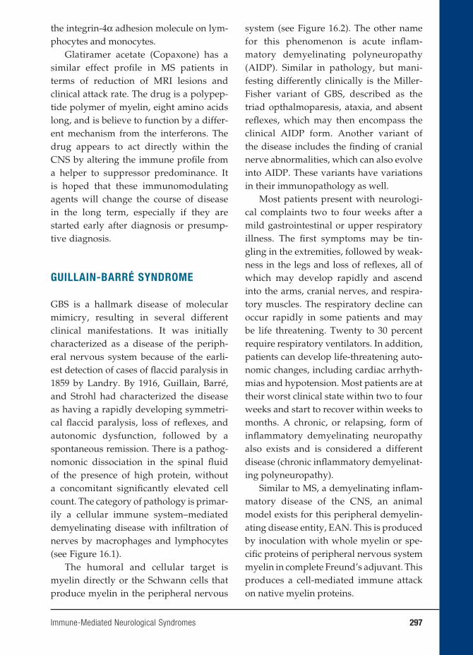
Immune-Mediated Neurological Syndromes 297
the integrin-4α adhesion molecule on lym-phocytes and monocytes.
Glatiramer acetate (Copaxone) has a similar effect profi le in MS patients in terms of reduction of MRI lesions and clinical attack rate. The drug is a polypep-tide polymer of myelin, eight amino acids long, and is believe to function by a differ-ent mechanism from the interferons. The drug appears to act directly within the CNS by altering the immune profi le from a helper to suppressor predominance. It is hoped that these immunomodulating agents will change the course of disease in the long term, especially if they are started early after diagnosis or presump-tive diagnosis.
GUILLAIN-BARRÉ SYNDROME
GBS is a hallmark disease of molecular mimicry, resulting in several different clinical manifestations. It was initially characterized as a disease of the periph-eral nervous system because of the earli-est detection of cases of fl accid paralysis in 1859 by Landry. By 1916, Guillain, Barré, and Strohl had characterized the disease as having a rapidly developing symmetri-cal fl accid paralysis, loss of refl exes, and autonomic dysfunction, followed by a spontaneous remission. There is a pathog-nomonic dissociation in the spinal fl uid of the presence of high protein, without a concomitant signifi cantly elevated cell count. The category of pathology is primar-ily a cellular immune system–mediated demyelinating disease with infi ltration of nerves by macrophages and lymphocytes (see Figure 16.1).
The humoral and cellular target is myelin directly or the Schwann cells that produce myelin in the peripheral nervous
system (see Figure 16.2). The other name for this phenomenon is acute infl am-matory demyelinating polyneuropathy (AIDP). Similar in pathology, but mani-festing differently clinically is the Miller-Fisher variant of GBS, described as the triad opthalmoparesis, ataxia, and absent refl exes, which may then encompass the clinical AIDP form. Another variant of the disease includes the fi nding of cranial nerve abnormalities, which can also evolve into AIDP. These variants have variations in their immunopathology as well.
Most patients present with neurologi-cal complaints two to four weeks after a mild gastrointestinal or upper respiratory illness. The fi rst symptoms may be tin-gling in the extremities, followed by weak-ness in the legs and loss of refl exes, all of which may develop rapidly and ascend into the arms, cranial nerves, and respira-tory muscles. The respiratory decline can occur rapidly in some patients and may be life threatening. Twenty to 30 percent require respiratory ventilators. In addition, patients can develop life-threatening auto-nomic changes, including cardiac arrhyth-mias and hypotension. Most patients are at their worst clinical state within two to four weeks and start to recover within weeks to months. A chronic, or relapsing, form of infl ammatory demyelinating neuropathy also exists and is considered a different disease (chronic infl ammatory demyelinat-ing polyneuropathy).
Similar to MS, a demyelinating infl am-matory disease of the CNS, an animal model exists for this peripheral demyelin-ating disease entity, EAN. This is produced by inoculation with whole myelin or spe-cifi c proteins of peripheral nervous system myelin in complete Freund’s adjuvant. This produces a cell-mediated immune attack on native myelin proteins.
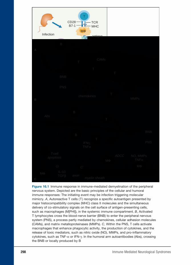
298 Immune-Mediated Neurological Syndromes
A
CD28 TCR
T
T
TT
T
T
T
B
BAbs
T-cellapoptosis
axonmyelin sheath
ADCC
B
C’
NO, MMPsTNFα
IL-10TGFβ
IFNγ,TNFα
MHC
CAMs
BNB
PNS
BNB
PNS
chemokinesMMPs
antigenInfection
B7-1
MΦ
MΦ
MΦ
− MΦ
B
C
Figure 16.1 Immune response in immune-mediated demyelination of the peripheral nervous system. Depicted are the basic principles of the cellular and humoral immune responses: The initiating event may be infection triggering molecular mimicry. A, Autoreactive T cells ( T ) recognize a specifi c autoantigen presented by major histocompatibility complex (MHC) class II molecules and the simultaneous delivery of co-stimulatory signals on the cell surface of antigen-presenting cells, such as macrophages (M[PHI]), in the systemic immune compartment. B, Activated T lymphocytes cross the blood-nerve barrier (BNB) to enter the peripheral nervous system (PNS), a process partly mediated by chemokines, cellular adhesion molecules (CAMs), and matrix metalloproteinases (MMPs). C, Within the PNS, T cells activate macrophages that enhance phagocytic activity, the production of cytokines, and the release of toxic mediators, such as nitric oxide (NO), MMPs, and pro-infl ammatory cytokines, such as TNF-α or IFN-γ. In the humoral arm autoantibodies (Abs), crossing the BNB or locally produced by B
Immune-Mediated Neurological Syndromes 299
However, more recently, basic prem-ises of this disease have been redefi ned. It is now recognized that there may be a dis-tinct axonal form of GBS. Electromyogra-phy has been able to distinguish these two forms. An attack against myelin and axons may be found concurrently in the same patient, with both demyelination and peri-axonal macrophages.
Molecular mimicry has been found to be the major pathogenic force and the basis for autoimmunity. Various preced-ing infections can initiate the immune response that causes the clinical state of GBS. The two major agents are Campylo-bacter and Cytomegalovirus, with Epstein-Barr virus considered an inciting agent as well. Specifi c features of the incit-ing organism may play a major role in the development of GBS. For instance, although Campylobacter jejuni often may cause diarrhea, the strains that have been linked to GBS (O-serotypes) are different from those strains causing diarrhea only but are genetically similar to each other. Only 70 percent of those infected with Campylobacter in the presence of GBS give a previous history of gastrointestinal com-plaints in the three months before the neu-rological onset, but the risk of developing GBS within two months of a symptomatic Campylobacter infection is 100-fold higher than the general population. GBS follow-ing C. jejuni and intestinal symptoms are
felt to have a more dire clinical course, involving axonal destruction in addition to demyelination.
The Miller-Fisher variant, which involves primarily the cranial nerves, dem-onstrates the close association between antiganglioside antibodies and a specifi c syndrome. Anti-GQ1b has a high specifi city and sensitivity for this symptom complex. GQ1b is a ganglioside that is a component of cranial nerve myelin. Similarly, anti-GM1 (anti-GD1a) antibodies have been linked specifi cally to the axonal form of the disease, acute motor axonal neuropathy. Although GM1 antigens can be found on motor and sensory nerves, there are vari-able reports in the literature about whether these antigens are on the axon or in the myelin sheath. However, antibodies to GM1 can bind to the nodes of Ranvier and activate complement. Other factors may be necessary for the disease to occur, as many patients with Campylobacter infections pro-duce antibodies to GM1 ganglioside but do not develop neurological symptoms. Because the disease can improve rapidly, an association between sodium channels and the antibody has been studied, but there has been no consistent association with sodium channels.
Infl uenza vaccine has been implicated as a causative agent for GBS as well. How-ever, careful epidemiological studies have not borne this out. Combining the 1992–1993
lymphocytes (B), contribute to the process of demyelination and axonal damage. Abs can mediate demyelination by antibody-dependent cell-mediated cytotoxicity (ADCC), can block functionally relevant epitopes for nerve conduction, and can activate the complement system (C′) by the classic pathway, yielding pro-infl ammatory mediators and the lytic C5b-9 terminal complex. The termination of the infl ammatory response is partly mediated by macrophages, inducing T-cell apoptosis and the release of anti-infl ammatory cytokines, such as IL-10 and transforming growth factor-β (TGF-β). Modifi ed from Hartung, H. et al., Acute immunoinfl ammatory neuropathy: update on Guillain-Barre´ syndrome: Curr Opin Neurol. 2002;15(5):571–577.
Figure 16.1 (Caption continued)
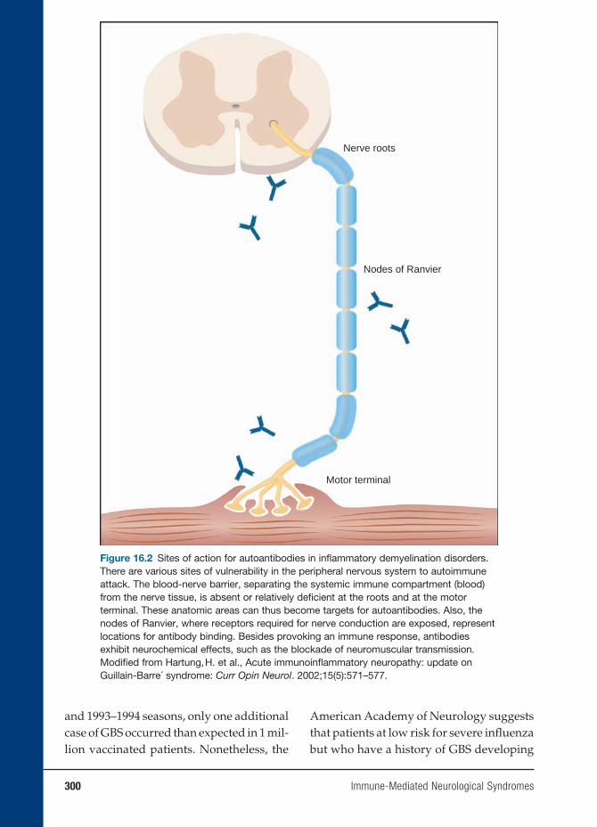
300 Immune-Mediated Neurological Syndromes
Nodes of Ranvier
Nerve roots
Motor terminal
Figure 16.2 Sites of action for autoantibodies in infl ammatory demyelination disorders. There are various sites of vulnerability in the peripheral nervous system to autoimmune attack. The blood-nerve barrier, separating the systemic immune compartment (blood) from the nerve tissue, is absent or relatively defi cient at the roots and at the motor terminal. These anatomic areas can thus become targets for autoantibodies. Also, the nodes of Ranvier, where receptors required for nerve conduction are exposed, represent locations for antibody binding. Besides provoking an immune response, antibodies exhibit neurochemical effects, such as the blockade of neuromuscular transmission. Modifi ed from Hartung, H. et al., Acute immunoinfl ammatory neuropathy: update on Guillain-Barre´ syndrome: Curr Opin Neurol. 2002;15(5):571–577.
and 1993–1994 seasons, only one additional case of GBS occurred than expected in 1 mil-lion vaccinated patients. Nonetheless, the
American Academy of Neurology suggests that patients at low risk for severe infl uenza but who have a history of GBS developing
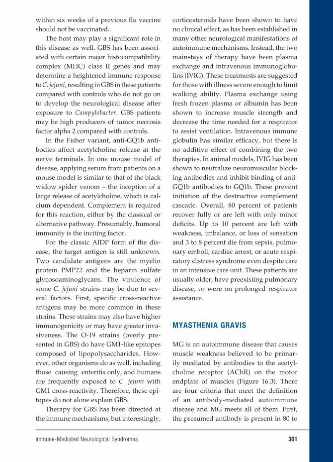
Immune-Mediated Neurological Syndromes 301
within six weeks of a previous fl u vaccine should not be vaccinated.
The host may play a signifi cant role in this disease as well. GBS has been associ-ated with certain major histocompatibility complex (MHC) class II genes and may determine a heightened immune response to C. jejuni, resulting in GBS in these patients compared with controls who do not go on to develop the neurological disease after exposure to Campylobacter. GBS patients may be high producers of tumor necrosis factor alpha 2 compared with controls.
In the Fisher variant, anti-GQ1b anti-bodies affect acetylcholine release at the nerve terminals. In one mouse model of disease, applying serum from patients on a mouse model is similar to that of the black widow spider venom – the inception of a large release of acetylcholine, which is cal-cium dependent. Complement is required for this reaction, either by the classical or alternative pathway. Presumably, humoral immunity is the inciting factor.
For the classic AIDP form of the dis-ease, the target antigen is still unknown. Two candidate antigens are the myelin protein PMP22 and the heparin sulfate glycosoaminoglycans. The virulence of some C. jejuni strains may be due to sev-eral factors. First, specifi c cross-reactive antigens may be more common in these strains. These strains may also have higher immunogenicity or may have greater inva-siveness. The O-19 strains (overly pre-sented in GBS) do have GM1-like epitopes composed of lipopolysaccharides. How-ever, other organisms do as well, including those causing enteritis only, and humans are frequently exposed to C. jejuni with GM1 cross-reactivity. Therefore, these epi-topes do not alone explain GBS.
Therapy for GBS has been directed at the immune mechanisms, but interestingly,
corticosteroids have been shown to have no clinical effect, as has been established in many other neurological manifestations of autoimmune mechanisms. Instead, the two mainstays of therapy have been plasma exchange and intravenous immunoglobu-lins (IVIG). These treatments are suggested for those with illness severe enough to limit walking ability. Plasma exchange using fresh frozen plasma or albumin has been shown to increase muscle strength and decrease the time needed for a respirator to assist ventilation. Intravenous immune globulin has similar effi cacy, but there is no additive effect of combining the two therapies. In animal models, IVIG has been shown to neutralize neuromuscular block-ing antibodies and inhibit binding of anti-GQ1b antibodies to GQ1b. These prevent initiation of the destructive complement cascade. Overall, 80 percent of patients recover fully or are left with only minor defi cits. Up to 10 percent are left with weakness, imbalance, or loss of sensation and 3 to 8 percent die from sepsis, pulmo-nary emboli, cardiac arrest, or acute respi-ratory distress syndrome even despite care in an intensive care unit. These patients are usually older, have preexisting pulmonary disease, or were on prolonged respirator assistance.
MYASTHENIA GRAVIS
MG is an autoimmune disease that causes muscle weakness believed to be primar-ily mediated by antibodies to the acetyl-choline receptor (AChR) on the motor endplate of muscles (Figure 16.3). There are four criteria that meet the defi nition of an antibody-mediated autoimmune disease and MG meets all of them. First, the presumed antibody is present in 80 to
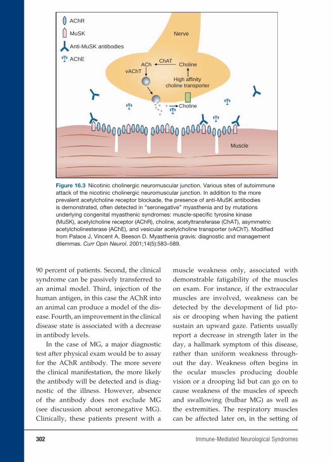
302 Immune-Mediated Neurological Syndromes
90 percent of patients. Second, the clinical syndrome can be passively transferred to an animal model. Third, injection of the human antigen, in this case the AChR into an animal can produce a model of the dis-ease. Fourth, an improvement in the clinical disease state is associated with a decrease in antibody levels.
In the case of MG, a major diagnostic test after physical exam would be to assay for the AChR antibody. The more severe the clinical manifestation, the more likely the antibody will be detected and is diag-nostic of the illness. However, absence of the antibody does not exclude MG (see discussion about seronegative MG). Clinically, these patients present with a
muscle weakness only, associated with demonstrable fatigability of the muscles on exam. For instance, if the extraocular muscles are involved, weakness can be detected by the development of lid pto-sis or drooping when having the patient sustain an upward gaze. Patients usually report a decrease in strength later in the day, a hallmark symptom of this disease, rather than uniform weakness through-out the day. Weakness often begins in the ocular muscles producing double vision or a drooping lid but can go on to cause weakness of the muscles of speech and swallowing (bulbar MG) as well as the extremities. The respiratory muscles can be affected later on, in the setting of
AChR
MuSK Nerve
ChATACh
vAChTCholine
Choline
Muscle
High affinitycholine transporter
Anti-MuSK antibodies
AChE
Figure 16.3 Nicotinic cholinergic neuromuscular junction. Various sites of autoimmune attack of the nicotinic cholinergic neuromuscular junction. In addition to the more prevalent acetylcholine receptor blockade, the presence of anti-MuSK antibodies is demonstrated, often detected in “seronegative” myasthenia and by mutations underlying congenital myasthenic syndromes: muscle-specifi c tyrosine kinase (MuSK), acetylcholine receptor (AChR), choline, acetyltransferase (ChAT), asymmetric acetylcholinesterase (AChE), and vesicular acetylcholine transporter (vAChT). Modifi ed from Palace J, Vincent A, Beeson D. Myasthenia gravis: diagnostic and management dilemmas. Curr Opin Neurol. 2001;14(5):583–589.
Immune-Mediated Neurological Syndromes 303
a “myasthenic crisis,” which can be life threatening.
In infants, congenital myasthenia occurs from transplacental passage of anti-bodies from a woman with MG or may be caused by a mutation of the neuromuscu-lar junction itself. It is accepted as conven-tional wisdom in the medical community that MG is antibody mediated because an autoantibody specifi cally reactive to the acetylcholine receptor in the neuromus-cular junction is present in 80–90 percent of patients. Reduction of this antibody is associated with improvement in clinical symptoms. A decrease in the number of acetylcholine receptors has been shown to occur directly because of binding of anti-body to the receptors, possibly by cluster-ing these receptors. These receptors may be destroyed ultimately via the activation of the complement system.
The Lambert-Eaton syndrome is an immunological and clinical variation of MG and is a paraneoplastic syndrome in the setting of small cell lung cancer (70 percent of patients). More than 90 percent of patients with this syndrome have autoanti-bodies to P/Q-type calcium channels.
Patients usually present with hip girdle weakness, which is more notable in the morning and improves with exercise and over the course of the day. Autonomic dysfunction is also found in addition to muscle weakness.
Animal models for myasthenia gravis can be produced by injecting the human antigen or by passively transferring the disease to an animal by injection of the antibodies. However, the antibodies are far from homogeneous. The subtypes of anti-bodies may vary among patients and even among muscles within the same patient. The antibody may also have variations in light chain type and subclass within one
patient. Most likely these variations are due to the receptor on the muscle, which consists of fi ve subunits. These subunits associate to form a transmembrane ion channel. It is likely that the populations of B lymphocytes, producing the receptor antibodies, are also heterogeneous.
T lymphocytes are also felt to play an important role in MG, although they are not found in biopsy specimens. Their main function in this setting might be to stimu-late the B cells. It has been demonstrated that anti-T-cell antibodies may be used for immunoregulation in this disease. The naturally occurring anti-T-cell receptor (anti-V beta 5.1) IgG antibody is found to occur in those patients with milder disease and less so in more severe MG. Although this antibody occurs in higher titers in MG patients than in controls, the higher the antibody titer, the less severe are the clinical symptoms, suggesting that therapy targeting pathogenic T-cell receptors (TCRs) may be a useful avenue of exploration.
The thymus gland has been considered to be the source of the autoimmune state of patients with MG. Most patients with MG have thymic hyperplasia and 10 to 12 per-cent have a thymoma. The thymus gland contains myoid-type cells, which have striations and acetylcholine receptors. The thymic cells may act as antigen-present-ing cells with MHC class II molecules. Cathepsin V, an enzyme responsible for cleaving the invariant chain in the antigen-presenting cleft of the MCH II molecule is overexpressed in MG patients in the thy-mic tissue and in the thymoma when pres-ent. However, mRNA and cathepsin V pro-tein are not expressed in patients who have a thymoma but do not have MG. Patients who have thymoma also may have anti-bodies directed against muscle antigens as
304 Immune-Mediated Neurological Syndromes
titin or the ryanodine receptor in addition to the acetylcholine receptor.
The thymus gland may also play a role because of the presence of acetylcho-line receptor on the myoid cells. Theories include the possibility that a virus might alter the myoid cells, and the proximity to the antigen presenting cells and helper T cells in the gland increases the possibil-ity of an autoimmune response. Molecular mimicry may play a role as well.
Herpes viruses and bacteria have been shown to share cross-reactivity with the acetylcholine receptor. A genetic pre-disposition is most likely a requisite of acquiring the disease. HLA types have been associated with MG such as HLA-B8, DRw3, and DQw2. Other autoimmune dis-eases are found to occur concomitantly in these patients or in their families such as lupus, rheumatoid arthritis, and Graves’ disease.
There is also a subset of patients (10–20 percent) with clinical MG, who do not produce antibodies to the acetyl-choline receptor. They are designated as “antibody-negative myasthenia.” This may be a misnomer. Some patients (40–70 percent) with this disorder produce anti-bodies to another antigen, the MuSK. The antibodies are of the subclass IgG4 and strongly activate complement cascade. In contrast, the ACh-R-Abs are primarily of the IgG1 and IgG3 subclasses, which can also fi x complement. The MuSK antibody is also found in 10 percent of patients with antibody-positive MG, or alone without the AChR-Ab.
Of interest, although no animal studies have shown that this particular antibody can cause muscle weakness, a myasthenic syndrome can be passively transferred with plasma from these patients into an animal model. In addition, seronegative
patients respond to plasmapheresis and immunosuppressive therapy, similar to seropositive MG.
The experimental animal model of MG, experimental autoimmune MG (EAMG) requires CD4+ helper T cells needed for the antibody-mediated autoimmune response. In mice, the T-cell response to the AchR is predominantly to a single peptide in about 50 percent of the cells. These T cells use a restricted set of TCR genes and have a conserved CDR3 region. This is in contrast to the human manifes-tation of the disease, where AChR-specifi c T cells are very low in frequency. The spe-cifi c T cells, when they have been cloned from patients are heterogeneous in MHC restriction, and recognize various epitopes of the AChR.
There are three levels to treatment of myasthenic patients, cholinesterase inhibitors, thymectomy, and immuno-suppression. The mainstay of medica-tions that treat the symptoms, but not the course of the disease, are the cholin-esterase inhibitors. These medications increase the availability of acetylcholine in the neuromuscular junction and thereby help to overcome the effect of decreased acetylcholine receptors due to the autoan-tibodies, AChR-Ab.
However, while this medication may maintain patients on a stable course for some time, they will not induce remis-sion of the disease. The two approaches to this goal are thymectomy and immuno-suppressants. The reason for thymectomy is that most MG patients have thymic abnormalities, either hyperplasia (60–70 percent) or a thymoma (10–12 percent). Computerized tomography or MRI scan-ning of the mediastinum should be routine in all patients suspected of MG. Although the clinical response is very variable
Immune-Mediated Neurological Syndromes 305
according to the literature, thymectomy is the most frequently used treatment for myasthenia and is generally suggested to be performed very early. The rationale is that the thymus gland serves as a source for the antigens involved in the autoim-mune response detected in patients, even without the presence of thymoma.
The third arm of treatment is immuno-suppression. Steroids improve 45 percent of patients and cause remission in 30 per-cent. Steroids can initially cause a wors-ening of the clinical symptoms in the fi rst few weeks, which is unique to this dis-ease, and patients are often initiated on them in a hospital setting. Plasmaphore-sis may be performed preventively before steroids are initiated. Patients are often on steroids for one or two years before tapering is begun. Immunosuppressive agents, such as azathioprine or cyclospo-rine, are also administered in some cases because they act as T-cell suppressors. The antibody production in this disease is T-cell dependent, as demonstrated in the animal model.
Plasmapheresis or intravenous immu-noglobulin is sometimes used in the set-ting of a clinical crisis. The plasmapho-resis removes the AChR-antibody from the circulation and the clinical response correlates with the decrease in antibody titer. Sometimes the plasma is processed through a staph-protein A immunoabsor-bant column to more effectively remove the IgG. Clinical trials are also under way using an AChR-Ab specifi c immunoab-sorbant column. Intravenous immuno-globulin also has been demonstrated to reverse an acute exacerbation for reasons unknown. For both treatments, while the response time is rapid, a matter of days, the result is short lived, lasting only a few months. It is hoped that within that time
other modalities of treatment have begun to be effective.
CENTRAL NERVOUS SYSTEM
SYSTEMIC LUPUS ERYTHEMATOSUS
CNS SLE is an example of an autoim-mune pathology with abnormalities also detected in other organs outside the cen-tral or peripheral nervous system. Over ten years’ follow-up, approximately 25 percent of SLE patients will develop CNS manifes-tations.
One hypothesis is that the neuropsy-chiatric symptoms of SLE may result from secondary causes. Infarctions may be experienced by patients, felt to be related to antibodies found in SLE, which affect the clotting system, such as circulating lupus anticoagulant or antiphospholipid antibodies (APLAs). Another mechanism may be symptoms secondary to CNS infec-tions such as meningitis or encephalitis. Treatments of SLE require immunosup-pressive medications or steroids, which may predispose patients to these etiolo-gies. Metabolic issues may also present as neuropsychiatric symptoms, such as in the case of uremia secondary to renal involve-ment of SLE.
Lupus cerebritis is the term used to con-vey that the CNS symptomatology is due to the SLE immune process. This broad etiology of neuropsychiatric symptoms may result from a demyelinating pathol-ogy. The symptoms associated with CNS SLE may be diffuse in nature, rather than a focal symptom suggestive of a vascu-lar territory and therefore stroke. These symptoms may include aseptic meningi-tis, headache, chorea, myelopathy, cranial neuropathy, seizures, confusion, and cog-nitive dysfunction. Also felt to be due to
306 Immune-Mediated Neurological Syndromes
diffuse etiology are psychiatric symptoms, such as mood disorders and psychosis. Other neurological symptoms outside the CNS include peripheral neuropathy or mononeuritis multiplex.
The combination of pathological stud-ies and positron emission tomography scanning form a hypothesis: hypoperfu-sion causes a breakdown of the blood-brain barrier. The loss of integrity allows antineuronal antibodies to cross over and ultimately lead to demyelination, similar to theories in MS. The inciting factors that would cause hypoperfusion are unknown. Evidence of a vasculopathy is present in pathological specimens, which dem-onstrate a perivascular accumulation of mononuclear cells, without destruction of the blood vessels. There may also be small infarctions secondary to occlusion of the vessel lumen. APLAs are associated with stroke syndromes and may play a role in the vasculopathy of CNS SLE.
The effects of these APLAs may include a prothrombotic state with coagu-lation pathway abnormalities and cause thromboembolism, stroke, focal seizures, migraines, and multi-infarct dementia. In this setting, the treatment of the disease would be directed more at the antithrom-botic state, with aspirin and consideration of anticoagulation, rather than with ste-roids or immunosuppressants.
Steroids are used only if there is coexist-ing serological evidence of active SLE. Ste-roids may be detrimental in a pure stroke setting, without evidence of active immune disease. Clinically, CNS diffuse vasculitis presents as fever, severe headaches, and confusion, with symptoms that progress rapidly to psychosis, seizures, or coma. Serologically active SLE is detected with elevated anti-dsDNA (double-stranded DNA) and hypocomplementemia.
MRI scanning is the clinical tool of choice in investigating the diagnosis of CNS SLE. Infarctions can be identifi ed, as well as diffuse demyelinating disease. Some lesions may resolve on subsequent MRI testing and therefore point away from infarction as the etiology of the clinical manifestation. Infarctions are permanent, whereas demyelination may be reversible. In fact, clinical improvement has been cor-related with resolving lesions, following immunosuppressive therapy on MRI scan-ning, and resolving lesions may actually predict an improved clinical course.
Diffuse CNS SLE is diffi cult to diagnose and manage. CSF studies, anti-DNA anti-body titers, complement levels, and even imaging may be nonspecifi c or insensitive. For instance, if a psychosis develops in the SLE patient, it may be due to the SLE, or the use of steroids, a mainstay of the phar-macopoeia in this disease. Depression may also be a manifestation of lupus cerebritis or may be a reactive depression to having the disease. However, missing the diagno-sis of lupus cerebritis or mistreating it car-ries high mortality and morbidity.
In 1986, the fi rst reports of an antibody population to ribosomal P proteins and association to lupus cerebritis came to the literature. This work was based on earlier fi ndings that patients produced antibod-ies that bound to ribosomes. The antigens were found to be phosphoproteins or “P proteins” located on the 60S subunit of ribosomes. These three proteins were labeled P0 (35 kD molecular weight), P1 (19 kD), and P2 (17 kD). These proteins are felt to be involved in protein synthesis; monoclonal antibodies to these proteins inhibit the elongation factors EF-1 and EF-2 to ribosomes and inhibit protein synthe-sis. In addition to being on ribosomes, the P proteins are also on the cell surfaces of
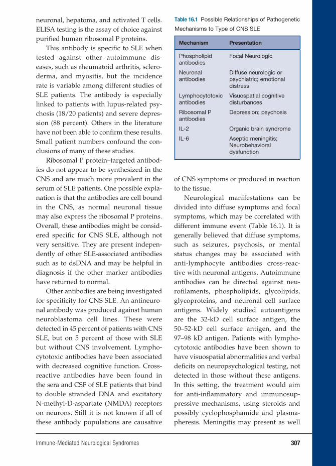
Immune-Mediated Neurological Syndromes 307
neuronal, hepatoma, and activated T cells. ELISA testing is the assay of choice against purifi ed human ribosomal P proteins.
This antibody is specifi c to SLE when tested against other autoimmune dis-eases, such as rheumatoid arthritis, sclero-derma, and myositis, but the incidence rate is variable among different studies of SLE patients. The antibody is especially linked to patients with lupus-related psy-chosis (18/20 patients) and severe depres-sion (88 percent). Others in the literature have not been able to confi rm these results. Small patient numbers confound the con-clusions of many of these studies.
Ribosomal P protein–targeted antibod-ies do not appear to be synthesized in the CNS and are much more prevalent in the serum of SLE patients. One possible expla-nation is that the antibodies are cell bound in the CNS, as normal neuronal tissue may also express the ribosomal P proteins. Overall, these antibodies might be consid-ered specifi c for CNS SLE, although not very sensitive. They are present indepen-dently of other SLE-associated antibodies such as to dsDNA and may be helpful in diagnosis if the other marker antibodies have returned to normal.
Other antibodies are being investigated for specifi city for CNS SLE. An antineuro-nal antibody was produced against human neuroblastoma cell lines. These were detected in 45 percent of patients with CNS SLE, but on 5 percent of those with SLE but without CNS involvement. Lympho-cytotoxic antibodies have been associated with decreased cognitive function. Cross-reactive antibodies have been found in the sera and CSF of SLE patients that bind to double stranded DNA and excitatory N-methyl-D-aspartate (NMDA) receptors on neurons. Still it is not known if all of these antibody populations are causative
of CNS symptoms or produced in reaction to the tissue.
Neurological manifestations can be divided into diffuse symptoms and focal symptoms, which may be correlated with different immune event (Table 16.1). It is generally believed that diffuse symptoms, such as seizures, psychosis, or mental status changes may be associated with anti-lymphocyte antibodies cross-reac-tive with neuronal antigens. Autoimmune antibodies can be directed against neu-rofi laments, phospholipids, glycolipids, glycoproteins, and neuronal cell surface antigens. Widely studied autoantigens are the 32-kD cell surface antigen, the 50–52-kD cell surface antigen, and the 97–98 kD antigen. Patients with lympho-cytotoxic antibodies have been shown to have visuospatial abnormalities and verbal defi cits on neuropsychological testing, not detected in those without these antigens. In this setting, the treatment would aim for anti-infl ammatory and immunosup-pressive mechanisms, using steroids and possibly cyclophosphamide and plasma-pheresis. Meningitis may present as well
Table 16.1 Possible Relationships of Pathogenetic
Mechanisms to Type of CNS SLE
Mechanism Presentation
Phospholipid antibodies
Focal Neurologic
Neuronal antibodies
Diffuse neurologic or psychiatric; emotional distress
Lymphocytotoxic antibodies
Visuospatial cognitive disturbances
Ribosomal P antibodies
Depression; psychosis
IL-2 Organic brain syndrome
IL-6 Aseptic meningitis; Neurobehavioral dysfunction
308 Immune-Mediated Neurological Syndromes
and may be secondary to bacterial infec-tions due to immunosuppression. In addi-tion, aseptic meningitis may be caused by medications, such as ibuprofen and immu-nosuppressive medication.
Partial or generalized seizures may also occur in the setting of SLE and gen-erally portend a poor prognosis. Patients are generally treated with anticonvul-sants, and steroids are only added if it is felt that the patient is in an active fl are, to prevent potential permanent scarring and a seizure focus.
Although migraine headaches occur frequently in the setting of SLE, a causative effect has not been determined. More seri-ously, patients may have other types of headache, such as pseudotumor cerebri, cerebral venous, or sagittal sinus throm-bosis. This is usually due to a secondary cause of SLE, such as renal dysfunction and a hypercoagulable state. The sudden development of headaches in a previously headache-free patient or neurological symptoms or signs requires rapid work-up and imaging.
More uncommon CNS syndromes occur and may be varied. Movement disorders such as chorea or ataxia point to disease in the basal ganglia or cerebellum. These dis-orders are usually self-limited to two to six weeks and may not require therapy. If they are associated with APLA, anticoagulation may be considered.
Patients may have cranial neuropa-thies causing symptoms such as double vision or hearing loss, trigeminal neural-gia, or dysarthria. Steroids are generally used in these conditions, and if refractory, cyclophosphamide, an immunosuppres-sant medication, is used. When patients present with these symptoms, the question of MS should be ruled out. Both diseases may have optic neuritis, transverse myelitis,
or infl ammation of the spinal cord (which must be treated aggressively with, at the very least, steroids, and possibly plasma-pheresis and cyclophosphamide for recov-ery), or internuclear ophthalmoplegia, and may have similar MRI scans. Patients who do have similar scans and who have lupus are referred to as having “lupus sclerosis.” APLA in this setting often help with dif-ferentiation of the syndromes. MS should have no peripheral manifestations out-side of the CNS.
Cytokine release may play a role in these pathologies. IL-1, IL-2, IL-6, TNF-α, IFN-α, and IFN-γ have all been demon-strated to be released by CNS microglial cells and astrocytes by LPS stimulation. In the CNS, a cascade of pro-infl ammatory cytokines causes events of infl ammation similar to peripheral macrophages and monocytes. These cytokines not only medi-ate infl ammation but also may directly affect brain function, as there are cytokine receptors on the hypothalamus. Antibod-ies can cause functional dysregulation by blocking the release of neurotransmitters and neuropeptides, resulting in abnormal electrophysiological testing, such as the electroencephalogram.
Animal models have been used to investigate the human disease with mixed success. The MRL/P mice have neurobe-havioral abnormalities such as timidity, phobic behavior, and anxiety. This mani-fests at age 7 to 8 weeks, when autoanti-bodies to brain tissue also present. The pathology demonstrates B lymphocytes around the choroid plexus with MHC class II antigen presentation. This differs from the vasculitis that is more typical of the human SLE syndrome. However, like humans with an exacerbation of SLE, IL-6 is elevated in the CSF and the animal behavior can be signifi cantly improved
Immune-Mediated Neurological Syndromes 309
by treating with immunosuppression and steroids. The clinical improvement is cor-related with a decrease in IL-6 levels in the CSF.
The SLE syndrome of focal events sec-ondary to APLA can also be demonstrated in a mouse model by injecting MRL/pr mice with APLA and anti-neuronal anti-bodies. Antigen derived from limbic brain structures, which also react to human sera, the B2 glycoprotein, causes production of APLA and ischemic events/stokes.
The future of CNS SLE may lie in stem cell transplant. One study of seven patients using chemotherapy followed by autolo-gous stem cell transplantation yielded excellent clinical results at a median follow-up of twenty-fi ve months. The patients had initially suffered from cerebritis, myelitis, vasculitis, or glomerulonephritis and were unresponsive to multiple courses of cyclo-phosphamide. At follow-up, all patients were free of signs of active lupus and sero-logical markers had improved. The theory of the treatment is that when memory T cells are removed from infl uence, maturation of new lymphocyte progenitor cells occurs without recruiting autoimmune activity.
ANIMAL MODELS
A unique diffi culty in developing treatment strategies and pathogenic theories of neu-rological immune diseases is that often the human end organ is unavailable for study. The morbidity of taking CNS tissue purely for research places that option beyond ethical boundaries in most cases. There-fore, reliance is mainly on autopsy mate-rial, when usually only end-stage disease pathology is available, keeping the natural history of the disease unknown. Therefore, reliance on animal models in these diseases
is paramount in testing treatment strate-gies and theories of etiology. Controversy arises when one considers how closely the animal model follows the human dis-ease state under study. The animal model should refl ect the susceptibility of the host, and the clinical fi ndings of the disease, and should be similar to the histopathological fi ndings in the human.
MS and EAE provide good examples of strengths and diffi culties of animal models based on differences between human and murine immunology. For example, in EAE IFN-γ was demonstrated in several studies to be a protective cytokine. Antibodies to the cytokine exacerbated EAE, possibly by blocking induction and activation of sup-pressor activity. However, when this prin-ciple was applied to patients, the study was discontinued because of exacerbation of MS by administration of IFN-γ in a clini-cal trial.
IFN-α is another example of differ-ing cytokine responses. In humans, this cytokine is secreted by several types of cells, inclining macrophages in response to exposure to viral antigens. The cyto-kine induces T cells to TH1 development, a process that depends on signal transducer and transcription-4 (STAT-4) activation. In mice, however, INF-α dose not activate STAT-4, therefore; TH1 is not induced by IFN-α. Multiple other differences abound in the immune systems between mice and humans; therefore, responses to treatment protocols in models compared with human disease may vary greatly.
Differences in the immunology of mice and human are numerous (see Table 16.2). For example, in delayed-type hypersensi-tivity (DTH) in humans, neutrophils are the fi rst responders, followed by a mix of mononuclear cells composed of T cells and macrophages. In mice, there are relatively
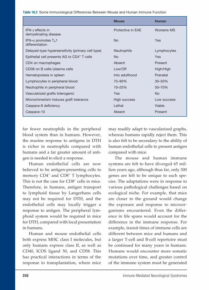
310 Immune-Mediated Neurological Syndromes
far fewer neutrophils in the peripheral blood system than in humans. However, the murine response to antigens in DTH is richer in neutrophils compared with humans and a far greater amount of anti-gen is needed to elicit a response.
Human endothelial cells are now believed to be antigen-presenting cells to memory CD4+ and CD8+ T lymphocytes. This is not the case for CD8+ cells in mice. Therefore, in humans, antigen transport to lymphoid tissue by Langerhans cells may not be required for DTH, and the endothelial cells may locally trigger a response to antigen. The peripheral lym-phoid system would be required in mice for DTH, compared with local presentation in humans.
Human and mouse endothelial cells both express MHC class I molecules, but only humans express class II, as well as CD40, ICOS ligand 50, and CD58. This has practical interactions in terms of the response to transplantation, where mice
may readily adapt to vascularized graphs, whereas humans rapidly reject them. This is also felt to be secondary to the ability of human endothelial cells to present antigen compared with mice.
The mouse and human immune systems are felt to have diverged 65 mil-lion years ago, although thus far, only 300 genes are felt to be unique to each spe-cies. The adaptations were in response to various pathological challenges based on ecological niche. For example, that mice are closer to the ground would change the exposure and response to microor-ganisms encountered. Even the differ-ence in life spans would account for the difference in the immune response. For example, transit times of immune cells are different between mice and humans and a larger T-cell and B-cell repertoire must be continued for many years in humans. Humans would encounter more somatic mutations over time, and greater control of the immune system must be generated
Table 16.2 Some Immunological Differences Between Mouse and Human Immune Function
Mouse Human
IFN-γ effects indemyelinating disease
Protective in EAE Worsens MS
IFN-α promotes TH1differentiation
No Yes
Delayed-type hypersensitivity (primary cell type) Neutrophils Lymphocytes
Epithelial cell presents AG to CD4+ T cells No Yes
CD4 on macrophages Absent Present
CD38 on B cells / plasma cells Low/Off High/High
Hematopoiesis in spleen Into adulthood Prenatal
Lymphocytes in peripheral blood 75–90% 30–50%
Neutrophils in peripheral blood 10–25% 50–70%
Vascularized grafts tolerogenic Yes No
Microchimerism induces graft tolerance High success Low success
Caspace-8 defi ciency Lethal Viable
Caspace-10 Absent Present
Immune-Mediated Neurological Syndromes 311
to control for autoimmunity and to con-trol larger, widely varied antigen-specifi c clones.
Thus, one can see multiple reasons for often wide discrepancies between animal models and the human condition, particu-larly in the response to potential therapeu-tic treatments. Perhaps the focus of effort and funding should be placed on direct human studies, whether on the molecular, tissue, or organism level, to unravel the designs of these diseases.
BIBLIOGRAPHY
REVIEW ARTICLESChitnis T, Khoury SJ. Immunologic neu-
romuscular disorders. J Allergy Clin Immunol. 2003;111(2 suppl):S659–S668.
Denburg J, Denburg S, Carbotte R, and Szechtman H. Nervous system lupus: pathogenesis and rationale for therapy. Scand J Rheum. 1995;12:263–273.
Ho T, Grifi n J. Guilain-Barré syndrome. Curr Opin Neurol. 1999;12(4):389–394.
Huizinga T, Steens S, van Buchem M. Imaging modalities in central nervous system systemic lupus erythematosis. Curr Opin Rheum. 2001;13:383–388.
Lucchinetti C, Bruck W, Parisi J, et al. Heterogeneity of multiple sclerosis lesions: implications for the patho-genesis of demyelination. Ann Neurol. 2000;47:707–717.
McDonald W, Compston A, Edan G, et al. Recommended diagnostic criteria for multiple sclerosis: guidelines from the
International Panel on the Diagnosis of Multiple Sclerosis. Ann Neurol. 2001;50:121–127.
Newswanger D, Warren C. Practical thera-peutics: Guillain-Barré syndrome. Am Fam Phys. 2004;69(10):2405–2410.
Palace J, Vincent A, Beeson D. Myasthe-nia gravis: diagnostic and manage-ment dilemmas. Curr Opin Neurol. 2001;14(5):583–589.
Schur P, Khoshbin S Neurologic manifes-tations of systemic lupus erythema-tosis. Online: http:/uptodateonline.com/application/topicText.asp?fi le =rheumatic/1974&type=A&selecte…; 2004.
SUGGESTED READINGMestas J, Hughes C. Of mice and not men:
differences between mouse and human immunology. J Immunol. 2004;172:2731–2738.
’t Hart B, Amor S. The use of animal mod-els to investigate pathogenesis of neu-roinfl ammatory disorders of the cen-tral nervous system. Curr Opin Neurol. 2003;16(3):375–383.
’t Hart B, Laman J, Bauer J, Blezer E, van Kooyk Y, Hintzen R. Modelling of multiple sclerosis: lessons learned in a non-human primate. Lancet Neurol. 2004;3:588–597.
Infante AJ, Kraig E. Myasthenia gravis and its animal model: T cell receptor expres-sion in antibody mediated autoimmune disease. Int Rev Immunol. 1999;18(1–2):83–109.

17. Immunological Aspects of Renal Disease
Gil Cu, M.D., and John B. Zabriskie, M.D.
313
INTRODUCTION
Human kidneys play an integral role in the development of primary or secondary immunologic diseases. As a major fi lter-ing organ, the kidneys, which represent about 0.5 percent of the human body mass, receive 20 percent of the total cardiac out-put. The enormous blood fl ow (1 L/min) to the renal microcirculation exceeds that observed in other major vascular organs (heart, liver, and brain). Urine is produced after a complex process of glomerular fi l-tration, tubular transport, and reabsorption at a rate of 1 ml/min. Cellular elements involved in immunity thereby have a high probability of interacting with glomeru-lar and tubular cells that may or may not cause renal disease.
Suffi cient knowledge of the anatomy and histology of the kidney is vital in understanding the pathogenesis of renal diseases. The renal corpuscle, or glomeru-lus (Figure 17.1), is composed of capillary tuft lined by a thin layer of endothelial cells; central region of mesangial cells and its surrounding matrix; and visceral and parietal epithelial cells with their respec-tive basement membranes. The glomeru-lus is primarily responsible for production of ultrafi lrate from the circulating plasma. The fi ltration barrier between the blood-stream and urinary space is made up by the fenestrated endothelium, the glomeru-lar basement membrane (GBM), and the
slit pores seen between the foot proc esses of the visceral epithelium (Figure 17.2).
The endothelial cells form as initial barriers to cellular elements of the blood (red blood cells, leucocytes, and platelets) in reaching the subendothelial space. The endothelial cells produce nitric oxide (a vasodilator) and endothelin-1 (a potent vasoconstrictor), chemical substances implicated in infl ammatory processes. The surface of the endothelial cells is negatively charged, which may contribute to the charge-selective properties of the glomeru-lar capillary wall.
The GBM is composed of a central dense layer called the lamina densa and two thin layers called the lamina rara externa and lamina rara interna. The GBM is formed by the fusion of the endothelial and epithe-lial basement membrane during develop-ment. Biochemical analyses of the GBM have identifi ed the presence of glycopro-teins (type IV collagen, laminin, fi bronec-tin, and endotactin/nidogen) and heparan sulfate proteoglycans (perlecan and agrin). Type IV collagen is the major constituent of the GBM. Gene mutations involving those encoding α3, α4, and α5 isomeric chains of the type IV collagen can cause Alport’s syndrome. This is a progressive form of glomerulopathy associated with ocular abnormalities, hearing loss, and microscopic hematuria. Electron micro-scopic examination shows thinning of the GBM in early stages of the disease. With
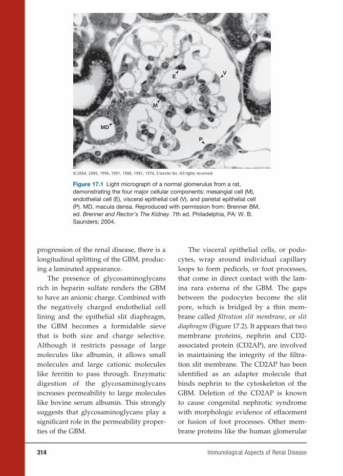
314 Immunological Aspects of Renal Disease
progression of the renal disease, there is a longitudinal splitting of the GBM, produc-ing a laminated appearance.
The presence of glycosaminoglycans rich in heparin sulfate renders the GBM to have an anionic charge. Combined with the negatively charged endothelial cell lining and the epithelial slit diaphragm, the GBM becomes a formidable sieve that is both size and charge selective. Although it restricts passage of large molecules like albumin, it allows small molecules and large cationic molecules like ferritin to pass through. Enzymatic digestion of the glycosaminoglycans increases permeability to large molecules like bovine serum albumin. This strongly suggests that glycosaminoglycans play a signifi cant role in the permeability proper-ties of the GBM.
The visceral epithelial cells, or podo-cytes, wrap around individual capillary loops to form pedicels, or foot processes, that come in direct contact with the lam-ina rara externa of the GBM. The gaps between the podocytes become the slit pore, which is bridged by a thin mem-brane called fi ltration slit membrane, or slit diaphragm (Figure 17.2). It appears that two membrane proteins, nephrin and CD2-associated protein (CD2AP), are involved in maintaining the integrity of the fi ltra-tion slit membrane. The CD2AP has been identifi ed as an adapter molecule that binds nephrin to the cytoskeleton of the GBM. Deletion of the CD2AP is known to cause congenital nephrotic syndrome with morphologic evidence of effacement or fusion of foot processes. Other mem-brane proteins like the human glomerular
MD
M
© 2004, 2000, 1996, 1991, 1986, 1981, 1976, Elsevier Inc. All rights reserved.
P
VE
Figure 17.1 Light micrograph of a normal glomerulus from a rat, demonstrating the four major cellular components: mesangial cell (M), endothelial cell (E), visceral epithelial cell (V), and parietal epithelial cell (P). MD, macula densa. Reproduced with permission from: Brenner BM, ed. Brenner and Rector’s The Kidney. 7th ed. Philadelphia, PA: W. B. Saunders; 2004.
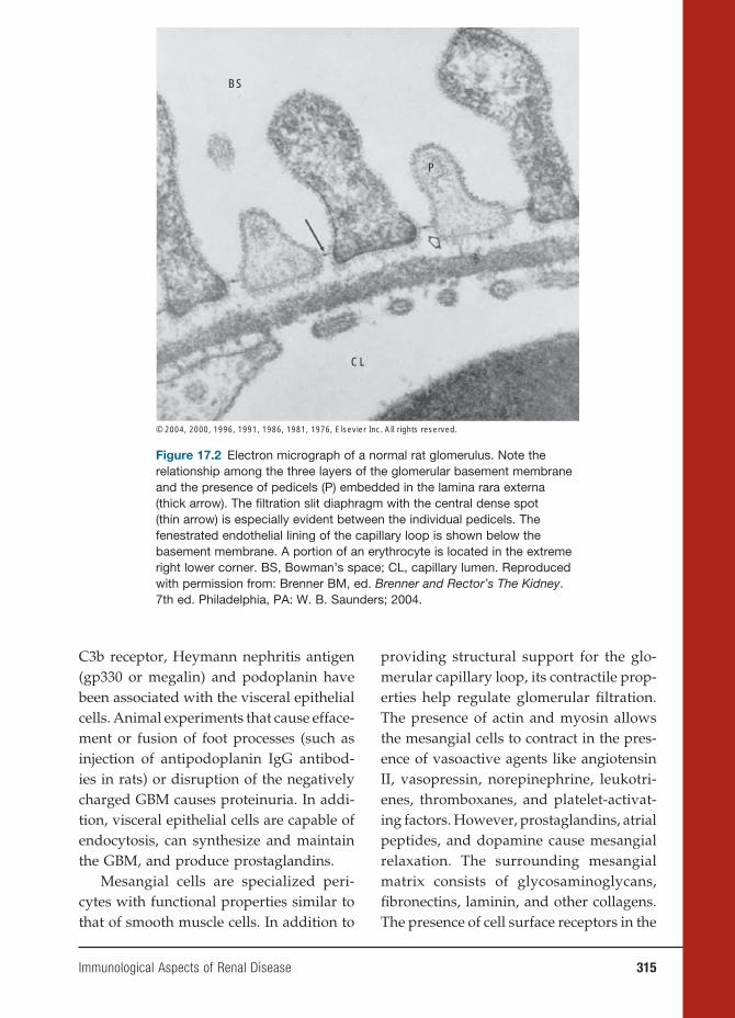
Immunological Aspects of Renal Disease 315
C3b receptor, Heymann nephritis antigen (gp330 or megalin) and podoplanin have been associated with the visceral epithelial cells. Animal experiments that cause efface-ment or fusion of foot processes (such as injection of antipodoplanin IgG antibod-ies in rats) or disruption of the negatively charged GBM causes proteinuria. In addi-tion, visceral epithelial cells are capable of endocytosis, can synthesize and maintain the GBM, and produce prostaglandins.
Mesangial cells are specialized peri-cytes with functional properties similar to that of smooth muscle cells. In addition to
providing structural support for the glo-merular capillary loop, its contractile prop-erties help regulate glomerular fi ltration. The presence of actin and myosin allows the mesangial cells to contract in the pres-ence of vasoactive agents like angiotensin II, vasopressin, norepinephrine, leukotri-enes, thromboxanes, and platelet-activat-ing factors. However, prostaglandins, atrial peptides, and dopamine cause mesangial relaxation. The surrounding mesangial matrix consists of glycosaminoglycans, fi bronectins, laminin, and other collagens. The presence of cell surface receptors in the
BS
P
CL
© 2004, 2000, 1996, 1991, 1986, 1981, 1976, Elsevier Inc. All rights reserved.
Figure 17.2 Electron micrograph of a normal rat glomerulus. Note the relationship among the three layers of the glomerular basement membrane and the presence of pedicels (P) embedded in the lamina rara externa (thick arrow). The fi ltration slit diaphragm with the central dense spot (thin arrow) is especially evident between the individual pedicels. The fenestrated endothelial lining of the capillary loop is shown below the basement membrane. A portion of an erythrocyte is located in the extreme right lower corner. BS, Bowman’s space; CL, capillary lumen. Reproduced with permission from: Brenner BM, ed. Brenner and Rector’s The Kidney. 7th ed. Philadelphia, PA: W. B. Saunders; 2004.
316 Immunological Aspects of Renal Disease
matrix (e.g., β-integrin receptor) are impli-cated in signal transduction mechanisms that promote synthesis of various infl am-matory cytokines, vasoactive substances, and growth factors. Finally, mesangial cells have phagocytic properties. During endocytosis of immune complexes, intra-cellular production of prostaglandins and reactive oxygen species cause injury to the glomerulus.
Parietal cells are squamous cells that make up the epithelium that forms the outer wall of the Bowman’s capsule. At the vascular pole, the parietal epithelium is continuous with the visceral epithelium. At the urinary pole, there is a transition to the cuboidal cells of the proximal tubule. The exact role of parietal cells is not well defined. However, as pointed out by Brenner (2004), these cells can proliferate and become crescents like in rapidly pro-gressive glomerulonephritis (RPGN).
MECHANISMS OF IMMUNE INJURY
TO THE KIDNEYS
Cells of both innate and adaptive immu-nity mediate human defense against microbes. The principal components of innate immunity are composed of physical and chemical barriers (e.g., epithelial cells and antimicrobial substances produced by the cells), phagocytic cells (neutrophils and macrophages), natural killer cells, comple-ment systems, and cytokines. These repre-sent the fi rst line of defense against micro-bial agents. In contrast, adaptive immunity is represented by B and T lymphocytes. These cells are initially stimulated by expo-sure to offending agents. Subsequent expo-sure to similar agents cause specifi c cells to mount a defense in increasing magnitude.
Adaptive immunity is distinguished as having the ability to “remember” specifi c molecules and therefore provide specifi c immunity. Both types of immunity work in tandem to provide comprehensive defense against offending microbes.
Diseases caused by immune responses are often called hypersensitivity diseases. These are classifi ed according to the type of immune responses and the effector mecha-nisms that are responsible for cell and tis-sue injury. In this chapter, we will attempt to elucidate the nature of renal diseases in relation to the immune system.
Immediate hypersensitivity or type I disease is mediated by immunoglobulin E (IgE), which is produced in response to an allergen. After exposure to a spe-cifi c allergen, TH2 cells specifi c for the allergen are activated. IgE antibodies are then produced, which subsequently bind to the Fc receptor of mast cells and basophils. This binding releases biogenic amines (histamine), neutral serine prote-ases, lipid mediators, and cytokines. Bio-genic amines and lipid mediators cause vascular leakage, vasodilation, and air-way bronc hoconstriction. Serine prote-ases cause tissue damage. Cytokines are implicated in late phase reaction. Certain drugs, especially methicillin, nonsteroi-dal agents, rifampin, sulfa, cimetidine, cephalosporins, and 5-aminosalicylates, can cause allergic interstitial nephritis. Affected patients present with acute renal failure three to fi ve days after intake of the offending drug. Fever, rash, hematu-ria, proteinuria, and eosinophiluria are typical fi ndings. On kidney biopsy, tubu-lointerstitial infl ammation is prominent with occasional demonstration of eosino-phils. The glomeruli appear spared of any infl ammatory effects. Treatment consists
Immunological Aspects of Renal Disease 317
of withholding the offending drug and use of steroids.
Antibody-mediated or type II disease is caused by antibodies against fi xed cell and tissue antigens. Although most cases demonstrate the presence of autoantibod-ies, some antibodies can be produced by a foreign antigen that is immunologically cross-reactive to human tissue. Three mechanisms have been described to explain this phenomenon. First, antibodies may opsonize cells or activate the complement system that eventually produces comple-ment proteins that assist in opsonization of cells. Macrophages bind to Fc receptors of antibodies or complement protein receptors to cause endocytosis and destruction of the offending antigen. This appears to be the main mechanism in hemolytic anemia and autoimmune thrombocytopenic purpura. Second, antibodies bound in target tissues recruit neutrophils and macrophages by binding to Fc receptors or by activating complement. Activated neutrophils and macrophages release intracellular enzymes and reactive oxygen radicals that cause tissue injury. Examples of glomerular dis-ease that can be explained by this pathway include Goodpasture’s syndrome and anti-neutrophil cytoplasmic antibody (ANCA) mediated disease. Third, antibodies may bind to normal cellular receptors and inter-fere with their function to cause disease. However, no actual tissue injury is dem-onstrated. Graves’ disease represents this mechanism where the TSH receptor is tar-geted by the anti-TSH-receptor antibody. This mechanism causes hyperthyroidism. No glomerular disease has been associated with this mechanism (see Table 17.1).
Immune-complex-mediated, or type III, disease is caused by deposition of antibod-ies bound to self- or foreign antigens into
target tissues. Although the glomerulus is a common target for immune complexes, other organ systems are involved, which suggests the systemic nature of this dis-ease. A classic example is the serum dis-ease or serum sickness, which was origi-nally described by Clemens von Pirquet in 1911. During his time, diphtheria infec-tions were treated with passive immuniza-tion using serum from horses immunized with diphtheria toxin. He noted that joint infl ammation (arthritis), rash, and fever occurred in patients injected with the anti-toxin-containing horse serum. On further investigation, similar symptoms were seen in patients injected with horse serum with-out the antitoxin. The symptoms usually occurred at least one week after the fi rst injection and more rapidly on subsequent injections. He concluded that the host had developed antibodies to the horse serum, and deposition of antibody–serum protein complexes (immune complexes) to differ-ent tissues of the host caused the symp-toms described earlier. In experimental animals like the rabbit, injection of a large dose of a foreign protein antigen leads to the formation of antibodies against the antigen (see Figure 17.3). Subsequent for-mation of antibody–antigen complexes leads to enhanced phagocytosis and clear-ance of the antigen by the macrophages in the liver and the kidney. With subse-quent injection of the antigen, more of the immune complexes are formed and may be deposited in the vascular bed, renal glomeruli, and synovia. These activate the complement, which leads to recruitment of inflammatory cells (predominantly neutrophils) to cause injury to the affected tissues. Clinical and pathological manifes-tations are vasculitis, glomerulonephri-tis, and arthritis. Clinical symptoms are
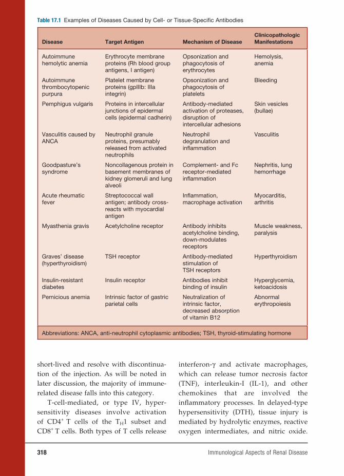
318 Immunological Aspects of Renal Disease
short-lived and resolve with discontinua-tion of the injection. As will be noted in later discussion, the majority of immune-related disease falls into this category.
T-cell-mediated, or type IV, hyper-sensitivity diseases involve activation of CD4+ T cells of the TH1 subset and CD8+ T cells. Both types of T cells release
interferon-γ and activate macrophages, which can release tumor necrosis factor (TNF), interleukin-I (IL-1), and other chemokines that are involved the infl ammatory processes. In delayed-type hypersensitivity (DTH), tissue injury is mediated by hydrolytic enzymes, reactive oxygen intermediates, and nitric oxide.
Table 17.1 Examples of Diseases Caused by Cell- or Tissue-Specifi c Antibodies
Disease Target Antigen Mechanism of DiseaseClinicopathologic Manifestations
Autoimmune hemolytic anemia
Erythrocyte membrane proteins (Rh blood group antigens, I antigen)
Opsonization and phagocytosis of erythrocytes
Hemolysis, anemia
Autoimmunethrombocytopenic purpura
Platelet membrane proteins (gpIIIb: IIIa integrin)
Opsonization and phagocytosis of platelets
Bleeding
Pemphigus vulgaris Proteins in intercellular junctions of epidermal cells (epidermal cadherin)
Antibody-mediated activation of proteases, disruption of intercellular adhesions
Skin vesicles (bullae)
Vasculitis caused by ANCA
Neutrophil granule proteins, presumably released from activated neutrophils
Neutrophil degranulation and infl ammation
Vasculitis
Goodpasture’s syndrome
Noncollagenous protein in basement membranes of kidney glomeruli and lung alveoli
Complement- and Fc receptor-mediated infl ammation
Nephritis, lung hemorrhage
Acute rheumatic fever
Streptococcal wall antigen; antibody cross-reacts with myocardial antigen
Infl ammation, macrophage activation
Myocarditis, arthritis
Myasthenia gravis Acetylcholine receptor Antibody inhibits acetylcholine binding, down-modulates receptors
Muscle weakness, paralysis
Graves’ disease (hyperthyroidism)
TSH receptor Antibody-mediated stimulation of TSH receptors
Hyperthyroidism
Insulin-resistant diabetes
Insulin receptor Antibodies inhibit binding of insulin
Hyperglycemia, ketoacidosis
Pernicious anemia Intrinsic factor of gastric parietal cells
Neutralization of intrinsic factor, decreased absorption of vitamin B12
Abnormal erythropoiesis
Abbreviations: ANCA, anti-neutrophil cytoplasmic antibodies; TSH, thyroid-stimulating hormone
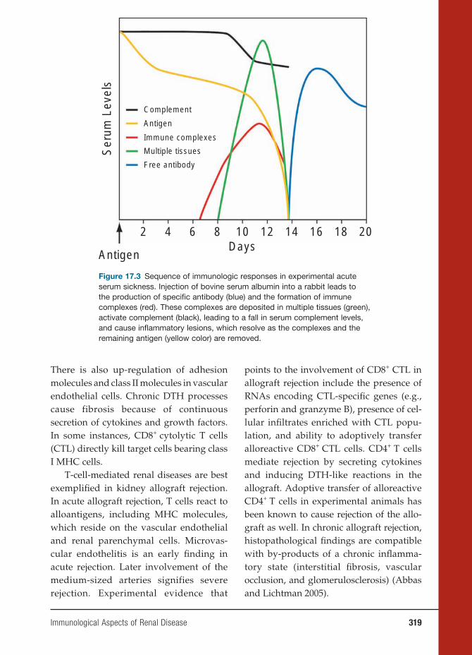
Immunological Aspects of Renal Disease 319
There is also up-regulation of adhesion molecules and class II molecules in vascular endothelial cells. Chronic DTH processes cause fibrosis because of continuous secretion of cytokines and growth factors. In some instances, CD8+ cytolytic T cells (CTL) directly kill target cells bearing class I MHC cells.
T-cell-mediated renal diseases are best exemplifi ed in kidney allograft rejection. In acute allograft rejection, T cells react to alloantigens, including MHC molecules, which reside on the vascular endothelial and renal parenchymal cells. Microvas-cular endothelitis is an early fi nding in acute rejection. Later involvement of the medium-sized arteries signifies severe rejection. Experimental evidence that
points to the involvement of CD8+ CTL in allograft rejection include the presence of RNAs encoding CTL-specifi c genes (e.g., perforin and granzyme B), presence of cel-lular infi ltrates enriched with CTL popu-lation, and ability to adoptively transfer alloreactive CD8+ CTL cells. CD4+ T cells mediate rejection by secreting cytokines and inducing DTH-like reactions in the allograft. Adoptive transfer of alloreactive CD4+ T cells in experimental animals has been known to cause rejection of the allo-graft as well. In chronic allograft rejection, histopathological fi ndings are compatible with by-products of a chronic infl amma-tory state (interstitial fi brosis, vascular occlusion, and glomerulosclerosis) (Abbas and Lichtman 2005).
Complement
Free antibody
Multiple tissues
Immune complexes
Antigen
Antigen
2
Ser
um L
evel
s
4 6 8 10Days
12 14 16 18 20
Figure 17.3 Sequence of immunologic responses in experimental acute serum sickness. Injection of bovine serum albumin into a rabbit leads to the production of specifi c antibody (blue) and the formation of immune complexes (red). These complexes are deposited in multiple tissues (green), activate complement (black), leading to a fall in serum complement levels, and cause infl ammatory lesions, which resolve as the complexes and the remaining antigen (yellow color) are removed.
320 Immunological Aspects of Renal Disease
SPECIFIC IMMUNE-RELATED
RENAL DISEASES
IgA Nephropathy
IgA nephropathy or Berger’s disease is the most common cause of glomerulonephritis in the world. Rarely seen in blacks, this dis-ease affl icts mainly Asians and Caucasians. Clinical features include gross hematuria (40–50 percent), microscopic hematuria (30–40 percent), and rarely, renal insuffi -ciency with edema and hypertension (less than 10 percent). These usually occur after a respiratory infection. Minimal protein-uria is noted. IgA nephropathy is made by kidney biopsy only. The hallmark of this disease is mesangial deposition of IgA, which is prominently noted on immuno-fl uorescence. This deposition is associated with focal mesangial expansion, which can be seen on light microscopy. Electron microscopy confi rms the presence of elec-tron-dense deposits in the mesangium, which correspond to the immune complex deposits. An abnormality in the regula-tion of IgA may exist because of an envi-ronmental stimuli. This is supported by higher circulating levels of IgA in about 50 percent of cases, circulating immune com-plexes that parallel the duration of the dis-ease, and increased number of IgA-specifi c B and T lymphocytes. Its strong association with respiratory infection has led authori-ties to believe that mucosal immunity plays a role in this disease. Exaggerated muco-sal IgA response or decreased clearance may explain the elevated levels of plasma IgA in this disease. In the glomeruli, IgA-containing immune complexes activate the alternative complement pathway but do not bind C1, which may slow the removal of circulating complexes and thereby pro-mote deposition in the glomeruli.
The role of fi bronectin and uteroglobin (an anti-infl ammatory protein) in IgA nephropathy remains unclear. Most of the IgA-containing complexes are bound to fi bronectin. In experimental animals, uteroglobin–fi bronectin heteromerization prevented formation of fi bronectin–IgA complexes and binding to glomeruli. Dele-tion of the uteroglobin gene in mice by gene knockout or suppression of the uteroglobin gene by antisense technology has histolog-ical features similar to IgA nephropathy. Interestingly, polymeric but not monomeric IgA binds only to mesangial cells, which stimulate release of IL-6, further leading to mesangial expansion.
IgA nephropathy usually follows a benign course, although about 15 per-cent of cases may reach end-stage renal disease at ten years and 20 percent at twenty years. Clinical and histopatho-logical predictors of progression of renal disease include worsening proteinuria (>1 g/day of proteinuria) and biopsy fi ndings of glomerular sclerosis, tubular atrophy, interstitial fi brosis, and crescent forma-tion. Treatment of IgA nephropathy has radically changed over the past decade. Aggressive use of angiotensin-converting enzyme (ACE) with and without angio-tensin-receptor blocker (ARB) is recom-mended to decrease the proteinuria and for their renoprotective properties. Cyto-toxic agents and steroids are reserved for aggressive renal disease.
MINIMAL CHANGE DISEASE
Minimal change disease (MCD), or nil disease, is the most common cause of nephrotic syndrome in children under age 10. It accounts for about 90 percent of cases
Immunological Aspects of Renal Disease 321
in children and about 10–15 percent in adult cases. Patients usually present with heavy proteinuria (>3 g/day), hypoalbuminemia, dyslipidemia, and anasarca. Urine sedi-ment is benign. Diagnosis rests on kidney biopsy. Under light microscopy, the glom-eruli appear normal. There is absence of immune deposits by immunofl uorescence. Electron microscopic fi ndings show efface-ment of the epithelial foot processes. Inves-tigators believe that MCD is a T-lymphocyte disease because of its sensitive response to steroids and cyclosporine. It is strongly associated with Hodgkin’s lymphoma, a hematologic malignancy. Proteinuria appears to disappear when the malignancy goes into remission. A glomerular perme-ability factor (GPF) derived from a T-cell hybridoma has been identifi ed to cause fusion of the foot processes and protein-uria, when injected into experimental rats. The T-cell hybridoma was made from T cells harvested from a patient with minimal change disease. It is thought that this GPF has TNF-like activity. In addition, epithe-lial damage causes loss of polyanions like heparin sulfate. This loss of charge selectiv-ity allows macromolecules like albumin to pass through the fi ltration barrier and leak into the urine.
In children with idiopathic nephrotic syndrome, treatment with steroids pro-duces immediate results with about 50 percent of cases going into remission in two weeks, and almost all within eight weeks of treatment. Clinical response in adults tends to be slower, but the majority will achieve remission after twelve to six-teen weeks of treatment. Cytotoxic agents, which include cyclophosphamide, chlo-rambucil, azathioprine, mycophenolate mofetil, and cyclosporine are reserved for relapsers and steroid-resistant cases.
FOCAL SCLEROSING
GLOMERULOSCLEROSIS
Focal glomerulosclerosis or focal segmen-tal glomerulosclerosis (FSGS) is a common cause of idiopathic nephrotic syndrome in adults, accounting for about 35 percent of all cases. About 50 percent of all FSGS cases appear to affl ict black people, suggest-ing possible involvement of their genetic background and socioeconomic status. FSGS is classifi ed as primary or second-ary. Primary or idiopathic FSGS usually presents with overt nephrotic syndrome. Secondary FSGS presents with nonne-phrotic-range proteinuria (<3.5 g/day) and renal dysfunction. The latter form has been considered to be a physiologic response to hyperfi ltration or glomerular hypertro-phy as a result of nephron loss. This loss can be seen in unilateral nephrectomy, unilateral agenesis of one kidney, refl ux nephropathy, renal vasodilation, obesity, and preeclampsia. Secondary FSGS may be seen as a response to a prior injury of any cause (e.g., acute glomerulonephritis, or vasculitis). The transforming growth fac-tor–beta (TGF-β), which is released from affected glomerular cells and platelets, appears to contribute to the development of glomerulosclerosis. TGF-β promotes extracellular matrix production, prevents matrix degradation, and facilitates migra-tion and adhesion of infl ammatory cells to the matrix. In experimental animals, inhi-bition of TGF-β with use of antibodies or inhibitors appears to decrease or abrogate glomerular scarring.
FSGS is diagnosed with a kidney biopsy. On light microscopy, some of the glomeruli show segmental areas of mesangial collapse and sclerosis. There is minimal mesangial hypercellularity. No immune deposits can
322 Immunological Aspects of Renal Disease
be found except for nonspecifi c binding of IgM and complements in sclerotic lesions. Like MCD, there is diffuse fusion of the epithelial foot processes. The histologic similarities between FSGS and MCD have led many investigators to believe that the former may be a more severe form of the latter. Like MCD, a circulating toxin has been identifi ed from the serum of FSGS patients that can increase permeability of isolated glomeruli to albumin. This toxin (or cytokine) appears to cause injury to the podocytes similar to the GPF found in MCD. This fi nding is also supported by rapid recurrence of FSGS in kidney trans-plant patients.
Primary FSGS can cause progressive renal disease if not treated aggressively. Like MCD, primary FSGS will respond to steroids and immunosuppressive agents, albeit it entails a more prolonged therapy. The degree of proteinuria is an important prognostic factor. Patients with nonne-phrotic proteinuria have better survival rates that those with nephrotic-range pro-teinuria. Poor prognostic factors include advanced renal insuffi ciency at the out-set of treatment and presence of intersti-tial fi brosis on the biopsy. Treatment of secondary FSGS is directed to modify-ing the underlying primary disease (e.g., antiviral therapy for HIV-associated nephropathy). In both types of FSGS, use of ACE inhibitors and ARBs are used for their antiproteinuric properties and renoprotective potentials.
MEMBRANOUS NEPHROPATHY
Membranous nephropathy is the most common cause of nephrotic syndrome in adults in developed countries. This renal disease affects males more than females.
The term membranous refers to the thicken-ing of the capillary walls of the glomeruli as seen on light microscopy. The thicken-ing appears to be seen on the epithelial side of the basement membrane where electron-dense deposits can be found. These deposits appear to represent in situ formation or deposition of immune com-plexes. Immunofl uorescence studies show granular deposits of IgG and C3 (including the membrane attack complex) along the capillary walls. There is minimal mesan-gial involvement. In about 75–80 percent of cases, the disease is idiopathic. Second-ary causes are associated with systemic lupus erythematosus (SLE), malignancy, infections (hepatitis B and C viruses and syphilis), and drugs (gold, penicillamine, mercury compounds, tiopronin, capto-pril, diclofenac, and other nonsteroidal agents).
The presence of subepithelial deposits suggests that the offending antigen may be cationic in its charge to be able to cross the anionic GBM. Antibodies to endogenous antigens (e.g., megalin or gp 330) have been implicated in the Heymann nephri-tis model of membranous nephropathy. Megalin appears to function as a receptor for aminoglycosides, advanced glycation products, and vitamin D.
The benign course of idiopathic mem-branous nephropathy has led clinicians to withhold treatment. About 5 to 20 percent of cases go into spontaneous remission with-out treatment. Partial remissions (<2 g/day proteinuria) can be seen in another 25 to 40 percent of cases. However, if progressive renal disease is noted, use of steroids and cytotoxic agents are recommended. Treat-ment of secondary membranous nephrop-athy involves treatment of the underlying disease (e.g., treatment of malignancy) or withholding offending drugs.
Immunological Aspects of Renal Disease 323
MEMBRANOPROLIFERATIVE
GLOMERULONEPHRITIS
Membranoproliferative glomerulonephri-tis (MPGN) or mesangiocapillary glo-merulonephritis is a severe type of glo-merulonephritis that is often idiopathic in nature. It affects patients from age 8 to 30. Patients usually present with a nephritic picture (hematuria with red cell casts, non-nephrotic-range proteinuria, hypertension, and renal insuffi ciency). Characteristic histologic changes seen on light micros-copy include thickening of the GBM, hypercellularity as a result of mesangial cell proliferation and infl ux of monocytes, and narrowing of the capillary lumens. Immunofluorescence shows extensive complement deposition along the capillary wall. MPGN is classifi ed into three types, depending on the location of dense depos-its seen on electron microscopy. In type I MPGN, immune deposits are found in the mesangium and subendothelial space. This type is a rare fi nding but appears to be the most benign lesion. Systemic diseases associated with type I MPGN include hep-atitis C infection with and without mixed cryoglobulinemia, SLE, hepatitis B infec-tion, subacute bacterial infection (SBE), and infected ventriculo-peritoneal shunt (VP shunt). Type II MPGN is also called dense-deposit disease due to the presence of continuous dense, ribbonlike mate-rial in the subendothelial area, tubules, and Bowman’s capsule. Although C3 has been detected on immunofl uorescence, no immune complexes have been demon-strated. Its high recurrence after kidney transplantation suggests that a circulating factor is present. This factor was previously identifi ed as a circulating C3 nephritic fac-tor (C3NeF), an IgG autoantibody against the alternative complement pathway C3
convertase that normally cleaves C3 into C3a and C3b. This binding to the enzyme creates a stable C3bBb complex that is resistant to enzymatic inactivation but allows consumption of C3. The exact role of C3NeF remains to be defi ned because it does not correlate with the activity of the disease. It is proposed that persistent hypocomplementemia and accumulation of terminal C5b-9 complement complex deposits in the glomeruli will eventually cause tissue damage. Type III MPGN is characterized by marked thickening of and disruption of the GBM. Subepithe-lial deposits are found that are suggestive of immune complexes. Causes of type III MPGN are unknown. A report links a gene that causes familial MPGN type III in an Irish family to chromosome 1.
The clinical course of MPGN varies according to the type and etiology. Type I MPGN has the best prognosis and appears to respond to steroids. Treatment of the underlying disease with antiviral agents (for hepatitis B and C) and antibacterial agents (for SBE and infected VP shunt) is effective in stabilizing renal function. Use of antiplatelet agents has limited use.
RAPIDLY PROGRESSIVE
GLOMERULONEPHRITIS
RPGN, or crescentic glomerulonephritis, is a severe form of glomerulonephritis that leads to end-stage renal disease invariably in a matter of weeks. Patients present with acute nephritis picture associated with progressive loss of renal function. The his-tology on kidney biopsy shows extensive presence of circumferential crescents that tends to compress the glomerular tuft and occlusion of capillary lumens. Rents along the glomerular capillary wall allow
324 Immunological Aspects of Renal Disease
infl ammatory cells such as macrophages that release cytokines such as IL-1 and TNF. Also, movement of plasma products into the Bowman’s space induces fi brin for-mation. This process is usually followed by formation of fi brocellular crescents, which are not amenable to therapy. Prognosis is worse when about 80 percent of glomeruli are affected.
There are three types of RPGN, depend-ing on the mechanism of injury. RPGN type I or anti-GBM disease (Goodpas-ture’s syndrome) refers to the presence of circulating antibodies to an antigen in the GBM. The antigen has been identifi ed to be the NC1 domain of the alpha-3 chain of type IV collagen. In addition to the role of B cells in producing this antibody, T cells isolated from affected patients also reacted to the same antigen. An anti-CD8 mono-clonal antibody appears to prevent anti-GBM disease in a rodent model (Reynolds et al. 2002). This fi nding suggests that T cells may enhance production of the anti-body to the alpha-3 chain of the GBM. In addition to renal failure, pulmonary hem-orrhage and hemoptysis can be seen in affected patients. The circulating antibody binds to the alveolar basement membrane, which contains the same alpha-3 chain and causes lung injury. Chest radiograph will show bilateral infi ltrates because of pulmonary hemorrhage. The presence of pulmonary involvement is variable, which appears to refl ect access of the cir-culating antibody to the alveolar basement membrane. Recent reports have indicated genetic susceptibility to this disease, which is supported by a murine model. In mice immunized with the alpha-3(IV) NC1, development of crescentic glomeru-lonephritis and lung hemorrhage appears in mice with MHC haplotypes H-2s, b and d. Laboratory diagnosis can be made by
measurement of anti-GBM antibody titer. Indirect immunofl uorescence is done by incubating the patient’s serum with nor-mal renal tissue. Fluorescein-labeled anti-human IgG is then added and checked if linear deposition of IgG can be seen. An ELISA test has been developed using native and recombinant alpha-3(IV) NCI. This test appears to be more accurate. If the tests described are negative, a kidney biopsy is performed. Immunofl uorescence will show the characteristic linear deposi-tion of IgG along the glomerular capillar-ies and tubules. The main treatment for anti-GBM disease includes a combination of plasmapheresis, steroids, and cyclo-phosphamide. Plasmapheresis is designed to remove circulating antibodies. Ste-roids and cyclophosphamide will prevent further antibody production. About 40 percent of cases respond to this regimen. Early diagnosis is crucial because a serum creatinine of 5 mg/dl or greater suggests irreversible damage.
Type II RPGN is often considered an immune-complex RPGN. Immune com-plex deposits in the glomeruli suggest the presence of a systemic disease. Diseases associated with this type include PSGN, post-infectious glomerulonephritis, IgA nephropathy, lupus nephritis, and mixed cryoglobulinemia. Treatment of the under-lying disease may or may not improve the clinical picture, especially in advanced renal failure.
Type III RPGN, or pauci-immune glomerulonephritis, is a form of necrotiz-ing glomerulonephritis characterized by minimal or absence of immune depos-its on immunofl uorescence or electron microscopy. In addition to renal failure, affected patients will exhibit systemic symptoms related to the respiratory tract, skin, nervous system, and musculoskeletal
Immunological Aspects of Renal Disease 325
system. Most patients will have ANCA, an antibody that is invariably present in other form of vasculitides (Wegen-er’s granulomatosis, polyarteritis nodosa, Churg-Strauss syndrome, microscopic polyangiitis, and drug-induced vasculi-tis). Two target antigens have been identi-fi ed by ANCA – proteinase 3 (PR-3) and myeloperoxidase. Both can be found in the azurophilic granules of neutrophils and lysosomes of monocytes. When etha-nol-fi xed neutrophils are incubated with serum from affected patients, two distinct immunofl uorescent patterns can be identi-fi ed. The fi rst is C-ANCA, which pertains to the diffuse staining of the cytoplasm, in contrast to clear nuclear staining. Antibodies against PR-3 antigen appear to exhibit this pattern. The majority of active Wegener’s granulomatosis will exhibit the C-ANCA pattern. P-ANCA refers to the perinuclear staining around the nucleus, which appears to be an artifact of etha-nol fi xation. Antibodies to the myeloper-oxidase antigen are associated with this pattern. About 75–80 percent of pauci-immune glomerulonephritis cases exhibit the P-ANCA pattern. ANCA patterns for the other vasculitides are variable, mak-ing it a diffi cult marker for diagnostic use. Like anti-GBM disease, use of steroids, plasmapheresis, and cytotoxic agents can be attempted to treat this disease.
LUPUS NEPHRITIS
SLE is an autoimmune disease character-ized by overproduction of antibodies to self-antigens, which are mostly derived from cell components like the nucleus, cytoplasm, ribosomes, and cell mem-branes. Most affected patients are women of childbearing age. In the United States,
SLE is more common in black people. Clinical symptoms include polyarthral-gias, malar rash, photosensitivity, alope-cia, serositis, myocarditis and endocarditis, anemia, and thrombocytopenia. Although about 50–70 percent of SLE cases will have renal involvement (abnormal urinalysis, hematuria, proteinuria, or pyuria), the incidence may actually reach 100 percent because kidney biopsies on patients with-out clinical evidence of lupus nephritis often show mesangial and even prolif-erative glomerulonephritis. Hypertension and renal insuffi ciency occur in later stage of the disease. The degree of proteinuria correlates with the severity of the renal disease. Nephrotic-range proteinuria is more common in membranous and dif-fuse proliferative forms.
Immune complex deposits in the glomeruli are primarily responsible for the infl ammatory process that causes glo-merular damage. If deposited into the mesangium and subendothelial space, immune complexes will activate the com-plement (cause hypocomplementemia) and generate chemoattractants (C3a and C5a). The result is an infl ux of neutro-phils and mononuclear cells that secrete proteases, reactive oxygen species, and cytokines, causing glomerular injury. His-tologically, these can be seen in mesan-gial, focal, or diffuse proliferative lesions. However, subepithelial deposits do not cause infl ux of infl ammatory cells because of the restriction of the chemoattractants from reaching the subepithelial space. The urine sediment is benign. Protein-uria is often in the nephrotic range and is mainly seen in membranous type (Roseet al. 2007). Immune complexes identifi ed in lupus nephritis include DNA-anti-DNA, nucleosomes, chromatin, C1q, laminin, Sm, La (SS-B), Ro (SS-A), ubiquitin, and
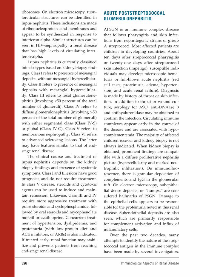
326 Immunological Aspects of Renal Disease
ribosomes. On electron microscopy, tubu-loreticular structures can be identifi ed in lupus nephritis. These inclusions are made of ribonucleoproteins and membranes and appear to be synthesized in response to interferon-alpha. Similar structures can be seen in HIV-nephropathy, a renal disease that has high levels of circulating inter-feron-alpha.
Lupus nephritis is currently classifi ed into six types based on kidney biopsy fi nd-ings. Class I refers to presence of mesangial deposits without mesangial hypercellular-ity. Class II refers to presence of mesangial deposits with mesangial hypercellular-ity. Class III refers to focal glomerulone-phritis (involving <50 percent of the total number of glomeruli). Class IV refers to diffuse glomerulonephritis (involving >50 percent of the total number of glomeruli) with either segmental class (Class IV-S) or global (Class IV-G). Class V refers to membranous nephropathy. Class VI refers to advanced sclerosing lesions. The latter may have features similar to that of end-stage renal disease.
The clinical course and treatment of lupus nephritis depends on the kidney biopsy fi ndings and presence of systemic symptoms. Class I and II lesions have good prognosis and do not require treatment. In class V disease, steroids and cytotoxic agents can be used to induce and main-tain remission. Likewise, class III and IV require more aggressive treatment with pulse steroids and cyclophosphamide, fol-lowed by oral steroids and mycophenolate mofetil or azathioprine. Concurrent treat-ment of hypertension, dyslipidemia, and proteinuria (with low-protein diet and ACE inhibitors, or ARBs) is also indicated. If treated early, renal function may stabi-lize and prevents patients from reaching end-stage renal disease.
ACUTE POSTSTREPTOCOCCAL
GLOMERULONEPHRITIS
APSGN is an immune complex disease that follows pharyngitis and skin infec-tions from nephritogenic strains of group A streptococci. Most affected patients are children in developing countries. About ten days after streptococcal pharyngitis or twenty-one days after streptococcal skin infection (impetigo), susceptible indi-viduals may develop microscopic hema-turia or full-blown acute nephritis (red cell casts, proteinuria, edema, hyperten-sion, and acute renal failure). Diagnosis is made by history of throat or skin infec-tion. In addition to throat or wound cul-ture, serology for ASO, anti-DNAase B and antihyaluronidase may be obtained to confi rm the infection. Circulating immune complexes appear early in the course of the disease and are associated with hypo-complementemia. The majority of affected children recover and kidney biopsy is not always indicated. When kidney biopsy is obtained, prominent fi ndings are compat-ible with a diffuse proliferative nephritis picture (hypercellularity and marked neu-trophilic infi ltration). On immunofl uo-rescence, there is granular deposition of complements and IgG in the glomerular tuft. On electron microscopy, subepithe-lial dense deposits, or “humps,” are con-sidered hallmarks of PSGN. Damage to the epithelial cells appears to be respon-sible for the proteinuria noted in this renal disease. Subendothelial deposits are also seen, which are primarily responsible for complement activation and infl ux of infl ammatory cells.
Over the past two decades, many attempts to identify the nature of the strep-tococcal antigen in the immune complex have been made by several investigators.
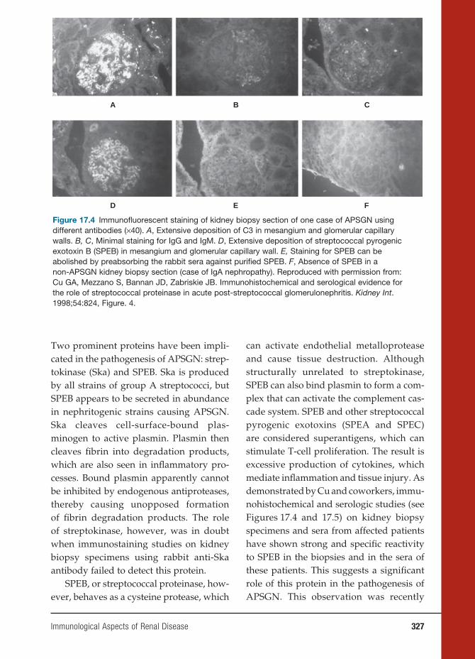
Immunological Aspects of Renal Disease 327
Two prominent proteins have been impli-cated in the pathogenesis of APSGN: strep-tokinase (Ska) and SPEB. Ska is produced by all strains of group A streptococci, but SPEB appears to be secreted in abundance in nephritogenic strains causing APSGN. Ska cleaves cell-surface-bound plas-minogen to active plasmin. Plasmin then cleaves fi brin into degradation products, which are also seen in infl ammatory pro-cesses. Bound plasmin apparently cannot be inhibited by endogenous antiproteases, thereby causing unopposed formation of fi brin degradation products. The role of streptokinase, however, was in doubt when immunostaining studies on kidney biopsy specimens using rabbit anti-Ska antibody failed to detect this protein.
SPEB, or streptococcal proteinase, how-ever, behaves as a cysteine protease, which
can activate endothelial metalloprotease and cause tissue destruction. Although structurally unrelated to streptokinase, SPEB can also bind plasmin to form a com-plex that can activate the complement cas-cade system. SPEB and other streptococcal pyrogenic exotoxins (SPEA and SPEC) are considered superantigens, which can stimulate T-cell proliferation. The result is excessive production of cytokines, which mediate infl ammation and tissue injury. As demonstrated by Cu and coworkers, immu-nohistochemical and serologic studies (see Figures 17.4 and 17.5) on kidney biopsy specimens and sera from affected patients have shown strong and specifi c reactivity to SPEB in the biopsies and in the sera of these patients. This suggests a signifi cant role of this protein in the pathogenesis of APSGN. This observation was recently
A B C
D E F
Figure 17.4 Immunofl uorescent staining of kidney biopsy section of one case of APSGN using different antibodies (×40). A, Extensive deposition of C3 in mesangium and glomerular capillary walls. B, C, Minimal staining for IgG and IgM. D, Extensive deposition of streptococcal pyrogenic exotoxin B (SPEB) in mesangium and glomerular capillary wall. E, Staining for SPEB can be abolished by preabsorbing the rabbit sera against purifi ed SPEB. F, Absence of SPEB in a non-APSGN kidney biopsy section (case of IgA nephropathy). Reproduced with permission from: Cu GA, Mezzano S, Bannan JD, Zabriskie JB. Immunohistochemical and serological evidence for the role of streptococcal proteinase in acute post-streptococcal glomerulonephritis. Kidney Int. 1998;54:824, Figure. 4.
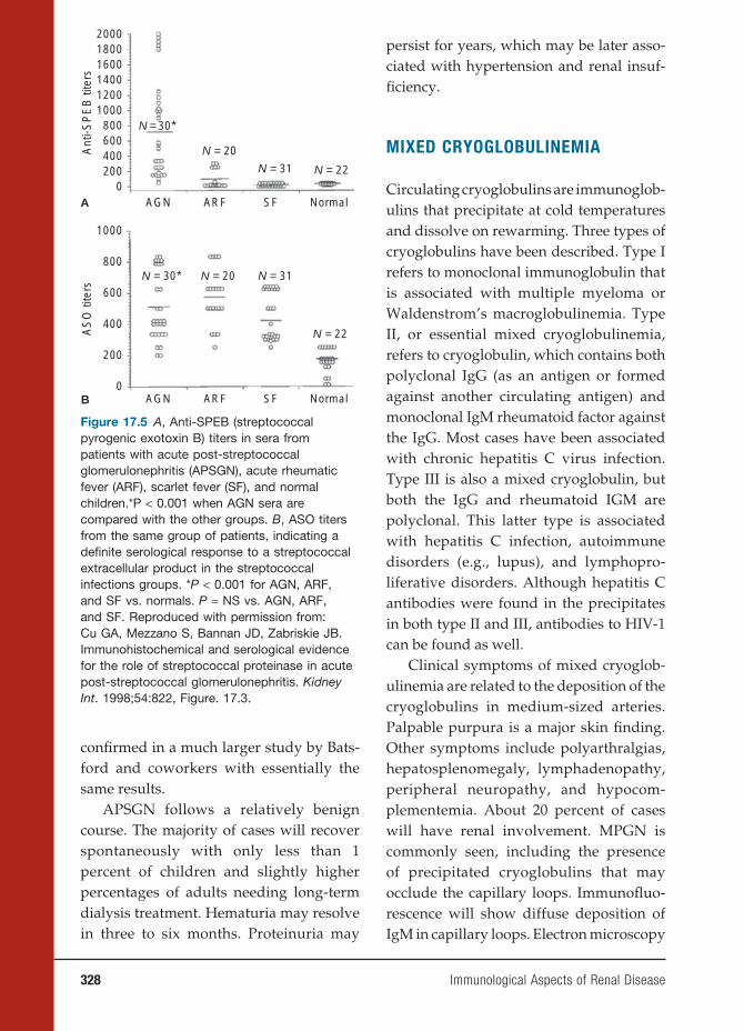
328 Immunological Aspects of Renal Disease
confi rmed in a much larger study by Bats-ford and coworkers with essentially the same results.
APSGN follows a relatively benign course. The majority of cases will recover spontaneously with only less than 1 percent of children and slightly higher percentages of adults needing long-term dialysis treatment. Hematuria may resolve in three to six months. Proteinuria may
persist for years, which may be later asso-ciated with hypertension and renal insuf-fi ciency.
MIXED CRYOGLOBULINEMIA
Circulating cryoglobulins are immunoglob-ulins that precipitate at cold temperatures and dissolve on rewarming. Three types of cryoglobulins have been described. Type I refers to monoclonal immunoglobulin that is associated with multiple myeloma or Waldenstrom’s macroglobulinemia. Type II, or essential mixed cryoglobulinemia, refers to cryoglobulin, which contains both polyclonal IgG (as an antigen or formed against another circulating antigen) and monoclonal IgM rheumatoid factor against the IgG. Most cases have been associated with chronic hepatitis C virus infection. Type III is also a mixed cryoglobulin, but both the IgG and rheumatoid IGM are polyclonal. This latter type is associated with hepatitis C infection, autoimmune disorders (e.g., lupus), and lymphopro-liferative disorders. Although hepatitis C antibodies were found in the precipitates in both type II and III, antibodies to HIV-1 can be found as well.
Clinical symptoms of mixed cryoglob-ulinemia are related to the deposition of the cryoglobulins in medium-sized arteries. Palpable purpura is a major skin fi nding. Other symptoms include polyarthralgias, hepatosplenomegaly, lymphadenopathy, peripheral neuropathy, and hypocom-plementemia. About 20 percent of cases will have renal involvement. MPGN is commonly seen, including the presence of precipitated cryoglobulins that may occlude the capillary loops. Immunofl uo-rescence will show diffuse deposition of IgM in capillary loops. Electron microscopy
A
N = 30*
N = 31 N = 22
N = 20
200018001600140012001000
Ant
i-SP
EB
tite
rs
800600400200
0
0
AGN ARF SF Normal
B
N = 30* N = 31
N = 22
N = 20
1000
AS
O ti
ters 600
200
400
800
AGN ARF SF Normal
Figure 17.5 A, Anti-SPEB (streptococcal pyrogenic exotoxin B) titers in sera from patients with acute post-streptococcal glomerulonephritis (APSGN), acute rheumatic fever (ARF), scarlet fever (SF), and normal children.*P < 0.001 when AGN sera are compared with the other groups. B, ASO titers from the same group of patients, indicating a defi nite serological response to a streptococcal extracellular product in the streptococcal infections groups. *P < 0.001 for AGN, ARF, and SF vs. normals. P = NS vs. AGN, ARF, and SF. Reproduced with permission from: Cu GA, Mezzano S, Bannan JD, Zabriskie JB. Immunohistochemical and serological evidence for the role of streptococcal proteinase in acute post-streptococcal glomerulonephritis. Kidney Int. 1998;54:822, Figure. 17.3.
Immunological Aspects of Renal Disease 329
will show subendothelial deposits, which may have a “fi ngerprint” appearance.
Until the discovery of hepatitis C and HIV-1 in cryglobulin precipitates, steroids and cytotoxic drugs (cyclophos-phamide or chlorambucil) were main-stays of treatment. These drugs are now reserved for severe cases of progressive renal failure, necrotic extremities that may require amputation, and severe neuropa-thy. Plasmapheresis is initially done to remove circulating cryoglobulins. Pulse steroids and cytotoxic agents are used to prevent new antibody formation. Anti-viral therapy is directed to hepatitis C and HIV disease to prevent further cry-oglobulin formation.
BIBLIOGRAPHY
REVIEWSAbbas AK, Lichtman AH, Pober JS, eds. Cel-
lular and Molecular Immunology. . Phila-delphia, PA: W. B. Saunders; 2005.
Brenner BM, ed. Brenner and Rector’s The Kidney. 7th ed. Philadelphia, PA: W. B. Saunders; 2004.
Lewis EJ, Schwartz MM, Korbet SM, eds. Lupus Nephritis. Oxford, UK: Oxford University Press; 1999.
Pereira BJG, Sayegh MH, Blake P, eds. Chronic Kidney Disease, Dialysis & Transplantation. 2nd ed. Elsevier Health Sciences; 2004.
Rose BD, ed. UpToDate Online 15.3. Waltham, MA; 2007.
SUGGESTED READINGSBatsford SR, Mezzano S, Mihatsch M,
Schiltz E, Rodríguez-Iturbe B. Is the nephritogenic antigen in post-streptococcal glomerulonephritis pyro-genic exotoxin B (SPE B) or GAPDH? Kidney Int. 2005;68:1120–1129.
Cu GA, Mezzano S, Bannan JD, Zabriskie JB. Immunohistochemical and serologi-cal evidence for the role of streptococcal proteinase in acute post-streptococcal glomerulonephritis. Kidney Int. 1998;54:819–826.
Farquhar MG, Saito A, Kerjaschki D, Orlando RA. The Heymann nephritis antigenic complex: megalin (gp330) and RAP. J Am Soc Nephrol. 1995; 6:35–47.
Reynolds J, Norgan VA, Bhambra U, Smith J, Cook HT, Pusey CD. Anti-CD8 monoclonal antibody therapy is effec-tive in the prevention and treatment of experimental autoimmune glomeru-lonephritis. J Am Soc Nephrol. 2002;13(2):359–369.
Johnson RJ, Feehally J, eds. Comprehensive Clinical Nephrology, 2nd ed. Elsevier Health Sciences; 2003.
Rose BD, Appel GB, Schur PH. Types of renal disease in systemic lupus ery-thematosus. In: Rose BD, ed. UpToDate. Waltham, MA; 2007.


18. Immunological Aspects of Transplantation
Jeffrey M. Venstrom, M.D., and James W. Young, M.D.
331
INTRODUCTION
More than fi fty years have passed since the fi rst identical twin transplant was per-formed, and we can now look on organ transplantation as an extremely success-ful treatment for hundreds of thousands of patients who would otherwise have been doomed to organ failure or to death. This successful kidney transplant between identical twins in 1954, performed at the Peter Bent Brigham Hospital by Dr. Joseph Murray and his colleagues, helped to usher in the fi eld of solid organ transplantation. Within the ensuing decade, this transplant was followed by the fi rst successful trans-plants from a non-twin living donor and later on from a cadaveric donor. The short-age of donor organs is now the chief stum-bling block to further development.
The fi rst twin transplant confi rmed that a young, healthy donor could survive with one kidney with no disability, as long as the single remaining kidney was not dam-aged. Indeed, the donor of the fi rst twin transplant is still alive more than fi fty years later. This achievement, which was quickly repeated in a number of other twin trans-plants, confi rmed the surgical aspects of this operation. Two other important obser-vations were made. First, if the patient suf-fered from an autoimmune nephritis, this could recur in the transplanted kidney. The same disease could also develop in the donor’s remaining kidney because of the
genetic susceptibility of identical twins to similar diseases.
The immunological obstacles, however, proved to be stubborn. Medawar and colleagues had shown that skin grafts were destroyed by a mechanism that had immune characteristics. Having reacted against one skin graft, the recipient of a second skin graft from the same donor would react more violently, demonstrating an acquired immunity and a memory for the fi rst tissue. A few years later, Medawar and colleagues demonstrated a natural way of overcom-ing graft rejection, which occurs in fraternal animal twins. The defi nitive experiment, injecting cells from an inbred strain of mice into the fetus of another strain, resulted in graft acceptance in survivors of this proce-dure and established the concept of immu-nological plasticity in the embryo before the immune system fully develops. Although this had no obvious clinical application, it raised the question about whether this state of immunological plasticity could be tem-porarily induced in a potential organ graft recipient so that the graft would be accepted even though the immune defenses would be rapidly restored.
The ensuing story of immunosuppres-sion still has this aim of “tolerance” in view. Clinical immunological tolerance occurs when the recipient’s immune system is ablated and replaced by hematopoietic stem cells from a donor matched according to expression of human leucocyte antigens
331
332 Immunological Aspects of Transplantation
(HLAs). Refi nements in immunosuppres-sion include more precisely dosed irradia-tion as well as drug and antibody treatment to support engraftment. Clinicians tend to add more potent immunosuppressive agents to the patient’s therapy. More recent attempts to achieve graft acceptance with minimal immunosuppression are begin-ning to meet with some success. One exam-ple is the use of the lympholytic monoclo-nal antibody, alemtuzumal (Campath-1H), as an inducing agent, followed by mainte-nance therapy with reduced-dose calcineu-rin inhibitor monotherapy.
The interest in organ transplantation greatly stimulated the science of immunol-ogy. Mechanisms of graft rejection are now well understood, and the fate of a graft depends not only on surgical technique but also on the degree of HLA matching between donor and recipient. The specifi c transplanted organ also has relevance, such as the liver, which is more tolerogenic than others. The HLA system of antigenic deter-minants arises from genes on human chro-mosome 6. The ABO antigen system is also important in solid organ graft outcome but does not directly affect hematopoietic stem cell transplantation (HSCT).
Of the drugs used to suppress the immune system, each has some side effects specifi c to the agent in question and oth-ers common to all immunosuppressive agents, namely, increased susceptibility to infections and tumors, especially lym-phoproliferative disorders. A strategy of using different agents together to maximize immunosuppression and minimize side effects has been partially successful, and the new approach of minimal immunosuppres-sion should be of additional benefi t. Active agents vary from small molecules (azathio-prine and corticosteroids) to complicated peptides and macrolides (cyclosporine,
tacrolimus, and sirolimus) and onto large polyclonal and monoclonal antibodies.
Much interest has recently focused on blocking the secondary and tertiary signals of antigen recognition. In animal models, such blockade can result in long-term graft acceptance without other potentially toxic immunosuppressive agents. These and other immunosuppressive agents are in the process of early clinical trials and no doubt will have an effect on immunosuppression in the future.
IMMUNOLOGICAL CONSIDERATIONS
OF TRANSPLANTATION
The human major histocompatibility com-plex (MHC) comprises a set of genes on the short arm of chromosome 6. These genes are highly polymorphic and are codominantly expressed, each individual having inher-ited one set of MHC genes from each par-ent. MHC molecules present foreign anti-gens, as well as self-peptides, to T cells for recognition and response. In general, class I MHC molecules present peptides to CD8+ T cells, while class II MHC molecules present peptides to CD4+ T cells. In the context of transplantation, this process can determine acceptance or rejection of the donor graft.
Class I and II M HC loci carry the great-est signifi cance for transplantation com-pared with other loci within the complex. While their polymorphisms are inherently critical to the species’ ability to resist various pathogens, these polymorphisms also pose one of the major obstacles to transplantation tolerance. Every individual expresses three different pairs of class I molecules (designated as HLA-A, HLA-B, and HLA-C) and three different pairs of class II molecules (designated as HLA-DP, HLA-DQ, and HLA-DR). In addition, the MHC contains
332 Immunological Aspects of Transplantation
Immunological Aspects of Transplantation 333
other genes that encode complement pro-teins, cytokines, and other proteins involved in antigen processing. Class I MHC molecules are expressed on all nucleated cells, whereas class II MHC molecules are expressed on antigen-presenting cells like macrophages, dendritic cells, B lymphocytes, endothelial cells, and thymic epithelial cells. Expression of MHC gene products is greatly enhanced by cytokines, especially interferon, which stimulates transcription of MHC genes.
Matching the class I and class II MHC genes between the graft and recipient has varying degrees of infl uence on graft–host interactions and tolerance, differing some-what with the inherent immunogenicity or tolerogenicity of the organ being trans-planted. In kidney transplantation, renal allograft survival directly correlates with the degree of similarity between graft and recipient MHC antigens. Complete HLA matching in liver transplantation is less criti-cal for long-term graft acceptance. Clinically, the HLA-A, HLA-B, and HLA-DR loci are the most relevant for predicting outcome, although the importance of other loci and allelic subtypes is increasingly recognized. Minor histocompatibility antigens are self-peptides derived from nucleotide polymor-phisms that differ between HLA identical siblings yet require HLA identity for presen-tation. These cannot be routinely typed in the laboratory but can greatly infl uence graft–host interactions in transplantation, espe-cially hematopoietic stem cell transplants.
LABORATORY TESTING FOR
COMPATIBILITY IN
TRANSPLANTATION
HLA Typing
Histocompatibility between the donor and recipient is primarily defi ned by
genes of the MHC. MHC is a general term for the gene complex encoding the HLA, and hence the two acronyms are used interchangeably in humans. Historically, HLA typing was performed by serologic evaluation, using a panel of anti-HLA antibody containing sera derived from multiparous women. Most clinical HLA laboratories now use DNA-based HLA typing methods. By using the polymerase chain reaction (PCR), polymorphic resi-dues of both class I and class II HLA or MHC molecules can be amplifi ed using primers that bind to the conserved regions of the gene. The precise nucleic acid sequence of the HLA alleles can be accu-rately determined using sequence-based typing. More commonly, partial ampli-fi cation of polymorphic residues using sequence-specifi c primer pairs (PCR-SSP or PCR-SSOP) can identify HLA alleles. Depending on the DNA probe, PCR-based HLA typing is described as either “intermediate-resolution” (beyond the serologic level, but short of the allelic subtype level) or “high-resolution” (allele- or allelic subtype–level typing). The resolution of the HLA typing defi nes the level of HLA mismatch. Low-resolu-tion serologic typing can identify antigen mismatch (HLA-A2 vs. -A11), whereas high-resolution typing can identify an allele- or allelic subtype-level mismatch (HLA-A*0201 vs. -A*0205).
ABO Blood Typing
ABO blood typing is one of the most important tests in human solid organ trans-plantation. ABO antigens are primarily expressed by erythrocytes but also can be expressed by platelets and glandular epi-thelial and endothelial cells. Incompatibility occurs when recipients lacking a certain
Immunological Aspects of Transplantation 333
334 Immunological Aspects of Transplantation
blood type produce IgM antibodies against that antigen and cause subsequent activation of complement and lysis of transfused incompatible red blood cells. Because ABO antigens are also expressed by endothelial cells, incompatibilities can lead to hyperacute vascular rejection.
In contrast to solid organ transplan-tation, ABO matching does not directly affect donor selection for allogeneic HSCT. Because inheritance of blood group anti-gens is independent of HLA antigens, fully HLA-matched donor-recipient pairs are often ABO incompatible. ABO incompat-ibility requires red blood cell depletion of a stem cell allograft but does not affect myeloid or megakaryocytic engraftment, graft rejection, or graft-versus-host disease (GvHD). Upon engraftment, the recipient transitions to the donor’s ABO blood type. During the conversion from recipient to donor blood type, transient hemolysis may occur.
Screening for Preformed Antibodies
Patients awaiting cadaveric organs un-dergo screening of their blood for pre-formed antibodies against HLA molecules. These antibodies may be produced by pregnancy, prior blood transfusions, or prior organ transplantation. An aliquot of the patient’s serum is mixed in separate wells with cells of at least forty donors assumed to be representative of the donor population. Complement-mediated lysis or fl ow cytometry with fl uorescent-labeled secondary antibodies to human IgG quantifi es the number of reactive cells as a proportion of the number of panel cells (percent reactive antibody, or PRA). Because transplantation across a posi-tive cross match can cause hyperacute or
acute rejection, patients with high PRAs typically experience longer waiting times on the cadaveric organ donor list. Many centers are therefore currently exploring strategies to remove these antibodies, by plasmapheresis, for example, either preop-eratively or perioperatively.
Cross Matching
In solid organ transplants, cross matching is usually done after a potential donor is identifi ed. The recipient’s serum is tested for reactivity to the donor’s lymphocytes using the previously mentioned comple-ment-mediated lysis or fl ow cytometric assays. A negative cross match means that there is no recipient antibody that is reac-tive with donor cells or graft. A positive cross match portends severe rejection if the donor organ is transplanted.
TYPES OF SOLID ORGAN
ALLOGRAFT REJECTION
Hyperacute Rejection
In hyperacute rejection, thrombosis of the vasculature occurs immediately after anasto-mosis of the solid organ graft blood vessels and release of the surgical clamp, which is caused by the presence of preformed anti-bodies that bind to the endothelial anti-gens. In the early days of transplantation, ABO incompatibility was the main culprit. Because ABO incompatibility is largely avoided now, hyperacute rejection usually occurs when IgG antibodies react with for-eign M HC antigens or lesser-known alloan-tigens in the endothelium. The latter may be associated with accelerated hyperacute rejec-tion that lasts a few days. Adequate screen-ing for preformed anti-HLA antibodies and
Immunological Aspects of Transplantation 335
ABO testing should prevent hyperacute rejection.
Acute Rejection
Acute rejection is an infl ammatory process affecting the vasculature and parenchyma of the allograft, which occurs a few days to a week after transplantation. Activated T cells can cause direct lysis of the graft or release cytokines that promote infl ammation in the allograft. The endothelial cells appear to be the earliest targets for rejection with re-sultant endotheliitis. A humoral-mediated response causes binding of antibodies to the blood vessel wall with resultant necro-sis and occlusion of the arteries.
Chronic Rejection
Fibrosis with intimal thickening and even-tual occlusion of medium-sized arteries that supply the graft characterizes chronic rejec-tion. The fi brosis seen in chronic rejection is presumed to be a by-product of previous infl ammation from acute rejection or from cytokines that stimulated fi broblast produc-tion. Chronic allograft rejection accounts for the majority of allograft failures.
PREVENTION AND MANAGEMENT
OF SOLID ORGAN ALLOGRAFT
REJECTION
Induction Agents
Induction agents are immunosuppressive drugs given intraoperatively and immedi-ately postoperatively to deplete the T-cell population. OKT3 is a murine monoclonal antibody to the CD3 complex of the T cell, an intrinsic part of the T-cell receptor. The exact mechanism by which T cells are then
cleared from the circulation may occur by FcR-mediated clearance or complement-mediated lysis. OKT3 also blocks the func-tion of killer T cells. Because of its murine origin, recipients often develop anti-murine antibodies precluding subsequent use for rejection episodes. Patients also experience cytokine release syndrome, with release of TNF-α, IL-2, and interferon-gamma into the circulation. Symptoms include fever, rigors, pulmonary edema, aseptic menin-gitis, and myalgias. Basiliximab (Simulect) and daclizumab (Zenapax) are humanized anti-CD25 monoclonal antibodies directed against the α chain of the high-affi nity IL-2 receptor. Such humanized antibodies are engineered to contain a part of the human antibody, rendering them less immuno-genic. Alemtuzumab (Campath 1H) is an anti-CD52 monoclonal antibody that is gaining acceptance as an induction agent for steroid-free protocols. This is another humanized antibody, initially designed to treat chronic lymphocytic leukemia. A single dose given intraoperatively should eliminate CD52-positive B and T cells, as well as monocytes and monocyte-derived dendritic cells, from the bone marrow and peripheral circulation. Rituximab (Rituxan) is a monoclonal antibody against the CD20 antigen on B lymphocytes. Originally used to treat non-Hodgkin’s lymphoma, it is being used to treat B-cell- or humoral-mediated allograft rejection. Rabbit anti-thymocyte globulin has replaced equine anti-thymocyte globulin as the primary polyclonal antibody to treat acute episodes of solid organ allograft rejection. This drug is made by injecting human lymphocytes into rabbits with subsequent collection and purifi cation of the antiserum. This drug is given for seven to ten days with elimination of T cells from the circulation.
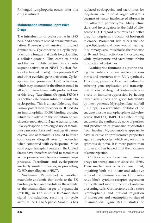
336 Immunological Aspects of Transplantation
Prolonged lymphopenia occurs after this drug is infused.
Maintenance Immunosuppressive
Drugs
The introduction of cyclosporine in 1983 heralded a new era of solid organ transplan-tation. Five-year graft survival improved dramatically. Cyclosporine is a cyclic pep-tide from a fungus that binds to cyclophilin, a cellular protein. This complex binds and further inhibits calcineurin and sub-sequent activation of NFAT (nuclear fac-tor of activated T cells). This prevents IL-2 and other cytokine gene activation. Cyclo-sporine also promotes TGF-β activation, which may account for the fi brosis noted in allograft parenchyma with prolonged use of this drug. Tacrolimus (Prograf, FK506 ) is another calcineurin inhibitor similar to cyclosporine. This is a macrolide drug that is more potent than cyclosporine. It binds to an immunophilin, FK506 binding protein, which is involved in the inhibition of cal-cineurin-mediated IL-2 gene transcription. Like cyclosporine, prolonged use of tacroli-mus can cause fi brosis of the allograft paren-chyma. Use of tacrolimus has led to fewer solid organ allograft rejection episodes when compared with cyclosporine. Most solid organ transplant centers in the United States have therefore shifted to tacrolimus as the primary maintenance immunosup-pressant. Tacrolimus and cyclosporine are fairly similar, however, in preventing GvHD after allogeneic HSCT.
Sirolimus (Rapamune) is another macrolide antibiotic that binds to the FK binding protein and modulates the activity of the mammalian target of rapamycin (mTOR); mTOR inhibits IL-2-mediated signal transduction, resulting in cycle arrest at the G1 to S phase. Sirolimus has
replaced cyclosporine and tacrolimus for long-term use in solid organ allografts because of lesser incidence of fi brosis in the allograft parenchyma. Many clini-cians and investigators in the fi eld of allo-geneic HSCT regard sirolimus as a better drug for long-term induction of host-graft tolerance. Prominent side effects include hyperlipidemia and poor wound healing. In summary, sirolimus blocks the response of B- and T-cell activation by cytokines, while cyclosporine and tacrolimus inhibit production of cytokines.
Azathioprine (Imuran) is a purine ana-log that inhibits purine nucleotide syn-thesis and interferes with RNA synthesis. This drug prevents T-cell activation by affecting gene replication and transcrip-tion. It is an old drug that continues to play a role in immunosuppression by having fewer side effects and good tolerability by most patients. Mycophenolate mofetil (CellCept) is a reversible inhibitor of the enzyme inosine monophosphate dehydro-genase (IMPDH). IMPDH is a rate-limiting enzyme in the synthesis de novo of purines and production of guanosine nucleotides from inosine. Mycophenolate appears to have selective antiproliferative properties against lymphocytes, which rely on purine synthesis de novo. It is more potent than Imuran and has helped limit the incidence of acute rejection.
Corticosteroids have been mainstay drugs for transplantation since the 1960s. Their mechanisms of action are broad, impairing both the innate and adaptive arms of the immune system. Corticoste-roids block cytokine-receptor expression by T cells and inhibit function of antigen-presenting cells. Corticosteroids also cause lymphophenia and prevent migration of monocytes and neutrophils to sites of infl ammation. Figure 18.1 illustrates the
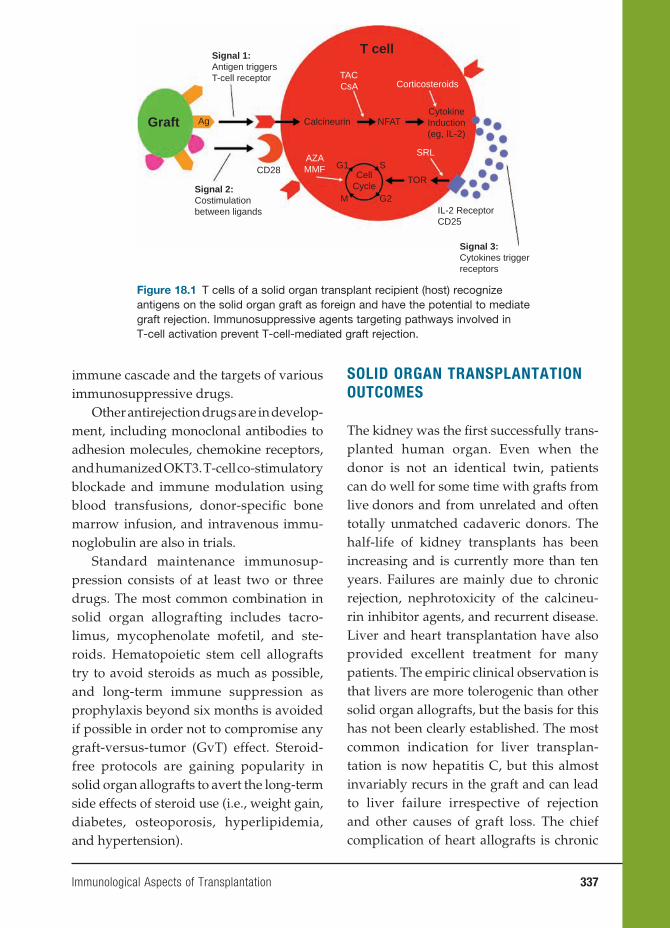
Immunological Aspects of Transplantation 337
immune cascade and the targets of various immunosuppressive drugs.
Other antirejection drugs are in develop-ment, including monoclonal antibodies to adhesion molecules, chemokine receptors, and humanized OKT3. T-cell co-stimulatory blockade and immune modulation using blood transfusions, donor-specifi c bone marrow infusion, and intravenous immu-noglobulin are also in trials.
Standard maintenance immunosup-pression consists of at least two or three drugs. The most common combination in solid organ allografting includes tacro-limus, mycophenolate mofetil, and ste-roids. Hematopoietic stem cell allografts try to avoid steroids as much as possible, and long-term immune suppression as prophylaxis beyond six months is avoided if possible in order not to compromise any graft-versus-tumor (GvT) effect. Steroid-free protocols are gaining popularity in solid organ allografts to avert the long-term side effects of steroid use (i.e., weight gain, diabetes, osteoporosis, hyperlipidemia, and hypertension).
SOLID ORGAN TRANSPLANTATION
OUTCOMES
The kidney was the fi rst successfully trans-planted human organ. Even when the donor is not an identical twin, patients can do well for some time with grafts from live donors and from unrelated and often totally unmatched cadaveric donors. The half-life of kidney transplants has been increasing and is currently more than ten years. Failures are mainly due to chronic rejection, nephrotoxicity of the calcineu-rin inhibitor agents, and recurrent disease. Liver and heart transplantation have also provided excellent treatment for many patients. The empiric clinical observation is that livers are more tolerogenic than other solid organ allografts, but the basis for this has not been clearly established. The most common indication for liver transplan-tation is now hepatitis C, but this almost invariably recurs in the graft and can lead to liver failure irrespective of rejection and other causes of graft loss. The chief complication of heart allografts is chronic
Figure 18.1 T cells of a solid organ transplant recipient (host) recognize antigens on the solid organ graft as foreign and have the potential to mediate graft rejection. Immunosuppressive agents targeting pathways involved in T-cell activation prevent T-cell-mediated graft rejection.
T cell
TACCsA Corticosteroids
AZAMMF
SRL
Calcineurin NFAT
S
M
CellCycle
G1
G2
TOR
IL-2 ReceptorCD25
CD28
Ag
Signal 1:Antigen triggersT-cell receptor
Signal 2:Costimulationbetween ligands
Signal 3:Cytokines triggerreceptors
GraftCytokineInduction(eg, IL-2)
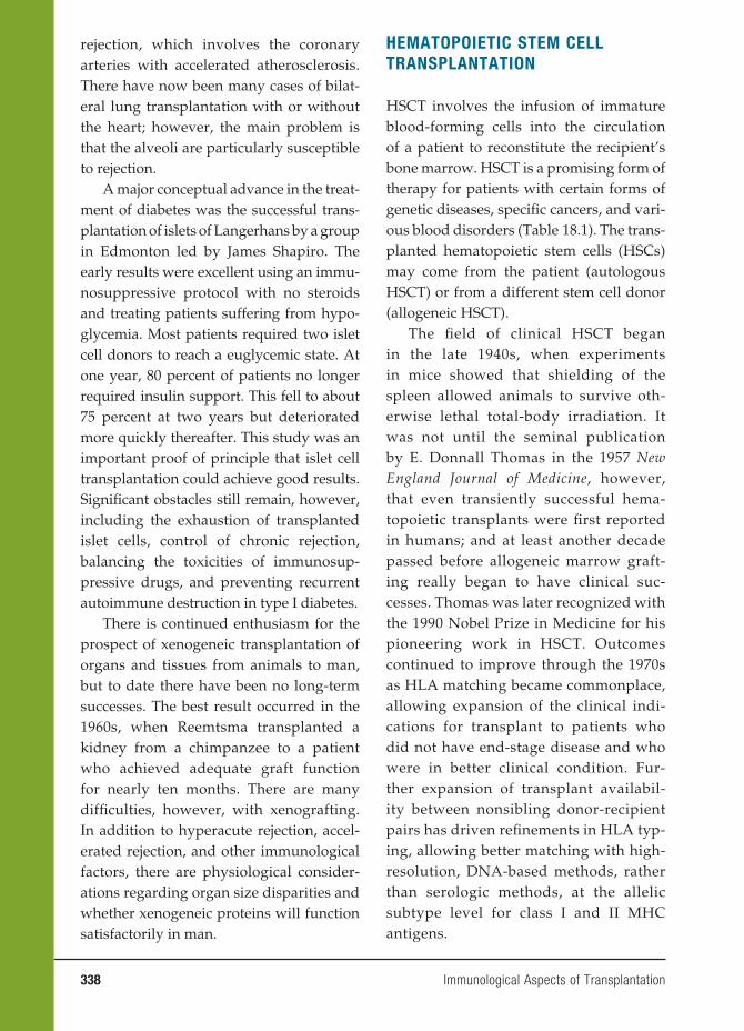
338 Immunological Aspects of Transplantation
rejection, which involves the coronary arteries with accelerated atherosclerosis. There have now been many cases of bilat-eral lung transplantation with or without the heart; however, the main problem is that the alveoli are particularly susceptible to rejection.
A major conceptual advance in the treat-ment of diabetes was the successful trans-plantation of islets of Langerhans by a group in Edmonton led by James Shapiro. The early results were excellent using an immu-nosuppressive protocol with no steroids and treating patients suffering from hypo-glycemia. Most patients required two islet cell donors to reach a euglycemic state. At one year, 80 percent of patients no longer required insulin support. This fell to about 75 percent at two years but deteriorated more quickly thereafter. This study was an important proof of principle that islet cell transplantation could achieve good results. Signifi cant obstacles still remain, however, including the exhaustion of transplanted islet cells, control of chronic rejection, balancing the toxicities of immunosup-pressive drugs, and preventing recurrent autoimmune destruction in type I diabetes.
There is continued enthusiasm for the prospect of xenogeneic transplantation of organs and tissues from animals to man, but to date there have been no long-term successes. The best result occurred in the 1960s, when Reemtsma transplanted a kidney from a chimpanzee to a patient who achieved adequate graft function for nearly ten months. There are many diffi culties, however, with xenografting. In addition to hyperacute rejection, accel-erated rejection, and other immunological factors, there are physiological consider-ations regarding organ size disparities and whether xenogeneic proteins will function satisfactorily in man.
HEMATOPOIETIC STEM CELL
TRANSPLANTATION
HSCT involves the infusion of immature blood-forming cells into the circulation of a patient to reconstitute the recipient’s bone marrow. HSCT is a promising form of therapy for patients with certain forms of genetic diseases, specifi c cancers, and vari-ous blood disorders (Table 18.1). The trans-planted hematopoietic stem cells (HSCs) may come from the patient (autologous HSCT) or from a different stem cell donor (allogeneic HSCT).
The fi eld of clinical HSCT began in the late 1940s, when experiments in mice showed that shielding of the spleen allowed animals to survive oth-erwise lethal total-body irradiation. It was not until the seminal publication by E. Donnall Thomas in the 1957 New England Journal of Medicine, however, that even transiently successful hema-topoietic transplants were fi rst reported in humans; and at least another decade passed before allogeneic marrow graft-ing really began to have clinical suc-cesses. Thomas was later recognized with the 1990 Nobel Prize in Medicine for his pioneering work in HSCT. Outcomes continued to improve through the 1970s as HLA matching became commonplace, allowing expansion of the clinical indi-cations for transplant to patients who did not have end-stage disease and who were in better clinical condition. Fur-ther expansion of transplant availabil-ity between nonsibling donor-recipient pairs has driven refi nements in HLA typ-ing, allowing better matching with high-resolution, DNA-based methods, rather than serologic methods, at the allelic subtype level for class I and II MHC antigens.
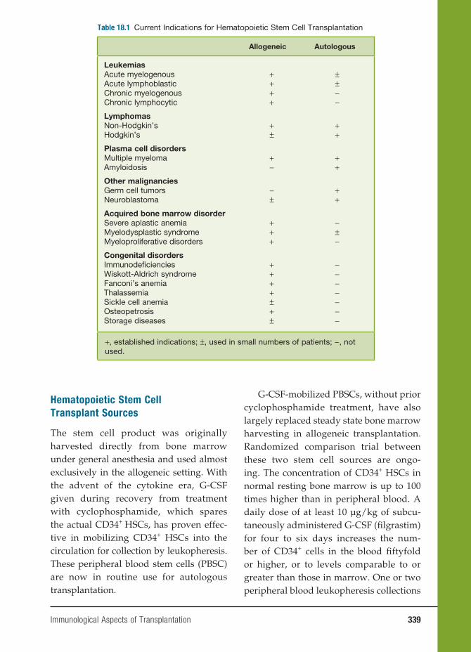
Immunological Aspects of Transplantation 339
Hematopoietic Stem Cell
Transplant Sources
The stem cell product was originally harvested directly from bone marrow under general anesthesia and used almost exclusively in the allogeneic setting. With the advent of the cytokine era, G-CSF given during recovery from treatment with cyclophosphamide, which spares the actual CD34+ HSCs, has proven effec-tive in mobilizing CD34+ HSCs into the circulation for collection by leukopheresis. These peripheral blood stem cells (PBSC) are now in routine use for autologous transplantation.
G-CSF-mobilized PBSCs, without prior cyclophosphamide treatment, have also largely replaced steady state bone marrow harvesting in allogeneic transplantation. Randomized comparison trial between these two stem cell sources are ongo-ing. The concentration of CD34+ HSCs in normal resting bone marrow is up to 100 times higher than in peripheral blood. A daily dose of at least 10 µg/kg of subcu-taneously administered G-CSF (fi lgrastim) for four to six days increases the num-ber of CD34+ cells in the blood fi ftyfold or higher, or to levels comparable to or greater than those in marrow. One or two peripheral blood leukopheresis collections
Table 18.1 Current Indications for Hematopoietic Stem Cell Transplantation
Allogeneic Autologous
LeukemiasAcute myelogenousAcute lymphoblasticChronic myelogenousChronic lymphocytic
++++
±±−−
LymphomasNon-Hodgkin’sHodgkin’s
+±
++
Plasma cell disordersMultiple myelomaAmyloidosis
+−
++
Other malignanciesGerm cell tumorsNeuroblastoma
−±
++
Acquired bone marrow disorderSevere aplastic anemiaMyelodysplastic syndromeMyeloproliferative disorders
+++
−±−
Congenital disordersImmunodefi cienciesWiskott-Aldrich syndromeFanconi’s anemiaThalassemiaSickle cell anemiaOsteopetrosisStorage diseases
++++±+ ±
−−−−−−−
+, established indications; ±, used in small numbers of patients; −, not used.
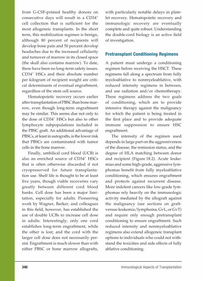
340 Immunological Aspects of Transplantation
from G-CSF-primed healthy donors on consecutive days will result in a CD34+ cell collection that is suffi cient for the most allogeneic transplants. In the short term, this mobilization regimen is benign, although 80 percent of recipients will develop bone pain and 50 percent develop headaches due to the increased cellularity and turnover of marrow in its closed space (the skull also contains marrow). To date, there have been no long-term safety issues. CD34+ HSCs and their absolute number per kilogram of recipient weight are criti-cal determinants of eventual engraftment, regardless of the stem cell source.
Hematopoietic recovery occurs earlier after transplantation of PBSC than bone mar-row, even though long-term engraftment may be similar. This seems due not only to the dose of CD34+ HSCs but also to other lymphocyte subpopulations included in the PBSC graft. An additional advantage of PBSCs, at least in autografts, is the lower risk that PBSCs are contaminated with tumor cells in the bone marrow.
Finally, umbilical cord blood (UCB) is also an enriched source of CD34+ HSCs that is often otherwise discarded if not cryopreserved for future transplanta-tion use. Shelf life is thought to be at least fi ve years, though viable recoveries vary greatly between different cord blood banks. Cell dose has been a major limi-tation, especially for adults. Pioneering work by Wagner, Barker, and colleagues in this fi eld, however, has established the use of double UCBs to increase cell dose in adults. Interestingly, only one cord establishes long-term engraftment, while the other is lost; and the cord with the larger cell dose does not necessarily per-sist. Engraftment is much slower than with either PBSC or bone marrow allografts,
with particularly notable delays in plate-let recovery. Hematopoietic recovery and immunologic recovery are eventually complete and quite robust. Understanding the double-cord biology is an active fi eld of investigation.
Pretransplant Conditioning Regimens
A patient must undergo a conditioning regimen before receiving the HSCT. These regimens fall along a spectrum from fully myeloablative to nonmyeloablative, with reduced intensity regimens in between, and use radiation and/or chemotherapy. These regimens address the two goals of conditioning, which are to provide intensive therapy against the malignancy for which the patient is being treated in the fi rst place and to provide adequate immune suppression to ensure HSC engraftment.
The intensity of the regimen used depends in large part on the aggressiveness of the disease, the remission status, and the degree of HLA matching between donor and recipient (Figure 18.2). Acute leuke-mias and some high-grade, aggressive lym-phomas benefi t from fully myeloablative conditioning, which ensures engraftment and protects against recurrent disease. More indolent cancers like low-grade lym-phomas rely heavily on the immunologic activity mediated by the allograft against the malignancy (see sections on graft-versus-leukemia/lymphoma, GvL, or GvT) and require only enough pretransplant conditioning to ensure engraftment. Such reduced intensity and nonmyeloablative regimens also extend allogeneic transplant options to individuals who could not with-stand the toxicities and side effects of fully ablative conditioning.
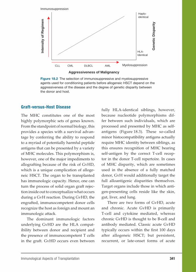
Immunological Aspects of Transplantation 341
Graft-versus-Host Disease
The MHC constitutes one of the most highly polymorphic sets of genes known. From the standpoint of normal biology, this provides a species with a survival advan-tage by conferring the ability to respond to a myriad of potentially harmful peptide antigens that can be presented by a variety of MHC molecules. This polymorphism is, however, one of the major impediments to allografting because of the risk of GvHD, which is a unique complication of alloge-neic HSCT. The organ to be transplanted has immunologic capacity. Hence, one can turn the process of solid organ graft rejec-tion inside out to conceptualize what occurs during a GvH reaction. During GvHD, the engrafted, immunocompetent donor cells recognize the host as foreign and mount an immunologic attack.
The dominant immunologic factors underlying GvHD are the HLA compat-ibility between donor and recipient and the presence of immunocompetent T cells in the graft. GvHD occurs even between
fully HLA-identical siblings, however, because nucleotide polymorphisms dif-fer between such individuals, which are processed and presented by MHC as self-antigens (Figure 18.3). These so-called minor histocompatibility antigens actually require MHC identity between siblings, as this ensures recognition of MHC bearing self-antigen by the correct T-cell recep-tor in the donor T-cell repertoire. In cases of MHC disparity, which are sometimes used in the absence of a fully matched donor, GvH would additionally target the full alloantigenic disparities themselves. Target organs include those in which anti-gen-presenting cells reside like the skin, gut, liver, and lung.
There are two forms of GvHD, acute and chronic. Acute GvHD is primarily T-cell and cytokine mediated, whereas chronic GvHD is thought to be B-cell and antibody mediated. Classic acute GvHD typically occurs within the fi rst 100 days after allogeneic HSCT; but persistent, recurrent, or late-onset forms of acute
Myelosuppression
Gen
etic
Dis
par
ity
Aggressiveness of Malignancy
HLA-Identical
Haplo-identical
Immunosuppression
CLL CML DLBCL AML
Inte
nsi
ty o
f R
egim
en
Figure 18.2 The selection of immunosuppressive and myelosuppressive agents used for conditioning patients before allogeneic HSCT depend on the aggressiveness of the disease and the degree of genetic disparity between the donor and host.
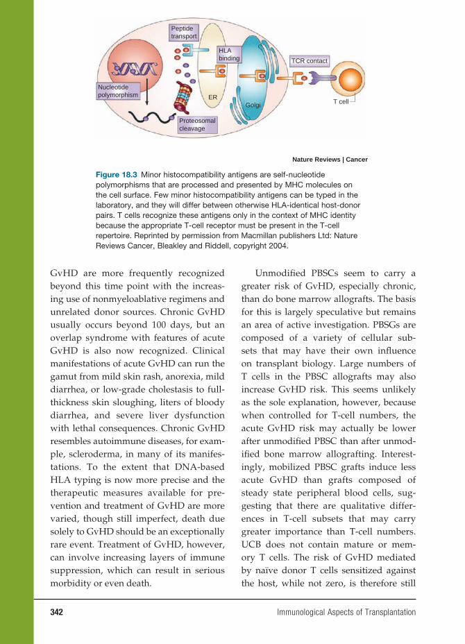
342 Immunological Aspects of Transplantation
GvHD are more frequently recognized beyond this time point with the increas-ing use of nonmyeloablative regimens and unrelated donor sources. Chronic GvHD usually occurs beyond 100 days, but an overlap syndrome with features of acute GvHD is also now recognized. Clinical manifestations of acute GvHD can run the gamut from mild skin rash, anorexia, mild diarrhea, or low-grade cholestasis to full-thickness skin sloughing, liters of bloody diarrhea, and severe liver dysfunction with lethal consequences. Chronic GvHD resembles autoimmune diseases, for exam-ple, scleroderma, in many of its manifes-tations. To the extent that DNA-based HLA typing is now more precise and the therapeutic measures available for pre-vention and treatment of GvHD are more varied, though still imperfect, death due solely to GvHD should be an exceptionally rare event. Treatment of GvHD, however, can involve increasing layers of immune suppression, which can result in serious morbidity or even death.
Unmodifi ed PBSCs seem to carry a greater risk of GvHD, especially chronic, than do bone marrow allografts. The basis for this is largely speculative but remains an area of active investigation. PBSGs are composed of a variety of cellular sub-sets that may have their own infl uence on transplant biology. Large numbers of T cells in the PBSC allografts may also increase GvHD risk. This seems unlikely as the sole explanation, however, because when controlled for T-cell numbers, the acute GvHD risk may actually be lower after unmodifi ed PBSC than after unmod-ifi ed bone marrow allografting. Interest-ingly, mobilized PBSC grafts induce less acute GvHD than grafts composed of steady state peripheral blood cells, sug-gesting that there are qualitative differ-ences in T-cell subsets that may carry greater importance than T-cell numbers. UCB does not contain mature or mem-ory T cells. The risk of GvHD mediated by naïve donor T cells sensitized against the host, while not zero, is therefore still
Peptidetransport
HLAbinding
ER
TCR contact
T cellGolgi
Proteosomalcleavage
Nucleotidepolymorphism
Nature Reviews | Cancer
Figure 18.3 Minor histocompatibility antigens are self-nucleotide polymorphisms that are processed and presented by MHC molecules on the cell surface. Few minor histocompatibility antigens can be typed in the laboratory, and they will differ between otherwise HLA-identical host-donor pairs. T cells recognize these antigens only in the context of MHC identity because the appropriate T-cell receptor must be present in the T-cell repertoire. Reprinted by permission from Macmillan publishers Ltd: Nature Reviews Cancer, Bleakley and Riddell, copyright 2004.

Immunological Aspects of Transplantation 343
considerably less than that which occurs after transplantation of other unmodifi ed transplant products, even for a greater degree of host-donor mismatch.
Prevention of GvHD requires either depletion of T cells from the allograft or pharmacologic prophylaxis to limit sen-sitization of donor T cells against host antigens. Drugs for GvHD prophylaxis include those used for prevention of solid organ graft rejection such as cyclospo-rine, tacrolimus, sirolimus, mycopheno-lic acid, and methotrexate (Figure 18.4). Corticosteroids are used acutely for treat-ment of GvHD if it occurs; but there are no proven therapies for steroid-refrac-tory GvHD, whether acute or chronic. T-cell depletion is extremely effective in reducing acute GvHD and is more effec-tive in preventing chronic GvHD than is pharmacologic prophylaxis. Techniques to decrease the number of T cells in the HSC allograft have been developed for application in vitro and in vivo. Methods
that achieve less T-cell depletion than others may sometimes require pharmaco-logic GvHD prophylaxis as well.
Graft-versus-Host Disease and
Graft-versus-Leukemia/Lymphoma
It was well after transplants were under-taken in humans that seminal work by Korngold and Sprent in the late 1970s and early 1980s established that donor T cells mediated the onset of acute GvHD between mice that were MHC identical but mismatched for minor histocompat-ibility antigens. We also now know that T cells are important for the GvL effect. In fact, donor leucocyte infusions (DLI) after allogeneic HSCT can often eradicate minimal residual disease recurrences and convert mixed-to full-donor chimeras. These DLIs are dosed according to their T-cell contents; but the products are usually not purifi ed T cells and therefore contain additional potential effector populations
DONORT cell
TACCsA costeroids
Corticosteroids
AZAMMF
SRL
Calcineurin NFAT
S
M
CellCycle
G1
G2
TOR
IL-2 ReceptorCD25
CD28
Ag
Signal 1:Antigen triggersT-cell receptor
Signal 2:Costimulationbetween ligands
Signal 3:Cytokines triggerreceptors
HOSTCytokineInduction(eg, IL-2)
Figure 18.4 Alloreactive T cells of the hematopoietic stem cell (HSC) graft recognize recipient antigens as foreign, and mediate graft-versus-host disease (GvHD) and graft-versus-leukemia/lymphoma. Immunosuppressive agents targeting T-cell activation pathways are administered to modulate the graft response and to prevent or treat GvHD.
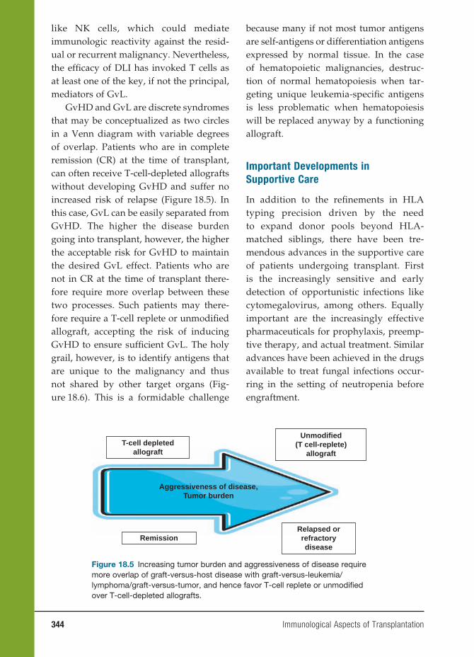
344 Immunological Aspects of Transplantation
like NK cells, which could mediate immunologic reactivity against the resid-ual or recurrent malignancy. Nevertheless, the effi cacy of DLI has invoked T cells as at least one of the key, if not the principal, mediators of GvL.
GvHD and GvL are discrete syndromes that may be conceptualized as two circles in a Venn diagram with variable degrees of overlap. Patients who are in complete remission (CR) at the time of transplant, can often receive T-cell-depleted allografts without developing GvHD and suffer no increased risk of relapse (Figure 18.5). In this case, GvL can be easily separated from GvHD. The higher the disease burden going into transplant, however, the higher the acceptable risk for GvHD to maintain the desired GvL effect. Patients who are not in CR at the time of transplant there-fore require more overlap between these two processes. Such patients may there-fore require a T-cell replete or unmodifi ed allograft, accepting the risk of inducing GvHD to ensure suffi cient GvL. The holy grail, however, is to identify antigens that are unique to the malignancy and thus not shared by other target organs (Fig-ure 18.6). This is a formidable challenge
because many if not most tumor antigens are self-antigens or differentiation antigens expressed by normal tissue. In the case of hematopoietic malignancies, destruc-tion of normal hematopoiesis when tar-geting unique leukemia-specifi c antigens is less problematic when hematopoiesis will be replaced anyway by a functioning allograft.
Important Developments in
Supportive Care
In addition to the refi nements in HLA typing precision driven by the need to expand donor pools beyond HLA-matched siblings, there have been tre-mendous advances in the supportive care of patients undergoing transplant. First is the increasingly sensitive and early detection of opportunistic infections like cytomegalovirus, among others. Equally important are the increasingly effective pharmaceuticals for prophylaxis, preemp-tive therapy, and actual treatment. Similar advances have been achieved in the drugs available to treat fungal infections occur-ring in the setting of neutropenia before engraftment.
Aggressiveness of disease,Tumor burden
Unmodified(T cell-replete)
allograftT-cell depleted
allograft
RemissionRelapsed orrefractorydisease
Figure 18.5 Increasing tumor burden and aggressiveness of disease require more overlap of graft-versus-host disease with graft-versus-leukemia/lymphoma/graft-versus-tumor, and hence favor T-cell replete or unmodifi ed over T-cell-depleted allografts.
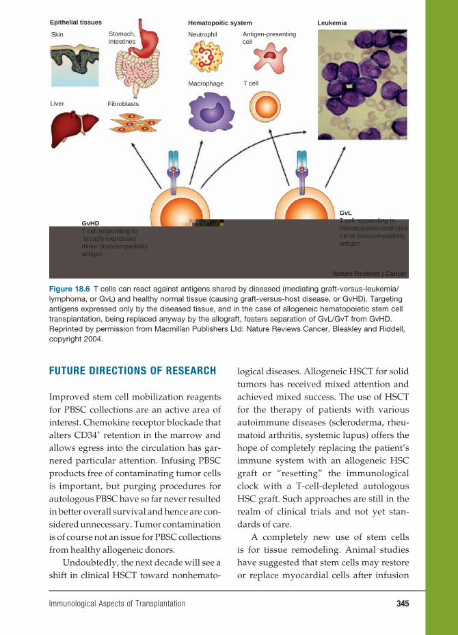
Immunological Aspects of Transplantation 345
FUTURE DIRECTIONS OF RESEARCH
Improved stem cell mobilization reagents for PBSC collections are an active area of interest. Chemokine receptor blockade that alters CD34+ retention in the marrow and allows egress into the circulation has gar-nered particular attention. Infusing PBSC products free of contaminating tumor cells is important, but purging procedures for autologous PBSC have so far never resulted in better overall survival and hence are con-sidered unnecessary. Tumor contamination is of course not an issue for PBSC collections from healthy allogeneic donors.
Undoubtedly, the next decade will see a shift in clinical HSCT toward nonhemato-
logical diseases. Allogeneic HSCT for solid tumors has received mixed attention and achieved mixed success. The use of HSCT for the therapy of patients with various autoimmune diseases (scleroderma, rheu-matoid arthritis, systemic lupus) offers the hope of completely replacing the patient’s immune system with an allogeneic HSC graft or “resetting” the immunological clock with a T-cell-depleted autologous HSC graft. Such approaches are still in the realm of clinical trials and not yet stan-dards of care.
A completely new use of stem cells is for tissue remodeling. Animal studies have suggested that stem cells may restore or replace myocardial cells after infusion
Epithelial tissues Hematopoitic system Leukemia
Skin Stomach,intestines
Liver Fibroblasts
Neutrophil Antigen-presentingcell
T cellMacrophage
GvHDT cell responding to broadly expressed minor histocompatibility antigen
GvLT cell responding tohematopoietic-restrictedminor histocompatibilityantigen
Nature Reviews | Cancer
Figure 18.6 T cells can react against antigens shared by diseased (mediating graft-versus-leukemia/lymphoma, or GvL) and healthy normal tissue (causing graft-versus-host disease, or GvHD). Targeting antigens expressed only by the diseased tissue, and in the case of allogeneic hematopoietic stem cell transplantation, being replaced anyway by the allograft, fosters separation of GvL/GvT from GvHD. Reprinted by permission from Macmillan Publishers Ltd: Nature Reviews Cancer, Bleakley and Riddell, copyright 2004.
346 Immunological Aspects of Transplantation
into the coronary arteries after myocardial damage. Early studies are ongoing in humans to see whether this approach is clinically feasible and effective in patients after myocardial infarction.
As technologies to isolate individual cell populations have improved, adjuvant cellular therapies are also gaining ground. Current protocols are evaluating adoptive cellular immunotherapies using regulatory T cells, viral-specifi c cytotoxic T cells, mes-enchymal stem cells, and natural killer cells.
In addition to HLA, other genetic loci are being evaluated as part of donor selection for allogeneic HSCT. One par-ticular genetic region under consideration is that which encodes natural killer cell receptors. The natural killer cell immu-noglobulin-like receptors (KIR) bind HLA epitopes and are encoded by a highly polymorphic gene region similar to the MHC. KIR-HLA combinations have been shown to predict NK cell alloreactivity and survival outcomes for some diseases, including acute myeloid leukemia. KIR genotyping is currently available in most clinical HLA laboratories, although there is insuffi cient data yet to recommend broad KIR genotyping for selection of unrelated donors. In the rare instances where more than one unrelated donor is available, choice based on KIR genotyping may be reasonable, all else being equal between the donor choices.
BIBLIOGRAPHY
REVIEWSAppelbaum FR. The current status of hema-
topoietic cell transplantation. Annu Rev Med. 2003;54:491–512.
Barker JN, Wagner JE. Umbilical-cord blood transplantation for the treat-
ment of cancer. Nat Rev Cancer. 2003;3(7):526–532.
Bleakley M, Riddell SR. Molecules and mechanisms of the graft-versus-leuke-mia effect. Nat Rev Cancer. 2004;4:371–380.
Copelan EA. Hematopoietic stem-cell transplantation. N Engl J Med. 2006;354(17):1813–1826.
Danovitch G. Handbook of Kidney Trans-plantation. Philadelphia, PA: Lippincott Williams & Wilkins; 2005.
Halloran, PF. Immunosuppressive drugs for kidney transplantation. N Engl J Med. 2004;351:2715.
Meier-Kriesche HU. Immunosuppression: evolution in practice and trends. Am J Transplant. 2006;6(5 Pt 2):1111–1131.
Stuart F, Abecassis MM, Kaufman DB, eds. Organ Transplantation. Georgetown, TX: Landes Bioscience; 2003.
LANDMARK PAPERSBillingham RE, Brent L, Medawar PB.
Actively acquired tolerance of foreign cells. Nature. 1953;172:603–606.
Gibson T, Medawar PB. The fate of skin homografts in man. J Anat. 1943;77:299–310.
Hale DA, Dhanireddy K, Bruno D, Kirk AD. Induction of transplanta-tion tolerance in non-human primate preclinical models. Philos Trans R Soc B. 2005;360:1723–1737.
Knechtle SJ. Development of tolerogenic strategies in the clinic. Philos Trans R Soc B. 2005;1739–1746.
Korngold B, Sprent J. Lethal graft-versus-host disease after bone marrow trans-plantation across minor histocompat-ibility barriers in mice: prevention by removing mature T cells from marrow J Exp Med. 1978;148:1687–1698.
Immunological Aspects of Transplantation 347
Murray JE, Merrill JP, Harrison JH. Renal homotransplantations in identical twins. Surg Forum. 1955;6:432–436.
Reemtsma K, McCracken BH, Schlegel JU, et al. Renal heterotransplantation in man. Ann Surg. 1964;160:384.
Shapiro AM, Lakey JR, Ryan EA, Korbutt GS. Islet transplantation in seven patients with type I diabetes mellitus using a glucocorticoid-free immuno-suppressive regimen. N Engl J Med. 2000;343:230–238.
Thomas ED, Buckner CD, Rudolph RH, et al. Allogeneic marrow grafting for hematologic malignancy using HLA matched donor-recipient sibling pairs. Blood. 1971;38:267–287.
Thomas ED, Lochte HL, Lu WC, Ferrebee JW. Intravenous infusion of bone marrow in patients receiving radiation and chemotherapy. N Engl J Med. 1957;257(11):491–496.

349
Index
AA. See Alopecia areataAbnormal host immune
response, 203Absent B cells, antibody
defi ciency from, 64, 66BTK genes and, 64X-linked
agammaglobulinemia, 66Acquired immune defi ciency
syndrome (AIDS), 131. See also Human immunodefi ciency virus
TB and, 233Activation-induced cell death
(AICD), 39Active immunization, 37–38Acute infl ammatory
demyelinating polyneuropathy (AIDP), 297. See also Guillian Barré syndrome
Acute post streptococcal glomerulonephritis (APSGN), 326–328
antigens in, 326–328biopsy staining for, 327fimmune response sequences
in, 319fSPEB and, 327streptokinase and, 327
Acute rheumatic fever (ARF), 199–211
animal models of, 207–208anorexia nervosa and, 210epidemiology of, 199–200etiology of, 203–207
abnormal host immune response, 203
mononuclear cellular infi ltrates in, 206f
future research for, 209–210genetic predisposition to,
201–202HLA and, 202
heart reactive antibody titers in, 204–205, 204f, 205f
incidence rates for, 199mortality rates for, decline of,
200f
M type strains of, 200OCD and, 206pathogenesis of, 200–201
group A streptococcus in, 200–201
staining for, 204fimmunofl uorescent, 207f
streptococcal vaccines for, 208–209, 210t
Tourette’s syndrome and, 210Adaptive immunity, 47–49
DC and, 47to HIV, 135–136, 137fMHC and, 47TLRs and, 47–49, 48t
signaling for, 48–49Adenosine nucleotide
translocator (ANT), 223Adhesion molecules, 10Adjuvant arthritis, 180Adoptive cell transfer, 13fAdoptive immunotherapy, 37AICD. See Activation-induced cell
deathAIDP. See Acute infl ammatory
demyelinating polyneuropathy
AIDS. See Acquired immune defi ciency syndrome
AIH. See Autoimmune hepatitisAIHA. See Autoimmune
hemolytic anemiaAK. See Atopic
keratoconjunctivitisAlkylating agents, 33–34Allergens and, 145Allergic asthma. See AsthmaAllergic conjunctivitis, 150–152
AK, 151animal models of, 151–152GPC, 151histopathological/lab fi ndings
for, 152tperennial, 150seasonal, 150vernal, 150–151
Allergic reactions, 145–160. See also Anaphylaxis; Respiratory tract,
allergic diseases of; Skin diseases, allergic reactions and
allergens and, 145anaphylaxis and, 147–149
animal models of, 148causes of, 148–149, 149tdefi nition of, 147systemic, 147–149treatment of, 148
atopy and, 146–147genetic component of, 146
classifi cation of, 146tconjunctivitis, 150–152
AK, 151animal models of, 151–152GPC, 151histopathological/lab
fi ndings for, 152tperennial, 150seasonal, 150vernal, 150–151
contact dermatitis, 145from food, 156–157
diagnosis of, 156food tolerance v., 157incidence rates for, 156from peanuts, 156–157RAST for, 156
hypersensitivity and, 145delayed type, 145immediate type, 145
incidence rates for, 145of respiratory tract, 152–156
allergic asthma, 154–156allergic rhinitis, 152–154
skin diseases, 157–160AK, 159angioedema, 157–158atopic dermatitis, 158atopic eczema, 158–159contact agent locations for,
160bcontact dermatitis, 145,
159–160urticaria, 157–158
Allergic rhinitis, 152–154causes of, 153disease states of, 153
350 Index
experimental models of, 153–154
incidence rates of, 153treatment of, 153
Alopecia areata (AA), 169–171autoantibodies and, 170clinical features of, 170fpathogenesis of, 170treatment for, 171
ALPS. See Autoimmune lymphoproliferative syndrome
Amino acid domains, 3Anaphylaxis, 147–149
animal models of, 148causes of, 148–149, 149t
food, 149insect venom, 149latex as, 149penicillin, 148
defi nition of, 147systemic, 147–149
basophils in, 147mast cells and, 147
treatment of, 148Angioedema, 157–158Animal models
of AIH, 274of allergic conjunctivitis,
151–152of anaphylaxis, 148of ARF, 207–208for autoimmune diseases, 102,
103t, 104–107for autoimmune thyroiditis,
107BXSB, 102, 104, 186MRL, 104, 185–186for MS, 106NZB, 104RA, 105–106for SLE, 102, 104for TID, 106–107for TMPD-induced lupus,
104–105of B-CLL, 126–127for Chagas’ disease, 214–216
T cells in, 215–216of DCM, 224–225of HIV, 138–139for IDDM, 278–281
BB rat, 279–280NOD mouse, 278–279RIP mice, 280T-cell receptor transgenic
mice, 280of MG, 303–304of MS, 295–296of myocarditis, 224–225of neurological syndromes,
309–311
human models v., 310tof psoriasis, 166for rheumatic diseases,
175–177, 179–182AS, 196BXSB, 102, 104, 186congenic, 177F1 hybrid, 184–185inducible, 176knockout, 176–177MRL, 102, 104, 185–186NZM2410, 186–187PM, 193–194PSS, 192RA, 179–182spontaneous, 175–176SS, 189transgenic, 176–177
of TB, 236–237, 246–247Cornell model, 237, 241in vaccines, 246–247
Ankylosing spondylitis (AS), 194–196
animal models of, 196clinical features of, 195epidemiology of, 194etiology of, 194–195pathogenesis of, 194–195symptoms of, 195treatment of, 195–196
Anorexia nervosa, 210ANT. See Adenosine nucleotide
translocatorAnti-B-lymphocyte therapy, 114Antibodies, 3–7, 12–17. See also
Antibody defi ciencies; Thymus lymphocyte cells
amino acid domains in, 3defi ciencies of, 63–69, 65t
from absent B cells, 64, 66clinical manifestations of, 63from CVID, 67–68from IgA defi ciency, 68–69from IgG subclass
defi ciency, 69from immunoglobulin
isotope switching, 66–67microbial infection patterns
in, 64ttreatment of, 69
EBV and, 53IgG, 53IgM, 53VCA and, 53
entrons in, 5–6exons in, 5–6genetic production of, 6fhypervariable regions of, 3idiotype determinant in, 3IgG molecules, 4
IgM molecules, 4immune regulation and, 34–35
monoclonal, 35, 35flight chains in, 3–4, 4ffor MG, 303–304monoclonal, 22
immune regulation with, 35, 35f
TNF and, 35polyclonal, 21–22production of, 12–17recombination events with, 6ffor SLE, 306–307T cells, 2, 13–14transplantation and, screening
of, 334variable regions of, 3
Antibody defi ciencies, 63–69, 65tfrom absent B cells, 64, 66
BTK genes and, 64X-linked
agammaglobulinemia, 66clinical manifestations of, 63
in respiratory tract, 63from CVID, 67–68
ICOS and, 68incidence rates for, 67
from IgA defi ciency, 68–69race as factor in, 68
from IgG subclass defi ciency, 69
from immunoglobulin isotope switching, 66–67
gene defects in, 66microbial infection patterns
in, 64ttreatment of, 69
Antibody-induced bullous lesions, 171–173
bullous pemphigoid, 172dermatitis herpetiformis,
172–173treatment for, 173
pemphigus vulgaris, 171–172, 172f
treatment for, 172Antibody production, 12–17
adoptive cell transfer in, 13fof B cells, 12–13cellular immunity in, 14–15clonal selection theory and, 12immunological techniques in,
21–22nonspecifi c effector molecules
in, 15–17complement component
system with, 15–17of plasma cells, 12of T cells, 2, 13–14
adoptive cell transfer with, 13f
Index 351
differentiation of, 13–14production of, 13–14TNF and, 14
Anticytokine agents, 113Antigen-induced arthritis,
180–181Antigen-presenting cells (APCs),
8, 11Antigens, 2–3
antigen-induced arthritis, 180–181
in APSGN, 326–327for β cells, 282–283in B-CLL, 122–123
foreign, 123binding of, 6–7for Chagas’ disease, 219tEA, 53EBNA, 53for GBS, 301for GvHD, 342f, 343fhaptens and, 3Langerhans cells in, 12MHC and, 9in mucosal immune system,
255in organ allograft rejection,
337fstreptococcal, 49
in kidneys, 328fsuperantigens, 50TNF and, 9
Antimalarial drugs, 113Anti-T-lymphocyte therapy,
113–114APCs. See Antigen-presenting
cellsAPSGN. See Acute post
streptococcal glomerulonephritis
ARF. See Acute rheumatic feverArthritis reaction, 19Assays, 22–32. See also
Lymphocytic assaysimmunological, 22–25
complement, 24–25ELISAs, 23immunoblots, 23–24, 25fimmunoglobin
measurements in, 22–23radioimmunoassays, 23, 24f
lymphocytic, 25–32with DNA technology,
28–32fl uorescein-activated cell
scanner, 25–26proliferation, 26–28
Asthma (allergic), 154–156causes of, 154–155experimental models of,
155–156
immunology of, 155treatment of, 155
with corticosteroids, 155AT. See Ataxia telangiectasiaAtaxia telangiectasia (AT), 79–80Atopic dermatitis, 158Atopic eczema, 158–159
common types, 159tweather as trigger for, 159
Atopic keratoconjunctivitis (AK), 151
allergic skin diseases and, 159
Atopy, 146–147genetic component of, 146
Autoantibodies, 96AA and, 170in GBS, 300f
Autoimmune hemolytic anemia (AIHA), 92, 94–96
Coombs test for, 94findications for, 95treatment for, 95–96
Autoimmune hepatitis (AIH), 273–274
animal models of, 274classifi cation of, 273environmental triggers of,
273treatment for, 274
Autoimmune lymphoproliferative syndrome (ALPS), 83–84
Autoimmune rheumatic diseases. See Rheumatic diseases, from autoimmunity
Autoimmune thyroiditis, animal model for, 107
Autoimmunity, diseases of, 91–117. See also Autoimmune hemolytic anemia; Hashimoto’s thyroiditis; Rheumatoid arthritis; Systemic lupus erythematosus
animal models of, 102, 103t, 104–107
for autoimmune thyroiditis, 107
BXSB, 102, 104, 186MRL, 104for MS, 106NZB, 104for RA, 105–106for SLE, 102, 104for TID, 106–107for TMPD-induced lupus,
104–105from B cells, 91defi nition of, 91development of, 83–84
autoimmune lymphoproliferative syndrome, 83–84
complement system defi ciency and, 86
immune dysregulation polyendocrinopathy, 83
XLP, 82–83EAE, 91epidemiology of, 102organ-specifi c, 92pathogenesis of, 107–112
environmental triggers in, 109–110
genetic predisposition in, 107–109, 110t
infections in, 110self-tolerance maintenance
in, 111–112twin studies for, 108t
rheumatic, 175–197SLE, 91systemic, 92from T cells, 91tissue damage from, 92,
94–102treatment of, 112–115
with anti-B-lymphocyte therapy, 113–114
with anticytokine agents, 113
with anti-infl ammatory agents, 112–113
with antimalarial drugs, 113with anti-T-lymphocyte
therapy, 113–114with cell therapy, 116with corticosteroids,
112–113with gene therapy, 116with HSCT, 115infl ammatory pathway
therapy, 115–116with IVIG, 115with methotrexate, 113with NSAIDs, 112with stem cell therapy,
116–117Type II reactions, 92, 94–96
AIHA, 92, 92.94–96, 94–96Graves’ disease, 96immune complex disease,
96–97MG, 96
Type III reactions, 96–99from autoantibodies, 96SLE, 91, 97–99
Type IV reactions, 99–102Hashimoto’s thyroiditis,
100–102MHC and, 99
352 Index
Autoinfl ammatory syndromes, 84–85
Bacille Calmette-Guérin (BCG), 242–243
Bacterial evasion mechanisms, from infections, 51–53
antigenic variation as, 51M protein in, 51
capsules and, 51parasitic, 57–58
Bacterial infections, 49–50from endotoxins, 50M proteins and, 5o, 51ffrom streptococcal antigens, 49
Bacterial killing defects, 77–78CGD, 77–78
diagnosis of, 78Basophils, 147β cells, 279f, 282–283
antigens for, 282–283intrathymic negative selection
and, 282islet-specifi c response by,
282–283T cells and, 282–283
BB rat. See Bio-breeding ratB cell receptor (BCR), 119, 124,
125fB cells. See Bursa lymphocyte
cellsB-cell type chronic lymphocytic
leukemia (B-CLL), 119–128
animal models of, 126–127BCR role in, 119, 124, 125fclinical features of, 124–125development of, 122–126
antigen stimulation in, 122–123
mutation status in, 120–122, 122f
cloning and, 121germinal center reactions
and, 121ftherapeutic development for,
127–128BCG. See Bacille Calmette-GuérinBCKD. See Branched-chain
ketodehydrogenaseB-CLL. See B-cell type chronic
lymphocytic leukemiaBCR. See B cell receptorBerger’s disease, 320. See also IgA
defi ciencyBio-breeding (BB) rat (animal
model), 279–280Bishai, William, 241Branched-chain
ketodehydrogenase (BCKD), 223
Bruton’s tyrosine kinase (BTK) genes, 64
BTK genes. See Bruton’s tyrosine kinase genes
Bullous pemphigoid, 172Burkitt’s lymphoma, 54Bursa (B) lymphocyte cells, 2,
12–13, 120f. See also B-cell type chronic lymphocytic leukemia
activation of, 119adoptive cell transfer with, 13fantibody defi ciencies and,
64, 66BTK genes and, 64
autoimmunity from, 91B-CLL and, 119–128
animal models of, 126–127BCR role in, 119, 124, 125fclinical features of, 124–125development of, 122–126mutation status in, 120–122,
122ftherapeutic development
for, 127–128BCR and, 119maturation of, 119
BXSB animal model (mice), 102, 104, 186
C1 inhibitor defi ciency, 87Cadherins, 10Cardiac disease, immunological
aspects of, 199–227. See also Acute rheumatic fever; Chagas’ disease; Dilated cardiomyopathy; Myocarditis
ARF, 199–211animal models of, 207–208anorexia nervosa and, 210epidemiology of, 199–200etiology of, 203–207future research for, 209–210genetic predisposition to,
201–202heart reactive antibody
titers in, 204–205, 204f, 205f
incidence rates for, 199mortality rates for, decline
of, 200fM type strains of, 200OCD and, 206pathogenesis of, 200–201staining for, 204fstreptococcal vaccines for,
208–209, 210tTourette’s syndrome and,
210Chagas’ disease, 211–221
animal models of, 214–216epidemiology of, 211–212future research for, 221immune response
mediators for, 220finnate immune response
for, 212–213mode of transmission for,
211–212molecular mimicry in, 218pathogenesis of, 216–218,
221T. cruzi and, 211–221, 219ttissue tropism for, 213–214
DCM/myocarditis, 222–227animal models of, 224–225autoimmune considerations
of, 222–223clinical features of, 223–224environmental features of,
224etiology of, 222future research for, 226–227genetic features of, 223–224treatment for, 225–226
Cell-surfaced-based inactivators, 86–87
Cell therapy, for autoimmune diseases, 116
Cellular immunity, 14–15Cellular response, in immune
system, 1Cellular vaccines, 42–43, 141–142
DC and, 42–43with T cells, 142
Central nervous system. See Neurological syndromes, immune-mediated
Cerebral malaria, 57Cerebrospinal fl uid (CSF)
analysis, 294CGD. See Chronic granulomatous
diseaseChagas, Carlos, 211Chagas’ disease, 211–221
animal models of, 214–216T cells in, 215–216
epidemiology of, 211–212future research for, 221immune response mediators
for, 220finnate immune response for,
212–213with GM-CSF, 212–213with LPS, 212–213
mode of transmission for, 211–212
molecular mimicry in, 218pathogenesis of, 216–218, 221
autoimmunity and, 217–218parasite-directed, 218, 221
Index 353
T. cruzi and, 211–221antigens for, 219t
tissue tropism for, 213–214Chains, in antibodies, 3–4, 4fChan, John, 241Chase, Merrill, 1Chemokines, 10Chronic gastritis, 256Chronic granulomatous disease
(CGD), 77–78diagnosis of, 78
CIA. See Collagen-induced arthritis
Cirrhosis. See Primary biliary cirrhosis
Clonal selection theory, 12CMIS. See Common mucosal
immune systemCollagen-induced arthritis (CIA),
105, 179–180Combination vaccines, 246Common mucosal immune
system (CMIS), 254Common variable immune
defi ciency (CVID), 67–68
ICOS and, 68incidence rates for, 67
Complement assays, 24–25elevation for, 25
Complement component system, 15–17
opsonization in, 15pathways in, 15–17, 16f
Complement system defi ciency, 85–87, 86t
autoimmunity and, 86C1 inhibitor defi ciency, 87cell-surfaced-based
inactivators, 86–87factor H defi ciency and, 86MBL and, 85
Contact dermatitis, 145, 159–160testing for, 160
Coombs test, for AIHA, 94fCord blood cells, 136fCornell model, for TB, 237, 241Corticosteroids, 33
for asthma, 155for autoimmune diseases,
112–113side effects of, 33
Crohn’s disease, 261–262characteristics of, 262fincidence rates for, 261management of, 262
CSF analysis. See Cerebrospinal fl uid analysis
CVID. See Common variable immune defi ciency
Cyclosporin, 34
Cytokine immunomodulation, 39–41
AICD and, 39interleukins and, 40–41, 41fstructure/signaling pathways
for, 39fTNF and, 40–41
Cytokines, 3, 10–11chemokines, 10
Cytokine therapy, 36–37interferons and, 36–37
interleukins, 37Cytotoxic T cells, 14
DC. See Dendritic cellsDC vaccines. See Dendritic cell
vaccinesDelayed type hypersensitivity
(DTH), 14, 145latent tuberculosis and, 235
Delayed-type hypersensitivity (DTH), 318–319
Dendritic cells (DC), 17–18adaptive immunity and, 47follicular, 12HIV and, role in, 134–135immature/mature, 17macropinocytosis and, 17–18in skin, 163–164vaccines with, 42–43
Dendritic cell (DC) vaccines, 42–43
FCS and, 42Deoxyribonucleic nucleic acid
(DNA). See also DNA technology assays
immunodefi ciency repair defects, 79–80
AT, 79–80Nijmegen breakage
syndrome, 80Dermatitis herpetiformis,
172–173treatment for, 173
DGS. See DiGeorge’s syndromeDiabetes. See Insulin-dependent
diabetes mellitusDiGeorge’s syndrome (DGS), 74Dilated cardiomyopathy (DCM),
222–227animal models of, 224–225autoimmune considerations
of, 222–223ANT and, 223BCKD and, 223
clinical features of, 223–224environmental features of, 224etiology of, 222future research for, 226–227genetic features of, 223–224treatment for, 225–226
Disease-modifying antirheumatic drugs (DMARDS), 179
DMARDS. See Disease-modifying antirheumatic drugs
DNA. See Deoxyribonucleic nucleic acid
DNA hybridization, 28, 29fDNA technology assays, 28–32
for analysis, 28–29hybridization, 29f
histocompatibility in, 30–31MHC in, 30–31microarray, 31–32, 31ffor PCR, 29–30
DNA vaccines, 245–246DTH. See Delayed type
hypersensitivity; Delayed-type hypersensitivity
EA. See Early antigensEAE. See Experimental
autoimmune encephalomyelitis
EAM. See Experimental autoimmune myositis
Early antigens (EA), 53EBNA. See Epstein-Barr nuclear
antigenEBV. See Epstein-Barr virusEhrlich, Paul, 91ELISAs. See Enzyme-linked
immunosorbent assaysEndocrine disease,
immunological aspects of, 277–290. See also Insulin-dependent diabetes mellitus
Endotoxins, 50Entrons, 5–6Enzyme-linked immunosorbent
assays (ELISAs), 23OD readings for, 23
Epstein-Barr nuclear antigen (EBNA), 53
Epstein-Barr virus (EBV), 53–55antibody response to, 53
EBNA, 53IgG, 53IgM, 53VCA, 53
Burkitt’s lymphoma and, 54homeostasis disorders and,
80, 83immunosuppressive therapy
for, 54as lymphoma, transformation
of, 55flymphoproliferative syndrome
and, 54
354 Index
viral bystander damage for, 55–56
ESN women. See Exposed seronegative women
Exons, 5–6Experimental autoimmune
encephalomyelitis (EAE), 91
Experimental autoimmune myositis (EAM), 193–194
Exposed seronegative (ESN) women, 134–136
F1 hybrid animal model, for SLE, 184–185
FACS. See Fluorescein-activated cell sorter
Factor H defi ciency, 86Fang, Ferric, 239FCS. See Fetal calf serumFetal calf serum (FCS), 42Flow cytometers, 25–26
cell separation under, 26fFluorescein-activated cell scanner,
25–26FACS and, 26, 27f, 28flymphocyte identifi cation
with, 25Fluorescein-activated cell sorter
(FACS), 26, 27f, 28fFlynn, Joan, 241Focal glomerulosclerosis (FSGS),
321–322diagnosis of, 321–322primary, 322race/genetic factors for, 321secondary, 321
Follicular dendritic cells, 12Food allergies, 156–157
diagnosis of, 156food tolerance v., 157incidence rates for, 156from peanuts, 156–157RAST for, 156
Food tolerance, 157FSGS. See Focal
glomerulosclerosisFungal infections, 56–57
mortality rates for, 56outcomes to, 56superfi cial, 56T cell defi ciencies and, 70
GALT. See Gut-associated lymphoid tissue
Gastrointestinal tract diseases, 255–263. See also Crohn’s disease; Ulcerative colitis
anatomy, 252fGSE, 258–260
cause of, 258
diagnosis of, 259–260genetic role in, 258MHC and, 259symptoms of, 258
IBD and, 260–263Crohn’s disease, 261–262PSC and, 272ulcerative colitis, 262–263
PA, 255–258, 256fchronic gastritis and, 256diagnosis of, 257treatment of, 257
GBM. See Glomerular basement membrane
GBS. See Guillian Barré syndromeGene therapy
for autoimmune diseases, 116for psoriasis, 169
Genomics, psoriasis and, 166–167Giant papillary conjunctivitis
(GPC), 151Glomerular basement membrane
(GBM), 313–315structure of, 313–314, 314f, 315f
Glucocorticoids, 193Gluten-sensitive enteropathy
(GSE), 258–260cause of, 258diagnosis of, 259–260genetic role in, 258MHC and, 259symptoms of, 258
GM-CSF. See Granulocyte-macrophage colony-stimulating factor
GPC. See Giant papillary conjunctivitis
Graft-versus-host-disease (GvHD), 341–343
antigens for, 342f, 343fforms of, 341–342prevention of, 343
Graft-versus-leukemia/lymphoma (GvL), 343–344
Granulocyte-macrophage colony-stimulating factor (GM-CSF), 212–213
Graves’ disease, 96Group A streptococcus, 200–201
schematic cross-section of, 202fGSE. See Gluten-sensitive
enteropathyGuillian Barré syndrome (GBS),
63, 297–301antigens for, 301autoantibody sites of action,
300fimmune response in, 298fMHC and, 301molecular mimicry in, 299
symptoms of, 297treatment for, 301vaccines as possible cause of,
299–300Gut-associated lymphoid tissue
(GALT), 251–254anatomy of, 251, 252f, 253lymphocyte population of,
253–254intraepithelial, 253lamina propria, 253TH3 cells, 253–254
GvHD. See Graft-versus-host-disease
GvL. See Graft-versus-leukemia/lymphoma
Haptens, 3Hashimoto’s thyroiditis, 100–102
cause of, 101imaging studies for, 100–101indications for, 101f
HAV. See Hepatitis A virusHBV. See Hepatitis B virusHCV. See Hepatitis C virusHDV. See Hepatitis D virusHeart reactive antibody titers,
204–205, 204f, 205fHelper T cells, 33Hematopoietic stem cell
transplantation (HSCT), 73
Hemophagocytic lymphohistiocytosis (HLH), 80
Hemopoietic stem cell transplantation (HSCT), 339–345
for autoimmune diseases, 115GvHD and, 341–343
antigens for, 342f, 343fforms of, 341–342prevention of, 343
GvL and, 343–344indications for, 339tpretransplant regimens for,
340, 341fsources for, 339–340
Hench, Philip, 112Hepatitis A virus (HAV), 268Hepatitis B virus (HBV), 265–266
incidence rates for, 265stages of, 265vaccines for, 266
Hepatitis C virus (HCV), 266–268biopsy for, 268incidence rates for, 266–267pathology of, 267symptoms of, 267treatment for, 268
Hepatitis D virus (HDV), 268
Index 355
Herpes simplex virus, 87–88HIES. See Hyper-IgE syndromeHistocompatibility assays, 30–31Histocytes, 45HIV. See Human
immunodefi ciency virusHLA. See Human leukocyte
antigensHLH. See Hemophagocytic
lymphohistiocytosisHomeostasis disorders, 80–85, 81t
for autoimmunity development, 83–84
ALPS, 83–84immune dysregulation
polyendocrinopathy, 83XLP, 82–83
autoinfl ammatory syndromes, 84–85
in cystic pathways, 80, 82EBV and, 80, 83HLH, 80
Horror autotoxicus, 91HPV. See Human papilloma virusHSCT. See Hemopoietic stem cell
transplantationHuman immunodefi ciency virus
(HIV), 54, 56, 131–142animal models of, 138–139future research for, 142genome of, 140infection course of, 133finfection demographics for,
131maternal-infant models for,
135–136progression patterns for, 131,
134–138adaptive immunity in,
135–136, 137fcord blood cells in, 136fDC role in, 134–135in ESN women, 134–136NEF protein marker in,
131, 134SIV and, 131, 134
TB and, 233types of, 139–140vaccines for, 139–142
cellular, 141–142inactivated, 141live attenuated, 140–141protein subunit, 141
vaginal transmission of, 132fHuman leukocyte antigens
(HLA), 8. See also Major histocompatibility complex
ARF and, 202transplantation and,
331–332
Human papilloma virus (HPV), 38
Humoral response, in immune system, 1
“Hygiene hypothesis,” 48Hyper-IgE syndrome (HIES), 88
IBD. See Infl ammatory bowel disease
ICOS. See Inducible co-stimulating receptor
IDDM. See Insulin-dependent diabetes mellitus
Idiotype determinant, 3IgA defi ciency, 68–69
diagnosis of, 320in mucosal immune system,
254–255race as factor in, 20, 68as renal disease, 319
IgG molecules, 4antibody defi ciencies from, 69EBV and, 53
IgM molecules, 4EBV and, 53
Immediate type hypersensitivity, 145
Immune complex disease, 96–97Immune dysregulation
polyendocrinopathy, 83Immune-mediated neurological
syndromes. See Neurological syndromes, immune-mediated
Immune regulation, 33–43. See also Immunopotentiation
antibodies and, 34–35in cancer therapies, 35tmonoclonal, 35, 35f
cellular vaccines and, 42–43DC and, 42–43
cytokine immunomodulation and, 39–41
AICD and, 39interleukins and, 40–41, 41fstructure/signaling
pathways for, 39fTNF and, 40–41
immunopotentiation, 36–38adoptive immunotherapy
and, 37cytokine therapy and, 36–37immunization and, 37–38
immunosuppression, 33–36with immunosuppressive
drugs, 33–34, 36twith plasmapheresis, 35–36with total lymphoid
irradiation, 36monoclonal antibodies and,
35, 35f
Immune system. See also Antibody production; Tissue damage pathways
adhesion molecules, 10antibodies, 3–7, 12–17
amino acid domains in, 3entrons in, 5–6exons in, 5–6genetic production of, 6fhypervariable regions of, 3idiotype determinant in, 3IgG molecules, 4IgM molecules, 4light chains in, 3–4, 4frecombination events with,
6fvariable regions of, 3
antigens, 2–3binding of, 6–7haptens and, 3MHC and, 9
APCs in, 8, 11cellular response in, 1components of, 1–19cytokines, 3, 10–11
chemokines, 10humoral response, 1interleukins, 3lymphocytes in, 2, 11t
bursa, 2, 12–13development/
differentiation of, 2fthymus, 2, 13–14
macrophages in, 17–18DC, 17–18mature, 18
MHC, 2, 8–9antigens and, 9APC and, 8polymorphism and, 8representation of, 8frestriction of antigen
recognition in, 9neutrophils in, 18NK cells in, 18–19response initiation of, 11–12TCR in, 7–8
helper, 8structure of, 7f
tissue damage pathways in, 19–20
hypersensitivity reactions in, 19–20
Immunization, 37–38active, 37–38passive, 38with vaccines, 37–38
Immunoblots, 23–24, 25fImmunodefi ciency diseases,
61–89. See also Antibody defi ciencies;
356 Index
Homeostasis disorders; Phagocyte defi ciencies
from antibody defi ciencies, 63–69, 65t
from absent B cells, 64, 66clinical manifestations of, 63from CVID, 67–68from IgA defi ciency, 68–69from IgG subclass
defi ciency, 69from immunoglobulin
isotope switching, 66–67microbial infection patterns
in, 64ttreatment of, 69
causes of, 62tcomplement system defi ciency
and, 85–87, 86tautoimmunity and, 86C1 inhibitor defi ciency, 87cell-surfaced-based
inactivators, 86–87factor H defi ciency and, 86MBL and, 85
from DNA repair effects, 79–80AT, 79–80Nijmegen breakage
syndrome, 80HIV, 54, 56homeostasis disorders, 80–85,
81tfor autoimmunity
development, 83–84autoinfl ammatory
syndromes, 84–85in cystic pathways, 80, 82
from infection barrier defects, 61–63
from integumentary damage, 61
from tissue damage, 62from vascular perfusion, 61
innate immunity defects, 87–88herpes simplex virus and,
87–88HIES, 88WHIM syndrome, 88
operational classifi cation of, 63t
from phagocyte defi ciencies, 74–79
bacterial killing defects, 77–78
leukocyte migration defects, 75–77
macrophage defects, 78–79neutropenia, 74–75
PID, 61incidence rates for, 61
SCID, 70–74classifi cation of, 71–73, 71t
clinical features of, 71incidence rates for, 70Omenn’s syndrome, 72treatment of, 73–74
from T-cell defi ciency, 69–74causes of, 70tDGS, 74fungal infections and, 70SCID and, 73–74
Wiskott-Aldrich syndrome, 79Immunoglobin measurements,
22–23Immunoglobin superfamily, 10Immunoglobulin isotope
switching, 66–67gene defects in, 66
Immunological assays, 22–25complement, 24–25ELISAs, 23
OD readings for, 23immunoblots, 23–24, 25fimmunoglobin measurements
in, 22–23radioimmunoassays, 23, 24f
Immunological techniques, 21–32. See also Assays; Immunological assays; Lymphocytic assays
in antibody production, 21–22assays, 22–32
immunological, 22–25lymphocytic, 25–32
Immunopotentiation, 36–38adoptive immunotherapy
and, 37cytokine therapy and, 36–37
interferons and, 36–37immunization and, 37–38
active, 37–38passive, 38with vaccines, 37–38
Immunosuppression, 33–36with immunosuppressive
drugs, 33–34, 36talkylating agents, 33–34corticosteroids, 33cyclosporin, 34thiopurines, 33for transplantation, 336–337
with plasmapheresis, 35–36with total lymphoid
irradiation, 36Immunosuppressive drugs,
33–34, 36talkylating agents, 33–34corticosteroids, 33cyclosporin, 34thiopurines, 33
Immunosuppressive therapy, for EBV, 54
Inactivated viral vaccines, 141
Inducible co-stimulating receptor (ICOS), 68
Induction agents, 335–336Infection barrier defects, 61–63
from integumentary damage, 61
from tissue damage, 62from vascular perfusion, 61
Infections, 45–58. See also Viral infections
adaptive immunity to, 47–49DC and, 47MHC and, 47TLRs and, 47–49, 48t
autoimmunity diseases and, in pathogenesis of, 110
from bacteria, 49–50from endotoxins, 50M proteins and, 50, 51ffrom streptococcal antigens,
49from bacterial evasion, 51–53
antigenic variation as, 51capsules and, 51parasitic, 57–58
fungal, 56–57nonspecifi c resistance to,
45–47from macrophages, 45organs/systems for, 46ffrom phagocytes, 45from
polymorpholeukocytes, 45
from toll receptors, 46parasitic, 57–58from superantigens, 50, 52t
binding of, 51fS. aureus, 50
viral, 53–56EBV, 53–55herpes simplex, 87–88HIV, 54, 56
Infl ammatory bowel disease (IBD), 260–263
Crohn’s disease, 261–262characteristics of, 262fincidence rates for, 261management of, 262
PSC and, 272ulcerative colitis, 262–263
characteristics of, 262fenvironmental causes of,
262–263treatment for, 263
Innate immunity defects, 87–88herpes simplex virus and,
87–88HIES, 88WHIM syndrome, 88
Insect venom, 149
Index 357
Insulin-dependent diabetes mellitus (IDDM), 277–290
animal models of, 278–281BB rat, 279–280NOD mouse, 278–279RIP mice, 280T-cell receptor transgenic
mice, 280β cells, 279f, 282–283
antigens for, 282–283intrathymic negative
selection and, 282islet-specifi c response by,
282–283T cells and, 282
clinical presentation of, 277–278
development rate for, 278fdiagnosis of, 287–289
with immunoassays, 287–288
LADA and, 288environmental factors for,
284–285epidemiology of, 278fetiology of, 283–285, 283f, 287genetic factors for, 283–284in immigrant population, in
England, 286fMS and, 286fprediabetes and, 288–289STZ model for, 280–281treatment for, 289viruses and, 285, 285f, 287
Integrins, 10Interferons, 36–37
interleukins, 37types of, 36–37
Interleukins, 3cytokine immunomodulation
and, 40–41, 41finterferons and, 37psoriasis and, in genomics
for, 167Intraepithelial lymphocytes, 253Intrathymic negative selection,
282Intravenous immunoglobulin
(IVIG), 115
Janeway, Charles, 46
K/BxN model, for RA, 105–106, 181–182
Keratinocytes, 163Kidneys, 313–314. See also Renal
diseaseanatomy of, 313GBM in, 313–315
structure of, 313–314, 314f, 315f
immune injury mechanisms, 316–319
cell-specifi c, 317, 317ttissue-specifi c, 317, 318t
streptococcal cells in, 328fKilled vaccines, 38Koch, Robert, 236Kupffer cells, 18, 45
in liver immunology, 264
LADA, 288Lambert-Eaton syndrome, 303Lamina propria lymphocytes, 253Langerhans cells, 12, 45
in skin, 163Latent tuberculosis, 235–236
DTH and, 235Lesions. See Antibody-induced
bullous lesionsLeukemias. See B-cell type
chronic lymphocytic leukemia
Leukocyte migration defects, 75–77
symptoms of, 77Lipopolysaccharide (LPS),
212–213Live attenuated vaccines, 38
for HIV, 140–141for TB, 244–245
Liver diseases, 265–274AIH, 273–274
animal models of, 274classifi cation of, 273environmental triggers of,
273treatment for, 274
HAV, 268HBV, 265–266
incidence rates for, 265stages of, 265vaccines for, 266
HCV, 266–268biopsy for, 268incidence rates for, 266–267pathology of, 267symptoms of, 267treatment for, 268
PBC, 269–271diagnosis of, 269–270symptoms of, 269treatment of, 271
PSC, 271–273clinical presentation of,
271–272IBD and, 272symptoms of, 272treatment of, 273
Liver immunology, 263–264, 264fKupffer cells in, 264LSECs in, 264
Liver sinusoidal endothelial cells (LSECs), 264
LPS. See LipopolysaccharideLSECs. See Liver sinusoidal
endothelial cellsLupus cerebritis, 305Lupus nephritis, 325–326
classifi cation of, 325–326treatment of, 326
Lymphocytes, 2, 11tB-cells, 2, 12–13development/differentiation
of, 2ffl ow cytometers and, 25–26under fl uorescein-activated
cell scanner, 25FACS and, 26, 27f, 28f
GALT and, 253–254intraepithelial, 253lamina propria, 253TH3 cells, 253–254
thymus, 2Lymphocytic assays, 25–32
with DNA technology, 28–32for analysis, 28–29histocompatibility in, 30–31MHC in, 30–31microarray, 31–32, 31ffor PCR, 29–30
fl uorescein-activated cell scanner, 25–26
FACS and, 26, 27f, 28fproliferation, 26–28
Lymphoproliferative syndrome, 54
Macrophages, 17–18DC, 17–18
immature/mature, 17macropinocytosis and,
17–18histocytes, 45Kupffer cells, 45Langerhans cells, 45mature, 18nonspecifi c resistance to
infections from, 45phagocyte defi ciencies and,
78–79TB and, 238–241
effector mechanisms in, 239–241
reactive micromolecules and, 240
Macropinocytosis, 17–18Major histocompatibility complex
(MHC), 2, 8–9adaptive immunity and, 47antigens and, 9
TNF and, 9APC and, 8
358 Index
autoimmune diseases and, 99in DNA technology assays,
30–31GBS and, 301GSE and, 259polymorphism and, 8representation of, 8frestriction of antigen
recognition in, 9transplantation and, 332–333
Malaria, 57–58cerebral, 57
MALT. See Mucosa-associated lymphoid tissue
Mannose-binding lectin (MBL), 85
Mast cells, 147Maternal-infant models, for HIV,
135–136MBL. See Mannose-binding lectinMCD. See Minimal change
diseaseMcKinney, John, 240Membranoproliferative
glomerulonephritis (MPGN), 323
classifi cation of, 323treatment of, 323
Membranous nephropathy, 322Mesenchymal cells, 117Methotrexate, 113MG. See Myasthenia gravisMHC. See Major
histocompatibility complex
Microarray assays, 31–32, 31fMicrobial infection patterns, 64tMinimal change disease (MCD),
320–321incidence rates for, 320–321treatment for, 321
Mixed cryoglobulinemia, 328–329Monoclonal antibodies, 22
immune regulation and, 35, 35fin cancer therapies, 34tTNF and, 35
Mononuclear cellular infi ltrates, 206f
MPGN. See Membranoproliferative glomerulonephritis
M proteins, 50, 51fin bacterial evasion
mechanisms, 51MRL animal model (mice), 102,
104, 185–186MS. See Multiple sclerosisMucosa-associated lymphoid
tissue (MALT), 254Mucosal immune system,
251–255. See also Gut-
associated lymphoid tissue
antigen sampling and, 255CMIS and, 254GALT and, 251–254
anatomy of, 251, 252f, 253lymphocyte population of,
253–254MALT and, 254OT in, 255secretory IgA and, 254–255
Multiple sclerosis (MS), 106, 293–297
animal models of, 295–296diagnosis of, 294
with CSF analysis, 294IDDM and, 286fincidence rates for, 293–294symptoms of, 294–295treatment of, 296–297
Myasthenia gravis (MG), 96, 301–305
animal models of, 303–304antibodies for, 303–304genetic factors for, 303Lambert-Eaton syndrome and,
303nicotinic cholinergic
neuromuscular junction in, 302f
treatment for, 304–305Mycobacterium tuberculosis,
46, 233–234. See also Tuberculosis
features of, 233–234genome for, 234
Myocarditis, 222–227animal models of, 224–225autoimmune considerations
of, 222–223ANT and, 223BCKD and, 223
clinical features of, 223–224environmental features of, 224etiology of, 222future research for, 226–227genetic features of, 223–224treatment for, 225–226
Nathan, Carl, 239Natural killer (NK) cells, 18–19NEF protein marker, in HIV, 131,
134Neurological syndromes,
immune-mediated, 293–311. See also Systemic lupus erythematosus
animal models of, 309–311human v., 310t
GBS, 63, 297–301antigens for, 301
autoantibody sites of action, 300f
immune response in, 298fMHC and, 301molecular mimicry in, 299symptoms of, 297treatment for, 301vaccines as possible cause
of, 299–300MG, 96, 301–305
animal models of, 303–304antibodies for, 303–304genetic factors for, 303Lambert-Eaton syndrome
and, 303nicotinic cholinergic
neuromuscular junction in, 302f
treatment for, 304–305MS, 106, 293–297
animal models of, 295–296diagnosis of, 294IDDM and, 286fincidence rates for, 293–294symptoms of, 294–295treatment of, 296–297
SLE, 91, 97–99, 182–187, 305–309
animal models of, 102, 104, 184–187, 308–309
antibodies for, 306–307clinical features of, 182–183,
183fclinical presentation of,
307–308cognitive disorders with,
183etiology of, 182–183,
305–306incidence rates for, 182indications for, 98fkidneys and, 183Lupus cerebritis and, 305neuropsychiatric
manifestations of, 183pathogenesis of, 182–183symptoms of, 305–306treatment for, 98–99,
183–184, 309types of, pathogenic
mechanisms in, 307tNeutropenia, 74–75
causes of, 75tNeutrophils, 18New Generation Vaccines, 209New York Times, 131Nicotinic cholinergic
neuromuscular junction, 302f
Nijmegen breakage syndrome, 80NK cells. See Natural killer cells
Index 359
NOD mouse (animal model), 278–279
Nonspecifi c effector molecules, 15–17
complement component system with, 15–17
opsonization in, 15pathways in, 15–17, 16f
Nonspecifi c resistance, to infection, 45–47
from macrophages, 45organs/systems for, 46ffrom phagocytes, 45from polymorpholeukocytes,
45from toll receptors, 46
TLR, 47Nonsteroidal anti-infl ammatory
drugs (NSAIDs), 112for PSS, 191for RA, 179
NSAIDs. See Nonsteroidal anti-infl ammatory drugs
NZB animal model (mice), 104NZM2410 animal model (mice),
186–187
Obsessive-compulsive disorder (OCD), 206
OCD. See Obsessive-compulsive disorder
OD. See Optical densityOmenn’s syndrome, 72Opsonization, 15Optical density (OD), 23Oral tolerance (OT), in mucosal
immune system, 255Organ allograft rejection,
334–337acute, 335antigens for, 337fchronic, 335hyperacute, 334–335immunosuppressive drugs for,
336–337induction agents for, 335–336management/prevention of,
335–337OT. See Oral tolerance, in mucosal
immune system
PA. See Pernicious anemiaPAMP. See Pathogen-associated
molecular patternsParasitic infections, 57–58
bacterial evasion with, 57–58malaria, 57–58
Passive immunization, 38Pathogen-associated molecular
patterns (PAMP), 46–47PRR and, 47
Pattern recognition receptors (PRR), 47
PBC. See Primary biliary cirrhosisPCR assays. See Polymerase chain
reaction assaysPemphigus vulgaris, 171–172,
172ftreatment for, 172
Penicillin, 148Perennial allergic conjunctivitis,
150Pernicious anemia (PA), 255–258,
256fchronic gastritis and, 256diagnosis of, 257treatment of, 257
with vitamin B12, 257Phagocyte defi ciencies, 74–79, 76t
bacterial killing defects, 77–78CGD, 77–78
leukocyte migration defects, 75–77
symptoms of, 77macrophage defects, 78–79neutropenia, 74–75
causes of, 75tPhagocytes, 45Piantadosi, Anne, 140PID. See Primary
immunodefi ciencyPlasma cells, 12Plasma exchange. See
PlasmapheresisPlasmapheresis, 35–36PM. See PolymyositisPolyclonal antibodies, 21–22Polymerase chain reaction (PCR)
assays, 29–30Polymorpholeukocytes, 45Polymyositis (PM), 192–194
animal models of, 193–194clinical features of, 193EAM and, 193–194epidemiology of, 192etiology of, 192–193pathogenesis of, 192–193symptoms of, 193treatment of, 193
with glucocorticoids, 193
Prediabetes, 288–289Primary biliary cirrhosis (PBC),
269–271diagnosis of, 269–270symptoms of, 269treatment of, 271
Primary immunodefi ciency (PID), 61
incidence rates for, 61Primary sclerosing cholangitis
(PSC), 271–273
clinical presentation of, 271–272
IBD and, 272symptoms of, 272treatment of, 273
Primary tuberculosis, 234–235Progressive systemic sclerosis
(PSS), 189–192animal models of, 192clinical features of, 190–191
pulmonary involvement in, 191
skin tightening/thickening as, 190, 191f
epidemiology of, 189–190etiology of, 190pathogenesis of, 190Raynaud’s phenomenon and,
190treatment of, 191–192
with NSAIDs, 191Protein subunit vaccines, 141
for TB, 245PRR. See Pattern recognition
receptorsPSC. See Primary sclerosing
cholangitisPsoriasis, 164–169
animal models of, 166cellular features of, 164–165,
165fT cells and, 164–165
genomics and, 166–167interleukins in, 167
incidence rates, 164pathogenesis of, 168–169
in gene therapy, 169in treatment therapies,
168–169treatment for, 168–169
pathogenesis and, 168–169PSS. See Progressive systemic
sclerosis
Radioallergosorbent test (RAST), 156
Radioimmunoassays, 23, 24fRapidly progressive
glomerulonephritis (RPGN), 323–325
types of, 323–325RAST. See Radioallergosorbent
testRaynaud’s phenomenon, 190Renal disease, 313–329
APSGN, 326–328antigens in, 326–327biopsy staining for, 327fimmune response
sequences in, 319fSPEB and, 327
360 Index
streptokinase and, 327DTH, 318–319FSGS, 321–322
diagnosis of, 321–322primary, 322race/genetic factors for, 321secondary, 321
IgA nephropathy, 319diagnosis of, 319race as factor in, 320
lupus nephritis, 325–326classifi cation of, 325–326treatment of, 326
MCD, 320–321incidence rates for, 319–320treatment for, 321
membranous nephropathy, 322mixed cryoglobulinemia,
328–329MPGN, 323
classifi cation of, 323treatment of, 323
RPGN, 323–325types of, 323–325
Respiratory tract, 231Respiratory tract, allergic diseases
of, 152–156allergic asthma, 154–156
causes of, 154–155experimental models of,
155–156immunology of, 155treatment of, 155
allergic rhinitis, 152–154causes of, 153disease states of, 153experimental model of,
153–154incidence rates of, 153treatment of, 153
Rheumatic diseases, from autoimmunity, 175–197. See also Progressive systemic sclerosis; Rheumatoid arthritis; Systemic lupus erythematosus
AS, 194–196animal models of, 196clinical features of, 195epidemiology of, 194etiology of, 194–195pathogenesis of, 194–195symptoms of, 195treatment of, 195–196
animal models of, 175–177congenic, 177inducible, 176knockout, 176–177spontaneous, 175–176transgenic, 176–177
clinical features of, 178–179future research applications
for, 196–197PM, 192–194
animal models of, 193–194clinical features of, 193EAM and, 193–194epidemiology of, 192etiology of, 192–193pathogenesis of, 192–193symptoms of, 193treatment of, 193
polymyositis, 192–194PSS, 189–192
animal models of, 192clinical features of, 190–191epidemiology of, 189–190etiology of, 190pathogenesis of, 190Raynaud’s phenomenon
and, 190treatment of, 191–192
RA, 105–106, 177–182animal models of, 179–182CIA and, 105, 179–180clinical features of, 178–179epidemiology of, 177etiology of, 177incidence rates for, 177K/BxN model for, 105–106,
181–182pathology of, 177–178symptoms of, 178treatment of, 179TTP defi ciency and, 105
SLE, 91, 97–99, 182–187animal models of, 102, 104,
184–187clinical features of, 182–183,
183fcognitive disorders with,
183etiology of, 182–183incidence rates for, 182indications for, 98fkidneys and, 183neuropsychiatric
manifestations of, 183pathogenesis of, 182–183treatment for, 98, 99,
183–184SS, 187–189
animal models of, 189clinical features of, 188epidemiology of, 187etiology of, 188neurological disease and,
188pathogenesis of, 188primary, 187secondary, 187
symptoms of, 187ftreatment of, 188–189
Rheumatoid arthritis (RA), 105–106, 177–182
animal models of, 179–182adjuvant arthritis, 180antigen-induced arthritis,
180–181CIA and, 105, 179–180clinical features of, 178–179
metacarpalphalangeal joint swelling, 178f
epidemiology of, 177etiology of, 177incidence rates for, 177K/BxN model for, 105–106,
181–182pathology of, 177–178symptoms of, 178treatment of, 179
with NSAIDs, 179TTP defi ciency and, 105
RIP mice (animal model), 280RPGN. See Rapidly progressive
glomerulonephritis
SCID. See Severe combined immunodefi ciency
Seasonal allergic conjunctivitis, 150
Selectins, 10Severe combined
immunodefi ciency (SCID), 70–74
classifi cation of, 71–73, 71tclinical features of, 71incidence rates for, 70Omenn’s syndrome, 72treatment of, 73–74
with gene therapy, 73–74with HSCT, 73
Signaling, for TLR, 48–49Simian immunodefi ciency virus
(SIV), 131, 134SIV. See Simian
immunodefi ciency virusSjögren’s syndrome (SS),
187–189animal models of, 189clinical features of, 188
extraglandular, 188epidemiology of, 187etiology of, 188neurological disease and, 188pathogenesis of, 188primary, 187secondary, 187symptoms of, 187ftreatment of, 188–189
Skin. See also Skin diseases, allergic reactions
Index 361
and; Skin diseases, immunological aspects of
DC in, 163–164keratinocytes in, 163Langerhans cells in, 163structure of, 163–164
Skin diseases, allergic reactions and, 157–160
AK, 159angioedema, 157–158atopic dermatitis, 158atopic eczema, 158–159
common types, 159tweather as trigger for, 159
contact agent locations for, 160b
contact dermatitis, 145, 159–160
testing for, 160urticaria, 157–158
types, 158bSkin diseases, immunological
aspects of, 163–174. See also Antibody-induced bullous lesions; Psoriasis
for AA, 169–171autoantibodies and, 170clinical features of, 170fpathogenesis of, 170treatment for, 171
for antibody-induced bullous lesions, 171–173
bullous pemphigoid, 172dermatitis herpetiformis,
172–173pemphigus vulgaris,
171–172, 172ffor psoriasis, 164–169
animal models of, 166cellular features of, 164–165,
165fgenomics and, 166–167incidence rates, 164pathogenesis of, 168–169treatment for, 168–169
SLE. See Systemic lupus erythematosus
SPEB. See Streptococcal pyrogenic exotoxin B
SS. See Sjögren’s syndromeStaphylococcal enterotoxins, 52tStaphylococcus aureus, 50Stem cell therapy, 116–117
for autoimmune diseases, 116–117
with mesenchymal cells, 117Streptococcal antigens, 49
in kidneys, 328fStreptococcal enterotoxins, 52tStreptococcal pyrogenic exotoxin
B (SPEB), 327
Streptococcal vaccines, 208–209, 210t
Streptokinase, 327Streptozotocin (STZ) model, for
IDDM, 280–281STZ model. See Streptozotocin
model, for IDDMSuperantigens, 50, 52t
binding of, 51fS. aureus, 50
Systemic anaphylaxis, 147–149basophils in, 147mast cells and, 147
Systemic lupus erythematosus (SLE), 91, 97–99, 182–187, 305–309
animal models of, 102, 104, 184–187, 308–309
BXSB, 102, 104, 186F1 hybrid, 184–185MRL, 102, 104, 185–186NZM2410, 186–187
antibodies for, 306–307clinical features of, 182–183, 183fclinical presentation of,
307–308cognitive disorders with, 183etiology of, 182–183, 305–306incidence rates for, 182indications for, 98fkidneys and, 183Lupus cerebritis and, 305neuropsychiatric
manifestations of, 183pathogenesis of, 182–183symptoms of, 305–306treatment for, 98–99, 183–184,
309types of, pathogenic
mechanisms in, 307t
TB. See TuberculosisT-cell receptors (TCR), 2, 7–8
helper, 8structure of, 7f
T-cell receptor transgenic mice (animal model), 280
T cells. See Thymus lymphocyte cells
TCR. See T-cell receptorsTetramethylpentadecane (TMPD)
-induced lupus, 104–105TH3 lymphocytes, 253–254Thiopurines, 33Thymic aplasia. See DiGeorge’s
syndromeThymus (T) lymphocyte cells,
2, 13–14. See also Severe combined immunodefi ciency
adoptive cell transfer with, 13f
autoimmunity from, 91β cells and, 282–283in cellular vaccines, 142Chagas' disease and, 215–216cytotoxic, 14defi ciencies of, 69–74
causes of, 70tDGS, 74fungal infections and, 70SCID and, 70–74
differentiation of, 13–14production of, 13–14psoriasis and, 164–165TCR, 2, 7–8
helper, 8structure of, 7f
TNF and, 14TID. See Type I diabetes, animal
model forTissue damage pathways, 19–20
hypersensitivity reactions in, 19–20
arthritis as, 19cell bound, 19delayed, 20immediate, 19immune complex, 19–20
TLR. See Toll-like receptorsTMPD-induced lupus. See
Tetramethylpentadecane-induced lupus
TNF. See Tumor necrosis factorToll-like receptors (TLR), 47–49,
48tadaptive immunity and, 47–49signaling for, 48–49
Toll receptors, 46TLR, 47–49, 48f
signaling for, 48–49Total lymphoid irradiation, 36Tourette’s syndrome, 210Transplantation, immunological
aspects of, 331–346. See also Graft-versus-host-disease; Hemopoietic stem cell transplantation
disease aggressiveness during, 344f
future research for, 345–346history of, 331HLA and, 331–332
typing of, 333HSCT and, 73, 339–345
for autoimmune diseases, 115
GvHD and, 341–343GvL and, 343–344indications for, 339tpretransplant regimens for,
340, 341fsources for, 339–340
362 Index
lab testing for compatibility in, 333–334
with antibody screening, 334
with blood typing, 333–334with cross matching, 334with HLA typing, 333
MHC and, 332–333organ allograft rejection in,
334–337acute, 335antigens for, 337fchronic, 335hyperacute, 334–335immunosuppressive drugs
for, 336–337induction agents for,
335–336management/prevention
of, 335–337of specifi c organs, 337–338supportive care for, 344
Tristetraproline (TTP) defi ciency, 105
Trypanosoma cruzi, 211–221. See also Chagas’ disease
antigens for, 219tTTP defi ciency. See
Tristetraproline defi ciency
Tuberculosis (TB), 231–248AIDS and, 233clinical, 234–236
latent, 235–236primary, 234–235
epidemiology of, 232–233experimental studies of, 236–242
in animal models, 236–237host-pathogen interactions
in, 237–242extracellular persistence in
vivo, 241–242human clinical trials for,
247–248immunity during infection
of, 241immunodefi ciency syndromes
for predisposition to, 232t
incidence rates for, 232macrophages and, 238–241
effector mechanisms of, 239–241
protective immune response to, 237–238, 238f
vaccination strategies for, 242–247
animal models of, 246–247with BCG, 242–243with combination vaccines,
246
development framework for, 243–244
with DNA vaccines, 245–246
evaluation of, 246–247with live vaccines, 244–245with protein subunit
vaccines, 245worldwide impact of, 231–232
Tumor necrosis factor (TNF)antigens and, 9cytokine immunomodulation
and, 40–41monoclonal antibodies and, 35T cells and, 14
Type I diabetes (TID), animal model for, 106–107
Ulcerative colitis, 262–263characteristics of, 262fenvironmental causes of,
262–263treatment for, 263
with surgery, 263Urticaria, 157–158
types, 158b
Vaccines, 37–38for ARF, 208–209cellular, 42–43DC, 42–43
FCS and, 42GBS and, as possible cause of,
299–300for HBV, 266for HIV, 139–142
cellular, 141–142inactivated, 141live attenuated, 140–141protein subunit, 141
for HPV, 38killed, 38live attenuated, 38for TB, 242–247
animal models of, 246–247with BCG, 242–243combination, 246development framework
for, 243–244with DNA, 245–246evaluation of, 246–247with live vaccines, 244–245with protein subunit
vaccines, 245VCA. See Viral capsid antigenVernal allergic conjunctivitis,
150–151Viral capsid antigen (VCA), 53Viral infections, 53–56. See
also Epstein-Barr virus; Human
immunodefi ciency virusEBV, 53–55
antibody response to, 53Burkitt’s lymphoma and, 54immunosuppressive
therapy for, 54as lymphoma,
transformation of, 55flymphoproliferative
syndrome and, 54viral bystander damage for,
55–56HAV, 268HBV, 265–266
incidence rates for, 265stages of, 265vaccines for, 266
HCV, 266–268biopsy for, 268incidence rates for, 266–267pathology of, 267symptoms of, 267treatment for, 268
HDV, 268herpes simplex, 87–88HIV, 54, 56, 131–142
animal models of, 138–139future research for, 142genome of, 140infection course of, 133finfection demographics
for, 131maternal-infant models for,
135–136progression patterns for,
131, 134–138TB and, 233types of, 139–140vaccines for, 139–142vaginal transmission of, 132f
Vitamin B12, 257von Pirquet, Clemens, 317
WASP. See Wiskott-Aldrich syndrome protein
WHIM syndrome, 88WHO. See World Health
OrganizationWiskott-Aldrich syndrome, 79
WASP for, 79Wiskott-Aldrich syndrome
protein (WASP), 79World Health Organization
(WHO), 232
X-linked agammaglobulinemia, 66X-linked lymphoproliferative
syndrome (XLP), 82–83XLP. See X-linked
lymphoproliferative syndrome


![[Visiting immunology]](https://img.dokumen.tips/doc/110x75/63453fbb38eecfb33a06782b/visiting-immunology.jpg)


