Embed Size (px)
Citation preview

Virology 379 (2008) 10–19
Contents lists available at ScienceDirect
Virology
j ourna l homepage: www.e lsev ie r.com/ locate /yv i ro
Biochemical and structural characterisation of membrane-containing icosahedraldsDNA bacteriophages infecting thermophilic Thermus thermophilus
S.T. Jaatinen, L.J. Happonen, P. Laurinmäki, S.J. Butcher, D.H. Bamford ⁎Department of Biological and Environmental Sciences and Institute of Biotechnology, Biocenter 2, FIN-00014, University of Helsinki, Finland
⁎ Corresponding author. Viikki, Biocenter, P.O. BoxUniversity of Helsinki, Finland. Fax: +358 9 19159098.
E-mail address: [email protected] (D.H. Ba
0042-6822/$ – see front matter © 2008 Elsevier Inc. Aldoi:10.1016/j.virol.2008.06.023
A B S T R A C T
A R T I C L E I N F OArticle history:
Icosahedral dsDNA viruses i Received 1 February 2008Returned to author for revision11 March 2008Accepted 8 June 2008Available online 25 July 2008Keywords:P23-77P23-72P23-65HThermusThermus thermophilusBacteriophageThermophilicMembrane-containing
solated from hot springs and proposed to belong to the Tectiviridae family infectthe Gram-negative thermophilic Thermus thermophilus bacterium. Seven such viruses were obtained fromthe Promega Corporation collection. The structural protein patterns of three of these viruses, growing to ahigh titer, appeared very similar but not identical. The most stable virus, P23-77, was chosen for moredetailed studies. Analysis of highly purified P23-77 by thin layer chromatography for neutral lipids showedlipid association with the virion. Cryo-EM based three-dimensional image reconstruction of P23-77 to 1.4 nmresolution revealed an icosahedrally-ordered protein coat, with spikes on the vertices, and an internalmembrane. The capsid architecture of P23-77 is most similar to that of the archaeal virus SH1. These findingsfurther complicate the grouping of icosahedrally-symmetric viruses containing an inner membrane. Wepropose a single superfamily or order with members in several viral families.
© 2008 Elsevier Inc. All rights reserved.
Introduction
Comparisons of high resolution capsid structures of viruses haveled to a surprising observation that viruses considered to be unrelatedand even infecting hosts in different domains of life (bacteria, archaea,eukarya) show strong structural conservation in their virion archi-tecture (Bamford et al., 2005a; Benson et al., 2004). Tectiviruses (typeorganism enterophage PRD1) are dsDNA phages with an internalmembrane vesicle that encloses the dsDNA genome (Bamford, 2005).There are Tectiviruses infecting both Gram-negative and Gram-positive bacterial hosts (Ravantti et al., 2003). Based on related coatprotein fold and virion architecture several viruses infecting archaealand eukaryotic hosts share these properties with the Tectiviruses. Ithas been postulated that such viruses have a common origin forming alineage or clade composed of structurally related viruses with adouble β-barrel coat protein fold (Bamford et al., 2005a; Benson et al.,2004). The viruses apart from Tectiviruses belonging to this lineageare adenovirus, although it does not have an internal membrane(Benson et al., 1999), Paramecium bursaria chlorella virus 1 (PBCV-1;(Nandhagopal et al., 2002), and Sulfolobus turreted icosahedral virus(STIV; (Khayat et al., 2005). A hypothesis has been put forward thatviruses may be ancient, already infecting early cells prior to the
56 (Viikinkaari 5), FIN-00014,
mford).
l rights reserved.
separation of the current domains of cellular life (Bamford et al.,2002).
Very recently we have determined the structure of the halophilic,icosahedral, membrane-containing archaeal dsDNA virus SH1 usingcryo-EM and three-dimensional image reconstruction (Jäälinoja et al.,2008). It has an unusual triangulation number (T=28) but in contrastto the PRD1-like viruses, it appears that SH1 has hexagonalcapsomers, that appear to be formed of six individual β-barrelsinstead of three double β-barrels. The capsomers of SH1 are decoratedby additional proteins in a conformation-dependent fashion.
Thermusbacteria,with optimal growth temperatures of 70–75 °C, arefound in alkaline hot springs, hot water heaters and natural waterssubjected to thermal pollution (Hreggvidsson et al., 2006; Pask-Hughesand Williams, 1975; Ramaley and Hixson, 1970). Thermus cells have anoutermembrane and a peptidoglycan layer in their cell envelope and arethus structurally similar to Gram-negative bacteria, although they residein the Thermus–Deinococcus phylum (Brock, 2005). Thermophilicbacteria and their phages have been actively studied due to theirpotential for biotechnological applications. Such studies have focused onthe exploitation of their enzymes, RNA, ribosomes and proteinsynthesizing machineries (Pantazaki et al., 2002).
About 5600 bacteriophages have been described so far (Ack-ermann, 2007), but only a few reports have been published on thoseinfecting Thermus species. The first reported isolate was the tailedicosahedral dsDNA phage phi-YS40 infecting T. thermophilus HB8(Sakaki and Oshima,1975). Other isolates have also been reported, e.g.

11S.T. Jaatinen et al. / Virology 379 (2008) 10–19
the filamentous phage PH75 infecting T. thermophilus (Pedersonet al., 2001) as well as phage TS2126 infecting T. scotoductus(Blondal et al., 2005). The most extensive survey so far conductedwas done by the Promega Corporation in which 115 bacteriophages,apparently belonging to the Myoviridae, Siphoviridae, Tectiviridae, andInoviridae families, were obtained from alkaline hot springs in NewZealand, Russia and the U.S.A. (Yu et al., 2006). We examined sevenicosahedral, nontailed, dsDNA phages infecting Thermus species,originating from the Promega collection. Three of them, were closelyrelated and grew to high titer. One of them, P23-77, was studiedfurther and had lipids as virion components. It was shown by electroncryo-microscopy (cryo-EM) and three-dimensional image reconstruc-tion to be closely related to other icosahedrally-symmetric dsDNAviruses with an internal membrane resembling most closely thearchaeal virus SH1 (Jäälinoja et al., 2008).
Results
Comparison of phages P23-77, P23-72 and P23-65H
We initially obtained seven tailless icosahedral dsDNA virusspecimens from the Promega collection. We were able to grow threeof them (P23-77, P23-72, P23-65H) to high titers (N1×1011 pfu/ml agarstocks) and these were analysed further. These viruses originate fromthe North Island of New Zealand (Yu, Slater, and Ackermann, 2006).Plaques were fully developed after 16 h at 70 °C; the diameter of theclear plaques was 0.5–2 mm depending on the humidity duringincubation. The infection was most efficient when the host cells werein the logarithmic growth phase (∼7×10⁸ cfu/ml). The cells weresusceptible to the phages for up to 4 h if stored at room temperaturewhere no further growth occurred.
In the single cycle growth experiment, the culture turbidity startedto decrease ∼90 min p.i. yielding ∼1×10¹¹ pfu/ml in the supernatantand giving an average burst size of ∼200 pfu/cell (Fig. 1A). All threephages had practically identical growth cycles and yields. They alsoappeared indistinguishable in negative stain (not shown) and thin-section electron microscopy as shown in Fig. 1B. Virus particles wereseen associated with the cell surface at 15 min p.i. similarly for allthree viruses. After 45 min p.i. progeny viruses were visible in cellinteriors, and 70 min p.i. lysing cells were detected. The maximaladsorption (∼70%) was reached in about 10 min at 70 °C (shown forP23-77 in Fig. 1C) giving an adsorption rate constant of 1.7×10⁻¹⁰ ml/min (using 5 min p.i. time) (Adams, 1959). At room temperature, themaximal adsorption was about 60% and the adsorption rate constantwas 7.3×10⁻¹¹ ml/min when determined 10 min p.i.
Phages P23-77, P23-72 andP23-65Hwere concentrated andpurifiedby rate zonal (1× purified) and equilibrium (2× purified) centrifugationsand the pelleted 2× purified particles were subjected to SDS-PAGEanalysis (Fig. 2). The protein profiles were very similar but not identicalbetween the viruses. The virions have two major protein species withapparent molecular masses of about 20 and 35 kDa respectively.
To determine the optimal storage temperature the virus stockswere stored at 37 °C, 28 °C, 22 °C, 4 °C and their infectivities wereassayed. P23-77 and P23-72 were most stable at 28 °C and P23-65H at22 °C (not shown). The stability of the three viruses was furtherdetermined by assaying the virus stock infectivity periodically at thedetermined optimal storage conditions. P23-72 and P23-65H bothsuffered from a drop in titre over a 72 h period, whereas P23-77 wasnot affected. However, when P23-77 was incubated at 70 °C ∼70% ofits infectivity was lost in 24 h. Due to its superior stability P23-77 waschosen for further analyses.
Properties of phage P23-77
P23-77 virus has a tendency to aggregate upon concentration andpurification. We established optimal buffer components using a
multivariate search of conditions for maximal solubility and stabilitybased on the titer after resuspension of PEG-concentrated viruses indifferent buffer conditions. Our optimal buffer contains 20 mM Tris–HCl, pH 7.5, 5 mMMgCl2 and 150 mMNaCl. To avoid aggregation PEG-concentrated virus was resuspended in no less than 1/20 of theoriginal lysate volume.
Rate zonal ultracentrifugation (linear 5–20% sucrose gradient) gaveonly a single virus-containing band composed mainly of DNA-containing virions. Based on cryo-EM we estimated that only 2% ofthe particles were empty. When the virus zone from the rate zonalcentrifugation was analyzed by equilibrium centrifugation a sharpinfectious virus zone was obtained at a density of 1.22 g/ml sucrose(Fig. 3A) and 1.21 g/ml CsCl (not shown). The recovery of infectiousviruses during the purification process is shown in Table 1. The specificinfectivity based on protein concentrations determined by theBradford assay (Bradford, 1976) was ∼4×10¹² pfu/mg protein for the1× virus and ∼3×10¹² pfu/mg protein for the 2× virus (in sucrose). Inboth cases the collection of viruses from gradient fractions bydifferential centrifugation led to about a 50% decrease in specificinfectivity.
Due to the low buoyant density, we expected P23-77 to containlipids. Consequently we determined the distribution of neutral lipidsin the equilibrium centrifugation fractions by thin layer chromato-graphy (Fig. 3B). The majority of lipids found in T. aquaticus areneutral lipids (Ray et al., 1971). Neutral lipids were detected only infractions containing the highly purified infectious virus particles. Weanalysed the major viral proteins in the purified virus preparation bytricine-SDS-PAGE (Fig. 3C) (Schägger and von Jagow, 1987) thatbetter separated the P23-77 virion proteins than our standard SDS-PAGE (Fig. 2). There are 10 identifiable protein species with apparentmasses ranging from 8 to 35 kDa of which two are major bands(apparent molecular masses 20 and 35 kDa). The proteins larger than35 kDa did not peak with the virus in the sucrose gradients, and arethus probably host-derived (Fig. 3). Also the comparison to highlypurified SH1 virion protein pattern is depicted in Fig. 3C. SH1 hastwo major coat proteins VP4 (25.7 kDa) and VP7 (20.0 kDa) (Bamfordet al., 2005b) although they migrate anomalously in gel electro-phoresis (Fig. 3C).
Electron cryo-microscopy and image reconstruction of P23-77
We analysed purified P23-77 virions from 1× and 2× purifiedsucrose zones using cryo-EM and three-dimensional image recon-struction in order to further characterize the virion. The majority ofthe P23-77 viral particles seen in the electron micrographs werespherical, intact, DNA-filled virions (Fig. 4A, white arrow). Very rarely,a few similar-sized DNA-lacking (empty) particles were also observed(Fig. 4A, inset). Approximately 15 nm long stick-like spikes can be seenextending from the surface of some of the virions (Fig. 4A, blackarrows).
An icosahedral three-dimensional reconstruction of intactparticles to 1.4 nm resolution was calculated from 880 imagesusing 25 micrographs (underfocus range 1.0–3.0 μm; Figs. 4B and5A). In addition, a reconstruction of empty particles to 3.0 nmresolution was calculated from 49 images taken from 44 CCD-micrographs (not shown). The two types of particles were highlyspherical, and of the same size with an average diameter of 78 nm(Fig. 4C).
The three-dimensional reconstruction of intact P23-77 particlesrevealed an icosahedrally-symmetric multilayered structure consist-ing of an outer protein capsid (6 nm thick) with several underlyinglayers (Fig. 4B). To determine which layers belong to DNA, wecompared radial profiles calculated from the intact and empty particlereconstructions (Fig. 4C). Since the two types of particles were of thesame size, we could identify that the membrane which follows theshape of the capsid, resides directly underneath the capsid (radii 25.5–

12 S.T. Jaatinen et al. / Virology 379 (2008) 10–19
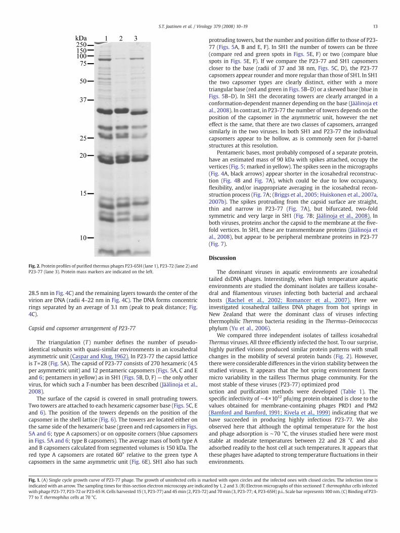
Fig. 2. Protein profiles of purified thermus phages P23-65H (lane 1), P23-72 (lane 2) andP23-77 (lane 3). Protein mass markers are indicated on the left.
13S.T. Jaatinen et al. / Virology 379 (2008) 10–19
28.5 nm in Fig. 4C) and the remaining layers towards the center of thevirion are DNA (radii 4–22 nm in Fig. 4C). The DNA forms concentricrings separated by an average of 3.1 nm (peak to peak distance; Fig.4C).
Capsid and capsomer arrangement of P23-77
The triangulation (T ) number defines the number of pseudo-identical subunits with quasi-similar environments in an icosahedralasymmetric unit (Caspar and Klug, 1962). In P23-77 the capsid latticeis T=28 (Fig. 5A). The capsid of P23-77 consists of 270 hexameric (4.5per asymmetric unit) and 12 pentameric capsomers (Figs. 5A, C and Eand 6; pentamers in yellow) as in SH1 (Figs. 5B, D, F) — the only othervirus, for which such a T-number has been described (Jäälinoja et al.,2008).
The surface of the capsid is covered in small protruding towers.Two towers are attached to each hexameric capsomer base (Figs. 5C, Eand 6). The position of the towers depends on the position of thecapsomer in the shell lattice (Fig. 6). The towers are located either onthe same side of the hexameric base (green and red capsomers in Figs.5A and 6; type A capsomers) or on opposite corners (blue capsomersin Figs. 5A and 6; type B capsomers). The average mass of both type Aand B capsomers calculated from segmented volumes is 150 kDa. Thered type A capsomers are rotated 60° relative to the green type Acapsomers in the same asymmetric unit (Fig. 6E). SH1 also has such
Fig. 1. (A) Single cycle growth curve of P23-77 phage. The growth of uninfected cells is maindicated with an arrow. The sampling times for thin-section electronmicroscopy are indicatwith phage P23-77, P23-72 or P23-65 H. Cells harvested 15 (1, P23-77) and 45min (2, P23-72)77 to T. thermophilus cells at 70 °C.
protruding towers, but the number and position differ to those of P23-77 (Figs. 5A, B and E, F). In SH1 the number of towers can be three(compare red and green spots in Figs. 5E, F) or two (compare bluespots in Figs. 5E, F). If we compare the P23-77 and SH1 capsomerscloser to the base (radii of 37 and 38 nm, Figs. 5C, D), the P23-77capsomers appear rounder andmore regular than those of SH1. In SH1the two capsomer types are clearly distinct, either with a moretriangular base (red and green in Figs. 5B–D) or a skewed base (blue inFigs. 5B–D). In SH1 the decorating towers are clearly arranged in aconformation-dependent manner depending on the base (Jäälinoja etal., 2008). In contrast, in P23-77 the number of towers depends on theposition of the capsomer in the asymmetric unit, however the neteffect is the same, that there are two classes of capsomers, arrangedsimilarly in the two viruses. In both SH1 and P23-77 the individualcapsomers appear to be hollow, as is commonly seen for β-barrelstructures at this resolution.
Pentameric bases, most probably composed of a separate protein,have an estimated mass of 90 kDa with spikes attached, occupy thevertices (Fig. 5; marked in yellow). The spikes seen in the micrographs(Fig. 4A, black arrows) appear shorter in the icosahedral reconstruc-tion (Fig. 4B and Fig. 7A), which could be due to low occupancy,flexibility, and/or inappropriate averaging in the icosahedral recon-struction process (Fig. 7A; (Briggs et al., 2005; Huiskonen et al., 2007a,2007b). The spikes protruding from the capsid surface are straight,thin and narrow in P23-77 (Fig. 7A), but bifurcated, two-foldsymmetric and very large in SH1 (Fig. 7B; Jäälinoja et al., 2008). Inboth viruses, proteins anchor the capsid to the membrane at the five-fold vertices. In SH1, these are transmembrane proteins (Jäälinoja etal., 2008), but appear to be peripheral membrane proteins in P23-77(Fig. 7).
Discussion
The dominant viruses in aquatic environments are icosahedraltailed dsDNA phages. Interestingly, when high temperature aquaticenvironments are studied the dominant isolates are tailless icosahe-dral and filamentous viruses infecting both bacterial and archaealhosts (Rachel et al., 2002; Romancer et al., 2007). Here weinvestigated icosahedral tailless DNA phages from hot springs inNew Zealand that were the dominant class of viruses infectingthermophilic Thermus bacteria residing in the Thermus–Deinococcusphylum (Yu et al., 2006).
We compared three independent isolates of tailless icosahedralThermus viruses. All three efficiently infected the host. To our surprise,highly purified virions produced similar protein patterns with smallchanges in the mobility of several protein bands (Fig. 2). However,there were considerable differences in the virion stability between thestudied viruses. It appears that the hot spring environment favorsmicro variability in the tailless Thermus phage community. For themost stable of these viruses (P23-77) optimized production and purification methods were developed (Table 1). Thespecific infectivity of ∼4×10¹² pfu/mg protein obtained is close to thevalues obtained for membrane-containing phages PRD1 and PM2(Bamford and Bamford, 1991; Kivela et al., 1999) indicating that wehave succeeded in producing highly infectious P23-77. We alsoobserved here that although the optimal temperature for the hostand phage adsorption is ∼70 °C, the viruses studied here were moststable at moderate temperatures between 22 and 28 °C and alsoadsorbed readily to the host cell at such temperatures. It appears thatthese phages have adapted to strong temperature fluctuations in theirenvironments.
rked with open circles and the infected ones with closed circles. The infection time ised by 1, 2 and 3. (B) Electron micrographs of thin sectioned T. thermophilus cells infectedand 70min (3, P23-77; 4, P23-65H) p.i.. Scale bar represents 100 nm. (C) Binding of P23-

Table 1Recovery of bacteriophage P23-77 during concentration and purification steps insucrose
Recovery of infectivity (pfu/Litre of starting culture)
Recovery ofinfectivity (%)
Specific infectivity(pfu/mg protein)
14 S.T. Jaatinen et al. / Virology 379 (2008) 10–19
We show that these Thermus viruses contain lipids organized as abilayer underneath the protein capsid. The large lipid-containingviruses, like PBCV-1 andChilo iridescent virus (Huiskonen and Butcher,2007), have their double β-barrel, pseudo-hexameric capsomersarranged as trigonal arrays forming the facets of the virus (Abrescia
Cell lysate 1.2×10¹⁴ 100 –
PEGprecipitate
8.3×10¹³ 69 –
1× virusgradient band
4.5×10¹³ 38 8.2×10¹²
1× viruspelleted
2.3×10¹³ 19 4.2×10¹²
2×virusgradient band
2.0×10¹³ 17 5.6×10¹²
2× viruspelleted
9.2×10¹² 8 3.1×10¹²
et al., 2004; Nandhagopal et al., 2002; Simpson et al., 2003; Yan et al.,2000). It was observed by Wrigley that when such viruses weredissociated, triangular “trisymmetrons” and pentagonal “pentasym-metrons” were visible (Wrigley, 1969). Detailed analysis of thecapsomer packing in PBCV-1 and Chilo iridescent virus from fitting ofthe capsomer X-ray structure into EM reconstructions, shows that acapsomer in the pentasymmetron is rotated by 72° relative to itsneighbouring pentasymmetron capsomer, and 60° relative to theadjacent trisymmetron capsomer; two interaction types with verydifferent buried surface areas (Simpson et al., 2003). The pseudo-hexameric capsomers in trisymmetrons are similarly oriented to eachother with one interaction type (Simpson et al., 2003). It has beenobserved that for T-numbers where h is odd, adjacent trisymmetronsmake contact across the two-fold axis of symmetry. However, when his evenWrigley predicted that linear disymmetrons would be requiredto form the contacts between trisymmetrons (Simpson et al., 2003;Wrigley, 1969). In the case where both h and k are even e.g. T=28, thecentral capsomer of the disymmetron would sit on the icosahedral 2-fold axis of symmetry. In such a case a virus with trimeric capsomerswould either have to abandon icosahedral symmetry or use a specialtwo- or six-fold symmetric capsomer (Simpson et al., 2003). In P23-77and SH1 we see the first examples, to our knowledge, of virusescontaining linear disymmetrons, formed from the B type capsomers(Figs. 5A and B, blue capsomers). As predicted, the central capsomer istwo-fold symmetric, but interestingly, so are the other disymmetroncapsomers on either side. The pentasymmetrons (red in Figs. 5A, 5Band 6E), now have an additional interaction with the adjacentdisymmetron capsomer (Fig. 6E). The P23-77 capsomers belonging tothe trisymmetron (green in Figs. 5A and 6E) are not pseudo-hexameric,and only face in the same direction within the asymmetric unit,rotating 120° around the icosahedral three-fold axis of symmetry. Thisis not apparent in SH1 or PBCV1-1where the trisymmetron capsomersare pseudo-hexameric. The Wrigley scheme was originally introducedin the context of the assembly of very large viruses, and there thenatural interpretation for the occurrence of di-, tri- and pentasymme-trons was as intermediates in virus assembly. In the case of a relativelysmall capsid this may not be true, but it does remind us of thedifferences in buried surface area that may occur between capsomersin different areas of the asymmetric unit assigned to di-, tri- orpentasymmetrons. These different interactions are potential sites for
Fig. 3. (A) Purification profile of P23-77 in a 20–70% equilibrium sucrose gradient. Thedensity of the gradient fractions (g/ml, circles), A260, (squares) and infectivity (pfu/ml,triangles) are depicted. (B) Thin-layer chromatograms of neutral lipids of fractions 8–19,the first lane is 2× purified P23-77. (C) Tricine SDS-PAGE gel comparing a proteinmarker(lane 1) with the protein profile from the P23-77 pooled peak infectivity fractions (14–17; lane 2) and highly purified SH1. P23-77 proteins are designated according to theirapparent molecular masses. SH1 proteins are indicated, the major SH1 capsomerproteins are VP4 (25.7 kDa) and VP7 (20.0 kDa). The position where the PRD1 majorcoat protein P3 (43.1 kDa) migrates is indicated to the left of lane 1.

Fig. 4. (A) Organization of P23-77. Cryo-electronmicrograph taken at 2.7 μmunderfocusshowing P23-77 viral particles (white arrow), with a diameter of 78 nm. Thin spikes,which might be used in the infection of the host, are visible on some particles (blackarrow). The viral membrane inside the protein capsid is visible in empty particles (inset,underfocus 2.2 μm). Bar, 100 nm. (B) A 0.28 nm thick central section through the virion.Symmetry axes are indicated with a black ellipse (2-fold), triangle (3-fold) andpentagon (5-fold). Proteins connecting the viral capsid and the underlying lipid bilayerat the five-fold vertexes are visible. D (DNA), M (membrane) and C (capsid shell). Bar,20 nm. Protein is black in A and B. (C) Radial density profiles of the icosahedralreconstruction of the intact virion (solid line) and the empty particle (dotted line). Forcalculation of the radial profiles both the full and the empty particle reconstructionswere calculated to 3.0 nm resolution.
15S.T. Jaatinen et al. / Virology 379 (2008) 10–19
interactions with minor capsid proteins important in regulatingassembly, e.g. in PRD1 there is an elongated, dimeric, minor proteinrunning between adjacent facets at the junction between twotrisymmetrons (Abrescia et al., 2004). In P23-77 it seems likely thatthe capsomers arebuilt from twodifferent protein species, one formingthe hexagonal bases, and the other the towers. The tower proteinbinding is related to the position of the underlying capsomer in theasymmetric unit. Two major bands (gp 20 and 35) are seen in SDS-PAGE analysis of P23-77 particles. They are of an appropriatemolecularweight and abundancy to be constituents of the capsomers. Whetherwe are dealing with trimeric or hexameric capsomers is intriquing.Obviously higher resolution data is needed to unequally determinethis. Given the occurrence of the type B capsomers (Figs. 5 and 6), it ismost likely that the capsomer has 6 subunits in the base, as wasconcluded for SH1 (Jäälinoja et al., 2008).
P23-77 has pentamers at the five-fold vertices, to which the spikesare attached. The average mass for the pentamer is 90 kDa, whichgives a mass of 18 kDa for the monomeric protein. This valueresembles those for the viruses of the PRD1-type such as Bam35, SH1,PRD1 and STIV (Abrescia et al., 2004; Jäälinoja et al., 2008; Laurinmakiet al., 2005; Rice et al., 2004). Based on SDS-PAGE analysis (Fig. 3C),there are three protein bands with apparent molecular weightsbetween 15–22 kDa, that could form the pentamer. P23-77 has longnarrow spikes on the vertices reminiscent of the receptor bindingcomplex in PRD1 (Huiskonen et al., 2007b). It is likely that the P23-77spikes are involved in host attachment. The P23-77 spikes are quitedissimilar to the horn-like spikes of SH1 (Jäälinoja et al., 2008) or theturreted spikes of STIV (Maaty et al., 2006) which both have archaealhosts. Hence spike protein evolution tends to reflect host specificityrather than reflecting the evolution of the capsid proteins.
Tectiviruses have membrane-containing procapsids that arepackaged by a viral ATPase without capsid expansion (Grahn et al.,2006). In P23-77, very few empty particles were identified in thepurified virus preparation and no empty particle band was observedduring gradient purification of cell lysates. This could indicate thatpackaging is very rapid and efficient in P23-77, that procapsids arevery unstable under our purification conditions, or that the virusdoes not go through a procapsid intermediate. The DNA in the capsidof P23-77 is highly ordered and the spacing between the concentricDNA rings is 3.1 nm. This spacing is relatively low compared to thatof the other dsDNA bacteriophages T7, P22 and PRD1, where theaverage spacing of the concentric DNA layers is approximately2.5 nm (Cerritelli et al., 1997; Chang et al., 2006; Cockburn et al.,2004). The P23-77 membrane localization and organization aresimilar to that which has been observed in other membrane-containing bacteriophages PRD1, Bam35 (Tectiviridae) and PM2(Corticoviridae) and the archaeal viruses STIV and SH1 (Abrescia etal., 2004; Huiskonen et al., 2004; Jäälinoja et al., 2008; Laurinmaki etal., 2005; Rice et al., 2004).
The discovery of two viruses, one infecting an archaeal and theother a bacterial host with similar capsid architecture reminiscent to,but deviating from, the canonical PRD1 architecture, is surprising andintriguing. Whether these Thermus phages can be positioned in theTectiviridae family remains an open question. As the closest structuralrelative to P23-77 is an archaeal virus and as the PRD1 virion has itsclosest structural counterparts in viruses infecting archaeal andeukaryotic hosts, we hope that further structural analyses reachingto higher resolution will shed more light on the relatedness of theseviruses with an internal membrane.
Materials and methods
Bacteria and phages
The icosahedral nontailed phages (P23-65H, P23-72, P23-77, P37-14, P78-65, P78-83 and P78-86) were obtained from the Promega

Fig. 5. Capsid architecture of P23-77 and SH1. (A) Surface representation of P23-77 drawn at 2 σ abovemean of the intact particle reconstruction viewed down a 3-fold symmetry axis.(B) Surface representation of SH1 (Accession number EMD1353). Symmetry axes are indicated in B with a white ellipse (2-fold), triangle (3-fold) and pentagon (5-fold). Thetriangulation number (T ) describes the geometrical arrangement of the capsomers, and is given by the relationship T=h2+hk+k2 (Caspar and Klug, 1962). The integers h and k thatdefine the lattice points for a triangulation number T=28 dextro lattice (h=4, k=2) are depicted with white dots in A and B. Icosahedrally independent capsomers of P23-77 and SH1have been manually segmented and are enumerated from 1 to 5. Wrigley (Wrigley, 1969) describes the arrangement of the icosahedral shell with the help of disymmetrons,trisymmetrons, and pentasymmetrons. Colored according toWrigley's theory, the green capsomers (2 and 3) of P23-77 and SH1 correspond to trisymmetrons, the blue capsomers (4and 5) to disymmetrons and the yellow and red (1) capsomers to pentasymmetrons. (C) A 0.28 nm thick spherical section of P23-77 at 37 nm and (D) of SH1 at 38 nm showing thehexameric bases of the capsomers. (E) A 0.28 nm thick spherical section of P23-77 at 40 nm and (F) of SH1 at 41 nm cutting through the decorating towers. (C-F) Bases and towersbelonging to one asymmetric unit are coloured as in A and B. Bar, 20 nm.
16 S.T. Jaatinen et al. / Virology 379 (2008) 10–19
corporation collection (Yu et al., 2006), and propagated in Thermusthermophilus (ATCC 33923). T. thermophilus cells were grown inthermus medium containing 4 g yeast extract, 8 g peptone, 2 g NaCl,(20 g agar when appropriate) per L at pH 7.5.
Virus propagation and purification
To obtain virus stocks, the soft-agar layer was harvested fromsemiconfluent plates incubated at 70 °C, 16 h in a humid

Fig. 6. Capsid architecture of P23-77. (A, B) The capsid of P23-77 is built up of twodifferent hexameric capsomers, type A (A) and type B (B), that are decorated with twotowers each (orange). (C) Schematic representation of the type A capsomers. (D)Schematic representation of the type B capsomers. (E) Schematic diagram of a P23-77facet, with one asymmetric unit coloured. Each icosahedrally independent capsomer isassigned a colour and a number corresponding to those in Fig. 5A. Type A capsomers arecolored in shades of green and red, and type B capsomers in shades of blue. The type Bcapsomer 5 is sharedwith the neighboring asymmetric unit. The black line indicates thelattice points for the T=28 dextro organisation of the P23-77 capsomers. Symmetry axesare indicated with an ellipse (2-fold), triangle (3-fold) and pentagon (5-fold). In C-E, theposition of the capsomer towers is indicated with black dots on top of a hexagonal base.
17S.T. Jaatinen et al. / Virology 379 (2008) 10–19
environment (sealed plastic bags), by adding 3 ml of thermusmedium per plate, followed by incubation for 4 h, with aeration, at70 °C. The cell debris was removed by differential centrifugation(Sorvall GSA rotor, 7000 rpm, 20 min, 25 °C) and the supernatantwas titered. For single cycle growth experiments, cells were infectedat a multiplicity of infection (MOI) of about 10 at a cell density of7×108 colony forming units (cfu)/ml. After lysis, cultures werecleared by centrifugation (Sorvall SLA3000 rotor, 7000 rpm, 20 min,25 °C), and virus particles were precipitated from the supernatantby the addition of 12% w/v polyethylene glycol (PEG) 6000 and0.5 M NaCl (final concentrations) with stirring for 35 min at 28 °C.The virus precipitate was collected by centrifugation (Sorvall
SLA3000 rotor, 7000 rpm, 20 min, 25 °C) and the pelletsubsequently rinsed with 20 mM Tris–HCl pH 7.5, 5 mM MgCl2,150 mM NaCl (TV buffer) and resuspended in 1/20 of the originallysate volume. All subsequent steps were carried out in the samebuffer. Viruses were purified by rate zonal centrifugation (linear 5–20% (w/v) sucrose gradient, Sorvall AH629 rotor, 23000 rpm,45 min, 25 °C). The light-scattering virus zone was collected (1×purified virus) or the gradient was fractionated to assay the proteincontent and infectivity. To produce 2× purified viruses, 10 ml of the1× band was further purified by equilibrium centrifugation (linear20–70% (w/v) sucrose gradient, Sorvall AH629 rotor, 23000 rpm,17 h, 25 °C) or alternatively in 1.30 mg/ml CsCl in TV buffer (SorvallAH629 rotor, 21000 rpm, 16 h, 25 °C). The light-scattering virus zonewas collected, an equal volume of buffer was added, and virusparticles were collected by differential centrifugation (Sorvall T647.5rotor, 32000 rpm, 4 h, 25 °C). The obtained virus pellet wasresuspended into TV buffer. Alternatively the gradients werefractionated and the fractions were assayed for protein content,density and infectivity.
Phage adsorption assay
The adsorption assay was carried out as previously described usingfresh virus stocks (Adams, 1959). T. thermophilus cells were grown to acell density of 7×10⁸ cfu/ml and 200 μl of the cell suspension wasmixed with ∼200 infective phage particles, and the mixture wasincubated at 70 or 22 °C. Samples were taken at different time points,cells removed by centrifugation (Eppendorf microcentrifuge,5000 rpm, 4 min, 22 °C) and the number of nonadsorbed phageparticles was determined in the supernatant by titering.
Protein analysis
The proteins in purified virus preparations were resolved usingsodium dodecyl sulfate-polyacrylamide gel electrophoresis (SDS-PAGE; 16% acrylamide, (Olkkonen and Bamford, 1989); or tricine-SDS-PAGE (17% acrylamide; (Schägger and von Jagow, 1987) using aBIO-RAD Precision Plus Protein standard as a marker. Proteinconcentrations were determined with Coomassie brilliant blue usingbovine serum albumin as a standard (Bradford, 1976).
Lipid analysis
Lipid analysis was carried out on all of the fractions (1 ml) from asucrose equilibrium gradient. Lipids were extracted essentially asdescribed previously (Folch et al., 1957), except that no NaCl wasincluded in the washing steps. A hexane, diethyl ether and acetic acidmixture (80:20:1; v/v) was used as the solvent system for neutrallipids on silica gel plates (Silica gel 60; Merck) (Kates, 1972). The lipidson the thin layer silica plates were visualized by iodine vapour andscanned.
Electron microscopy
For thin-section electron microscopy T. thermophilus cells weregrown to a density of 7×10⁸ cfu/ml and infected with freshly madeP23-77, P23-72 or P23-65H phage stocks at MOI 10 (45 and 70 minpost-infection (p.i.) samples) or MOI 40 (15 min p.i. sample) at 70 °C.Samples were taken at different time points after infection and fixedwith 3% (v/v) glutaraldehyde in 20mMphosphate buffer (pH 7.4). After20 min incubation at room temperature the fixed cells were collected,washed twice and prepared for transmission electron microscopy aspreviously described (Bamford and Mindich, 1980). Micrographs weretaken with a JEOL 1200 EX electron microscope operating at 60 kV.
For cryo-EM, 1× and 2× purified P23-77 viruses were concentratedfrom gradient fractions by differential centrifugation (Beckman

Fig. 7. Close-up of 0.84 nm thick central sections through the five-fold vertices of P23-77 (A) and SH1 (B). The position of the capsid (C) and membrane (M) are indicated. The P23-77spike is stick-like compared to the bifurcated SH1 spike. Strong densities connecting the capsid to the membrane are visible in both SH1 and P23-77. Bar, 10 nm.
18 S.T. Jaatinen et al. / Virology 379 (2008) 10–19
SW50.1 rotor, 40000 rpm, 40 min, 25 °C), resuspended in 20 mM Tris–HCl pH 7.5, 1 mMMgCl2, 0.1 mM CaCl2 (1× virus zone) or TV buffer (2×virus zone) and immediately used for the preparation of vitrifiedspecimens. Aliquots of virus (3 μl) were vitrified on holey carbon filmcoated grids (Quantifoil R 2/2) in liquid ethane as described previously(Adrian et al., 1984). The specimens were imagined at −180 °C in a FEITecnai F20 field emission gun transmission electron microscopeoperating at 200 kV and using a Gatan 626 cryoholder. Virus imageswere recorded on Kodak SO163 film or on a Gatan UltraScan 4000 CCDcamera under low dose conditions, at nominal magnifications of50,000× and 68,000× respectively. The micrographs recorded on filmwere developed in full-strength Kodak D19 film developer for 12 min.All electron microscopy data were collected in the Electron Micro-scopy Unit, Institute of Biotechnology, University of Helsinki.
Image processing
Films were digitized at 7-μm intervals on a Zeiss Photoscan TDscanner, resulting in a nominal sampling of 0.14 nm pixel−1 andbinned to 0.28 nm pixel−1. CCD sampling was 0.22 nm pixel−1.CTFFIND3 (Mindell and Grigorieff, 2003) was used to estimate thecontrast transfer function. Drifted and astigmatic pictures werediscarded. ETHAN (Kivioja et al., 2000) was used to locate the virusparticles, and particles were extracted in the EMAN program BOXER(Ludtke et al., 1999). Bsoft (Heymann, 2001) was used for furtherimage processing unless stated otherwise.
The three-dimensional structure of the archaeal virus SH1(Accession number EMD1353; (Jäälinoja et al., 2008), scaled to theappropriate size, was used as a starting model to determine theorientations and origins of the icosahedral particles in a model-basedapproach (Baker and Cheng, 1996). PFT2 and EM3DR2 (Baker andCheng, 1996) were used in the initial rounds of refinement, and PORand P3DR (Ji et al., 2006; Marinescu and Ji, 2003) for subsequentrounds. The contrast transfer function was fully corrected in P3DRusing a Wiener filter (Marinescu and Ji, 2003).
The spacing of the P23-77 protein and membrane layers wascalculated from spherically-averaged radial profiles of the reconstruc-tions using Bsoft (Heymann, 2001; Huiskonen et al., 2004). Bsoft(Heymann, 2001) was in addition used to create spherical and centralsections of the three-dimensional models of P23-77 and SH1(Accession number EMD1353).
Segmentation of the reconstruction and mass estimates of thecapsomers was carried out as described previously (Jäälinoja et al.,2008). Themolecular mass for the P23-77 capsomerswas estimated inEMAN using a density threshold of 2 standard deviations above themean and a protein density of 1.35 g/ml (Ludtke et al., 1999). Allsurface representations were created with the UCSF Chimera package(Pettersen et al., 2004).
The reconstruction of the virion has been deposited in the EMDB,at the European Bioinformatics Institute with the accession code:EMD1525.
Acknowledgments
The technical assistance of Anna Latva-Käyrä and Heini Hyvönenis gratefully acknowledged. Harri Jäälinoja and Jani Seitsonen areacknowledged for the useful discussions. We thank the ElectronMicroscopy Unit of the Institute of Biotechnology, University ofHelsinki for providing microscopy facilities. This study was sup-ported (to S.J.B and D.H.B) from the Finnish Center of ExcellenceProgram [2006–2011] grant 1213467. Further funding was obtainedfrom the Academy of Finland [grant 1210253] to D.H.B and grant1112244 to S. J. B. As part of the European Science FoundationEUROCORES Programme Euro-SCOPE, the work is also supported byfunds from the European Commission's Sixth Framework Programmeunder contract ERAS-CT-2003-980409. The Helsinki Graduate Schoolof Biotechnology and Molecular Biology is acknowledged for fundingof S.T.J.
References
Abrescia, N.G., Cockburn, J.J., Grimes, J.M., Sutton, G.C., Diprose, J.M., Butcher, S.J., Fuller,S.D., San Martin, C., Burnett, R.M., Stuart, D.I., Bamford, D.H., Bamford, J.K., 2004.Insights into assembly from structural analysis of bacteriophage PRD1. Nature 432,68–74.
Ackermann, H.W., 2007. 5500 Phages examined in the electron microscope. Arch. Virol.152, 227–243.
Adams, M.H., 1959. Bacteriophages. Interscience publishers, Inc., New York.Adrian, M., Dubochet, J., Lepault, J., McDowall, A.W., 1984. Cryo-electron microscopy of
viruses. Nature 308, 32–36.Baker, T.S., Cheng, R.H., 1996. A model-based approach for determining orientations of
biological macromolecules imaged by cryoelectron microscopy. J. Struct. Biol. 116,120–130.
Bamford, D.H., 2005. Tectiviridae. In: Fauquet, C.M., Mayo, M.A., Maniloff, J.,Desselberger, U., Ball, L.A. (Eds.), Virus taxonomy, Eighth report of the internationalcommittee on taxonomy of viruses. Elsevier Academic Press, London, pp. 81–85.
Bamford, J.K., Bamford, D.H., 1991. Large-scale purification of membrane-containingbacteriophage PRD1 and its subviral particles. Virology 181, 348–352.
Bamford, D.H., Mindich, L., 1980. Electron microscopy of cells infected with nonsensemutants of bacteriophage phi 6. Virology 107, 222–228.
Bamford, D.H., Burnett, R.M., Stuart, D.I., 2002. Evolution of viral structure. Theor. Popul.Biol 61, 461–470.
Bamford, D.H., Grimes, J.M., Stuart, D.I., 2005a. What does structure tell us about virusevolution? Curr. Opin. Struct. Biol. 15, 655–663.
Bamford, D.H., Ravantti, J.J., Ronnholm, G., Laurinavicius, S., Kukkaro, P., Dyall-Smith, M.,Somerharju, P., Kalkkinen, N., Bamford, J.K., 2005b. Constituents of SH1, a novellipid-containing virus infecting the halophilic euryarchaeon Haloarcula hispanica.J. Virol. 79, 9097–9107.
Benson, S.D., Bamford, J.K., Bamford, D.H., Burnett, R.M., 1999. Viral evolution revealedby bacteriophage PRD1 and human adenovirus coat protein structures. Cell 98,825–833.
Benson, S.D., Bamford, J.K., Bamford, D.H., Burnett, R.M., 2004. Does common architecturereveal a viral lineage spanning all three domains of life? Mol. Cell 16, 673–685.

19S.T. Jaatinen et al. / Virology 379 (2008) 10–19
Blondal, T., Thorisdottir, A., Unnsteinsdottir, U., Hjorleifsdottir, S., Aevarsson, A.,Ernstsson, S., Fridjonsson, O.H., Skirnisdottir, S., Wheat, J.O., Hermannsdottir, A.G.,Sigurdsson, S.T., Hreggvidsson, G.O., Smith, A.V., Kristjansson, J.K., 2005. Isolationand characterization of a thermostable RNA ligase 1 from a Thermus scotoductusbacteriophage TS2126 with good single-stranded DNA ligation properties. NucleicAcids Res. 33, 135–142.
Bradford, M.M., 1976. A rapid and sensitive method for the quantitation of microgramquantities of protein utilizing the principle of protein-dye binding. Anal. Biochem.72, 248–254.
Briggs, J.A., Huiskonen, J.T., Fernando, K.V., Gilbert, R.J., Scotti, P., Butcher, S.J., Fuller, S.D.,2005. Classification and three-dimensional reconstruction of unevenly distributedor symmetry mismatched features of icosahedral particles. J. Struct. Biol. 150,332–339.
Brock, T.D., 2005. Genus Thermus. In: Holt, J.G. (Ed.), Bergey's manual of systematicbacteriology, vol. 1. Williams & Wilkins, pp. 333–337.
Caspar, D.L., Klug, A., 1962. Physical principles in the construction of regular viruses.Cold Spring Harb. Symp. Quant. Biol. 27, 1–24.
Cerritelli, M.E., Cheng, N., Rosenberg, A.H., McPherson, C.E., Booy, F.P., Steven, A.C., 1997.Encapsidated conformation of bacteriophage T7 DNA. Cell 91, 271–280.
Chang, J., Weigele, P., King, J., Chiu, W., Jiang, W., 2006. Cryo-EM asymmetricreconstruction of bacteriophage P22 reveals organization of its DNA packagingand infecting machinery. Structure 14, 1073–1082.
Cockburn, J.J., Abrescia, N.G., Grimes, J.M., Sutton, G.C., Diprose, J.M., Benevides, J.M.,Thomas Jr., G.J., Bamford, J.K., Bamford, D.H., Stuart, D.I., 2004. Membrane structureand interactions with protein and DNA in bacteriophage PRD1. Nature 432,122–125.
Folch, J., Lees, M., Sloane Stanley, G.H., 1957. A simple method for the isolation andpurification of total lipides from animal tissues. J. Biol. Chem. 226, 497–509.
Grahn, M.A., Butcher, S.J., Bamford, J.H.K., Bamford, D.H., 2006. PRD1: Dissecting thegenome, structure and entry. In: Calendar, R. (Ed.), The Bacteriophages. OxfordUniversity Press, pp. 161–170.
Heymann, J.B., 2001. Bsoft: image and molecular processing in electron microscopy.J. Struct. Biol. 133, 156–169.
Hreggvidsson, G.O., Skirnisdottir, S., Smit, B., Hjorleifsdottir, S., Marteinsson, V.T.,Petursdottir, S., Kristjansson, J.K., 2006. Polyphasic analysis of Thermus isolatesfrom geothermal areas in Iceland. Extremophiles 10, 563–575.
Huiskonen, J.T., Butcher, S.J., 2007. Membrane-containing viruses with icosahedrallysymmetric capsids. Curr. Opin. Struct. Biol. 17, 229–236.
Huiskonen, J.T., Kivela, H.M., Bamford, D.H., Butcher, S.J., 2004. The PM2 virion has anovel organization with an internal membrane and pentameric receptor bindingspikes. Nat. Struct. Mol. Biol. 11, 850–856.
Huiskonen, J.T., Jaalinoja, H.T., Briggs, J.A., Fuller, S.D., Butcher, S.J., 2007a. Structure of ahexameric RNA packaging motor in a viral polymerase complex. J. Struct. Biol. 158,156–164.
Huiskonen, J.T., Manole, V., Butcher, S.J., 2007b. Tale of two spikes in bacteriophagePRD1. Proc. Natl. Acad. Sci. U S A 104, 6666–6671.
Jäälinoja, H.T., Roine, E., Laurinmäki, P., Kivelä, H.M., Bamford, D.H., Butcher, S.J., 2008.Structure and host cell interaction of SH1, a membrane-containing, halophiliceuryarchaeal virus. Proc. Natl. Acad. Sci. USA 105, 8008–8013.
Ji, Y., Marinescu, D.C., Zhang, W., Zhang, X., Yan, X., Baker, T.S., 2006. A model-basedparallel origin and orientation refinement algorithm for cryoTEM and itsapplication to the study of virus structures. J. Struct. Biol. 154, 1–19.
Kates, M., 1972. Techniques of lipidology: Isolation, analysis and identification of lipids.North-Holland Publishing Company, Amsterdam.
Khayat, R., Tang, L., Larson, E.T., Lawrence, C.M., Young, M., Johnson, J.E., 2005. Structureof an archaeal virus capsid protein reveals a common ancestry to eukaryotic andbacterial viruses. Proc. Natl. Acad. Sci. U S A 102, 18944–18949.
Kivela, H.M., Mannisto, R.H., Kalkkinen, N., Bamford, D.H., 1999. Purification and proteincomposition of PM2, the first lipid-containing bacterial virus to be isolated.Virology 262, 364–374.
Kivioja, T., Ravantti, J., Verkhovsky, A., Ukkonen, E., Bamford, D., 2000. Local averageintensity-based method for identifying spherical particles in electron micrographs.J. Struct. Biol. 131, 126–134.
Laurinmaki, P.A., Huiskonen, J.T., Bamford, D.H., Butcher, S.J., 2005. Membrane proteinsmodulate the bilayer curvature in the bacterial virus Bam35. Structure 13,1819–1828.
Ludtke, S.J., Baldwin, P.R., Chiu, W., 1999. EMAN: semiautomated software for high-resolution single-particle reconstructions. J. Struct. Biol. 128, 82–97.
Maaty, W.S., Ortmann, A.C., Dlakic, M., Schulstad, K., Hilmer, J.K., Liepold, L., Weidenheft,B., Khayat, R., Douglas, T., Young, M.J., Bothner, B., 2006. Characterization of thearchaeal thermophile Sulfolobus turreted icosahedral virus validates an evolu-tionary link among double-stranded DNA viruses from all domains of life. J. Virol.80, 7625–7635.
Marinescu, D.C., Ji, Y., 2003. A computational framework for the 3D structuredetermination of viruses with unknown symmetry. J. Parallel. Distrib. Comput.63, 738–758.
Mindell, J.A., Grigorieff, N., 2003. Accurate determination of local defocus and specimentilt in electron microscopy. J. Struct. Biol. 142, 334–347.
Nandhagopal, N., Simpson, A.A., Gurnon, J.R., Yan, X., Baker, T.S., Graves, M.V., VanEtten, J.L., Rossmann, M.G., 2002. The structure and evolution of the major capsidprotein of a large, lipid-containing DNA virus. Proc. Natl. Acad. Sci. U S A 99,14758–14763.
Olkkonen, V.M., Bamford, D.H., 1989. Quantitation of the adsorption and penetrationstages of bacteriophage phi 6 infection. Virology 171, 229–238.
Pantazaki, A.A., Pritsa, A.A., Kyriakidis, D.A., 2002. Biotechnologically relevant enzymesfrom Thermus thermophilus. Appl. Microbiol. Biotechnol. 58, 1–12.
Pask-Hughes, R., Williams, R.A., 1975. Extremely thermophilic gram-negative bacteriafrom hot tap water. J. Gen. Microbiol. 88, 321–328.
Pederson, D.M., Welsh, L.C., Marvin, D.A., Sampson, M., Perham, R.N., Yu, M., Slater, M.R.,2001. The protein capsid of filamentous bacteriophage PH75 from Thermusthermophilus. J. Mol. Biol. 309, 401–421.
Pettersen, E.F., Goddard, T.D., Huang, C.C., Couch, G.S., Greenblatt, D.M., Meng, E.C.,Ferrin, T.E., 2004. UCSF Chimera–a visualization system for exploratory researchand analysis. J. Comput. Chem. 25, 1605–1612.
Rachel, R., Bettstetter, M., Hedlund, B.P., Haring, M., Kessler, A., Stetter, K.O., Prangishvili,D., 2002. Remarkable morphological diversity of viruses and virus-like particles inhot terrestrial environments. Arch. Virol. 147, 2419–2429.
Ramaley, R.F., Hixson, J., 1970. Isolation of a nonpigmented, thermophilic bacteriumsimilar to Thermophilic bacterium similar to Thermus aquaticus. J. Bacteriol. 103,527–528.
Ravantti, J.J., Gaidelyte, A., Bamford, D.H., Bamford, J.K., 2003. Comparative analysis ofbacterial viruses Bam35, infecting a gram-positive host, and PRD1, infecting gram-negative hosts, demonstrates a viral lineage. Virology 313, 401–414.
Ray, P.H., White, D.C., Brock, T.D., 1971. Effect of growth temperature on the lipidcomposition of Thermus aquaticus. J. Bacteriol. 108, 227–235.
Rice, G., Tang, L., Stedman, K., Roberto, F., Spuhler, J., Gillitzer, E., Johnson, J.E., Douglas, T.,Young, M., 2004. The structure of a thermophilic archaeal virus shows a double-stranded DNA viral capsid type that spans all domains of life. Proc. Natl. Acad. Sci.U S A 101, 7716–7720.
Romancer, M., Gaillard, M., Geslin, C., Prieur, D., 2007. Viruses in extreme environmets.Rev. Environ. Sci. Biotechnol. 6, 17–31.
Sakaki, Y., Oshima, T., 1975. Isolation and characterization of a bacteriophage infectiousto an extreme thermophile, Thermus thermophilus HB8. J. Virol. 15, 1449–1453.
Schägger, H., von Jagow, G., 1987. Tricine-sodium dodecyl sulfate-polyacrylamide gelelectrophoresis for the separation of proteins in the range from 1 to 100 kDa. Anal.Biochem. 166, 368–379.
Simpson, A.A., Nandhagopal, N., Van Etten, J.L., Rossmann, M.G., 2003. Structuralanalyses of Phycodnaviridae and Iridoviridae. Acta Crystallogr. D Biol. Crystallogr.59, 2053–2059.
Wrigley, N.G., 1969. An electron microscope study of the structure of Sericesthisiridescent virus. J. Gen. Virol. 5, 123–134.
Yan, X., Olson, N.H., Van Etten, J.L., Bergoin, M., Rossmann, M.G., Baker, T.S., 2000.Structure and assembly of large lipid-containing dsDNA viruses. Nat. Struct. Biol. 7,101–103.
Yu, M.X., Slater, M.R., Ackermann, H.W., 2006. Isolation and characterization of Thermusbacteriophages. Arch. Virol. 151, 663–679.


