Embed Size (px)
Citation preview
Biochem. J. (1989) 262, 409-416 (Printed in Great Britain)
Caldolase, a chelator-insensitive extracellular serine proteinasefrom a Thermus spp.
Gholam-Ali SARAVANI,* Don A. COWAN,t Roy M. DANIELt and Hugh W. MORGANThermophile Research Group, School of Science, University of Waikato, Hamilton, New Zealand
409
An extracellular alkaline serine proteinase from Thermus strain ToK3 was isolated and purified tohomogeneity by (NH4)2S04 precipitation followed by ion-exchange chromatography on DEAE-cellulose andQAE-Sephadex, affinity chromatography on N-benzyloxycarbonyl-D-phenylalanyl-triethylenetetraminyl-Sepharose 4B and gel-filtration chromatography on Sephadex G-75. The purified enzyme had a pl of 8.9 andan Mr determined by gel-permeation chromatography of 25000. The specific activity was about 37700proteolytic units/mg with casein as substrate, and the pH optimum was 9.5. Proteolytic activity wasinhibited by low concentrations of di-isopropyl phosphorofluoridate and phenylmethanesulphonyl fluoride,but was unaffected by EDTA, EGTA, o-phenanthroline, N-ethyl-5-phenylisoxazolium-3'-sulphonate, Na-p-tosyl-L-phenylalanylchloromethane, M=-p-tosyl-L-lysylchloromethane, trypsin inhibitors and pepstatin A.The enzyme contained approx. 10% carbohydrate and four disulphide bonds. No Ca2", Zn2+ or free thiolgroups were detected. It hydrolysed several native and dye-linked proteins and synthetic chromogenicpeptides and esters. The enzyme was very thermostable (half-life values were 840 min at 80 °C, 45 min at90 °C and 5 min at 100 °C). The enzyme was unstable at low ionic strength: after 60 min at 75 °C in 0.1 M-Tris/acetate buffer, pH 8, only 20% activity remained, compared with no loss in 0.1 M-Tris/acetate buffer,pH 8, containing 0.4 M-NaCl.
INTRODUCTION
Attention is being increasingly focused upon enzymesfrom extremely thermophilic bacteria. Their thermo-stability and resistance to denaturing agents and organicsolvents make these enzymes increasingly attractive forbiotechnological processes (Doig, 1974; Daniel et al.,1981; Hartley & Payton, 1983; Sonnleitner & Fiechter,1983). Proteinases from extreme thermophiles have an
additional advantage in their high specific activity(Cowan et al., 1985, 1987a).To date, a limited number of proteinases from ex-
tremely thermophilic bacteria have been reported andinvestigated (Heinen & Heinen, 1972; Matsuzawa et al.,1983; Taguchi et al., 1983). Only caldolysin, the extra-cellular proteinase from Thermus aquaticus strain T351(Cowan & Daniel, 1982a,b; Khoo et al., 1984), andarchaelysin, the proteinase from a New Zealand strain ofDesulfurococcus (Cowan et al., 1987b), have beencharacterized in detail.
In the present paper the production, purification andcharacterization of an extracellular proteinase fromThermus strain Tok3 are described. For convenience theproteinase was assigned the trivial name of caldolase.The prefix 'caldo' is derived from the Latin caldus (hot)and the suffix 'ase' is a general term for enzymes.
METHODS
Bacterial cultureThermus strain Tok3 was isolated from the Tokaanu
thermal region, New Zealand. Cells were grown aerobic-
ally at 75 °C on Thermus medium (Hickey & Daniel,1979). Large-scale growth (600 litres) of Thermus strainTok3 was carried out in an 800-litre fermenter.
Enzyme recovery and purificationThe maximum enzyme activity in the culture super-
natant occurred after 9 h, during late exponential phase.The culture was harvested by continuous-flow centrifug-ation in a Sharples model 6 centrifuge at 17000 g with aflow rate of 5 litres/min. Protein in the cell-free super-natant was precipitated by addition of crude (NH4)2SO4to a concentration of 70 % saturation (20 °C). Theprecipitated protein, containing crude extracellular pro-teinase, was then pelleted in a Sharples model 6 centrifugeat 17000 g, and the pellet was homogenized with 1 litreof 0.1 M-Tris/acetate buffer, pH 7, in a Waring blender athigh speed for 2-3 min, and left overnight at 4 'C. Thesuspension was then centrifuged at 10000 g in a typeGSA rotor in Sorvall RC-2B centrifuge for 30 min. Thesupernatant, which contained most of the proteinase,was removed and freeze-dried. The enzyme extractionwas repeated twice more as above. About 5 g of thefreeze-dried crude proteinase was dissolved in 200 mlof 0.1 M-Tris/acetate buffer, pH 8. The enzyme sol-ution was then applied to a DEAE-cellulose column(1.6 cm x 40 cm) equilibrated with 0.1 M-Tris/acetatebuffer, pH 8. The non-adsorbed protein fractions,including proteinase, were pooled and applied to QAE-Sephadex in accordance with the above procedure. Thenon-adsorbed protein, including proteinase, was thenapplied to a column of Z-D-Phe-TETA-Sepharose 4B
Abbreviations used: Z-, benzyloxycarbonyl-; Bz-, benzoyl-; Tos-, tosyl-; Suc-, 3-carboxypropionyl; Ac-, acetyl-; -NH-Np, p-nitroanilide; -ONp,p-nitrophenyl ester; TETA, triethylenetetramine.
* Present address: National Health Institute, P.O. Box 50348, Porirua, New Zealand.t Present address: Department of Biochemistry, University College London, Gower Street, London WC1E 6BT, U.K.t To whom correspondence should be addressed.
Vol. 262

G.-A. Saravani and others
affinity-chromatography gel (Pierce Biochemicals). Theadsorbed proteinase was eluted with 0.1 M-acetic acid,pH 2.8, and freeze-dried. The freeze-dried enzyme wasdissolved in 4 ml of 0.1 M-Tris/acetate buffer, pH 8.0,and then applied to a Sephadex G-75 gel-filtrationcolumn (1.6 cm x 100 cm) equilibrated with 0.1 M-Tris/acetate buffer, pH 8.0, containing 0.1 M-NaCl and10 mM-CaCl2.
Proteinase assayThe modified Kunitz (1947) method was used for
hydrolysis of casein, albumin, ovalbumin, fibrin andcollagen (Cowan & Daniel, 1982a). Hydrolysis of dye-linked proteins was determined by measuring the increasein trichloroacetic acid-soluble chromophores in the super-natant at 440 nm (azo-casein, azo-albumin and Azocoll)or at 495 nm (elastin-Congo Red) (Shotton, 1970;Cowan, 1980). Chromogenic peptides (Bz-Phe-Val-Arg-NH-Np, Bz-Val-Gly-Arg-NH-Np, Tos-Gly-Pro-Arg-NH-Np, Z-Gly-Pro-Arg-NH-Np, Suc-Ala-Ala-Ala-NH-Np, Bz-Pro-Phe-Arg-NH-Np, Bz-Arg-NH-Np, Suc-Phe-NH-Np, Ac-Phe-NH-Np and Z-Phe-NA-Np) wereobtained from Sigma Chemical Co. The rate of cleavagewas determined at 410 nm (Bergstrom, 1977). Esteraseactivity of caldolase was determined with synthetic estersubstrates (Z-Ala-ONp, Z-Tyr-ONp, Z-Phe-ONp, Z-Leu-ONp, Z-Trp-ONp, Z-Lys-ONp, Z-Asn-ONp, Z-fl-Cys-ONp, Z-J-Asn-ONp, Z-Val-ONp, Z-/3-Ala-ONp, Z-D-Nle-ONp, Z-Ile-ONp, and Z-Pro-ONp obtained fromSigma Chemical Co.) by using the method describedby Abdelal et al. (1977). Hydrolysis of bradykininand insulin B-chain (substrate/enzyme ratio approx.10000:1, w/w) was assayed at intervals ranging from30 s to 30 min at 50 'C.
Protein determinationProtein concentration was determined by using the
modified Lowry method (Peterson,1977), with bovineserum albumin as standard.
Mr determinationThe Mr of caldolase was determined by SDS/
polyacrylamide-gel electrophoresis (Laemmli, 1970) bycomparison with the migration of protein markers ofknown Mr values (bovine serum albumin, ovalbumin, a-chymotrypsinogen and cytochrome c). The Mr of theenzyme was also determined by gel-filtration chromato-graphy on a Sephadex G-75 column (1.6 cm x 100 cm)and gel-permeation chromatography on a 60 cm TSKG3000 SW column (Toyo Soda Co.) with the above-mentioned proteins as standard. Blue Dextran was usedfor determination of the void volume.
pl determinationIsoelectric focusing was carried out on commercially
prepared Servalyt Precotes (pH 3-10; Serva). The voltagewas gradually increased from 250 to 1200 V during a110 min focusing period, while the current declined from23 mA to below 10 mA. The gel was fixed with 200%(w/v) trichloroacetic acid and then stained withCoomassie Brilliant Blue G-250. A mixture of 11 standardproteins with known pl values (Pharmacia Fine Chemi-cals) was run in parallel in the same gel.
Carbohydrate determinationThe carbohydrate content of the purified enzyme was
analysed by the phenol/H2SO4 method (Dubois et al.,1956).
Thiol groups and disulphide bonds in caldolaseFree thiol groups were determined colorimetrically
with 5,5'-dithiobis-(2-nitrobenzoic acid) by using themethod of Robyt et al. (1971) at an enzyme concentrationof 35 ,ug/ml. For the determination of cystine contentof caldolase (35 ,ug/ml) the method of Anderson &Wetlaufer (1975) was employed.
Presence of metal ions in caldolasePurified freeze-dried enzyme was dissolved in 10 mM-
EDTA and incubated at room temperature for 30 min.The mixture was then applied to a Sephadex G-75column (1.6 cm x 65 cm) equilibrated with 0.1 M-Tris/acetate buffer, pH 8.0, containing 0.5 M-NaCl. The frac-tions of enzyme and EDTA peaks were tested for Ca2"and Zn2+ by atomic absorption spectrometry, with Ca2+and Zn2+ as the standards. Thermolysin was used as apositive control.
Amino acid composition of caldolaseProtein hydrolysis was carried out with 6 M-HCI con-
taining 700 (v/v) thioglycollic acid (B6hlen, 1983) insealed evacuated ampoules for 24 and 72 h at 110 'C.The amino acid composition of the enzyme was de-termined by using a Waters h.p.l.c. amino acid analyserwith post-column reaction with o-phthalaldehyde.
pH optimumThe influence of pH on enzyme activity was examined
with 0.20% (w/v) casein in the following buffers: Uni-versal buffer (Dawson et al., 1969) (pH 6-11), 0.1 M-Hepes (pH 6.5-10), 0.1 M-Bicine (pH 6.5-9.5) and 0.1 M-Na2CO3/NaHCO3 (pH 8.5-10.3). The pH of buffers wasadjusted at 75 'C.
ThermostabilitySamples of caldolase (24 ,tg/ml in 0.1 M-Tris/acetate
buffer, pH 8.0, containing 0.5 M-NaCl and 10 mM-CaCl2)were incubated in Kimax Hungate tubes or in sealedmelting-point capillary tubes at temperatures from 75to 110 °C. The residual proteinase activity was measuredat 75 'C by the standard modified Kunitz method.
Effect of pH on caldolase stabilityThe enzyme in Universal buffer (Dawson et al., 1969),
at pH values ranging from 3 to 12, was incubated for90 min at room temperature (22 'C) before assay asdescribed above.
Effect of ionic strength on enzyme stabilityCaldolase (in 0.1 M-Tris/acetate buffer, pH 8, con-
taining 0.1 M- to 0.7 M-NaCl) was incubated at 22 °C or75 'C for 60 min. The enzyme was also incubated at75 'C for 60 min in the presence of various buffers. Forthe analysis of denaturation and autolysis the freeze-dried salt-free enzyme was dissolved in the followingbuffers: 0.1 M-Tris/acetate buffer, pH 8, 0.1 M-Tris/acetate buffer, pH 8, containing 10 mM-CaCl2 and 0.1 M-,0.3 M- or 0.5 M-NaCl. Each enzyme solution (40 ,ug/ml)was then incubated in Kimax Hungate tubes at a chosen
1989
410
Caldolase: extracellular Thermus serine proteinase
Table 1. Purification of caldolase
For experimental details see the text. One proteolytic unit of activity is defined as 1 ,ug ofabsorbance at 280 nm.
tyrosine released/min measured as
Total SpecificTotal activity activity
Volume protein (proteolytic (proteolytic Purification RecoveryStep (ml) (mg) units) units/mg) (fold) (%)
600-litre fermenter 600000 - 8491 500 - - 100Extracted enzyme 3000 - 3358400 - - 40
5 g of freeze-driedextracted enzyme*DEAE-cellulosechromatographyQAE-Sephadex anion-exchangechromatography
Affinity chromatography(Z-D-Phe-TETA-Sepharose-4B)
Sephadex G-75chromatography
200 694
208 321
208 166
48 17
50 7.
497 300
461300
441500
414900
.4 279200
* A 5 g portion of the freeze-dried extracted enzyme was dissolved in 200 ml of 0.1 M-Tris/acetate buffer, pH 8.0. This sample wasabout 15% of the total enzyme extracted from the (NH4)2SO4-precipitated sample.
temperature (in the range 75-95 °C). Samples of enzymewere removed periodically in order to measure theproteinase activity and to determine the Mr distributionof components (by gel-permeation chromatography) on
a 60 cm TSK G3000 SW column.
RESULTS AND DISCUSSION
Enzyme purificationAbout 60 % of the original proteinase activity was lost
during the (NH4)2SO4 precipitation and extraction steps(Table 1). A large portion of the non-enzyme protein andcoloured material was removed by the subsequent ion-exchange chromatography stages, during which little ofthe proteinase was lost. Affinity chromatography withZ-D-Phe-TETA-Sepharose 4B was the most effectivepurification step. The enzyme obtained from the laststage of purification (Sephadex G-75) had a specificactivity of about 37 700 proteolytic units/mg. The resultsof the enzyme purification are presented in Table 1. Thepurified caldolase was shown to be homogeneous bySDS/polyacrylamide-gel electrophoresis and isoelectricfocusing.
MlThe Mr of caldolase was estimated by SDS/
polyacrylamide-gel electrophoresis to be 32000, whereasboth the Sephadex G-75 gel-filtration chromatographyand TSK G3000 SW h.p.l.c. showed it to be 25000 on
the basis of comparison with standards of known Mrvalues. Substantial differences in values for Mr deter-mined by different methods have also been describedby other authors (Voordouw et al., 1974; Leach et al.,1980; Roitsch & Hageman, 1983; Zlotnik et al., 1984).Anomalous behaviour of glycoproteins on SDS/poly-acrylamide-gel electrophoresis has been discussed pre-
viously (Segrest & Jackson, 1972). This is mainly due tothe lower SDS binding of glycoproteins compared withstandard proteins (Pitt-Rivers & Impiombato, 1968;Reynolds & Tanford, 1970; Clarke, 1975), resulting ina decreased mobility during SDS/polyacrylamide-gelelectrophoresis, and thus a higher apparent Mr (lowerSDS binding could also be caused by incomplete un-
folding of the highly stable caldolase). In view of the highcarbohydrate content of caldolase the Mr derived by gelpermeation is probably the more reliable of the twovalues.
PIIsoelectric focusing of the purified proteinase revealed
a single band with a pl of 8.9 when compared withstandards of known pl values (in the range 3.50-9.50).
Carbohydrate contentThe carbohydrate content of caldolase (70,g/ml),
with glucose as a standard, was determined to be approx.10 %, equivalent to 16 mol of hexose/mol of protein onthe basis ofan M, of 25 000. The presence ofcarbohydratehas been also reported in other proteinases (Cowan &Daniel, 1982a; Ogrydziak & Scharf, 1982; Roitsch &Hageman, 1983), but is unusual in a prokaryotic enzyme.Although this carbohydrate may help to stabilize caldol-ase, its function is perhaps more likely to be connectedwith secretion. In caldolysin (Cowan & Daniel, 1982a),another extracellular Thermus proteinase, which alsocontains about 10 % carbohydrate, the high stabilityseems to be due to Ca2" binding.
Free thiol groups and disulphide bonds
It was calculated that the ratio of 3-carboxylato-4-nitrothiophenolate produced per mol of enzyme (Robyt
Vol. 262
1 40
2
716
1440
2690
24200
37700
37
3.8 36
33 33
53 22
411
G.-A. Saravani and others
I VV
80 F
60 _
-
0s
(U
E
N
c
40k
20 F
v--5 6 7 8 9 10 11
pH
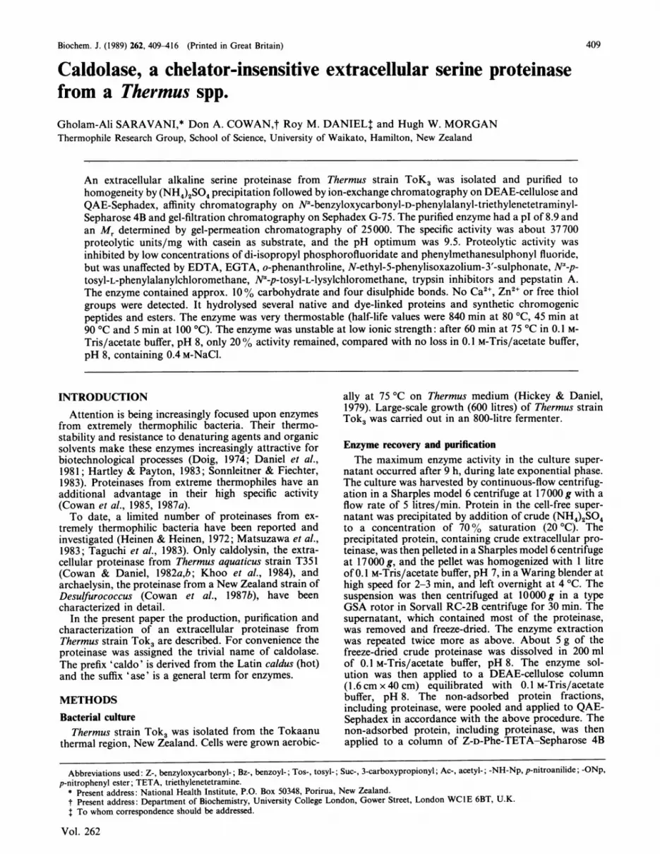
Fig. 1. Effect of pH on proteolytic activity of caldolaseThe enzyme reaction was carried out in the followingbuffers at the indicated pH values with 0.2% (w/v) caseinas outlined in the Methods section: Hepes buffer (0),Bicine buffer (El), Na2CO3/NaHCO3 buffer (A) andUniversal buffer (0).
et al., 1971) corresponded to less than 0.1 thiol group permolecule. This result, suggesting the absence of free thiolgroups, is in agreement with the conclusion based on theresponse of the enzyme to cysteine-proteinase inhibitors(see below).On the basis of the molar absorption coefficient of
3-carboxy-4-nitrothiophenolate (e 1 1 400 M-1 * cm-')(Robyt et al., 1971) and the Anderson & Wetlaufer(1975) stoichiometric reaction of 1.2 + 0.3 mol of 3-carboxy-4-nitrothiophenolate/mol of disulphide groups,the presence of four disulphide bonds per molecule ofcaldolase was inferred. However, neither 2-mercapto-ethanol nor dithiothreitol had a significant effect oncaldolase stability. It may be that the disulphide bondsare well protected from the reagents. Bridgen et al. (1973)have reported that thiol groups in alcohol dehydrogenasefrom Bacillus stearothermophilius were unreactive to-wards iodoacetate, and attributed this to the localizationof these groups within the protein molecule.
Metal ions in caldolaseNeither Ca2+ nor Zn2+ was present in caldolase
(64,ug/ml) in significant amounts (< 0.2 mol of Ca2"/mol and < 0.03 mol of Zn21/mol of enzyme). Caldolaseretained full activity and thermostability after EDTAtreatment (the enzyme was incubated at 75 °C for 30 minand the proteinase activity was measured), indicatingthat no EDTA-susceptible essential metal ion was presentin the enzyme. In the thermolysin sample used as apositive control (Feder et al., 1971; Dalquist et al.,1976) both Ca2` and Zn2+ were present in the EDTAfractions but not in the enzyme fractions (83 ,ug/ml) (seethe Methods section). Treatment by EDTA entirelyinactivated thermolysin.Amino acid composition of caldolaseThe partial amino acid composition of caldolase based
on an Mr of 25000 was Lys5His6Arg7Asx2,Thrl9-Serl7GlxloGly29Ala3lVallgMet2Ile8Leul6TyrgPhe4Trp3.Cysteine, proline and hydroxyproline residues were notdetermined.
a
EI
a
N
wr
Temperature (°C)
60
3.0
103/T (K-1)
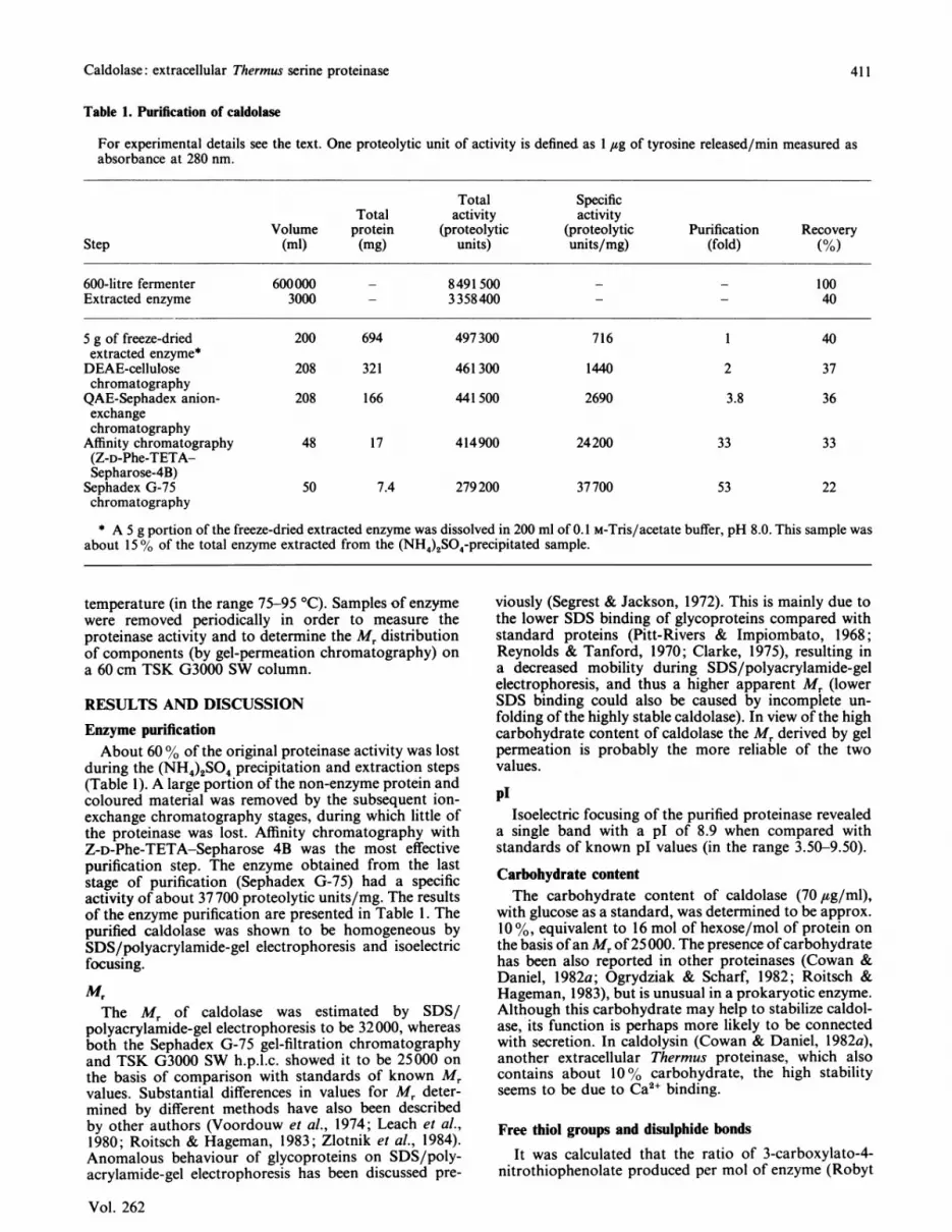
Fig. 2. Arrhenius plot for the hydrolysis of casein by caldolase
The temperature-activity relationship of the enzyme wasdetermined at the indicated temperatures with 0.2% (w/v)casein in 0.1 M-Tris/acetate buffer, pH 8.0.
0.5
0.4
-
E-
I
.H
co
EN
.u
0.3
0.2
Temperature (OC)98 90 80 70 60 50 40 30
2.8 3.0 3.2
103/T (K1)
he hydrolysis of Suc-Ala-Ala-Ala-
0.12.6
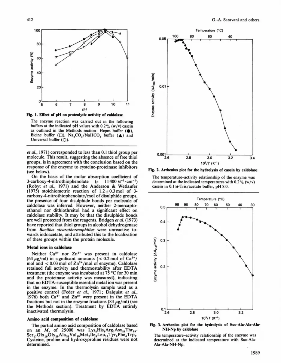
Fig. 3. Arrhenius plot for dNH-Np by caldolase
The temperature-activity relationship of the enzyme was
determined at the indicated temperature with Suc-Ala-Ala-Ala-NH-Np.
1989
,~~~~~~~r,:-|-It ,Z
0~~~~
0
4 AA I
nX 1
412
Caldolase: extracellular Thermus serine proteinase
Table 2. Inhibition of caldolase
Standard error values in all determinations were less than 5 %. The following inhibitors had negligible (< 5 %) effect: soya-beantrypsin inhibitor, lima-bean trypsin inhibitor, chicken egg-white trypsin inhibitor, antipain, a-antitrypsin, leupeptin, gramicidinS (each at 0.1 mg/ml), Tos-Lys-CH2CI at 5 mM, p-chloromercuribenzoate at 10 mM, 4-hydroxymercuribenzoate at 10 mm andpepstatin A at 2 mm concentration.
Preincubation EnzymeInhibitor time Concn. of inhibition(class) Inhibitor (at 20°C) inhibitor (Mo)
Serine-proteinaseinhibitor
Phenylmethane-sulphonyl fluoride
Di-isopropyl phosphoro-fluoridate
Chymotrypsininhibitors
Metal-ion chelators
Cysteine-proteinaseinhibitors
Heavy-metal ions
Chymostatin
EDTA (disodium salt)EGTAO-PhenanthrolineN-Ethyl-5-phenyliso-xazolium-3'-sulphonatelodoacetic acid
CuCl2
HgCl2
60 min
105 min
60 min60 min120 min120 min120 min120 min
120 min
60 min (75 °C)
60 min
60 min
pH optimumCaldolase exhibited a broad pH-activity profile with
an apparent pH optimum of about 9.5 (Fig. 1). However,in view of the sensitivity of caldolase activity to changesin ionic strength (see below), this datum must be viewedwith caution. The ionic strengths of Universal buffer(calculated by using the Henderson-Hasselbach equa-tion) at pH 6, 8, and 10 are 0.114, 0.243 and 0.368respectively. Increasing ionic strength might account forthe increased pH-dependence of activity below pH 7 inUniversal buffer, but probably has little effect on enzymeactivity above pH 9.
Temperature-activity relationship in caldolase
The Arrhenius plots for casein and peptide substrate(Suc-Ala-Ala-Ala-NH-Np) exhibit a sharp discontinuityat about 92 'C, due to denaturation (Figs. 2 and 3).Neither of the plots is a straight line, but the evidence fora discontinuity at any particular temperature or tempera-tures below 90 'C seems poor (e.g. see Wolfe & Bagnall,1979). For the casein plot it is difficult to separatethe effect of temperature on the enzyme from that on thesubstrate. However, the Ea at 80 'C is 58 kJ/mol. For the
peptide substrate there is a declining apparent Ea withincreasing temperature (Ea at 40 'C 22 kJ/mol and
at 80 'C 6 kJ/mol): the Q10 at 80 'C, approx. 1.1, issurprisingly low.
Effect of inhibitors on caldolase activityCaldolase was strongly inhibited by low concentra-
tions of di-isopropyl phosphorofluoridate and phenyl-methanesulphonyl fluoride and inhibited by relativelyhigh concentrations ofdiphenylcarbamoyl fluoride, iodo-acetic acid, HgCl2, CuCl2 and chymostatin (Table 2). Itwas not inhibited by metal-ion chelators, trypsin inhibi-tors Tos-Phe-CH2Cl, Tos-Lys-CH2Cl and pepstatin A.The above results indicate that caldolase is a serineproteinase. The chelating agents had no effect on caldol-ase, confirming that metal ions, in particular Ca2+ andZn2+, are not required for enzyme activity or stability.Tos-Phe-CH2Cl and Tos-Lys-CH2Cl, which inhibitchymotrypsin and trypsin respectively (Walsh, 1970),had no effect on caldolase. lodoacetic acid was only ableto inhibit caldolase at high concentration and hightemperature. At a high concentration iodoacetic acid canreact with the side groups of a number of amino acids(Means & Feeney, 1971).
Substrate specificity(a) Native proteins. Caldolase hydrolysed several pro-
tein substrates (Table 3). There was significant hydrolysisof collagen even at 35 °C, but only a very weak activitytowards elastin.
Vol. 262
0.1 mM1 mM5 mM
0.1 mM1 mM5 mM
0.1 mg/ml0.5 mg/ml10mM10mM5 mM5 mM
1 mM10 mM
1 mM10mM1 mM10mM
1 mM10mM
8492100309010066783020
3583
9703970100
413

G.-A. Saravani and others
Table 3. Relative rates of protein hydrolysis by caldolase
Actual rates of casein hydrolysis at 75 °C and 35 °C wereAA280/min = 0.069 and A280/min = 0.0027 respectively.Actual rates of azo-casein hydrolysis at 75 °C and 35 °Cwere AA440/min = 0.061 and AA440/min = 0.0024 re-spectively.
Relative rate ofhydrolysis
Substrate At 75 °C At 35 °C
Casein 100 100Bovine serum albumin 62 34Ovalbumin 31 8Haemoglobin 99 68Collagen type I 61 31Fibrin 52 39Elastin < 1 < 1
Azo-casein* 100 100Azo-albumin* 133 127Azocoll* 124 115Elastin-Congo Redt 1.8 1.2
* Measured at 440 nm.t Measured at 495 nm.
100
C
00
*E 5 L
N
a~~~~
ui
1 ! I
0 4 8 12 16 20 24Incubation time (h)
Fig. 4. Stability of caldolase at temperatures between 75 °C and100 OC
Purified enzyme (24 ,ug/ml in 0.1 M-Tris/acetate buffer,pH 8.0, containing 0.5 M-NaCl and 10 mM-CaCl2) wasincubated at 75 °C (El), at 80 °C (-), at 85 °C (A), at90 °C (A), at 95 °C (0) and at 100 °C (*). Samples ofenzyme solution were removed at intervals, and the residualproteinase activity was determined at 75 °C by the modifiedKunitz method.
(b) Dye-linked proteins. The susceptibility of azo-albumin and Azocoll was greater than that of azo-casein,in contrast with the results for the unmodified substrates.There was little hydrolysis of elastin-Congo Red(Table 3).
(c) Chromogenic peptide substrates. Caldolase wascapable of hydrolysing several synthetic N-terminallyblocked tripeptide p-nitroanilide substrates. Reactionrates followed the order: Bz-Phe-Val-Arg-NH-Np > Bz-Val-Gly-Arg-NH-Np > Tos-Gly-Pro-Arg-NH-Np >Suc-Ala-Ala-Ala-NH-Np > Bz-Pro-Phe-Arg-H-Np; theenzyme failed to hydrolyse any of the single amino acidp-nitroanilide substrates.
(d) Chromogenic ester substrates. Caldolase hydrolysedall synthetic ester substrates tested (see the Methodssection). Reaction rates followed the order: Z-Ala-ONp> Z-Tyr-ONp - Z-Phe-ONp - Z-Leu-ONp > Z-Trp-ONp > Z-lys-ONp > Z-Asn-ONp > Z-,6-Cys-ONp c
Z-,1-Asn-ONp > Z-Val-ONp > Z-,J-Ala-ONp - Z-D-Nle-ONp > Z-Ile-ONp - Z-Pro-ONp. Esterase activitywas not the result of a contaminant, since the ratio ofproteinase to esterase activity was the same for all thelater purification stages, and these ratios remained un-changed after partial thermal denaturation and aftertreatment with EDTA, p-chloromercuribenzoate andphenylmethanesulphonyl fluoride (results not shown).
(e) Peptides. Caldolase was not able to hydrolysebradykinin (Arg-Pro-Pro-Gly-Phe-Ser-Pro-Phe-Arg)during the reaction periods tested, suggesting that theenzyme lacked specificity towards non-polar amino acids.In contrast, cleavage of insulin B-chain even for shortperiods resulted in a very complex pattern (results notshown), indicating a low degree of enzyme specificity.
Effect of denaturing and reducing agentsCaldolase activity was affected little by incubation
in 8 M-urea and 6 M-guanidinium chloride at low tem-perature, but was decreased significantly after incubationat high temperatures (69% loss and 800 loss respectivelyafter 60 min incubations at 75 °C).
2-Mercaptoethanol and dithiothreitol had no signifi-cant effect on enzyme stability at either low or hightemperatures This suggests either that the disulphidebonds are protected from the solvent, or that they are notinvolved in the stability of caldolase.
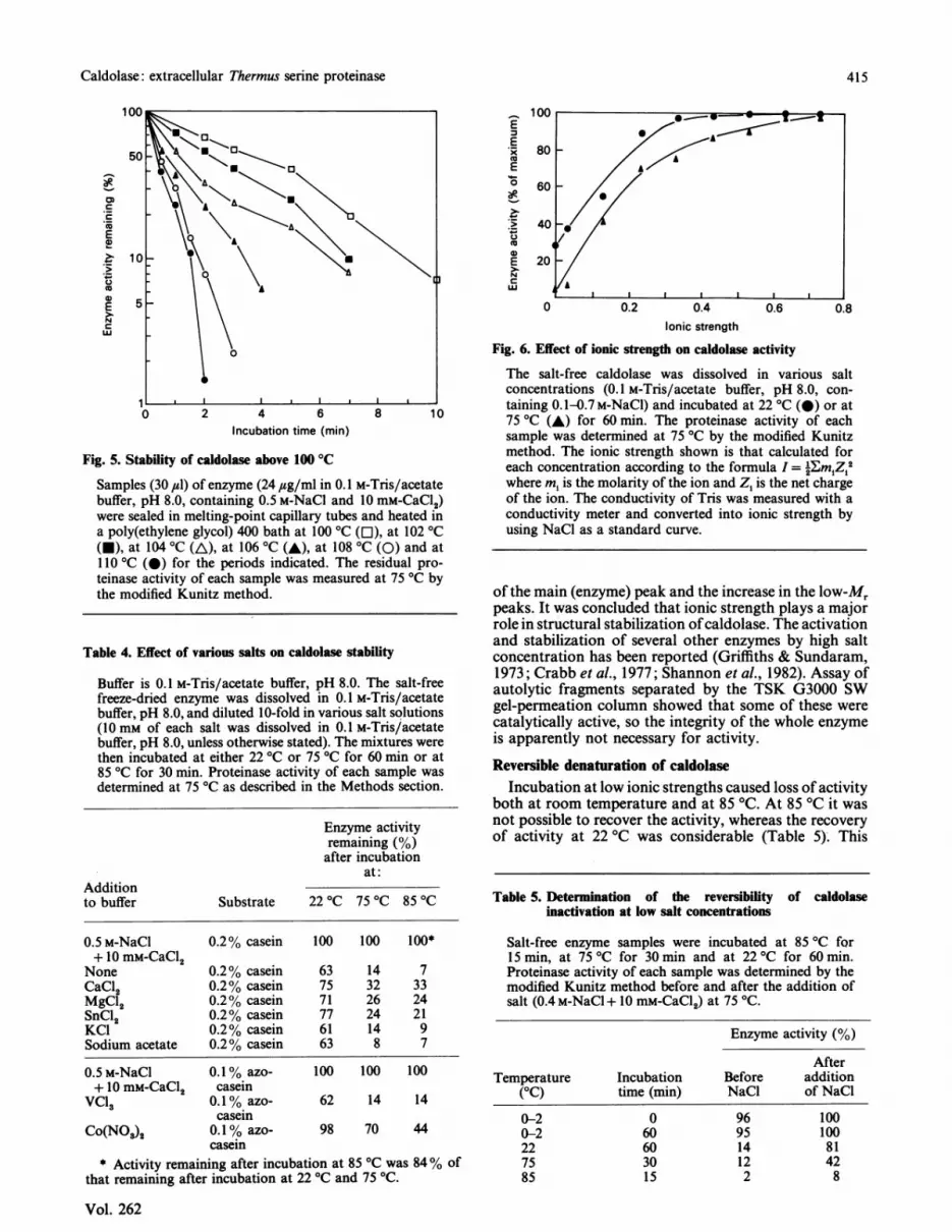
StabilityStability profiles at temperatures between 75 °C and
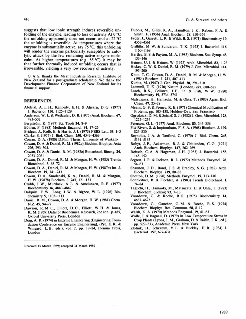
110 °C are shown in Figs. 4 and 5. The half-life of theenzyme was about 840 min at 80 °C and 45 min at 90 'C.Loss of proteinase activity above 100 'C was very rapid,and at 106 'C the half-life of the enzyme was less than1 min.Caldolase was unstable at low salt concentration
(Fig. 6), and was stabilized by v'arious salts (Table 4). Ananalysis of denaturation and autolysis of caldolase in thepresence of various salt concentrations (0.1 M-Tris/acetate buffer, pH 8, and 0.1 M-Tris/acetate buffer, pH 8,containing 0.1 M-, 0.3 M- or 0.5 M-NaCl plus 10 mM-CaCl2) and at temperatures ranging from 75 to 95 'Crevealed that at high ionic strength and temperatures upto 85 'C second-order kinetics were predominant (auto-lysis was dominant), whereas at 90 'C and 95 'C first-order kinetics were significant (denaturation was import-ant) (Voordouw & Roche, 1975). At low ionic strengthsecond-order kinetics were predominant up to 95 °C.TSK gel-filtration chromatography demonstrated that,by increasing the incubation time, by; raising the tem-perature or by a combination of both, autolysis productswere increased. This was shown by the decrease in height
1989
414
Caldolase: extracellular Thermus serine proteinase
0
, *0 2 4 6 8 10
Incubation time (min)
Fig. 5. Stability of caldolase above 100 °C
Samples (30 ,ul) of enzyme (24 ,usg/ml in 0.1 M-Tris/acetatebuffer, pH 8.0, containing 0.5 M-NaCl and 10 mM-CaCl2)were sealed in melting-point capillary tubes and heated ina poly(ethylene glycol) 400 bath at 100 °C (El), at 102 °C(-), at 104 °C (A), at 106 °C (A), at 108 °C (0) and at110 °C (0) for the periods indicated. The residual pro-teinase activity of each sample was measured at 75 °C bythe modified Kunitz method.
Table 4. Effect of various salts on caldolase stability
Buffer is 0.1 M-Tris/acetate buffer, pH 8.0. The salt-freefreeze-dried enzyme was dissolved in 0.1 M-Tris/acetatebuffer, pH 8.0, and diluted 10-fold in various salt solutions(10 mm of each salt was dissolved in 0.1 M-Tris/acetatebuffer, pH 8.0, unless otherwise stated). The mixtures werethen incubated at either 22 °C or 75 °C for 60 min or at85 °C for 30 min. Proteinase activity of each sample wasdetermined at 75 °C as described in the Methods section.
Enzyme activityremaining (%)after incubation
at:Additionto buffer Substrate 22 °C 75 °C 85 °C
0.5 M-NaCl 0.2% casein 100 100 100*+ 10 mM-CaCl2
None 0.2% casein 63 14 7CaC12 0.2% casein 75 32 33MgCl2 0.2% casein 71 26 24SnC12 0.2% casein 77 24 21KCI 0.2% casein 61 14 9Sodium acetate 0.2% casein 63 8 7
0.5 M-NaCI+ 10 mM-CaCl2
VCl3
Co(N03)2
0.1% azo-casein0.1% azo-casein0.1% azo-casein
100 100 100
62 14 14
98 70 44
* Activity remaining after incubation at 85 °C was 84% ofthat remaining after incubation at 22 °C and 75 'C.
100
Eu.
E0
E
C.,._
cuE
N-C
w5i~
Ionic strength
Fig. 6. Effect of ionic strength on caldolase activity
The salt-free caldolase was dissolved in various saltconcentrations (0.1 M-Tris/acetate buffer, pH 8.0, con-taining 0.1-0.7M-NaCI) and incubated at 22 °C (@) or at75 °C (A) for 60 min. The proteinase activity of eachsample was determined at 75 °C by the modified Kunitzmethod. The ionic strength shown is that calculated foreach concentration according to the formula I = 2Em Z 2where m, is the molarity of the ion and Z1 is the net chargeof the ion. The conductivity of Tris was measured with aconductivity meter and converted into ionic strength byusing NaCl as a standard curve.
of the main (enzyme) peak and the increase in the low-Mrpeaks. It was concluded that ionic strength plays a majorrole in structural stabilization ofcaldolase. The activationand stabilization of several other enzymes by high saltconcentration has been reported (Griffiths & Sundaram,1973; Crabb et al., 1977; Shannon et al., 1982). Assay ofautolytic fragments separated by the TSK G3000 SWgel-permeation column showed that some of these werecatalytically active, so the integrity of the whole enzymeis apparently not necessary for activity.
Reversible denaturation of caldolaseIncubation at low ionic strengths caused loss of activity
both at room temperature and at 85 'C. At 85 'C it wasnot possible to recover the activity, whereas the recoveryof activity at 22 'C was considerable (Table 5). This
Table 5. Determination of the reversibility of caldolaseinactivation at low salt concentrations
Salt-free enzyme samples were incubated at 85 °C for15 min, at 75 °C for 30 min and at 22 °C for 60 min.Proteinase activity of each sample was determined by themodified Kunitz method before and after the addition ofsalt (0.4 M-NaCl +10 mM-CaCl2) at 75 'C.
Enzyme activity (%)
AfterTemperature Incubation Before addition
(OC) time (min) NaCl of NaCl
0-20-2227585
060603015
969514122
10010081428
Vol. 262
415
G.-A. Saravani and others
suggests that low ionic strength induces reversible un-folding of the enzyme, leading to loss of activity At 0 °Cthe unfolding apparently does not occur, and at 22 ICthe unfolding is reversible. At temperatures where theenzyme is substantially active, say 75 °C, this unfoldingwill render the enzyme particularly susceptible to auto-lytic attack by the few remaining active enzyme mole-cules. At higher temperatures (e.g. 85 °C) it may bethat further thermally induced unfolding occurs that isirreversible, yielding a very low recovery of activity.
G. A. S. thanks the Meat Industries Research Institute ofNew Zealand for a post-graduate scholarship. We thank theDevelopment Finance Corporation of New Zealand for itsfinancial support.
REFERENCESAbdelal, A. T. H., Kennedy, E. H. & Aheam, D. G. (1977)
J. Bacteriol. 130, 1125-1129Anderson, W. L. & Wetlaufer, D. B. (1975) Anal. Biochem. 67,493-502
Bergstr6m, K. (1977) Sci. Tools 24, 8-9Bohlen, P. (1983) Methods Enzymol. 91, 17-26Bridgen, J., Kolb, E. & Harris, J. I. (1973) FEBS Lett. 33, 1-3Clarke, S. (1975) J. Biol. Chem. 250, 4549-4569Cowan, D. A. (1980) D.Phil. Thesis, University of WaikatoCowan, D. A. & Daniel, R. M. (1982a) Biochim. Biophys. Acta
705, 293-305.Cowan, D. A. & Daniel, R. M. (1982b) Biotechnol. Bioeng. 24,
2053-2061Cowan, D. A., Daniel, R. M. & Morgan, H. W. (1985) Trends
Biotechnol. 3, 68-72Cowan, D. A., Daniel, R. M. & Morgan, H. W. (1987a) Int. J.
Biochem. 19, 741-743Cowan, D. A., Smolenski, K. A., Daniel, R. M. & Morgan,
H. W. (1987b) Biochem. J. 247, 121-133Crabb, J. W., Murdock, A. L. & Amelunxen, R. E. (1977)
Biochemistry 16, 4840-4847Dalquist, F. W., Long, J. W. & Bigbee, W. L. (1976) Bio-
chemistry 5, 1103-1111Daniel, R. M., Cowan, D. A. & Morgan, H. W. (1981) Chem.
N.Z. 45, 94-97Dawson, R. M. C., Elliott, D. C., Elliott, W. H. & Jones,
K. M. (1969) Data for Biochemical Research, 2nd edn., p.485,Oxford University Press, London
Doig, A. R. (1974) in Enzyme Engineering (Engineering Foun-dation Conference on Enzyme Engineering), (Pye, E. K. &Wingard, L. B., eds.), vol. 2, pp. 17-24, Plenum Press,London
Dubois, M., Gilles, K. A., Hamilton, J. K., Rebers, P. A. &Smith, F. (1956) Anal. Biochem. 28, 350-356
Feder, J., Garrett, L. R. & Wildi, B. S. (1971) Biochemistry 10,4552-4561
Griffiths, M. W. & Sundaram, T. K. (1973) J. Bacteriol. 116,1160-1169
Hartley, B. S. & Payton, M. A. (1983) Biochem. Soc. Symp. 48,133-146
Heinen, U. J. & Heinen, W. (1972) Arch. Microbiol. 82, 1-23Hickey, C. W. & Daniel, R. M. (1979) J. Gen. Microbiol. 114,
195-200Khoo, T. C., Cowan, D. A., Daniel, R. M. & Morgan, H. W.
(1984) Biochem. J. 221, 407-413Kunitz, M. (1947) J. Gen. Physiol. 30, 291-310Laemmli, U. K. (1970) Nature (London) 227, 680-685Leach, B. S., Collawn, J. F., Jr. & Fish, W. W. (1980)
Biochemistry 19, 5734-5741Matsuzawa, H., Hamaoki, M. & Ohta, T. (1983) Agric. Biol.Chem. 47, 25-28
Means, G. F. & Feeney, R. E. (1971) Chemical Modification ofProteins, pp. 105-138, Holden-Day, San Francisco
Ogrydziak, D. M. & Scharf, S. J. (1982) J. Gen. Microbiol. 128,1225-1234
Peterson, G. L. (1977) Anal. Biochem. 83, 346-356Pitt-Rivers, S. & Impiombato, F. S. A. (1968) Biochem. J. 109,
825-830Reynolds, J. A. & Tanford, C. (1970) J. Biol. Chem. 245,
5161-5165Robyt, J. F., Ackerman, R. J. & Chittenden, C. G. (1971)
Arch. Biochem. Biophys. 147, 262-269Roitsch, C. A. & Hageman, J. H. (1983) J. Bacteriol. 155,
145-152Segrest, J. P. & Jackson, R. L. (1972) Methods Enzymol. 28,
54-63Shannon, J. D., Bond, J. S. & Bradley, S. G. (1982) Arch.
Biochem. Biophys. 219, 80-88Shotton, D. M. (1970) Methods Enzymol. 19, 113-140Sonnleitner, B. & Fiechter, A. (1983) Trends Biotechnol. 1,
74-84Taguchi, H., Hamaoki, M., Matsuzata, H. & Ohta, T. (1983)
J. Biochem. (Tokyo) 93, 7-13Voordouw, G. & Roche, R. S. (1975) Biochemistry 14,4667-4673
Voordouw, G., Gaucher, G. M. & Roche, R. S. (1974)Biochem. Biophys. Res. Commun. 58, 8-12
Walsh, K. A. (1970) Methods Enzymol. 19, 41-63Wolfe, J. & Bagnall, D. (1979) in Low Temperature Stress inCrop Plants (Lyons, J. M., Graham, D. & Raisin, J. K., ed.),pp. 527-533, Academic Press, New York
Zlotnik, H., Schramm, V. L. & Buckley, H. R. (1984) J.Bacteriol. 157, 627-631
1989
Received 15 March 1989; accepted 31 March 1989
416


