Embed Size (px)
Citation preview
Chemistry & Biology
Article
Defining Criteria for Oligomannose Immunogensfor HIV Using Icosahedral Virus Capsid ScaffoldsRena D. Astronomo,1,8 Eiton Kaltgrad,2,3,9 Andrew K. Udit,2,3,10 Sheng-Kai Wang,2,3 Katie J. Doores,1,4
Cheng-Yuan Huang,2,3,11 Ralph Pantophlet,1,12 James C. Paulson,5,6 Chi-Huey Wong,2,3 M.G. Finn,2,3,*and Dennis R. Burton1,4,7,*1Department of Immunology and Microbial Science2Department of Chemistry3The Skaggs Institute for Chemical Biology
The Scripps Research Institute, La Jolla, CA 92037, USA4Ragon Institute of Massachusetts General Hospital, MIT, and Harvard, Boston, MA 02129, USA5Department of Molecular Biology6Department of Chemical Physiology7IAVI Neutralizing Antibody Center
The Scripps Research Institute, La Jolla, CA 92037, USA8Present address: Fred Hutchinson Cancer Research Center, 1100 Fairview Avenue North, Seattle, WA 98109, USA9Present address: Calmune Corporation, 9393 Towne Center Drive, San Diego, CA 92121, USA10Present address: Department of Chemistry, Occidental College, 1600 Campus Road, Los Angeles, CA 90041, USA11Present address: Research Center for Materials Science, Nagoya University, Nagoya 464-15 8602, Japan12Present address: Faculty of Health Sciences, Simon Fraser University, Blusson Hall, 8888 University Drive, Burnaby, BC V5A 1S6, Canada
*Correspondence: [email protected] (M.G.F.), [email protected] (D.R.B.)
DOI 10.1016/j.chembiol.2010.03.012
SUMMARY
The broadly neutralizing antibody 2G12 recognizes aconserved cluster of high-mannose glycans on thesurface envelope spike of HIV, suggesting that the‘‘glycan shield’’ defense of the virus can be breachedand may, under the right circumstances, serve asa vaccine target. In an attempt to recreate featuresof the glycan shield semisynthetically, oligomanno-sides were coupled to surface lysines on the icosa-hedral capsids of bacteriophage Qb and cowpeamosaic virus (CPMV). The Qb glycoconjugates, butnot CPMV, presented oligomannose clusters thatbind the antibody 2G12 with high affinity. However,antibodies against these 2G12 epitopes were notdetected in immunized rabbits. Rather, alternativeoligomannose epitopes on the conjugates wereimmunodominant and elicited high titers of anti-mannose antibodies that do not crossreact with theHIV envelope. The results presented reveal importantdesign considerations for a carbohydrate-basedvaccine component for HIV.
INTRODUCTION
A protective vaccine remains widely accepted as the best
weapon to combat the spread of HIV-1, but despite tremendous
effort a vaccine that induces a neutralizing antibody (Nab)
response against a broad range of isolates has yet to be realized
(Virgin and Walker, 2010). Difficulties in the elicitation of anti-
bodies (Abs) to conserved regions of the functional envelope
spike (Env), responsible for viral infectivity, may largely be attrib-
Chemistry & Biology 17,
uted to the nature of this target: it is an unstable heterodimeric
trimer, composed of glycoproteins gp120 and gp41, in which
conserved epitopes are recessed, transiently exposed, or other-
wise occluded by highly variable immunodominant loops and
a dense glycan shield (Chen et al., 2005; Kwong et al., 1998;
Wyatt et al., 1998; Wyatt and Sodroski, 1998). Despite these
formidable defenses, a handful of monoclonal broadly neutral-
izing Abs (bNabs) (Burton et al., 1994; Corti et al., 2010; Trkola
et al., 1996; Walker et al., 2009; Zwick et al., 2001) and polyclonal
sera (Binley, 2009; Binley et al., 2008; Dhillon et al., 2007; Gray
et al., 2009; Li et al., 2009; Stamatatos et al., 2009) from HIV-1-
infected individuals suggest that a crossreactive Nab response
against HIV-1 can be achieved.
Monoclonal bNabs are potentially valuable tools for the design
of effective vaccine components, as their epitopes reveal
conserved chinks in the armor of Env that may be exploited.
For example, although the ‘‘glycan shield’’ of gp120 is crucial
to immune evasion, the bNab 2G12 binds a cluster of oligoman-
nose glycans on the shield, making it a potential vaccine target
(Sanders et al., 2002; Scanlan et al., 2002; Trkola et al., 1996).
In addition to a broad neutralization profile (Binley et al., 2004;
Trkola et al., 1995, 1996), 2G12 protects against infection in
non-human primate studies (Hessell et al., 2009; Mascola
et al., 2000) and exerts selection pressure on HIV-1 in humans
while being well tolerated (Mehandru et al., 2007; Trkola et al.,
2005). Thus, the ability to elicit 2G12-like Abs is an important
goal for vaccine researchers.
2G12 is specific for terminal Mana1-2Man residues on high-
mannose glycans, particularly on the D1 and D3 arms (Calarese
et al., 2003, 2005; Scanlan et al., 2002). A variable heavy-
domain-exchanged Ab structure creates a compact multivalent
binding surface which allows 2G12 to bind its glycan epitope
with high affinity (Calarese et al., 2003, 2005). The clustered
presentation of the high-mannose glycans on gp120, comprising
357–370, April 23, 2010 ª2010 Elsevier Ltd All rights reserved 357
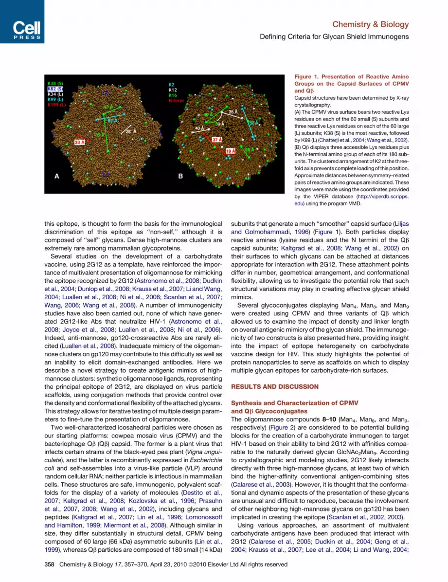
Figure 1. Presentation of Reactive Amino
Groups on the Capsid Surfaces of CPMV
and Qb
Capsid structures have been determined by X-ray
crystallography.
(A) The CPMV virus surface bears two reactive Lys
residues on each of the 60 small (S) subunits and
three reactive Lys residues on each of the 60 large
(L) subunits; K38 (S) is the most reactive, followed
by K99 (L) (Chatterji et al., 2004; Wang et al., 2002).
(B) Qb displays three accessible Lys residues plus
the N-terminal amino group of each of its 180 sub-
units. The clustered arrangement of K2 at the three-
fold axis prevents complete loading of this position.
Approximate distances betweensymmetry-related
pairs of reactive amino groups are indicated. These
images were made using the coordinates provided
by the VIPER database (http://viperdb.scripps.
edu) using the program VMD.
Chemistry & Biology
Defining Criteria for Glycan Shield Immunogens
this epitope, is thought to form the basis for the immunological
discrimination of this epitope as ‘‘non-self,’’ although it is
composed of ‘‘self’’ glycans. Dense high-mannose clusters are
extremely rare among mammalian glycoproteins.
Several studies on the development of a carbohydrate
vaccine, using 2G12 as a template, have reinforced the impor-
tance of multivalent presentation of oligomannose for mimicking
the epitope recognized by 2G12 (Astronomo et al., 2008; Dudkin
et al., 2004; Dunlop et al., 2008; Krauss et al., 2007; Li and Wang,
2004; Luallen et al., 2008; Ni et al., 2006; Scanlan et al., 2007;
Wang, 2006; Wang et al., 2008). A number of immunogenicity
studies have also been carried out, none of which have gener-
ated 2G12-like Abs that neutralize HIV-1 (Astronomo et al.,
2008; Joyce et al., 2008; Luallen et al., 2008; Ni et al., 2006).
Indeed, anti-mannose, gp120-crossreactive Abs are rarely eli-
cited (Luallen et al., 2008). Inadequate mimicry of the oligoman-
nose clusters on gp120 may contribute to this difficulty as well as
an inability to elicit domain-exchanged antibodies. Here we
describe a novel strategy to create antigenic mimics of high-
mannose clusters: synthetic oligomannose ligands, representing
the principal epitope of 2G12, are displayed on virus particle
scaffolds, using conjugation methods that provide control over
the density and conformational flexibility of the attached glycans.
This strategy allows for iterative testing of multiple design param-
eters to fine-tune the presentation of oligomannose.
Two well-characterized icosahedral particles were chosen as
our starting platforms: cowpea mosaic virus (CPMV) and the
bacteriophage Qb (Qb) capsid. The former is a plant virus that
infects certain strains of the black-eyed pea plant (Vigna ungui-
culata), and the latter is recombinantly expressed in Escherichia
coli and self-assembles into a virus-like particle (VLP) around
random cellular RNA; neither particle is infectious in mammalian
cells. These structures are safe, immunogenic, polyvalent scaf-
folds for the display of a variety of molecules (Destito et al.,
2007; Kaltgrad et al., 2008; Kozlovska et al., 1996; Prasuhn
et al., 2007, 2008; Wang et al., 2002), including glycans and
peptides (Kaltgrad et al., 2007; Lin et al., 1996; Lomonossoff
and Hamilton, 1999; Miermont et al., 2008). Although similar in
size, they differ substantially in structural detail, CPMV being
composed of 60 large (66 kDa) asymmetric subunits (Lin et al.,
1999), whereas Qb particles are composed of 180 small (14 kDa)
358 Chemistry & Biology 17, 357–370, April 23, 2010 ª2010 Elsevier
subunits that generate a much ‘‘smoother’’ capsid surface (Liljas
and Golmohammadi, 1996) (Figure 1). Both particles display
reactive amines (lysine residues and the N termini of the Qb
capsid subunits; Kaltgrad et al., 2008; Wang et al., 2002) on
their surfaces to which glycans can be attached at distances
appropriate for interaction with 2G12. These attachment points
differ in number, geometrical arrangement, and conformational
flexibility, allowing us to investigate the potential role that such
structural variations may play in creating effective glycan shield
mimics.
Several glycoconjugates displaying Man4, Man8, and Man9
were created using CPMV and three variants of Qb which
allowed us to examine the impact of density and linker length
on overall antigenic mimicry of the glycan shield. The immunoge-
nicity of two constructs is also presented here, providing insight
into the impact of epitope heterogeneity on carbohydrate
vaccine design for HIV. This study highlights the potential of
protein nanoparticles to serve as scaffolds on which to display
multiple glycan epitopes for carbohydrate-rich surfaces.
RESULTS AND DISCUSSION
Synthesis and Characterization of CPMVand Qb GlycoconjugatesThe oligomannose compounds 8–10 (Man4, Man8, and Man9,
respectively) (Figure 2) are considered to be potential building
blocks for the creation of a carbohydrate immunogen to target
HIV-1 based on their ability to bind 2G12 with affinities compa-
rable to the naturally derived glycan GlcNAc2Man9. According
to crystallographic and modeling studies, 2G12 likely interacts
directly with three high-mannose glycans, at least two of which
bind the higher-affinity conventional antigen-combining sites
(Calarese et al., 2003). However, it is thought that the conforma-
tional and dynamic aspects of the presentation of these glycans
are unusual and difficult to reproduce, because the involvement
of other neighboring high-mannose glycans on gp120 has been
implicated in creating the epitope (Scanlan et al., 2002, 2003).
Using various approaches, an assortment of multivalent
carbohydrate antigens have been produced that interact with
2G12 (Calarese et al., 2005; Dudkin et al., 2004; Geng et al.,
2004; Krauss et al., 2007; Lee et al., 2004; Li and Wang, 2004;
Ltd All rights reserved

Figure 2. Synthesis of Virus Glycoconjugates
Top: synthesis of Qbwt, QbK16M, and CPMV conjugates. Bottom: stepwise synthesis of QbHPG glycoconjugates QbHPG-Man8 (11) and QbHPG-
Man8/Man9 (12).
Chemistry & Biology
Defining Criteria for Glycan Shield Immunogens
Luallen et al., 2008; Ni et al., 2006; Scanlan et al., 2007; Wang,
2006; Wang et al., 2004, 2007, 2008). However, all of these
approaches have employed flexible and/or structurally hetero-
geneous platforms. Virus particles, in contrast, are relatively rigid
and structurally homogeneous to atomic resolution. The antici-
pated importance of oligomannose conformation and clustering
therefore prompted us to test virus scaffolds that permit
the creation of glycoconjugates presenting oligomannose in
different spatial arrangements that are constrained by the posi-
tions of the amino acid residues to which attachments are
made (Figure 1). In addition to the Qbwt capsid protein shown
(Figure 1), two other Qb variants were prepared: QbK16M, a point
mutant in which the most exposed Lys was changed to Met, and
QbHPG, an expression variant of QbK16M in which an alkyne-
Chemistry & Biology 17,
containing unnatural amino acid was incorporated instead of
Met at position 16 (Strable et al., 2008). The QbHPG scaffold
enabled us to attach different glycans at different positions in
a sequential fashion, a capability that improves the overall homo-
geneity of mixed-glycan conjugates.
High-density glycan display by chemical coupling of the
sugars to a scaffold requires a conjugation reaction that is
sufficiently strongly driven to overcome unfavorable steric inter-
actions that such crowding may create. In this case, efficient
conjugation is particularly important because these oligomanno-
sides are the products of lengthy and technically demanding
syntheses. We implemented a two-step strategy, first acylating
the surface amino groups with a large excess of alkynyl
N-hydroxysuccinimide ester 1 (Figure 2). Following purification
357–370, April 23, 2010 ª2010 Elsevier Ltd All rights reserved 359

Figure 3. Representations of the Display Patterns
around the Three-Fold Symmetry Axis on the
Surface of Qbwt and QbK16M
(A) Qbwt.
(B) QbK16M.
The colored spheres represent triazole linkages to the
following: K12 (blue), K16 (green), K2 (red), and the
N terminus (black). Because K12 and K16 are very close
to each other (�8 A apart), it is unlikely that both bear
attached glycans in each subunit of the wild-type struc-
ture. The oval shapes in (A), therefore, represent a triazole
linkage made at one of the two residues (K12 or K16).
Based on this model, the overall glycan display patterns
for Qbwt and QbK16M glycoconjugates are very similar.
Chemistry & Biology
Defining Criteria for Glycan Shield Immunogens
from excess linker, the desired azides were attached with the
copper-catalyzed azide-alkyne cycloaddition (CuAAC) reaction
(Rostovtsev et al., 2002), enhanced by a ligand known to strongly
accelerate the process (Lewis et al., 2004; Sen Gupta et al.,
2005). Azidoalkyl linkers are commonly employed in glycan
synthesis as precursors to terminal amines and are stable
toward extended storage, as are the alkyne groups placed on
the protein. The CuAAC ‘‘click reaction’’ allows these stable
reactive groups to be employed at low concentrations to achieve
reproducibly high loadings.
The resulting constructs 3, 4, and 5 (Figure 2), differing in the
starting platform (Qbwt, QbK16M, and CPMV, respectively)
and, therefore, in the number and geometry of available conjuga-
tion points, were characterized by size-exclusion chromatog-
raphy, polyacrylamide gel electrophoresis, and transmission
electron microscopy (see Figure S1 available online). The final
conjugates were isolated in 60%–70% yields. The number of
triazole-tethered glycans on each particle was estimated by
parallel reactions using the selenomethionine azide derivative
7a, which more closely mimics the hydrophilicity of carbohy-
drates than dyes (e.g., fluorescein) previously used for this
purpose (Figure 2). Reproducible measurements of 230 ± 30
attachments to CPMV, 470 ± 50 to Qbwt VLP, and 450 ± 50 to
QbK16M particles were made using inductively coupled plasma
optical emission spectroscopy. These values provide an upper
limit for the numbers of bulkier glycans attached, because
steric crowding will be more pronounced in these cases than
for the SeMet label 7a. CPMV bears fewer reactive amines on
its surface than does Qb, accounting for the lower density of
attached glycans (Figure 1). Although the QbK16M has 180 fewer
lysine attachment points than the Qbwt capsid, their loading
values were very similar, suggesting that the closely spaced
K12 and K16 residues (�8 A apart) were not both derivatized
on Qbwt, and that loss of K16 may have allowed the K12 to be
fully accessed (Figure 3).
To minimize the length of the linker connecting the scaffold to
the glycans, which may impact the antigenicity of resultant
conjugates, we expressed the QbK16M mutant construct in an
E. coli Met auxotroph in the presence of the alkyne-containing
amino acid homopropargyl glycine (HPG) in the manner of Tirrell
360 Chemistry & Biology 17, 357–370, April 23, 2010 ª2010 Elsevier
and coworkers (Strable et al., 2008). HPG was incorporated at
position 16 with 50% efficiency as determined by mass spec-
trometry (MS) and CuAAC reaction with SeMet 7a (Strable
et al., 2008) (Figure S2). Conjugation of azide 9 under the same
conditions provided conjugate 11 (QbHPG-Man8) (Figure 2).
Conjugate 12 (QbHPG-Man8/Man9) was produced by subjecting
11 to another round of elaboration with NHS alkyne 1 and azide
10. By parallel loading with SeMet 7a, we estimated 270 ± 30
Man9 glycans were added to the capsid surface in addition to
the 90 Man8 glycans attached at position 16. MALDI MS analysis
of QbHPG-Man8/Man9, after dissociation of the particles into
their component subunit proteins, also showed only a small
amount of nonmannosylated protein, with major components
bearing 1–3 oligomannose glycans (Figure S2).
QbHPG-Man8/Man9, therefore, displays Man8 on a shortened
tether at the most highly exposed position, with numerous Man9
glycans distributed over the rest of the surface on the longer
conventional linker. We reasoned that this type of mixed particle
may be beneficial for mimicking the microheterogeneity found on
HIV. The QbHPG scaffold also provided a unique opportunity to
study a controlled, mixed conjugate where the position of one of
the two glycans is fixed (in this case, Man8). In contrast, CPMV-
Man4/Man9 (5f) was the product of a single conjugation using
a mixture of Man4 and Man9 azides, resulting in a random distri-
bution of the two oligosaccharides over the particle surface. The
CPMV and Qb scaffolds thereby provide examples of different
arrangements and densities of oligomannose display, whereas
the QbHPG scaffold affords the opportunity to investigate what
effect a more rigid display format may have on 2G12 binding.
Interaction of Capsid Conjugates with 2G12The presence of the attached oligomannose ligands was first
verified by the observation of rapid aggregation of dilute samples
when mixed with the tetravalent mannose-binding lectin conca-
navalin A (ConA) (Figure S3). A conventional ELISA, in which
serial dilutions of 2G12 were allowed to bind antigen coated
directly onto wells, was then used to compare the affinity of
2G12 for the capsid conjugates with that of gp120JR-FL (Fig-
ure 4A). Qbwt and QbK16M conjugates of Man4 and Man9 inter-
acted with 2G12 with nanomolar apparent affinities (�50-fold
Ltd All rights reserved
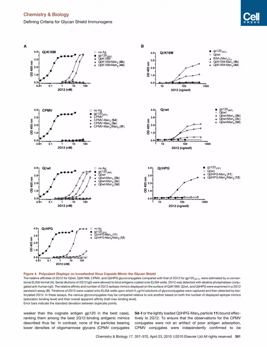
Figure 4. Polyvalent Displays on Icosahedral Virus Capsids Mimic the Glycan Shield
The relative affinities of 2G12 for Qbwt, QbK16M, CPMV, and QbHPG glycoconjugates compared with that of 2G12 for gp120JR-FL were estimated by a conven-
tional ELISA format (A). Serial dilutions of 2G12 IgG were allowed to bind antigens coated onto ELISA wells. 2G12 was detected with alkaline phosphatase-conju-
gated anti-human IgG. The relative affinity and number of 2G12 epitope mimics displayed on the surface of QbK16M, Qbwt, and QbHPG were examined in a 2G12
sandwich assay (B). Titrations of 2G12 were coated onto ELISA wells upon which 5 mg/ml solutions of glycoconjugates were captured and then detected by bio-
tinylated 2G12. In these assays, the various glycoconjugates may be compared relative to one another based on both the number of displayed epitope mimics
(saturation binding level) and their overall apparent affinity (half-max binding level).
Error bars indicate the standard deviation between duplicate points.
Chemistry & Biology
Defining Criteria for Glycan Shield Immunogens
weaker than the cognate antigen gp120 in the best case),
ranking them among the best 2G12-binding antigenic mimics
described thus far. In contrast, none of the particles bearing
lower densities of oligomannose glycans (CPMV conjugates
Chemistry & Biology 17,
5d–f or the lightly loaded QbHPG-Man8 particle 11) bound effec-
tively to 2G12. To ensure that the observations for the CPMV
conjugates were not an artifact of poor antigen adsorption,
CPMV conjugates were independently confirmed to be
357–370, April 23, 2010 ª2010 Elsevier Ltd All rights reserved 361
Chemistry & Biology
Defining Criteria for Glycan Shield Immunogens
362 Chemistry & Biology 17, 357–370, April 23, 2010 ª2010 Elsevier Ltd All rights reserved
Chemistry & Biology
Defining Criteria for Glycan Shield Immunogens
deposited onto ELISA wells in quantities comparable to naked
CPMV particles by detection with anti-CPMV polyclonal Ab
(data not shown).
Qbwt-Man9 (3d), QbK16M-Man9 (4d), and especially Qbwt-
Man8 (3c) showed weaker binding than the corresponding Qb
conjugates of Man4 (3b and 4b), which was unexpected because
the larger oligomannosides are structurally more similar to the
high-mannose glycans comprising the epitope of 2G12 on HIV
(Figure 4A). This suggested that the highly glycosylated nature
of these conjugates may have impeded their adsorption onto
the plastic wells, more so for those displaying the larger glycans.
Thus, this assay potentially underestimates the affinities of 2G12
for these VLP glycoconjugates, especially for the high-density
Man8 and Man9 conjugates.
To address this possibility, a modified 2G12 sandwich ELISA
was employed which takes into account both the affinity and
the number of 2G12-like glycan epitopes (Figure 4B). Virus
conjugates were captured onto ELISA wells by titrated amounts
of immobilized 2G12. The antigens were then detected with
a saturating concentration of biotinylated 2G12. A previously
described synthetic antigen for 2G12, BSA-(Man4)14, and
gp120JR-FL were included for comparison (Astronomo et al.,
2008). Only one high-affinity epitope is present on wild-type
gp120 (despite the presence of �13 high-mannose glycans on
its surface) (Cutalo et al., 2004), giving rise to the observed
lack of biotinylated 2G12 binding to gp120 in this assay format
(Figure 4B). Likewise, multiple 2G12-like epitopes are not evident
for BSA-(Man4)14. In contrast, the Qbwt, QbK16M, and QbHPG-
Man8/Man9 conjugates display multiple high-affinity binding
sites, as shown in Figure 4B. Here, QbK16M-Man9 (4d) proved
to be superior to its Man4 analog; on wild-type Qb, the difference
was less pronounced in favor of Man4. Qbwt-Man8 (3c) was
much less effective than both Man4 and Man9 conjugates.
The contrasting performance of CPMV and Qb is striking and
suggests that the manner of glycan display plays a critical role
in their recognition by 2G12; for example, one may surmise
that the arrangement of glycans on the CPMV scaffold does
not adequately mimic that of gp120. Lysines K38 and K99 are
the most reactive on the CPMV surface, accounting for approx-
imately 120 displayed glycans at distances shown in Figure 1A.
The remaining �110 glycans would be distributed among the
other sites, with poor conjugation efficiency on K82 because
of its passivation in a 5-fold salt-bridge interaction with neigh-
boring D81 residues (Wang et al., 2002). The overall result is
expected to be a surface decorated with pairs or trios of glycans
at appropriate distances from one another to interact with
2G12. However, additional neighboring glycans, which may play
a role in buttressing these epitopes, would be rare (Figure 1A).
Qbwt or QbK16M should provide appropriately spaced glycans
closely surrounded by additional glycans restricting their confor-
mational flexibility, enhancing their affinity for 2G12 (Figure 1B).
The similar level of antigenic mimicry observed, based on 2G12
binding, between analogous QbK16M and Qbwt conjugates
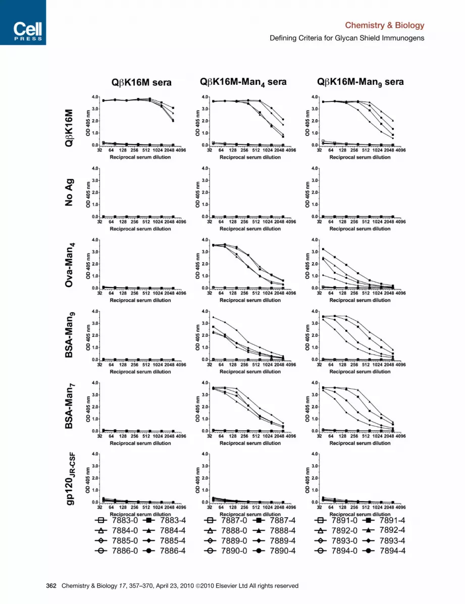
Figure 5. Representative Immunogenicity Profiles for QbK16M-Man4 a
Serum Ab titers from rabbits immunized with QbK16M virus glycoconjugates or na
ID numbers; open symbols correspond to preimmune sera and filled symbols cor
graph. Man7 corresponds to the D1/D2 motif, that is, Mana1-2Mana1-2Mana1-3
Chemistry & Biology 17,
corresponds well with the grossly similar glycan arrangements
we predicted for these scaffolds.
More precisely controlled conjugation (only at position 16 of
Qb) of Man8 was accomplished on a shorter tether (directly to
the amino acid side chain) by using QbHPG to give QbHPG-
Man8 (11). Given the poor recognition of Qbwt-Man8 (3c) by
2G12, and the lower loading of Man8 on QbHPG (�90 glycans),
it was not surprising to find QbHPG-Man8 to be a poor antigen
for 2G12 (Figure 4). However, when the rest of the platform
was ‘‘filled in’’ with Man9 residues, the combined display of
Man8 and Man9 gave rise to the most effective glycoconjugate
antigen: QbHPG-Man8/Man9 (12) bound 2G12 as well as the
best K16M and wild-type conjugates despite having approxi-
mately 25% fewer glycans (Figure 4). Such a result suggests
that shorter linkers, in addition to a threshold density of attach-
ment, may improve the presentation of high-mannose glycans
for 2G12 binding and, consequently, antigenic mimicry of the
gp120 glycan shield. The shorter linker may decrease the overall
flexibility of the glycan epitopes and thereby reduce the entropic
penalty associated with 2G12 binding. The synthesis and
binding studies of the QbHPG conjugates presented here consti-
tute the first exploration to our knowledge, albeit preliminary in
nature, of the impact of linker length and controlled combinato-
rial display.
Immunogenicity of QbK16M GlycoconjugatesBecause the QbK16M-Man4 and QbK16M-Man9 conjugates dis-
played multiple high-affinity epitopes for 2G12 with potentially
less heterogeneity compared with the corresponding Qbwt
conjugates, these constructs were tested for their immunogenic
properties in rabbits. The rabbit IgG response against QbK16M-
Man4 and QbK16M-Man9 peaked after the second immunization
and showed no further boosting effects from the third and fourth
immunizations. Preimmune and fourth-bleed sera were titrated
against the naked carrier protein QbK16M and a series of glyco-
conjugates that utilized a different linker (Figure S4) to better
measure the overall Ab response against the carbohydrate
portion of the displayed ligands (Figure 5). Overall, the anti-
mannose Abs elicited against QbK16M-Man4 reacted most
effectively with Man4, followed very closely by Man7, the D1/
D2 biantennary glycan. In contrast, the serum Abs elicited by
QbK16M-Man9 reacted most effectively with Man9 and nearly
as well with Man7, but only moderately with Man4. Similar trends
were observed when IgG titers against analogous CPMV conju-
gates were measured (Table S1). These results suggest that
immunization with QbK16M-Man4 or QbK16M-Man9 elicits
strong anti-mannose responses with distinct Ab specificities.
The polyclonal Ab response to the Man4 glycoconjugate appears
to be tailored toward recognition of the D1 structure in simpler
contexts (i.e., Man4 and Man7). The response to the Man9 glyco-
conjugate reacts better with branched structures that include
more structural elements (arms) in common with the glycan
immunogen. This may reflect the presence of Abs against
nd QbK16M-Man9
ked QbK16M particles. Symbol keys below each panel of graphs indicate rabbit
respond to fourth-bleed immune sera. Antigens are denoted on the left of each
Man(Mana1-2Mana1-3Mana1-6)Man.
357–370, April 23, 2010 ª2010 Elsevier Ltd All rights reserved 363

Chemistry & Biology
Defining Criteria for Glycan Shield Immunogens
branched structures in Man9 and/or Abs against individual arms
of Man9.
The reactivity of anti-mannose serum Abs with HIV-1 gp120
glycans was initially assessed with recombinant gp120JR-FL,
which corresponds to a primary HIV-1 isolate that is potently
neutralized by 2G12 (Binley et al., 2004). No reactivity was
observed above background levels with gp120JR-FL or with two
additional recombinant gp120s: one derived from a virus isolate,
JR-CSF, sensitive to 2G12 neutralization, and the other, YU2,
neutralized poorly by 2G12 (Binley et al., 2004; Trkola et al.,
1996). These two gp120 antigens were chosen because they
present different degrees of high-mannose glycosylation, the
gp120YU2 presumably having less high-mannose clustering
and the gp120JR-CSF having more high-mannose clustering.
Pseudovirus neutralization assays also confirmed that the anti-
mannose Abs elicited by both of the Qb glycoconjugates could
not interact with the glycan shield of HIV either in the context
of monomeric gp120 or the envelope spike (Table S1). These
results were especially striking for the QbK16M-Man9 group,
given the high titers of Man9-reactive Abs observed. The immune
sera also did not react with two other glycoproteins known to
display lower densities of high-mannose glycans (Table S1)
(Iacob et al., 2008; Joao and Dwek, 1993).
Only moderate reactivity was observed with CPMV control
particles bearing hydroxypentyl (from 5b) or single mannose
(from 5c) groups, suggesting that the majority of Abs were eli-
cited against the oligomannose structures rather than the linker
motifs, although the possibility of some linker dependency
cannot be excluded. Taken together, these results suggest
that the Qb conjugates induce anti-mannose antibodies that
recognize epitopes displayed on the synthetic neoglycoconju-
gates, but not on the mammalian and viral glycoproteins tested
here. In order to further probe the fine specificities of the anti-
body responses to these capsid glycoconjugates, the immune
serum IgGs were also analyzed on the printed glycan array
created by the Consortium for Functional Glycomics.
The serum reactivity profiles indicate that both Qb glycoconju-
gates elicit IgGs that strongly recognize synthetic fragments of
high-mannose oligosaccharides terminating in Mana1/2Man
and/or containing Mana1/3Man motifs (Figure 6). In contrast,
no substantial binding was observed with the naturally derived
N-linked high-mannose glycans, including GlcNAc2Man8 (193)
and GlcNAc2Man9 (194) (Figure 6). These results are consistent
with the ELISA results presented above, and suggest that the anti-
body specificities elicited by both Qb glycoconjugates discrimi-
nate between truncated synthetic oligomannose and full-length
N-linked high-mannose glycans containing the chitobiose core.
Dissection of the antibody response to Qb-displayed Man4
versus Man9 was also intriguing. The QbK16M-Man9 response
was much more specific, showing consistently high titers in the
sera of all animals against only Man9 (314 on the array). The spec-
ificity profiles of these rabbits against the other high-mannose
sugars varied significantly. In contrast, the individual profiles for
the QbK16M-Man4 group were very similar to one another and
to the BSA-(Man4)14 immune serum profiles described previously
(Astronomo et al., 2008) for a variety of high-mannose fragments.
Aside from the natural glycans, all immune sera in the Man9
group generally reacted poorly to a-D-Man (9) and oligomanno-
sides corresponding to internal structures shared by Man9 and
364 Chemistry & Biology 17, 357–370, April 23, 2010 ª2010 Elsevier
(GlcNAc)2Man9, namely 195, 311, and 312 (Figure 6B). These
results suggest that immunization with QbK16M-Man9 elicits
Ab specificities geared predominantly toward the terminal
Mana1/2Man motifs of branched high-mannose glycans and
not toward the internal motifs and linker. Immune sera 7891
and 7893, especially, exhibited preferential recognition of Man9
(314) over all other related synthetic derivatives including Man8
(313), which differs from Man9 only by the loss of the terminal
D2 mannose residue (Figure 6). Overall, these results suggest
that the immune system can discriminate between the terminal
structures of synthetic Man9 and GlcNAc2Man9 through differ-
ences in presentation and/or conformation. Accordingly,
synthetic Man9 seems to adopt structural presentations and/or
conformations in addition to those recognized by 2G12.
We next used competition ELISAs to determine whether the
rabbit Abs raised against our Qb conjugates recognized the
same synthetic oligomannose epitopes as 2G12. Preincubation
of immune sera with excess naked Qb particles successfully
dampened reactivity to the Qb scaffold (Figure 7A). To assess
epitope overlap, the binding of these serum IgG samples to the
Qb glycoconjugates was tested after exposure of the particles
to serially diluted 2G12, but no inhibition by 2G12 was observed
(Figure 7B). Similarly, 2G12 binding to the glycoconjugates was
also unaffected by the presence of the immune sera (Figure 7C).
These competition results suggest the existence of epitope
heterogeneity on the Qb glycoconjugates, and immunization
with these constructs elicits anti-glycan Ab responses primarily
against epitopes (i.e., structural presentations/conformations)
different from those recognized by 2G12.
The striking inability of antibodies elicited against Qb-high-
mannose conjugates to bind GlcNAc2Man9 suggests that the
presentation of the glycan might be influenced by its manner of
connection to the scaffold or the nature of that scaffold. Perhaps
the difference in flexibility between the linkers is responsible (the
synthetic oligomannose glycans being tethered to the carrier
protein particle by flexible polycarbon chains compared with
the shorter direct connection with the chitobiose core). Further-
more, or alternatively, the extensive internal hydrogen-bonding
network that maintains the overall topology of GlcNAc2Man9
(Woods et al., 1998) may not be preserved in the absence
of the chitobiose core (Shenoy et al., 2002). Both of these
scenarios would lead to differential access to various mannosyl
faces as potential epitopes, especially if the glycans (particularly
the Mana1/2Man motifs) are not closely spaced together.
However, it should be noted that immunization with synthetic
GlcNAc2Man9-based mimitopes in an attempt to elicit 2G12-
like antibodies has also failed to elicit convincing gp120 cross-
reactive Ab responses (Joyce et al., 2008; Ni et al., 2006).
Our efforts to limit the flexibility of the displayed glycans by
using carriers with densely clustered attachment sites and by
making use of the branching of Man9 itself did not give rise to
2G12-like antibody responses. It would therefore seem that the
synthetic clusters on our conjugates are all too reminiscent of
the natural glycan shield, in that the high-affinity 2G12 binding
sites are poorly immunogenic in comparison with alternative
epitopes not recognized by 2G12 (i.e., presumably less clustered
glycans), and no evidence of the elicitation of domain-exchanged
antibodies was gained. This suggests that altering the pre-
sentation of glycans on Qb to limit the display of inadequately
Ltd All rights reserved
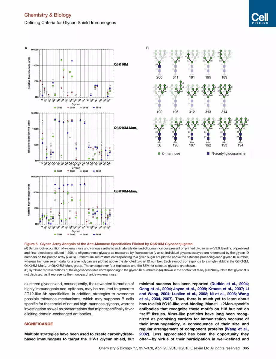
Figure 6. Glycan Array Analysis of the Anti-Mannose Specificities Elicited by QbK16M Glycoconjugates
(A) Serum IgG recognition of a-D-mannose and various synthetic and naturally derived oligomannosides present on printed glycan array V3.0. Binding of prebleed
and final-bleed sera, diluted 1:200, to oligomannose glycans as measured by fluorescence (y axis). Individual glycans assayed are referenced by the glycan ID
numbers on the printed array (x axis). Preimmune serum data corresponding to a given sugar are plotted above the asterisks preceding each glycan ID number,
whereas immune serum data for a given glycan are plotted above the denoted glycan ID number. Each symbol corresponds to a single rabbit in the QbK16M,
QbK16M-Man4, or QbK16M-Man9 group. The average over four replicates and the SEM for selected glycans are shown.
(B) Symbolic representations of the oligosaccharides corresponding to the glycan ID numbers in (A) shown in the context of Man9 (GlcNAc)2. Note that glycan 9 is
not depicted, as it represents the monosaccharide a-D-mannose.
Chemistry & Biology
Defining Criteria for Glycan Shield Immunogens
clustered glycans and, consequently, the unwanted formation of
highly immunogenic neo-epitopes, may be required to generate
2G12-like Ab specificities. In addition, strategies to overcome
possible tolerance mechanisms, which may suppress B cells
specific for the termini of natural high-mannose glycans, warrant
investigation as well as presentations that might specifically favor
eliciting domain-exchanged antibodies.
SIGNIFICANCE
Multiple strategies have been used to create carbohydrate-
based immunogens to target the HIV-1 glycan shield, but
Chemistry & Biology 17,
minimal success has been reported (Dudkin et al., 2004;
Geng et al., 2004; Joyce et al., 2008; Krauss et al., 2007; Li
and Wang, 2004; Luallen et al., 2008; Ni et al., 2006; Wang
et al., 2004, 2007). Thus, there is much yet to learn about
how to elicit 2G12-like, end-binding, Mana1/2Man-specific
antibodies that recognize these motifs on HIV but not on
‘‘self’’ tissues. Virus-like particles have long been recog-
nized as promising carriers for immunization because of
their immunogenicity, a consequence of their size and
regular arrangement of component proteins (Wang et al.,
2002). Less appreciated has been the opportunity they
offer—by virtue of their participation in well-defined and
357–370, April 23, 2010 ª2010 Elsevier Ltd All rights reserved 365

Chemistry & Biology
Defining Criteria for Glycan Shield Immunogens
366 Chemistry & Biology 17, 357–370, April 23, 2010 ª2010 Elsevier Ltd All rights reserved
Chemistry & Biology
Defining Criteria for Glycan Shield Immunogens
robust chemistry, their relatively rigid structures, and knowl-
edge of these structures at atomic resolution—to test previ-
ously unapproachable design aspects of tailored antigens.
For carbohydrates, which are typically poorly immunogenic,
such details of presentation are crucial and likely amenable
to optimization.
Immunization with QbK16M-Man4 and QbK16M-Man9
provided novel insights into the nature of this challenge.
Despite the presentation of multiple oligomannose clusters
that structurally mimic the HIV glycan shield, as judged by
their high affinity for 2G12, the Ab responses in the immu-
nized animals were directed toward alternative epitopes,
presumably associated with glycans separate from these
clusters. This immunological discrimination may stem from
differences in presentation and/or conformation between
truncated oligomannose tethered by a synthetic linker and
Asn-(GlcNAc)2-linked high-mannose glycans. These studies
strongly suggest that refinements to improve epitope homo-
geneity and/or the immunogenicity of 2G12 epitope mimics
on Qb scaffolds may be crucial for directing the immune
system to recognize the glycan shield of infectious HIV and
thus lead to effective vaccine immunogens.
EXPERIMENTAL PROCEDURES
Oligomannose-Azide Ligands
Detailed descriptions of the syntheses (except for Man7; see Supplemental
Information) and characterization of these glycans have been published previ-
ously (Calarese et al., 2005; Lee et al., 2004).
Synthesis of Glycoconjugates
CPMV-carbohydrate conjugates 5a–5f: CPMV-alkyne 2 derived from the
reaction of CPMV with NHS ester 1 was pelleted by ultracentrifugation and
resuspended in degassed 0.1 M Tris buffer (pH 8.0) in a nitrogen-filled glove
box. This solution (2 mg/ml, 21 mM in protein, 0.35 mM in particles) was treated
with 7a–7c, 8, 10, or a 1:1 mixture of 8 and 10 (0.3 mM), in each case mixed
with the components of Cu complex 6 (1 mM CuOTf + 2 mM sulfonated bath-
ophenthroline ligand), in degassed 0.1 M Tris buffer (pH 8.0), all under an inert
atmosphere. Gentle agitation on a slow rotor for 12–18 hr under nitrogen was
followed by purification of the virus-triazole conjugates as described above. All
Qb-carbohydrate conjugates were prepared in analogous fashion, using a VLP
concentration of 2 mg/ml (140 mM in protein subunits, 0.78 mM in VLPs) and
glycan-azide concentration of 0.3 mM. Note that we now recommend
a more convenient benchtop procedure involving a different Cu-binding ligand
for bioconjugation reactions such as these (Hong et al., 2009).
2G12 ELISA
Two assay formats were employed to measure binding of 2G12 to glycocon-
jugate antigens. Ab binding was visualized with p-nitrophenol phosphate
substrate (Sigma), and development was monitored at 405 nm.
Conventional Assay
Flat-bottomed microtiter plate wells (Costar type 3690; Corning) were coated
with 250 ng of antigen in PBS overnight at 4�C. Subsequent steps were done
at room temperature. The plates were washed with PBS/0.05% Tween 20
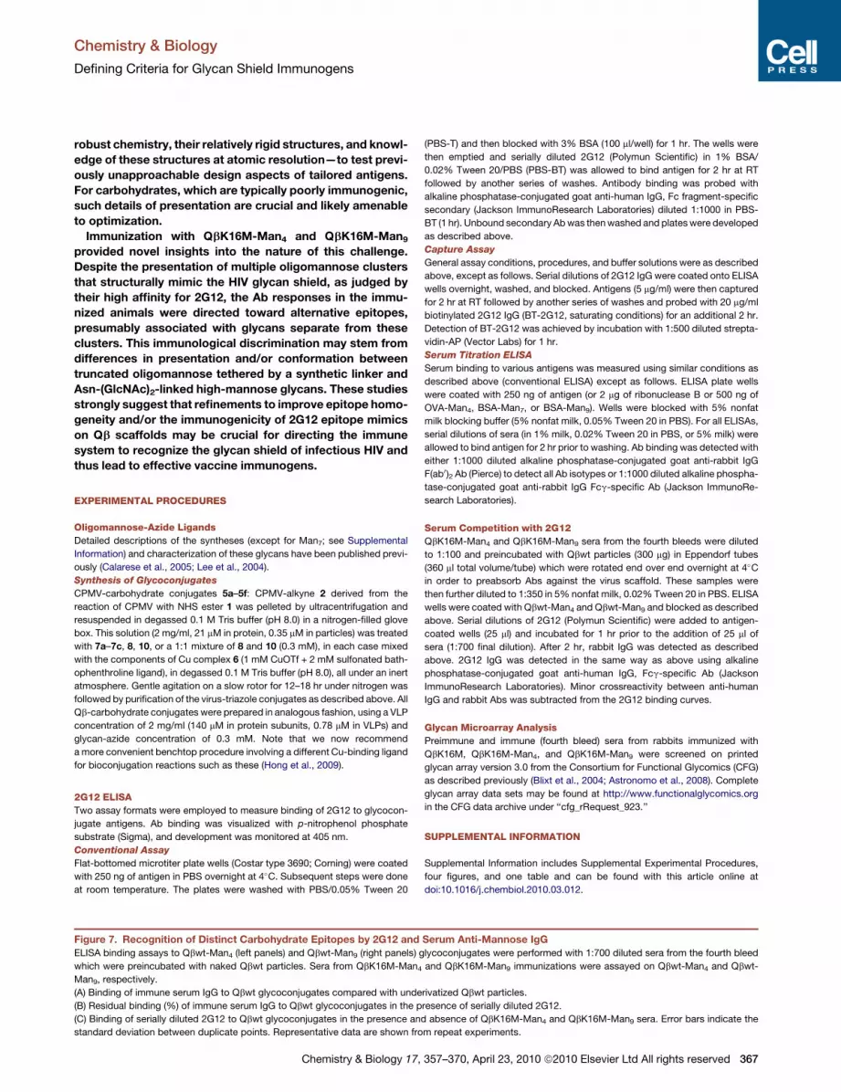
Figure 7. Recognition of Distinct Carbohydrate Epitopes by 2G12 and
ELISA binding assays to Qbwt-Man4 (left panels) and Qbwt-Man9 (right panels) g
which were preincubated with naked Qbwt particles. Sera from QbK16M-Man4
Man9, respectively.
(A) Binding of immune serum IgG to Qbwt glycoconjugates compared with unde
(B) Residual binding (%) of immune serum IgG to Qbwt glycoconjugates in the p
(C) Binding of serially diluted 2G12 to Qbwt glycoconjugates in the presence an
standard deviation between duplicate points. Representative data are shown fro
Chemistry & Biology 17,
(PBS-T) and then blocked with 3% BSA (100 ml/well) for 1 hr. The wells were
then emptied and serially diluted 2G12 (Polymun Scientific) in 1% BSA/
0.02% Tween 20/PBS (PBS-BT) was allowed to bind antigen for 2 hr at RT
followed by another series of washes. Antibody binding was probed with
alkaline phosphatase-conjugated goat anti-human IgG, Fc fragment-specific
secondary (Jackson ImmunoResearch Laboratories) diluted 1:1000 in PBS-
BT (1 hr). Unbound secondary Ab was then washed and plates were developed
as described above.
Capture Assay
General assay conditions, procedures, and buffer solutions were as described
above, except as follows. Serial dilutions of 2G12 IgG were coated onto ELISA
wells overnight, washed, and blocked. Antigens (5 mg/ml) were then captured
for 2 hr at RT followed by another series of washes and probed with 20 mg/ml
biotinylated 2G12 IgG (BT-2G12, saturating conditions) for an additional 2 hr.
Detection of BT-2G12 was achieved by incubation with 1:500 diluted strepta-
vidin-AP (Vector Labs) for 1 hr.
Serum Titration ELISA
Serum binding to various antigens was measured using similar conditions as
described above (conventional ELISA) except as follows. ELISA plate wells
were coated with 250 ng of antigen (or 2 mg of ribonuclease B or 500 ng of
OVA-Man4, BSA-Man7, or BSA-Man9). Wells were blocked with 5% nonfat
milk blocking buffer (5% nonfat milk, 0.05% Tween 20 in PBS). For all ELISAs,
serial dilutions of sera (in 1% milk, 0.02% Tween 20 in PBS, or 5% milk) were
allowed to bind antigen for 2 hr prior to washing. Ab binding was detected with
either 1:1000 diluted alkaline phosphatase-conjugated goat anti-rabbit IgG
F(ab0)2 Ab (Pierce) to detect all Ab isotypes or 1:1000 diluted alkaline phospha-
tase-conjugated goat anti-rabbit IgG Fcg-specific Ab (Jackson ImmunoRe-
search Laboratories).
Serum Competition with 2G12
QbK16M-Man4 and QbK16M-Man9 sera from the fourth bleeds were diluted
to 1:100 and preincubated with Qbwt particles (300 mg) in Eppendorf tubes
(360 ml total volume/tube) which were rotated end over end overnight at 4�C
in order to preabsorb Abs against the virus scaffold. These samples were
then further diluted to 1:350 in 5% nonfat milk, 0.02% Tween 20 in PBS. ELISA
wells were coated with Qbwt-Man4 and Qbwt-Man9 and blocked as described
above. Serial dilutions of 2G12 (Polymun Scientific) were added to antigen-
coated wells (25 ml) and incubated for 1 hr prior to the addition of 25 ml of
sera (1:700 final dilution). After 2 hr, rabbit IgG was detected as described
above. 2G12 IgG was detected in the same way as above using alkaline
phosphatase-conjugated goat anti-human IgG, Fcg-specific Ab (Jackson
ImmunoResearch Laboratories). Minor crossreactivity between anti-human
IgG and rabbit Abs was subtracted from the 2G12 binding curves.
Glycan Microarray Analysis
Preimmune and immune (fourth bleed) sera from rabbits immunized with
QbK16M, QbK16M-Man4, and QbK16M-Man9 were screened on printed
glycan array version 3.0 from the Consortium for Functional Glycomics (CFG)
as described previously (Blixt et al., 2004; Astronomo et al., 2008). Complete
glycan array data sets may be found at http://www.functionalglycomics.org
in the CFG data archive under ‘‘cfg_rRequest_923.’’
SUPPLEMENTAL INFORMATION
Supplemental Information includes Supplemental Experimental Procedures,
four figures, and one table and can be found with this article online at
doi:10.1016/j.chembiol.2010.03.012.
Serum Anti-Mannose IgG
lycoconjugates were performed with 1:700 diluted sera from the fourth bleed
and QbK16M-Man9 immunizations were assayed on Qbwt-Man4 and Qbwt-
rivatized Qbwt particles.
resence of serially diluted 2G12.
d absence of QbK16M-Man4 and QbK16M-Man9 sera. Error bars indicate the
m repeat experiments.
357–370, April 23, 2010 ª2010 Elsevier Ltd All rights reserved 367
Chemistry & Biology
Defining Criteria for Glycan Shield Immunogens
ACKNOWLEDGMENTS
We greatly appreciate the gift of 2G12 from Gabriela Stiegler and Hermann
Katinger (Polymun Scientific, Vienna, Austria; University of Natural Resources
and Applied Life Sciences, Vienna, Austria) and the glycan array resources
provided by Core D and Core H of the CFG funded by NIGMS (GM062116).
We also thank Michael Huber for critical reading of the manuscript. The
work was supported by the Natural Sciences and Engineering Research
Council of Canada (to R.D.A.), the Canadian Institutes of Health Research
(to A.K.U.), the Neutralizing Antibody Consortium of the International AIDS
Vaccine Initiative (to D.R.B.), the Skaggs Institute for Chemical Biology (to
M.G.F.), the National Institutes of Health (AI33292 and AI060425 to D.R.B.
and GM083658 to M.G.F.), and the Ragon Institute (to D.R.B. and K.J.D.).
Received: February 25, 2009
Revised: March 24, 2010
Accepted: March 25, 2010
Published: April 22, 2010
REFERENCES
Astronomo, R.D., Lee, H.K., Scanlan, C.N., Pantophlet, R., Huang, C.Y.,
Wilson, I.A., Blixt, O., Dwek, R.A., Wong, C.H., and Burton, D.R. (2008).
A glycoconjugate antigen based on the recognition motif of a broadly neutral-
izing human immunodeficiency virus antibody, 2G12, is immunogenic but
elicits antibodies unable to bind to the self glycans of gp120. J. Virol. 82,
6359–6368.
Binley, J. (2009). Specificities of broadly neutralizing anti-HIV-1 sera. Curr.
Opin. HIV AIDS 4, 364–372.
Binley, J.M., Wrin, T., Korber, B., Zwick, M.B., Wang, M., Chappey, C.,
Stiegler, G., Kunert, R., Zolla-Pazner, S., Katinger, H., et al. (2004). Compre-
hensive cross-clade neutralization analysis of a panel of anti-human immuno-
deficiency virus type 1 monoclonal antibodies. J. Virol. 78, 13232–13252.
Binley, J.M., Lybarger, E.A., Crooks, E.T., Seaman, M.S., Gray, E., Davis, K.L.,
Decker, J.M., Wycuff, D., Harris, L., Hawkins, N., et al. (2008). Profiling the
specificity of neutralizing antibodies in a large panel of plasmas from patients
chronically infected with human immunodeficiency virus type 1 subtypes B
and C. J. Virol. 82, 11651–11668.
Blixt, O., Head, S., Mondala, T., Scanlan, C., Huflejt, M.E., Alvarez, R., Bryan,
M.C., Fazio, F., Calarese, D., Stevens, J., et al. (2004). Printed covalent glycan
array for ligand profiling of diverse glycan binding proteins. Proc. Natl. Acad.
Sci. USA 101, 17033–17038.
Burton, D.R., Pyati, J., Koduri, R., Sharp, S.J., Thornton, G.B., Parren, P.W.,
Sawyer, L.S., Hendry, R.M., Dunlop, N., Nara, P.L., et al. (1994). Efficient
neutralization of primary isolates of HIV-1 by a recombinant human mono-
clonal antibody. Science 266, 1024–1027.
Calarese, D.A., Scanlan, C.N., Zwick, M.B., Deechongkit, S., Mimura, Y.,
Kunert, R., Zhu, P., Wormald, M.R., Stanfield, R.L., Roux, K.H., et al. (2003).
Antibody domain exchange is an immunological solution to carbohydrate
cluster recognition. Science 300, 2065–2071.
Calarese, D.A., Lee, H.K., Huang, C.Y., Best, M.D., Astronomo, R.D., Stanfield,
R.L., Katinger, H., Burton, D.R., Wong, C.H., and Wilson, I.A. (2005). Dissection
of the carbohydrate specificity of the broadly neutralizing anti-HIV-1 antibody
2G12. Proc. Natl. Acad. Sci. USA 102, 13372–13377.
Chatterji, A., Ochoa, W.F., Paine, M., Ratna, B.R., Johnson, J.E., and Lin, T.
(2004). New addresses on an addressable virus nanoblock: uniquely reactive
Lys residues on cowpea mosaic virus. Chem. Biol. 11, 855–863.
Chen, B., Vogan, E.M., Gong, H., Skehel, J.J., Wiley, D.C., and Harrison, S.C.
(2005). Structure of an unliganded simian immunodeficiency virus gp120 core.
Nature 433, 834–841.
Corti, D., Langedijk, J.P., Hinz, A., Seaman, M.S., Vanzetta, F., Fernandez-
Rodriguez, B.M., Silacci, C., Pinna, D., Jarrossay, D., Balla-Jhagjhoorsingh,
S., et al. (2010). Analysis of memory B cell responses and isolation of novel
monoclonal antibodies with neutralizing breadth from HIV-1-infected individ-
uals. PLoS One 5, e8805.
368 Chemistry & Biology 17, 357–370, April 23, 2010 ª2010 Elsevier
Cutalo, J.M., Deterding, L.J., and Tomer, K.B. (2004). Characterization of
glycopeptides from HIV-I(SF2) gp120 by liquid chromatography mass spec-
trometry. J. Am. Soc. Mass Spectrom. 15, 1545–1555.
Destito, G., Yeh, R., Rae, C.S., Finn, M.G., and Manchester, M. (2007). Folic
acid-mediated targeting of cowpea mosaic virus particles to tumor cells.
Chem. Biol. 14, 1152–1162.
Dhillon, A.K., Donners, H., Pantophlet, R., Johnson, W.E., Decker, J.M., Shaw,
G.M., Lee, F.H., Richman, D.D., Doms, R.W., Vanham, G., et al. (2007). Dis-
secting the neutralizing antibody specificities of broadly neutralizing sera
from human immunodeficiency virus type 1-infected donors. J. Virol. 81,
6548–6562.
Dudkin, V.Y., Orlova, M., Geng, X., Mandal, M., Olson, W.C., and Danishefsky,
S.J. (2004). Toward fully synthetic carbohydrate-based HIV antigen design: on
the critical role of bivalency. J. Am. Chem. Soc. 126, 9560–9562.
Dunlop, D.C., Ulrich, A., Appelmelk, B.J., Burton, D.R., Dwek, R.A., Zitzmann,
N., and Scanlan, C.N. (2008). Antigenic mimicry of the HIV envelope by AIDS-
associated pathogens. AIDS 22, 2214–2217.
Geng, X., Dudkin, V.Y., Mandal, M., and Danishefsky, S.J. (2004). In pursuit of
carbohydrate-based HIV vaccines, part 2: the total synthesis of high-
mannose-type gp120 fragments—evaluation of strategies directed to maximal
convergence. Angew. Chem. Int. Ed. Engl. 43, 2562–2565.
Gray, E.S., Taylor, N., Wycuff, D., Moore, P.L., Tomaras, G.D., Wibmer, C.K.,
Puren, A., DeCamp, A., Gilbert, P.B., Wood, B., et al. (2009). Antibody
specificities associated with neutralization breadth in plasma from human
immunodeficiency virus type 1 subtype C-infected blood donors. J. Virol. 83,
8925–8937.
Hessell, A.J., Rakasz, E.G., Poignard, P., Hangartner, L., Landucci, G., Forthal,
D.N., Koff, W.C., Watkins, D.I., and Burton, D.R. (2009). Broadly neutralizing
human anti-HIV antibody 2G12 is effective in protection against mucosal
SHIV challenge even at low serum neutralizing titers. PLoS Pathog. 5,
e1000433.
Hong, V., Presolski, S.I., Ma, C., and Finn, M.G. (2009). Analysis and optimiza-
tion of copper-catalyzed azide-alkyne cycloaddition for bioconjugation.
Angew. Chem. Int. Ed. Engl. 48, 9879–9883.
Iacob, R.E., Perdivara, I., Przybylski, M., and Tomer, K.B. (2008). Mass spec-
trometric characterization of glycosylation of hepatitis C virus E2 envelope
glycoprotein reveals extended microheterogeneity of N-glycans. J. Am. Soc.
Mass Spectrom. 19, 428–444.
Joao, H.C., and Dwek, R.A. (1993). Effects of glycosylation on protein structure
and dynamics in ribonuclease B and some of its individual glycoforms. Eur.
J. Biochem. 218, 239–244.
Joyce, J.G., Krauss, I.J., Song, H.C., Opalka, D.W., Grimm, K.M., Nahas, D.D.,
Esser, M.T., Hrin, R., Feng, M., Dudkin, V.Y., et al. (2008). An oligosaccharide-
based HIV-1 2G12 mimotope vaccine induces carbohydrate-specific anti-
bodies that fail to neutralize HIV-1 virions. Proc. Natl. Acad. Sci. USA 105,
15684–15689.
Kaltgrad, E., Sen Gupta, S., Punna, S., Huang, C.-Y., Chang, A., Wong, C.-H.,
Finn, M.G., and Blixt, O. (2007). Anti-carbohydrate antibodies elicited by
polyvalent display on a viral scaffold. ChemBioChem 8, 1455–1462.
Kaltgrad, E., O’Reilly, M.K., Liao, L., Han, S., Paulson, J., and Finn, M.G.
(2008). On-virus construction of polyvalent glycan ligands for cell-surface
receptors. J. Am. Chem. Soc. 130, 4578–4579.
Kozlovska, T.M., Cielens, I., Vasiljeva, I., Strelnikova, A., Kazaks, A., Dislers, A.,
Dreilina, D., Ose, V., Gusars, I., and Pumpens, P. (1996). RNA phase Q b coat
protein as a carrier for foreign epitopes. Intervirology 39, 9–15.
Krauss, I.J., Joyce, J.G., Finnefrock, A.C., Song, H.C., Dudkin, V.Y., Geng, X.,
Warren, J.D., Chastain, M., Shiver, J.W., and Danishefsky, S.J. (2007). Fully
synthetic carbohydrate HIV antigens designed on the logic of the 2G12
antibody. J. Am. Chem. Soc. 129, 11042–11044.
Kwong, P.D., Wyatt, R., Robinson, J., Sweet, R.W., Sodroski, J., and
Hendrickson, W.A. (1998). Structure of an HIV gp120 envelope glycoprotein
in complex with the CD4 receptor and a neutralizing human antibody. Nature
393, 648–659.
Ltd All rights reserved
Chemistry & Biology
Defining Criteria for Glycan Shield Immunogens
Lee, H.K., Scanlan, C.N., Huang, C.Y., Chang, A.Y., Calarese, D.A., Dwek,
R.A., Rudd, P.M., Burton, D.R., Wilson, I.A., and Wong, C.H. (2004). Reac-
tivity-based one-pot synthesis of oligomannoses: defining antigens recog-
nized by 2G12, a broadly neutralizing anti-HIV-1 antibody. Angew. Chem.
Int. Ed. Engl. 43, 1000–1003.
Lewis, W.G., Magallon, F.G., Fokin, V.V., and Finn, M.G. (2004). Discovery and
characterization of catalysts for azide-alkyne cycloaddition by fluorescence
quenching. J. Am. Chem. Soc. 126, 9152–9153.
Li, H., and Wang, L.X. (2004). Design and synthesis of a template-assembled
oligomannose cluster as an epitope mimic for human HIV-neutralizing anti-
body 2G12. Org. Biomol. Chem. 2, 483–488.
Li, Y., Svehla, K., Louder, M.K., Wycuff, D., Phogat, S., Tang, M., Migueles,
S.A., Wu, X., Phogat, A., Shaw, G.M., et al. (2009). Analysis of neutralization
specificities in polyclonal sera derived from human immunodeficiency virus
type 1-infected individuals. J. Virol. 83, 1045–1059.
Liljas, L., and Golmohammadi, R. (1996). The crystal structure of bacterio-
phage Q b at 3.5 A resolution. Structure 4, 543–554.
Lin, T., Porta, C., Lomonossoff, G., and Johnson, J.E. (1996). Structure-based
design of peptide presentation on a viral surface: the crystal structure of
a plant/animal virus chimera at 2.8 A resolution. Fold. Des. 1, 179–187.
Lin, T., Chen, Z., Usha, R., Stauffacher, C.V., Dai, J.-B., Schmidt, T., and John-
son, J.E. (1999). The refined crystal structure of cowpea mosaic virus at 2.8 A
resolution. Virology 265, 20–34.
Lomonossoff, G.P., and Hamilton, W.D.O. (1999). Cowpea mosaic virus-based
vaccines. Curr. Top. Microbiol. Immunol. 240, 177–189.
Luallen, R.J., Lin, J., Fu, H., Cai, K.K., Agrawal, C., Mboudjeka, I., Lee, F.H.,
Montefiori, D., Smith, D.F., Doms, R.W., et al. (2008). An engineered Saccha-
romyces cerevisiae strain binds the broadly neutralizing human immunodefi-
ciency virus type 1 antibody 2G12 and elicits mannose-specific gp120-binding
antibodies. J. Virol. 82, 6447–6457.
Mascola, J.R., Stiegler, G., VanCott, T.C., Katinger, H., Carpenter, C.B., Han-
son, C.E., Beary, H., Hayes, D., Frankel, S.S., Birx, D.L., et al. (2000). Protec-
tion of macaques against vaginal transmission of a pathogenic HIV-1/SIV
chimeric virus by passive infusion of neutralizing antibodies. Nat. Med. 6,
207–210.
Mehandru, S., Vcelar, B., Wrin, T., Stiegler, G., Joos, B., Mohri, H., Boden, D.,
Galovich, J., Tenner-Racz, K., Racz, P., et al. (2007). Adjunctive passive immu-
notherapy in human immunodeficiency virus type 1-infected individuals
treated with antiviral therapy during acute and early infection. J. Virol. 81,
11016–11031.
Miermont, A., Barnhill, H., Strable, E., Lu, X., Wall, K.A., Wang, Q., Finn, M.G.,
and Huang, X. (2008). Cowpea mosaic virus capsid, a promising carrier
towards the development of carbohydrate based anti-tumor vaccines.
Chem. Eur. J. 14, 4939–4947.
Ni, J., Song, H., Wang, Y., Stamatos, N.M., and Wang, L.X. (2006).
Toward a carbohydrate-based HIV-1 vaccine: synthesis and immunological
studies of oligomannose-containing glycoconjugates. Bioconjug. Chem. 17,
493–500.
Prasuhn, D.E., Jr., Yeh, R.M., Obenaus, A., Manchester, M., and Finn, M.G.
(2007). Viral MRI contrast agents: coordination of Gd by native virions and
attachment of Gd complexes by azide-alkyne cycloaddition. Chem. Commun.
(Camb), 1269–1271.
Prasuhn, D.E., Jr., Singh, P., Strable, E., Brown, S., Manchester, M., and Finn,
M.G. (2008). Plasma clearance of bacteriophage Qb particles as a function of
surface charge. J. Am. Chem. Soc. 130, 1328–1334.
Rostovtsev, V.V., Green, L.G., Fokin, V.V., and Sharpless, K.B. (2002).
A stepwise Huisgen cycloaddition process: copper(I)-catalyzed regioselective
ligation of azides and terminal alkynes. Angew. Chem. Int. Ed. Engl. 41,
2596–2599.
Sanders, R.W., Venturi, M., Schiffner, L., Kalyanaraman, R., Katinger, H.,
Lloyd, K.O., Kwong, P.D., and Moore, J.P. (2002). The mannose-dependent
epitope for neutralizing antibody 2G12 on human immunodeficiency virus
type 1 glycoprotein gp120. J. Virol. 76, 7293–7305.
Chemistry & Biology 17,
Scanlan, C.N., Pantophlet, R., Wormald, M.R., Ollmann Saphire, E., Stanfield,
R., Wilson, I.A., Katinger, H., Dwek, R.A., Rudd, P.M., and Burton, D.R. (2002).
The broadly neutralizing anti-human immunodeficiency virus type 1 antibody
2G12 recognizes a cluster of a1/2 mannose residues on the outer face of
gp120. J. Virol. 76, 7306–7321.
Scanlan, C.N., Pantophlet, R., Wormald, M.R., Saphire, E.O., Calarese, D.,
Stanfield, R., Wilson, I.A., Katinger, H., Dwek, R.A., Burton, D.R., et al.
(2003). The carbohydrate epitope of the neutralizing anti-HIV-1 antibody
2G12. Adv. Exp. Med. Biol. 535, 205–218.
Scanlan, C.N., Ritchie, G.E., Baruah, K., Crispin, M., Harvey, D.J.,
Singer, B.B., Lucka, L., Wormald, M.R., Wentworth, P., Jr., Zitzmann, N.,
et al. (2007). Inhibition of mammalian glycan biosynthesis produces non-self
antigens for a broadly neutralising, HIV-1 specific antibody. J. Mol. Biol. 372,
16–22.
Sen Gupta, S., Kuzelka, J., Singh, P., Lewis, W.G., Manchester, M., and Finn,
M.G. (2005). Accelerated bioorthogonal conjugation: a practical method for
the ligation of diverse functional molecules to a polyvalent virus scaffold.
Bioconjug. Chem. 16, 1572–1579.
Shenoy, S.R., Barrientos, L.G., Ratner, D.M., O’Keefe, B.R., Seeberger, P.H.,
Gronenborn, A.M., and Boyd, M.R. (2002). Multisite and multivalent binding
between cyanovirin-N and branched oligomannosides: calorimetric and
NMR characterization. Chem. Biol. 9, 1109–1118.
Stamatatos, L., Morris, L., Burton, D.R., and Mascola, J.R. (2009). Neutralizing
antibodies generated during natural HIV-1 infection: good news for an HIV-1
vaccine? Nat. Med. 15, 866–870.
Strable, E., Prasuhn, D.E., Jr., Udit, A.K., Brown, S., Link, A.J., Ngo, J.T.,
Lander, G., Quispe, J., Potter, C.S., Carragher, B., et al. (2008). Unnatural
amino acid incorporation into virus-like particles. Bioconjug. Chem. 19,
866–875.
Trkola, A., Pomales, A.B., Yuan, H., Korber, B., Maddon, P.J., Allaway, G.P.,
Katinger, H., Barbas, C.F. III, Burton, D.R., Ho, D.D., et al. (1995). Cross-clade
neutralization of primary isolates of human immunodeficiency virus type 1
by human monoclonal antibodies and tetrameric CD4-IgG. J. Virol. 69,
6609–6617.
Trkola, A., Purtscher, M., Muster, T., Ballaun, C., Buchacher, A., Sullivan, N.,
Srinivasan, K., Sodroski, J., Moore, J.P., and Katinger, H. (1996). Human
monoclonal antibody 2G12 defines a distinctive neutralization epitope on the
gp120 glycoprotein of human immunodeficiency virus type 1. J. Virol. 70,
1100–1108.
Trkola, A., Kuster, H., Rusert, P., Joos, B., Fischer, M., Leemann, C., Manrique,
A., Huber, M., Rehr, M., Oxenius, A., et al. (2005). Delay of HIV-1 rebound after
cessation of antiretroviral therapy through passive transfer of human neutral-
izing antibodies. Nat. Med. 11, 615–622.
Virgin, H.W., and Walker, B.D. (2010). Immunology and the elusive AIDS
vaccine. Nature 464, 224–231.
Walker, L.M., Phogat, S.K., Chan-Hui, P.Y., Wagner, D., Phung, P., Goss, J.L.,
Wrin, T., Simek, M.D., Fling, S., Mitcham, J.L., et al. (2009). Broad and potent
neutralizing antibodies from an African donor reveal a new HIV-1 vaccine
target. Science 326, 285–289.
Wang, L.X. (2006). Toward oligosaccharide- and glycopeptide-based HIV
vaccines. Curr. Opin. Drug Discov. Devel. 9, 194–206.
Wang, Q., Lin, T., Tang, L., Johnson, J.E., and Finn, M.G. (2002). Icosahedral
virus particles as addressable nanoscale building blocks. Angew. Chem. Int.
Ed. Engl. 41, 459–462.
Wang, L.X., Ni, J., Singh, S., and Li, H. (2004). Binding of high-mannose-type
oligosaccharides and synthetic oligomannose clusters to human antibody
2G12: implications for HIV-1 vaccine design. Chem. Biol. 11, 127–134.
Wang, J., Li, H., Zou, G., and Wang, L.X. (2007). Novel template-assembled
oligosaccharide clusters as epitope mimics for HIV-neutralizing antibody
2G12. Design, synthesis, and antibody binding study. Org. Biomol. Chem. 5,
1529–1540.
Wang, S.K., Liang, P.H., Astronomo, R.D., Hsu, T.L., Hsieh, S.L., Burton, D.R.,
and Wong, C.H. (2008). Targeting the carbohydrates on HIV-1: interaction of
357–370, April 23, 2010 ª2010 Elsevier Ltd All rights reserved 369

Chemistry & Biology
Defining Criteria for Glycan Shield Immunogens
oligomannose dendrons with human monoclonal antibody 2G12 and
DC-SIGN. Proc. Natl. Acad. Sci. USA 105, 3690–3695.
Woods, R.J., Pathiaseril, A., Wormald, M.R., Edge, C.J., and Dwek, R.A.
(1998). The high degree of internal flexibility observed for an oligomannose
oligosaccharide does not alter the overall topology of the molecule. Eur.
J. Biochem. 258, 372–386.
Wyatt, R., and Sodroski, J. (1998). The HIV-1 envelope glycoproteins: fuso-
gens, antigens, and immunogens. Science 280, 1884–1888.
370 Chemistry & Biology 17, 357–370, April 23, 2010 ª2010 Elsevier
Wyatt, R., Kwong, P.D., Desjardins, E., Sweet, R.W., Robinson, J., Hendrick-
son, W.A., and Sodroski, J.G. (1998). The antigenic structure of the HIV gp120
envelope glycoprotein. Nature 393, 705–711.
Zwick, M.B., Labrijn, A.F., Wang, M., Spenlehauer, C., Saphire, E.O., Binley,
J.M., Moore, J.P., Stiegler, G., Katinger, H., Burton, D.R., et al. (2001). Broadly
neutralizing antibodies targeted to the membrane-proximal external region
of human immunodeficiency virus type 1 glycoprotein gp41. J. Virol. 75,
10892–10905.
Ltd All rights reserved


