Embed Size (px)
Citation preview
Antibodies Elicited in Response to EBNA-1 May Cross-React with dsDNAPragya Yadav1,2, Hoa Tran3, Roland Ebegbe3, Paul Gottlieb2,3,4, Hui Wei4, Rita H. Lewis3, Alice Mumbey-
Wafula4, Atira Kaplan4, Elina Kholdarova4, Linda Spatz2,3,4*
1 Department of Chemistry, The City College of New York and the Graduate Center of the City University of New York, New York, New York, United States of America,
2 The Ph.D. program in Biochemistry, The City College of New York and the Graduate Center of the City University of New York, New York, New York, United States of
America, 3 The Graduate School of Biology, The City College of New York, New York, New York, United States of America, 4 Department of Microbiology and Immunology,
Sophie Davis School of Biomedical Education, The City College of New York, New York, New York, United States of America
Abstract
Background: Several genetic and environmental factors have been linked to Systemic Lupus Erythematosus (SLE). Oneenvironmental trigger that has a strong association with SLE is the Epstein Barr Virus (EBV). Our laboratory previouslydemonstrated that BALB/c mice expressing the complete EBNA-1 protein can develop antibodies to double stranded DNA(dsDNA). The present study was undertaken to understand why anti-dsDNA antibodies arise during the immune response toEBNA-1.
Methodology/Principal Findings: In this study, we demonstrated that mouse antibodies elicited in response to EBNA-1cross-react with dsDNA. First, we showed that adsorption of sera reactive with EBNA-1 and dsDNA, on dsDNA cellulosecolumns, diminished reactivity with EBNA-1. Next, we generated mononclonal antibodies (MAbs) to EBNA-1 and showed, byseveral methods, that they also reacted with dsDNA. Examination of two cross-reactive MAbs—3D4, generated in thislaboratory, and 0211, a commercial MAb—revealed that 3D4 recognizes the carboxyl region of EBNA-1, while 0211recognizes both the amino and carboxyl regions. In addition, 0211 binds moderately well to the ribonucleoprotein, Sm,which has been reported by others to elicit a cross-reactive response with EBNA-1, while 3D4 binds only weakly to Sm. Thissuggests that the epitope in the carboxyl region may be more important for cross-reactivity with dsDNA while the epitopein the amino region may be more important for cross-reactivity with Sm.
Conclusions/Significance: In conclusion, our results demonstrate that antibodies to the EBNA-1 protein cross-react withdsDNA. This study is significant because it demonstrates a direct link between the viral antigen and the development ofanti-dsDNA antibodies, which are the hallmark of SLE. Furthermore, it illustrates the crucial need to identify the epitopes inEBNA-1 responsible for this cross-reactivity so that therapeutic strategies can be designed to mask these regions from theimmune system following EBV exposure.
Citation: Yadav P, Tran H, Ebegbe R, Gottlieb P, Wei H, et al. (2011) Antibodies Elicited in Response to EBNA-1 May Cross-React with dsDNA. PLoS ONE 6(1):e14488. doi:10.1371/journal.pone.0014488
Editor: Paulo Lee Ho, Instituto Butantan, Brazil
Received May 3, 2010; Accepted December 9, 2010; Published January 4, 2011
Copyright: � 2011 Yadav et al. This is an open-access article distributed under the terms of the Creative Commons Attribution License, which permitsunrestricted use, distribution, and reproduction in any medium, provided the original author and source are credited.
Funding: This study was supported by grants SO6 GM 08168 from the National Institute of General Medical Sciences (NIGMS)/Support of Continuous ResearchExcellence (SCORE), NIH/NCRR/RCMI 5G12 RR03060 from the National Center for Research Resources Centers in Minority Institutions and the Professional StaffCongress at the City University of New York (PSC-CUNY) foundation. The funders had no role in study design, data collection and analysis, decision to publish, orpreparation of the manuscript.
Competing Interests: The authors have declared that no competing interests exist.
* E-mail: [email protected]
Introduction
Systemic Lupus Erythematosus (SLE) is a chronic autoimmune
disease characterized by the production of antibodies to double
stranded DNA (dsDNA) and ribonucleoproteins. The etiology of
SLE is unknown, although genetic and environmental causes have
been implicated. Several viruses have been linked to SLE,
however, the strongest association has been made with the
Epstein-Barr virus (EBV). EBV is a lymphotropic, dsDNA herpes
virus that infects 90–95% of adults in the United States [1].
Despite this high incidence of infection, only a small subset of
infected individuals will develop SLE [2]. Epidemiological studies
have demonstrated a higher incidence of EBV infection and
higher titers of antibodies to EBV in both young and adult lupus
patients relative to healthy individuals. James et al., observed
seroconversion (development of IgG antibodies to EBV viral
capsid antigen) in 99% of adolescent SLE patients compared to
70% of healthy adolescents and 72% of adolescents with other
rheumatic diseases [3]. In addition, they observed by PCR
analysis, the presence of EBV DNA in lymphocytes of 100% of
SLE patients tested, compared to 72% of controls. McClain et.al.
observed that antibodies to a major EBV nuclear antigen, EBNA-
1, which is continuously expressed in latently infected B cells, arose
in all pediatric SLE patients examined compared to only 69% of
healthy pediatric controls [4].
EBNA-1 is a DNA binding protein that maintains replication of
the EBV genome within infected cells. It is also required for
maintaining viral latency. Several studies suggest that exposure to
PLoS ONE | www.plosone.org 1 January 2011 | Volume 6 | Issue 1 | e14488
EBNA-1 following EBV infection, can lead to an autoimmune
response in some individuals, which may play a role in SLE disease
etiology. It has been reported that antibodies to epitopes on
EBNA-1 cross-react with epitopes on Sm, a ribonucleoprotein
complex consisting of a core of polypeptides (B/B9, D, E, F, G)
[5,6]. Sabbatini et al. demonstrated that antibodies to Sm D could
be generated in mice immunized with a Gly-Arg rich peptide
derived from the amino terminal end of EBNA-1 [7]. James et al
revealed that antibodies to Sm B/B9 could be elicited in rabbits
and mice following immunization with a proline rich peptide in
the carboxyl end of EBNA-1 (PPPGRRP) that has homology to a
proline rich region (PPPGMRPP) found in Sm [8]. In addition,
they observed that some animals subsequently developed antibod-
ies to dsDNA , which they hypothesized arose as a consequence of
epitope spreading, although this was not proven. More recently,
Poole et al showed that rabbits and mice injected with the proline
rich peptide of EBNA-1, subsequently develop antibodies to U1
ribonucleoproteins, RNP A and RNP C as a consequence of
epitope spreading [9].
Our laboratory previously reported, that BALB/c mice
immunized with an EBNA-1 expression vector that expressed
either the entire EBNA-1 protein or EBNA-1 lacking the Gly-Ala
repeat, developed antibodies to dsDNA as well as to Sm [10]. It
was assumed that the antibodies to Sm arose because of cross-
reactivity with EBNA-1 as previously reported, however, the basis
for the anti-dsDNA response was unknown. The present study was
undertaken to address this issue. Our results strikingly reveal that
many antibodies elicited in response to EBNA-1 actually cross-
react with dsDNA.
Results
Mice injected with purified recombinant EBNA-1 proteindevelop antibodies to dsDNA
We were interested in determining how anti-dsDNA antibodies
could arise in mice that develop anti-EBNA-1 antibodies upon
exposure to EBNA-1 protein. In our previous study, we generated
an anti-EBNA-1 response in mice by injecting them with an
EBNA-1 expression vector. However, not all mice developed anti-
EBNA-1 antibodies, presumably because they did not all express
an adequate concentration of the EBNA-1 protein [10]. In the
present study, in order to examine the EBNA-1 response, we
decided to inject mice with purified recombinant EBNA-1 protein
(rEBNA-1) rather than the EBNA-1 expression vector. EBNA-1
protein used for injections was prepared in our laboratory from a
baculovirus vector obtained from Lori Frappier (McMaster
University, Ontario, Canada). The rEBNA-1 protein encoded by
this vector lacks most of the Gly-Ala repeat. It has been shown that
the Gly-Ala repeat is not required for the replication, transactiva-
tion or segregation function of EBNA-1, although, it does enable
EBNA-1 to escape detection by cytotoxic CD8+ T cells [11,12].
The MW of the rEBNA-1 protein lacking the Gly-Ala repeat is
approximately 52Kda.
Five, 6 week old, female, BALB/c mice were injected with
50 mg of rEBNA-1 protein in CFA and were boosted 2 times at
weeks 3 and 9 with 25 mg of rEBNA-1 in IFA. Five age and sex
matched control BALB/c mice were immunized with CFA alone
and boosted with IFA and 5 mice were used as uninjected age
matched controls. We observed that all 5 mice injected with
rEBNA-1 developed IgG antibodies to EBNA-1 within the first 3
weeks (Figure 1A). In addition, mice developed antibodies to
dsDNA, although, the kinetics of the anti-dsDNA response lagged
behind that of the anti-EBNA-1 response suggesting that anti-
dsDNA antibodies may have developed over time as a conse-
quence of epitope spreading or somatic mutation (Figure 1B).
Some mice immunized with adjuvant only, also developed
antibodies to dsDNA but with the exception of one mouse, their
levels of anti-dsDNA antibody were never as high as that of mice
injected with rEBNA-1 in adjuvant. Intraperitoneal delivery of
CFA has been shown by others to elicit the production of
autoantibodies in mice, including the production of anti-DNA
antibodies [13]. It is extremely unlikely that the anti-dsDNA
response in rEBNA-1 injected mice was due primarily to adjuvant,
as our previous DNA based inoculation studies using EBNA-1
expression vectors in the absence of adjuvant, also elicited the
production of anti-dsDNA antibodies [10].
Week 12 sera from all 5 rEBNA-1 injected mice were serially
diluted and tested for binding to EBNA-1 (Figure 1C) and dsDNA
(Figure 1D) by ELISA. All rEBNA-1 injected mice developed high
titers of antibody to EBNA-1 and dsDNA. However, the anti-
EBNA-1 titers were higher than the anti-dsDNA titers suggesting
that either the concentration of antibodies to EBNA-1 were higher
than the concentration of antibodies to dsDNA or the affinities of
the antibodies to EBNA-1 were higher than for dsDNA. At a
dilution of 1:6400, the anti-EBNA-1 response in all mice was
greater than 3 standard deviations above the mean of similarly
diluted uninjected control mice (dotted line). At a dilution of 1:800,
the anti-dsDNA response was greater than 3 standard deviations
above the mean of uninjected control mice (dotted line). No anti-
dsDNA response was observed at a dilution of 1:6400.
To confirm the specificity of the anti-dsDNA response, week 12
sera from mice injected with rEBNA-1 were diluted 1:50 and used
to immunostain Crithidia luciliae slides. The presence of antibody
to dsDNA was indicated by binding to the kinetoplast (Figure 1E,
left panel). In contrast, sera from adjuvant immunized mice did
not reveal kinetoplast binding (Figure 1E, right panel ) indicating
that either the anti-DNA antibodies present in these mice were of
lower affinity than the anti-dsDNA antibodies obtained from
rEBNA-1 injected mice or they were not specific for dsDNA.
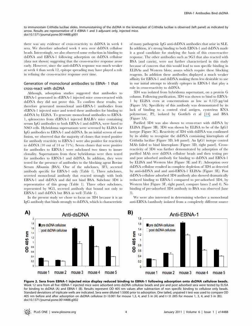
To determine whether any of the antibodies to EBNA-
1generated in rEBNA-1 injected mice also cross-reacted with
dsDNA, week 12 sera from all 5 EBNA-1 injected mice were
adsorbed over dsDNA-cellulose beads to remove dsDNA reactive
antibodies and then sera were tested by ELISA to determine if
adsorbed sera showed reduced binding to EBNA-1. Loss of
antibody in the sera due to non specific sticking to cellulose was
determined by adsorbing sera to cellulose only beads. Figure 2,
represents the OD 405 nm of anti-dsDNA (A) and anti-EBNA-1
antibody (B), pre and post adsorption onto dsDNA cellulose beads,
after the value for non specific binding to cellulose was subtracted.
We observed a significant decrease in anti-dsDNA antibody
activity following adsorption on dsDNA cellulose beads, in mouse
1, 3, 4, and 5 (p,0.001) (Figure 2A). In a similar trend, we
observed a significant decrease in anti-EBNA-1 activity in the sera
from mouse 1, 3, 4, and 5, following adsorption on dsDNA
cellulose (p,0.005) (Figure 2B). Mouse 2 showed a small decrease
in anti-dsDNA and anti-EBNA-1 activity following adsorption on
dsDNA cellulose although it was not significant. This is likely
because mouse 2 developed a negligible response to dsDNA
following injection with rEBNA-1 although the anti-EBNA-1
response was significant. The observation that a reduction of anti-
dsDNA antibody on a dsDNA cellulose column, led to a parallel
reduction of anti-EBNA-1 activity, suggests that anti-EBNA-1
antibody in the sera of some rEBNA-1 injected mice, cross-reacts
with dsDNA.
Since week 4 rEBNA-1 injected mice displayed a significant
delay in the development of a high titer anti-dsDNA but not anti-
EBNA-1 response (Figure 1B), we wanted to examine whether
EBNA-1 Antibodies Bind dsDNA
PLoS ONE | www.plosone.org 2 January 2011 | Volume 6 | Issue 1 | e14488
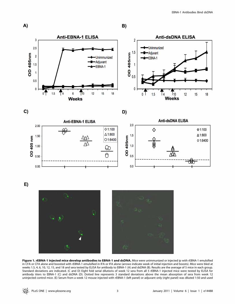
Figure 1. rEBNA-1 injected mice develop antibodies to EBNA-1 and dsDNA. Mice were unimmunized or injected ip with rEBNA-1 emulsifiedin CFA or CFA alone and boosted with rEBNA-1 emulsified in IFA or IFA alone (arrows indicate week of initial injection and boosts). Mice were bled atweeks 1.5, 4, 6, 10, 12, 15, and 18 and sera tested by ELISA for antibody to EBNA-1 (A) and dsDNA (B). Results are the average of 5 mice in each group.Standard deviations are indicated. (C and D) Eight fold serial dilutions of week 12 sera from all 5 rEBNA-1 injected mice were tested by ELISA forantibody titers to EBNA-1 (C) and dsDNA (D). Dotted line represents 3 standard deviations above the mean absorption of sera from week 12uninjected control mice. (E) Serum from a week 12 mouse injected with rEBNA-1 (left panel) or adjuvant only (right panel) was diluted 1:50 and used
EBNA-1 Antibodies Bind dsDNA
PLoS ONE | www.plosone.org 3 January 2011 | Volume 6 | Issue 1 | e14488
there was any evidence of cross-reactivity to dsDNA in week 4
sera. We therefore adsorbed week 4 sera over dsDNA cellulose
beads. Interestingly, we also observed some reduction in binding to
dsDNA and EBNA-1 following adsorption on dsDNA cellulose
(data not shown) suggesting that the cross-reactive response arose
early. However, since the anti-dsDNA response was much weaker
at week 4 than week 12, epitope spreading may have played a role
in refining the cross-reactive response over time.
Generation of monoclonal antibodies to EBNA- 1 thatcross-react with dsDNA
Although, adsorption studies suggested that antibodies to
EBNA-1 generated in rEBNA-1 injected mice cross-creacted with
dsDNA they did not prove this. To confirm these results, we
therefore generated monoclonal anti-EBNA-1 antibodies from
rEBNA-1 injected mice and tested these antibodies for binding to
dsDNA by ELISA. To generate monoclonal antibodies to EBNA-
1, splenocytes from rEBNA-1 injected BALB/c mice containing
serum IgG antibodies to both EBNA-1 and dsDNA, were fused to
NSO cells. Hybridoma supernatants were screened by ELISA for
IgG antibodies to EBNA-1 and dsDNA. In an initial screen of one
fusion, we observed that the majority of clones that tested positive
for antibody reactivity to EBNA-1 were also positive for reactivity
to dsDNA (10 out of 14 or 71%). Seven clones that were positive
for antibodies to EBNA-1 were subcloned two times to insure
clonality. Supernatants from these hybridomas were then tested
for antibodies to EBNA-1 and dsDNA. In addition, they were
tested for the presence of antibodies to the blocking agent Bovine
Serum Albumin (BSA). One of the subclones, 3F3, secreted
antibody specific for EBNA-1 only (Table 1). Three subclones,
secreted monoclonal antibody that reacted strongly with both
EBNA-1 and dsDNA and did not bind BSA. Subclone 3D4 is
representative of this group (Table 1). Three other subclones,
represented by 9G3, secreted antibody that bound not only to
EBNA-1 and dsDNA but BSA as well (Table 1).
In the present study we chose to focus on 3D4 because it is an
IgG antibody that binds strongly to dsDNA, which is characteristic
of many pathogenic IgG anti-dsDNA antibodies that arise in SLE.
In addition, it’s strong binding to both EBNA-1 and dsDNA made
it a good candidate for studying the basis of this cross-reactive
response. The other antibodies such as 9G3 that also reacted with
BSA (and casein), were not further characterized in this study
because of concern that this would lead to non specific binding in
ELISAs and Western blots; assays which require these blocking
reagents. In addition these antibodies displayed a much weaker
affinity for EBNA-1 and dsDNA making them less desirable to use
in our initial attempt to identify epitopes in EBNA-1 that play a
role in cross-reactivity to dsDNA.
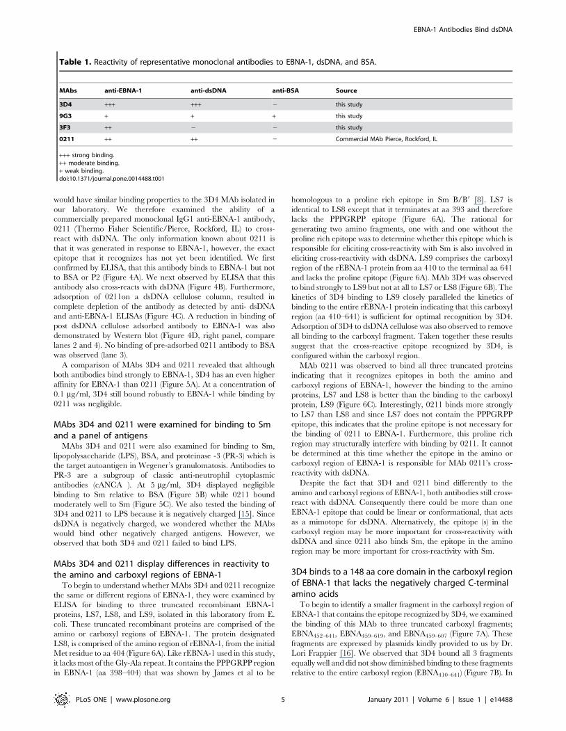
3D4 was isolated from hybridoma supernatant, on a protein G
column. Following purification, 3D4 was shown to bind to EBNA-
1 by ELISA even at concentrations as low as 0.125 mg/ml
(Figure 3A). Specificity of this antibody was demonstrated by its
lack of binding to a control viral antigen, cystovirus RNA
polymerase, P2, isolated by Gottlieb et al [14] and BSA
(Figure 3A).
Purified 3D4 was also shown to cross-react with dsDNA by
ELISA (Figure 3B). 3D4 was shown by ELISA to be of the IgG1
isotype (Figure 3C). Reactivity of 3D4 with dsDNA was confirmed
by its ability to recognize the dsDNA containing kinetoplasts of
Crithidia luciliae (Figure 3D, left panel). An IgG1 isotype control
MAb failed to bind kinetoplasts (Figure 3D, right panel). Cross-
reactivity of 3D4 was further demonstrated by adsorption of the
purified MAb over dsDNA cellulose beads and then testing pre
and post adsorbed antibody for binding to dsDNA and EBNA-1
by ELISA and Western blot (Figure 3E and F). Adsorption over
dsDNA cellulose resulted in complete depletion of 3D4 as detected
by anti-dsDNA and and anti-EBNA-1 ELISAs (Figure 3E). Post
dsDNA-cellulose adsorbed 3D4 antibody also showed dramatically
reduced binding to EBNA-1 compared to pre-adsorbed 3D4, by
Western blot (Figure 3F, right panel, compare lanes 2 and 4). No
binding of pre-adsorbed 3D4 antibody to BSA was observed (lane
3).
We were also interested in determining whether a monoclonal
anti-EBNA-1antibody isolated from a completely different source
Figure 2. Sera from EBNA-1 injected mice display reduced binding to EBNA-1 following adsorption onto dsDNA cellulose beads.Week 12 sera from all five rEBNA-1 injected mice were adsorbed onto dsDNA cellulose beads and pre and post adsorbed sera were tested by ELISAfor binding to dsDNA (A) and EBNA-1 (B). Results represent OD 405 nm values after subtraction of non specific binding to cellulose only beads.Standard deviations of triplicate wells are indicated. Sera were diluted 1:5000 prior to adsorption. One tailed, unpaired t test was used to compare OD405 nm before and after adsorption on dsDNA cellulose (t,0.001 for mouse 1,3, 4, and 5 in (A) and t,0 .005 for mouse 1, 3, 4, and 5 in (B)).doi:10.1371/journal.pone.0014488.g002
to immunostain Crithidia luciliae slides. Immunostaining of the dsDNA in the kinetoplast of Crithidia luciliae is observed (left panel) as indicated byarrow. Results are representative of 3 rEBNA-1 and 3 adjuvant only, injected mice.doi:10.1371/journal.pone.0014488.g001
EBNA-1 Antibodies Bind dsDNA
PLoS ONE | www.plosone.org 4 January 2011 | Volume 6 | Issue 1 | e14488
would have similar binding properties to the 3D4 MAb isolated in
our laboratory. We therefore examined the ability of a
commercially prepared monoclonal IgG1 anti-EBNA-1 antibody,
0211 (Thermo Fisher Scientific/Pierce, Rockford, IL) to cross-
react with dsDNA. The only information known about 0211 is
that it was generated in response to EBNA-1, however, the exact
epitope that it recognizes has not yet been identified. We first
confirmed by ELISA, that this antibody binds to EBNA-1 but not
to BSA or P2 (Figure 4A). We next observed by ELISA that this
antibody also cross-reacts with dsDNA (Figure 4B). Furthermore,
adsorption of 0211on a dsDNA cellulose column, resulted in
complete depletion of the antibody as detected by anti- dsDNA
and anti-EBNA-1 ELISAs (Figure 4C). A reduction in binding of
post dsDNA cellulose adsorbed antibody to EBNA-1 was also
demonstrated by Western blot (Figure 4D, right panel, compare
lanes 2 and 4). No binding of pre-adsorbed 0211 antibody to BSA
was observed (lane 3).
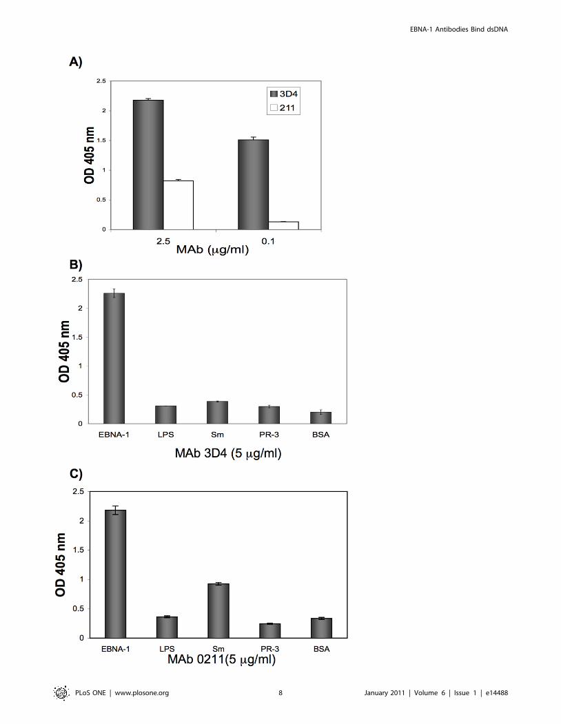
A comparison of MAbs 3D4 and 0211 revealed that although
both antibodies bind strongly to EBNA-1, 3D4 has an even higher
affinity for EBNA-1 than 0211 (Figure 5A). At a concentration of
0.1 mg/ml, 3D4 still bound robustly to EBNA-1 while binding by
0211 was negligible.
MAbs 3D4 and 0211 were examined for binding to Smand a panel of antigens
MAbs 3D4 and 0211 were also examined for binding to Sm,
lipopolysaccharide (LPS), BSA, and proteinase -3 (PR-3) which is
the target autoantigen in Wegener’s granulomatosis. Antibodies to
PR-3 are a subgroup of classic anti-neutrophil cytoplasmic
antibodies (cANCA ). At 5 mg/ml, 3D4 displayed negligible
binding to Sm relative to BSA (Figure 5B) while 0211 bound
moderately well to Sm (Figure 5C). We also tested the binding of
3D4 and 0211 to LPS because it is negatively charged [15]. Since
dsDNA is negatively charged, we wondered whether the MAbs
would bind other negatively charged antigens. However, we
observed that both 3D4 and 0211 failed to bind LPS.
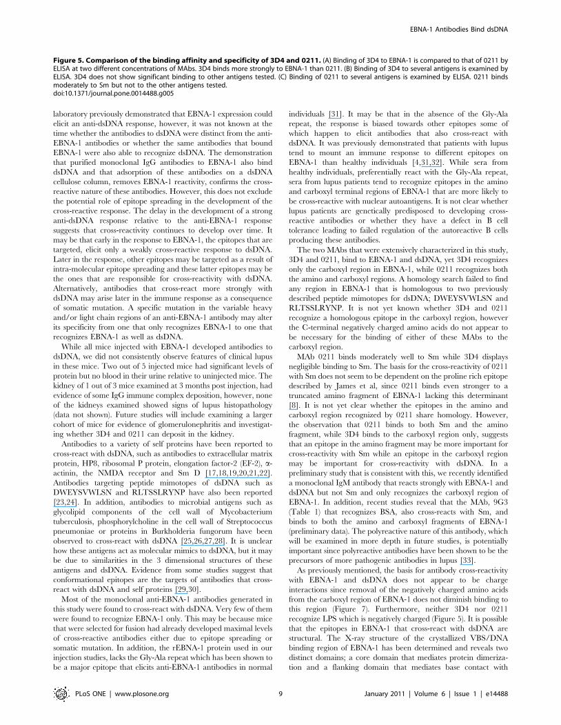
MAbs 3D4 and 0211 display differences in reactivity tothe amino and carboxyl regions of EBNA-1
To begin to understand whether MAbs 3D4 and 0211 recognize
the same or different regions of EBNA-1, they were examined by
ELISA for binding to three truncated recombinant EBNA-1
proteins, LS7, LS8, and LS9, isolated in this laboratory from E.
coli. These truncated recombinant proteins are comprised of the
amino or carboxyl regions of EBNA-1. The protein designated
LS8, is comprised of the amino region of rEBNA-1, from the initial
Met residue to aa 404 (Figure 6A). Like rEBNA-1 used in this study,
it lacks most of the Gly-Ala repeat. It contains the PPPGRPP region
in EBNA-1 (aa 398–404) that was shown by James et al to be
homologous to a proline rich epitope in Sm B/B9 [8]. LS7 is
identical to LS8 except that it terminates at aa 393 and therefore
lacks the PPPGRPP epitope (Figure 6A). The rational for
generating two amino fragments, one with and one without the
proline rich epitope was to determine whether this epitope which is
responsible for eliciting cross-reactivity with Sm is also involved in
eliciting cross-reactivity with dsDNA. LS9 comprises the carboxyl
region of the rEBNA-1 protein from aa 410 to the terminal aa 641
and lacks the proline epitope (Figure 6A). MAb 3D4 was observed
to bind strongly to LS9 but not at all to LS7 or LS8 (Figure 6B). The
kinetics of 3D4 binding to LS9 closely paralleled the kinetics of
binding to the entire rEBNA-1 protein indicating that this carboxyl
region (aa 410–641) is sufficient for optimal recognition by 3D4.
Adsorption of 3D4 to dsDNA cellulose was also observed to remove
all binding to the carboxyl fragment. Taken together these results
suggest that the cross-reactive epitope recognized by 3D4, is
configured within the carboxyl region.
MAb 0211 was observed to bind all three truncated proteins
indicating that it recognizes epitopes in both the amino and
carboxyl regions of EBNA-1, however the binding to the amino
proteins, LS7 and LS8 is better than the binding to the carboxyl
protein, LS9 (Figure 6C). Interestingly, 0211 binds more strongly
to LS7 than LS8 and since LS7 does not contain the PPPGRPP
epitope, this indicates that the proline epitope is not necessary for
the binding of 0211 to EBNA-1. Furthermore, this proline rich
region may structurally interfere with binding by 0211. It cannot
be determined at this time whether the epitope in the amino or
carboxyl region of EBNA-1 is responsible for MAb 0211’s cross-
reactivity with dsDNA.
Despite the fact that 3D4 and 0211 bind differently to the
amino and carboxyl regions of EBNA-1, both antibodies still cross-
react with dsDNA. Consequently there could be more than one
EBNA-1 epitope that could be linear or conformational, that acts
as a mimotope for dsDNA. Alternatively, the epitope (s) in the
carboxyl region may be more important for cross-reactivity with
dsDNA and since 0211 also binds Sm, the epitope in the amino
region may be more important for cross-reactivity with Sm.
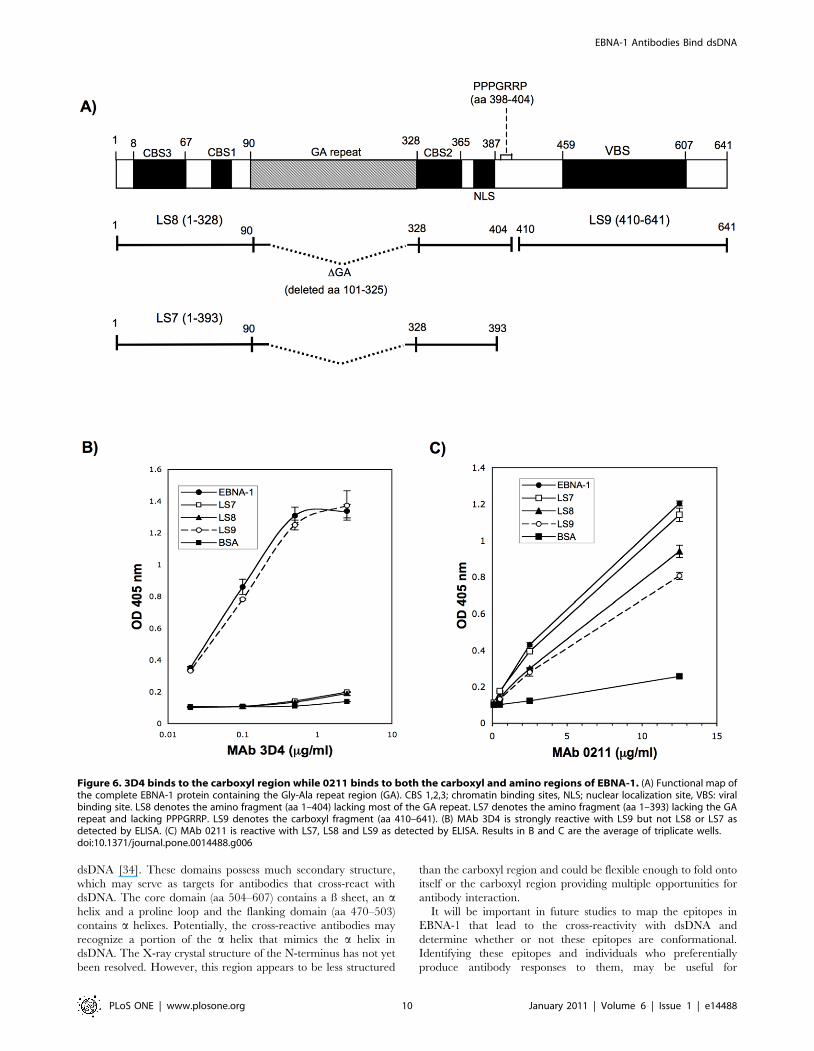
3D4 binds to a 148 aa core domain in the carboxyl regionof EBNA-1 that lacks the negatively charged C-terminalamino acids
To begin to identify a smaller fragment in the carboxyl region of
EBNA-1 that contains the epitope recognized by 3D4, we examined
the binding of this MAb to three truncated carboxyl fragments;
EBNA452–641, EBNA459–619, and EBNA459–607 (Figure 7A). These
fragments are expressed by plasmids kindly provided to us by Dr.
Lori Frappier [16]. We observed that 3D4 bound all 3 fragments
equally well and did not show diminished binding to these fragments
relative to the entire carboxyl region (EBNA410–641) (Figure 7B). In
Table 1. Reactivity of representative monoclonal antibodies to EBNA-1, dsDNA, and BSA.
MAbs anti-EBNA-1 anti-dsDNA anti-BSA Source
3D4 +++ +++ 2 this study
9G3 + + + this study
3F3 ++ 2 2 this study
0211 ++ ++ 2 Commercial MAb Pierce, Rockford, IL
+++ strong binding.++ moderate binding.+ weak binding.doi:10.1371/journal.pone.0014488.t001
EBNA-1 Antibodies Bind dsDNA
PLoS ONE | www.plosone.org 5 January 2011 | Volume 6 | Issue 1 | e14488
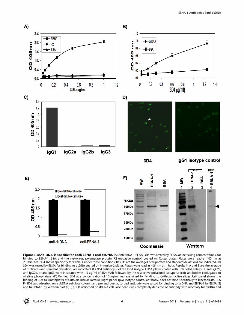
Figure 3. MAb, 3D4, is specific for both EBNA-1 and dsDNA. (A) Anti-EBNA-1 ELISA. 3D4 was tested by ELISA, at increasing concentrations, forbinding to EBNA-1, BSA, and the cystovirus, polymerase protein, P2 (negative control) coated on Costar plates. Plates were read at 405 nm at20 minutes. 3D4 shows specificity for EBNA-1 under these conditions. Results are the averages of triplicates and standard deviations are indicated. (B)3D4 was tested by ELISA for binding to dsDNA coated on Immulon-2 plates. Plates were read at 405 nm at 1 hour. Results in A and B are the averageof triplicates and standard deviations are indicated. (C) 3D4 antibody is of the IgG1 isotype. ELISA plates coated with unlabeled anti-IgG1, anti-IgG2a,anti-IgG2b, or anti-IgG3 were incubated with 1.5 mg/ml of 3D4 MAb followed by the respective polyclonal isotype specific antibodies conjugated toalkaline phosphatase. (D) Purified 3D4 at a concentration of 10 mg/ml was examined for binding to Crithidia luciliae slides. Left panel shows thebinding of 3D4 to kinetoplasts of Crithidia luciliae (arrow). Right panel; IgG1 isotype control antibody, does not bind specifically to kinetoplasts. (E &F) 3D4 was adsorbed on a dsDNA cellulose column and pre and post adsorbed antibody were tested for binding to dsDNA and EBNA-1 by ELISA (E)and to EBNA-1 by Western blot (F). (E) 3D4 adsorbed on dsDNA cellulose beads was completely depleted of antibody with reactivity for dsDNA and
EBNA-1 Antibodies Bind dsDNA
PLoS ONE | www.plosone.org 6 January 2011 | Volume 6 | Issue 1 | e14488
fact 3D4 displayed optimal binding to the smallest fragment,
EBNA459–607 suggesting that the cross-reactive epitope lies within
this 148 aa region. The carboxyl region of EBNA-1 has a net
negative charge due to the high frequency of negatively charged
amino acids at the C-terminus (aa 619–641). Twelve out of 22 of the
C terminal amino acids are either glutamic or aspartic acid. Both,
EBNA459–619, and EBNA459–607 lack these negatively charged
amino acids. Since removal of these negatively charged amino acids
did not diminish recognition by 3D4, this suggests that charge
interaction is not the basis for 3D4’s binding to EBNA-1. MAb,
0211 displays a similar binding pattern to the truncated carboxyl
fragments of EBNA-1 with maximal binding to the two smallest
fragments EBNA459–619 and EBNA459–607 (data not shown).
Discussion
This study demonstrates for the first time, that some antibodies
that arise in response to EBNA-1 cross-react with dsDNA. Our
Figure 4. Commercial MAb, 0211 cross-reacts with dsDNA. (A) MAb 0211 binds to EBNA-1 as detected by ELISA but not P2, or BSA. (B) MAb0211 binds to dsDNA as detected by ELISA. (C & D) MAb 0211 was adsorbed onto dsDNA cellulose beads and pre and post adsorbed antibody weretested for binding to dsDNA and EBNA-1 by ELISA. (C) MAb 0211 adsorbed on dsDNA cellulose beads, was completely depleted of antibody reactivityfor dsDNA and EBNA-1. Anti-dsDNA and anti-EBNA-1 ELISAs were performed on separate ELISA plates and ELISAs were developed when ODs reachedmaximal values. Results represent OD 405 nm values after subtraction of non specific binding to cellulose only beads. (D) Post dsDNA celluloseadsorbed MAb 0211 shows reduced binding to rEBNA-1 by Western blot. Left panel: Coomassie stained polyacrylamide gel. Right panel: Western blot:filters were immunostained with pre (lanes 2 and 3) or post dsDNA cellulose adsorbed 0211 (lane 4) as indicated.doi:10.1371/journal.pone.0014488.g004
EBNA-1 as detected by ELISA. Results represent OD 405 nm values after subtraction of non specific binding to cellulose only beads. Standarddeviations of triplicate wells are indicated. Anti-dsDNA and anti-EBNA-1 ELISAs were performed on Immulon-2 and Costar plates respectively andELISAs were developed when ODs on each plate reached maximal values. (F) Post dsDNA cellulose adsorbed 3D4 shows reduced binding to EBNA-1by Western blot. Left panel: Coomassie stained polyacrylamide gel. Right panel: Western blot: filters were immunostained with pre (lanes 2 and 3) orpost dsDNA cellulose adsorbed 3D4 MAb (lane 4). Molecular weight markers used in Western blot were conjugated to strep-tag and were detectedwith Strep-Tactin-HRP.doi:10.1371/journal.pone.0014488.g003
EBNA-1 Antibodies Bind dsDNA
PLoS ONE | www.plosone.org 7 January 2011 | Volume 6 | Issue 1 | e14488
EBNA-1 Antibodies Bind dsDNA
PLoS ONE | www.plosone.org 8 January 2011 | Volume 6 | Issue 1 | e14488
laboratory previously demonstrated that EBNA-1 expression could
elicit an anti-dsDNA response, however, it was not known at the
time whether the antibodies to dsDNA were distinct from the anti-
EBNA-1 antibodies or whether the same antibodies that bound
EBNA-1 were also able to recognize dsDNA. The demonstration
that purified monoclonal IgG antibodies to EBNA-1 also bind
dsDNA and that adsorption of these antibodies on a dsDNA
cellulose column, removes EBNA-1 reactivity, confirms the cross-
reactive nature of these antibodies. However, this does not exclude
the potential role of epitope spreading in the development of the
cross-reactive response. The delay in the development of a strong
anti-dsDNA response relative to the anti-EBNA-1 response
suggests that cross-reactivity continues to develop over time. It
may be that early in the response to EBNA-1, the epitopes that are
targeted, elicit only a weakly cross-reactive response to dsDNA.
Later in the response, other epitopes may be targeted as a result of
intra-molecular epitope spreading and these latter epitopes may be
the ones that are responsible for cross-reactivity with dsDNA.
Alternatively, antibodies that cross-react more strongly with
dsDNA may arise later in the immune response as a consequence
of somatic mutation. A specific mutation in the variable heavy
and/or light chain regions of an anti-EBNA-1 antibody may alter
its specificity from one that only recognizes EBNA-1 to one that
recognizes EBNA-1 as well as dsDNA.
While all mice injected with EBNA-1 developed antibodies to
dsDNA, we did not consistently observe features of clinical lupus
in these mice. Two out of 5 injected mice had significant levels of
protein but no blood in their urine relative to uninjected mice. The
kidney of 1 out of 3 mice examined at 3 months post injection, had
evidence of some IgG immune complex deposition, however, none
of the kidneys examined showed signs of lupus histopathology
(data not shown). Future studies will include examining a larger
cohort of mice for evidence of glomerulonephritis and investigat-
ing whether 3D4 and 0211 can deposit in the kidney.
Antibodies to a variety of self proteins have been reported to
cross-react with dsDNA, such as antibodies to extracellular matrix
protein, HP8, ribosomal P protein, elongation factor-2 (EF-2), a-
actinin, the NMDA receptor and Sm D [17,18,19,20,21,22].
Antibodies targeting peptide mimotopes of dsDNA such as
DWEYSVWLSN and RLTSSLRYNP have also been reported
[23,24]. In addition, antibodies to microbial antigens such as
glycolipid components of the cell wall of Mycobacterium
tuberculosis, phosphorylcholine in the cell wall of Streptococcus
pneumoniae or proteins in Burkholderia fungorum have been
observed to cross-react with dsDNA [25,26,27,28]. It is unclear
how these antigens act as molecular mimics to dsDNA, but it may
be due to similarities in the 3 dimensional structures of these
antigens and dsDNA. Evidence from some studies suggest that
conformational epitopes are the targets of antibodies that cross-
react with dsDNA and self proteins [29,30].
Most of the monoclonal anti-EBNA-1 antibodies generated in
this study were found to cross-react with dsDNA. Very few of them
were found to recognize EBNA-1 only. This may be because mice
that were selected for fusion had already developed maximal levels
of cross-reactive antibodies either due to epitope spreading or
somatic mutation. In addition, the rEBNA-1 protein used in our
injection studies, lacks the Gly-Ala repeat which has been shown to
be a major epitope that elicits anti-EBNA-1 antibodies in normal
individuals [31]. It may be that in the absence of the Gly-Ala
repeat, the response is biased towards other epitopes some of
which happen to elicit antibodies that also cross-react with
dsDNA. It was previously demonstrated that patients with lupus
tend to mount an immune response to different epitopes on
EBNA-1 than healthy individuals [4,31,32]. While sera from
healthy individuals, preferentially react with the Gly-Ala repeat,
sera from lupus patients tend to recognize epitopes in the amino
and carboxyl terminal regions of EBNA-1 that are more likely to
be cross-reactive with nuclear autoantigens. It is not clear whether
lupus patients are genetically predisposed to developing cross-
reactive antibodies or whether they have a defect in B cell
tolerance leading to failed regulation of the autoreactive B cells
producing these antibodies.
The two MAbs that were extensively characterized in this study,
3D4 and 0211, bind to EBNA-1 and dsDNA, yet 3D4 recognizes
only the carboxyl region in EBNA-1, while 0211 recognizes both
the amino and carboxyl regions. A homology search failed to find
any region in EBNA-1 that is homologous to two previously
described peptide mimotopes for dsDNA; DWEYSVWLSN and
RLTSSLRYNP. It is not yet known whether 3D4 and 0211
recognize a homologous epitope in the carboxyl region, however
the C-terminal negatively charged amino acids do not appear to
be necessary for the binding of either of these MAbs to the
carboxyl region.
MAb 0211 binds moderately well to Sm while 3D4 displays
negligible binding to Sm. The basis for the cross-reactivity of 0211
with Sm does not seem to be dependent on the proline rich epitope
described by James et al, since 0211 binds even stronger to a
truncated amino fragment of EBNA-1 lacking this determinant
[8]. It is not yet clear whether the epitopes in the amino and
carboxyl region recognized by 0211 share homology. However,
the observation that 0211 binds to both Sm and the amino
fragment, while 3D4 binds to the carboxyl region only, suggests
that an epitope in the amino fragment may be more important for
cross-reactivity with Sm while an epitope in the carboxyl region
may be important for cross-reactivity with dsDNA. In a
preliminary study that is consistent with this, we recently identified
a monoclonal IgM antibody that reacts strongly with EBNA-1 and
dsDNA but not Sm and only recognizes the carboxyl region of
EBNA-1. In addition, recent studies reveal that the MAb, 9G3
(Table 1) that recognizes BSA, also cross-reacts with Sm, and
binds to both the amino and carboxyl fragments of EBNA-1
(preliminary data). The polyreactive nature of this antibody, which
will be examined in more depth in future studies, is potentially
important since polyreactive antibodies have been shown to be the
precursors of more pathogenic antibodies in lupus [33].
As previously mentioned, the basis for antibody cross-reactivity
with EBNA-1 and dsDNA does not appear to be charge
interactions since removal of the negatively charged amino acids
from the carboxyl region of EBNA-1 does not diminish binding to
this region (Figure 7). Furthermore, neither 3D4 nor 0211
recognize LPS which is negatively charged (Figure 5). It is possible
that the epitopes in EBNA-1 that cross-react with dsDNA are
structural. The X-ray structure of the crystallized VBS/DNA
binding region of EBNA-1 has been determined and reveals two
distinct domains; a core domain that mediates protein dimeriza-
tion and a flanking domain that mediates base contact with
Figure 5. Comparison of the binding affinity and specificity of 3D4 and 0211. (A) Binding of 3D4 to EBNA-1 is compared to that of 0211 byELISA at two different concentrations of MAbs. 3D4 binds more strongly to EBNA-1 than 0211. (B) Binding of 3D4 to several antigens is examined byELISA. 3D4 does not show significant binding to other antigens tested. (C) Binding of 0211 to several antigens is examined by ELISA. 0211 bindsmoderately to Sm but not to the other antigens tested.doi:10.1371/journal.pone.0014488.g005
EBNA-1 Antibodies Bind dsDNA
PLoS ONE | www.plosone.org 9 January 2011 | Volume 6 | Issue 1 | e14488
dsDNA [34]. These domains possess much secondary structure,
which may serve as targets for antibodies that cross-react with
dsDNA. The core domain (aa 504–607) contains a ß sheet, an ahelix and a proline loop and the flanking domain (aa 470–503)
contains a helixes. Potentially, the cross-reactive antibodies may
recognize a portion of the a helix that mimics the a helix in
dsDNA. The X-ray crystal structure of the N-terminus has not yet
been resolved. However, this region appears to be less structured
than the carboxyl region and could be flexible enough to fold onto
itself or the carboxyl region providing multiple opportunities for
antibody interaction.
It will be important in future studies to map the epitopes in
EBNA-1 that lead to the cross-reactivity with dsDNA and
determine whether or not these epitopes are conformational.
Identifying these epitopes and individuals who preferentially
produce antibody responses to them, may be useful for
Figure 6. 3D4 binds to the carboxyl region while 0211 binds to both the carboxyl and amino regions of EBNA-1. (A) Functional map ofthe complete EBNA-1 protein containing the Gly-Ala repeat region (GA). CBS 1,2,3; chromatin binding sites, NLS; nuclear localization site, VBS: viralbinding site. LS8 denotes the amino fragment (aa 1–404) lacking most of the GA repeat. LS7 denotes the amino fragment (aa 1–393) lacking the GArepeat and lacking PPPGRRP. LS9 denotes the carboxyl fragment (aa 410–641). (B) MAb 3D4 is strongly reactive with LS9 but not LS8 or LS7 asdetected by ELISA. (C) MAb 0211 is reactive with LS7, LS8 and LS9 as detected by ELISA. Results in B and C are the average of triplicate wells.doi:10.1371/journal.pone.0014488.g006
EBNA-1 Antibodies Bind dsDNA
PLoS ONE | www.plosone.org 10 January 2011 | Volume 6 | Issue 1 | e14488
determining those who are at risk of developing lupus so that early
treatment strategies can be initiated. In addition, knowledge of
these epitopes may help in the design of therapeutic strategies that
can mask these epitopes thereby preventing the immune system
from mounting a cross-reactive response to them.
Materials and Methods
All animals were handled in strict accordance with good animal
practice as defined by federal and state policies set forth by The
Public Health Service Policy on the Humane Care and Use of
Laboratory Animals (PHS 1986), The Guide for the Care and Use
of Laboratory Animals (ILAR 1996), and The USDA Animal
Welfare Act (CFR 1985). All work done with animals in this study,
was approved by The Institutional Animal Care and Use
Committee (IACUC) at The City College of New York, (approval
numbers 626 and 828).
Extraction, Purification and Characterization of rEBNA-1lacking the Gly-Ala repeat
The EBNA-1 baculovirus expression vector used in this study
was a generous gift from Dr. Lori Frappier (McMaster University,
Ontario, Canada). Recombinant EBNA-1 protein (rEBNA-1) was
isolated from this baculovirus expression vector according to Lori
Frappier (personal communication and modifications of Frappier
and O’Donnell) [35]. This vector encodes an EBNA-1 protein that
has a deletion of most of the Gly-Ala repeat and has a 66His tag
on the N-terminus, which allows for the protein’s isolation on a
Ni2+ metal affinity column. Briefly, SF9 cells were grown in serum-
free insect cell culture medium, Sf-900 II SFM (Invitrogen,
Carlsbad, CA) at 27uC. Cells were resuspended at a concentration
of 16106 cells per ml and 100 ml of cells (1006106 cells total) were
infected with 500 ml of high titer recombinant EBNA-1 baculo-
virus and grown in 500 ml Erlenmeyer flasks (Corning, Acton,
MA) at 27uC in an air shaker for 60 hours. The cells were then
harvested by centrifuging at 2000 rpm at 4uC for 10 minutes. The
cell pellets were resuspended in 25 ml of a hypotonic buffer
(20 mM HEPES pH 7.8, 1 mM MgCl2, 1 mM PMSF and 10 mM
leupeptin) and allowed to swell on ice for 10 minutes. Cells were
then dounced 20 times on ice and centrifuged at 4uC at 3000 rpm
for 10 minutes. Supernatant was discarded. The pellet containing
intact nuclei was resuspended in 25 ml of hypotonic buffer
containing 2.7 ml of 5 M NaCl. After douncing on ice to open the
nuclear envelope, the fraction was centrifuged at 18,000 rpm for
20 minutes and the supernatant containing rEBNA-1 protein was
collected. Further purification of rEBNA-1 was performed
employing a nickel agarose (Ni2+-NTA) (QIAGEN, Valencia,
CA) column according to modifications of Ceccarelli and Frappier
[12]. Ni2+-NTA agarose (1ml) was equilibrated in column buffer
(0.2 M Hepes pH 7.8, 0.5 M NaCl, 10% glycerol) containing
5 mM imidazole, at room temperature. The nuclear extract was
incubated with pre-equilibrated Ni2+-NTA at room temperature
for 2 hours, with rocking. After incubation, a column was packed
with the nuclear extract/Ni2+-NTA slurry. The column was
washed slowly with column buffer containing 5 mM imidazole
followed by column buffer containing 25 mM imidazole. Next, the
EBNA-1 protein was eluted with column buffer containing
300 mM imidazole. The protein was then concentrated and the
buffer exchanged with PBS, 250 mM NaCl using an Amicon
Centrifugal filter (10,000 molecular weight cut off) (Millipore,
Billerica, MA). The protein was then resolved by 12% SDS-PAGE
followed by a Western blot and immunostaining with a
monoclonal antibody to EBNA-1.
Injection of mice with rEBNA-1 proteinFifteen, six week old, female BALB/c mice were used for
injection studies. Five mice were injected intraperitoneally (ip) with
50 mg of rEBNA-1 protein in complete Freund’s adjuvant (CFA)
(Sigma, St Louis, MO) in a 1:1 (v/v) ratio and boosted twice (at
weeks 3 and 9) with 25 mg of rEBNA-1 in incomplete Freund’s
adjuvant (IFA). Five mice were injected with CFA only and
boosted with IFA and 5 age-matched control mice remained
uninjected throughout the study. The mice were bled immediately
before injection and at weeks 1.5, 4, 6, 10, 12, 15 and 18. The sera
obtained, from these mice were tested for anti-EBNA-1 and anti-
dsDNA antibodies by ELISA.
Figure 7. 3D4 recognizes a 148 aa core domain in the carboxyl region of EBNA-1. (A) Map of the carboxyl region of EBNA-1 and 3truncated carboxyl fragments. (B) 3D4 was tested by ELISA for binding to the 3 truncated fragments of EBNA-1. 3D4 binds strongly to all 3 fragments,the smallest being EBNA459–607.doi:10.1371/journal.pone.0014488.g007
EBNA-1 Antibodies Bind dsDNA
PLoS ONE | www.plosone.org 11 January 2011 | Volume 6 | Issue 1 | e14488
Construction of plasmids encoding the amino andcarboxyl regions of EBNA-1
Truncated EBNA-1 proteins were isolated from plasmid
transformed E. coli cells. The pLS8 expression plasmid carries
the encoding sequence for the amino terminus of the EBNA-1
antigen, from the initial Met residue to amino acid position 404
and lacks virtually all of the Gly-Ala repeat. It was prepared by
PCR amplification of the EBNA-1 gene from pMRC72 [10]
which contains the EBNA-1 coding sequence, but lacks the Gly-
Ala repeat, using the following primer pair; EBV7, 5 –
CATATGTCTGACGAGGGGC CAGGT-39 (forward primer)
and EBV6, 59-CTCGAGTTATGGCCTTCTACCTGG-39 (re-
verse primer). The pLS7 expression plasmid also carries the
encoding sequence for the amino terminus of the EBNA-1 protein,
from the initial Met residue but it terminates at amino acid
position 393. Like pLS8, it lacks most of the Gly-Ala repeat.
However, unlike pLS8, it is missing the PPPGRRP epitope (aa
398–404). It was prepared from pMRC72 using the following
primer pair; EBV7 (see above) and EBV5, 59 CTCGAGTTAA-
GACCCGGAT GATGA 39 (reverse primer). The pLS9 expres-
sion plasmid carries the EBNA-1 encoding sequence for the
carboxyl terminus of EBNA-1 from amino acids 410 to 641. It was
also prepared by PCR amplification of the EBNA-1 gene from
pMRC72 using the following primer pair; EBV3, 59-CAT-
ATGGGGGAA GCCGATTA TTTTGAAT-39 (forward primer)
and EBV 4, 59-CTCGAGTTACTCCTGCCCTTCCTC-39 (re-
verse primer). The PCR amplifications were performed for 30
cycles. The amino and carboxyl PCR fragments were digested
with Nde1 and Xho1 and inserted into the pET28A expression
vector (Novagen, San Diego, CA) which contains an N-terminal
66His tag.
Isolation of truncated recombinant EBNA-1 proteinsE. coli colonies transformed with pLS7, pLS8, or pLS9 (see
above) were selected on LB ampicillin plates and grown at 37uC in
50 ml of LB media containing 1% glucose. Cultures were diluted
in 490 ml LB with 0.1 mM IPTG and grown for several hours at
20uC to a final OD600 of approximately 0.6. Cultures were
harvested and re-suspended in lysis buffer (50 mM Tris-Hcl,
pH 7.8, 250 mM NaCl) containing 1.0 mM PMSF. Cells were
sonicated for 15 minutes on ice with a 4 second on pulse,
6 seconds off at a 30% amplitude. The cell lysate was cleared by
centrifugation at 10,000 rpm for 30 minutes at 4uC and filtered
through a 45 mM filter. Five mls of Ni2+-NTA beads equilibrated
with lysis buffer were added to the cleared supernatant and
incubated with gentle rocking at room temperature. The beads
(bound to the recombinant protein) were separated from the
supernatant by low- speed centrifugation. They were then washed
6 times with wash buffer (50 mM Tris-HCl, ph 7.8, 250 mM
Nacl, 60 mM imidazol, and 10% glycerol). Two ml of elution
buffer (50 mM Tris-Hcl, pH 7.8, 250 mM NaCl, 250 mM
imidazol, 10% glycerol) were added to the beads and beads were
rocked for 15 minutes. The beads were removed from the reaction
by low-speed centrifugation. Supernatants containing the recom-
binant protein were concentrated and the buffer was exchanged
with PBS, 250 mM NaCl using an Amicon Centrifugal filter.
Proteins were analyzed by SDS-PAGE and Western Blot.
Plasmids (vector pET15b) expressing the following EBNA-1 amino
acid sequences; EBNA452–641, EBNA459–607, and EBNA459–619 were
gifts from Dr. Lori Frappier [16]. Soluble truncated EBNA-1 proteins
were produced in Escherichia coli strain BL21 (DE3) and isolated
from cell-lysates. Proteins were then purified over a Ni-NTA agarose
column as described above. Proteins were analyzed by SDS-PAGE
and Western Blot.
ELISAsDetection of antibodies to EBNA-1, dsDNA, Sm, LPS,
Proteinase 3 and BSA. Diluted serum samples from EBNA-1
injected mice, hybridoma supernatants, or purified monoclonal
antibodies were tested for binding to EBNA-1, dsDNA, Sm, LPS,
or PR-3 by ELISA as previously described [10,36]. For the
detection of antibodies to EBNA-1, LPS, Proteinase 3, and BSA,
Costar plates (Corning Incorporated, Corning, NY) were coated in
PBS with 2.0–5.0 mg/ml of antigen. Costar plates were coated
overnight with 5.0 mg/ml of Sm (Immunovision, Springdale, AR)
in 0.1M carbonate buffer for the detection of antibodies to Sm.
For the detection of antibodies to dsDNA, Immulon-2 plates
(Dynatech Laboratories, Inc., Chantilly, VA) were coated with
100 mg/ml of calf thymus dsDNA.
Detection of antibody binding to truncated amino or
carboxyl fragments of EBNA-1. Purified monoclonal anti-
bodies were tested for binding to truncated amino (LS7 and LS8)
and carboxyl regions (LS9, EBNA452–641, EBNA459–619, and
EBNA459–607) of EBNA-1. ELISA plates were coated with
2.0 mg/ml of the purified, truncated recombinant proteins
isolated in this laboratory. Subsequent steps in the ELISA were
performed according to Sundar et al [10].
Isotype ELISA. ELISA plates were coated with 50 ml of a
1:1000 dilution of either unlabeled goat anti-mouse IgG1, IgG2a,
IgG2b or IgG3 (Southern Biotech, Birmingham, Alabama) and
incubated at 37uC for one hour and overnight at 4uC. Monoclonal
3D4 antibody was diluted to 1.5 mg/ml and incubated on the plate
for one hour at 37uC. Next, 50 ml of a 1:1000 dilution of goat anti-
mouse IgG1 conjugated to alkaline phosphatase (AP), anti-IgG2a-
AP, anti-IgG2b-AP, or anti-IgG3-AP (Southern Biotech) was
added to wells coated with unlabeled anti-IgG1, anti-IgG2a, anti-
IgG2b, or anti- IgG3 respectively. Color development was
measured following the addition of 4-nitrophenyl-phosphate
disodium salt as substrate and plates were read at 405 nm on a
Titertek Multiscan ELISA plate reader.
Quantitative ELISA. A quantitative ELISA was performed,
as previously described, to determine the concentration of purified
monoclonal IgG antibodies in hybridoma supernatants [36].
Briefly, ELISA plates were coated overnight with 1.0 mg/well of
goat anti-mouse IgG antibody (Southern Biotechnology). A com-
mercial mouse monoclonal IgG antibody (Sigma) was serially
diluted, beginning at a concentration of 200 ng/ml and used to
generate a standard curve. Serial dilutions of monoclonal antibody
purified in this laboratory were applied to the anti-IgG coated
wells and the concentration of antibody was extrapolated from the
standard curve. Monoclonal antibodies were detected with goat
anti-mouse IgG antibody conjugated to AP followed by the
addition of 4-nitrophenyl-phosphate disodium salt as substrate.
Crithidia AssayReady to use Crithidia slides from the CrithiDNA Anti-nDNA
Antibody Test Kit from Antibodies Inc. (Davis, CA), were
immunostained either with mouse sera from EBNA-1 injected
mice, diluted 1/50 or with purified monoclonal antibody diluted
to 10 mg/ml. Slides were incubated in a moist, dark chamber for
30 minutes at room temperature (RT). A positive control anti-
dsDNA antibody was provided with the kit. A nonspecific
monoclonal mouse IgG1 antibody was used as an isotype control
(Sigma). Next, the slides were extensively washed with PBS and
immunostained for 30 minutes at RT with a 1:250 dilution of
biotinylated goat anti mouse IgG (Southern Biotech). This was
followed by 20 ml of a 1:500 dilution of Streptavidin-FITC
(Southern Biotech) for 30 minutes at RT. Slides were washed
again and Prolong Gold Antifade, (Invitrogen, Carlsbad, CA) was
EBNA-1 Antibodies Bind dsDNA
PLoS ONE | www.plosone.org 12 January 2011 | Volume 6 | Issue 1 | e14488
added prior to examination by fluorescence microscopy using a
Nikon Eclipse microscope, model, TE 2000-S at a magnification
of 4006.
Western BlotProteins were analyzed by SDS-PAGE on a 12% gel and
transferred to a nitrocellulose membrane using a Bio-Rad wet
transfer apparatus (BioRad, Hercules, CA). After transfer, the
membranes were blocked with 3% Milk-PBS for one hour at RT
with shaking. The blot was incubated overnight at 4uC with a
MAb generated in our laboratory (3D4), diluted to 1 mg/ml or a
commercially prepared MAb, 0211 (Thermo Fisher Scientific/
Pierce, Rockford, IL) diluted to 10 mg/ml according to the
manufacturers protocol. The membrane was washed 6 times in
wash buffer (PBS, 0.05% Tween-20). Bound MAbs antibodies
were detected with HRP-conjugated goat anti mouse IgG
(Southern Biotech) diluted 1:20,000, followed by chemilumines-
cence using the Pierce ECL kit according to the manufacturers
protocol (Pierce, Rockford, IL). Molecular weight markers
conjugated to strep-tag (Precision plus protein WesternC) (Biorad,
Hercules, CA) were detected with a 1:20,000 dilution of Strep-
Tactin-HRP (Biorad).
Somatic Cell FusionBALB/c mice were immunized intraperitoneally (ip) with
50 mg/ml of rEBNA-1 in CFA and then boosted at 3, 7, and 12
weeks with 25 mg/ml of rEBNA-1 in IFA. Three to four days
following the third boost, splenocytes were fused with NSO cells
according to Iliev et al [37]. They were grown in complete HAT
media supplemented with 20% FBS, 10% NCTC, 1% Penicillin-
Streptomycin, 1% non-essential amino acids and 1% L-glutamine.
Supernatants from hybridomas were tested for IgG anti-EBNA-1
and anti-dsDNA antibodies by ELISAs as described above.
Purification of Monoclonal AntibodiesHybridomas producing a MAb to EBNA-1 were grown in
serum free media (Hyclone, Logan, Utah) and 400 ml of
supernatant were collected for IgG purification. Antibody was
purified from the supernatant by eluting it off a protein G
Sepharose column (Gamma BindTM Plus Sepharose TM gel beads,
Amersham Pharmacia, Uppsala, Sweden) with 0.1M glycine
pH 2.5, according to the manufacturer’s protocol. Column eluate
was neutralized with 1M Tris-HCl. The purified antibody was
dialyzed overnight with PBS and antibody concentration was
determined by a quantitative ELISA (above).
Antibody adsorption on dsDNA-cellulose columnsColumns were packed with 0.5ml of calf thymus dsDNA-
cellulose or cellulose beads (Sigma, St.Louis, MO) according to the
manufacturer’s protocol. The columns were washed with 10 mM
Tris buffer pH 7.9 containing 1 mM EDTA. Columns were then
blocked with 5% FBS-PBS overnight at 4uC. A 1/1,000 dilution of
week 4 and a 1/5000 dilution of week 12, rEBNA-1 injected
mouse sera or 5 mg/ml of MAbs, 3D4 or 0211 were slowly loaded
onto cellulose and dsDNA cellulose columns and allowed to sit for
1 hour at 4uC. The flow through was collected and pre and post
adsorbed sera or monoclonal antibody were tested for binding to
dsDNA and EBNA-1 by ELISA and Western blot as described
above.
Acknowledgments
We wish to thank Dr. Lori Frappier for supplying us with the recombinant
EBNA-1 baculovirus. We also wish to thank Dr. David Fox Schechter for
his advice and assistance in the isolation of the recombinant EBNA-1
protein.
Author Contributions
Conceived and designed the experiments: PY PG LAS. Performed the
experiments: PY HT RE HW AMW AK EK. Analyzed the data: PY HT
RE LAS. Contributed reagents/materials/analysis tools: PG. Wrote the
paper: PY PG LAS. Assisted teaching students how to perform somatic cell
fusions: RHL.
References
1. Evans AS, Niederman JC (1989) Epstein Barr virus. In ASEvans, ed. Viral
Infections of Humans, Epidemiology and Control. New York: Plenum
Publishing Corp. pp 265–292.
2. James JA, Neas BR, Moser KL, Bruner GR, Sestak AL, et al. (2001) Systemic
lupus erythematosus in adults is associated with previous Epstein-Barr virus
exposure. Arthr Rheum 44: 1122–1126.
3. James JA, Kaufman KM, Farris AD, Taylor-Albert E, Lehman TJA, et al. (1997)
An increased prevalence of Epstein-Barr virus infection in young patients
suggests a possible etiology for Systemic Lupus Erythematosus. J Clin Invest 100:
3019–3026.
4. McClain MT, Poole BD, Bruner BF, Kaufman KM, Harley JB, et al. (2006) An
altered immune response to Epstein-B nuclear antigen 1 in pediatric Systemic
Lupus Erythematosus. Arthritis & Rheumatism 54: 360–368.
5. Poole BD, Templeton AK, Guthridge JM, Brown EJ, Harley JB, et al. (2009)
Aberrant Epstein-Barr viral infection in Systemic Lupus Erythematosus.
Autoimmunity Rev 8: 337–342.
6. Harley J, Scofield RH, James JA (2000) Peptide induction of Systemic Lupus
autoimmunity. In: MWCunningham, RSFujinami, eds. Molecular Mimicry,
Microbes and Autoimmunity ASM Press. pp 109–126.
7. Sabbatini A, Bombardiera S, Migliorini P (1993) Autoantibodies from patients
with systemic lupus erythematosus bind a shared sequence of SmD and Epstein-
Barr virus-encoded nuclear antigen EBNA 1. Eur J Immunol 23: 1146–1152.
8. James JA, Scofield RH, Harley JB (1997b) Lupus humoral autoimmunity after
short peptide immunization. Ann NY Acad Sci 815: 124–127.
9. Poole BD, Gross T, Maier S, Harley JB, James JA (2008) Lupus-like
autoantibody development in rabbits and mice after immunization with
EBNA-1 fragments. J Autoimmun 31: 362–371.
10. Sundar K, Jacques S, Gottlieb P, Villars R, Benito M-E, et al. (2004) Expression
of the Epstein-Barr virus nuclear antigen-1 (EBNA-1) in the mouse can elicit the
production of anti-dsDNA and anti-Sm antibodies. J Autoimmun 23: 127–140.
11. Levitskaya J, Coram M, Levisky V, Imreh S, Steigerwald-Mullen PM, et al.
(1995) Inhibition of antigen processing by the internal repeat region of the
Epstein-Barr virus nuclear antigen-1. Nature 375: 685–688.
12. Ceccarelli DFJ, Frappier L (2000) Functiional analyses of the EBNA-1 origin
DNA binding protein of Epstein-Barr virus. J Virol 74: 4939–4948.
13. Fawcett PT, Dubbs SB, Fawcett LB, Doughty RA (1990) Induction of humoral
manifestations of autoimmunity following intraperitoneal injection of complete
Freund’s adjuvant in mice. Autoimmunity 6: 249–256.
14. Gottlieb P, Potgieter C, Wei H, Toporovsky I (2002) Characterization of ø12, a
bacteriophage related to ø6: nucleotide sequence of the large double-stranded
RNA (dsRNA). Virology 295: 266–271.
15. Rana FR, Sultany CM, Blazyk J (1990) Interactions between Salmonella
typhimurium lipopolysaccharide and the antimicrobial peptide magainin 2
amide. FEBS Lett 261: 464–467.
16. Summers H, Barwell JA, Pfuetzner RA, Edwards AM, Frappier L (1996)
Cooperative assembly of EBNA1 on the Epstein-Barr virus latent origin of
replication. J Virol 70: 1228–1231.
17. Zack DJ, Yamamoto K, Wong AL, Stempniak M, French C, et al. (1995) DNA
mimics a self-protein that may be a target for some anti-DNA antibodies in
systemic lupus erythematosus. J Immunol 154: 1987–1994.
18. Takeda I, Ravno K, Wolfson-Reichlin M, Reichlin M (1999) Heterogeneity of
anti-dsDNA antibodies in their cross-reaction with ribosomal P protein.
J Autoimmun 13: 423–428.
19. Deocharan B, Qing X, Lichauco J, Putterman C (2002) a-actinin is a cross-
reactive renal target for pathogenic anti-DNA antibodies. J Immunol 168:
3072–3078.
20. DeGiorgio LA, Konstantinov KN, Lee SC, Hardin JA, Volpe BT, et al.
(2001) A subset of lupus anti-DNA antibodies cross-reacts with the NR2
glutamate receptor in systemic lupus erythematosus. Nature Medicine 7:
1189–1193.
EBNA-1 Antibodies Bind dsDNA
PLoS ONE | www.plosone.org 13 January 2011 | Volume 6 | Issue 1 | e14488
21. Alberdi F, Dadone J, Ryazanov A, Isenberg DA, Ravirajan C, et al. (2001)
Cross-reaction of lupus anti-dsDNA antibodies with protein translation factor
EF-2. Clin Immunol 98: 293–300.
22. Jiang C, Deshmukh US, Gaskin F, Bagavant H, Hanson J, et al. (2010)
Differential responses to Smith D autoantigen by mice with HLA-DR and HLA-
DQ transgenes: dominant responses by HLA-DR3 transgenic mice with
diversification of autoantibodies to small nuclear ribonucleoprotein, double-
stranded DNA, and nuclear antigens. J Immunol 184: 1085–1091.
23. Sun Y, Fong KY, Chung MC, Yao ZJ (2001) Peptide mimicking antigenic and
immunogenic epitope of double-stranded DNA in systemic lupus erythematosus.
Int Immunol 13: 223–232.
24. Gaynor B, Putterman C, Valadon P, Spatz L, Scharff MD, et al. (1997) Peptide
inhibition of glomerular deposition of an anti-DNA antibody. Proc Natl Acad
Sci USA 94: 1955–1960.
25. Zhang W, Reichlin M (2008) A possible link between infection with
Burkholderia bacteria and systemic lupus erythematosus based on epitope
mimicry. Clin Develop Immunol 2008: 1–7.
26. Shoenfeld Y, Vilner Y, Coates ARM, Rauch J, Lavie G, et al. (1986)
Monoclonal anti-tuberculosis antibodies react with DNA and monoclonal anti-
DNA antibodies react with Mycobacterium tuberculosis. Clin Exp Immunol 66:
255–261.
27. Sharma A, Isenberg DA, Diamnod B (2001) Crossreactivity of human anti-
dsDNA antibodies to phosphorylcholine: clues to their origin. J Autoimmun 16:
479–484.
28. Limpanasithikul W, Ray S, Diamond B (1995) Cross-reactive antibodies have
both protective and pathogenic potential. J Immunol 155: 967–973.
29. Riemekasten G, Marell J, Trebeljahr G, Klein R, Hausdorf G, et al. (1998) A
novel epitope on the C-terminus of SmD1 is recognized by the majority of serafrom patients with systemic lupus erythematosus. J Clin Invest 102: 754–763.
30. Workman CJ, Pfund WP, Voss EWJ (1998) Two dual specific (anti-IgG and anti-
dsDNA) monoclonal autoantibodies derived from the NZB/NZW F1 recognizean epitope in the hinge region. J Protein Chem 17: 599–606.
31. Petersen J, Rhodes G, Roudier J, Vaughan JH (1990) Altered immune responseto glycine-rich sequences of Epstein-Barr nuclear antigen-1 in patients with
Rheumatoid Arthritis and Systemic Lupus Erytematosus. Arthr Rheum 33:
993–1000.32. Marchini B, Dolcher MP, Sabbatini A, Klein G, Migliorini P (1994) Immune
response to different sequences of the EBNA1 molecule in Epstein-Barr Virus-related disorders and in autoimmune diseases. J Autoimmun 7: 179–191.
33. Zhang J, Jacobi AM, Wang T, Berlin R, Volpe BT, et al. (2009) Polyreactiveautoantibodies in systemic lupus erythematosus have pathogenic potential.
J Autoimmun 2009: 270–274.
34. Bochkarev A, Barwell JA, Pfuetzner E, Bochkarev E, Frappier L, et al. (1996)Crystal structure of the DNA-binding domain of the Epstein-Barr virus origin-
binding protein, EBNA-1, bound to DNA. Cell 84: 791–800.35. Frappier L, O’Donnell M (1991) Overproduction, purification, and character-
ization of EBNA-1, the origin binding protein of Epstein-Barr V\virus. J Biol
Chem 266: 7819–7826.36. Taylor DK, Ito E, Thorn M, Sundar K, Tedder T, et al. (2006) Loss of tolerance
of anti-dsDNA B cells in mice overexpressing CD19. Molec Immunol 43:1776–1790.
37. Iliev A, Spatz L, Ray S, Diamond B (1994) Lack of allelic exclusion permitsautoreactive B cells to escape deletion. J Immunol 153: 3551–3556.
EBNA-1 Antibodies Bind dsDNA
PLoS ONE | www.plosone.org 14 January 2011 | Volume 6 | Issue 1 | e14488