Embed Size (px)
Citation preview


Absorption:
It is defined as “the process of movement of unchanged drug from the site of administration to systemic circulation.”
Absorption can also be defined as “the process of movement of unchanged drug from the site of administration to the site of measurement i.e.plasma

A drug that is completely but slowly absorbed may fail to show therapeutic response as the plasma concentration for desired effect is never achieved.
On the contrary,a rapidly absorbed drug attains the
therapeutic level easily to elicit pharmacologic effect.
Thus both the rate & the extent of drug absorption are important.

Gastrointestinal absorption of drugs: The oral route of drug administration is the most
common for systemically acting drugs & therefore more emphasis will be given to gastrointestinal (GI) absorption of drugs .
Drug Transport:
For a drug to be absorbed & distributed into organs & tissues & eliminated from the body, it must pass through one or more biological membranes/barriers at various location. Such a movement of drug across membrane is called as Drug Transport.

Membranes are major structure in cells, surrounding the entire cell (plasma membrane)
Membrane act as a boundary between the cell & the interstitial fluid.
In addition, the membrane enclose the most of the cell organelles (e.g. the mitochondrion membrane, nuclear membrane)
Cell Membrane: structure & physiology

Cell:

Functionally membrane acts as a selective barrier to the passage of molecules.
Cell membranes acts as Semi-permeable Membranes

The Trans membrane movement of drugs is influenced by the composition & structure of the cell membranes.
Cell membranes are generally thin, approximately 70 to 100 Angstrom in thickness.
Composition of Cell membrane : Cell membranes are primarily composed of
phospholipids: with inter-dispersed carbohydrates The integral proteins that protrude all the way through
the membranes The peripheral proteins that are attached only to the
surface of the membrane & do not penetrate.

Active transport : some of the integral proteins acts acts carrier proteins for transporting substances against the concentration gradient, which is called as active transport.
Peripheral proteins: These occurs either entirely or almost on the
inside of the cell membrane, & they are normally attached to one of the integral proteins.
These peripheral proteins functions almost entirely as enzymes.

Membrane Carbohydrates : 1.These occur almost invariably in combination
with proteins & lipids in the form of glycoprotein & glycolipids.
2. In fact, the most of the integral proteins, & about 1/10 th of the lipid molecule are glycolipids.
3. The glycoportion of these molecules almost invariably protrude to the outside of the cell, dangling outward from the cell surface
4.Proteoglycans


Anatomical & Physiological considerations of the gastrointestinal tract :

The gastrointestinal tract (GI tract), also called the digestive tract, or the alimentary canal, is the system of organs within multicellular animals that takes in food, digests it to extract energy and nutrients, and expels the remaining waste. The major functions of the GI tract are digestion and excretion

Upper gastrointestinal tract The upper GI tract consists of the mouth, pharynx,
esophagus, and stomach. The mouth contains the buccal mucosa, which
contains the openings of the salivary glands; the tongue; and the teeth.
Behind the mouth lies the pharynx, which leads to a hollow muscular tube, the esophagus.
Peristalsis takes place, which is the contraction of muscles to propel the food down the esophagus which extends through the chest and pierces the diaphragm to reach the stomach.
The stomach, in turn, leads to the small intestine.
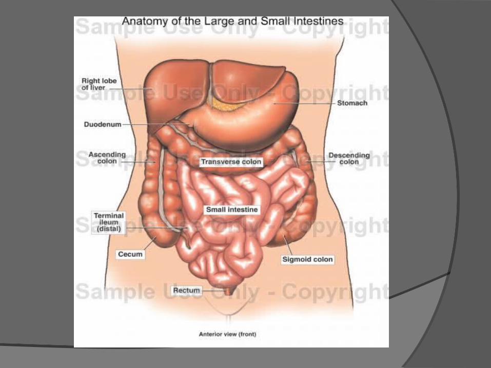
Lower gastrointestinal tract The lower GI tract comprises the intestines and anus. Bowel or intestine
small intestine, which has three parts: ○ duodenum ○ jejunum ○ ileum
large intestine, which has three parts: ○ caecum (the vermiform appendix is attached to the cecum). ○ colon (ascending colon, transverse colon, descending colon and
sigmoid flexure) ○ rectum
anus

Common anatomical features of GIT. Histology The GI tract has a uniform general histology with
some differences which reflect the specialization in functional anatomy.The GI tract can be divided into 4 concentric layers:
mucosa submucosa Muscularis externa (the external muscle layer) adventitia or serosa
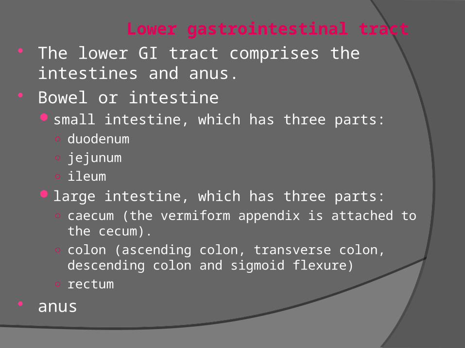
Mucosa The mucosa is the innermost layer of the GI tract,
surrounding the lumen, or space within the tube. This layer comes in direct contact with the food (or bolus), and is responsible for absorption and secretion, important processes in digestion.
The mucosa can be divided into: epithelium lamina propria muscularis mucosae

Submucosa The submucosa consists of a dense irregular layer of connective
tissue with large blood vessels, lymphatics and nerves branching into the mucosa and muscularis.

Muscularis externa The muscularis externa consists of a
circular inner muscular layer and a longitudinal outer muscular layer. The circular muscle layer prevents the food from going backwards and the longitudinal layer shortens the tract. The coordinated contractions of these layers is called peristalsis.
Adventitia/Serosa The adventitia consists of several layers
of epithelia
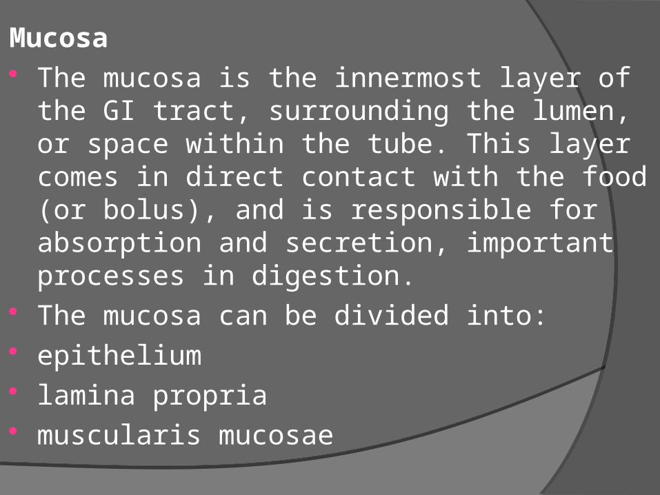
The pH of the organs of GI tract

Region Mean (cm)Over all length (nose to anus)
Duodenum
Jejunum & Ileum
Colon
451
22
255
100
Lengths of various regions of the human GI tract :

Stomach The stomach functions both as a reservoir and as a
digestive organ. It empties its contents in small portions (suitable for continued digestion) into the small intestine.
Anatomically, the stomach is divided into a cardiac part, fundus, body or corpus, and a pyloric part (pyloric antrum and pyloric canal) Histologically, most of the layers of the wall of the
stomach appear similar in its different parts. Regional differences are mainly restricted to the appearance of the gastric mucosa
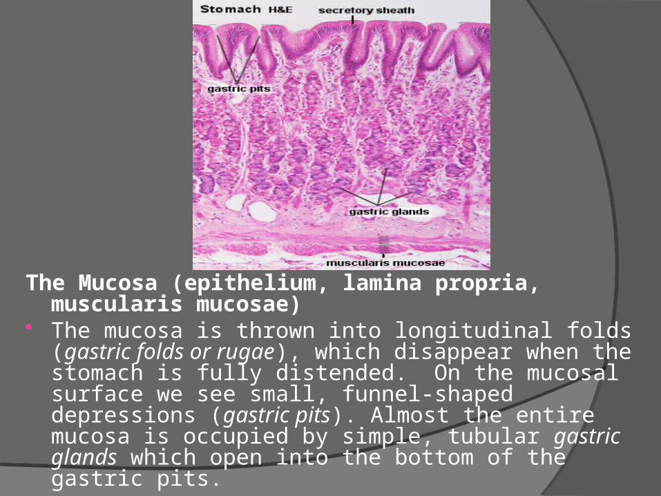
The Mucosa (epithelium, lamina propria, muscularis mucosae)
The mucosa is thrown into longitudinal folds (gastric folds or rugae), which disappear when the stomach is fully distended. On the mucosal surface we see small, funnel-shaped depressions (gastric pits). Almost the entire mucosa is occupied by simple, tubular gastric glands which open into the bottom of the gastric pits.
Pyloric glands Pyloric glands are more coiled than principal glands,
and they may be branched. The lamina propria is formed by a very cell-rich loose
connective tissue (fibroblasts, lymphocytes, plasma cells, macrophages, eosinophilic leukocytes and mast cells). The muscularis mucosae of the stomach contains both circular and longitudinal layers of muscle cells. Its organization is somewhat variable depending on the location in the stomach.
Large blood vessels, lymph vessels and nerves are located in the submucosa which consists of loose connective tissue.
Note that the muscularis externa consists of three layers of muscles: an inner oblique layer, a middle circular layer and an outer longitudinal layer.

Small Intestine The small intestine is divided into three
parts
1 duodenum (25-30 cm),
2 jejunum (about first two-fifths of the rest),
3 ileum.

Structure of small intestine
Peritoneum: a double layer peritoneum called the mesentery
attach the jejunum and ileum to posterior abdominal wall
The Mucosa The mucosa of the small intestine has various
structural features which considerably increase the luminal surface area and consequently support the main function of the small intestine - the absorption of the degraded components of the food.


.
The entire intestinal mucosa forms intestinal villi , which is tiny figure like projection of mucosal layer in to intestinal lumen, about 0.5 to 1 mm long.
The wall consist of columnar epithelial cells, or enterocytes, with tiny microvilli on their free borders.
Blood supply : The superior artery supplies the whole of the intestine and venous drainage is by the superior mesenteric vein which joins other veins to form the portal vein.

The Submucosa The submucosa contains glands only in
the duodenum. Submucosal glands of the duodenum are also called Brunner's glands.

Large intestine

Large Intestine The large intestine constitutes the terminal part of the
digestive system. It is divided into three main sections: caecum, colon, and rectum with the anal canal. The primary function of the large intestine is the reabsorption of water and inorganic salts.
The surface of the mucosa is relatively smooth as there are no plicae circulares or intestinal villi. Crypts of Lieberkühn are present and usually longer and straighter than those of the small intestine. .
There is only little lamina propria squeezed between the glands. The muscularis mucosae again forms two layers.
Considerable amounts of fat may be found in the submucosa.
The appearance of the muscularis externa is different from that of the small intestine.


References 1. Fundamentals of Biopharmaceutics &
Pharmacokinetics by V.Venkateswarlu 2. Biopharmaceutics & Pharmacokinetics by
D.M. Brahmankar 3.Google images 4. Google. COM 5.Ross and Wilson.


![Bharti Telecom Performance Appraisal in Bharti Telecom[1]](https://img.dokumen.tips/doc/110x75/547f3ee15806b5b35e8b47d6/bharti-telecom-performance-appraisal-in-bharti-telecom1.jpg)