Embed Size (px)
Citation preview

Clairembault et al. Acta Neuropathologica Communications (2015) 3:12 DOI 10.1186/s40478-015-0196-0
RESEARCH Open Access
Structural alterations of the intestinal epithelialbarrier in Parkinson’s diseaseThomas Clairembault1,2,3†, Laurène Leclair-Visonneau1,2,4†, Emmanuel Coron1,2,3,4, Arnaud Bourreille1,3,4, Séverine Le Dily4,Fabienne Vavasseur3,4, Marie-Françoise Heymann2,5,6, Michel Neunlist1,2,3 and Pascal Derkinderen1,2,4,7*
Abstract
Functional and morphological alterations of the intestinal epithelial barrier (IEB) have been consistently reported indigestive disorders such as irritable bowel syndrome and inflammatory bowel disease. There is mounting evidencethat Parkinson’s disease (PD) is not only a brain disease but also a digestive disorder. Gastrointestinal involvement isa frequent and early event in the course of PD, and it may be critically involved in the early development of thedisease. We therefore undertook the present survey to investigate whether changes in the IEB function and/ormorphology occur in PD. Colonic biopsies were performed in 31 PD patients and 11 age-matched healthy controls.The para- and transcellular permeability were evaluated by measuring sulfonic acid and horseradish peroxidase fluxrespectively, in colonic biopsies mounted in Ussing chambers. The expression and localization of the two tightjunctions proteins ZO-1 and occludin were analyzed by Western blot and immunofluorescence, respectively. Thepara- and transcellular permeability were not different between PD patients and controls. The expression of occludin,but not ZO-1, was significantly lower in colonic samples from PD patients as compared to controls and the cellulardistribution of both proteins was altered in colonic mucosal specimens from PD patients. Our findings provideevidence that the IEB is morphologically altered in PD and further reinforce the potential role of the gastrointestinaltract in the initiation and/or the progression of the disease.
Keywords: Parkinson’s disease, Intestinal epithelial barrier, Enteric nervous system, Tight junctions, Occludin, ZO-1
IntroductionThe intestinal epithelium forms a regulated barrier, knownas intestinal epithelial barrier (IEB), between the blood cir-culation and the contents of the intestinal lumen [1]. Itprevents the passage of noxious contents while allowingthe absorption and secretion of nutrients [1]. Penetrationof this barrier occurs via two routes, either between epi-thelial cells via the paracellular pathway, or through epi-thelial cell via the transcellular pathway [1]. Among themost important structures of the intestinal barrier are theepithelial tight junctions (TJs) that connect adjacent enter-ocytes together to determine paracellular permeabilitythrough the lateral intercellular space [2]. They are formedby transmembrane proteins such as claudins and occlu-dins connected to the actin cytoskeleton via high
* Correspondence: [email protected]†Equal contributors1Inserm U913, 1 rue Gaston Veil, Nantes F-44035, France2University Nantes, Nantes F-44093, FranceFull list of author information is available at the end of the article
© 2015 Clairembault et al.; licensee BioMed CeCommons Attribution License (http://creativecreproduction in any medium, provided the orDedication waiver (http://creativecommons.orunless otherwise stated.
molecular weight proteins called zona occludens (ZO-1,ZO-2 and 3) [2]. Increased permeability of the IEB alongwith changes in the expression levels of TJs proteins havebeen consistently reported in several digestive disorderssuch as inflammatory bowel disease [3,4] and irritablebowel syndrome [5,6].It has become evident over the last 20 years that PD is
a gut disorder (reviewed in [7]). Gastrointestinal symp-toms occur in almost every PD patient at some pointand are among the most debilitating non-motor featuresof the disease [8]. These clinical data have been supportedby post mortem studies that demonstrated the presence ofLewy bodies and neurites in the enteric neurons in nearlyevery case examined pathologically [9,10]. The Germanpathologist Heiko Braak suggested that the appearance ofLewy pathology in enteric neurons develop early in thecourse of disease, prior to the involvement of the centralnervous system [11]. This led him to suggest that thegastrointestinal tract might be a portal of entry for a puta-tive pathogen that would breach the IEB to induce the
ntral. This is an Open Access article distributed under the terms of the Creativeommons.org/licenses/by/4.0), which permits unrestricted use, distribution, andiginal work is properly credited. The Creative Commons Public Domaing/publicdomain/zero/1.0/) applies to the data made available in this article,
Clairembault et al. Acta Neuropathologica Communications (2015) 3:12 Page 2 of 9
formation of Lewy bodies and neurites in the entericneurons [11].The high prevalence of gastrointestinal symptoms and
pathology in PD and the possible derangement of gastro-intestinal permeability in the pathogenesis of the diseaseprompted several groups to investigate IEB permeabilityin parkinsonian patients. The three studies, which havebeen carried out to date have all used absorption of sugarprobes as a means to investigate non-invasively the para-cellular permeability [12]. These studies, which have in-cluded a small number of patients, led to conflictingresults. Two studies found a pattern of sugar absorptionreminiscent of small intestine hyperpermeability in a subsetof patients [13,14] while the third one showed an increasein sucralose excretion without changes in the lactulose/mannitol ratio, a pattern consistent with increased colonicpermeability [15]. We therefore undertook the present re-search to analyze in more details the IEB in PD. To thisend, a functional and structural characterization of the IEBwas performed in colonic biopsies from PD patients.
Materials and methodsSubjectsA total of 42 subjects participated in this study, 31 PDpatients and 11 healthy controls. PD patients aged 43–74years were recruited from the movement disorder clinic atNantes University Hospital, France. Diagnosis of PD wasmade according to criteria provided by the United King-dom Parkinson’s Disease Survey Brain Bank [16]. Col-lected demographic data included gender, age at onset anddisease duration, as well as age at colonoscopy. Completedrug history was obtained, and an approximation of thecumulative dose of L-dopa was made based on the equa-tion developed by Parkkinen and collaborators [17]. Con-trol subjects were healthy subjects who had a normalcolonoscopy performed for colorectal cancer screening.All controls subjects underwent a detailed neurologicalexamination to rule out PD symptoms and cognitive defi-ciency. Controls and PD patients were excluded if they suf-fered from irritable bowel syndrome and/or anorectaldysfunction. The study protocol was approved by the localCommittee on Ethics and Human Research (Comité deProtection des Personnes Ouest VI) and registered on Clin-icalTrials.gov (identifier NCT01748409). Written informedconsent was obtained from each patient and from each nor-mal volunteer according to the principles of Helsinki.
Endoscopic procedure and colonic biopsiesFor each subject, nine biopsies were taken in the sigmoid/descending colon during the course of a rectosigmoido-scopy for PD patients and during a colonoscopy for con-trol subjects. Five biopsies were immersed in 4°C Hank’sBalanced Salt Solution (Life Technologies, Saint Aubin,France): three of these biopsies were immediately processed
for the assessment of para- and transcellular permeabilityin Ussing chambers while the two other biopsies were usedfor immunohistochemistry experiments. Two biopsies werestored at −80°C in lysis buffer RA1 (Macherey-Nagel,Hoerdt, France) with 1% (v/v) β-mercaptoethanol (Sigma,Saint Quentin Fallavier, France) for further analysis byimmunoblotting. The two remaining biopsies weresnap frozen in liquid nitrogen at the time of collectionand kept at −80°C.
Para- and transcellular permeability of colonic biopsies inUssing chambersThree biopsies were mounted in Ussing chambers (WorldPrecision Instruments; WPI, Hertfordshire, UK) exposinga surface of 0.011 cm2. Tissues were bathed on each sidewith 3 ml of F12 supplemented Dulbecco’s Modified Eaglemedium (Invitrogen, France) containing 0.1% (v/v)fetal bovine serum, 200 mM Glutamine and 45 g/L ofNaHCO3. The medium was continuously oxygenatedand maintained at 37°C by a gas flow (95% O2/5%CO2). After a 30 min baseline period, 275 μL of apicalmedium was replaced with 200 μL of media containing1 mg/mL of fluorescein-5,6-sulfonic acid (molecularweight: 400 Da) (Life Technologies) for a final concen-tration of 0.1 mg/mL to assess paracellular permeabil-ity. Seventy-five microliters of media with 10 mg/mLof Horse Radish Peroxydase (HRP) (Sigma) were alsoadded to the basolateral chamber for a final concentra-tion of 0.375 mg/mL to measure transcellular perme-ability in a subset of PD patients and control subjects.The fluorescence level of basolateral aliquots of 150 μl,reflecting paracellular transit from the luminal surfacewas measured every 30 min over a 3-hour period using afluorimeter (Varioskan®, ThermoFisher Scientific, Cillebonsur Yvette, France). HRP quantities in the basolateralchamber, reflecting transcellular transit from the apicalsurface, was measured using an enzymatic activity assaywith 3,3’,5,5’-tetramethylbenzidine reagent (BD Bioscience,Le Pont de Claix, France). Paracellular and transcellularpermeabilities were determined by averaging the gradientof change in fluorescence intensity over time in the threebiopsies that were analyzed per patient, using a linear re-gression fit model (GraphPad Prism 5, La Jolla, USA).
Western blotFor the analysis of ZO-1 expression, total proteins fromthe 2 biopsies stored in RA1 buffer were precipitatedand prepared for Polyacrylamide Gel Electrophoresis(PAGE) using protein precipitator and resuspensionbuffer (Protein solving buffer and (tris(2-carboxyethyl)phosphine) TCEP reducing agent, PSB/TCEP) fromNucleoSpin Triprep Kit (Macherey-Nagel, Hoerdt, France)according to the manufacturer’s instructions. For experi-ments on the transmembrane protein occludin, the two
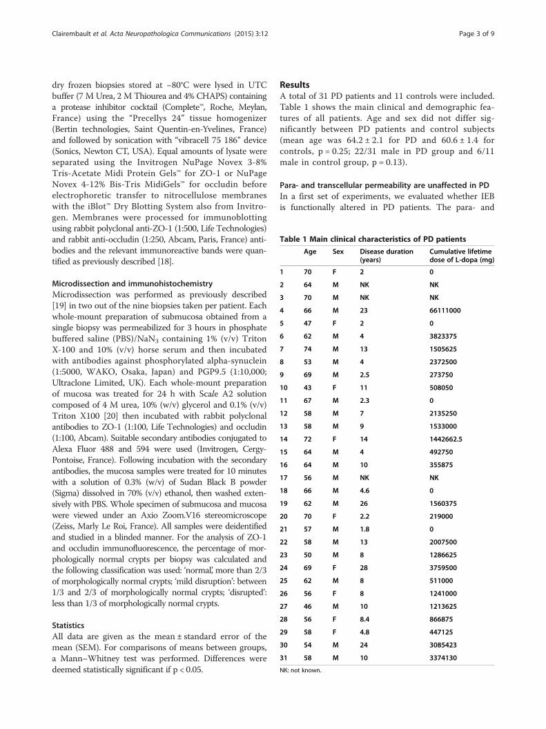
Table 1 Main clinical characteristics of PD patients
Age Sex Disease duration(years)
Cumulative lifetimedose of L-dopa (mg)
1 70 F 2 0
2 64 M NK NK
3 70 M NK NK
4 66 M 23 66111000
5 47 F 2 0
6 62 M 4 3823375
7 74 M 13 1505625
8 53 M 4 2372500
9 69 M 2.5 273750
10 43 F 11 508050
11 67 M 2.3 0
12 58 M 7 2135250
13 58 M 9 1533000
14 72 F 14 1442662.5
15 64 M 4 492750
16 64 M 10 355875
17 56 M NK NK
18 66 M 4.6 0
19 62 M 26 1560375
20 70 F 2.2 219000
21 57 M 1.8 0
22 58 M 13 2007500
23 50 M 8 1286625
24 69 F 28 3759500
25 62 M 8 511000
26 56 F 8 1241000
27 46 M 10 1213625
28 56 F 8.4 866875
29 58 F 4.8 447125
30 54 M 24 3085423
31 58 M 10 3374130
NK: not known.
Clairembault et al. Acta Neuropathologica Communications (2015) 3:12 Page 3 of 9
dry frozen biopsies stored at −80°C were lysed in UTCbuffer (7 M Urea, 2 M Thiourea and 4% CHAPS) containinga protease inhibitor cocktail (Complete™, Roche, Meylan,France) using the “Precellys 24” tissue homogenizer(Bertin technologies, Saint Quentin-en-Yvelines, France)and followed by sonication with “vibracell 75 186” device(Sonics, Newton CT, USA). Equal amounts of lysate wereseparated using the Invitrogen NuPage Novex 3-8%Tris-Acetate Midi Protein Gels™ for ZO-1 or NuPageNovex 4-12% Bis-Tris MidiGels™ for occludin beforeelectrophoretic transfer to nitrocellulose membraneswith the iBlot™ Dry Blotting System also from Invitro-gen. Membranes were processed for immunoblottingusing rabbit polyclonal anti-ZO-1 (1:500, Life Technologies)and rabbit anti-occludin (1:250, Abcam, Paris, France) anti-bodies and the relevant immunoreactive bands were quan-tified as previously described [18].
Microdissection and immunohistochemistryMicrodissection was performed as previously described[19] in two out of the nine biopsies taken per patient. Eachwhole-mount preparation of submucosa obtained from asingle biopsy was permeabilized for 3 hours in phosphatebuffered saline (PBS)/NaN3 containing 1% (v/v) TritonX-100 and 10% (v/v) horse serum and then incubatedwith antibodies against phosphorylated alpha-synuclein(1:5000, WAKO, Osaka, Japan) and PGP9.5 (1:10,000;Ultraclone Limited, UK). Each whole-mount preparationof mucosa was treated for 24 h with Scale A2 solutioncomposed of 4 M urea, 10% (w/v) glycerol and 0.1% (v/v)Triton X100 [20] then incubated with rabbit polyclonalantibodies to ZO-1 (1:100, Life Technologies) and occludin(1:100, Abcam). Suitable secondary antibodies conjugated toAlexa Fluor 488 and 594 were used (Invitrogen, Cergy-Pontoise, France). Following incubation with the secondaryantibodies, the mucosa samples were treated for 10 minuteswith a solution of 0.3% (w/v) of Sudan Black B powder(Sigma) dissolved in 70% (v/v) ethanol, then washed exten-sively with PBS. Whole specimen of submucosa and mucosawere viewed under an Axio Zoom.V16 stereomicroscope(Zeiss, Marly Le Roi, France). All samples were deidentifiedand studied in a blinded manner. For the analysis of ZO-1and occludin immunofluorescence, the percentage of mor-phologically normal crypts per biopsy was calculated andthe following classification was used: ‘normal’, more than 2/3of morphologically normal crypts; ‘mild disruption’: between1/3 and 2/3 of morphologically normal crypts; ‘disrupted’:less than 1/3 of morphologically normal crypts.
StatisticsAll data are given as the mean ± standard error of themean (SEM). For comparisons of means between groups,a Mann–Whitney test was performed. Differences weredeemed statistically significant if p < 0.05.
ResultsA total of 31 PD patients and 11 controls were included.Table 1 shows the main clinical and demographic fea-tures of all patients. Age and sex did not differ sig-nificantly between PD patients and control subjects(mean age was 64.2 ± 2.1 for PD and 60.6 ± 1.4 forcontrols, p = 0.25; 22/31 male in PD group and 6/11male in control group, p = 0.13).
Para- and transcellular permeability are unaffected in PDIn a first set of experiments, we evaluated whether IEBis functionally altered in PD patients. The para- and
Clairembault et al. Acta Neuropathologica Communications (2015) 3:12 Page 4 of 9
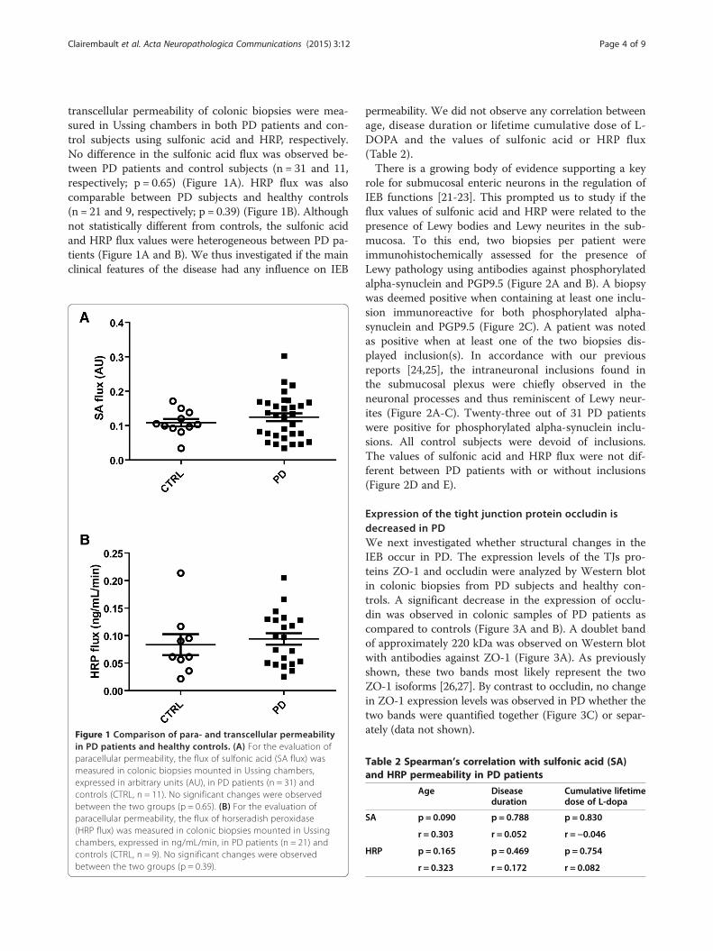
transcellular permeability of colonic biopsies were mea-sured in Ussing chambers in both PD patients and con-trol subjects using sulfonic acid and HRP, respectively.No difference in the sulfonic acid flux was observed be-tween PD patients and control subjects (n = 31 and 11,respectively; p = 0.65) (Figure 1A). HRP flux was alsocomparable between PD subjects and healthy controls(n = 21 and 9, respectively; p = 0.39) (Figure 1B). Althoughnot statistically different from controls, the sulfonic acidand HRP flux values were heterogeneous between PD pa-tients (Figure 1A and B). We thus investigated if the mainclinical features of the disease had any influence on IEB
Figure 1 Comparison of para- and transcellular permeabilityin PD patients and healthy controls. (A) For the evaluation ofparacellular permeability, the flux of sulfonic acid (SA flux) wasmeasured in colonic biopsies mounted in Ussing chambers,expressed in arbitrary units (AU), in PD patients (n = 31) andcontrols (CTRL, n = 11). No significant changes were observedbetween the two groups (p = 0.65). (B) For the evaluation ofparacellular permeability, the flux of horseradish peroxidase(HRP flux) was measured in colonic biopsies mounted in Ussingchambers, expressed in ng/mL/min, in PD patients (n = 21) andcontrols (CTRL, n = 9). No significant changes were observedbetween the two groups (p = 0.39).
permeability. We did not observe any correlation betweenage, disease duration or lifetime cumulative dose of L-DOPA and the values of sulfonic acid or HRP flux(Table 2).There is a growing body of evidence supporting a key
role for submucosal enteric neurons in the regulation ofIEB functions [21-23]. This prompted us to study if theflux values of sulfonic acid and HRP were related to thepresence of Lewy bodies and Lewy neurites in the sub-mucosa. To this end, two biopsies per patient wereimmunohistochemically assessed for the presence ofLewy pathology using antibodies against phosphorylatedalpha-synuclein and PGP9.5 (Figure 2A and B). A biopsywas deemed positive when containing at least one inclu-sion immunoreactive for both phosphorylated alpha-synuclein and PGP9.5 (Figure 2C). A patient was notedas positive when at least one of the two biopsies dis-played inclusion(s). In accordance with our previousreports [24,25], the intraneuronal inclusions found inthe submucosal plexus were chiefly observed in theneuronal processes and thus reminiscent of Lewy neur-ites (Figure 2A-C). Twenty-three out of 31 PD patientswere positive for phosphorylated alpha-synuclein inclu-sions. All control subjects were devoid of inclusions.The values of sulfonic acid and HRP flux were not dif-ferent between PD patients with or without inclusions(Figure 2D and E).
Expression of the tight junction protein occludin isdecreased in PDWe next investigated whether structural changes in theIEB occur in PD. The expression levels of the TJs pro-teins ZO-1 and occludin were analyzed by Western blotin colonic biopsies from PD subjects and healthy con-trols. A significant decrease in the expression of occlu-din was observed in colonic samples of PD patients ascompared to controls (Figure 3A and B). A doublet bandof approximately 220 kDa was observed on Western blotwith antibodies against ZO-1 (Figure 3A). As previouslyshown, these two bands most likely represent the twoZO-1 isoforms [26,27]. By contrast to occludin, no changein ZO-1 expression levels was observed in PD whether thetwo bands were quantified together (Figure 3C) or separ-ately (data not shown).
Table 2 Spearman’s correlation with sulfonic acid (SA)and HRP permeability in PD patients
Age Diseaseduration
Cumulative lifetimedose of L-dopa
SA p = 0.090 p = 0.788 p = 0.830
r = 0.303 r = 0.052 r = −0.046
HRP p = 0.165 p = 0.469 p = 0.754
r = 0.323 r = 0.172 r = 0.082
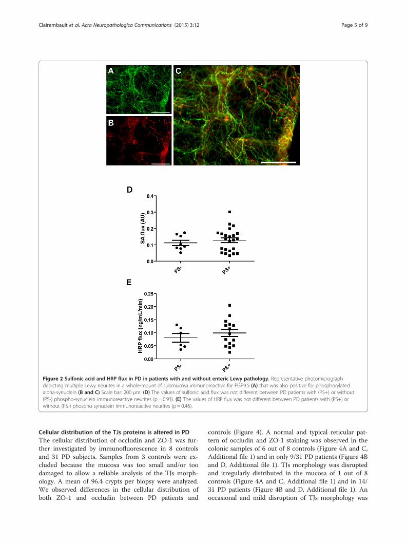
Figure 2 Sulfonic acid and HRP flux in PD in patients with and without enteric Lewy pathology. Representative photomicrographdepicting multiple Lewy neurites in a whole-mount of submucosa immunoreactive for PGP9.5 (A) that was also positive for phosphorylatedalpha-synuclein (B and C) Scale bar: 200 μm. (D) The values of sulfonic acid flux was not different between PD patients with (PS+) or without(PS-) phospho-synuclein immunoreactive neurites (p = 0.93). (E) The values of HRP flux was not different between PD patients with (PS+) orwithout (PS-) phospho-synuclein immunoreactive neurites (p = 0.46).
Clairembault et al. Acta Neuropathologica Communications (2015) 3:12 Page 5 of 9
Cellular distribution of the TJs proteins is altered in PDThe cellular distribution of occludin and ZO-1 was fur-ther investigated by immunofluorescence in 8 controlsand 31 PD subjects. Samples from 3 controls were ex-cluded because the mucosa was too small and/or toodamaged to allow a reliable analysis of the TJs morph-ology. A mean of 96.4 crypts per biopsy were analyzed.We observed differences in the cellular distribution ofboth ZO-1 and occludin between PD patients and
controls (Figure 4). A normal and typical reticular pat-tern of occludin and ZO-1 staining was observed in thecolonic samples of 6 out of 8 controls (Figure 4A and C,Additional file 1) and in only 9/31 PD patients (Figure 4Band D, Additional file 1). TJs morphology was disruptedand irregularly distributed in the mucosa of 1 out of 8controls (Figure 4A and C, Additional file 1) and in 14/31 PD patients (Figure 4B and D, Additional file 1). Anoccasional and mild disruption of TJs morphology was
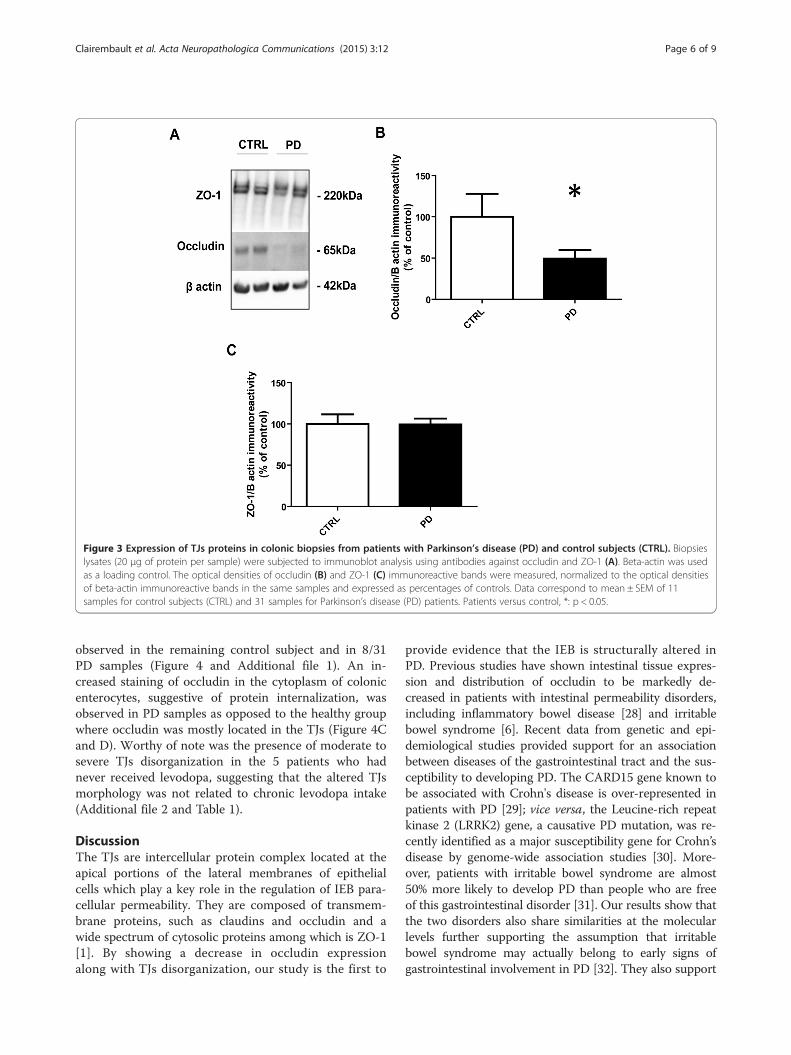
Figure 3 Expression of TJs proteins in colonic biopsies from patients with Parkinson’s disease (PD) and control subjects (CTRL). Biopsieslysates (20 μg of protein per sample) were subjected to immunoblot analysis using antibodies against occludin and ZO-1 (A). Beta-actin was usedas a loading control. The optical densities of occludin (B) and ZO-1 (C) immunoreactive bands were measured, normalized to the optical densitiesof beta-actin immunoreactive bands in the same samples and expressed as percentages of controls. Data correspond to mean ± SEM of 11samples for control subjects (CTRL) and 31 samples for Parkinson’s disease (PD) patients. Patients versus control, *: p < 0.05.
Clairembault et al. Acta Neuropathologica Communications (2015) 3:12 Page 6 of 9
observed in the remaining control subject and in 8/31PD samples (Figure 4 and Additional file 1). An in-creased staining of occludin in the cytoplasm of colonicenterocytes, suggestive of protein internalization, wasobserved in PD samples as opposed to the healthy groupwhere occludin was mostly located in the TJs (Figure 4Cand D). Worthy of note was the presence of moderate tosevere TJs disorganization in the 5 patients who hadnever received levodopa, suggesting that the altered TJsmorphology was not related to chronic levodopa intake(Additional file 2 and Table 1).
DiscussionThe TJs are intercellular protein complex located at theapical portions of the lateral membranes of epithelialcells which play a key role in the regulation of IEB para-cellular permeability. They are composed of transmem-brane proteins, such as claudins and occludin and awide spectrum of cytosolic proteins among which is ZO-1[1]. By showing a decrease in occludin expressionalong with TJs disorganization, our study is the first to
provide evidence that the IEB is structurally altered inPD. Previous studies have shown intestinal tissue expres-sion and distribution of occludin to be markedly de-creased in patients with intestinal permeability disorders,including inflammatory bowel disease [28] and irritablebowel syndrome [6]. Recent data from genetic and epi-demiological studies provided support for an associationbetween diseases of the gastrointestinal tract and the sus-ceptibility to developing PD. The CARD15 gene known tobe associated with Crohn's disease is over-represented inpatients with PD [29]; vice versa, the Leucine-rich repeatkinase 2 (LRRK2) gene, a causative PD mutation, was re-cently identified as a major susceptibility gene for Crohn’sdisease by genome-wide association studies [30]. More-over, patients with irritable bowel syndrome are almost50% more likely to develop PD than people who are freeof this gastrointestinal disorder [31]. Our results show thatthe two disorders also share similarities at the molecularlevels further supporting the assumption that irritablebowel syndrome may actually belong to early signs ofgastrointestinal involvement in PD [32]. They also support
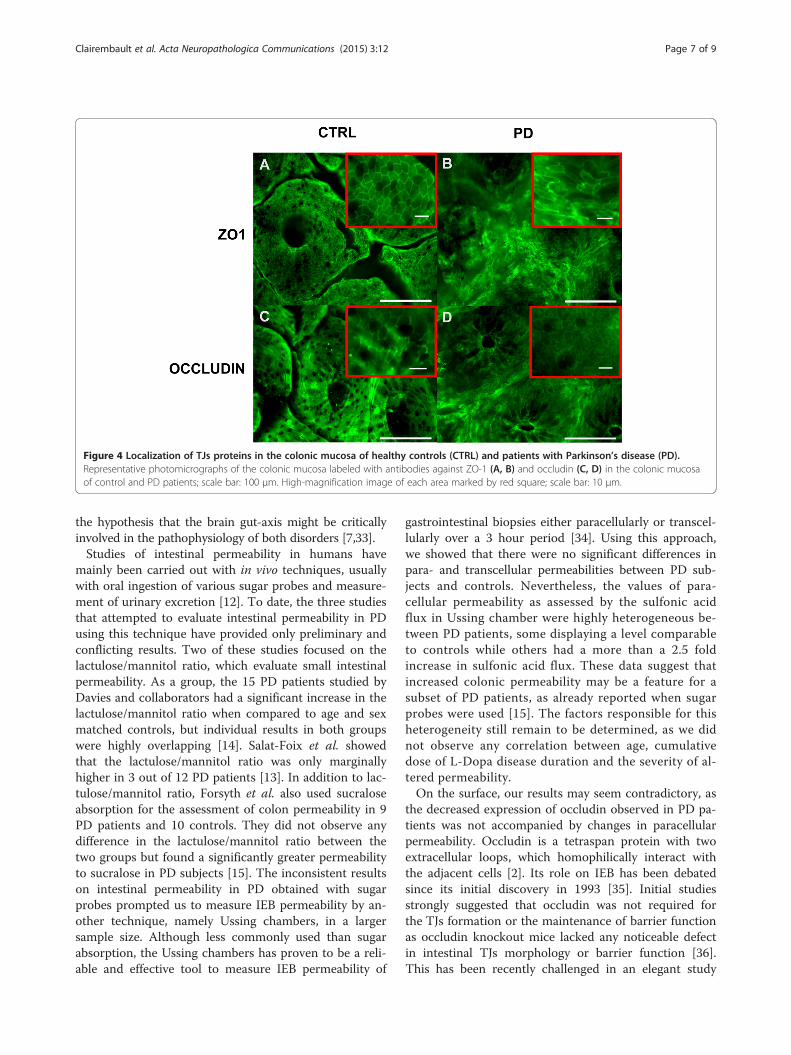
Figure 4 Localization of TJs proteins in the colonic mucosa of healthy controls (CTRL) and patients with Parkinson’s disease (PD).Representative photomicrographs of the colonic mucosa labeled with antibodies against ZO-1 (A, B) and occludin (C, D) in the colonic mucosaof control and PD patients; scale bar: 100 μm. High-magnification image of each area marked by red square; scale bar: 10 μm.
Clairembault et al. Acta Neuropathologica Communications (2015) 3:12 Page 7 of 9
the hypothesis that the brain gut-axis might be criticallyinvolved in the pathophysiology of both disorders [7,33].Studies of intestinal permeability in humans have
mainly been carried out with in vivo techniques, usuallywith oral ingestion of various sugar probes and measure-ment of urinary excretion [12]. To date, the three studiesthat attempted to evaluate intestinal permeability in PDusing this technique have provided only preliminary andconflicting results. Two of these studies focused on thelactulose/mannitol ratio, which evaluate small intestinalpermeability. As a group, the 15 PD patients studied byDavies and collaborators had a significant increase in thelactulose/mannitol ratio when compared to age and sexmatched controls, but individual results in both groupswere highly overlapping [14]. Salat-Foix et al. showedthat the lactulose/mannitol ratio was only marginallyhigher in 3 out of 12 PD patients [13]. In addition to lac-tulose/mannitol ratio, Forsyth et al. also used sucraloseabsorption for the assessment of colon permeability in 9PD patients and 10 controls. They did not observe anydifference in the lactulose/mannitol ratio between thetwo groups but found a significantly greater permeabilityto sucralose in PD subjects [15]. The inconsistent resultson intestinal permeability in PD obtained with sugarprobes prompted us to measure IEB permeability by an-other technique, namely Ussing chambers, in a largersample size. Although less commonly used than sugarabsorption, the Ussing chambers has proven to be a reli-able and effective tool to measure IEB permeability of
gastrointestinal biopsies either paracellularly or transcel-lularly over a 3 hour period [34]. Using this approach,we showed that there were no significant differences inpara- and transcellular permeabilities between PD sub-jects and controls. Nevertheless, the values of para-cellular permeability as assessed by the sulfonic acidflux in Ussing chamber were highly heterogeneous be-tween PD patients, some displaying a level comparableto controls while others had a more than a 2.5 foldincrease in sulfonic acid flux. These data suggest thatincreased colonic permeability may be a feature for asubset of PD patients, as already reported when sugarprobes were used [15]. The factors responsible for thisheterogeneity still remain to be determined, as we didnot observe any correlation between age, cumulativedose of L-Dopa disease duration and the severity of al-tered permeability.On the surface, our results may seem contradictory, as
the decreased expression of occludin observed in PD pa-tients was not accompanied by changes in paracellularpermeability. Occludin is a tetraspan protein with twoextracellular loops, which homophilically interact withthe adjacent cells [2]. Its role on IEB has been debatedsince its initial discovery in 1993 [35]. Initial studiesstrongly suggested that occludin was not required forthe TJs formation or the maintenance of barrier functionas occludin knockout mice lacked any noticeable defectin intestinal TJs morphology or barrier function [36].This has been recently challenged in an elegant study
Clairembault et al. Acta Neuropathologica Communications (2015) 3:12 Page 8 of 9
published by Al-Sadi et al. [37]. The purpose of their re-search was to better delineate the involvement of occlu-din in IEB by studying the transepithelial flux ofvarious-sized probes after knocking down occludin bothin vitro and in vivo. They showed that the occludinknock down caused a marked increase in the flux ratesof macromolecules above 5 kDa such as inulin and dex-tran but had only modest effect on flux of smaller-sizedprobes under 200 Da such as mannitol and urea [36].Fluorescein-5,6-sulfonic acid, which was used for the as-sessment of paracellular permeability in our study has amolecular weight of 400 Da, likely to be too small fordetecting defects in IEB permeability induced by a meredown regulation of occludin. This may explain the lackof significant changes in IEB permeability observed inPD patients in our study in spite of the occurrence ofstructural changes.The question arises as to what might be the clinical
relevance of our experimental findings. A current theory,the so-called Braak’s theory, assumes that PD originatesin the gastrointestinal tract [11]. Braak and co-workerssuggested that the appearance of Lewy pathology occursin the earliest stage of PD in both the enteric nervoussystem and the dorsal motor nucleus of the vagus[11,38]. This led Braak to postulate that a pathogen maybreach the IEB to trigger Lewy pathology in the terminalaxons of the enteric neurons, further spreading to thecentral nervous system via the vagal preganglionic in-nervation of the gut [11,39]. In light of these consider-ations, our results demonstrating altered intestinal TJsstructure in PD gain in importance as the down regula-tion of occludin may favor the entry of a putative patho-gen. This must be however balanced, as the stomach incontrast to the colon appears to be the most suitable tar-get for the pathologic insult to occur in Braak’s scenario.Several studies have indeed described that Lewy path-ology is distributed following a rostro-caudal gradient inPD, with the lower esophagus and stomach having thegreatest involvement and the colon and rectum the low-est [9,10], a distribution that parallels the vagal innerv-ation of the gastrointestinal tract [40]. Further studiesare therefore warranted to analyze the mucosal barrierpermeability and morphology in gastric and duodenalsamples from PD patients.
ConclusionsIn conclusion, we provide evidence for the first time thatmorphological changes in the IEB occur in PD patients.Our results further reinforce the possible role of thegastrointestinal tract in the pathophysiology of PD.Further work is needed to determine if occludin downregulation in the gut might facilitate the spreading of PDpathology in the enteric nervous system and in thebrain.
Additional files
Additional file 1: Percentage of morphologically normal crypts inthe colonic mucosa of healthy controls (CTRL) and patients withParkinson’s disease (PD).
Additional file 2: Localization of TJs proteins in the colonic mucosaof the 5 with Parkinson’s disease (PD) who had never receivedlevodopa. Scale bar: 100 μm.
Competing interestsThe authors declare that they have no competing interests.
AcknowledgementsThis work was supported by a grant from the Michael J. Fox Foundation forParkinson’s Research (Rapid Response Innovation Award 2013) and FranceParkinson to PD. LLV is supported by a grant from Nantes University Hospital(Appel d'offre interne 2012, Grant number RC12_0264) and France Parkinson.TC is supported by a grant from centre d’entraide et de coordination desassociations de parkinsoniens (CECAP).
Author details1Inserm U913, 1 rue Gaston Veil, Nantes F-44035, France. 2University Nantes,Nantes F-44093, France. 3CHU Nantes, Institut des Maladies de l’AppareilDigestif, Nantes F-44093, France. 4Inserm, CIC-04, Nantes F-44093, France.5CHU Nantes, Service d’Anatomie Pathologique, Nantes F-44093, France.6Inserm, UMR957, Nantes F-44093, France. 7CHU Nantes, Department ofNeurology, Nantes F-44093, France.
Received: 8 February 2015 Accepted: 10 February 2015
References1. Marchiando AM, Graham WV, Turner JR (2010) Epithelial barriers in
homeostasis and disease. Annu Rev Pathol 5:119–442. Suzuki T (2013) Regulation of intestinal epithelial permeability by tight
junctions. Cell Mol Life Sci 70:631–593. Peeters M, Ghoos Y, Maes B, Hiele M, Geboes K, Vantrappen G et al (1994)
Increased permeability of macroscopically normal small bowel in Crohn’sdisease. Dig Dis Sci 39:2170–6
4. Katz KD, Hollander D, Vadheim CM, McElree C, Delahunty T, Dadufalza VDet al (1989) Intestinal permeability in patients with Crohn’s disease and theirhealthy relatives. Gastroenterology 97:927–31
5. Piche T, Barbara G, Aubert P, Bruley des Varannes S, Dainese R, Nano JL et al(2009) Impaired intestinal barrier integrity in the colon of patients withirritable bowel syndrome: involvement of soluble mediators. Gut 58:196–201
6. Bertiaux-Vandaële N, Youmba SB, Belmonte L, Lecleire S, Antonietti M,Gourcerol G et al (2011) The expression and the cellular distribution of thetight junction proteins are altered in irritable bowel syndrome patients withdifferences according to the disease subtype. Am J Gastroenterol 106:2165–73
7. Derkinderen P, Rouaud T, Lebouvier T, Bruley des Varannes S, Neunlist M,De Giorgio R (2011) Parkinson disease: the enteric nervous system spills itsguts. Neurology 77:1761–7
8. Cloud LJ, Greene JG (2011) Gastrointestinal features of Parkinson’s disease.Curr Neurol Neurosci Rep 11:379–84
9. Wakabayashi K, Takahashi H, Takeda S, Ohama E, Ikuta F (1988) Parkinson’sdisease: the presence of Lewy bodies in Auerbach’s and Meissner’s plexuses.Acta neuropathologica 76:217–21
10. Beach TG, Adler CH, Sue LI, Vedders L, Lue L, White Iii CL et al (2009) Multi-organdistribution of phosphorylated alpha-synuclein histopathology in subjects withLewy body disorders. Acta neuropathologica 119:689–702
11. Braak H, de Vos RA, Bohl J, Del Tredici K (2006) Gastric alpha-synucleinimmunoreactive inclusions in Meissner’s and Auerbach’s plexuses in casesstaged for Parkinson’s disease-related brain pathology. Neurosci Lett396:67–72
12. Hollander D (1999) Intestinal permeability, leaky gut, and intestinaldisorders. Curr Gastroenterol Rep 1:410–6
13. Salat-Foix D, Tran K, Ranawaya R, Meddings J, Suchowersky O (2012)Increased intestinal permeability and Parkinson disease patients: chicken oregg? Can J Neurol Sci 39:185–8
Clairembault et al. Acta Neuropathologica Communications (2015) 3:12 Page 9 of 9
14. Davies KN, King D, Billington D, Barrett JA (1996) Intestinal permeability andorocaecal transit time in elderly patients with Parkinson’s disease. PostgradMed J 72:164–7
15. Forsyth CB, Shannon KM, Kordower JH, Voigt RM, Shaikh M, Jaglin JA et al(2011) Increased intestinal permeability correlates with sigmoid mucosaalpha-synuclein staining and endotoxin exposure markers in early Parkinson’sdisease. PLoS One 6:e28032
16. Hughes AJ, Daniel SE, Lees AJ (2001) Improved accuracy of clinical diagnosisof Lewy body Parkinson’s disease. Neurology 57:1497–9
17. Parkkinen L, O'Sullivan SS, Kuoppamaki M, Collins C, Kallis C, Holton JL et al(2011) Does levodopa accelerate the pathologic process in Parkinsondisease brain? Neurology 77:1420–6
18. Clairembault T, Kamphuis W, Leclair-Visonneau L, Rolli-Derkinderen M, Coron E,Neunlist M et al. Enteric GFAP expression and phosphorylation in Parkinson’sdisease. J Neurochem 2014; doi:10.1111/jnc.12742.
19. Lebouvier T, Coron E, Chaumette T, Paillusson S, Bruley des Varannes S,Neunlist M et al (2010) Routine colonic biopsies as a new tool to study theenteric nervous system in living patients. Neurogastroenterol Motil 22:e11–4
20. Hama H, Kurokawa H, Kawano H, Ando R, Shimogori T, Noda H et al (2011)Scale: a chemical approach for fluorescence imaging and reconstruction oftransparent mouse brain. Nat Neurosci 14:1481–8
21. Neunlist M, Aubert P, Toquet C, Oreshkova T, Barouk J, Lehur PA et al (2003)Changes in chemical coding of myenteric neurones in ulcerative colitis. Gut52:84–90
22. Toumi F, Neunlist M, Cassagnau E, Parois S, Laboisse CL, Galmiche JP et al(2003) Human submucosal neurones regulate intestinal epithelial cellproliferation: evidence from a novel co-culture model. NeurogastroenterolMotil 15:239–42
23. Cameron HL, Perdue MH (2007) Muscarinic acetylcholine receptor activationincreases transcellular transport of macromolecules across mouse andhuman intestinal epithelium in vitro. Neurogastroenterol Motil 19:47–56
24. Lebouvier T, Neunlist M, Bruley des Varannes S, Coron E, Drouard A,N'Guyen JM et al (2010) Colonic biopsies to assess the neuropathology ofParkinson’s disease and its relationship with symptoms. PLoS One 5:e12728
25. Pouclet H, Lebouvier T, Coron E, Des Varannes SB, Neunlist M, DerkinderenP (2012) A comparison between rectal and colonic biopsies to detect Lewypathology in Parkinson’s disease. Neurobiol Dis 45:305–9
26. Ciana A, Meier K, Daum N, Gerbes S, Veith M, Lehr CM et al (2010) Adynamic ratio of the alpha + and alpha- isoforms of the tight junctionprotein ZO-1 is characteristic of Caco-2 cells and correlates with theirdegree of differentiation. Cell Biol Int 34:669–78
27. Willott E, Balda MS, Heintzelman M, Jameson B, Anderson JM (1992)Localization and differential expression of two isoforms of the tight junctionprotein ZO-1. Am J Physiol 262:C1119–24
28. Gassler N, Rohr C, Schneider A, Kartenbeck J, Bach A, Obermüller N et al(2001) Inflammatory bowel disease is associated with changes ofenterocytic junctions. Am J Physiol Gastrointest Liver Physiol 281:G216–28
29. Bialecka M, Kurzawski M, Klodowska-Duda G, Opala G, Juzwiak S, KurzawskiG et al (2007) CARD15 variants in patients with sporadic Parkinson’s disease.Neurosci Res 57:473–6
30. Barrett JC, Hansoul S, Nicolae DL, Cho JH, Duerr RH, Rioux JD et al (2008)Genome-wide association defines more than 30 distinct susceptibility locifor Crohn’s disease. Nat Genet 40:955–62
31. Lai S-W, Liao K-F, Lin C-L, Sung F-C (2014) Irritable bowel syndrome correlateswith increased risk of Parkinson’s disease in Taiwan. Eur J Epidemiol 29:57–62,doi:10.1007/s10654-014-9878-3
32. Cersosimo MG, Raina GB, Pecci C, Pellene A, Calandra CR, Gutiérrez C et al(2013) Gastrointestinal manifestations in Parkinson’s disease: prevalence andoccurrence before motor symptoms. J Neurol 260:1332–8
33. Coss-Adame E, Rao SSC (2014) Brain and gut interactions in irritable bowelsyndrome: new paradigms and new understandings. Curr GastroenterolRep 16:379
34. Wallon C, Braaf Y, Wolving M, Wolving M, Olaison G, Söderholm JD (2005)Endoscopic biopsies in Ussing chambers evaluated for studies ofmacromolecular permeability in the human colon. Scand J Gastroenterol40:586–95
35. Furuse M, Hirase T, Itoh M, Nagafuchi A, Yonemura S, Tsukita S et al (1993)Occludin: a novel integral membrane protein localizing at tight junctions. JCell Biol 123:1777–88
36. Saitou M, Furuse M, Sasaki H, Schulzke JD, Fromm M, Takano H et al (2000)Complex phenotype of mice lacking occludin, a component of tightjunction strands. Mol Biol Cell 11:4131–42
37. Al-Sadi R, Khatib K, Guo S, Ye D, Youssef M, Ma T (2011) Occludin regulatesmacromolecule flux across the intestinal epithelial tight junction barrier. AmJ Physiol Gastrointest Liver Physiol 300:G1054–64
38. Braak H, Del Tredici K, Rub U, de Vos RA, Jansen Steur EN, Braak E (2003)Staging of brain pathology related to sporadic Parkinson’s disease.Neurobiol Aging 24:197–211
39. Braak H, Rub U, Gai WP, Del Tredici K (2003) Idiopathic Parkinson’s disease:possible routes by which vulnerable neuronal types may be subject toneuroinvasion by an unknown pathogen. J Neural Transm 110:517–36
40. Hopkins DA, Bieger D, deVente J, Steinbusch WM (1996) Vagal efferentprojections: viscerotopy, neurochemistry and effects of vagotomy. ProgBrain Res 107:79–96
Submit your next manuscript to BioMed Centraland take full advantage of:
• Convenient online submission
• Thorough peer review
• No space constraints or color figure charges
• Immediate publication on acceptance
• Inclusion in PubMed, CAS, Scopus and Google Scholar
• Research which is freely available for redistribution
Submit your manuscript at www.biomedcentral.com/submit