Embed Size (px)
Citation preview
Intestinal epithelial cells as producers but nottargets of chronic TNF suffice to cause murineCrohn-like pathologyManolis Roulis, Maria Armaka, Menelaos Manoloukos, Maria Apostolaki1, and George Kollias2
Institute of Immunology, Biomedical Sciences Research Center Alexander Fleming, Vari 16672, Greece
Edited by Fabio Cominelli, Case Western Reserve University, Cleveland, OH, and accepted by the Editorial Board February 24, 2011 (received for review June2, 2010)
TNF plays a crucial role in the pathogenesis of Crohn disease. Dys-regulated TNF production in mice that bear the genetic deletion ofthe TNF AU-rich regulatory elements (ARE) (TnfΔARE/+ mice) resultsin TNF receptor I (TNFRI)-dependent spontaneous Crohn-like pa-thology. Current concepts consider intestinal epithelial cell (IEC)responses to TNF to be critical for intestinal pathology, but thepotential contribution of IEC-derived TNF in disease pathogenesishas not been addressed. In this study we examined whether IECare sufficient as cellular targets or sources of TNF in the develop-ment of intestinal pathology. Using IEC-specific reactivation ofa hypomorphic TnfΔAREneo allele in mice, we show that selectivechronic overproduction of TNF by IEC suffices to cause full devel-opment of Crohn-like pathology. Epithelial TNF overexpressionleads to early activation of the underlying intestinal myofibro-blast, a cell type previously identified as a sufficient target ofTNF for disease development in the TnfΔARE model. By contrast,restricted TNFRI expression on IEC although sufficient to confer IECapoptosis after acute exogenous TNF administration, fails to in-duce pathology following chronic specific targeting of IEC by en-dogenous TNF in TnfΔARE/+ mice. Our results argue against IECbeing early and sufficient responders to chronic TNF-mediatedpathogenic signals and suggest that proinflammatory aberrationsleading to chronic TNF production by IEC may initiate pathology inCrohn disease.
inflammatory bowel disease | ileitis | mucosal | mesenchymal
Inflammatory bowel disease (IBD) is a chronic inflammatorydisorder of the gastrointestinal tract resulting from inappro-
priate and sustained activation of the mucosal immune systemagainst the bacterial microflora of the gut (1). IBD is representedby two major forms: ulcerative colitis (UC), which is manifestedin the colon, and Crohn disease (CD), which is manifested pri-marily in the ileum but also in the colon (2). Genome-wideassociation studies in patients with CD indicate a putative rolefor genes of the innate and adaptive immunity system and anemerging role for genes implicated in autophagy (3). Animalmodels of disease suggest that adaptive immune responses arerequired for disease manifestation and that the gut microfloraappear to drive inflammation (4). However, the exact mecha-nisms underlying IBD pathogenesis and especially the earlypathogenic events remain largely obscure.TNF is critically implicated in IBD pathophysiology, high-
lighted by the successful use of anti-TNF therapies for thetreatment of patients who have CD (5). TNF is shown to exertpathogenic activities in several animal models of IBD (6),whereas protective roles have been described also (7, 8). Thecrucial pathogenic role of TNF in CD has been verified experi-mentally in mice that bear the genetic deletion of the TNF AU-rich regulatory elements (ARE) that leads to defective post-transcriptional regulation of tnf expression and chronic TNFoverproduction (9). TnfΔARE/+ mice chronically overproduceTNF and spontaneously develop CD8+ T lymphocyte-dependentCrohn-like IBD pathology in the ileum (9–11). Restricting TNFoverexpression in myeloid cells or T lymphocytes suffices for the
induction of intestinal pathology in this model, indicating thepathogenic potential of TNF derived from innate or adaptiveeffectors (10). The dominant role of TNF receptor I (TNFRI) inmediating TNF pathogenic signals leading to intestinal pathologyhas been established previously in this model (9). Most impor-tantly, by restricting TNFRI expression in mesenchymal cells, theintestinal myofibroblast (IMF) was identified as a primary andsufficient cellular target of TNF for intestinal pathology in theTnfΔARE model (12).An important cell type prominently linked to IBD pathogen-
esis is the intestinal epithelial cell (IEC), which lies at the in-terphase between the huge luminal bacterial microflora and thegut-associated lymphoid tissues (13). IEC participate in luminalmicrobiota recognition by the innate immune system and theclearance of bacterial pathogens, either through the productionof antimicrobial peptides or through autophagy (14–16). Im-portantly, mutations associated with CD pathology in genessuch as nucleotide-binding oligomerization domain-containing 2(NOD2), autophagy-related 16-like 1 (ATG16L1), and immunity-related GTPase family M (IRGM) have been associated withdefective bacterial clearance by epithelial cells (14, 17, 18), andthe NOD2 and ATG16L1 mutations have been associated withinflammatory activation of macrophages in mice (19, 20). Al-though human IEC have been reported to overexpress TNF inpatients who have CD (21), whether IEC have a role in initiatinginflammatory responses in the gut was unknown. In turn, aprominent role for the IEC as a target of TNF has been reported,associating TNF with either the alteration of the epithelialjunctions (22) or the induction of IEC apoptosis (23). In thiscontext we aimed to identify the role of IEC as a potential sourceand target of TNF in a model of TNF-driven Crohn-like IBD.Here we show that TNF overexpression specifically by IEC leadsto the early activation of the underlying mesenchymal cells andis sufficient for the full induction of Crohn-like IBD pathologyin the mouse. By contrast, although IEC-restricted TNFRI ex-pression is sufficient for the induction of IEC apoptosis in re-sponse to exogenous TNF administration, chronic targeting ofIEC by endogenous TNF is not sufficient to induce IBD pa-thology. These findings provide insight into the mechanisms ofCD pathogenesis by showing that IEC are potential TNF pro-ducers and that IEC and mesenchymal cells can form a cellularaxis of TNF function in the gut that is sufficient to cause the fullspectrum of pathology seen in human disease.
Author contributions: M.R., M. Armaka, M.M., M. Apostolaki, and G.K. designed research;M.R., M. Armaka, M.M., and M. Apostolaki performed research; M.R., M. Armaka, M.M.,and M. Apostolaki analyzed data; and M.R., M. Apostolaki, and G.K. wrote the paper.
The authors declare no conflict of interest.
This article is a PNAS Direct Submission. F.C. is a guest editor invited by the EditorialBoard.1Deceased December 10, 2010.2To whom correspondence should be addressed. E-mail: [email protected].
This article contains supporting information online at www.pnas.org/lookup/suppl/doi:10.1073/pnas.1007811108/-/DCSupplemental.
5396–5401 | PNAS | March 29, 2011 | vol. 108 | no. 13 www.pnas.org/cgi/doi/10.1073/pnas.1007811108
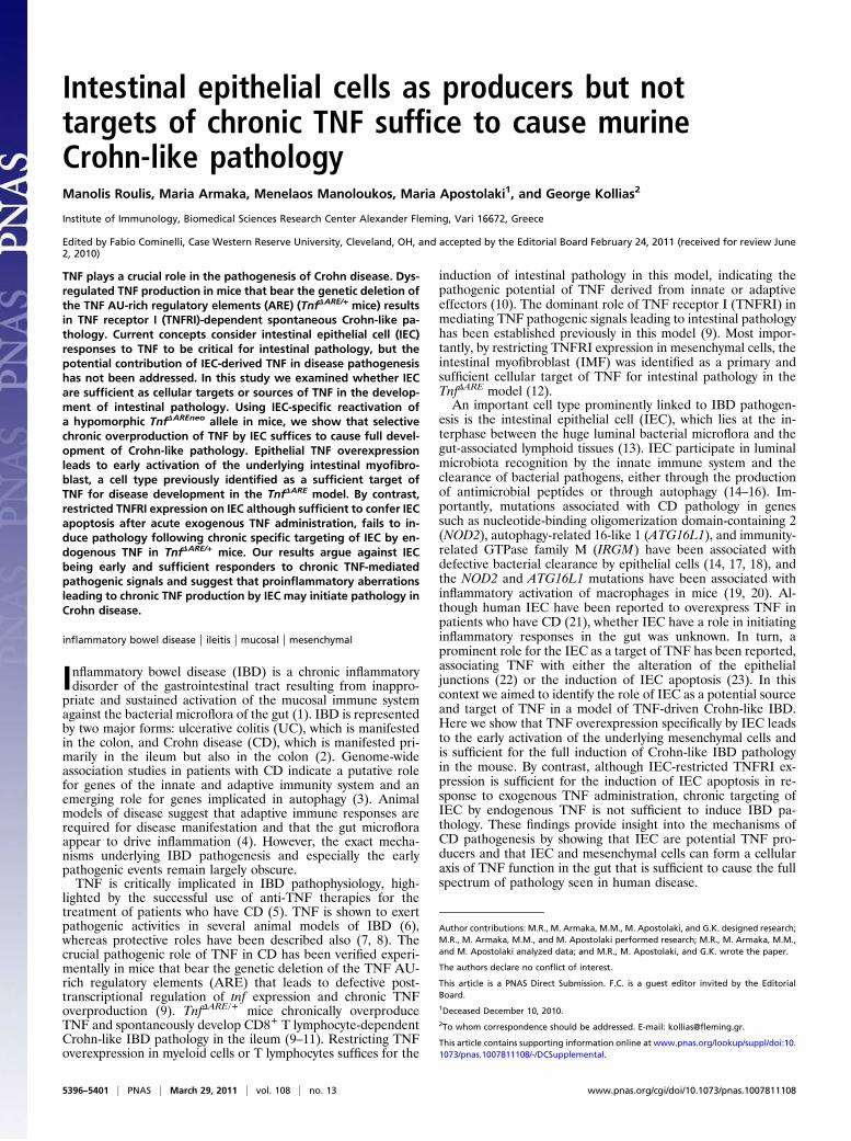
ResultsIEC-Specific TNF Overexpression Is Sufficient for the Induction ofCrohn-Like IBD. IEC, and in particular Paneth cells, from patientswho have CD have been reported to overexpress TNF mRNA(21). Therefore, we examined whether IEC may constitute anadditional source of TNF overproduction in the TnfΔARE model.By performing RT-PCR in primary ileal IEC, we established thatTnfΔARE/+ IEC significantly overexpress TNF as compared withWT IEC (Fig. S1). To estimate the impact of IEC as an earlylocal cellular source of TNF as compared with the other celltypes in the ileum, we directly compared TNF expression in IECand in nonepithelial tissue in WT and TnfΔARE/+ mice at age4 wk, a stage of disease development at which no inflammatoryinfiltrates are observed. Although IEC in WT mice appear toexpress less TNF than do the other intestinal cell types, IEC inTnfΔARE/+ mice express significantly more TNF than does therest of the tissue and indeed constitute the major source of TNF(Fig. 1A). These observations show that in TnfΔARE/+ mice IECare the major local source of TNF overproduction before therecruitment of immune effector cells.
To investigate the potential role of IEC in introducing in-flammatory signals in the intestinal mucosa, we generated miceoverproducing TNF specifically by IEC. For this purpose, wecrossed the IEC-specific VillinCre transgenic mouse (24) withTnfΔAREneo/+ mice bearing a hypomorphic TnfΔAREneo allele inwhich a LoxP-flanked neomycin (neo) cassette is inserted next tothe tnfΔARE mutation but can be activated upon cre-mediateddeletion of the neo cassette (9, 10). In accordance with thereported pattern of expression of the Villin-Cre transgene, weobserved in VillinCreTnfΔAREneo/+ mice the activation of theTnfΔARE allele in isolated IEC from the ileum and in all parts ofthe small intestine and colon but not in other tissues examined(Fig. 1B). Performing quantitative RT-PCR analysis in primaryIEC isolated from the ileum, we verified that IEC of Villin-CreTnfΔAREneo/+ mice express significantly higher levels of TNFthan do IEC from WT, VillinCre, and TnfΔAREneo/+ controls (Fig.1C). To confirm the specificity of TNF overexpression in IEC, weexamined TNF production from macrophages and T-cell receptorβ–positive (TCRβ+) splenic T lymphocytes and observed that,unlike control TnfΔARE/+ mice, VillinCreTnfΔAREneo/+ mice did notdisplay increased TNF levels in these cell types (Fig. 1 D and E).Interestingly, IEC-specific TNF overproduction in Villin-
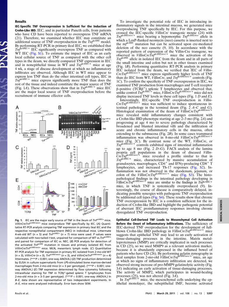
CreTnfΔAREneo/+ mice was sufficient to induce spontaneous in-testinal pathology in the terminal ileum (Fig. 2 A–C and G).Histological examination of the ileum of VillinCreTnfΔAREneo/+mice revealed mild inflammatory changes consistent witha Crohn-like IBD phenotype starting at age 2–3 mo (Fig. 2A) andprogressing at age 4 mo to severe pathology characterized bybroadened and blunted intestinal villi and the infiltration ofacute and chronic inflammatory cells in the mucosa, oftenextending to the submucosa (Fig. 2B). In some cases transmuralinflammation was observed in 8-mo-old VillinCreTnfΔAREneo/+
mice (Fig. 2C). By contrast none of the WT, VillinCre, orTnfΔAREneo/+ controls exhibited signs of intestinal inflammationup to age 8 mo (Fig. 2 D–G). FACS analysis of the laminapropria cell populations in the ileum of diseased Villin-CreTnfΔAREneo/+ mice revealed a profile similar to that inTnfΔARE/+ mice, characterized by massive accumulation ofgranulocytes, macrophages, CD4+ and IFNγ-producing CD8+ Tlymphocytes, and increased Th-17 responses (Fig. S2). In-flammation was not observed in the duodenum, jejunum, orcolon of the VillinCreTnfΔAREneo/+ mice (Fig. S3). The histo-pathological findings in the intestinal pathology developing inVillinCreTnfΔAREneo/+ mice are similar to the findings in TnfΔARE/+mice, in which TNF is systemically overproduced (9). In-terestingly, the course of disease is comparatively delayed, in-dicating possible synergies with pathogenic TNF overproductionby additional cell types (Fig. S4). These results show that chronicTNF overexpression by IEC is a condition sufficient for the in-duction of Crohn-like IBD and highlight the pathogenic potentialof aberrant IEC proinflammatory responses involving chronicdysregulated TNF overproduction.
Epithelial Cell-Derived TNF Leads to Mesenchymal Cell ActivationBefore the Onset of Inflammatory Infiltrations. The sufficiency ofIEC-derived TNF overproduction for the development of full-blown Crohn-like IBD pathology in VillinCreTnfΔAREneo/+ micesuggests that epithelial TNF may lead to an early activation oftissue-damaging processes in the intestine. Matrix metal-loproteinases (MMP) are critically implicated in such processesin CD (25), so we used MMP9 as a relevant activation marker,because it is abundantly expressed in the inflamed bowel ofpatients who have CD (26). By performing gelatin zymography inileal samples from 2-mo-old VillinCreTnfΔAREneo/+ mice, an ageat which no signs of inflammatory infiltration are detected, weobserved strong increase of pro-MMP9 gelatinolytic activity (Fig.3A) indicating an early activation of tissue-damaging processes.The activity of MMP2, which participates in wound-healingprocesses (25), was not altered (Fig. 3A).In TnfΔARE/+ mice, the mesenchymal cells underlying the ep-
ithelial monolayer, the subepithelial IMF, become activated
Fig. 1. IEC are the major early source of TNF in the ileum of TnfΔARE/+ mice.VillinCreTnfΔAREneo/+ mice overproduce TNF specifically by IEC. (A) Quanti-tative RT-PCR analysis comparing TNF expression in primary ileal IEC and therespective nonepithelial compartment (NEC) in individual mice. Littermate4-wk-old WT (n = 5) and TnfΔARE/+ (n = 7) mice were used. P values werecalculated with two-tailed t test, unpaired for comparison of WT vs.TnfΔARE/+
and paired for comparison of IEC vs. NEC. (B) PCR analysis for detection ofthe activated TnfΔARE mutation in tissues and primary isolated IEC fromVillinCreTnfΔAREneo/+ mice. MLN, mesenteric lymph node. (C) QuantitativeRT-PCR analysis for TNF expression in primary IEC isolated from 2-mo-old WT(n = 3), VillinCre (n = 3), TnfΔAREneo/+ (n = 3), and VillinCreTnfΔAREneo/+ (n = 4)littermates. (***P < 0.001; one-way ANOVA.) (D) TNF production determinedby ELISA in culture supernatants from LPS-stimulated bone marrow-derivedmacrophages from 2-mo-old mice (n = 3 per genotype). (***P < 0.001; one-way ANOVA.) (E) TNF expression determined by flow cytometry followingintracellular staining for TNF in TCRβ+-gated splenic T lymphocytes from2-mo-old mice (n = 3–5 per genotype). (***P < 0.001; one-way ANOVA.) InB–E, data shown are representative of two independent experiments. InA–E, mice were analyzed individually. Error bars show SEM.
Roulis et al. PNAS | March 29, 2011 | vol. 108 | no. 13 | 5397
MED
ICALSC
IENCE
S
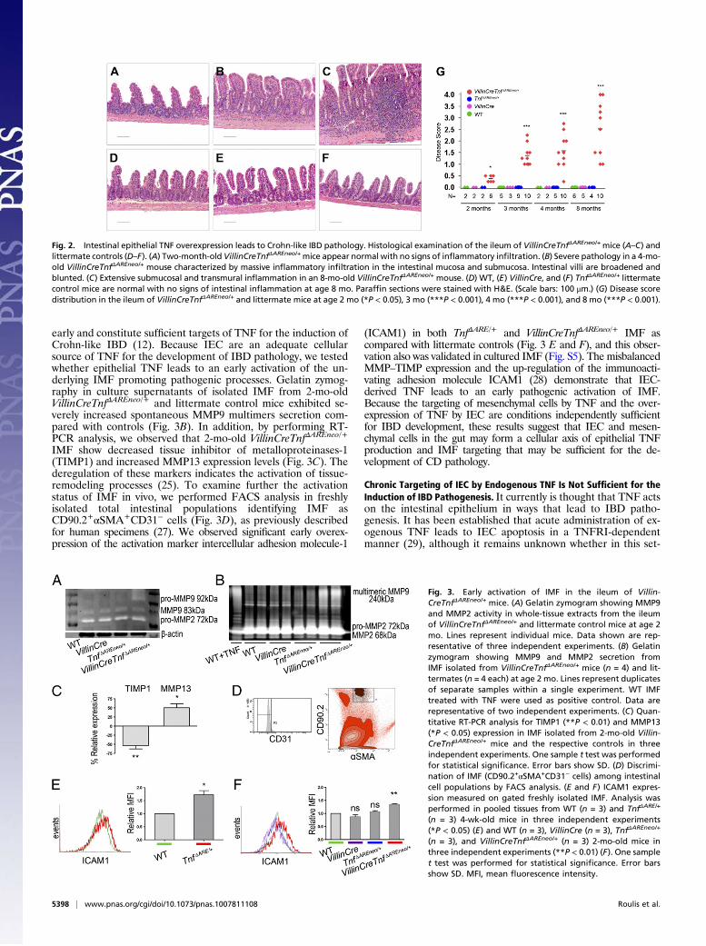
early and constitute sufficient targets of TNF for the induction ofCrohn-like IBD (12). Because IEC are an adequate cellularsource of TNF for the development of IBD pathology, we testedwhether epithelial TNF leads to an early activation of the un-derlying IMF promoting pathogenic processes. Gelatin zymog-raphy in culture supernatants of isolated IMF from 2-mo-oldVillinCreTnfΔAREneo/+ and littermate control mice exhibited se-verely increased spontaneous MMP9 multimers secretion com-pared with controls (Fig. 3B). In addition, by performing RT-PCR analysis, we observed that 2-mo-old VillinCreTnfΔAREneo/+
IMF show decreased tissue inhibitor of metalloproteinases-1(TIMP1) and increased MMP13 expression levels (Fig. 3C). Thederegulation of these markers indicates the activation of tissue-remodeling processes (25). To examine further the activationstatus of IMF in vivo, we performed FACS analysis in freshlyisolated total intestinal populations identifying IMF asCD90.2+αSMA+CD31− cells (Fig. 3D), as previously describedfor human specimens (27). We observed significant early overex-pression of the activation marker intercellular adhesion molecule-1
(ICAM1) in both TnfΔARE/+ and VillinCreTnfΔAREneo/+ IMF ascompared with littermate controls (Fig. 3 E and F), and this obser-vation also was validated in cultured IMF (Fig. S5). The misbalancedMMP–TIMP expression and the up-regulation of the immunoacti-vating adhesion molecule ICAM1 (28) demonstrate that IEC-derived TNF leads to an early pathogenic activation of IMF.Because the targeting of mesenchymal cells by TNF and the over-expression of TNF by IEC are conditions independently sufficientfor IBD development, these results suggest that IEC and mesen-chymal cells in the gut may form a cellular axis of epithelial TNFproduction and IMF targeting that may be sufficient for the de-velopment of CD pathology.
Chronic Targeting of IEC by Endogenous TNF Is Not Sufficient for theInduction of IBD Pathogenesis. It currently is thought that TNF actson the intestinal epithelium in ways that lead to IBD patho-genesis. It has been established that acute administration of ex-ogenous TNF leads to IEC apoptosis in a TNFRI-dependentmanner (29), although it remains unknown whether in this set-
Fig. 2. Intestinal epithelial TNF overexpression leads to Crohn-like IBD pathology. Histological examination of the ileum of VillinCreTnfΔAREneo/+mice (A–C) andlittermate controls (D–F). (A) Two-month-old VillinCreTnfΔAREneo/+mice appear normal with no signs of inflammatory infiltration. (B) Severe pathology in a 4-mo-old VillinCreTnfΔAREneo/+ mouse characterized by massive inflammatory infiltration in the intestinal mucosa and submucosa. Intestinal villi are broadened andblunted. (C) Extensive submucosal and transmural inflammation in an 8-mo-old VillinCreTnfΔAREneo/+ mouse. (D) WT, (E) VillinCre, and (F) TnfΔAREneo/+ littermatecontrol mice are normal with no signs of intestinal inflammation at age 8 mo. Paraffin sections were stained with H&E. (Scale bars: 100 μm.) (G) Disease scoredistribution in the ileum of VillinCreTnfΔAREneo/+ and littermate mice at age 2 mo (*P < 0.05), 3 mo (***P < 0.001), 4 mo (***P < 0.001), and 8 mo (***P < 0.001).
Fig. 3. Early activation of IMF in the ileum of Villin-CreTnfΔAREneo/+ mice. (A) Gelatin zymogram showing MMP9and MMP2 activity in whole-tissue extracts from the ileumof VillinCreTnfΔAREneo/+ and littermate control mice at age 2mo. Lines represent individual mice. Data shown are rep-resentative of three independent experiments. (B) Gelatinzymogram showing MMP9 and MMP2 secretion fromIMF isolated from VillinCreTnfΔAREneo/+ mice (n = 4) and lit-termates (n = 4 each) at age 2 mo. Lines represent duplicatesof separate samples within a single experiment. WT IMFtreated with TNF were used as positive control. Data arerepresentative of two independent experiments. (C) Quan-titative RT-PCR analysis for TIMP1 (**P < 0.01) and MMP13(*P < 0.05) expression in IMF isolated from 2-mo-old Villin-CreTnfΔAREneo/+ mice and the respective controls in threeindependent experiments. One sample t test was performedfor statistical significance. Error bars show SD. (D) Discrimi-nation of IMF (CD90.2+αSMA+CD31− cells) among intestinalcell populations by FACS analysis. (E and F) ICAM1 expres-sion measured on gated freshly isolated IMF. Analysis wasperformed in pooled tissues from WT (n = 3) and TnfΔARE/+
(n = 3) 4-wk-old mice in three independent experiments(*P < 0.05) (E) and WT (n = 3), VillinCre (n = 3), TnfΔAREneo/+
(n = 3), and VillinCreTnfΔAREneo/+ (n = 3) 2-mo-old mice inthree independent experiments (**P < 0.01) (F). One samplet test was performed for statistical significance. Error barsshow SD. MFI, mean fluorescence intensity.
5398 | www.pnas.org/cgi/doi/10.1073/pnas.1007811108 Roulis et al.
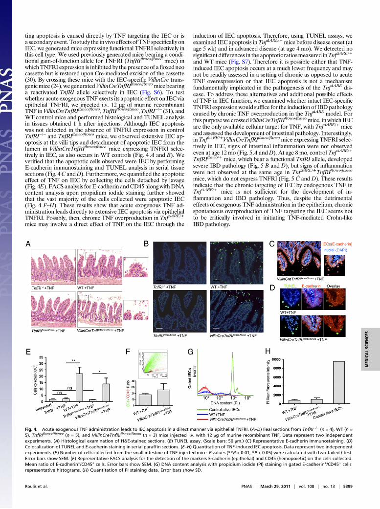
ting apoptosis is caused directly by TNF targeting the IEC or isa secondary event. To study the in vivo effects ofTNF specifically onIEC, we generated mice expressing functional TNFRI selectively inthis cell type. We used previously generated mice bearing a condi-tional gain-of-function allele for TNFRI (TnfRIflxneo/flxneo mice) inwhichTNFRI expression is inhibited by thepresence of afloxedneocassette but is restored upon Cre-mediated excision of the cassette(30). By crossing these mice with the IEC-specific VillinCre trans-genicmice (24), we generatedVillinCreTnfRIflxneo/flxneomice bearinga reactivated TnfRI allele selectively in IEC (Fig. S6). To testwhether acute exogenous TNF exerts its apoptotic effect on IEC viaepithelial TNFRI, we injected i.v. 12 μg of murine recombinantTNF inVillinCreTnfRIflxneo/flxneo,TnfRIflxneo/flxneo,TnfRI−/− (31), andWT control mice and performed histological and TUNEL analysisin tissues obtained 1 h after injections. Although IEC apoptosiswas not detected in the absence of TNFRI expression in controlTnfRI−/− and TnfRIflxneo/flxneo mice, we observed extensive IEC ap-optosis at the villi tips and detachment of apoptotic IEC from thelumen in VillinCreTnfRIflxneo/flxneo mice expressing TNFRI selec-tively in IEC, as also occurs in WT controls (Fig. 4 A and B). Weverified that the apoptotic cells observed were IEC by performingE-cadherin immunostaining and TUNEL analysis in serial tissuesections (Fig. 4C andD). Furthermore, we quantified the apoptoticeffect of TNF on IEC by collecting the cells detached by lavage(Fig. 4E). FACS analysis forE-cadherin andCD45 alongwithDNAcontent analysis upon propidium iodide staining further showedthat the vast majority of the cells collected were apoptotic IEC(Fig. 4 F–H). These results show that acute exogenous TNF ad-ministration leads directly to extensive IEC apoptosis via epithelialTNFRI. Possibly, then, chronic TNF overproduction in TnfΔARE/+
mice may involve a direct effect of TNF on the IEC through the
induction of IEC apoptosis. Therefore, using TUNEL assays, weexamined IEC apoptosis inTnfΔARE/+mice before disease onset (atage 5 wk) and in advanced disease (at age 4 mo). We detected nosignificant differences in the apoptotic ratiosmeasured inTnfΔARE/+
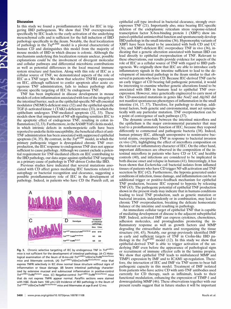
and WT mice (Fig. S7). Therefore it is possible either that TNF-induced IEC apoptosis occurs at a much lower frequency and maynot be readily assessed in a setting of chronic as opposed to acuteTNF overexpression or that IEC apoptosis is not a mechanismfundamentally implicated in the pathogenesis of the TnfΔARE dis-ease. To address these alternatives and additional possible effectsof TNF in IEC function, we examined whether intact IEC-specificTNFRI expressionwould suffice for the induction of IBDpathologycaused by chronic TNF overproduction in the TnfΔARE model. Forthis purpose we crossedVillinCreTnfRIflxneo/flxneomice, in which IECare the only available cellular target for TNF, with TnfΔARE/+ miceand assessed the development of intestinal pathology. Interestingly,inTnfΔARE/+VillinCreTnfRIflxneo/flxneomice expressing TNFRI selec-tively in IEC, signs of intestinal inflammation were not observedeven at age 12 mo (Fig. 5A andD). At age 8 mo, control TnfΔARE/+
TnfRIflxneo/+ mice, which bear a functional TnfRI allele, developedsevere IBD pathology (Fig. 5 B and D), but signs of inflammationwere not observed at the same age in TnfΔARE/+TnfRIflxneo/flxneo
mice, which do not express TNFRI (Fig. 5 C and D). These resultsindicate that the chronic targeting of IEC by endogenous TNF inTnfΔARE/+ mice is not sufficient for the development of in-flammation and IBD pathology. Thus, despite the detrimentaleffects of exogenous TNF administration in the epithelium, chronicspontaneous overproduction of TNF targeting the IEC seems notto be critically involved in initiating TNF-mediated Crohn-likeIBD pathology.
Fig. 4. Acute exogenous TNF administration leads to IEC apoptosis in a direct manner via epithelial TNFRI. (A–D) Ileal sections from TnfRI−/− (n = 4), WT (n =5), TnfRIflxneo/flxneo (n = 5), and VillinCreTnfRIflxneo/flxneo (n = 3) mice injected i.v. with 12 μg of murine recombinant TNF. Data represent two independentexperiments. (A) Histological examination of H&E-stained sections. (B) TUNEL assay. (Scale bars: 50 μm.) (C) Representative E-cadherin immunostaining. (D)Colocalization of TUNEL and E-cadherin staining in serial paraffin sections. (E–H) Quantitation of TNF-induced IEC apoptosis. Data represent two independentexperiments. (E) Number of cells collected from the small intestine of TNF-injected mice. P values (**P < 0.01, *P < 0.05) were calculated with two-tailed t test.Error bars show SEM. (F) Representative FACS analysis for the detection of the markers E-cadherin (epithelial) and CD45 (hemopoietic) on the cells collected.Mean ratio of E-cadherin+/CD45+ cells. Error bars show SEM. (G) DNA content analysis with propidium iodide (PI) staining in gated E-cadherin+/CD45− cells:representative histograms. (H) Quantitation of PI staining data. Error bars show SD.
Roulis et al. PNAS | March 29, 2011 | vol. 108 | no. 13 | 5399
MED
ICALSC
IENCE
S
DiscussionIn this study we found a proinflammatory role for IEC in trig-gering IBD pathogenesis. We show that TNF overexpressionspecifically by IEC leads to the early activation of the underlyingmesenchymal cells and is sufficient for the full induction of IBDpathology specifically in the ileum. Notably, the ileal localizationof pathology in the TnfΔARE model is a pivotal characteristic ofhuman CD and distinguishes this model from the majority ofmouse models of IBD in which disease is colonic. Although themechanisms underlying this localization remain unclear, possibleexplanations could be the involvement of divergent molecularand cellular pathways and differential microbiota contributionsas well as potential differences in the local mucosal immunesystem structure and function. Along with this role of IEC as acellular source of TNF, we demonstrated aspects of the role ofIEC as a TNF target. We show that selective TNFRI expressionon IEC, although sufficient to confer apoptosis after acute ex-ogenous TNF administration, fails to induce pathology afterchronic specific targeting of IEC by endogenous TNF.TNF has been implicated in disease development in mouse
models of colon inflammation associatedwith loss of the integrity ofthe intestinal barrier, such as the epithelial-specific NF-κB essentialmodulator (NEMO)-deficient mice (32) and the epithelial-specificTGF-β–activated kinase 1 (TAK1)-deficientmice (33), in which theepithelium undergoes TNF-mediated apoptosis (32, 33). Thesemodels show that impairment of NF-κB signaling sensitizes IEC tothe apoptotic effect of endogenous TNF, resulting in colon in-flammation (32, 33).Furthermore, in theSAMP/YitFc ileitismodel,in which intrinsic defects in nonhemopoietic cells have beenreported tounderlie ileitis susceptibility, thebeneficial effect of anti-TNF administration has been associated with suppressed epithelialapoptosis (34, 35). By contrast, in the TnfΔARE model, in which theprimary pathogenic trigger is dysregulated chronic TNF over-production, the IEC response to endogenous TNF does not appearsufficient to cause pathology. Although we cannot exclude a poten-tial requirement for TNF-mediated effects on IEC contributing tothe IBD pathology, our data argue against epithelial TNF targetingas a primary cause of pathology in TNF-driven Crohn-like IBD.Previous studies have indicated that several mutations asso-
ciated with CD affect genes mediating IEC functions related toautophagy or bacterial recognition and clearance, suggesting apossible proinflammatory role of IEC in the development ofpathology. Indeed, in patients who have CD the Paneth cell, an
epithelial cell type involved in bacterial clearance, strongly over-expresses TNF (21). Importantly also, mice bearing IEC-specificdeletion of the endoplasmic reticulum stress response-relatedtranscription factor X-box-binding protein 1 (XBP1) show im-paired epithelial antimicrobial function and spontaneously developIBD pathology in the small intestine (36). Hypomorphic variants ofXBP1 have been found to be associated with both CD and UC(36), and XBP1-deficient IEC overproduce TNF in vivo (36), in-dicating that a genetic alteration associated with human IBD canlead directly to epithelial overproduction of TNF. Relevant tothese observations, our results provide evidence for aspects of therole of IEC as a cellular source of TNF with regard to IBD path-ogenesis. We originally show that IEC are able to introduce earlyinflammatory signals in the intestine sufficient to lead to the de-velopment of intestinal pathology in the ileum similar to that ob-served in patients who have CD. Because IEC-derived TNF can bean early trigger of CD bearing full pathogenic potential, it wouldbe interesting to examine whether genetic alterations found to beassociated with IBD in humans lead to epithelial TNF over-expression. However, mice genetically engineered to carry most ofthe CD-associated mutations in genes like Nod2 and Atg16l1 didnot manifest spontaneous phenotypes of inflammation in the smallintestine (14, 17, 37). Therefore, for pathology to develop, addi-tional factors, both genetic and environmental, must act synergis-tically with any particular mutation. TNF overproduction can bea point of convergence of such pathways (37).The dynamic cross-talk between the intestinal microflora and
the epithelium is the major environmental parameter that maylead to a proinflammatory function of IEC, because they responddifferently to commensal and pathogenic bacteria (38). Indeed,human primary IEC, although unresponsive to noninvasive bac-terial strains, overproduce TNF in response to bacterial invasionex vivo (39), highlighting the effect of microflora composition onthe tolerant or inflammatory character of IEC. On the other hand,important differences are observed in the composition of the in-testinal microflora in patients who have CD or UC and healthycontrols (40), and infections are considered to be implicated inboth disease onset and relapse in humans (41). Interestingly, it hasbeen shown that Escherichia coli bacterial isolates from inflamedtissues of patients who have CD trigger NF-κB activation and TNFsecretion by IEC (42). Furthermore, the hypoxia generated underconditions of infection, tissue damage, and inflammation can be anadditional trigger or positive-feedback mechanism for epithelialTNF production, because IEC respond to hypoxia by releasingTNF (43). The pathogenic potential of epithelial TNF productionshown in the present study may indicate that in humans conditionsleading to local TNF production, such as genetic mutation orbacterial invasion, independently or in combination, may lead tochronic TNF overproduction, breaking the delicate homeostaticbalance of the intestine and resulting in pathology.An immediate cellular target of epithelial TNF that is capable
of mediating development of disease is the adjacent subepithelialIMF. Indeed, activated IMF can express cytokines, chemokines,adhesion molecules, and prostaglandins promoting the in-flammatory response as well as growth factors and MMPdegrading the extracellular matrix and reorganizing the tissuestructure (44, 45). Notably, our group previously identified IMFas early and sufficient targets of TNF in Crohn-like IBD pa-thology in the TnfΔARE model (12). In this study we show thatepithelial-derived TNF is able to trigger activation of the un-derlying IMF even before the appearance of pathological signsor recruitment of immune effector cells in the lamina propria.We show that epithelial TNF leads to misbalanced MMP andTIMP1 expression by IMF and to ICAM1 up-regulation. There-fore, the interaction of IEC and IMF via TNF seems to bear fullpathogenic capacity in this model. Treatment of IMF isolatedfrom patients who have active CD with anti-TNF antibodies usedcurrently for CD therapy, such as infliximab, leads to theirfunctional modulation, enhancing the expression of TIMP-1 anddownregulating MMP (46). These observations together with ourpresent results suggest that in future studies it will be important
Fig. 5. Chronic selective targeting of IEC by endogenous TNF in TnfΔARE/+
mice is not sufficient for the development of intestinal pathology. (A–C) Histo-logical examination of the ileum of 8-mo-old TnfΔARE/+VillinCreTnfRIflxneo/flxneo
mice and littermate controls. (A) TnfΔARE/+VillinCreTnfRIflxneo/flxneo mice thatexpress TNFRI selectively in IEC show normal tissue structure without signs ofinflammation or tissue damage. (B) Severe intestinal pathology character-ized by extensive mucosal and submucosal inflammation in positive-controlTnfΔARE/+TnfRIflxneo/+ mice. (C) Negative-control TnfΔARE/+TnfRIflxneo/flxneo micethat do not express TNFRI appear normal. Paraffin sections were stainedwith H&E. (Scale bars: 100 μm.) (D) Incidence of IBD pathology in the ileum ofTnfΔARE/+VillinCreTnfRIflxneo/flxneo mice and littermates at age 8 and 12 mo.
5400 | www.pnas.org/cgi/doi/10.1073/pnas.1007811108 Roulis et al.
to examine genetic and/or environmental parameters that lead toepithelial TNF overproduction in the human intestine. Suchstudies should provide essential information on the early eventsleading to CD pathology in humans and may lead to novel ap-plications in disease prevention and therapy.
Materials and MethodsMice. The TnfΔARE/+, TnfΔAREneo/+ (9), TnfRIflxneo/flxneo (30), and TnfRI−/− (31)mice have been described previously. VillinCre transgenic mice (24) wereprovided by D. L. Gumucio (Department of Cell and Developmental Biology,University of Michigan Medical School, Ann Arbor, MI). All mice were bredand maintained on a C57BL/6J or on a mixed C57BL/6J×129S6 geneticbackground in the animal facilities of the Biomedical Sciences ResearchCenter (BSRC) Alexander Fleming under specific pathogen-free conditions.All mice were used in accordance with the guidance of the InstitutionalAnimal Care and Use Committee of BSRC Alexander Fleming.
Histological Assessment of Intestinal Pathology. Histological analysis wasperformed in the terminal ileum and parts of the colon, duodenum, and
jejunum. The tissue was embedded in paraffin and stained with H&E.Semiquantitative assessment of intestinal pathology was performed ina blinded fashion. The following scoring system was used: 0 = normal; 1 =villi blunting and mucosal inflammation; 2 = villi blunting, extensive mucosalinflammation, and submucosal inflammation; 3 = extensive submucosal in-flammation; 4 = transmural inflammation.
Statistical Analysis. Statistical analysis was performed using GraphPad Prismsoftware with P values <0.05 regarded as statistically significant.
ACKNOWLEDGMENTS. We thank D. L. Gumucio for providing the VillinCretransgenic mice; T. Fotsis and A. S. Politou for advice; Ksanthi Kranidioti fordiscussions and technical advice; Spiros Lalos for excellent technical assis-tance in histopathology; and Panos Athanasakis, Nikos Giannakas, andPeggy Andriopoulou for technical assistance. This work was supported byGrant LSHG-CT-2005–005203 from the European Commission IntegratedFunctional Genomics in Mutant Mouse Models as Tools to Investigate theComplexity of Human Immunological Disease (MUGEN) program, Grant223151 from the European Commission program Inflammation and CancerResearch in Europe (INFLACARE), and by Grant GSRT-PENED-03EΔ770 fromthe Hellenic Ministry for Development.
1. Xavier RJ, Podolsky DK (2007) Unravelling the pathogenesis of inflammatory boweldisease. Nature 448:427–434.
2. Podolsky DK (2002) Inflammatory bowel disease. N Engl J Med 347:417–429.3. Cho JH (2008) The genetics and immunopathogenesis of inflammatory bowel disease.
Nat Rev Immunol 8:458–466.4. Strober W, Fuss IJ, Blumberg RS (2002) The immunology of mucosal models of
inflammation. Annu Rev Immunol 20:495–549.5. Targan SR, et al.; Crohn’s Disease cA2 Study Group (1997) A short-term study of
chimeric monoclonal antibody cA2 to tumor necrosis factor alpha for Crohn’s disease.N Engl J Med 337:1029–1035.
6. Apostolaki M, Armaka M, Victoratos P, Kollias G (2010) Cellular mechanisms of TNFfunction in models of inflammation and autoimmunity. Curr Dir Autoimmun 11:1–26.
7. Noti M, Corazza N, Mueller C, Berger B, Brunner T (2010) TNF suppresses acuteintestinal inflammation by inducing local glucocorticoid synthesis. J Exp Med 207:1057–1066.
8. Pagnini C, et al. (2010) Probiotics promote gut health through stimulation ofepithelial innate immunity. Proc Natl Acad Sci USA 107:454–459.
9. Kontoyiannis D, Pasparakis M, Pizarro TT, Cominelli F, Kollias G (1999) Impaired on/offregulation of TNF biosynthesis in mice lacking TNF AU-rich elements: Implications forjoint and gut-associated immunopathologies. Immunity 10:387–398.
10. Kontoyiannis D, et al. (2002) Genetic dissection of the cellular pathways and signalingmechanisms in modeled tumor necrosis factor-induced Crohn’s-like inflammatorybowel disease. J Exp Med 196:1563–1574.
11. Apostolaki M, et al. (2008) Role of beta7 integrin and the chemokine/chemokinereceptor pair CCL25/CCR9 in modeled TNF-dependent Crohn’s disease. Gastroen-terology 134:2025–2035.
12. Armaka M, et al. (2008) Mesenchymal cell targeting by TNF as a common pathogenicprinciple in chronic inflammatory joint and intestinal diseases. J Exp Med 205:331–337.
13. Artis D (2008) Epithelial-cell recognition of commensal bacteria and maintenance ofimmune homeostasis in the gut. Nat Rev Immunol 8:411–420.
14. Kobayashi KS, et al. (2005) Nod2-dependent regulation of innate and adaptiveimmunity in the intestinal tract. Science 307:731–734.
15. Rioux JD, et al. (2007) Genome-wide association study identifies new susceptibility locifor Crohn disease and implicates autophagy in disease pathogenesis. Nat Genet 39:596–604.
16. Cario E (2008) Innate immune signalling at intestinal mucosal surfaces: A fine linebetween host protection and destruction. Curr Opin Gastroenterol 24:725–732.
17. Cadwell K, et al. (2008) A key role for autophagy and the autophagy gene Atg16l1 inmouse and human intestinal Paneth cells. Nature 456:259–263.
18. McCarroll SA, et al. (2008) Deletion polymorphism upstream of IRGM associated withaltered IRGM expression and Crohn’s disease. Nat Genet 40:1107–1112.
19. Maeda S, et al. (2005) Nod2 mutation in Crohn’s disease potentiates NF-kappaBactivity and IL-1beta processing. Science 307:734–738.
20. Saitoh T, et al. (2008) Loss of the autophagy protein Atg16L1 enhances endotoxin-induced IL-1beta production. Nature 456:264–268.
21. Lala S, et al. (2003) Crohn’s disease and the NOD2 gene: A role for Paneth cells.Gastroenterology 125:47–57.
22. Turner JR (2006) Molecular basis of epithelial barrier regulation: From basicmechanisms to clinical application. Am J Pathol 169:1901–1909.
23. Zeissig S, et al. (2004) Downregulation of epithelial apoptosis and barrier repair inactive Crohn’s disease by tumour necrosis factor alpha antibody treatment. Gut 53:1295–1302.
24. Madison BB, et al. (2002) Cis elements of the villin gene control expression inrestricted domains of the vertical (crypt) and horizontal (duodenum, cecum) axes ofthe intestine. J Biol Chem 277:33275–33283.
25. Ravi A, Garg P, Sitaraman SV (2007) Matrix metalloproteinases in inflammatory boweldisease: Boon or a bane? Inflamm Bowel Dis 13:97–107.
26. Baugh MD, et al. (1999) Matrix metalloproteinase levels are elevated in inflammatorybowel disease. Gastroenterology 117:814–822.
27. Saada JI, et al. (2006) Subepithelial myofibroblasts are novel nonprofessional APCs inthe human colonic mucosa. J Immunol 177:5968–5979.
28. Lebedeva T, Dustin ML, Sykulev Y (2005) ICAM1 co-stimulates target cells to facilitateantigen presentation. Curr Opin Immunol 17:251–258.
29. Piguet PF, Vesin C, Guo J, Donati Y, Barazzone C (1998) TNF-induced enterocyteapoptosis in mice is mediated by the TNF receptor 1 and does not require p53. Eur JImmunol 28:3499–3505.
30. Victoratos P, et al. (2006) FDC-specific functions of p55TNFR and IKK2 in thedevelopment of FDC networks and of antibody responses. Immunity 24:65–77.
31. Pfeffer K, et al. (1993) Mice deficient for the 55 kd tumor necrosis factor receptor areresistant to endotoxic shock, yet succumb to L. monocytogenes infection. Cell 73:457–467.
32. Nenci A, et al. (2007) Epithelial NEMO links innate immunity to chronic intestinalinflammation. Nature 446:557–561.
33. Kajino-Sakamoto R, et al. (2008) Enterocyte-derived TAK1 signaling preventsepithelium apoptosis and the development of ileitis and colitis. J Immunol 181:1143–1152.
34. Marini M, et al. (2003) TNF-alpha neutralization ameliorates the severity of murineCrohn’s-like ileitis by abrogation of intestinal epithelial cell apoptosis. Proc Natl AcadSci USA 100:8366–8371.
35. Olson TS, et al. (2006) The primary defect in experimental ileitis originates froma nonhematopoietic source. J Exp Med 203:541–552.
36. Kaser A, et al. (2008) XBP1 links ER stress to intestinal inflammation and confersgenetic risk for human inflammatory bowel disease. Cell 134:743–756.
37. Kaser A, Zeissig S, Blumberg RS (2010) Inflammatory bowel disease. Annu RevImmunol 28:573–621.
38. Hill DA, Artis D (2010) Intestinal bacteria and the regulation of immune cellhomeostasis. Annu Rev Immunol 28:623–667.
39. Jung HC, et al. (1995) A distinct array of proinflammatory cytokines is expressed inhuman colon epithelial cells in response to bacterial invasion. J Clin Invest 95:55–65.
40. Qin J, et al.; MetaHIT Consortium (2010) A human gut microbial gene catalogueestablished by metagenomic sequencing. Nature 464:59–65.
41. Irving PM, Gibson PR (2008) Infections and IBD. Nat Clin Pract Gastroenterol Hepatol5:18–27.
42. La Ferla K, Seegert D, Schreiber S (2004) Activation of NF-kappaB in intestinalepithelial cells by E. coli strains isolated from the colonic mucosa of IBD patients. Int JColorectal Dis 19:334–342.
43. Taylor CT, Fueki N, Agah A, Hershberg RM, Colgan SP (1999) Critical role of cAMPresponse element binding protein expression in hypoxia-elicited induction ofepithelial tumor necrosis factor-alpha. J Biol Chem 274:19447–19454.
44. Powell DW, et al. (1999) Myofibroblasts. II. Intestinal subepithelial myofibroblasts. AmJ Physiol 277:C183–C201.
45. Andoh A, Bamba S, Brittan M, Fujiyama Y, Wright NA (2007) Role of intestinalsubepithelial myofibroblasts in inflammation and regenerative response in the gut.Pharmacol Ther 114:94–106.
46. Di Sabatino A, et al. (2007) Functional modulation of Crohn’s disease myofibroblastsby anti-tumor necrosis factor antibodies. Gastroenterology 133:137–149.
Roulis et al. PNAS | March 29, 2011 | vol. 108 | no. 13 | 5401
MED
ICALSC
IENCE
S